Embed Size (px)
Citation preview

Connective
1. Loose (areolar, adipose, reticular)2. Dense (dense irregular, white fibrous)3. Cartilage (hyaline, elastic, fibrocartilage)4. Bone5. Blood6. Mesenchyme
FunctionsA. SupportB. Connecting partsC. Storage (calcium, energy)D. Protection (skull, ribs, kidneys)E. Movement (muscle attachments)F. Blood formation and transport
1.28

CT Glue
Chondroitin sulfate
Common, long, white, wavy
Thin, wire-like, flexible
Short, branching (netting)
1.30

Copyright © 2010 Pearson Education, Inc.
Figure 4.7 Areolar connective tissue: A prototype (model) connective tissue.
Macrophage
Fibroblast
Lymphocyte
Fat cell
Mast cell
Neutrophil
Capillary
Cell types Extracellularmatrix
Fibers• Collagen fiber• Elastic fiber• Reticular fiber
Ground substance
Pg 128
Cells?Blood vessels?Nerves?

Copyright © 2010 Pearson Education, Inc.
Figure 4.13 Embryonic germ layers and the primary tissue types they produce.
MesodermEndoderm
16-day-old embryo(dorsal surface view)
EpitheliumNervous tissue(from ectoderm)
Muscle and connectivetissue (mostly frommesoderm)Ectoderm
Pg 144
Mesenchyme -ameboidStem cellsFlexible

Copyright © 2010 Pearson Education, Inc.
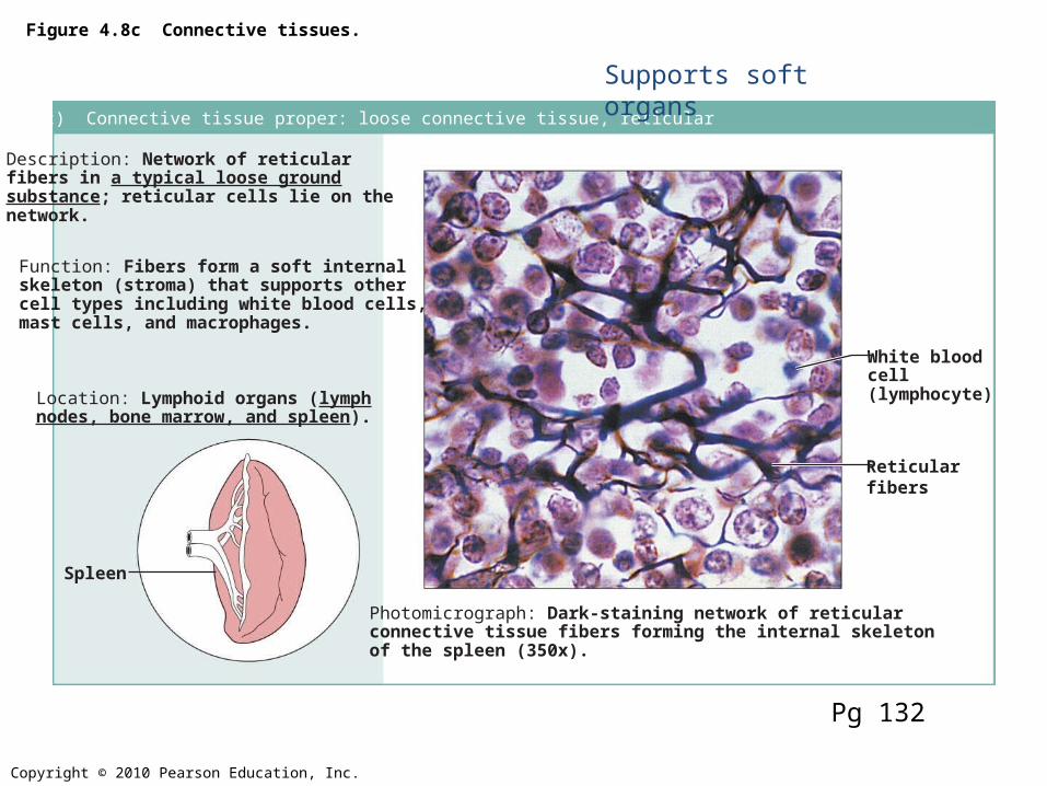
Figure 4.8k Connective tissues.
(k) Others: blood
Description: Red and whiteblood cells in a fluid matrix(plasma).
Function: Transport ofrespiratory gases, nutrients,wastes, and other substances.
Location: Contained withinblood vessels.
Photomicrograph: Smear of human blood (1860x); twowhite blood cells (neutrophil in upper left and lymphocytein lower right) are seen surrounded by red blood cells.
Neutrophil
Red bloodcells
Lymphocyte
Plasma
Pg 137
Liquid matrix called PlasmaWater soluble fibers

Copyright © 2010 Pearson Education, Inc.
Figure 4.8a Connective tissues.
(a) Connective tissue proper: loose connective tissue, areolar
Description: Gel-like matrix with allthree fiber types; cells: fibroblasts,macrophages, mast cells, and somewhite blood cells.
Function: Wraps and cushionsorgans; its macrophages phagocytizebacteria; plays important role ininflammation; holds and conveystissue fluid.
Location: Widely distributed underepithelia of body, e.g., forms laminapropria of mucous membranes;packages organs; surroundscapillaries.
Photomicrograph: Areolar connective tissue, asoft packaging tissue of the body (300x).
Epithelium
Laminapropria
Fibroblastnuclei
Elasticfibers
Collagenfibers
Pg 131
Jello! Filler tissue

Copyright © 2010 Pearson Education, Inc.
Figure 4.8b Connective tissues.
(b) Connective tissue proper: loose connective tissue, adipose
Description: Matrix as in areolar,but very sparse; closely packedadipocytes, or fat cells, havenucleus pushed to the side by largefat droplet.
Function: Provides reserve foodfuel; insulates against heat loss;supports and protects organs.
Location: Under skin in thehypodermis; around kidneys andeyeballs; within abdomen; in breasts.
Photomicrograph: Adipose tissue from thesubcutaneous layer under the skin (350x).
Nucleus offat cell
Vacuolecontainingfat droplet
Adiposetissue
Mammaryglands
Pg 131
Triglycerides

Copyright © 2010 Pearson Education, Inc.
Figure 4.8c Connective tissues.
(c) Connective tissue proper: loose connective tissue, reticular
Description: Network of reticularfibers in a typical loose groundsubstance; reticular cells lie on thenetwork.
Function: Fibers form a soft internalskeleton (stroma) that supports othercell types including white blood cells,mast cells, and macrophages.
Location: Lymphoid organs (lymphnodes, bone marrow, and spleen).
Photomicrograph: Dark-staining network of reticularconnective tissue fibers forming the internal skeletonof the spleen (350x).
Spleen
White bloodcell(lymphocyte)
Reticularfibers
Pg 132
Supports soft organs

Copyright © 2010 Pearson Education, Inc.
Figure 4.8e Connective tissues.
(e) Connective tissue proper: dense connective tissue, dense irregular
Description: Primarilyirregularly arranged collagenfibers; some elastic fibers;major cell type is the fibroblast.
Function: Able to withstandtension exerted in manydirections; provides structuralstrength.
Location: Fibrous capsules oforgans and of joints; dermis ofthe skin; submucosa ofdigestive tract.
Photomicrograph: Dense irregularconnective tissue from the dermis of theskin (400x).
Collagenfibers
Nuclei offibroblasts
Fibrousjointcapsule
Pg 133
Dermis, Deep fascia

Copyright © 2010 Pearson Education, Inc.
Figure 4.8d Connective tissues.
(d) Connective tissue proper: dense connective tissue, dense regular
Description: Primarily parallelcollagen fibers; a few elastic fibers;major cell type is the fibroblast.
Function: Attaches muscles tobones or to muscles; attaches bonesto bones; withstands great tensilestress when pulling force is appliedin one direction.
Location: Tendons, mostligaments, aponeuroses.
Photomicrograph: Dense regular connectivetissue from a tendon (500x).
Shoulderjoint
Ligament
Tendon
Collagenfibers
Nuclei offibroblasts
Pg 133
(White Fibrous)

Copyright © 2010 Pearson Education, Inc.
Figure 4.8f Connective tissues.
(f) Connective tissue proper: dense connective tissue, elastic
Description: Dense regularconnective tissue containing a highproportion of elastic fibers.
Function: Allows recoil of tissuefollowing stretching; maintainspulsatile flow of blood througharteries; aids passive recoil of lungsfollowing inspiration.
Location: Walls of large arteries;within certain ligaments associatedwith the vertebral column; within thewalls of the bronchial tubes.
Elastic fibers
Aorta
HeartPhotomicrograph: Elastic connective tissue inthe wall of the aorta (250x).
Pg 134

Clicker Question: Which of the following statements regarding connective tissue are true?
1. The 3 main components of connective tissue are: ground substance, fibers and proteoglycans.2. All connective tissue has a good blood supply.3. Collagen fibers provide a lot of strength within connective tissue.4. Connective tissue can have a nerve supply.
A. 1, 3, 4 B. 3, 4 C. 2, 3, 4 1, 2, 3