Embed Size (px)
Citation preview
Available online at www.sciencedirect.com
SciVerse ScienceDirectjournal homepage:www.elsevier.com/locate/jasi
Journal of the Anatomical Society of India 62 (2013) 90–92
Case report
Congenital pulmonary airway malformationDeepali Onkar*
Associate Professor, Department of Anatomy, NKP Salve Institute of Medical Sciences and Research Centre, Nagpur, Maharashtra
1. Introduction
Lung malformation constitutes a spectrum of lesions that originate in the embryonic period.1 The rate of diagnosis of malformations in prenatal and neonatal period has substan-tially increased in the recent times. Still most of the lung malformations are diagnosed at autopsy.2
Congenital pulmonary airway malformation (CPAM) is a hamartomatous lesion of the lung thought to result from ab-normal distal airway branching.3 The condition was reported first by Ch'in and Tang where the term congenital cystic ade-nomatoid malformation (CCAM) was used.4 This malforma-tion can spontaneously regress, increase in size, or cause nonimmune hydrops fetalis.5
We present a rare case of type-2 CPAM in a full-term still-born male fetus diagnosed on autopsy.
Case report
A full-term stillborn male fetus was referred to the Anatomy Department for autopsy. Maternal age was 25 years. She was primigravida with no history of diabetes, drug intake, and consanguineous marriage. Family history was insignificant. Parental consent was taken for autopsy. On external exami-nation, no obvious congenital anomaly was observed except
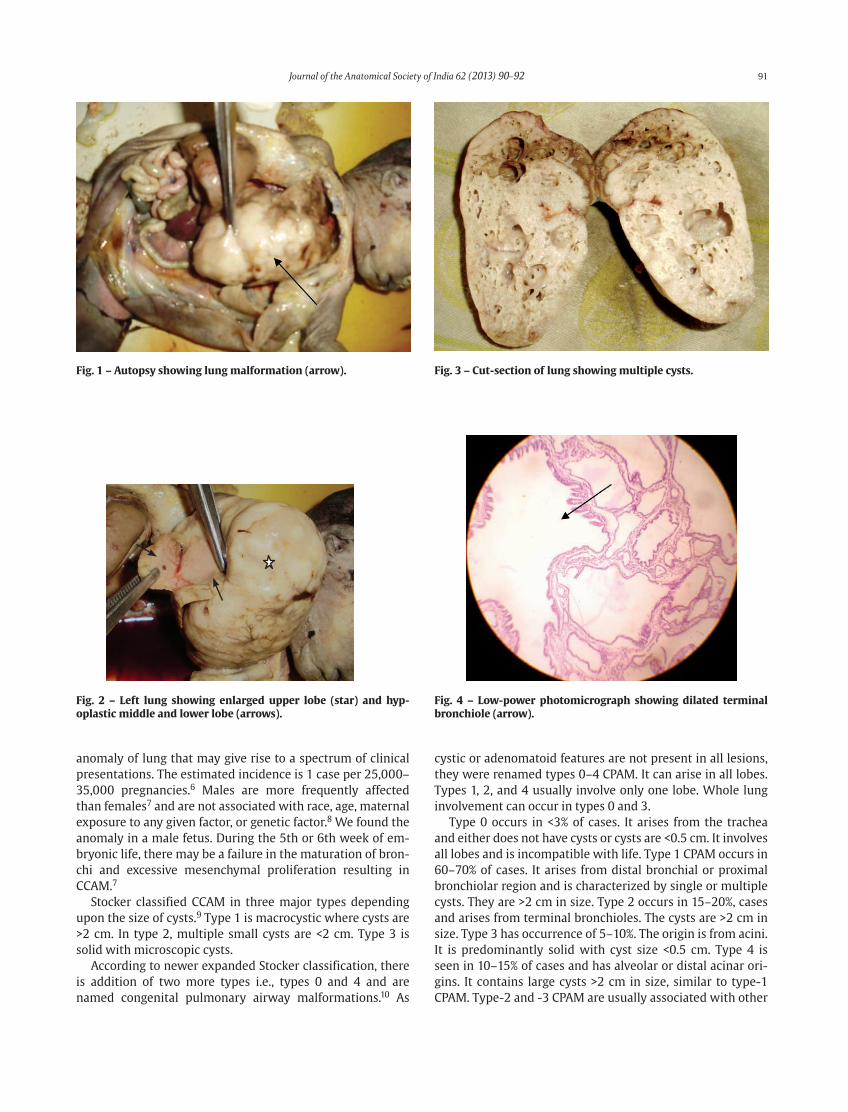
distended abdomen on the left side. On internal examination, a large mass was seen on the left side of the thorax extending into the abdomen (Fig. 1). The diaphragm was thinned, in-tact, and inverted. The abdominal contents were compressed by the pressure of the mass. The left lung showed three lobes. The mass was arising from the left upper pole of lung with dimensions 8 cm × 5 cm × 4 cm. The middle and lower lobes of the left lung were hypoplastic (Fig. 2). The heart was mark-edly displaced to the right side but was structurally normal. The right lung was hypoplastic. All other internal organs were normal.
The lung mass was spongy in consistency. Cut-section re-vealed multiple cysts of variable sizes. Their diameter ranged from 1 mm to 1.2 cm (Fig. 3).
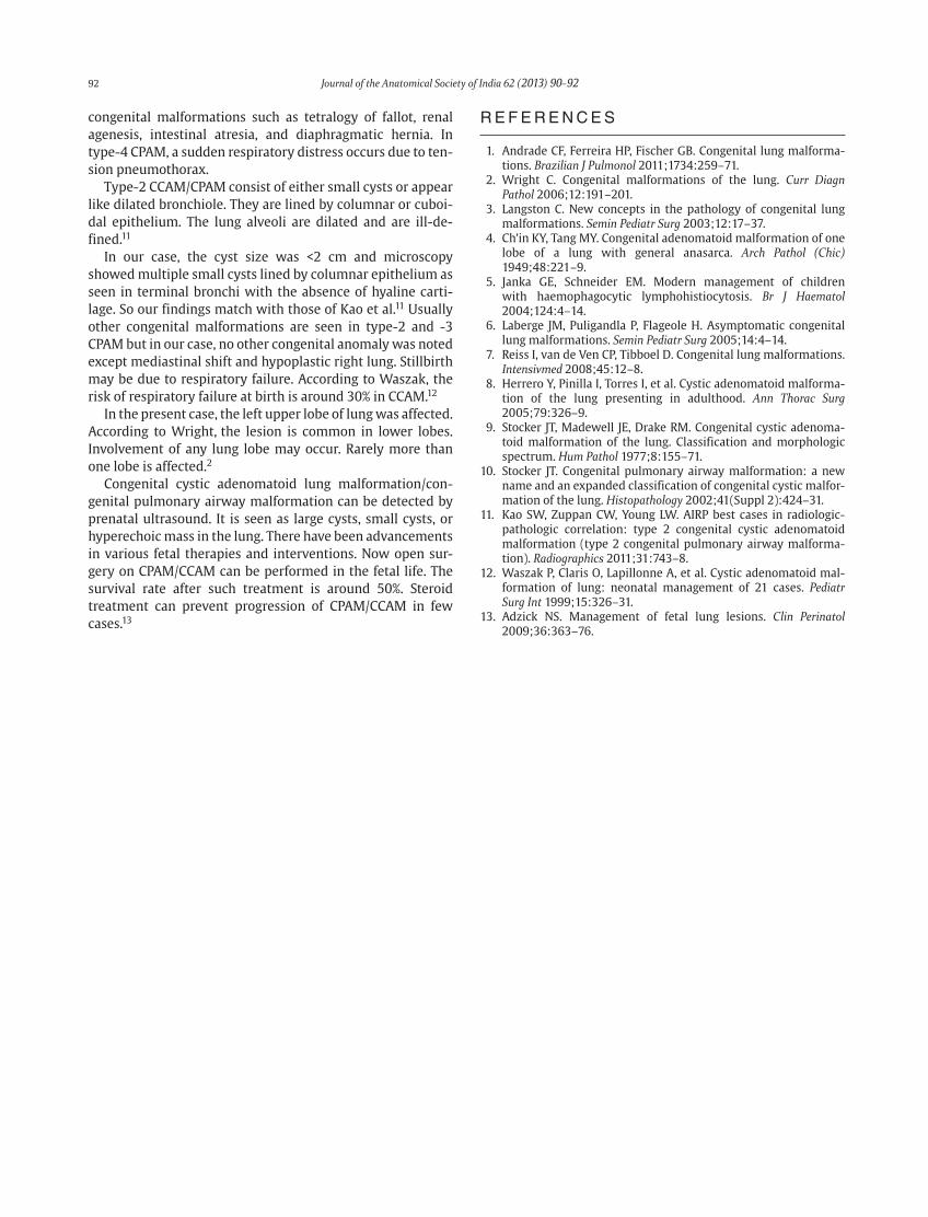
Histological findings showed multiple small cysts. They were lined by columnar epithelium and structurally appeared like distended terminal bronchioles. Few smooth muscle cells were seen surrounding the cyst. Hyaline cartilage was absent (Fig. 4). The diagnosis of type-2 CPAM was confirmed by mi-croscopy.
2. Discussion
Congenital pulmonary airway malformation or congenital cystic adenomatoid lung malformation is a developmental
A B S T R A C T
Congenital pulmonary airway malformation is a dysplastic lesion of the lung resulting from the overgrowth of terminal bronchiole. We present a rare case of type-2 congenital pulmonary airway malformation, diagnosed on autopsy in a full-term stillborn male fetus. The enlarged left upper lobe of lung showed multiple cysts with diameter <2 cm. Histologically, the cysts resembled bron-chioles. No other congenital anomaly was noted.
Copyright © 2013, The Anatomical Society of India. All rights reserved.
K E Y W O R D S
Congenital pulmonary airway malformation, Congenital cystic adenomatoid lung malformations, Autopsy.
0003-2778 Copyright © 2013. The Anatomical Society of India. All rights reserved.
* Corresponding author: Tel: +91 (0) 7122248590, 9822248570E-mail address: [email protected] (Deepali Onkar)
Journal of the Anatomical Society of India �� (2013) 90–92 91
anomaly of lung that may give rise to a spectrum of clinical presentations. The estimated incidence is 1 case per 25,000–35,000 pregnancies.6 Males are more frequently affected than females7 and are not associated with race, age, maternal exposure to any given factor, or genetic factor.8 We found the anomaly in a male fetus. During the 5th or 6th week of em-bryonic life, there may be a failure in the maturation of bron-chi and excessive mesenchymal proliferation resulting in CCAM.7
Stocker classified CCAM in three major types depending upon the size of cysts.9 Type 1 is macrocystic where cysts are >2 cm. In type 2, multiple small cysts are <2 cm. Type 3 is solid with microscopic cysts.
According to newer expanded Stocker classification, there is addition of two more types i.e., types 0 and 4 and are named congenital pulmonary airway malformations.10 As
cystic or adenomatoid features are not present in all lesions, they were renamed types 0–4 CPAM. It can arise in all lobes. Types 1, 2, and 4 usually involve only one lobe. Whole lung involvement can occur in types 0 and 3.
Type 0 occurs in <3% of cases. It arises from the trachea and either does not have cysts or cysts are <0.5 cm. It involves all lobes and is incompatible with life. Type 1 CPAM occurs in 60–70% of cases. It arises from distal bronchial or proximal bronchiolar region and is characterized by single or multiple cysts. They are >2 cm in size. Type 2 occurs in 15–20%, cases and arises from terminal bronchioles. The cysts are >2 cm in size. Type 3 has occurrence of 5–10%. The origin is from acini. It is predominantly solid with cyst size <0.5 cm. Type 4 is seen in 10–15% of cases and has alveolar or distal acinar ori-gins. It contains large cysts >2 cm in size, similar to type-1 CPAM. Type-2 and -3 CPAM are usually associated with other
Fig. 1 – Autopsy showing lung malformation (arrow). Fig. 3 – Cut-section of lung showing multiple cysts.
Fig. 2 – Left lung showing enlarged upper lobe (star) and hyp-oplastic middle and lower lobe (arrows).
Fig. 4 – Low-power photomicrograph showing dilated terminal bronchiole (arrow).
92 Journal of the Anatomical Society of India �� (2013) 90–92
congenital malformations such as tetralogy of fallot, renal agenesis, intestinal atresia, and diaphragmatic hernia. In type-4 CPAM, a sudden respiratory distress occurs due to ten-sion pneumothorax.
Type-2 CCAM/CPAM consist of either small cysts or appear like dilated bronchiole. They are lined by columnar or cuboi-dal epithelium. The lung alveoli are dilated and are ill-de-fined.11
In our case, the cyst size was <2 cm and microscopy showed multiple small cysts lined by columnar epithelium as seen in terminal bronchi with the absence of hyaline carti-lage. So our findings match with those of Kao et al.11 Usually other congenital malformations are seen in type-2 and -3 CPAM but in our case, no other congenital anomaly was noted except mediastinal shift and hypoplastic right lung. Stillbirth may be due to respiratory failure. According to Waszak, the risk of respiratory failure at birth is around 30% in CCAM.12
In the present case, the left upper lobe of lung was affected. According to Wright, the lesion is common in lower lobes. Involvement of any lung lobe may occur. Rarely more than one lobe is affected.2
Congenital cystic adenomatoid lung malformation/con-genital pulmonary airway malformation can be detected by prenatal ultrasound. It is seen as large cysts, small cysts, or hyperechoic mass in the lung. There have been advancements in various fetal therapies and interventions. Now open sur-gery on CPAM/CCAM can be performed in the fetal life. The survival rate after such treatment is around 50%. Steroid treatment can prevent progression of CPAM/CCAM in few cases.13
R E F E R E N C E S
1. Andrade CF, Ferreira HP, Fischer GB. Congenital lung malforma-tions. Brazilian J Pulmonol 2011;1734:259–71.
2. Wright C. Congenital malformations of the lung. Curr Diagn Pathol 2006;12:191–201.
3. Langston C. New concepts in the pathology of congenital lung malformations. Semin Pediatr Surg 2003;12:17–37.
4. Ch’in KY, Tang MY. Congenital adenomatoid malformation of one lobe of a lung with general anasarca. Arch Pathol (Chic) 1949;48:221–9.
5. Janka GE, Schneider EM. Modern management of children with haemophagocytic lymphohistiocytosis. Br J Haematol 2004;124:4–14.
6. Laberge JM, Puligandla P, Flageole H. Asymptomatic congenital lung malformations. Semin Pediatr Surg 2005;14:4–14.
7. Reiss I, van de Ven CP, Tibboel D. Congenital lung malformations. Intensivmed 2008;45:12–8.
8. Herrero Y, Pinilla I, Torres I, et al. Cystic adenomatoid malforma-tion of the lung presenting in adulthood. Ann Thorac Surg 2005;79:326 –9.
9. Stocker JT, Madewell JE, Drake RM. Congenital cystic adenoma-toid malformation of the lung. Classification and morphologic spectrum. Hum Pathol 1977;8:155–71.
10. Stocker JT. Congenital pulmonary airway malformation: a new name and an expanded classification of congenital cystic malfor-mation of the lung. Histopathology 2002;41(Suppl 2):424–31.
11. Kao SW, Zuppan CW, Young LW. AIRP best cases in radiologic-pathologic correlation: type 2 congenital cystic adenomatoid malformation (type 2 congenital pulmonary airway malforma-tion). Radiographics 2011;31:743–8.
12. Waszak P, Claris O, Lapillonne A, et al. Cystic adenomatoid mal-formation of lung: neonatal management of 21 cases. Pediatr Surg Int 1999;15:326–31.
13. Adzick NS. Management of fetal lung lesions. Clin Perinatol 2009;36:363–76.