Embed Size (px)
Citation preview

Congenital Heart Disease
Abdullah Alhuzaimi
MBBS, FRCPC, FACC
Congenital Heart disease
• Incidence:
• 1% of general population (excluding PDA in preterm babies)
• Recurrence rate :
• The risk of recurrence in siblings is about 3% ( up to 10% if HLHS)
• Higher recurrence if a parent is affected
• Higher recurrence for highly prevalent lesions (ex VSD)
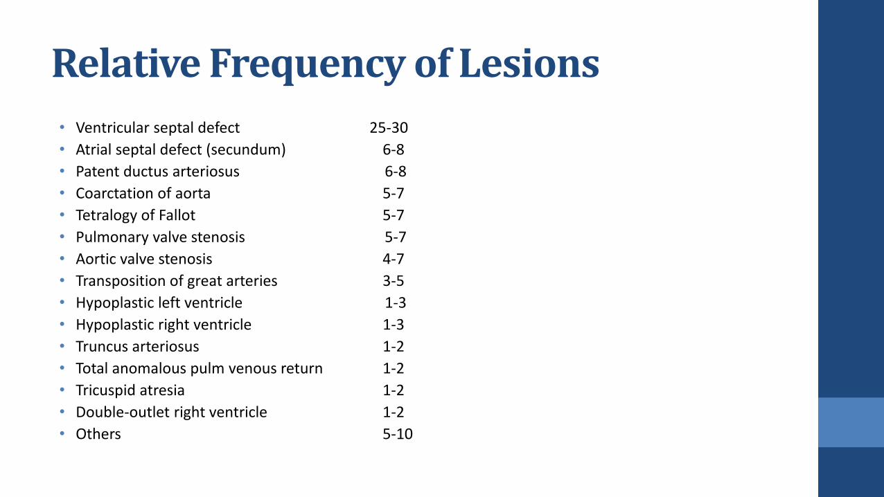
Relative Frequency of Lesions
• Ventricular septal defect 25-30
• Atrial septal defect (secundum) 6-8
• Patent ductus arteriosus 6-8
• Coarctation of aorta 5-7
• Tetralogy of Fallot 5-7
• Pulmonary valve stenosis 5-7
• Aortic valve stenosis 4-7
• Transposition of great arteries 3-5
• Hypoplastic left ventricle 1-3
• Hypoplastic right ventricle 1-3
• Truncus arteriosus 1-2
• Total anomalous pulm venous return 1-2
• Tricuspid atresia 1-2
• Double-outlet right ventricle 1-2
• Others 5-10

History Taking
• GESTATIONAL AND NATAL HISTORY:
• Infections, medications, excessive smoking or alcohol intake during pregnancy
• Birth weight
•
• POSTNATAL, PAST AND PRESENT HISTORY
• Weight gain, development, and feeding pattern
• Cyanosis, “cyanotic spells,” and squatting
• Tachypnea, dyspnea, puffy eyelids
• Frequency of respiratory infection
• Exercise intolerance
• Heart murmur
• Chest pain
• Syncope
• Palpitations
• Joint symptoms
• Neurologic symptoms
• Medications
FAMILY HISTORY
• Hereditary disease
• Congenital heart defect
• Rheumatic fever
• Sudden unexpected death
• Diabetes mellitus, arteriosclerotic heart disease, hypertension, and so on

Physical Examination
• Growth pattern :
• low birth weight – Congential anmolies or torch
• low weight : l-r shunt
• low weight and height : cyanotic heart disease
• Blood pressure measurement
• General appearance and nutritional state
• Color
• Clubbing
• Pulses
• Respiratory rate, dyspnea, and retraction
• Chest deformities
• Apical Impulse
• Point of Maximal Impulse
• Hyperactive Precordium
• Thrills
• Auscultation:
• Heart sounds
• Extra heart sounds
• Ejection click or opening snap
• Heart murmur
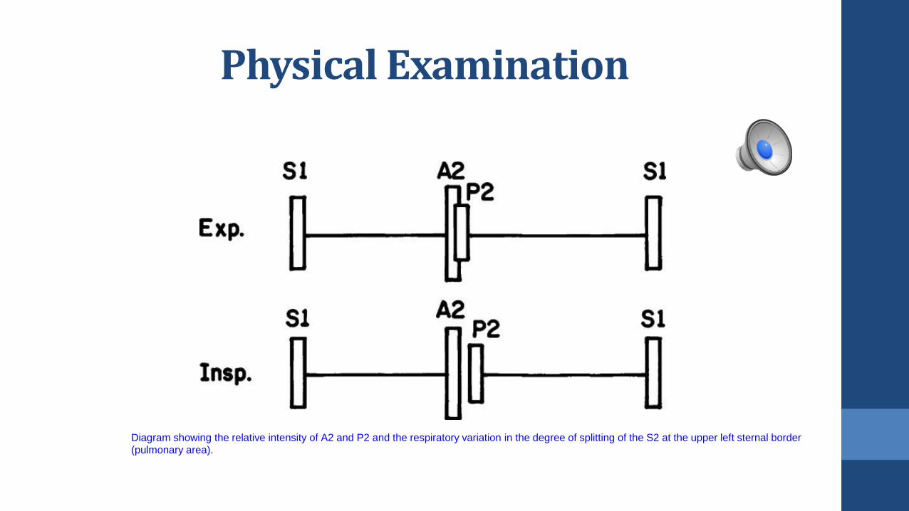
Physical Examination
Diagram showing the relative intensity of A2 and P2 and the respiratory variation in the degree of splitting of the S2 at the upper left sternal border
(pulmonary area).

Classification of CHD
• Left to Right Shunt Lesions
• Right to Left Shunt Lesions - Cyanotic heart diseases
• Obstructive lesions
• Valvular Regurgitation Lesions- Congenital MR or TR

Quiz
• 3 years old boy was noted to have heart murmur during routine medical checkup.
• He is asymptomatic.
• Cardiac examination showed normal satuation. Normal RR and HR. No signs of heart failure.
• CXR showed mild cardiomegaly
• Auscultation

Quiz
The most likely Lesion:
1. ASD
2. VSD
3. Aortic stenosis
4. TGA

Classification of CHD
• Left to Right Shunt Lesions
• Right to Left Shunt Lesions - Cyanotic heart diseases
• Obstructive lesions
• Valvular Regurgitation Lesions- Congenital MR or TR
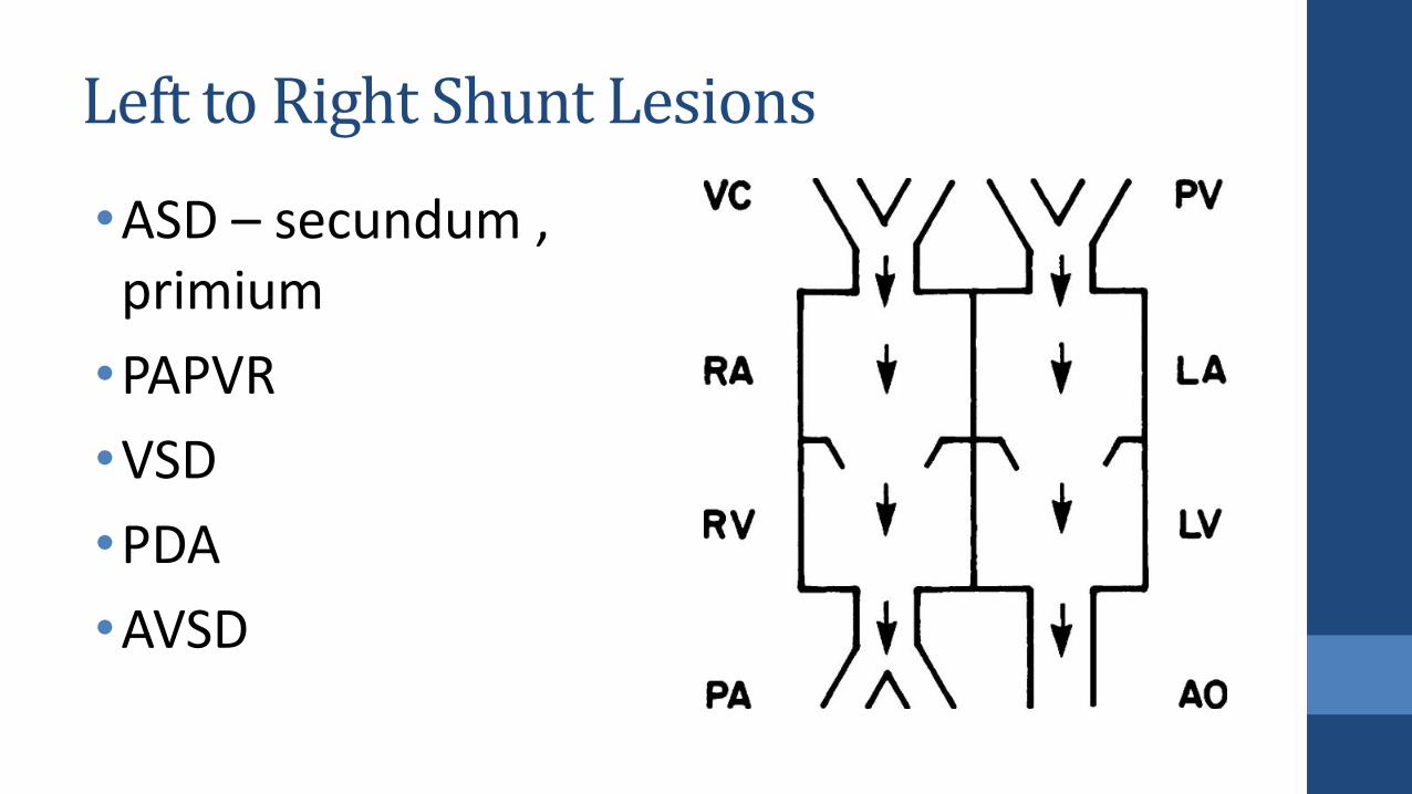
Left to Right Shunt Lesions
•ASD – secundum , primium
•PAPVR
•VSD
•PDA
•AVSD
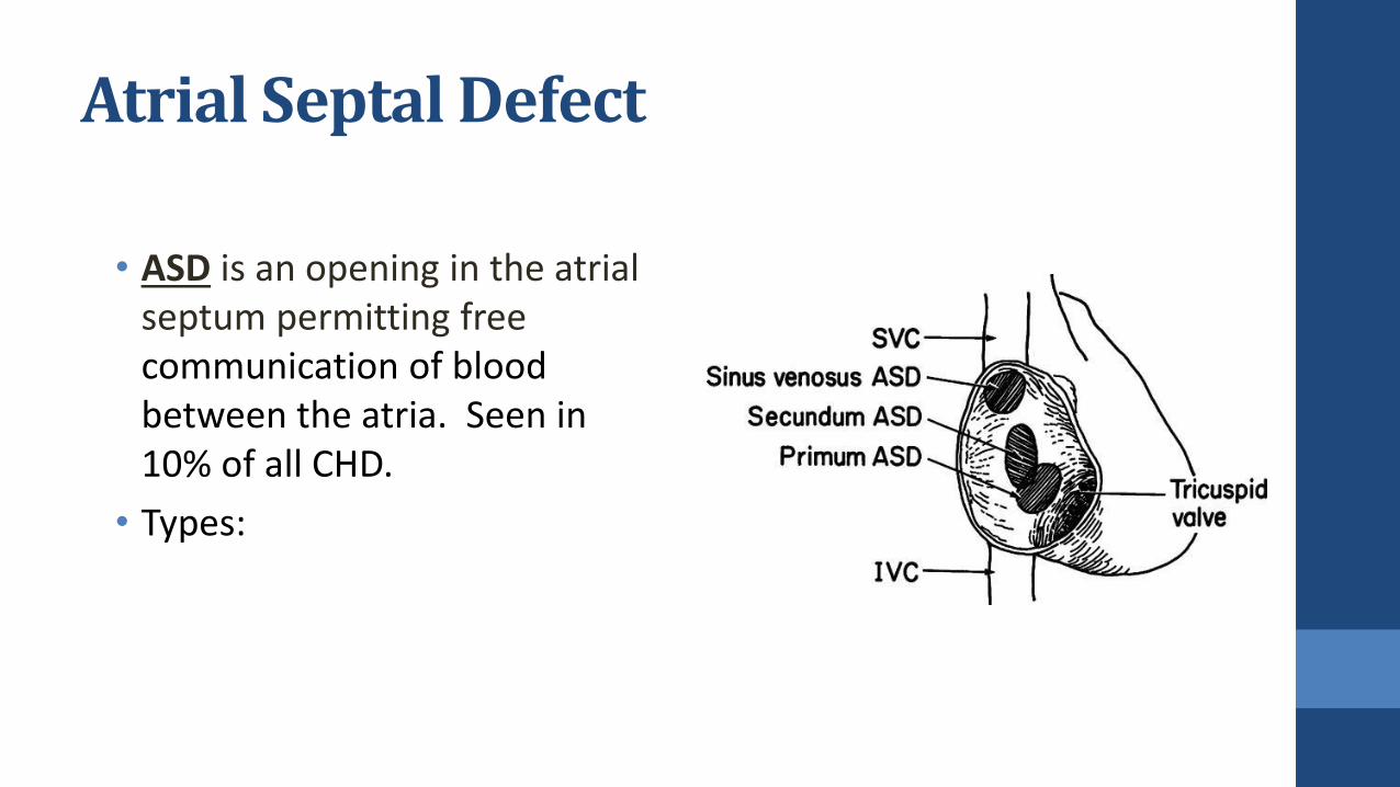
Atrial Septal Defect
• ASD is an opening in the atrial septum permitting free communication of blood between the atria. Seen in 10% of all CHD.
• Types:

Atrial Septal Defect
Large RA
Large RV
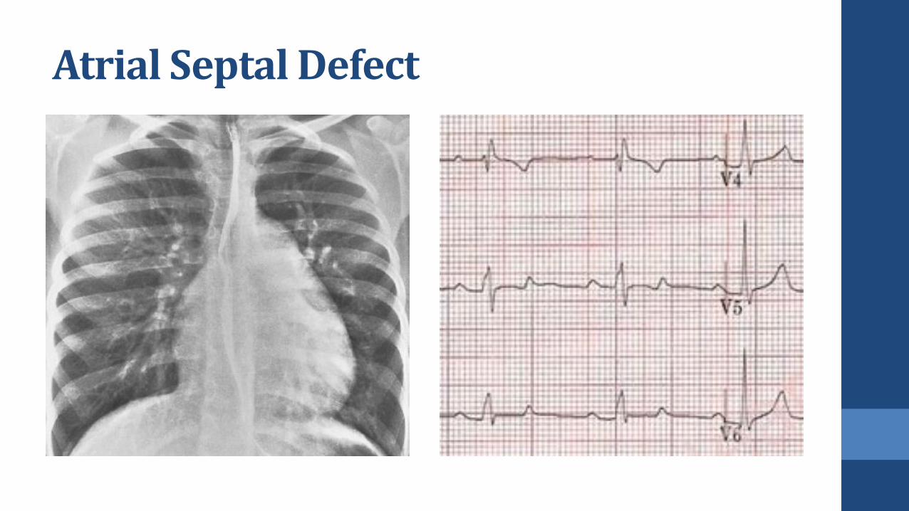
Atrial Septal Defect

Atrial Septal Defect
• History: usually asymptomatic.
• Examination: S1 + fixed split S2 + ESM LUSB
• Natural history: 40% close spontaneously.
• 20% develop pulmonary hypertension in 20s if not closed
• Tx: Device or surgical closure at pre-school age if Qp:Qs > 2:1 , pul
HTN, significant RV dilation
• Mortality is < 1%.
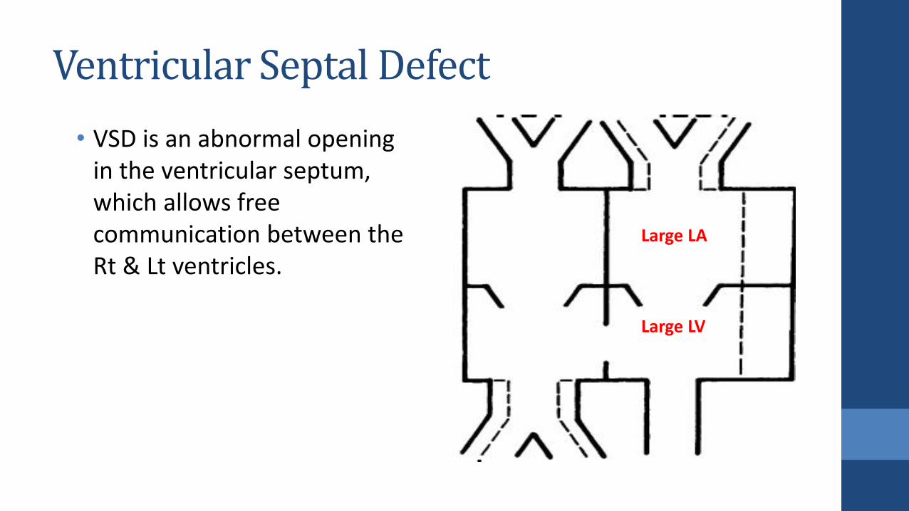
Ventricular Septal Defect
Large LA
Large LV
• VSD is an abnormal opening in the ventricular septum, which allows free communication between the Rt & Lt ventricles.

Types of Ventricular Septal Defect
1. Perimembranous– Most common.
2. Infundibular(subpulmonary) – involves the RV outflow tract.
3. Muscular VSD
4. Inlet
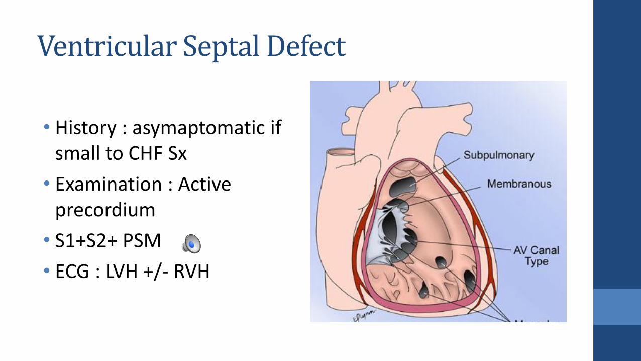
Ventricular Septal Defect
• History : asymaptomatic if small to CHF Sx
• Examination : Active precordium
• S1+S2+ PSM
• ECG : LVH +/- RVH

Ventricular Septal Defect
Ventricular Septal Defect
Natural History :
• depend on type of VSD and size ( inlet and outlet don’t close spontaneously)
• Spontaneous closure occurs in 30% to 40% of patients with membranous VSDs and muscular VSDs
Treatment :
• Medical therapy
• Device closure , PA band , surgical closure
• Surgical Indications : CHF, Qp:Qs > 2:1 , Early pulmonary hypertension
• Surgery not indicated if small VSD , asymptomatic and Qp:Qs < 1.5
Quiz
Infant with large VSD is likely to present:
1. at birth with cyanosis
2. at birth with loud murmur
3. At 6 weeks with CHF

You do not suffer from your beliefs.
You suffer from your disbeliefs.
If you have no hope inside of you, it’s not because there is no hope, it’s because you don’t
believe there is.
Gianpaolo Tucci’s work
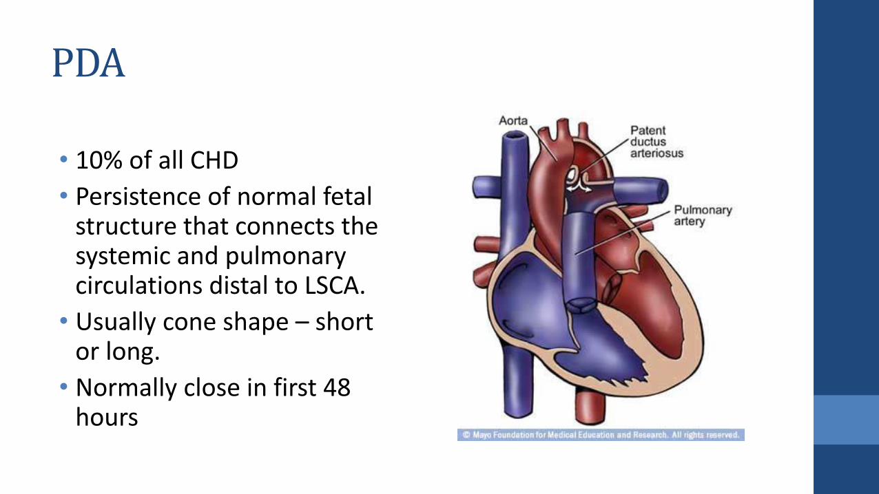
PDA
• 10% of all CHD
• Persistence of normal fetal structure that connects the systemic and pulmonary circulations distal to LSCA.
• Usually cone shape – short or long.
• Normally close in first 48 hours
PDA pathophysiology
• Left to right shunt at level of pulmonary artery.
• Shunt depend on PVR and size of the PDA.
• It increase the LA, LV and ascending aorta size
PDA
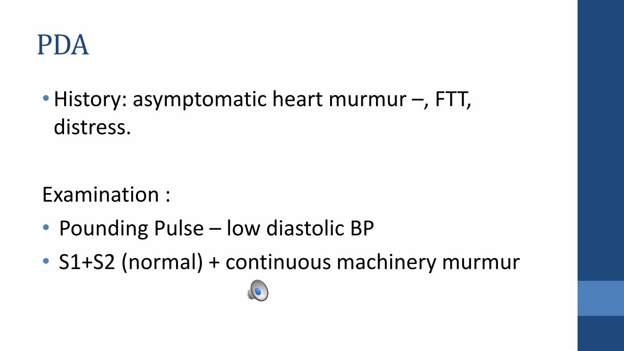
• History: asymptomatic heart murmur –, FTT, distress.
Examination :
• Pounding Pulse – low diastolic BP
• S1+S2 (normal) + continuous machinery murmur

PDA
• ECG: similar to VSD (N-LVH)
• CXR:
• Cardiomegaly
• enlargement of the LA, LV, and ascending aorta.
• Pulmonary vascular markings are increased
• Echo: diagnostic images
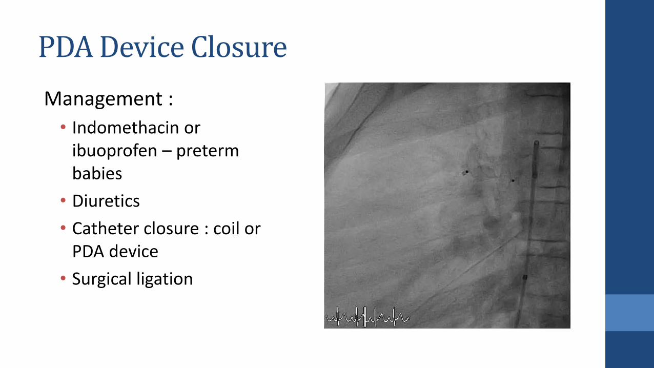
PDA Device Closure
Management :
• Indomethacin or ibuoprofen – preterm babies
• Diuretics
• Catheter closure : coil or PDA device
• Surgical ligation
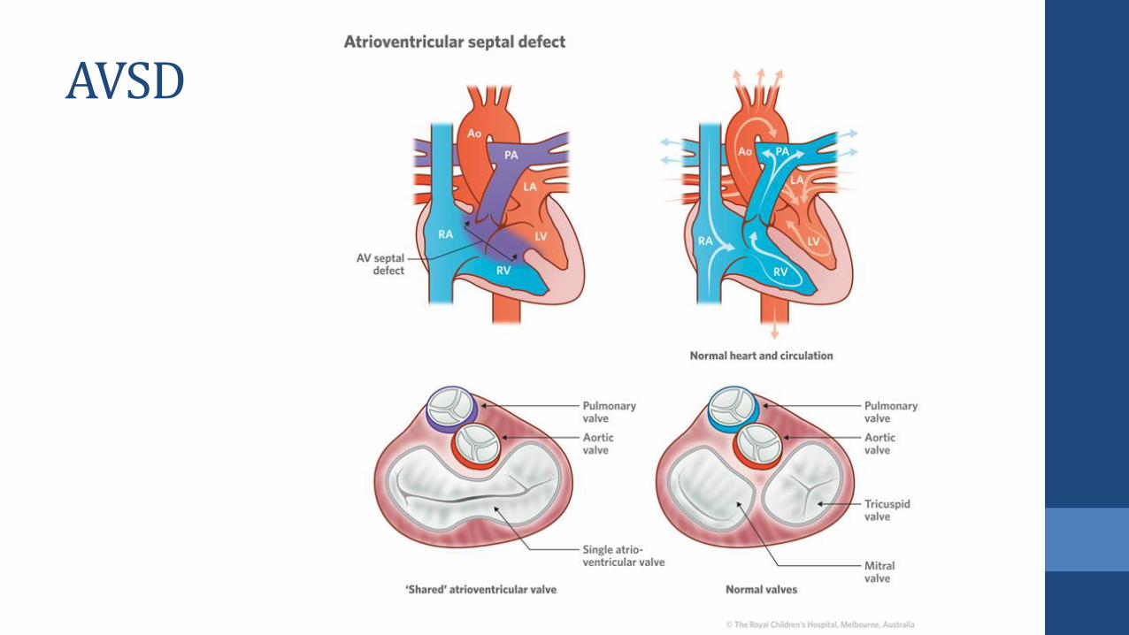
AVSD
Anatomy Play- Benard Salunga
Classification of CHD
• Left to Right Shunt Lesions
• Right to Left Shunt Lesions - Cyanotic heart diseases
• Obstructive lesions
• Valvular Regurgitation Lesions- Congenital MR or TR

Obstructive Lesions
• Pulmonary stenosis (PS)
• Aortic stenosis (AS)
• Coarctation of the aorta (COA)
• Interrupted aortic arch

Pulmonary Stenosis
• 10% of all CHD
• Types : valvular > subvavular > supra valvular
• Valvular : thickened, with fused or absent commissures and a small orifice. Or three leaflets but dysplastic
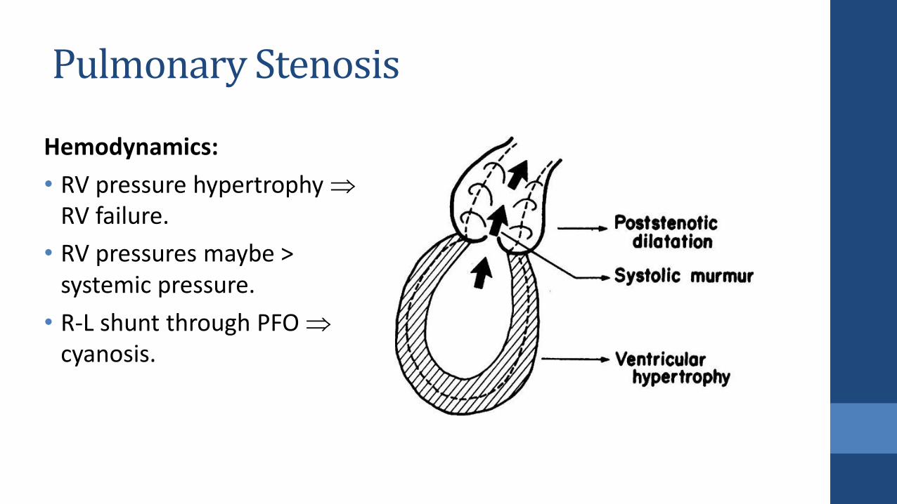
Pulmonary Stenosis
Hemodynamics:
• RV pressure hypertrophyRV failure.
• RV pressures maybe > systemic pressure.
• R-L shunt through PFO cyanosis.
Obstructive Lesions
Pulmonary Stenosis
• History : asymptomatic if small, exertional dyspnea and easy fatigability may be present in patients with moderately severe case.
• Cyanosis in neonate ( or with ASD)
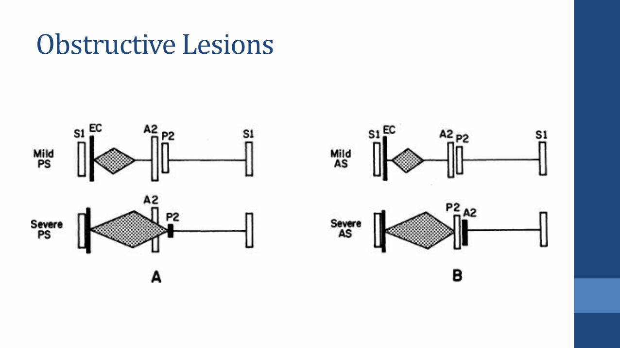
• Examination: Normal S1 + EC + Fixed split S2 or faint P2 + ESM
Pulmonary Stenosis
• ECG: RAD and RVH
• CXR: normal heart size
• Echo : estricted systolic motion (doming), Increased gradient across Valve
Pulmonary Stenosis
Management :
• Mild – No treatment required
• Moderate to severe : Balloon valvuloplasty is the procedure of choice for the valvular stenosis
• Indications : Symptomatic patients with catheterization gradient >=30 mm Hg or asymptomatic patients >=40 mm Hg
• Surgical valvotomy for dysplastic pulmonary valves resistant to dilatation.
Quiz
Q) Which of the following is correct pulmonary stenosis :
1. A2 is earlier
2. S2 is single
3. P2 is delayed
4. S1 is muffled

Aortic Stenosis
• 7% of all CHD
• Types: Valvular AS (71%), subvalvular stenosis (23%) and supravalvularstenosis (6%).
• Etiology: bicuspid aortic valve, a unicuspid aortic valve, or stenosis of the tricuspid aortic valve
• Types of Aortic stenosis :
Aortic Stenosis
History :
• Mild AS-usually asymptomatic.
• Moderate AS – Chest pain, dypsnea on exertion, dizziness & syncope.
• Severe or Critical AS or – Weak pulses, left sided heart failure, Sudden Death, shock.
Aortic stenosis
Examination :
• Narrow pulse pressure.
• LV heave at the Apex.
• Systolic thrill RUSB and suprasternal notch
• S1 + narrow split S2 or paradoxically splitting + ESM best heard at the second RUSB radiate to neck and apex.
• ECG : LVH , ischemia
• CXR : Normal heart size. Dialted aorta
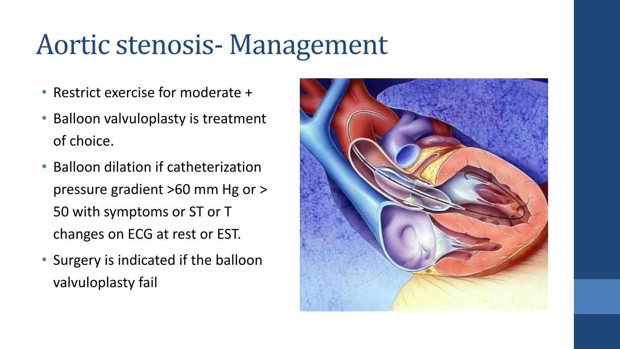
Aortic stenosis- Management
• Restrict exercise for moderate +
• Balloon valvuloplasty is treatment
of choice.
• Balloon dilation if catheterization
pressure gradient >60 mm Hg or >
50 with symptoms or ST or T
changes on ECG at rest or EST.
• Surgery is indicated if the balloon
valvuloplasty fail
Aortic stenosis- Management
Outcome:
• Worse outcome compare to pulmonary stenosis
• Higher risk of sudden death
• Risk of development of aortic regurgitation and re-
stenosis
• Might need valve replacement
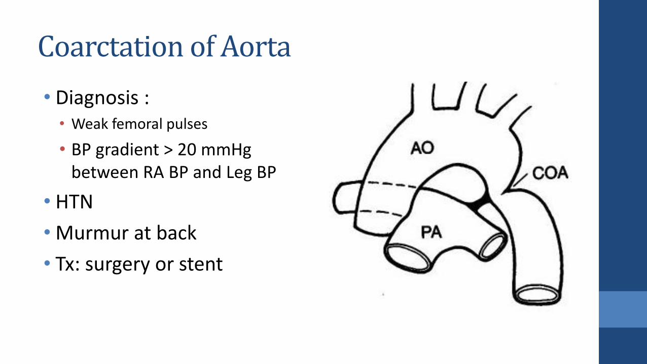
Coarctation of Aorta
• Diagnosis : • Weak femoral pulses
• BP gradient > 20 mmHg between RA BP and Leg BP
• HTN
• Murmur at back
• Tx: surgery or stent
Cyanotic Heart Disease
• Tetralogy of Fallot (TOF)
• Tricuspid atresia (TA)
• Total anomalous pulmonary venous return (TAPVR)
• Truncus arteriosus
• Transposition of the great vessels
• Hypoplastic left heart syndrome (HLH)
• Pulmonary atresia (PA) / critical PS
• Double outlet right ventricle (DORV)
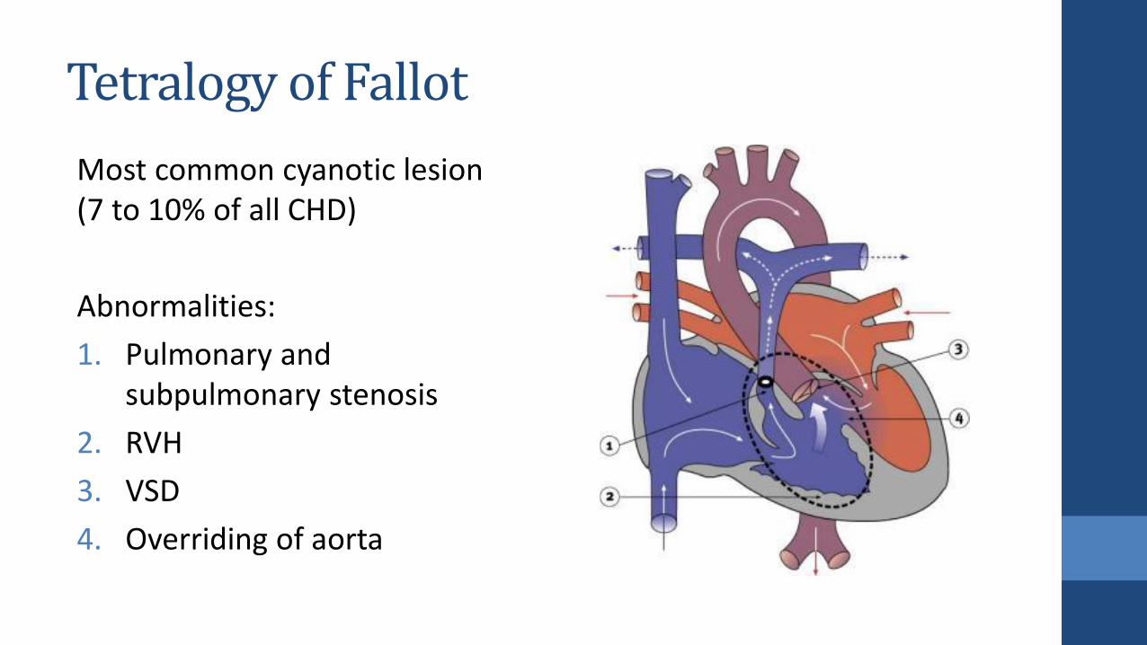
Tetralogy of Fallot
Most common cyanotic lesion (7 to 10% of all CHD)
Abnormalities:
1. Pulmonary and subpulmonary stenosis
2. RVH
3. VSD
4. Overriding of aorta
Tetralogy of Fallot- Clinical features
• Clinical findings vary depending on degree of RVOT obstruction
• Most patients are cyanotic by 4 months and it is usually progressive
• Hypoxemic spells (“tet spells”) are one of the hallmarks of severe tetralogy
• Examination: Systolic ejection murmur at the upper LSB
Tetralogy of Fallot- Clinical features
• Hypercyanotic (or "tet") spells present as periods of profound cyanosis that occur because of episodes of almost total RVOT obstruction. They typically arise when an infant becomes agitated or in older, uncorrected children after vigorous exercise.
• More blood going to systemic circulation and less blood going to lungs ..!

Tet Spells- Management
1. knee-chest position to increase systemic vascular resistance, which promotes movement of blood from the right ventricle into the pulmonary circulation rather than the aorta.
2. Oxygen
3. Fluid bolus – Increase RV filling
4. IV Morphine
5. IV Propranolol or esmolol
6. IV Phenylephrine
Tetralogy of Fallot- Management
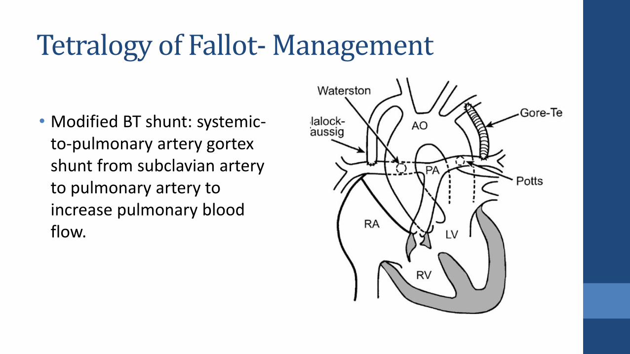
• Modified BT shunt: systemic-to-pulmonary artery gortexshunt from subclavian artery to pulmonary artery to increase pulmonary blood flow.
Tetralogy of Fallot- Management
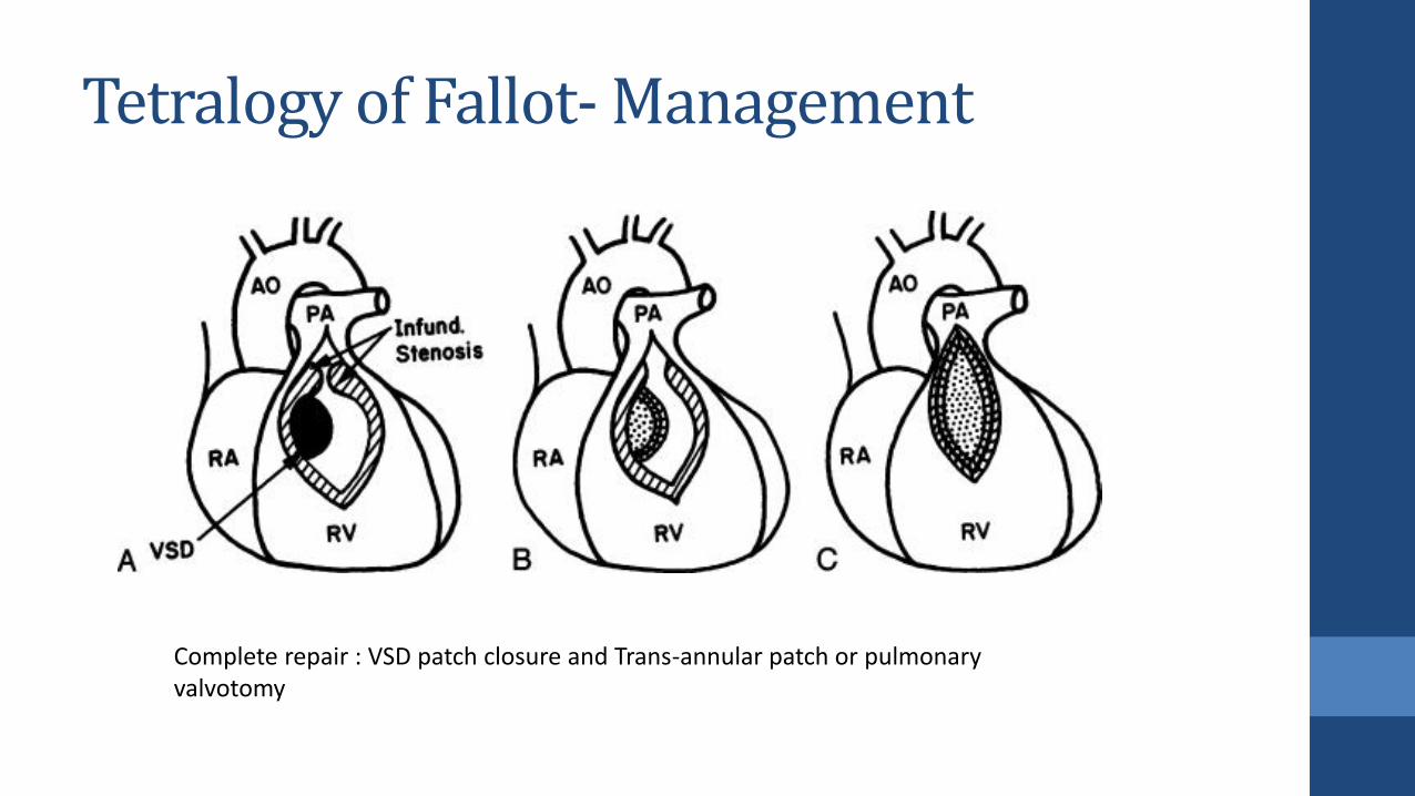
Complete repair : VSD patch closure and Trans-annular patch or pulmonary valvotomy