Embed Size (px)
Citation preview
12/9/2015
1
Congenital Heart Disease: A NICU Perspective
Kerri Carter, MDAssistant Professor
Division of Pediatric Cardiology, CHoR
November 13, 2015
12/9/2015
2
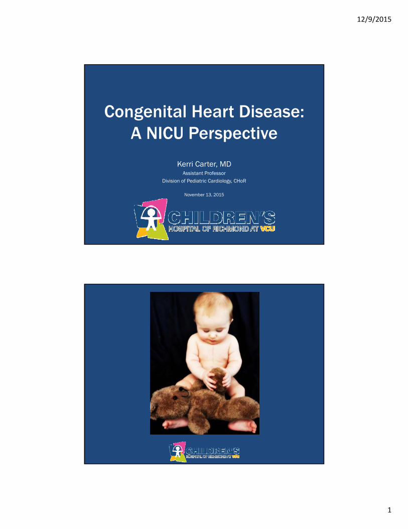
Objectives• To review the basics of cardiac anatomy and
embryology• To review the fetal and transitional circulation and
pertinent physiologic changes that occur at birth• To define basic murmur nomenclature and grading• To practice a systematic approach to differential
diagnosis of cardiac murmurs by location• To develop a strategy for identifying innocent
murmurs vs. those that need subspecialty evaluation• To introduce neonatal screening guidelines and
algorithms
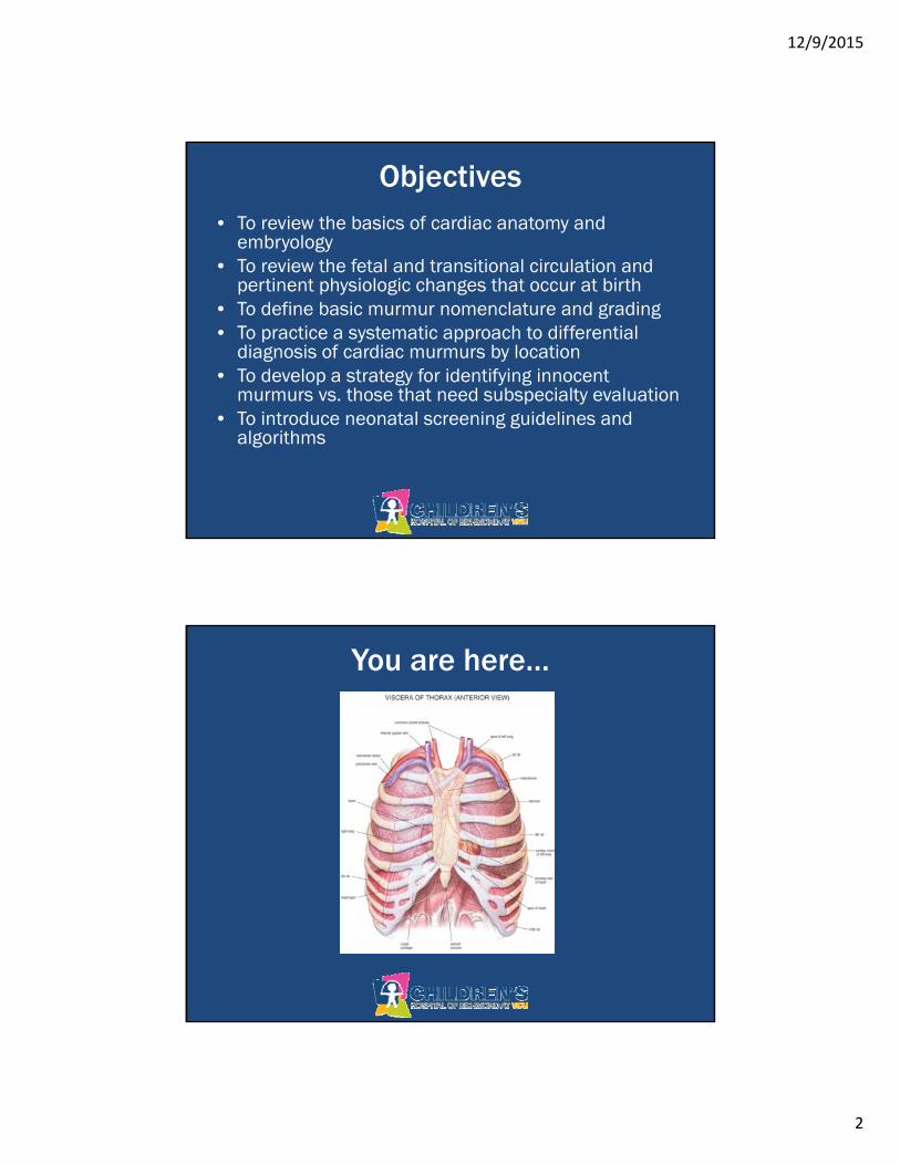
You are here…
12/9/2015
3
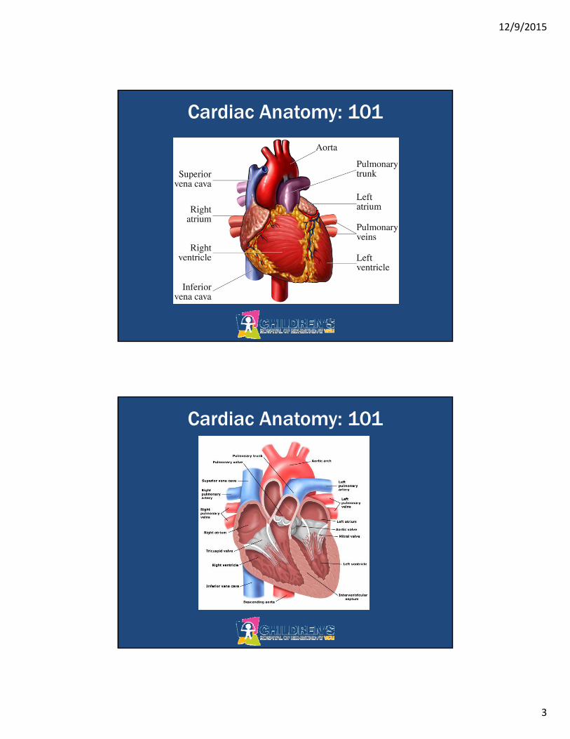
Cardiac Anatomy: 101
Cardiac Anatomy: 101
12/9/2015
4
But how the heck did we get here??
Sherman, set the Way-Back machine to the second week of gestation, and
HANG ON!!!
12/9/2015
5
12/9/2015
6
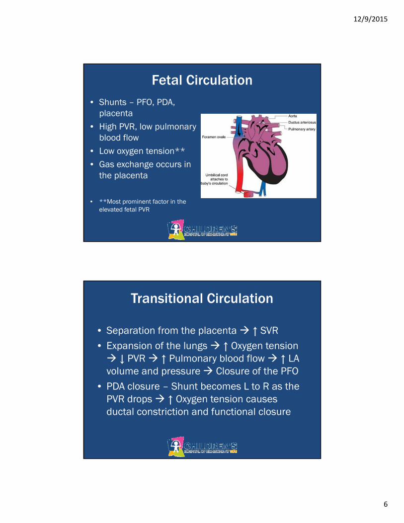
Fetal Circulation• Shunts – PFO, PDA,
placenta• High PVR, low pulmonary
blood flow• Low oxygen tension**• Gas exchange occurs in
the placenta
• **Most prominent factor in the elevated fetal PVR
Transitional Circulation
• Separation from the placenta ↑ SVR• Expansion of the lungs ↑ Oxygen tension ↓ PVR ↑ Pulmonary blood flow ↑ LA volume and pressure Closure of the PFO
• PDA closure – Shunt becomes L to R as the PVR drops ↑ Oxygen tension causes ductal constriction and functional closure
12/9/2015
7
What about that duct…
• Oxygen induces the initial constriction along with a normal fall in endogenous levels of prostaglandins after birth in the term baby
• Persistence of the ductus in preterm infants is likely secondary to a combination of persistently elevated prostaglandin levels and also relative hypoxemia
Transitional Circulation
• By 24h of life, the PVR and PA pressure will fall to about ½ systemic
• This continues through the first 6w of life • Related to involution of smooth muscle in
the walls of small pulmonary arteries
12/9/2015
8
And we’re back!
What about murmurs???
12/9/2015
9
Murmurs
• What is a murmur? – Sound created by blood moving through the
heart & blood vessels
• Timing - Systolic vs. Diastolic• Location• Grade• Quality• Radiation
Systolic Murmurs• Start with or after S1 & terminate before or with S2.• Graded I-VI
– I – Barely audible– II – As loud as heart sounds– III – Louder than heart sounds, no thrill– IV – Readily audible with palpable thrill– V – Loud enough to be heard with diaphragm barely on
chest wall; + palpable thrill– VI – Loud enough to be heard with the diaphragm off the
chest; + palpable thrill
• S1- Coincident (holosystolic) vs. Systolic Ejection
12/9/2015
10
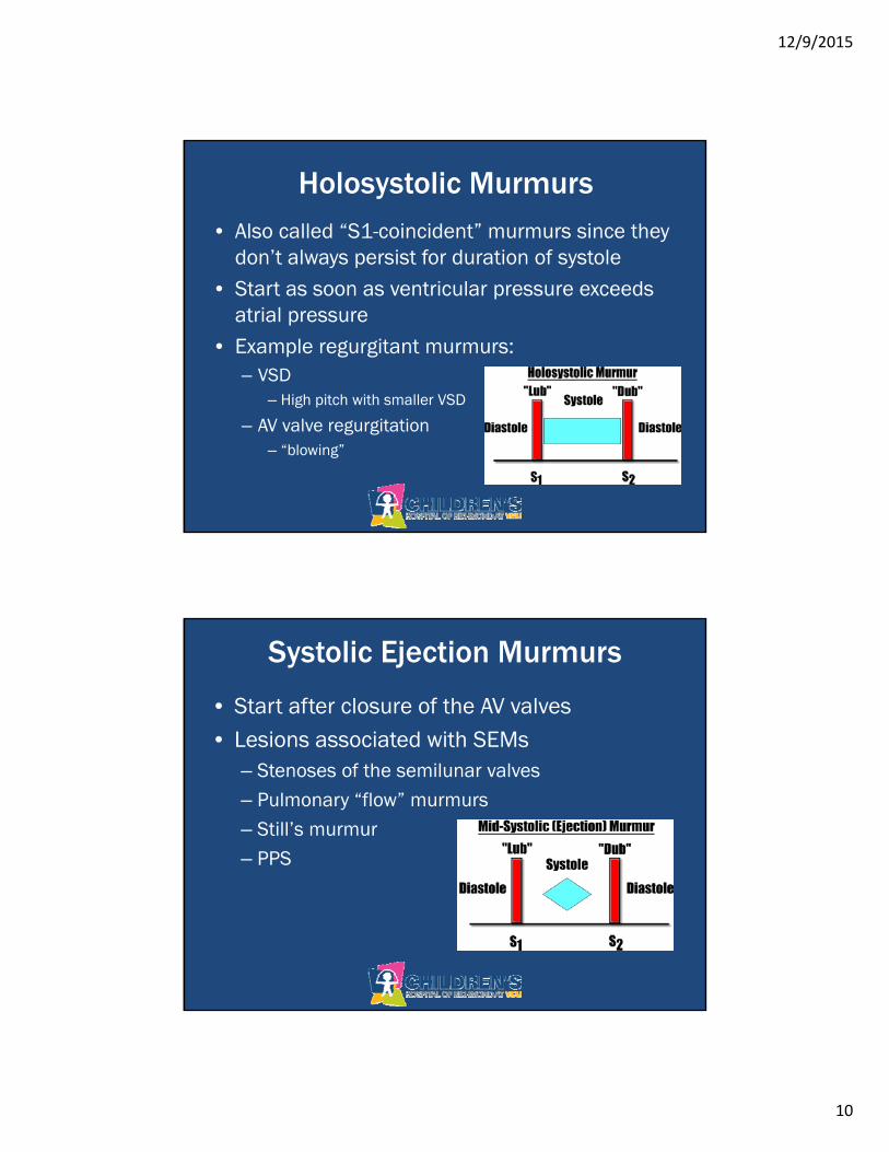
Holosystolic Murmurs• Also called “S1-coincident” murmurs since they
don’t always persist for duration of systole• Start as soon as ventricular pressure exceeds
atrial pressure • Example regurgitant murmurs:
– VSD– High pitch with smaller VSD
– AV valve regurgitation– “blowing”
Systolic Ejection Murmurs
• Start after closure of the AV valves• Lesions associated with SEMs
– Stenoses of the semilunar valves– Pulmonary “flow” murmurs – Still’s murmur– PPS
12/9/2015
11
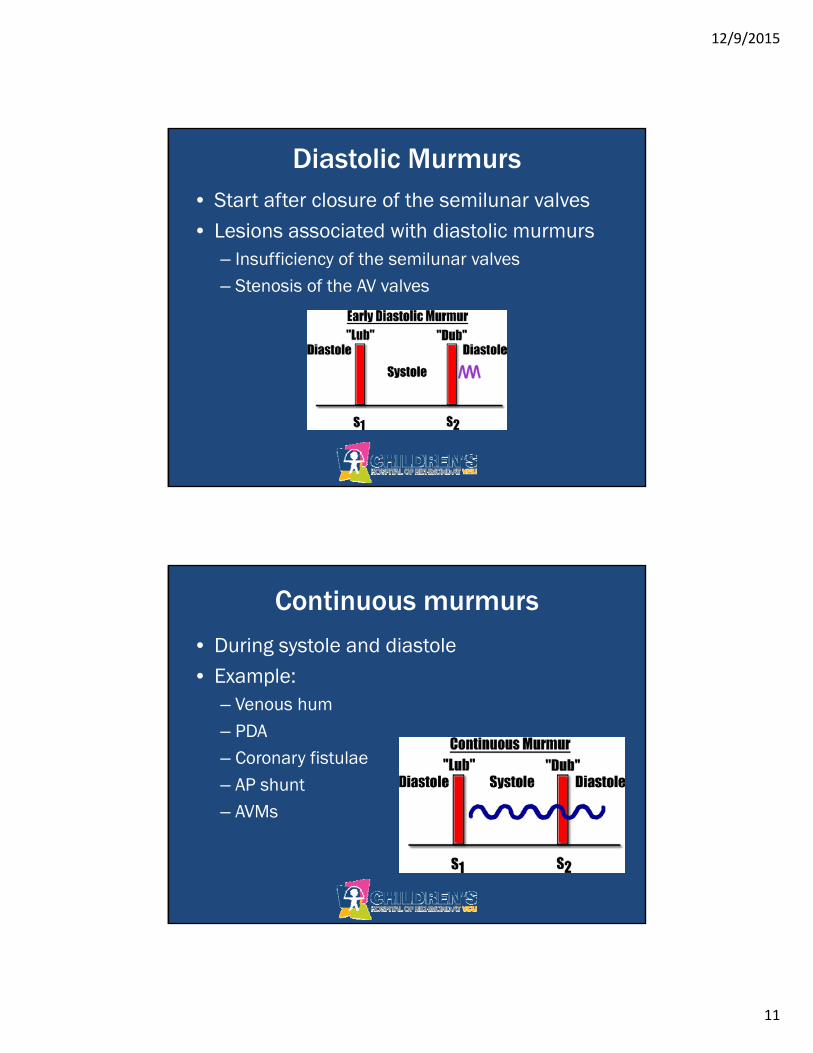
Diastolic Murmurs• Start after closure of the semilunar valves• Lesions associated with diastolic murmurs
– Insufficiency of the semilunar valves– Stenosis of the AV valves
Continuous murmurs
• During systole and diastole• Example:
– Venous hum– PDA– Coronary fistulae– AP shunt– AVMs
12/9/2015
12
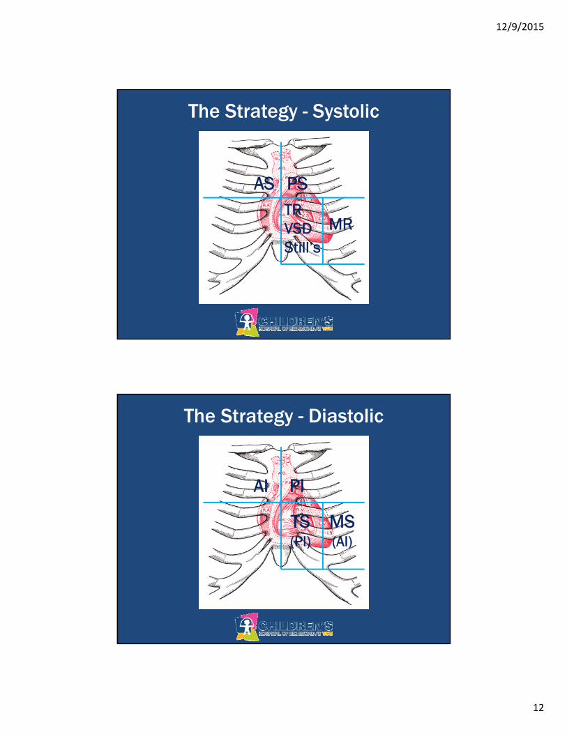
The Strategy - Systolic
AS PSTRVSDStill’s
MR
The Strategy - Diastolic
AI PI
TS(PI)
MS(AI)
12/9/2015
13
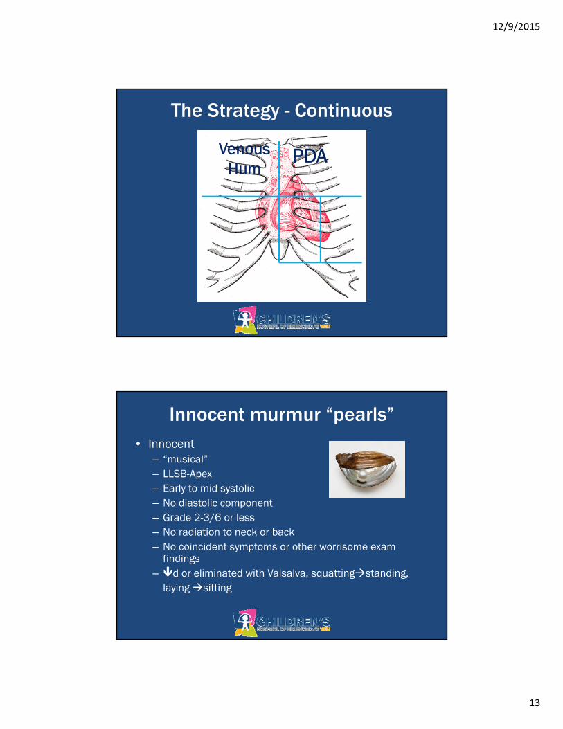
The Strategy - Continuous
Venous Hum
PDA
Innocent murmur “pearls”• Innocent
– “musical”– LLSB-Apex– Early to mid-systolic– No diastolic component– Grade 2-3/6 or less– No radiation to neck or back– No coincident symptoms or other worrisome exam
findings– d or eliminated with Valsalva, squattingstanding,
laying sitting
12/9/2015
14
Phone a friend..
• Presence of a thrill• Radiation to the neck or interscapular area• Clicks, gallops, or other concerning exam
findings• Diastolic murmurs• Hyperdynamic precordium or subxiphoid
impulse• Symptoms
So why don’t I care about murmurs???
• Can you hear anything with this thing??
• Lots of big bad heart disease has no associated pathologic murmur…
12/9/2015
15
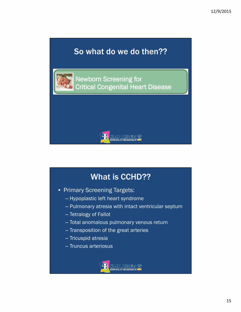
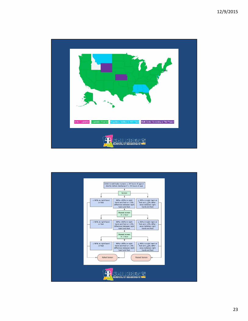
So what do we do then??
What is CCHD??
• Primary Screening Targets:– Hypoplastic left heart syndrome– Pulmonary atresia with intact ventricular septum– Tetralogy of Fallot– Total anomalous pulmonary venous return– Transposition of the great arteries– Tricuspid atresia– Truncus arteriosus
12/9/2015
16
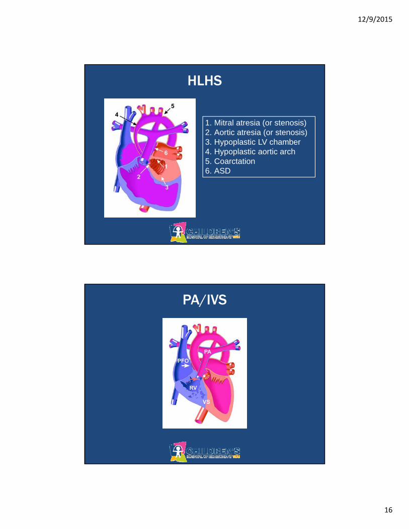
HLHS
1. Mitral atresia (or stenosis)2. Aortic atresia (or stenosis)3. Hypoplastic LV chamber4. Hypoplastic aortic arch5. Coarctation6. ASD
PA/IVS
12/9/2015
17
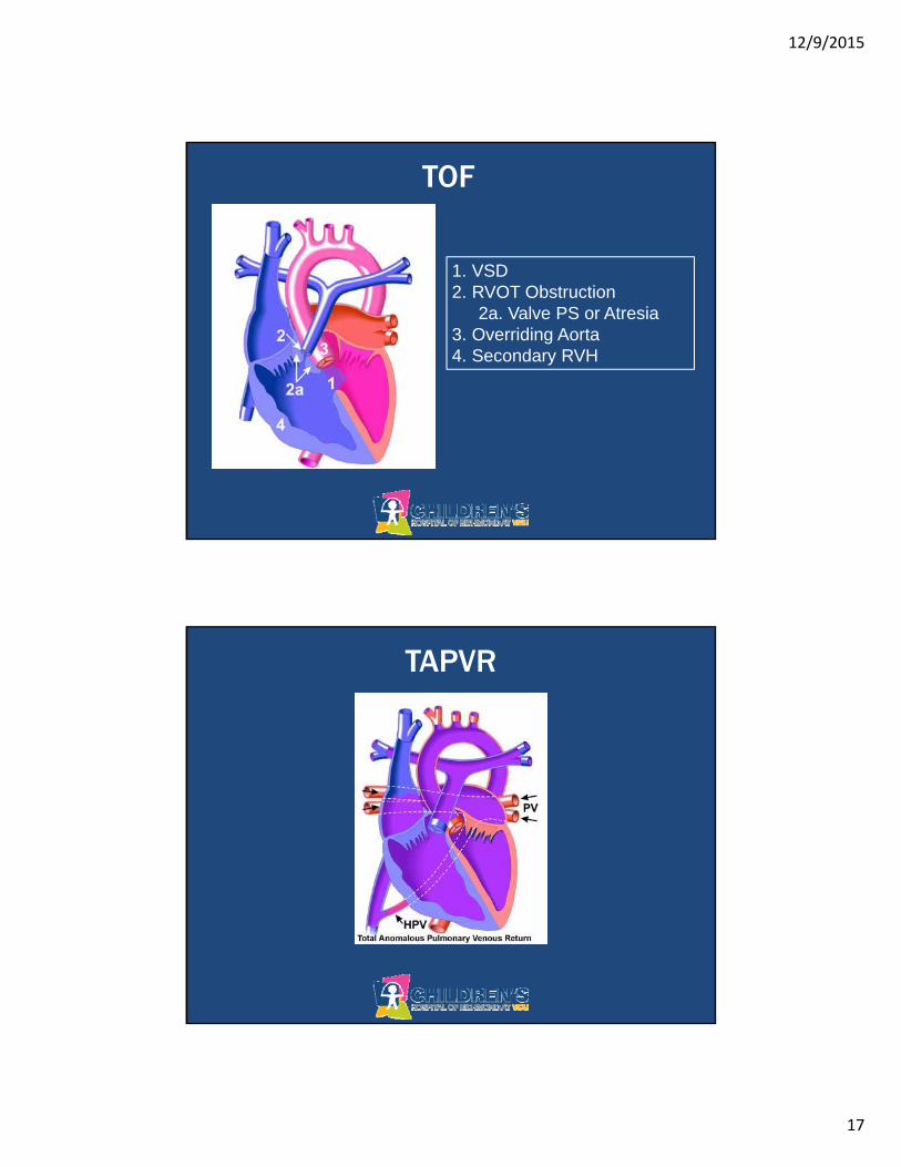
TOF
1. VSD2. RVOT Obstruction
2a. Valve PS or Atresia3. Overriding Aorta4. Secondary RVH
TAPVR
12/9/2015
18
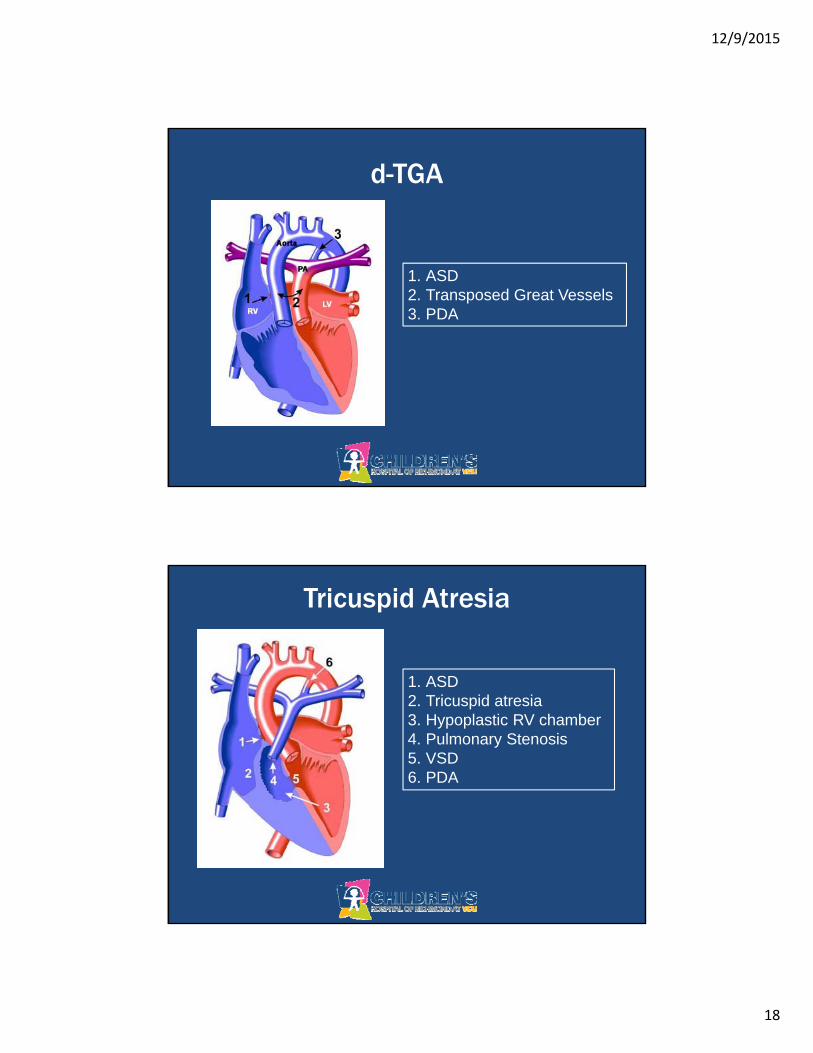
d-TGA
1. ASD2. Transposed Great Vessels3. PDA
Tricuspid Atresia
1. ASD2. Tricuspid atresia3. Hypoplastic RV chamber4. Pulmonary Stenosis5. VSD6. PDA
12/9/2015
19
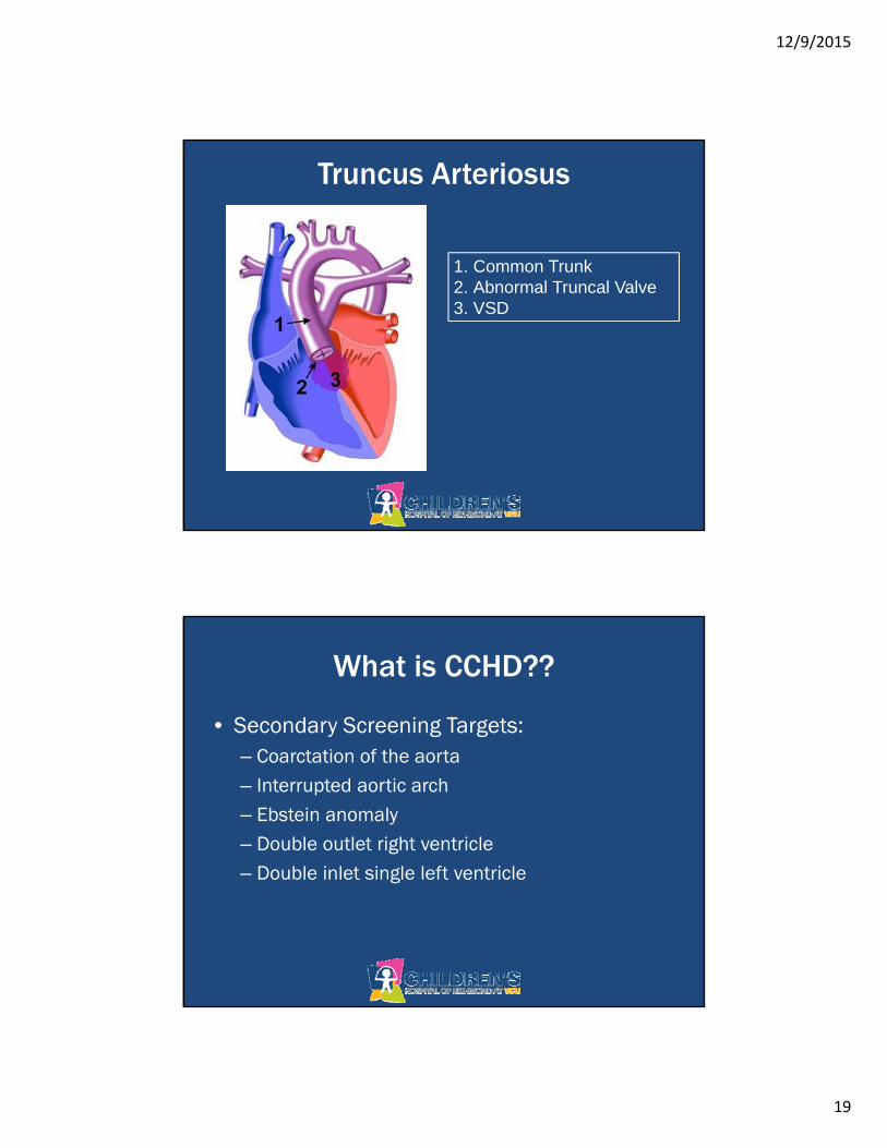
Truncus Arteriosus
1. Common Trunk2. Abnormal Truncal Valve3. VSD
What is CCHD??
• Secondary Screening Targets:– Coarctation of the aorta– Interrupted aortic arch– Ebstein anomaly– Double outlet right ventricle– Double inlet single left ventricle
12/9/2015
20
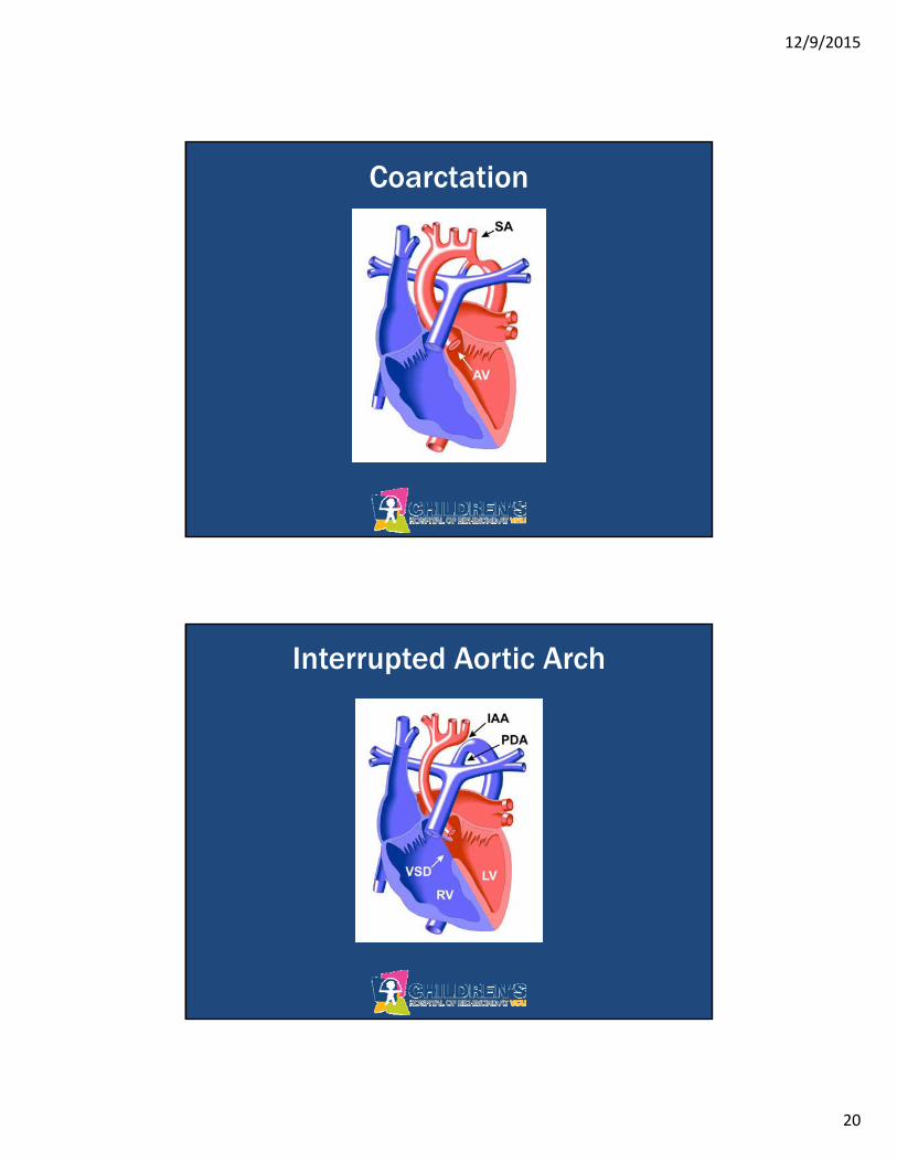
Coarctation
Interrupted Aortic Arch
12/9/2015
21
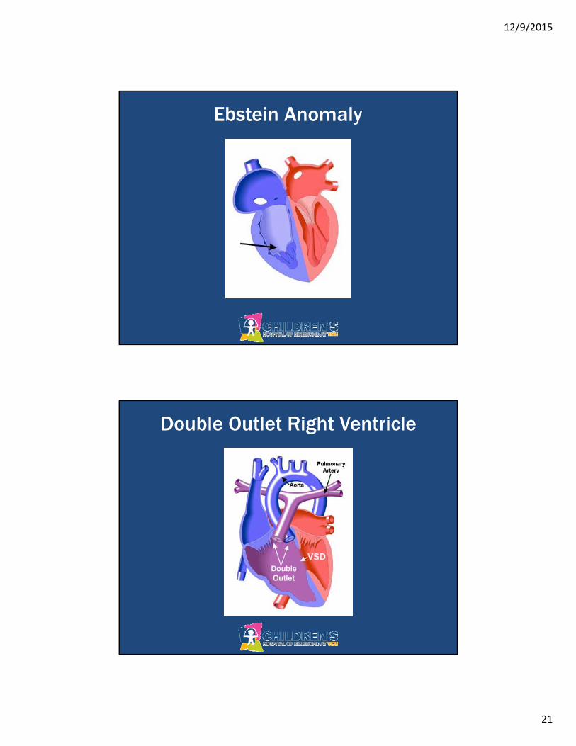
Ebstein Anomaly
Double Outlet Right Ventricle
12/9/2015
22
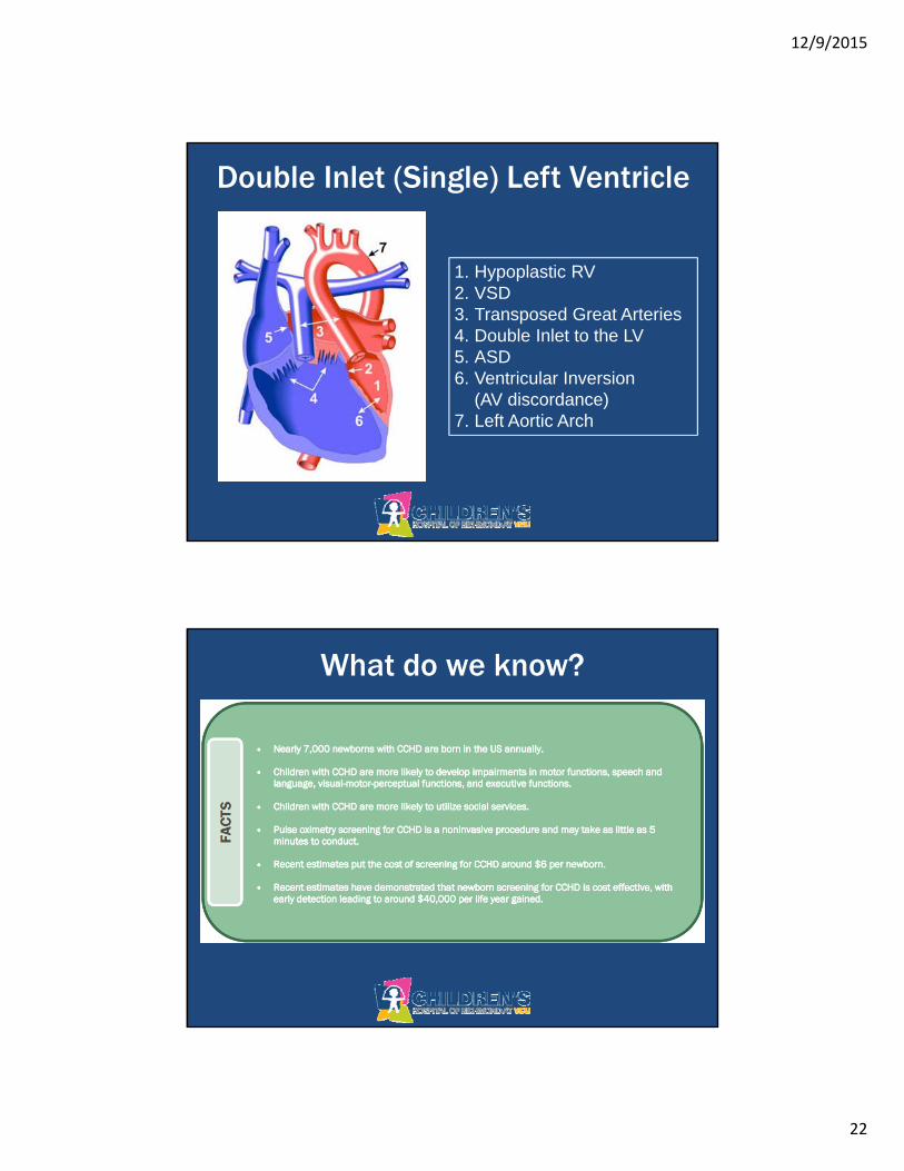
Double Inlet (Single) Left Ventricle
1. Hypoplastic RV2. VSD3. Transposed Great Arteries4. Double Inlet to the LV5. ASD6. Ventricular Inversion
(AV discordance)7. Left Aortic Arch
What do we know?
12/9/2015
23

12/9/2015
24
What if my baby fails??
• Call your friendly, local pediatric cardiologist to request a consultation including a complete congenital echocardiogram
• It’s NEVER the wrong answer to start PGE until a diagnosis can be confirmed or refuted
I’m friendly!
Is the test really that good?• Sensitivity – 76.5%
•SPECIFICITY – 99.9%• That means that if your baby passes the
screening, there is a 99.9% chance that they DO NOT have CCHD
• If your baby fails screening, there is a 76.5% chance that they DO have CCHD
12/9/2015
25
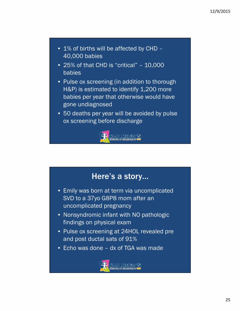
• 1% of births will be affected by CHD –40,000 babies
• 25% of that CHD is “critical” – 10,000 babies
• Pulse ox screening (in addition to thorough H&P) is estimated to identify 1,200 more babies per year that otherwise would have gone undiagnosed
• 50 deaths per year will be avoided by pulse ox screening before discharge
Here’s a story…
• Emily was born at term via uncomplicated SVD to a 37yo G8P8 mom after an uncomplicated pregnancy
• Nonsyndromic infant with NO pathologic findings on physical exam
• Pulse ox screening at 24HOL revealed pre and post ductal sats of 91%
• Echo was done – dx of TGA was made
12/9/2015
26
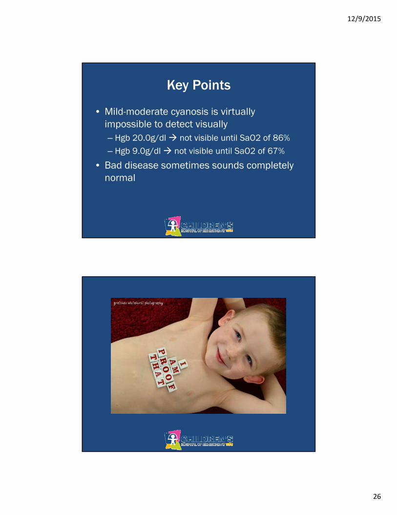
Key Points
• Mild-moderate cyanosis is virtually impossible to detect visually– Hgb 20.0g/dl not visible until SaO2 of 86%– Hgb 9.0g/dl not visible until SaO2 of 67%
• Bad disease sometimes sounds completely normal

12/9/2015
27
thanks.

12/9/2015
28


![NICU Database Appendices - CPQCC NICU Appendices.pdf2020 NICU Appendices| 4 1101 [K. oxytoca, K. pneumoniae and others] including Carbapenem-resistant Klebsiella and Cephalosporin-resistant](https://img.pdfslide.us/doc/110x75/5f0e96fb7e708231d43ff847/nicu-database-appendices-cpqcc-nicu-2020-nicu-appendices-4-1101-k-oxytoca.jpg)