Embed Size (px)
Citation preview
Congenital Gingival Vascular Hamartoma in a Calf
Sadık YAYLA*1, Enver BEYTUT2, Engin KILIÇ1, Mete CİHAN1, Uğur AYDIN1, Emin KARAKURT2
1Kafkas University, Faculty of Veterinary, Department of Surgery, Kars, Turkey. 2Kafkas University, Faculty of Veterinary, Department of Pathology, Kars, Turkey.
Geliş Tarihi: 19.11.2015 Kabul Tarihi: 01.02.2016
Abstract: The purpose of this case was to report clinical and pathological results of gingival vascular hamartoma in a calf and to evaluate the treatment results. A 25-day old native breed female calf was presented to our clinic with complaints of a mass at the level of the incisive teeth. In the clinical examination, the oval plum-sized mass was seen at the level of incisive teeth. The mass was connected to gingiva and was push forward one of the incisive teeth, and the incisive teeth was being held outside of the array. Following sedation and local anesthesia the mass were totally extirpated by using electrocautery. It was noticed that incisive tooth was dislocated to labial direction, and that was totally embedded into the mass. Affected-tooth was fixed to the other incisive tooth with the help of a cerclage wire. In the postoperative period, no any problem was observed in terms of the calf feeding. Based on the histopathological examination the mass was defined as vascular hamartoma. The gingival vascular hamartoma rarely formed in cattle could contribute to literature. Keywords: Congenital gingival vascular hamartoma, calf
Bir Buzağıda Kongenital Gingival Vasküler Hamartom
Özet: Bu olgu sunumu ile bir buzağıda karşılaşılan gingival vasküler hamartom olgusunun klinik ve patolojik olarak tanımlanması ile sağaltım sonuçlarının değerlendirilmesi amaçlandı. Olgumuzu kliniliklerimize insiciv dişler düzeyinde bir kitle şikayeti ile getirilen 25 günlük, dişi yerli bir buzağı oluşturdu. Klinik muayenede insiciv dişler düzeyinde erik büyüklüğünde oval yapılı bir kitle saptandı. Kitlenin gingivayla bağlantılı olduğu ve insiciv dişlerden birini öne doğru ittiği ve bu dişi sıra dizisinin dışında tuttuğu tespit edildi. Sedasyon ve lokal infiltrasyon anestezi altında kitle elektrokoterle total olarak ekstirpe edildi. Sıradizisi dışında kalan diş ise serklaj teli yardımıyla diğer dişlere fikse edildi. Buzağının postoperatif dönemde beslenmesinde herhangi bir sorun gözlenmedi. Kitle histopatolojik bulguları ile vasküler hamartoma olarak tanımlandı. Sığırlarda nadiren görülen vasküler hamartom olgusunun sunulmasının literatüre katkı sağlayacağı sonucuna varıldı. Anahtar Kelimeler: Kongenital gingival vasküler hamartom, buzağı Introduction
Bovine vascular tumors are generally benign
angiomatous lesions arose in different tissues and have been described as juvenile angiomatosis, hemangioma, or hamartoma (Mohammadi et al., 2007). It has been reported that hemangiomas are the most common tumors of infancy (Robbins and Cotran, 1979), but are uncommon in animal species and therefore, some authors consider them to be hamartomas or nevi (Mohammadi et al., 2007). The term hamartoma designates an excessive focal overgrowth of mature normal cells and tissues in an organ (Robbins and Cotran, 1979), and it is a convenient term for an ill-defined group of lesions that have some resemblance to tumors but are not real neoplastic tissue (Robbins and Cotran, 1979; Amniattalab et al., 2012). Hamartomas are caused from faulty development in an organ and is composed of an abnormal mixture of identical cellular elements (Robbins and Cotran, 1979; Misdorp, 2002). Most vascular hamartomas are present at or shortly after birth or during early
infancy and are considered to be developmental abnormalities (Gülbahar et al., 1999; Amniattalab et al., 2012). It is reported that gingival vascular tumors of the oral cavity are commonly seen at birth, and most are of blood-vessel origin (Nourani et al., 2007). The purpose of this case study is to clinically and pathologically describe a case of gingival vascular hamartoma in a calf and to evaluate the treatment results. Case History
Our case consisted of a 25-day-old female calf
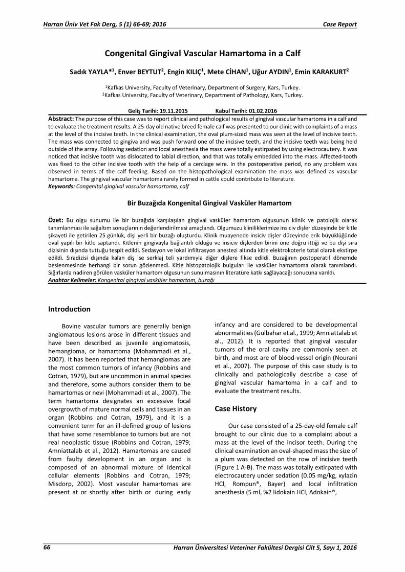
brought to our clinic due to a complaint about a mass at the level of the incisor teeth. During the clinical examination an oval-shaped mass the size of a plum was detected on the row of incisive teeth (Figure 1 A-B). The mass was totally extirpated with electrocautery under sedation (0.05 mg/kg, xylazin HCl, Rompun®, Bayer) and local infiltration anesthesia (5 ml, %2 lidokain HCl, Adokain®,
66 Harran Üniversitesi Veteriner Fakültesi Dergisi Cilt 5, Sayı 1, 2016
Harran Üniv Vet Fak Derg, 5 (1) 66-69; 2016 Case Report
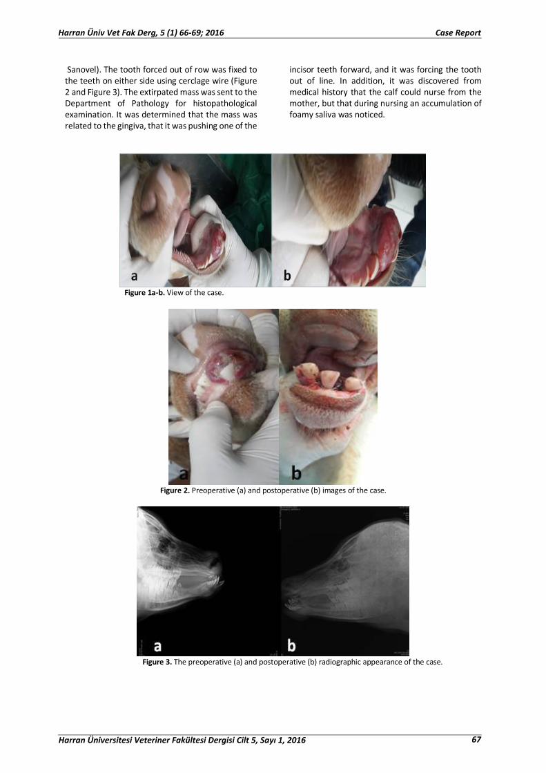
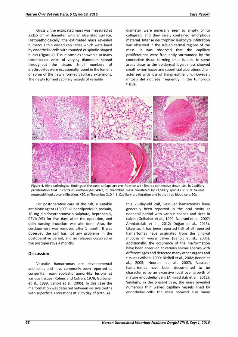
Sanovel). The tooth forced out of row was fixed to the teeth on either side using cerclage wire (Figure 2 and Figure 3). The extirpated mass was sent to the Department of Pathology for histopathological examination. It was determined that the mass was related to the gingiva, that it was pushing one of the
incisor teeth forward, and it was forcing the tooth out of line. In addition, it was discovered from medical history that the calf could nurse from the mother, but that during nursing an accumulation of foamy saliva was noticed.
Figure 1a-b. View of the case.
Figure 2. Preoperative (a) and postoperative (b) images of the case.
Figure 3. The preoperative (a) and postoperative (b) radiographic appearance of the case.
Harran Üniversitesi Veteriner Fakültesi Dergisi Cilt 5, Sayı 1, 2016 67
Harran Üniv Vet Fak Derg, 5 (1) 66-69; 2016 Case Report
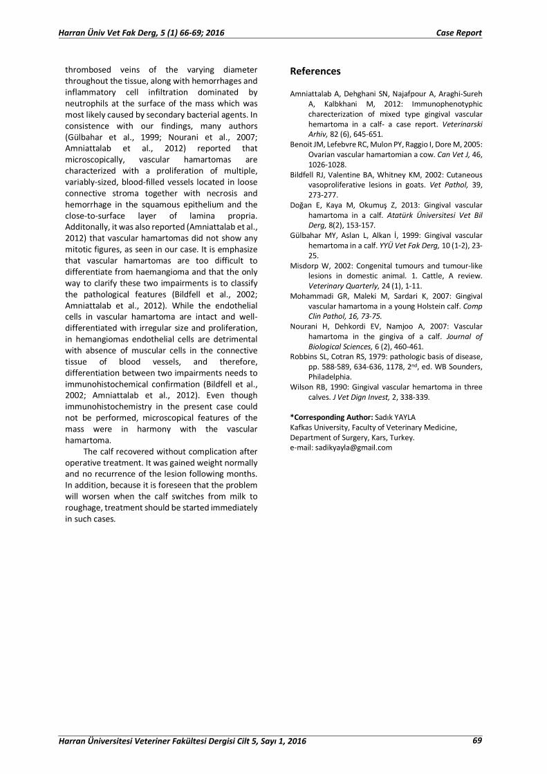
Grossly, the extirpated mass was measured at 2x3x5 cm in diameter with an ulcerated surface. Histopathologically, the extirpated mass revealed numerous thin walled capillaries which were lined by endothelial cells with rounded or spindle-shaped nuclei (Figure 4). Tissue samples showed also many thrombosed veins of varying diameters spread throughout the tissue. Small numbers of erythrocytes were occasionally found in the lumens of some of the newly formed capillary extensions. The newly formed capillary vessels of variable
diameter were generally seen to empty or to collapsed, and they rarely contained amorphous material. Intense neutrophilic leukocyte infiltration was observed in the sub-epidermal regions of the mass. It was observed that the capillary proliferations were frequently surrounded by the connective tissue forming small islands. In some areas close to the epidermal layer, mass showed small hemorrhages and superficial ulcerations char-acterized with loss of lining epithelium. However, mitosis did not see frequently in the tumorous tissue.
Figure 4. Histopathological findings of the case; a: Capillary proliferation with limited connective tissue 10x, b: Capillary proliferation that it contains erythrocytes 40x2, c: Thrombus mass translated by capillary sprouts x10, d: Severe neutrophil leukocyte infiltration. X20, e: Thrombus X10.4, f: Capillary proliferation and in their red blood cells 20x.
For postoperative care of the calf, a suitable
antibiotic agent (10,000 IU benzilpenicillin prokain, 10 mg dihidrostreptomycin sulphate, Reptopen S, CEVA-DIF) for five days after the operation, and daily nursing procedure was also done. Also, the cerclage wire was removed after 1 month. It was observed the calf has not any problems in the postoperative period, and no relapses occurred in the postoperative 4 months. Discussion
Vascular hamartomas are developmental
anomalies and have commonly been reported as congenital, non-neoplastic tumor-like lesions at various tissues (Robins and Cotran, 1979; Gülbahar et al., 1999; Benoit et al., 2005). In this case the malformation was detected between incisive tooths with superficial ulcerations at 25th day of birth. As
this 25-day-old calf, vascular hamartomas have generally been reported in the oral cavity at neonatal period with various shapes and sizes in calves (Gulbahar et al., 1999; Nourani et al., 2007; Amniattalab et al., 2012; Doğan et al., 2013). Likewise, it has been reported half of all reported hamartomas have originated from the gingival mucosa of young calves (Benoit et al., 2005). Additionally, the occurence of the malformation have been observed at various animal species with different ages and detected many other organs and tissues (Wilson, 1990; Bildfell et al., 2002; Benoit et al., 2005; Nourani et al., 2007). Vascular hamartomas have been documented to be characterize by an excessive focal over growth of mature endothelial cells (Amniattalab et al., 2012). Similarly, in the present case, the mass revealed numerous thin walled capillary vessels lined by endothelial cells. The mass showed also many
68 Harran Üniversitesi Veteriner Fakültesi Dergisi Cilt 5, Sayı 1, 2016
Harran Üniv Vet Fak Derg, 5 (1) 66-69; 2016 Case Report
thrombosed veins of the varying diameter throughout the tissue, along with hemorrhages and inflammatory cell infiltration dominated by neutrophils at the surface of the mass which was most likely caused by secondary bacterial agents. In consistence with our findings, many authors (Gülbahar et al., 1999; Nourani et al., 2007; Amniattalab et al., 2012) reported that microscopically, vascular hamartomas are characterized with a proliferation of multiple, variably-sized, blood-filled vessels located in loose connective stroma together with necrosis and hemorrhage in the squamous epithelium and the close-to-surface layer of lamina propria. Additonally, it was also reported (Amniattalab et al., 2012) that vascular hamartomas did not show any mitotic figures, as seen in our case. It is emphasize that vascular hamartomas are too difficult to differentiate from haemangioma and that the only way to clarify these two impairments is to classify the pathological features (Bildfell et al., 2002; Amniattalab et al., 2012). While the endothelial cells in vascular hamartoma are intact and well-differentiated with irregular size and proliferation, in hemangiomas endothelial cells are detrimental with absence of muscular cells in the connective tissue of blood vessels, and therefore, differentiation between two impairments needs to immunohistochemical confirmation (Bildfell et al., 2002; Amniattalab et al., 2012). Even though immunohistochemistry in the present case could not be performed, microscopical features of the mass were in harmony with the vascular hamartoma.
The calf recovered without complication after operative treatment. It was gained weight normally and no recurrence of the lesion following months. In addition, because it is foreseen that the problem will worsen when the calf switches from milk to roughage, treatment should be started immediately in such cases.
References Amniattalab A, Dehghani SN, Najafpour A, Araghi-Sureh
A, Kalbkhani M, 2012: Immunophenotyphic charecterization of mixed type gingival vascular hemartoma in a calf- a case report. Veterinarski Arhiv, 82 (6), 645-651.
Benoit JM, Lefebvre RC, Mulon PY, Raggio I, Dore M, 2005: Ovarian vascular hamartomian a cow. Can Vet J, 46, 1026-1028.
Bildfell RJ, Valentine BA, Whitney KM, 2002: Cutaneous vasoproliferative lesions in goats. Vet Pathol, 39, 273-277.
Doğan E, Kaya M, Okumuş Z, 2013: Gingival vascular hamartoma in a calf. Atatürk Üniversitesi Vet Bil Derg, 8(2), 153-157.
Gülbahar MY, Aslan L, Alkan İ, 1999: Gingival vascular hemartoma in a calf. YYÜ Vet Fak Derg, 10 (1-2), 23-25.
Misdorp W, 2002: Congenital tumours and tumour-like lesions in domestic animal. 1. Cattle, A review. Veterinary Quarterly, 24 (1), 1-11.
Mohammadi GR, Maleki M, Sardari K, 2007: Gingival vascular hamartoma in a young Holstein calf. Comp Clin Pathol, 16, 73-75.
Nourani H, Dehkordi EV, Namjoo A, 2007: Vascular hamartoma in the gingiva of a calf. Journal of Biological Sciences, 6 (2), 460-461.
Robbins SL, Cotran RS, 1979: pathologic basis of disease, pp. 588-589, 634-636, 1178, 2nd, ed. WB Sounders, Philadelphia.
Wilson RB, 1990: Gingival vascular hemartoma in three calves. J Vet Dign Invest, 2, 338-339.
*Corresponding Author: Sadık YAYLA Kafkas University, Faculty of Veterinary Medicine, Department of Surgery, Kars, Turkey. e-mail: [email protected]
Harran Üniversitesi Veteriner Fakültesi Dergisi Cilt 5, Sayı 1, 2016 69
Harran Üniv Vet Fak Derg, 5 (1) 66-69; 2016 Case Report


![Giant neurocristic hamartoma of scalp: a case report · diagnosis of NCH[4]. In congenital NCH, melanomas have been identified 15 to 60 years after birth[6]. Long-term follow-up with](https://img.pdfslide.us/doc/110x75/5ebcc4e25c9d7f25fb5b910a/giant-neurocristic-hamartoma-of-scalp-a-case-diagnosis-of-nch4-in-congenital.jpg)