Embed Size (px)
Citation preview
r e v b r a s o r t o p . 2 0 1 6;5 1(1):109–112
C
C
PJa
b
a
A
R
A
A
K
P
P
K
R
O
P
L
L
A
A
P
o
h2
www.rbo.org .br
ase Report
ongenital dislocation of the patella – clinical case�
edro Miguel Sáa,∗, Filipa Raposoa, Manuel Santos Carvalhob, Nuno Alegreteb,orge Coutinhob, Gilberto Costab
Unidade Local de Saúde Alto Minho, Viana do Castelo, PortugalCentro Hospitalar São João, Porto, Portugal
r t i c l e i n f o
rticle history:
eceived 11 February 2015
ccepted 13 February 2015
vailable online 30 December 2015
eywords:
atellar dislocation/surgery
atellar dislocation/congenital
nee joint/surgery
ange of joint motion
rthopedic procedures/methods
a b s t r a c t
Congenital patellar dislocation is a rare condition in which the patella is permanently dislo-
cated and cannot be reduced manually. The patella develops normally as a sesamoid bone
of the femur. This congenital dislocation results from failure of the internal rotation of the
myotome that forms the femur, quadriceps muscle and extensor apparatus. It usually man-
ifests immediately after birth, although in some rare cases, the diagnosis may be delayed
until adolescence or adulthood. Early diagnosis is important, thereby allowing surgical cor-
rection and avoiding late sequelae, including early degenerative changes in the knee. A case
of permanent dislocation of the patella is presented here, in a female child aged seven years.
© 2015 Sociedade Brasileira de Ortopedia e Traumatologia. Published by Elsevier Editora
Ltda. All rights reserved.
Luxacão congênita da patela – caso clínico
alavras chave:
uxacão patelar/cirurgia
uxacão patelar/congênito
rticulacão do joelho/cirurgia
mplitude de movimento articular
rocedimentos
rtopédicos/métodos
r e s u m o
A luxacão congênita da patela é uma patologia rara, em que a patela se encontra per-
manentemente luxada e manualmente irredutível. A patela desenvolve-se normalmente
como um osso sesamoide do fêmur. A luxacão congênita da patela resulta da falência da
rotacão interna do miótomo que forma o fêmur, músculo quadricípite e o aparelho exten-
sor. Usualmente manifesta-se imediatamente após o nascimento, embora em alguns casos
raros o diagnóstico possa ser adiado até a adolescência/idade adulta. O diagnóstico precoce é
importante, permite a correcão cirúrgica, evita as sequelas tardias, notadamente alteracões
degenerativas precoces do joelho. É apresentado um caso de luxacão permanente da patela,
numa crianca de sexo feminino, com sete anos.
rasil
© 2015 Sociedade B� Work performed in the Centro Hospitalar São João, Porto, Portugal.∗ Corresponding author.
E-mail: [email protected] (P. Miguel Sá).ttp://dx.doi.org/10.1016/j.rboe.2015.12.010255-4971/© 2015 Sociedade Brasileira de Ortopedia e Traumatologia. P
eira de Ortopedia e Traumatologia. Publicado por Elsevier Editora
Ltda. Todos os direitos reservados.
ublished by Elsevier Editora Ltda. All rights reserved.
p . 2 0 1 6;5 1(1):109–112
erally detected in the first decade of life. It usually affects bothlegs and in most cases is associated with polymalformative
110 r e v b r a s o r t o
Introduction
Congenital dislocation of the patella comprises a pathologi-cal condition of permanent lateral dislocation of this bone.It is impossible to reduce it through manual maneuvers.It may arise in isolation, associated with lower-limb mal-formations, or within the context of a polymalformativesyndrome.1
It is generally diagnosed at birth. These infants presentgenu valgum and contracture of the flexed knee, in associationwith external rotation of the tibia. When these deformities arenot present, this pathological condition may not be diagnoseduntil adulthood is reached.2
Radiological examinations, especially X-rays (XR), com-puted tomography (CT) and magnetic resonance imaging (MRI)are essential for identifying and characterizing lesions associ-ated with permanent patellar dislocation (trochlear dysplasiaor chondral lesions). However, the diagnosis for this patholog-ical condition is essentially clinical.3
This condition can only be corrected through a surgical pro-cedure. Several options exist. Early correction is important foravoiding sequelae.4
Case report
The patient was a seven-year-old white girl without any rele-vant antecedents who was examined in an external pediatricorthopedics consultation due to deformity of the left knee.She did not have any previous history of trauma. Her parentssaid that there had not been any initial episode of patellardislocation.
Upon objective examination, she presented irreducible lat-eral dislocation of the left patella. It was observed that, duringactive and passive movements, her extension and flexion didnot present any amplitude deficits and were not painful. Shepresented grade IV strength deficit during extension of the leftlower limb.
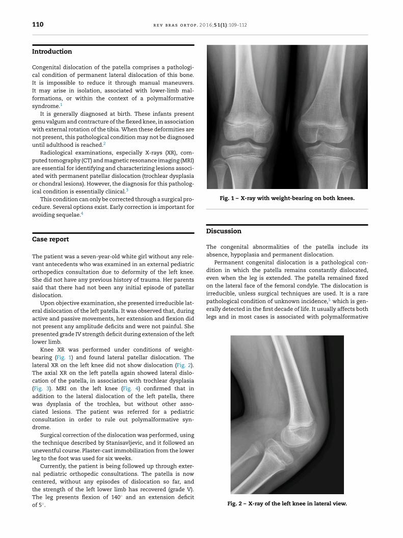
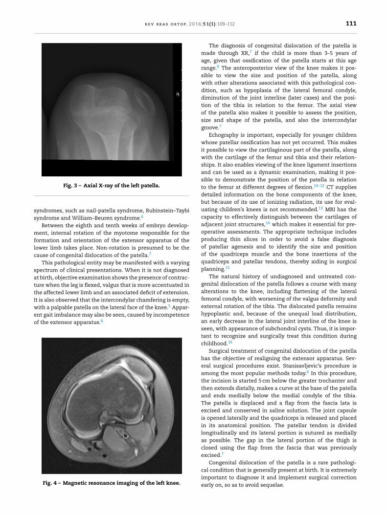
Knee XR was performed under conditions of weight-bearing (Fig. 1) and found lateral patellar dislocation. Thelateral XR on the left knee did not show dislocation (Fig. 2).The axial XR on the left patella again showed lateral dislo-cation of the patella, in association with trochlear dysplasia(Fig. 3). MRI on the left knee (Fig. 4) confirmed that inaddition to the lateral dislocation of the left patella, therewas dysplasia of the trochlea, but without other asso-ciated lesions. The patient was referred for a pediatricconsultation in order to rule out polymalformative syn-drome.
Surgical correction of the dislocation was performed, usingthe technique described by Stanisavljevic, and it followed anuneventful course. Plaster-cast immobilization from the lowerleg to the foot was used for six weeks.
Currently, the patient is being followed up through exter-nal pediatric orthopedic consultations. The patella is now
centered, without any episodes of dislocation so far, andthe strength of the left lower limb has recovered (grade V).The leg presents flexion of 140◦ and an extension deficitof 5◦.Fig. 1 – X-ray with weight-bearing on both knees.
Discussion
The congenital abnormalities of the patella include itsabsence, hypoplasia and permanent dislocation.
Permanent congenital dislocation is a pathological con-dition in which the patella remains constantly dislocated,even when the leg is extended. The patella remained fixedon the lateral face of the femoral condyle. The dislocation isirreducible, unless surgical techniques are used. It is a rarepathological condition of unknown incidence,5 which is gen-
Fig. 2 – X-ray of the left knee in lateral view.
r e v b r a s o r t o p . 2 0 1 6
ss
mflc
sattIweo
Fig. 3 – Axial X-ray of the left patella.
yndromes, such as nail-patella syndrome, Rubinstein–Taybiyndrome and William–Beuren syndrome.6
Between the eighth and tenth weeks of embryo develop-ent, internal rotation of the myotome responsible for the
ormation and orientation of the extensor apparatus of theower limb takes place. Non-rotation is presumed to be theause of congenital dislocation of the patella.7
This pathological entity may be manifested with a varyingpectrum of clinical presentations. When it is not diagnosedt birth, objective examination shows the presence of contrac-ure when the leg is flexed, valgus that is more accentuated inhe affected lower limb and an associated deficit of extension.t is also observed that the intercondylar chamfering is empty,
5
ith a palpable patella on the lateral face of the knee. Appar-nt gait imbalance may also be seen, caused by incompetencef the extensor apparatus.8Fig. 4 – Magnetic resonance imaging of the left knee.
;5 1(1):109–112 111
The diagnosis of congenital dislocation of the patella ismade through XR,7 if the child is more than 3–5 years ofage, given that ossification of the patella starts at this agerange.9 The anteroposterior view of the knee makes it pos-sible to view the size and position of the patella, alongwith other alterations associated with this pathological con-dition, such as hypoplasia of the lateral femoral condyle,diminution of the joint interline (later cases) and the posi-tion of the tibia in relation to the femur. The axial viewof the patella also makes it possible to assess the position,size and shape of the patella, and also the intercondylargroove.7
Echography is important, especially for younger childrenwhose patellar ossification has not yet occurred. This makesit possible to view the cartilaginous part of the patella, alongwith the cartilage of the femur and tibia and their relation-ships. It also enables viewing of the knee ligament insertionsand can be used as a dynamic examination, making it pos-sible to demonstrate the position of the patella in relationto the femur at different degrees of flexion.10–12 CT suppliesdetailed information on the bone components of the knee,but because of its use of ionizing radiation, its use for eval-uating children’s knees is not recommended.13 MRI has thecapacity to effectively distinguish between the cartilages ofadjacent joint structures,14 which makes it essential for pre-operative assessments. The appropriate technique includesproducing thin slices in order to avoid a false diagnosisof patellar agenesis and to identify the size and positionof the quadriceps muscle and the bone insertions of thequadriceps and patellar tendons, thereby aiding in surgicalplanning.15
The natural history of undiagnosed and untreated con-genital dislocation of the patella follows a course with manyalterations to the knee, including flattening of the lateralfemoral condyle, with worsening of the valgus deformity andexternal rotation of the tibia. The dislocated patella remainshypoplastic and, because of the unequal load distribution,an early decrease in the lateral joint interline of the knee isseen, with appearance of subchondral cysts. Thus, it is impor-tant to recognize and surgically treat this condition duringchildhood.16
Surgical treatment of congenital dislocation of the patellahas the objective of realigning the extensor apparatus. Sev-eral surgical procedures exist. Stanisavljevic’s procedure isamong the most popular methods today.6 In this procedure,the incision is started 5 cm below the greater trochanter andthen extends distally, makes a curve at the base of the patellaand ends medially below the medial condyle of the tibia.The patella is displaced and a flap from the fascia lata isexcised and conserved in saline solution. The joint capsuleis opened laterally and the quadriceps is released and placedin its anatomical position. The patellar tendon is dividedlongitudinally and its lateral portion is sutured as mediallyas possible. The gap in the lateral portion of the thigh isclosed using the flap from the fascia that was previouslyexcised.7
Congenital dislocation of the patella is a rare pathologi-
cal condition that is generally present at birth. It is extremelyimportant to diagnose it and implement surgical correctionearly on, so as to avoid sequelae.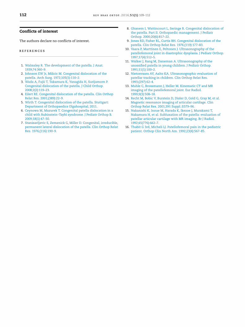
p . 2 0
r 1
1
1
1
1
1
112 r e v b r a s o r t o
Conflicts of interest
The authors declare no conflicts of interest.
e f e r e n c e s
1. Walmsley R. The development of the patella. J Anat.1939;74:360–9.
2. Johnson EW Jr, Milicic M. Congenital dislocation of thepatella. Arch Surg. 1972;105(1):110–2.
3. Wada A, Fujii T, Takamura K, Yanagida H, Surijamorn P.Congenital dislocation of the patella. J Child Orthop.2008;2(2):119–23.
4. Eilert RE. Congenital dislocation of the patella. Clin OrthopRelat Res. 2001;(389):22–9.
5. Wirth T. Congenital dislocation of the patella. Stuttgart:Department of Orthopaedics Olgahospital; 2011.
6. Ceynowa M, Mazurek T. Congenital patella dislocation in achild with Rubinstein–Taybi syndrome. J Pediatr Orthop B.
2009;18(1):47–50.7. Stanisavljevic S, Zemenick G, Miller D. Congenital, irreducible,permanent lateral dislocation of the patella. Clin Orthop RelatRes. 1976;(116):190–9.
1
1 6;5 1(1):109–112
8. Ghanem I, Wattincourt L, Seringe R. Congenital dislocation ofthe patella. Part II. Orthopaedic management. J PediatrOrthop. 2000;20(6):817–22.
9. Jones RD, Fisher RL, Curtis BH. Congenital dislocation of thepatella. Clin Orthop Relat Res. 1976;(119):177–83.
0. Vaara P, Marttinen E, Peltonen J. Ultrasonography of thepatellofemoral joint in diastrophic dysplasia. J Pediatr Orthop.1997;17(4):512–5.
1. Walker J, Rang M, Daneman A. Ultrasonography of theunossified patella in young children. J Pediatr Orthop.1991;11(1):100–2.
2. Nietosvaara AY, Aalto KA. Ultrasonographic evaluation ofpatellar tracking in children. Clin Orthop Relat Res.1993;(297):62–4.
3. Muhle C, Brossmann J, Heller M. Kinematic CT and MRimaging of the patellofemoral joint. Eur Radiol.1999;9(3):508–18.
4. Recht M, Bobic V, Burstein D, Disler D, Gold G, Gray M, et al.Magnetic resonance imaging of articular cartilage. ClinOrthop Relat Res. 2001;391 Suppl.:S379–96.
5. Nakanishi K, Inoue M, Harada K, Ikezoe J, Murakami T,Nakamura H, et al. Subluxation of the patella: evaluation of
patellar articular cartilage with MR imaging. Br J Radiol.1992;65(776):662–7.6. Thabit G 3rd, Micheli LJ. Patellofemoral pain in the pediatricpatient. Orthop Clin North Am. 1992;23(4):567–85.