Embed Size (px)
Citation preview

Congenital Aural AtresiaMiranda S. Dennis, M.D.April 6, 2011

Embryology
External Auditory Canal
first branchial groove
starts as a solid core of epithelial cells, which undergoes absorption in a medial to lateral direction
if this is arrested can result in a normal TM with bony external ear canal assoc with an atretic or very stenotic or membranous canal predisposing to canal cholesteatoma

Medial portion of the EAC
formed by tympanic bone and begins to ossify in the 3rd month
forming tympanic ring and osseous portion of EAC
Malformation produces atretic bone at the level of the tympanic membrane, resulting in atresia of the ear canal

Mastoid and Middle Ear
ET, middle ear, mastoid air cells: First pharyngeal pouch
Ossicles (except for vestibular portion of stapes footplate): First and second branchial arches
Membranous labryrinth is derived from the ectodermal otocyst, so hearing and vestibular function should be normal
Stapes footplate from otic capsule and is usually normally developed

Facial NerveTypically see bony dehiscence of the fallopian canal
Acute angle at the second genu
More anterior and lateral direction
Places the nerve at higher risk when drilling in the posterior inferior portion of the new ear canal
Nerve may lie just deep to the tragus as it exits the skull, making it susceptible to injury if undermining the auricle
There is a correlation between degree of microtia and extent of facial nerve abnormality
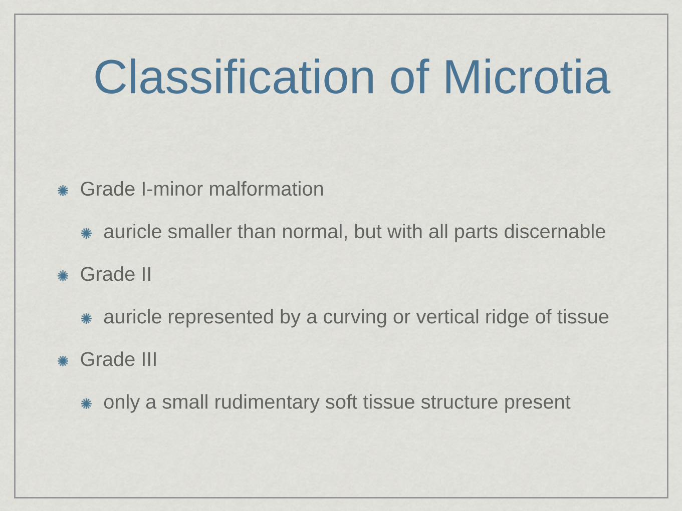
Classification of Microtia
Grade I-minor malformation
auricle smaller than normal, but with all parts discernable
Grade II
auricle represented by a curving or vertical ridge of tissue
Grade III
only a small rudimentary soft tissue structure present

Ombredanne’s CriteriaMajor Malformation
EAC and TM usually absent, and stenosis prevents visualization of the medial aspect of the ear canal
ME space is reduced, malleus and incus are deformed, fused, and fixed to the atretic bone
Severe cases-ME space is hypoplastic, and ossicles are rudimentary or absent. Dehiscence or displacement is expected in most major malformations
Commonly seen with grade II and III microtia with normal inner ear function

Minor Malformation
CHL secondary to absence or deformity of one or more ossicles, or fixation of the ossicular chain
Abnormalities of the stapes may be more severe in minor malformations
ME space and TM are normal, EAC is patent
Dehiscence or displacement of FN may occur
Pinna usually normally developed or grade I microtia

AudiometryIf canal patent can do electrocochleography
Patients with bilateral atresia difficult to test secondary to masking dilemma
Cannot assume that cochlear function is normal bilaterally even if inner ear normal on CT
Need bone conduction auditory brainstem response testing
Wave I of ABR is generated by the distal portion of the auditory nerve, with minimal crossover to the contralateral ear
Best measured by a recording electrode ipsilateral to the stimulated side

CT
Necessary for all patients being considered for surgery
In cases of stenosis can evaluate for possible cholesteatoma formation
Axial-body of malleus and incus, IS joint, round window
Coronal-stapes, oval window, vestibule
Both projections needed to evaluate the facial nerve

Medical ManagementUnilateral Atresia
If normal hearing in contralateral ear, no immediate intervention necessary
Preferential seating in school
Atresia-bone conduction HA
Stenosis-air-condution HA
Bilateral Atresia
need early amplification with bone conduction HA

Surgical ManagementReluctant to operate on unilateral cases until adulthood
hard to predict degree of hearing improvement, potential lifetime care of mastoid cavity, risk of injury to the facial nerve
Need to improve hearing threshold to 25dB or greater to eliminate handicap of unilateral hearing loss--can be achieved in about 50% of carefully selected patients
Goal in bilateral atresia is to restore sufficient hearing so amplification no longer needed
better ear (by CT evaluation) is selected for initial procedure
Selection Criteria
Patients typically have a 10dB residual conductive deficit after repair
SN function should normal in operative ear, and normal to near-normal in contralateral ear to avoid operating on better hearing ear
Mostly determined by CT evaluation
In unilateral cases, only ideal candidates are selected
In bilateral cases, minimal criteria are a middle ear of at least one half normal size, and presence of ossicular mass
Only 60% of patients with aural atresia are surgical candidates

Often CT findings can be predicted by physical exam--poor middle ear development seen more frequently in patients with craniofacial abnormalities than those with isolated aural atresia
Treacher Collins--often have bizzare middle ear findings
The better developed the auricle, the larger and better developed the middle ear

Jahrsdoefer Grading System
Total points:
8-good prognosis
7-fair prognosis
6-marginal candidate
5-poor prognosis
Parameter PointsStapes Present 2
Oval window patent 1Middle ear space 1
Facial Nerve 1Malleus/Incus Complex 1Mastoid Pneumatization 1Incus-Stapes Connection 1
Round Window 1External Ear Appearance 1

NOT A SURGICAL
CANDIDATE

CANDIDATES FOR SURGERY

Timing of SurgeryEarly as 6 to 7 years old, and microtia repair, if needed should be well underway, ensuring an unoperated field without compromised blood supply for the implanted auricular framework.
Typically atresia surgery is performed midway through the microtia repair, after the auricular framework has been implanted and the lobule transposed, but prior to reconstructing the tragus or elevating the auricle.
The auricle can be repositioned to align the meatus and EAC
CholesteatomaPatients with stenosis are at higher risk for cholesteatoma formation
Cole and Jahrsdoefer: of 50 pts with average canal diameter of 4mm or less, 50% developed cholesteatoma
Patient age and exact canal size were important variables: no cholesteatoma in age less than 3, and bone erosion and middle ear involvement not seen in patients under 12
Most cholesteatomas developed in canals 2mm or less
Commonly present with drainage or postauricular fistula track

Management Protocols in Stenosis
Patients with significant stenosis that prevents adequate cleaning of EAC and visualization of TM should have CT by age 5
If no cholesteatoma and favorable middle ear findings, can proceed with canal and middle ear surgery
If middle ear unfavorable, can offer canaloplasty
If parents opt for non-surgical management, follow with serial CTs every few years
Periodic CTs not indicated in patients with a completely atretic ear canal, as cholesteatomas are rare in this patient population

Surgical TechniqueTwo surgical approaches
Mastoid
Sinodural angle identified and followed to antrum, facial recess opened and IS joint separated. Atretic bone is then removed
Anterior (popularized by Jahrsdoefer)
Exposure of mastoid air cells is limited, and drilling is comfined to an area defined by TMJ anteriorly MCF dura superiorly, and mastoid air cells posteriorly
Large mastoid cavity is avoided, there is less surgical manipulation in the area of the mastoid segment of the facial nerve, and the more cylindrical canal better facilitates a STSG

Incision
Post-auricular
Soft tissues elevated anteriorly until a depression is encountered
In most major malformations, this is the TMJ, but occasionally a stenotic bony ear canal is encountered
Manipulation here should be limited as the facial nerve frequently exits the skull into the glenoid fossa

Drilling a Canal
Most atretic ears without a tympanic bone remnant
Sufficient space between the glenoid fossa anteriorly and mastoid air cells posteriorly for a canal
If tympanic remnant present, the atretic bone directs the drilling for the EAC
Concentrating the drilling superiorly along the MCF protects the facial nerve because it will always lie medial to the ossicular mass in the epitympanum
Because of the acute angle of the facial nerve at the 2nd genu, it is vulnerable to injury as the EAC is opened in a posteriorinferior direction
In the middle ear cavity, the nerve may lie lateral and be anteriorly displaced


Exposure of Ossicular Chain
Malleus neck or manubrium often fused to atretic bone
In order to free the ossicular chain, the overlying bone is carefully thinned then removed
Limit drilling or manipulation of ossicles to protect to the inner ear
Except for fossa inudis, which may be left intact, bone should be completely removed around the ossicles, leaving at least 2-3 mm space
Atretic bone is removed leaving the ossicular mass centered in the new canal
To ensure proper draping of STSG, the canal walls should be smooth and without ledges lateral to the ossicular mass


Middle Ear Surgery
Stapes often obscured by contracted middle ear cavity, malformed lateral ossicles, or overlying facial nerve
Enough usually seen in order to asses the IS joint
Stapes is often small, with a delicate misshapen crura, but normal oval window and stapes footplate are usually found
Lateral ossicular chain, although deformed, is often mobile and left in place for better hearing results

Tympanic Membrane GraftingThin fascia graft is placed over the mobilized ossicular chain
Manubrium of malleus often deformed making anchoring the graft beneath the ossicular chain difficult
Absence of TM graft remnant or annulus predisposes to lateralization
To prevent this the graft can be tucked under the anterior and superior edges of the bony wall or by covering the fascia graft with the STSC of the canal and then placing a Silastic button which has been contoured to the canal

Meatoplasty
Auricle undermined and soft tissue debulked at approximate area of meatus, preventing future stenosis
Extensive undermining can place the facial nerve at risk as prior auricular reconstruction may have caused scarring and tethering of the extratemporal facial nerve in a more superficial location

Skin Grafting0.01-inch thick, pie-crusted, graft from thigh or upper arm used to line the canal
Typically shaped like a hexagon 4x6cm with small wedges taken from the medial portion to facilitate placement at the TM
Overlaps the fascia graft, which helps facilitate epithelialization of the neo-TM
Sialastic disc placement helps prevent lateralization
Packing then placed


Postoperative Care
POD 10 packing and silastic disc are removed
If granulation tissue present, antibiotic-soaked gelfoam is placed in the canal and the patient is given drops for 7-10 days

Ossicles
Malleus/Incus Complex-malleus more deformed than the incus and has a short manubrium
Ossicular fixation usually between malleus neck or shortened manubrium and atretic bone
Fixation of stapes, or severe deformity, more likely to be seen in patients with a patent EAC than in those with major atresia


Hearing Results
Initial postoperative hearing level of 30dB or better can be achieved in approximately 50% of cases
Most of these patients continue to have improved SRT with long-term follow-up

Complications
Serious: SNHL and facial nerve injury
Other: Canal stenosis, chronic infection, recurrent CHL
Many of these patients require revision surgery (approximately 1/3). Stenosis and lateralization are the most common problems encountered.

Labyrinthine Injury
Anterior approach limits exposure of mastoid air cells and the horizontal SCC.
High-frequency SNHL noted in some patients postoperatively, but loss in the speech frequencies is rare.
Avoid by limiting drilling near the ossicles or excessive manipulation

Facial Nerve InjuryFacial Nerve monitoring is necessary
Usually occurs with transposition of the facial nerve to gain access to the oval window
Jahrsdoefer and Lambert: 1% incidence in 1000 cases
Concentrate drilling superiorly along MCF dural plate, with caution when enlarging the canal in the posterior-inferior direction
Nerve can also be injured in the extratemporal segment when making the postauricular incision or undermining the auricle

Canal StenosisOccurs in 25% of cases
If a large meatus is made, then not an issue
Significant stenosis can trap squamous epithelium and causing infection
Can attempt to dilate the canal with soft or hard stents, but usually ineffective, and a secondary meatoplasty with STSF is necessary
Pinna can become displaced, usually anteriorly or inferiorly, causing a malalignment of the meatus and bony canal. A permanent suspension suture can be placed from the framework of the auricle to the mastoid periosteum or to a hole in the mastoid cortex

Chronic Infection
Normal migration of keratin debris is absent in the skin-grafted ear canal as well as protective secretions from sebaceous and apocrine glands
Higher incidence of canal infections
Widely patent meatus and membranous canal are required for aeration and cleaning
Most patients not restricted from water activities

Conductive Hearing Loss
Persistent or recurrent
Most common negative outcome in aural atresia surgery
Inadequate mobilization of the ossicular mass from the atretic bone
Unrecognized IS joint discontinuity
Fixed stapes footplate
Recurrent CHL usually from refixation or TM lateralization

BAHA
Bone anchored hearing aid is an alternative, especially for marginal or poor surgical candidates
Osseointegrated titanium fixture with a sound processor atteached percutaneously
Allows more efficient transfer of sound than with bone conduction HA

ConclusionGoal in congenital ear surgery is to create a functional pathway by which sound have reach the cochlear fluids
Challenging surgery requiring thorough knowledge of the anatomic variations, audiometric and radiographic findings in these patients
With proper patient selection in cases of major atresia, it is possible to achieve a hearing level of 25 dB or better in approximately 50-70% of patients


![OAV dysplasia handout.ppt [Compatibiliteitsmodus] · maximum multipoint lod score of 3.00. familial simplex bilateral involvement 71% 46% hearing loss 26% 87% aural atresia 41% 79%](https://img.pdfslide.us/doc/110x75/5c68865809d3f206678ba166/oav-dysplasia-compatibiliteitsmodus-maximum-multipoint-lod-score-of-300.jpg)