Embed Size (px)
Citation preview
IOP PUBLISHING JOURNAL OF PHYSICS: CONDENSED MATTER
J. Phys.: Condens. Matter 19 (2007) 113102 (25pp) doi:10.1088/0953-8984/19/11/113102
TOPICAL REVIEW
Confocal microscopy of colloids
V Prasad, D Semwogerere and Eric R Weeks
Department of Physics, Emory University, Atlanta, GA 30322, USA
E-mail: [email protected]
Received 5 September 2006, in final form 5 January 2007Published 27 February 2007Online at stacks.iop.org/JPhysCM/19/113102
AbstractColloids have increasingly been used to characterize or mimic many aspectsof atomic and molecular systems. With confocal microscopy these colloidalparticles can be tracked spatially in three dimensions with great precisionover large time scales. This review discusses equilibrium phases such ascrystals and liquids, and non-equilibrium phases such as glasses and gels.The phases that form depend strongly on the type of particle interaction thatdominates. Hard-sphere-like colloids are the simplest, and interactions suchas the attractive depletion force and electrostatic repulsion result in morenon-trivial phases which can better model molecular materials. Furthermore,shearing or otherwise externally forcing these colloids while under microscopicobservation helps connect the microscopic particle dynamics to the macroscopicflow behaviour. Finally, directions of future research in this field are discussed.
(Some figures in this article are in colour only in the electronic version)
Contents
1. Introduction 22. Properties of colloids 3
2.1. Hard spheres and their phase behaviour 32.2. Attractive and repulsive interactions 4
3. Comparison of techniques to study colloidal suspensions 54. How a confocal microscope works 7
4.1. Resolution 84.2. Scanning speed 9
5. Observations of hard-sphere colloids 105.1. Hard-sphere colloidal crystals 105.2. Hard-sphere colloidal glasses 115.3. Ageing in colloidal glasses 135.4. Systems under shear 13
0953-8984/07/113102+25$30.00 © 2007 IOP Publishing Ltd Printed in the UK 1

J. Phys.: Condens. Matter 19 (2007) 113102 Topical Review
6. Observations of interacting systems 156.1. Colloidal gels 156.2. Attractive glasses 166.3. Liquid–liquid or liquid–solid coexistence 176.4. Ionic systems 176.5. Anisotropic systems 19
7. New directions 19Acknowledgments 20References 20
1. Introduction
Optical microscopy is widely used in many systems where the domain of interest lies in thesubmicrometre to micrometre range. These include biological systems such as cells or tissue aswell as areas of ‘soft’ physics such as complex fluids and colloidal dispersions, the main focusof this review. Since the important length scales are of the order of the wavelength of visiblelight, microscopy provides a powerful tool to obtain real-space and real-time information aboutthe complex mechanisms that govern these systems [1]. The ease of sample preparation at roomtemperature and atmospheric pressure (as opposed to, say, low temperature or low pressureconditions for electron microscopy) also makes optical microscopy a convenient technique.Nevertheless, certain problems exist for complex many-body systems that interact with light;the most prominent one being multiple scattering. Visualization deep within the sample isdifficult for an optically dense sample such as a biological tissue or a concentrated colloidaldispersion, as the light that enters the sample undergoes many scattering events resulting inblurriness of the image.
This problem was first faced by Marvin Minsky in the 1950s as he tried to visualize theway nerve cells were connected in the human nervous system [2]. His solution was two-fold:first, point by point illumination of the sample to minimize aberrant rays of scattered lightfrom regions outside the image plane of interest; and second, a pinhole aperture in the imageplane on the other side of the objective, to reject out-of-focus light. This prototype for thefirst confocal microscope used a carbon-arc lamp as the light source and a translating stage tovisualize each point of the sample. Subsequent improvements utilize a laser and scan the laserbeam rather than translating the sample. These two improvements enabled confocal microscopyto become a powerful tool for many scientific fields [3–5]; details of these improvements, aswell as advances in the speed and resolution of confocal imaging, are described in later sectionsof this review.
The use of confocal microscopy in colloidal systems is a relatively recent development,in part driven by the discovery that monodisperse colloids could mimic many of the phasesseen in atomic systems [6]. While these observations were performed by bulk techniquessuch as light scattering, further information about the microscopic details of these systemsrequired visualization in concentrated dispersions. One of the first studies to look at colloidswith a confocal microscope was by Yoshida et al in 1991 [7] who looked deep within asample of charged polystyrene latex colloids and observed hexagonal ordering of the spheres.van Blaaderen and Wiltzius [8] extended the capability of confocal microscopy to densersystems, imaging spheres with packing fractions of ∼60% to observe structure in the so-calledcolloidal glass phase. Such a high density of particles required a significant advance in image-analysis algorithms; the positions of thousands of particles separated by distances slightly largerthan their diameter had to be located to high accuracy. Once this was achieved almost anymicrometre-sized colloidal system could be studied by confocal microscopy.
2

J. Phys.: Condens. Matter 19 (2007) 113102 Topical Review
Advances in imaging and particle-tracking have led to rapid advances in our understandingof colloidal phenomena, and confocal microscopy is currently a tool in the laboratories of manyresearch groups. In subsequent sections we discuss the details of how confocal microscopesfunction and some of the interesting physics governing colloidal systems that can be capturedwith this technique.
2. Properties of colloids
2.1. Hard spheres and their phase behaviour
Colloids have been used as models for atomic and molecular systems as they demonstratemany of the phases observed in such systems. The simplest model system is the hard-sphere system [9–13] in which the colloids are non-interacting at all separations beyond theirradius and infinitely repulsive on contact. The sole control parameter that determines thephase behaviour of hard-sphere colloids is the sphere volume fraction φ, a non-dimensionalquantity related to the number density n (φ = (4π/3)a3n with particle radius a). Here wediscuss only the simple monodisperse case, although other parameters that could be consideredinclude polydispersity, the aspect ratio of ellipsoids, etc. The size of the sphere is not acontrol parameter for ideal hard spheres, but for colloidal suspensions it can strongly affectsedimentation or even the overall dynamics of the sphere. In the case of sedimentation this isbecause gravitational energy is still significant at the colloidal length scale when compared tothermal fluctuations. Balancing this energy with kBT gives a gravitational height h = kBT/Fg,where the gravitational force on a sphere of radius a is Fg = (4/3)πa3�ρg (�ρ is the densitydifference between the colloid and the surrounding fluid). The gravitational height can bevaried by many orders of magnitude, either by going to a low gravity environment, such as inspace [14], or a high gravity one, by mismatching the density of the colloid and solvent [15–17].For most systems it is desirable to have h larger than the sample chamber height as gravitationaleffects can then be neglected (for example, h = 1 mm for a ∼ 0.5 μm and �ρ = 1 kg m−3).The other effect of size arises due to diffusion; larger particles diffuse more slowly than smallerparticles. This can be quantified by the diffusion coefficient D = kBT/6πηa which determineshow fast a sphere diffuses in a fluid solvent of viscosity η. The time scale for a particle todiffuse a distance equal to its own radius is τB = a2/6D; this sets the dynamic time scale ofthe colloidal system in question (for a micrometre-sized particle in water τB is of the order ofseconds).
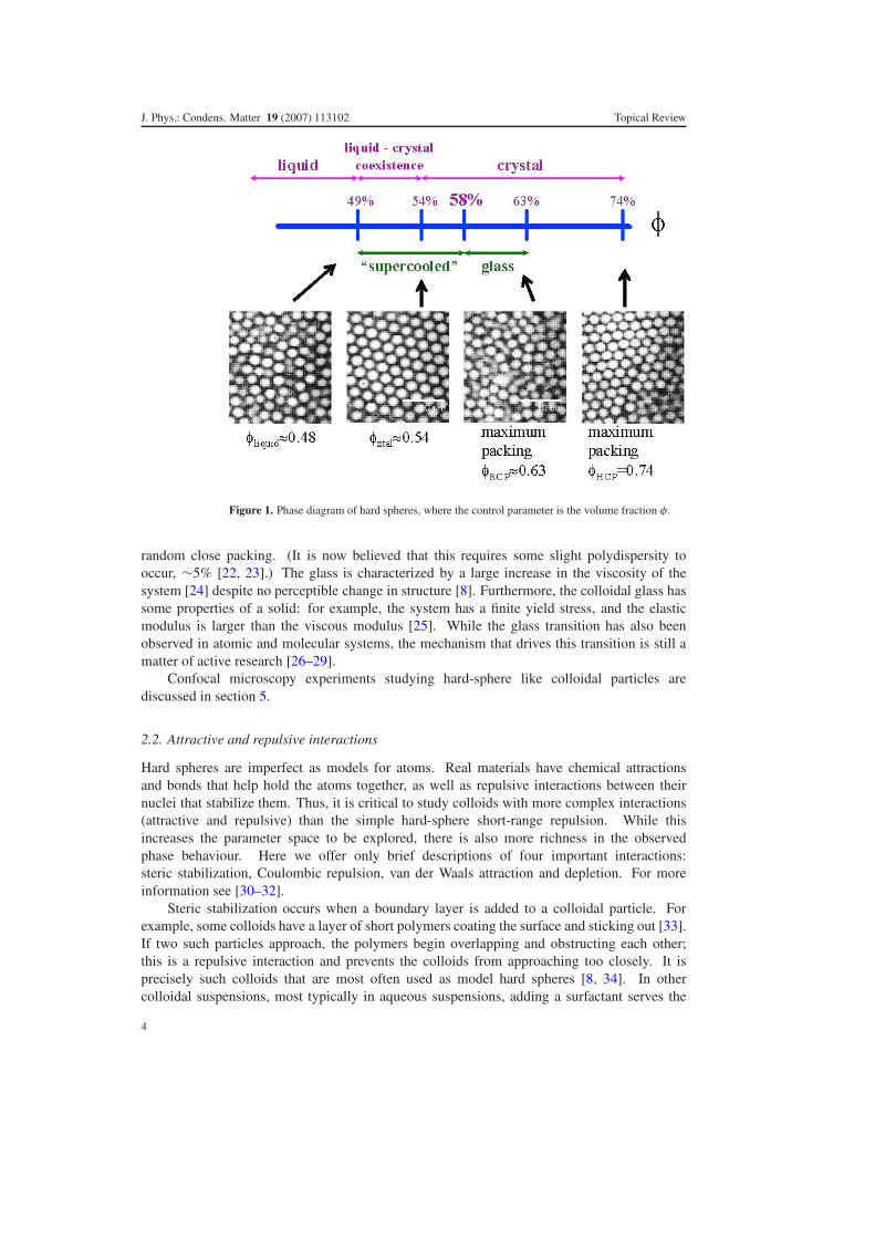
In a dilute system (φ → 0) the spheres are disordered and generally far from each otherlike a dilute gas. As the volume fraction increases from the dilute limit spheres remain in adisordered state. Denser states have spatial correlations between the positions of the spheresand thus these states are more analogous to liquids than to gases, although this distinction issomewhat arbitrary. The maximum amorphous packing is φ ≈ 0.64 [18–21], often termedRCP for ‘random close packing’ (see figure 1).
If the hard-sphere system is allowed to equilibrate as the volume fraction is increased,however, the system undergoes an entropy driven phase transition to a crystalline state [6].This transition starts at φ = 0.494 with a coexistence region up to φ = 0.545. States with0.494 < φ < 0.545 have crystalline domains (φ = 0.545) coexisting with liquid regions(φ = 0.494) (see figure 1). This phase persists from φ = 0.545 to 0.740, the maximal packinga 3D system of monodisperse spheres can obtain.
In addition to these equilibrium phases colloidal systems can also exhibit non-equilibriumbehaviour. For example, at φ = 0.58 the crystallization of the colloids can be arrested bythe appearance of a metastable, kinetically trapped state known as a glass that persists until
3
J. Phys.: Condens. Matter 19 (2007) 113102 Topical Review
Figure 1. Phase diagram of hard spheres, where the control parameter is the volume fraction φ.
random close packing. (It is now believed that this requires some slight polydispersity tooccur, ∼5% [22, 23].) The glass is characterized by a large increase in the viscosity of thesystem [24] despite no perceptible change in structure [8]. Furthermore, the colloidal glass hassome properties of a solid: for example, the system has a finite yield stress, and the elasticmodulus is larger than the viscous modulus [25]. While the glass transition has also beenobserved in atomic and molecular systems, the mechanism that drives this transition is still amatter of active research [26–29].
Confocal microscopy experiments studying hard-sphere like colloidal particles arediscussed in section 5.
2.2. Attractive and repulsive interactions
Hard spheres are imperfect as models for atoms. Real materials have chemical attractionsand bonds that help hold the atoms together, as well as repulsive interactions between theirnuclei that stabilize them. Thus, it is critical to study colloids with more complex interactions(attractive and repulsive) than the simple hard-sphere short-range repulsion. While thisincreases the parameter space to be explored, there is also more richness in the observedphase behaviour. Here we offer only brief descriptions of four important interactions:steric stabilization, Coulombic repulsion, van der Waals attraction and depletion. For moreinformation see [30–32].
Steric stabilization occurs when a boundary layer is added to a colloidal particle. Forexample, some colloids have a layer of short polymers coating the surface and sticking out [33].If two such particles approach, the polymers begin overlapping and obstructing each other;this is a repulsive interaction and prevents the colloids from approaching too closely. It isprecisely such colloids that are most often used as model hard spheres [8, 34]. In othercolloidal suspensions, most typically in aqueous suspensions, adding a surfactant serves the
4

J. Phys.: Condens. Matter 19 (2007) 113102 Topical Review
same purpose. The hydrophobic tail of the surfactant coats the colloidal particles, and the polarhead sticks into the water, thus providing the steric stabilization.
Coulombic repulsion occurs when surface groups on the colloidal particles dissociate,leaving the particles charged. Counterions in the water hover nearby, forming an electrostatic‘Debye double layer’ around the particle. Particles separated by distances much larger than thethickness of the Debye double layer feel essentially no electrostatic repulsion. By adding salt,the length of the Debye double layer can be reduced. The details of the interaction betweenthe charged colloidal particles and their counterions are complex; for more details see [30–32].Interesting phases seen with charged particles are discussed in section 6.4.
While repulsive forces prevent colloidal aggregation, and are thus often of industrial andcommercial interest, attractive interactions are often desirable as more closely modelling atomicinteractions. The van der Waals force is caused by fluctuating dipoles in the colloidal particles.The interaction potential is short-ranged, decaying as 1/r 6, but is very strong at short distances,with energies of interaction U � kBT . Particles which are not sterically stabilized or chargestabilized can approach each other at close distances and tend to stick irreversibly due to thevan der Waals force [30, 32]. This force can be diminished somewhat by using a solvent havingthe same refractive index as the colloidal particles.
The depletion force arises in the presence of a polymer or smaller species of colloid. Thesmaller entities create an osmotic pressure within the solvent that drives the large colloidstogether. The depth of the interaction can be changed by changing the polymer (small colloid)concentration while the range of the interaction can be varied by varying the size of the polymer(smaller colloid) [35–37]. The ability to change depth and range independently is the keyadvantage of the depletion force for studies of model systems. Typically the size of the smallerspecies is limited to at most ≈10% of the large particles.
Studies of colloids with attractive interactions are discussed in section 6.1. In general,understanding how the microscopic details of particle interactions relate to their mesoscopicstructure and macroscopic rheological properties [32] is one of the principal questions of thefield of soft condensed matter, and is highly relevant to industrial use of soft materials.
A brief mention should be made of the most commonly used colloids, poly-methylmethacrylate (PMMA) spheres of radius a = 0.2–2 μm coated with a stericallystabilizing agent that prevents aggregation [33]. Particles are dyed by introducing afluorophore into the spheres, either after swelling them or else when the colloids are originallysynthesized [38–40]. Some groups are partial to using silica spheres that have a fluorescentcore [41]. While PMMA spheres can easily be suspended in solvents that match them forindex and density, recent studies have shown that the colloids have a slight charge on them,which changes the inter-particle potential from hard-sphere-like to soft [42]. Silica spheres,escaping most of the effects of charge, cannot be easily density matched. The choice of colloid,therefore, depends on the details of the system to be studied and the relative importance ofinteraction potential versus density matching.
3. Comparison of techniques to study colloidal suspensions
The size (∼μm) and energy scale (∼kBT ) of colloidal particles decides the techniques usedto study such suspensions. These techniques typically fall into three categories: scattering,rheology and microscopy. Each of these techniques has distinct advantages and disadvantages,which we describe in detail below.
Scattering is the process where radiation incident on the sample is deflected from itstrajectory. Measuring the intensity of scattered radiation as a function of scattered angleand time gives information about the structure and dynamics of the sample. Typically, the
5
J. Phys.: Condens. Matter 19 (2007) 113102 Topical Review
types of radiation used are neutrons [43–47], x-rays [48–51] or laser light in the visiblespectrum [52–56]. Since the optimum size of colloidal particles lies near the visible spectrum oflight, laser light scattering is easily the most popular of these techniques [25]. Light scatteringcan be further subdivided into two broad categories: static and dynamic scattering. Staticlight scattering involves measuring the intensity of scattered light at different scattering wavevectors q (q = 4πn/λ sin(θ/2), where λ is the wavelength of incident light and θ is thescattering angle). In this way, the structure of colloidal suspensions can be measured at differentwave vectors and hence different length scales. Dynamic light scattering, on the other hand,measures the fluctuation of the scattered intensity of light, and therefore provides informationabout the motion of the particles in the suspension. Measuring the dynamics at different q-vectors provides information about local or collective motions in the suspension. Becauselight scattering averages over large ensembles of the system configurations, it provides highlyaccurate measurements of both structure and dynamics. However, because of this ensembleaverage, it also fails to accurately probe local properties. For example, light scattering canaccurately measure the fraction of a colloidal sample which is crystalline, but not determinethe shapes of individual crystalline domains. Another disadvantage of light scattering is itsreliance on single scattering events. For relatively turbid samples, multiple scattering causesdecoherence of the incident beam, and therefore lack of information about structure anddynamics present in the sample. Certain techniques such as two-colour light scattering [57–60]attempt to circumvent this problem by using two incident beams with the same q-vector butdifferent scattering geometries. Cross-correlating the two scattered beams gives informationmainly about single scattering events, as multiply scattered events are uncorrelated. Othertechniques such as diffusing-wave spectroscopy [61–64] use the property of multiple scatteringby assuming that light traversing the sample behaves like a random walk. The change in phaseof the beam as it traverses the sample is related to dynamical processes such as particle motionsor rearrangements. However, optical microscopy is still superior to these techniques in termsof providing quantitative information about local events.
Rheology is the study of deformation and flow of a material in response to an externalperturbation, such as an applied force or thermal fluctuations. Most colloidal systems areviscoelastic, that is, they have both elastic and viscous responses to such perturbations.Typically, the elastic response of a colloidal suspension is described by its elastic modulus,which has the same dimensions as energy density. Therefore, a crude estimate of the magnitudeof the elastic modulus can be given by kBT/a3, which is of the order of 1 Pa. The viscousresponse, on the other hand, is set by the viscosity of the background solvent, and is typicallyof the order of 1 mPa s and higher. The complex rheological behaviour of colloidal suspensionsarises from the coupling between the colloidal particles (or the networks and aggregates theyform) and the solvent around them [65, 66]. Conventional rheometers are capable of measuringthe bulk viscoelasticity of colloidal fluids [67], glasses [68] and gels [66, 69, 70]. Since thesemeasurements are macroscopic in nature, the structure and dynamics at short length scales canonly be indirectly inferred. This is usually done by comparison with a theoretical microscopicmodel, or complementary microscopic measurements by optical methods.
Conventional optical microscopy can easily resolve individual colloidal particles of size∼1 μm, which is of order the wavelength of visible light. Further, because of its relative easeof use and low cost, microscopy is a popular tool for the study of colloidal suspensions [1].The most common and basic mode of operation of a conventional microscope is bright field,where objects appear dark under bright illumination. For most colloidal suspensions, thismethod provides images of adequate quality, but since the particles are nearly transparent byvirtue of index matching, these images usually display poor contrast. Other techniques suchas phase contrast [71, 72] or differential interference contrast (DIC) microscopy [73] can be
6

J. Phys.: Condens. Matter 19 (2007) 113102 Topical Review
Figure 2. Schematic diagram of a conventional confocal microscope. The screen with the pinholelies in the back focal plane of the sample with respect to the objective, thus rejecting most out-of-focus light. The rotating mirrors scan the sample pixel by pixel, and are the rate-limiting step forobtaining an image.
used to enhance the contrast of such images. An extensive description of conventional opticalmicroscopy of colloids is beyond the scope of this review; an excellent article on this topic andrelated techniques is provided by Elliott and Poon [74]. However, conventional microscopysuffers from the same problem as light scattering, namely multiple scattering from objects thatare out of focus within the illuminated region prevents imaging deep within a sample. Further,if care is not taken, optical microscopy can lead to the observation of certain artefacts [75]which in turn leads to incorrect physical interpretation of the system in question.
Confocal microscopy provides a way to overcome many of the problems caused bymultiple scattering or low contrast images, the subsequent section details the internal workingsof a confocal microscope as well as its resolution and speed limitations.
4. How a confocal microscope works
A laser scanning confocal microscope (LSCM) incorporates two principal ideas: point by pointillumination of the sample and rejection of out of focus light [3–5]. Figure 2 shows the internalworkings of a confocal microscope. Laser light (blue line) is directed by a dichroic mirrortowards a pair of mirrors that scan the light in x and y. The light then passes through themicroscope objective and excites the fluorescent sample. The fluoresced (light green) lightfrom the sample passes back through the objective and is descanned by the same mirrors usedto scan the sample. The light then passes through the dichroic mirror through a pinhole placedin the conjugate focal (hence the term confocal) plane of the sample; the pinhole thus rejects allout-of-focus light arriving from the sample. The light that emerges from the pinhole is finallymeasured by a detector such as a photomultiplier tube.
At any particular instant only one point of the sample is observed; a computer reconstructsthe 2D image plane one pixel at a time. A 3D reconstruction of the sample can be performed bycombining a series of such slices at different depths. Figure 3 shows one such reconstructionof a sample consisting of PMMA spheres of diameter d = 2 μm. This 3D capability is one
7

J. Phys.: Condens. Matter 19 (2007) 113102 Topical Review
Figure 3. 3D reconstruction of a colloidal sample of PMMA spheres (d = 2 μm).
of the main advantages to confocal microscopy. Another, related, advantage is that the pinholefilters out background fluorescence that would normally prevent clear imaging of a high volumefraction sample. It is precisely this ‘sectioning’ ability that allows a crisp image at a given depthinto the sample that in turn enables the 3D imaging.
4.1. Resolution
Like a conventional optical microscope, the resolution of a confocal microscope is limited bydiffraction of light. The image of an ideal point viewed through a circular aperture is blurred,and the diffracted image is known as an Airy disc. The size of the Airy disc depends on thewavelength of the laser source and the numerical aperture of the objective lens [3–5]. This Airydisc limits the maximum resolution of the microscope in the sample plane due to the Rayleighcriterion, which states that two Airy discs must be separated by at least their radius in order to beresolved. For the optical setup of most commercially available confocal microscopes this limitis about 200 nm. More generally, the Airy disc is the image of a perfectly focused point; an out-of-focus image tends to be even more blurred due to diffraction. The 3D generalization of theAiry disc function is termed the ‘point-spread function’. Just as the intensity of light smoothlydecreases away from the centre of the Airy disc in x and y, the intensity also decreases in zfor the point spread function. Limitations in the optics make this decrease slower in z than inx or y and thus the z resolution is poorer, typically at best 500 nm [3–5]. (Note that in practicethe size of the confocal pinhole is set to be the size of the Airy disc after it is magnified bythe microscope optics. A larger pinhole allows too much out-of-focus light to pass through; asmaller pinhole degrades the signal to noise ratio.)
It is encouraging to notice that there is an important difference between resolution and‘ability to locate the position’. For a tiny and isolated fluorescent object, the position of thatobject can often be located to a precision better than the resolution. The image of the objectwill show up as a spatially extended Airy disc, and the ‘centre of mass’ of that round imagecan be found. If the disc is ∼N pixels wide and each pixel is M micrometres across, the centreof the disc can be estimated to about M/N accuracy, which often beats the optical resolution.This is a useful trick, but is not solving the same problem as resolution. Resolution lets youdecide whether you are looking at two closely positioned bright objects or just one big object.
8

J. Phys.: Condens. Matter 19 (2007) 113102 Topical Review
In some cases various tricks can be performed to make the spot size bigger (increase N) so thatthe centre can be located to even higher precision.
The magnification is something different altogether. The technical definition compares theapparent angular size of the image to the actual angular size of the object as it would appearif it were 25 cm away from your eye [4]. This is a somewhat arbitrary definition. In reality,one often takes pictures using a CCD camera on a microscope and projects them on a monitor.Using a larger monitor can certainly magnify the image further, but it will still be just as blurryor sharp as the resolution. Thus, when considering how ‘good’ a microscope is, the mostimportant question is what the resolution is. In general, high magnification lenses also havebetter resolutions. (More technically, a microscope objective’s resolution is quantified by thenumerical aperture; see [4] for details.)
4.2. Scanning speed
The speed of most confocal microscopes is limited by the rate at which the mirrors (see figure 2)can scan the entire sample plane. Typically this speed can range from 0.1 to 30 Hz. Untilrecently, standard confocal microscopes would use galvanometers to move the mirrors backand forth in a saw-tooth pattern. However, this is a slow process even for moderately sizedimages. For example, a 512 × 512 pixel image taken at video rates (30 frames s−1) requiresthe galvanometer to scan a single direction at a frequency of ∼30 × 512 = 15 kHz, muchfaster than its operating specifications of a few kilohertz. Two designs are used to overcomethis limitation and capture images at high speeds: (1) acousto-optic deflectors (AODs) and (2)Nipkow discs.
An AOD is a crystal that acts as an electronically tunable diffraction grating. Radiofrequency sound waves are sent through the crystal and change the local refractive index. Thissets up a standing wave pattern which acts as a diffraction grating to deflect the laser light.Therefore, changing the frequency and wavelength of the sound waves rapidly allows for quickand accurate steering of the laser beam. Because there are no moving parts the scanning is notlimited by inertia, and so the speeds obtained with an AOD for a 512 × 512 pixel image can beas fast as 30 Hz. The main disadvantage of an AOD is that different wavelengths are deflectedto different degrees. Since the excitation and the emitted light have different wavelengths, theAOD cannot be used to descan the light from the sample. This problem is partially resolvedby descanning in one direction with a slow galvanometer and collecting the light with a slitrather than a pinhole. This reduces the amount of optical sectioning and slightly distorts theimage due to the loss of circular symmetry. Nevertheless, it still produces high quality images(e.g. figure 3).
An even faster technique uses a Nipkow disc [3, 5, 76]. A Nipkow disc microscope createsan image by passing the laser light through a spinning mask of pinholes. The excitation lighttravels through the pinholes onto the sample and the fluoresced light returns through the samepinholes. The pinholes are arranged so that as the disc spins, they scan across every pixel inthe sample. This full-image scan only requires the disc to spin perhaps 1/15th of a revolution;a rotation rate of 40 revolutions per second results in up to 600 frames s−1. One disadvantageof the Nipkow disc, however, is that the pinhole size is fixed for a 100× oil objective whichmakes the technique less than optimum for lower magnifications. This is an issue when oneobtains a wide field of view by going to lower magnifications. Another disadvantage is thatlarge portions of the sample plane are illuminated simultaneously, increasing the backgroundfluorescence. This problem becomes especially severe deep within the sample.
The time it takes a colloid (a = 200 nm) to diffuse its radius in water is of the order ofτB = a2(6πηa/kBT ) ∼ 37 ms. Therefore, even at the limits of resolution and scanning speeds
9

J. Phys.: Condens. Matter 19 (2007) 113102 Topical Review
it is reasonable to expect accurate observations of thermal motion of colloidal particles withconfocal microscopes.
Once the colloids have been visualized, the positions within an image or sequence ofimages can be found to sub-pixel accuracy [77–79]. If particles move less than their typicalinter-particle spacing, then their locations between images can be connected and tracked overlong periods of time. Typically confocal microscopy is used to study high volume fractionsamples for which particle motion is inherently slower and thus more amenable to thesetracking methods [78]. However, recent techniques allow particle tracking in the presenceof rapid, non-uniform flow [80]. In general tracking can be done in either 2D or 3D; numerousexamples of its use are given below.
5. Observations of hard-sphere colloids
5.1. Hard-sphere colloidal crystals
At volume fractions 0.494 < φ < 0.545 a hard-sphere colloidal suspension spontaneouslyphase separates into a crystalline and a liquid phase (see figure 1). The microscopic detailsof this phase separation are not only interesting in their own right, but also provide uniqueinsights into the details of the nucleation dynamics and growth of solid state crystals. Sincecolloids are much larger, they can be observed in real space and real time, unlike atomicsystems. Crystalline regions in 2D can be identified with the local bond orientational parameterψ6 [81]. This parameter, sensitive to hexagonal order where the nearest neighbours of a givenparticle are spaced roughly 60◦ apart, ranges from 0 to 1 (ψ6 = 1 for a perfect hexagonal2D crystal). Three-dimensional crystalline regions have been explored with bond orientationalorder parameters that assumes that two neighbouring particles with similar orientation of theirneighbours are classified as ordered neighbours, and particles with eight or more orderedneighbours are identified as being crystal-like [82, 83].
The nucleation and growth of crystallites in a bulk system was observed in real time [84].There are two competing factors for free energy that determine these growth rates, chemicalpotential and surface tension. The difference in chemical potential lowers this energy in thecrystalline phase compared to the liquid phase, but the surface tension between the two phasesincreases it. For small crystalline regions surface tension dominates and the regions tendto shrink. Above a critical size, however, the regions grow as the chemical potential term(proportional to the volume) dominates the surface tension term (proportional to the surfacearea). These behaviours were observed using confocal microscopy and a critical size of roughly60–100 particles measured [84], in agreement with computer simulations [85]. The structureof the post-critical crystallites was also determined, with nuclei found to be random hexagonalclosed packed (rhcp) in agreement with crystal nuclei observed in computer simulations andlight scattering measurements [34]. Finally, crystallites were found to be slightly non-sphericalin shape, with rough interfaces with the fluid phase. This was considered to be indicative ofa low surface tension (γ ∼ 10−2kBT/a2), consistent with the small difference in free energybetween the fluid and the crystal phase.
In 2D systems the bulk crystal growth rate was found to decrease due to the presenceof impurities [86, 87]. The extent to which the growth rate decreased was larger nearer theimpurity, because of the incommensurate nature of the impurity with a crystal structure, furtherevidenced by a fluid layer of particles surrounding the impurity.
Because of the low interfacial tension between the fluid and crystal phases thermalfluctuations become important and can influence the sharpness of the interface. Studying theseeffects is relevant to other systems with low surface tension such as biological lipid interfaces.
10

J. Phys.: Condens. Matter 19 (2007) 113102 Topical Review
30z (µm)(001)
2520151060
30z (µm)(001)
25201510
5040
3020
100 0 10 20 30 40 50
y (µm) (110)
6050
4030
20
0
y (µm) (110)
x (µm) (110)− 0 10 20 30 40
x (µm) (110)−10
A B
Figure 4. From [89]. Reprinted with permission from AAAS. (A) 3D reconstruction of a 55 μm by55 μm by 17μm crystal grown on a template. The red spheres define stacking faults in an otherwiseperfect fcc crystal (described by the blue particles). (B) Particles that are adjacent to the stackingfault.
Dullens and co-workers [88] looked at crystal–fluid interfaces under the influence of gravity.This was done by controlling the buoyancy of the colloids and observing the width of theinterface. The interface width was quantified by identifying where the colloid number densitychanged from 10% to 90% of its value in the crystal relative to its value in the fluid phase.Counter-intuitively, they observed the interface to broaden (or become more ‘rough’) uponincreasing the density difference between the particles and the solvent (from 8 to nearly 15particle diameters in width for �ρ going from 0.024 to 0.256 g ml−1). The explanation forthis effect was suggested to be caused by the non-equilibrium nature of the interface. Theincreased density difference caused the flux of particles arriving at the interface to increase,thereby increasing both the local number density and also the fluctuations. It is anticipated thatsimilar effects may be seen in other interfacial systems such as the gas–fluid interface, and alsoin systems driven by external forces.
While the above experiments focused on non-equilibrium phenomena, the equilibriumstructures of colloidal crystals are also extremely interesting. In particular, they provide insightstowards understanding defects and grain boundaries [89, 90] commonly seen in atomic andmolecular crystals. Schall and co-workers grew large face-centred cubic (fcc) crystals bysedimenting colloids on a patterned surface [89, 91]. By changing the particle size with respectto the lattice parameter of the surface they were able to create dislocations in the crystal in acontrolled manner [89]. A 3D reconstruction of the dislocations can be seen in figure 4. Thenearest neighbour configuration was found to be hcp (hexagonal close packed) at the defects,as can be seen in figure 4(B) (in red). The red planes sandwich a stacking fault where the orderof the planes changes from ABCABCABC to ABCBCABCA. The authors also showed thatthe stacking faults culminated in a partial dislocation termed as a Shockley fault, also seen infcc metals. Therefore, these colloidal crystals were able to capture features seen in continuumsystems such as atomic crystals.
While monodisperse hard-sphere crystals are interesting in their own right, binary crystalsformed by mixtures of hard spheres [92–94] may also provide unique insights into atomicsystems such as alloys. The challenge in such bidisperse systems is to identify both speciesof particles simultaneously. This can be done with two different fluorescent dyes or imageanalysis techniques which can identify the two particle sizes separately.
5.2. Hard-sphere colloidal glasses
Colloidal glass was one of the first colloidal phases to be investigated with confocalmicroscopy [8]. When a glass is formed from a liquid, either by cooling (for an atomic
11

J. Phys.: Condens. Matter 19 (2007) 113102 Topical Review
A B
Figure 5. From [99]. Reprinted with permission from AAAS. Three-dimensional rendering ofcolloidal samples with locations of the fastest moving particles (large spheres) and other particles(smaller spheres), over a fixed time �t . The samples are (A) supercooled liquid with φ = 0.56 and(B) glassy sample with φ = 0.61. Clearly, in the supercooled fluid, one can see large clusters offast moving particles (there are 70 red particles clustered together), while these clusters are absentin the glassy sample.
or molecular glass) or by increasing the volume fraction (for a colloidal glass), its viscosityincreases by many orders of magnitude. The exact mechanism of this transition, whetherthermodynamic or kinetic, is still a matter of debate [26–29]. The consensus in recent yearsseems to be that the transition, at least for colloidal glasses, is primarily kinetic [95, 96]. Onereason for this is that no evidence of a diverging correlation length has been found in the staticlocal structure of glasses [8]. Most theories of the glass transition therefore look at microscopicdynamical mechanisms, the underlying concept of which involves some form of cooperativemotion between the molecules or colloids. The arrest of motion at the glass transition is said tobe caused by the divergence of the size of these cooperative regions [97].
Several groups [98, 99] used confocal microscopy to try to observe these ‘dynamicalheterogeneities’. Kegel and co-workers [98] obtained evidence of these spatially heterogeneousdynamics by measuring the van Hove correlation function Gs(�x, τ ) of the particletrajectories. This quantity is the ensemble averaged probability distribution for particledisplacements �x and is therefore a Gaussian for systems such as colloidal suspensions atvery dilute φ that are purely Brownian. Due to dynamical heterogeneities, however, thisquantity is no longer Gaussian for a glass. Kegel et al found that Gs(x, τ ) could be describedas a sum of two Gaussians—a wide one with fast-moving particles and a narrow one withslower particles [98]—thus obtaining indirect evidence of the presence of domains of differingmobilities.
Weeks et al [99] observed the dynamics of both the fast and the slow particles insupercooled colloidal liquids in 3D. In the supercooled phase the motions of the fast-movingparticles were strongly correlated spatially in clusters. As the glass transition was approachedthese domains grew in size, consistent with theoretical predictions of the Adams and Gibbshypothesis [100]. In the glass phase, however, the average size of these clusters was reduced,providing a dynamic signature of the glass transition. A comparison of the two phases is shownin figure 5 with the fastest particles being represented by large spheres. In the supercooledfluid two large clusters with 50–70 particles each can be seen while the glass has a largernumber of small clusters. The mobile particles are weakly correlated with regions of lowerdensity [101, 102], although this is not a strong enough correlation to be predictive of thedynamics in advance [103].
12

J. Phys.: Condens. Matter 19 (2007) 113102 Topical Review
The slowing of the dynamics is often thought of as the confinement of a particle by acage formed by its neighbours. While light scattering studies have provided indirect evidenceof cage motions and rearrangements [104–106], the actual motion of the particles trapped inthese cages has been observed only recently [101, 102, 107, 108]. Looking at the direction ofthe particle motion shows that neighbouring particles typically rearrange by moving in paralleldirections. A surprisingly non-trivial fraction of neighbouring particles, however, seem to movein antiparallel directions [109]. This is due to pairs of particles moving together to close a gap,often simultaneously with other pairs of particles moving away from the same gap [101]. Inother words, four particles at the corner of a square configuration in a plane move about to forma planar diamond configuration.
5.3. Ageing in colloidal glasses
An important difference between supercooled fluids and glasses is their history dependentbehaviour: particle motion in a colloidal glass slows down as the sample ages. This is mostapparent in the mean square displacement (MSD) of the particles, which can be measuredeither indirectly, by light scattering or by directly tracking the motions of the particles withconfocal microscopy [99, 110]. At short and intermediate lag times the MSD curves of glassesare indistinct from supercooled fluids, exhibiting linear behaviour at short times and a plateauat intermediate times due to confinement by neighbouring particles. At long times, however,the particles break free from their cages, and the MSD curve rises. For supercooled fluids,this increase indicates diffusive motion through the sample, albeit on very slow time scales.An asymptotic diffusion coefficient D∞ can be determined from these measurements and isindependent of the age of the sample [101, 102]. In glasses, however, the upturn indicates onlylocal rearrangements, and the time scale at which the upturn is seen depends on the age of thesample [111]. In older samples the upturn happens at later times and D∞ is not well definedbut rather depends on the age of the sample.
Ageing of colloidal glasses has been studied using confocal microscopy, and it was foundthat the local rearrangements take place in a spatially heterogeneous fashion [110] similar tothat shown in figure 5(A). This differs slightly from the conclusions of [99], which found thatthe clusters of mobile particles in the glass were small. The key improvement in the dataanalysis was to average each particle trajectory over time to filter out local Brownian motion andthus more clearly see the slight irreversible rearrangements that cause the sample to age [110].In the ageing sample, the size of the mobile regions of particles showed no dependence onthe age of the glass, a perhaps surprising result. Moreover, spatially heterogeneous motionwas found even on time scales significantly shorter than the age of the system. Locally, ageinghappens in intermittent bursts with periods of time where the sample does not locally age [110].This agrees with similar observations from novel light scattering techniques [112].
As the original observations found no link between the length scales of dynamicalheterogeneities and the age of the sample, the next confocal studies tried to link changes inlocal structure to ageing of a glass [113, 114]. Unfortunately, to date no correlations betweenthe age and structure have been found. The physical mechanism for ageing therefore remainsan active area of research for many groups.
5.4. Systems under shear
The relationship between the microscopic behaviour of a colloidal suspension and its flowproperties is of strong industrial interest. For many materials, the flow properties arecharacterized through rheology, as discussed in section 3. For example, a dense colloidal
13
J. Phys.: Condens. Matter 19 (2007) 113102 Topical Review
suspension (e.g. toothpaste) is placed between two parallel circular plates and the top platerotated in an oscillatory fashion while the torque required to cause these oscillations ismeasured. The relationship between the strain and the stress as a function of frequency is oneway to characterize the viscoelastic properties of a sample [32]. This macroscopic methodologyhas inspired several groups to study the microscopic behaviour of dense colloidal suspensionswhile they are being sheared.
The application of shear to a colloidal suspension can have a dramatic impact on itsmicrostructure and dynamics. Oscillatory shear applied to a dense monodisperse suspension,for example, results in the spheres forming hexagonally close packed (hcp) layers [115–118](first observed directly using light scattering and optical microscopy). There are significantchallenges to determining the structural and dynamical changes in the suspension when shearis applied, especially for optical methods. Particles subjected to shear rapidly move past thefield of view making visualization of individual trajectories difficult. This difficulty can becircumvented in certain cases by imaging immediately after stopping the shear [119]. Anotheringenious technique involves a counter-rotating shear cell, with the upper and lower parts of thecell moving in opposite directions, resulting in a stationary plane being formed in the interior.In a cone-plate geometry, for example, the angular velocity ratios of the two components can betuned to move the stationary plane within the bulk [120]. Other designs involve parallel platesthat move in opposite directions with different velocities so that the stationary plane can againbe varied as a function of height [121].
Rapid motion of particles out of the field of view has prevented systems under shear frombeing extensively studied, although fast confocal microscopes provide some hope to addressthis issue. Colloidal crystals are an exception, since the colloids are locked in to their latticepositions. Derks et al [120] used this property to measure the velocity of crystalline particlelayers about the zero-shear plane. The shear rate in these layers is much higher than the appliedshear; it was postulated that this is due to shear banding effects. The observed region wastoo small to give direct evidence of this effect, however, which had been previously seen byother indirect measurements [122]. They also observed that the velocity of a crystalline layerwas intermediate to its neighbouring two layers. To facilitate the sliding of the layers, theparticles performed a zigzag motion which was observed directly, similar to light scatteringmeasurements [122]. More recently, Solomon and Solomon [123] looked at the effects of shearon the stacking faults in colloidal crystals. They observed and quantified non-random spatialheterogeneity in these faults, especially at high amplitudes of strain (γ � 3), something notexpected by theory.
Other experiments on sheared colloidal crystals look at the effects of confinement on thestructures observed [124, 125]. When the gap between the shearing plates is below a criticalvalue and is incommensurate with the crystal structure the crystals break up to form a newordering. The particles form ‘buckled’ layers of one, two or three particle bands that areoriented parallel to the shearing direction with fluid voids between these bands. The positionsand velocities of these bands depend sensitively on the depth of the particles from the plates.
Recent studies [80, 121] have attempted to look at the effects of shear on non-crystallineglassy suspensions. In the study by Bessling et al [121] a fast confocal (VT-Eye, VisitechInternational) took an entire 3D stack of images within ∼2 s. Relatively small shear rates(γ ∼ 10−3 s−1) were employed and so individual particle trajectories could be visualized.Individual particle trajectories showed cage rattling followed by shear-induced plastic cage-breaking events. These plastic, non-affine rearrangements were heterogeneous and seemed tooccur cooperatively, similar to rearrangements seen in quiescent colloidal suspensions belowthe glass transition. Further, even though the shear was applied in the z-direction, no anisotropywas found in the diffusion of the colloids.
14

J. Phys.: Condens. Matter 19 (2007) 113102 Topical Review
6. Observations of interacting systems
6.1. Colloidal gels
Attractive interactions between colloids cause them to stick together, and sometimes aggregateto form an elastic solid or gel. The strength of the gel depends sensitively on both the depth andthe range of the interparticle interaction. As discussed in section 2.2, there are several causesof attractive interactions. If there are no significant steric or Coulombic interactions, the depthand range of the depletion interaction can be independently controllable. If Coulombic forcesare present, adding salt can shrink the size of the Debye double layer, diminishing the repulsionuntil the colloids aggregate by van der Waals attraction. Sterically stabilized colloids can becooled from high temperatures, causing the surface grafted polymer layers on the colloid tochange conformation, again allowing the colloids to approach close enough to aggregate by thevan der Waals attraction.
Gels created by addition of salt typically form at extremely low volume fraction (φ ∼10−4) with a fractal like structure and fractal clusters of well defined size. The van der Waalsattraction is extremely strong, which explains the ability to form such low-φ gels. While it takestime for the colloids to approach each other in such a dilute system, once they come close theystick essentially irreversibly. In time, the fractal clusters grow and span the system as a tenuousgel. These gels have been extensively studied with bulk techniques such as light scatteringand rheology and the relation between microstructure, dynamics and elastic properties hasbeen elucidated [126–128]. Gels formed by depletion or temperature driven destabilizationhave weaker attractive forces, and thus a stable gel phase forms at higher volume fractions.The topology of these gels at short length scales below a cluster size becomes important andconfocal microscopy provides valuable information on these dense systems.
Dinsmore et al [78, 129] have looked at the topology of depletion gels and the effectof changing the range of the depletion interaction on its elasticity. They found that the paircorrelation function g(r) displayed power-law behaviour at large r with g(r) ∼ rd f −3 whered f = 2.1 ± 0.1 is the fractal dimension. This was taken to be evidence of the gel consisting ofa network of intersecting chains. The contour length L (the shortest path between two particlesalong the gel) and the number of particles N in the path scale as rdb with db = 1.2 ± 0.1. Sincemultiple paths can exist between pairs of particles in the gel, the role of loops in the gel elasticitywas also probed, by looking at the second shortest non-intersecting path between particles.Unlike in the shortest path, here a difference was found for different ranges of interactions,with short-range interactions creating far fewer loops than longer-range interactions.
The elasticity of individual chains was determined by looking at statistics of particleseparations and determining the spring constant κ(r). The scaling forms of κ(r) showed thatlong-range interactions created chains that resist stress by bond bending while chains createdby short-range interactions did so by bond stretching between connected particles. In this waythe bulk elasticity of the gels was linked to the microstructure at the particle level.
An intriguing observation in colloidal systems with depletion interactions is the presenceof a ‘fluid cluster’ phase [130–133]. This phase consists of compact clusters of particles (seefigure 6) that do not form a network structure or percolate over extended periods of time.The morphology of these clusters is relatively independent of φ, but sensitively dependenton the range of the depletion potential [130]. Particles with a longer-range attraction typicallyhad 10–15 neighbours, while those with a shorter-range attraction typically had only three tofive neighbours. The internal structure of the clusters revealed a further difference: clustersof particles with long-range interactions had a volume fraction of φ ∼ 0.46, while those ofparticles with short-range interactions did not have a well defined volume fraction due to their
15

J. Phys.: Condens. Matter 19 (2007) 113102 Topical Review
A B
Figure 6. Reprinted with permission from [130]. Copyright 2006 by the American Physical Society.(A) Two-dimensional slice of a fluid cluster phase, where the clusters are in coexistence withindividual particles. (B) Three-dimensional reconstruction of (A), where the clusters that appearto be joined together are clearly distinct. The internal volume fraction of the clusters is φ = 0.46,which means they are fairly compact.
fractal nature. Some groups [132, 134, 135] have postulated that these fluid clusters persist dueto the presence of charge in these systems. In the absence of any long-range repulsion and thepresence of an attractive interaction [130], however, the underlying reason for their existenceremains a matter of debate and active research.
In dense colloidal gels the fractal nature of the clusters seen in low volume fraction gelsbreaks down. Varadan and Solomon [136] found that the overall shape of g(r) for gels withhigh volume fractions (φ > 0.25) was typical of those seen in dense liquid structures. Theheterogeneity in the structure of the gels was determined by measuring the distribution ofVoronoi polyhedra volumes of the particles. As φ increased, the distribution of these volumesshifted towards lower volumes. Hence, although clusters are absent in these gels, the numberdensity of the particles is redistributed by the creation of voids within the gels.
Recently, a novel means of controlling particle attraction and repulsion, termednanoparticle haloing, has been found [137, 138]. Strongly charged nanoparticles surroundnegligibly charged large colloidal particles resulting in a variety of phases. At lownanoparticle concentrations, the large colloidal particles aggregate due to the van der Waalsforce. At intermediate nanoparticle concentrations the large colloids become coated withthe nanoparticles, and effectively charge-stabilized. At high nanoparticle concentrations thisstabilization is reversed and the large colloids again flocculate. Confocal microscopy hasbeen used to investigate these phase behaviours [139, 140]. By careful tuning of nanoparticleconcentrations, dense or tenuous gels or crystalline phases can be formed [140]. Nanoparticlebridging can modify the gel structure of more strongly charged large particles as well [139].
6.2. Attractive glasses
The glass transition is typically achieved by increasing the packing fraction of colloids. Addingpolymer to a dilute hard-sphere colloidal suspension can result in the formation of a gel whichis characterized by a freezing of the dynamics. Recently, it was found that adding smallamounts of polymer to a colloidal glass caused it to melt with the particles becoming moremobile [141–145]. Further addition of polymer caused the system to re-enter the glassy state,although the arrest in dynamics was now caused by particle bonding (attractive glass), ratherthan a cage effect of neighbours (repulsive glass). The studies above were performed bylight scattering and visual observation, as well as a new technique called coherent anti-StokesRaman scattering (CARS) microscopy [145], but a recent confocal study [146] has verified
16

J. Phys.: Condens. Matter 19 (2007) 113102 Topical Review
this ‘devitrified’ glass as well as the return of the glass phase by adding more polymer. It isimportant to note that there were no significant differences in either structure or local densityfluctuations between any of the phases (repulsive glass, devitrifed glass, attractive glass).
6.3. Liquid–liquid or liquid–solid coexistence
As noted in section 2.2, attractive forces are important for modelling real materials. Forexample, modifying the ideal gas law with a van der Waals attractive force produces a phasetransition between a low-density gas state and a high-density liquid state [147]. Addingpolymer to a hard-sphere colloidal suspension results in an attractive depletion force whichthen produces a similar gas/liquid phase transition. These colloid–polymer mixtures can phaseseparate into coexisting phases of colloid-rich and colloid-poor phases [148]. The colloid-rich phase is either crystalline or fluid in nature [149], depending on the depth and rangeof the attractive potential, while the colloid-poor domain is always fluid. Fluid–fluid phaseseparation occurs rapidly followed by slow coarsening of the two phases. While the initialphase separation is too rapid to be observed by confocal microscopy, the subsequent coarseningprocess follows a three-stage process; interfacial-tension driven coarsening, gravity driven flowand finally interface formation [150]. The shape of the meniscus formed by the interface givesan estimate of the interfacial tension γ between the two fluid phases [151] that is extremely low(γ ∼ 0.2 μN m), especially when compared to fluid–fluid interfaces of molecular liquids (theair–water interfacial tension is of order γ ∼ 50 mN m−1).
The consequence of such low interfacial tension is that thermal fluctuations can createundulations, or roughness, at an interface of order ∼√
kBT/γ . A density mismatch between thesolvent and colloid can also create thermal capillary waves at the interface. The characteristiccapillary length ξ ∼ √
γ /g�ρ, which is in the μm regime, and the time scale associated withthe decay of interfacial fluctuations τ ∼ ξη/γ is of the order of seconds for colloidal systems.Hence, these thermal capillary waves can be observed by confocal microscopy, as was doneby Aarts and co-workers [152, 153]. Because the colloid-poor region is less intense than thecolloid-rich region, the position of the interface, h(x, t) (x is the direction along the interface),can be determined by where the pixel intensity of the images undergoes a sharp transition. Bycorrelating h(x, t) at different values of x and t different modes of the capillary waves wereobserved that agreed very well with theoretical predictions.
6.4. Ionic systems
For many years the model system for hard-sphere interactions has been PMMA spheressuspended in an index- and density-matching mixture of organic solvents [6]. Recentstudies [42] have observed that this system has electrostatic interactions induced by residualcharges left on the surface of the PMMA spheres. The organic solvents are polar enough (thedielectric constant is ε = 5–6) that this interaction is fairly long-ranged. The range of therepulsive interactions can be tuned by adding a salt (tetrabutylammonium chloride), and theinteractions changed from hard-sphere-like to soft and dipolar. Under certain circumstances,the charge of the colloid can also be changed or reversed from positive to negative at moderatesalt concentrations [154].
The effects of the interparticle potential are seen by observing the phase behaviour atvarying κa, where κ is the inverse electrostatic screening length and a is the radius of thecolloid. For large κa (hard potential), crystals form at high volume fraction (φ > 0.545), andare rhcp in structure. As κa is lowered the fluid-crystal phase boundary shifts to lower φ and,further, the structures obtained are fcc for a wide range of κa values. For values of κa ∼ 0.15
17
J. Phys.: Condens. Matter 19 (2007) 113102 Topical Review
A B
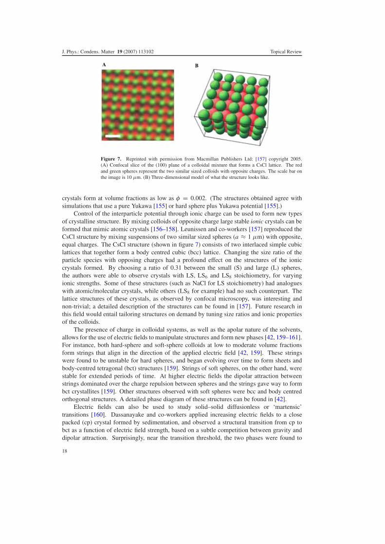
Figure 7. Reprinted with permission from Macmillan Publishers Ltd: [157] copyright 2005.(A) Confocal slice of the (100) plane of a colloidal mixture that forms a CsCl lattice. The redand green spheres represent the two similar sized colloids with opposite charges. The scale bar onthe image is 10 μm. (B) Three-dimensional model of what the structure looks like.
crystals form at volume fractions as low as φ = 0.002. (The structures obtained agree withsimulations that use a pure Yukawa [155] or hard sphere plus Yukawa potential [155].)
Control of the interparticle potential through ionic charge can be used to form new typesof crystalline structure. By mixing colloids of opposite charge large stable ionic crystals can beformed that mimic atomic crystals [156–158]. Leunissen and co-workers [157] reproduced theCsCl structure by mixing suspensions of two similar sized spheres (a ≈ 1 μm) with opposite,equal charges. The CsCl structure (shown in figure 7) consists of two interlaced simple cubiclattices that together form a body centred cubic (bcc) lattice. Changing the size ratio of theparticle species with opposing charges had a profound effect on the structures of the ioniccrystals formed. By choosing a ratio of 0.31 between the small (S) and large (L) spheres,the authors were able to observe crystals with LS, LS6 and LS8 stoichiometry, for varyingionic strengths. Some of these structures (such as NaCl for LS stoichiometry) had analogueswith atomic/molecular crystals, while others (LS8 for example) had no such counterpart. Thelattice structures of these crystals, as observed by confocal microscopy, was interesting andnon-trivial; a detailed description of the structures can be found in [157]. Future research inthis field would entail tailoring structures on demand by tuning size ratios and ionic propertiesof the colloids.
The presence of charge in colloidal systems, as well as the apolar nature of the solvents,allows for the use of electric fields to manipulate structures and form new phases [42, 159–161].For instance, both hard-sphere and soft-sphere colloids at low to moderate volume fractionsform strings that align in the direction of the applied electric field [42, 159]. These stringswere found to be unstable for hard spheres, and began evolving over time to form sheets andbody-centred tetragonal (bct) structures [159]. Strings of soft spheres, on the other hand, werestable for extended periods of time. At higher electric fields the dipolar attraction betweenstrings dominated over the charge repulsion between spheres and the strings gave way to formbct crystallites [159]. Other structures observed with soft spheres were bcc and body centredorthogonal structures. A detailed phase diagram of these structures can be found in [42].
Electric fields can also be used to study solid–solid diffusionless or ‘martensic’transitions [160]. Dassanayake and co-workers applied increasing electric fields to a closepacked (cp) crystal formed by sedimentation, and observed a structural transition from cp tobct as a function of electric field strength, based on a subtle competition between gravity anddipolar attraction. Surprisingly, near the transition threshold, the two phases were found to
18
J. Phys.: Condens. Matter 19 (2007) 113102 Topical Review
coexist for long periods of time. While this raises interesting questions about the order ofthe phase transition, further studies, similar to those seen in fluid–fluid phase separation, areneeded. Finally, these new structures formed by the subtle interplay between attractive forcesand repulsion or gravity, while having interesting physics, also have great application such asin photonic band gap structures.
6.5. Anisotropic systems
While attractive and repulsive interactions help make colloids better models for atomicsystems, the systems described above lack one interesting feature: the interactions are allspherically symmetric. With improving synthesis techniques, several groups now study thephase behaviour of non-spherical colloidal particles. Confocal microscopy has been used tocharacterize samples consisting of non-spherical colloids such as polyhedra [162] or anisotropicones such as dumbbells [163] and rods [164, 165].
It is crucial but non-trivial to identify the orientation of these anisotropic colloids in aconfocal image. Colloidal dumbbells [163] consist of dimers of silica spheres that have beenfused together. It is difficult to resolve these dumbbells using x-ray scattering except at highscattering wavenumbers. However, because the distance between two spheres that are part ofthe same dumbbell is less than any other separation between spheres in the sample, dumbbellscan be identified by measuring the distance between the fluorescent core centres of the silicaparticles. Using these identified centres, the position and orientation of all the dumbbells in a3D sample could be measured. A similar problem exists for the case of colloidal rods [164],made by uniaxially extending PMMA spheres. In this case an algorithm was devised to identifythe backbones of the rods, therefore associating a central axis with each rod. The centroid of therod was determined by averaging the pixel positions of the points comprising this central axis,and the rod was characterized by its length and orientation. This technique worked remarkablywell even for highly concentrated, nearly close packed rods.
Confocal microscopy has also been used to characterize non-spherical ‘polyhedral’colloids [162], where the particle shape is characterized by comparing the projected particlearea in 2D to its projected perimeter. The close packing of the particles showed deviations fromthose of spherical particles. Especially interesting was the fact that g(r) decayed much fasterfor polyhedral colloids than spheres, implying no long-range translational order. The bond-orientational correlation function g6(r) also decayed faster, implying frustration of hexagonalorder in these polyhedra. Conceivably, with advances in particle synthesis and new typesof particles being produced with ease, confocal microscopy can have great application indetermining unique structures and phases these particles may form, especially because of theease of 3D reconstruction.
7. New directions
In this review we have tried to touch upon many aspects of the phase behaviour of colloids thatinteract either as hard spheres or with repulsive/attractive potentials. Apart from these well-studied systems with thermodynamic analogies to atomic and molecular systems [166], colloidshave other applications as well. For instance, they are used as tracer particles in viscoelasticsolutions to probe the rheological properties of the environment around them. For spatiallyheterogeneous materials, confocal microscopy of the thermal motions of these particles in threedimensions can be very useful [167, 168]. Further, the effects of shear on these viscoelasticmaterials can also be probed [168].
Large colloids, such as emulsion droplets, can also be used to mimic granular systems.Emulsions are visualized either by dying the dispersed phase [169, 170] or by utilizing
19

J. Phys.: Condens. Matter 19 (2007) 113102 Topical Review
fluorescent surfactants that go to the interface between the dispersed phase and the continuousphase [171]. The force between two droplets that are in contact can be quantified bycharacterizing the shape of these compressible droplets [169, 171, 172]. Thus a spatialdistribution of ‘force chains’ in three dimensions can be obtained, which has analogies withsuch chains seen in granular materials.
In conclusion, colloidal systems show behaviour than spans the gamut from atomic systems(with length and time scales in the nanometre and picosecond range) to granular materials(millimetres and seconds). Confocal microscopy is key to the observation of these phenomenain real space and real time, and this powerful technique holds much promise for future research.
Acknowledgments
We thank Scott V Franklin and G C Cianci for enlightening discussions. E R Weeks andV Prasad thank NSF (DMR-0239109) for funding.
References
[1] Habdas P and Weeks E R 2002 Video microscopy of colloidal suspensions and colloidal crystals Curr. Opin.Colloid Interface Sci. 7 196–203
[2] Minsky M 1988 Memoir on inventing the confocal scanning microscope Scanning 10 128–38[3] Sheppard C J R and Shotton D M 1997 Confocal Laser Scanning Microscopy (New York: Springer)[4] Inoue S and Spring K R 1997 Video Microscopy: The Fundamentals (New York: Plenum)[5] Pawley J B 1995 Handbook of Biological Confocal Microscopy (New York: Plenum)[6] Pusey P N and van Megen W 1986 Phase-behavior of concentrated suspensions of nearly hard colloidal spheres
Nature 320 340–2[7] Yoshida H, Ito K and Ise N 1991 Localized ordered structure in polymer latex suspensions as studied by a
confocal laser scanning microscope Phys. Rev. B 44 435–8[8] van Blaaderen A and Wiltzius P 1995 Real-space structure of colloidal hard-sphere glasses Science 270 1177–9[9] Alder B J and Wainwright T E 1970 Decay of velocity autocorrelation function Phys. Rev. A 1 18–21
[10] Woodcock L V 1981 Glass-transition in the hard-sphere model and Kauzmann paradox Ann. NY Acad. Sci.371 274–98
[11] Speedy R J 1998 The hard sphere glass transition Mol. Phys. 95 169–78[12] Alder B J, Gass D M and Wainwright T E 1970 Studies in molecular dynamics.8. transport coefficients for a
hard-sphere fluid J. Chem. Phys. 53 3813–26[13] Alder B J and Wainwright T E 1957 Phase transition for a hard sphere system J. Chem. Phys. 27 1208–9[14] Zhu J X, Li M, Rogers R, Meyer W, Ottewill R H, Russell W B and Chaikin P M 1997 Crystallization of
hard-sphere colloids in microgravity Nature 387 883–5[15] Hoogenboom J P, Vergeer P and van Blaaderen A 2003 A real-space analysis of colloidal crystallization in a
gravitational field at a flat bottom wall J. Chem. Phys. 119 3371–83[16] Royall C P, van Roij R and van Blaaderen A 2005 Extended sedimentation profiles in charged colloids: the
gravitational length, entropy, and electrostatics J. Phys.: Condens. Matter 17 2315–26[17] Simeonova N B and Kegel W K 2004 Gravity-induced aging in glasses of colloidal hard spheres Phys. Rev. Lett.
93 035701[18] Bernal J D 1959 Geometrical approach to the structure of liquids Nature 183 141–7[19] Bernal J D 1960 Geometry of the structure of monatomic liquids Nature 185 68–70[20] Bernal J D 1964 Bakerian lecture 1962—structure of liquids Proc. R. Soc. A 280 299[21] Torquato S, Truskett T M and Debenedetti P G 2000 Is random close packing of spheres well defined? Phys.
Rev. Lett. 84 2064–7[22] Moriguchi I, Kawasaki K and Kawakatsu T 1993 The effects of size polydispersity in nearly hard-sphere colloids
J. Physique II 3 1179–84[23] Williams S R, Snook I K and van Megen W 2001 Molecular dynamics study of the stability of the hard sphere
glass Phys. Rev. E 6402 021506[24] Cheng Z D, Zhu J X, Chaikin P M, Phan S E and Russel W B 2002 Nature of the divergence in low shear
viscosity of colloidal hard-sphere dispersions Phys. Rev. E 65 041405[25] Pusey P N 1991 Liquids, Freezing and the Glass Transition (Amsterdam: Elsevier)
20

J. Phys.: Condens. Matter 19 (2007) 113102 Topical Review
[26] Angell C A 1995 Formation of glasses from liquids and biopolymers Science 267 1924–35[27] Angell C A 2000 Ten questions on glassformers, and a real space ‘excitations’ model with some answers on
fragility and phase transitions J. Phys.: Condens. Matter 12 6463–75[28] Stillinger F H 1995 A topographic view of supercooled liquids and glass-formation Science 267 1935–9[29] Ediger M D, Angell C A and Nagel S R 1996 Supercooled liquids and glasses J. Phys. Chem. 100 13200–12[30] Russel W B, Saville D A and Schowalter W R 1989 Colloidal Dispersions (Cambridge: Cambridge University
Press)[31] Jones R A L 2002 Soft Condensed Matter (Oxford: Oxford University Press)[32] Larson R G 1998 The Structure and Rheology of Complex Fluids (Oxford: Oxford University Press)[33] Antl L, Goodwin J W, Hill R D, Ottewill R H, Owens S M, Papworth S and Waters J A 1986 The preparation
of poly(methyl methacrylate) lattices in nonaqueous media Colloids Surf. 17 67–78[34] Pusey P N, van Megen W, Bartlett P, Ackerson B J, Rarity J G and Underwood S M 1989 Structure of crystals
of hard colloidal spheres Phys. Rev. Lett. 63 2753–6[35] Asakura S and Oosawa F 1958 Interaction between particles suspended in solutions of macromolecules J. Polym.
Sci. 33 183–92[36] Crocker J C, Matteo J A, Dinsmore A D and Yodh A G 1999 Entropic attraction and repulsion in binary colloids
probed with a line optical tweezer Phys. Rev. Lett. 82 4352–5[37] Verma R, Crocker J C, Lubensky T C and Yodh A G 2000 Attractions between hard colloidal spheres in
semiflexible polymer solutions Macromolecules 33 177–86[38] Bosma G, Pathmamanoharan C, de Hoog E H A, Kegel W K, van Blaaderen A and Lekkerkerker H N W 2002
Preparation of monodisperse, fluorescent pmma-latex colloids by dispersion polymerization J. ColloidInterface Sci. 245 292–300
[39] Dullens R P A, Claesson M, Derks D, van Blaaderen A and Kegel W K 2003 Monodisperse core-shellpoly(methyl methacrylate) latex colloids Langmuir 19 5963–6
[40] Campbell A I and Bartlett P 2002 Fluorescent hard-sphere polymer colloids for confocal microscopy J. ColloidInterface Sci. 256 325–30
[41] van Blaaderen A and Vrij A 1992 Synthesis and characterization of colloidal dispersions of fluorescent,monodisperse silica spheres Langmuir 8 2921–31
[42] Yethiraj A and van Blaaderen A 2003 A colloidal model system with an interaction tunable from hard sphere tosoft and dipolar Nature 421 513–7
[43] Stieger M, Richtering W, Pedersen J S and Lindner P 2004 Small-angle neutron scattering study of structuralchanges in temperature sensitive microgel colloids J. Chem. Phys. 120 6197–206
[44] Ye X, Narayanan T, Tong P, Huang J S, Lin M Y, Carvalho B L and Fetters L J 1996 Depletion interactions incolloid-polymer mixtures Phys. Rev. E 54 6500–10
[45] Ye X, Narayanan T and Tong P 1996 Neutron scattering study of depletion interactions in a colloid-polymermixture Phys. Rev. Lett. 76 4640–3
[46] Ottewill R H, Hanley H J M, Rennie A R and Straty G C 1995 Small-angle neutron-scattering studies on binary-mixtures of charged-particles Langmuir 11 3757–65
[47] Ashdown S, Markovic I, Ottewill R H, Lindner P, Oberthur R C and Rennie A R 1990 Small-angle neutron-scattering studies on ordered polymer colloid dispersions Langmuir 6 303–7
[48] Lal J, Abernathy D, Auvray L, Diat O and Grubel G 2001 Dynamics and correlations in magnetic colloidalsystems studied by x-ray photon correlation spectroscopy Eur. Phys. J. E 4 263–71
[49] Konishi T, Yamahara E and Ise N 1996 Characterization of colloidal silica particles by ultra-small-angle x-rayscattering Langmuir 12 2608–10
[50] Dierker S B, Pindak R, Fleming R M, Robinson I K and Berman L 1995 X-ray photon-correlation spectroscopystudy of Brownian-motion of gold colloids in glycerol Phys. Rev. Lett. 75 449–52
[51] Konishi T, Ise N, Matsuoka H, Yamaoka H, Sogami I S and Yoshiyama T 1995 Structural study of silica particledispersions by ultra-small-angle x-ray-scattering Phys. Rev. B 51 3914–7
[52] van Megen W, Underwood S M and Pusey P N 1991 Dynamics of hard spherical colloids from the fluid to theglass J. Chem. Soc.-Faraday Trans. 87 395–401
[53] van Megen W and Pusey P N 1991 Dynamic light-scattering study of the glass-transition in a colloidalsuspension Phys. Rev. A 43 5429–41
[54] Carpineti M and Giglio M 1992 Spinodal-type dynamics in fractal aggregation of colloidal clusters Phys. Rev.Lett. 68 3327–30
[55] Dhont J K G, Smits C and Lekkerkerker H N W 1992 A time resolved static light-scattering study on nucleationand crystallization in a colloidal system J. Colloid Interface Sci. 152 386–401
[56] Cipelletti L, Manley S, Ball R C and Weitz D A 2000 Universal aging features in the restructuring of fractalcolloidal gels Phys. Rev. Lett. 84 2275–8
21

J. Phys.: Condens. Matter 19 (2007) 113102 Topical Review
[57] Moussaid A and Pusey P N 1999 Multiple scattering suppression in static light scattering by cross-correlationspectroscopy Phys. Rev. E 60 5670–6
[58] Segre P N, van Megen W, Pusey P N, Schatzel K and Peters W 1995 2-color dynamic light-scattering J. Mod.Opt. 42 1929–52
[59] Stieber F and Richtering W 1995 Fiber-optic-dynamic-light-scattering and two-color-cross-correlation studiesof turbid, concentrated, sterically stabilized polystyrene latex Langmuir 11 4724–7
[60] Schatzel K, Drewel M and Ahrens J 1990 Suppression of multiple-scattering in photon-correlation spectroscopyJ. Phys.: Condens. Matter 2 (Suppl. A.) SA393–8
[61] Dasgupta B R, Tee S Y, Crocker J C, Frisken B J and Weitz D A 2002 Microrheology of polyethylene oxideusing diffusing wave spectroscopy and single scattering Phys. Rev. E 65 051505
[62] Durian D J, Weitz D A and Pine D J 1990 Dynamics and coarsening in 3-dimensional foams J. Phys.: Condens.Matter 2 (Suppl. A.) SA433–6
[63] Pine D J, Weitz D A, Zhu J X and Herbolzheimer E 1990 Diffusing-wave spectroscopy—dynamic light-scattering in the multiple-scattering limit J. Physique 51 2101–27
[64] Pine D J, Weitz D A, Chaikin P M and Herbolzheimer E 1988 Diffusing-wave spectroscopy Phys. Rev. Lett.60 1134–7
[65] Trappe V, Prasad V, Cipelletti L, Segre P N and Weitz D A 2001 Jamming phase diagram for attractive particlesNature 411 772–5
[66] Trappe V and Weitz D A 2000 Scaling of the viscoelasticity of weakly attractive particles Phys. Rev. Lett.85 449–52
[67] Fagan M E and Zukoski C F 1997 The rheology of charge stabilized silica suspensions J. Rheol. 41 373–97[68] Mason T G and Weitz D A 1995 Linear viscoelasticity of colloidal hard-sphere suspensions near the glass-
transition Phys. Rev. Lett. 75 2770–3[69] Gisler T, Ball R and Weitz D A 1999 Strain hardening of fractal colloidal gels Phys. Rev. Lett. 82 1064–7[70] Prasad V, Trappe V, Dinsmore A D, Segre P N, Cipelletti L and Weitz D A 2003 Universal features of the fluid
to solid transition for attractive colloidal particles Faraday Discuss. 123 1–12[71] Zernike F 1942 Phase contrast, a new method for the microsopic observation of transparent objects Physica
9 686–98[72] Zernike F 1942 Phase contrast, a new method for the microscopic observation of transparent objects part II
Physica 9 974–86[73] Nomarski M G 1955 Microinterferometre differentiel a ondes polarisees J. Physique Radium 16 (Suppl. S)
S9–13[74] Elliot M S and Poon W C K 2001 Conventional optical microscopy of colloidal suspensions Adv. Colloid
Interface Sci. 92 133–94[75] Baumgartl J, Arauz-Lara J L and Bechinger C 2006 Like-charge attraction in confinement: myth or truth? Soft
Matter 2 631–5[76] Xiao G Q, Corle T R and Kino G S 1988 Real-time confocal scanning optical microscope Appl. Phys. Lett.
53 716–8[77] Crocker J C and Grier D G 1996 Methods of digital video microscopy for colloidal studies J. Colloid Interface
Sci. 179 298–310[78] Dinsmore A D, Weeks E R, Prasad V, Levitt A C and Weitz D A 2001 Three-dimensional confocal microscopy
of colloids Appl. Opt. 40 4152–9[79] A tutorial on particle tracking can be found at http://www.physics.emory.edu/∼weeks/idl[80] Isa L, Besseling R, Weeks E R and Poon W C K 2006 Experimental studies of the flow of concentrated hard
sphere suspensions into a constriction J. Phys. Conf. Ser. 40 124–32[81] Nelson D R 2002 Defects and Geometry in Condensed Matter Physics (Cambridge: Cambridge University
Press)[82] ten Wolde P R, Ruiz-Montero M J and Frenkel D 1999 Numerical calculation of the rate of homogeneous
gas–liquid nucleation in a Lennard-Jones system J. Chem. Phys. 110 1591–9[83] Steinhardt P J, Nelson D R and Ronchetti M 1983 Bond-orientational order in liquids and glasses Phys. Rev. B
28 784–805[84] Gasser U, Weeks E R, Schofield A, Pusey P N and Weitz D A 2001 Real-space imaging of nucleation and
growth in colloidal crystallization Science 292 258–62[85] Auer S and Frenkel D 2001 Prediction of absolute crystal-nucleation rate in hard-sphere colloids Nature
409 1020–3[86] de Villeneuve V W A, Dullens R P A, Aarts D G A L, Groeneveld E, Scherff J H, Kegel W K and
Lekkerkerker H N W 2005 Colloidal hard-sphere crystal growth frustrated by large spherical impuritiesScience 309 1231–3
22

J. Phys.: Condens. Matter 19 (2007) 113102 Topical Review
[87] de Villeneuve V W A, Verboekend D, Dullens R P A, Aarts D G A L, Kegel W K andLekkerkerker H N W 2005 Hard sphere crystal nucleation and growth near large spherical impurities J.Phys.: Condens. Matter 17 (Sp. Iss. SI) S3371–8
[88] Dullens R P A, Aarts D G A L and Kegel W K 2006 Dynamic broadening of the crystal–fluid interface ofcolloidal hard spheres Phys. Rev. Lett. 97 228301
[89] Schall P, Cohen I, Weitz D A and Spaepen F 2004 Visualization of dislocation dynamics in colloidal crystalsScience 305 1944–8
[90] Schall P, Cohen I, Weitz D A and Spaepen F 2006 Visualizing dislocation nucleation by indenting colloidalcrystals Nature 440 319–23
[91] Lin K H, Crocker J C, Prasad V, Schofield A, Weitz D A, Lubensky T C and Yodh A G 2000 Entropically drivencolloidal crystallization on patterned surfaces Phys. Rev. Lett. 85 1770–3
[92] Hunt N, Jardine R and Bartlett P 2000 Superlattice formation in mixtures of hard-sphere colloids Phys. Rev. E62 900–13
[93] Schofield A B 2001 Binary hard-sphere crystals with the cesium chloride structure Phys. Rev. E6405 051403
[94] Bartlett P, Ottewill R H and Pusey P N 1992 Superlattice formation in binary-mixtures of hard-sphere colloidsPhys. Rev. Lett. 68 3801–4
[95] Gotze W and Sjogren L 1992 Relaxation processes in supercooled liquids Rep. Prog. Phys. 55 241–376[96] Bengtzelius U, Gotze W and Sjolander A 1984 Dynamics of supercooled liquids and the glass-transition J. Phys.
C: Solid State Phys. 17 5915–34[97] Ediger M D 2000 Spatially heterogeneous dynamics in supercooled liquids Annu. Rev. Phys. Chem. 51 99–128[98] Kegel W K and van Blaaderen A 2000 Direct observation of dynamical heterogeneities in colloidal hard-sphere
suspensions Science 287 290–3[99] Weeks E R, Crocker J C, Levitt A C, Schofield A and Weitz D A 2000 Three-dimensional direct imaging of
structural relaxation near the colloidal glass transition Science 287 627–31[100] Adam G and Gibbs J H 1965 On temperature dependence of cooperative relaxation properties in glass-forming
liquids J. Chem. Phys. 43 139[101] Weeks E R and Weitz D A 2002 Properties of cage rearrangements observed near the colloidal glass transition
Phys. Rev. Lett. 89 095704[102] Weeks E R and Weitz D A 2002 Subdiffusion and the cage effect studied near the colloidal glass transition
Chem. Phys. 284 (Sp. Iss. SI) 361–7[103] Conrad J C, Starr F W and Weitz D A 2005 Weak correlations between local density and dynamics near the
glass transition J. Phys. Chem. B 109 21235–40[104] Pusey P N and van Megen W 1987 Observation of a glass-transition in suspensions of spherical colloidal
particles Phys. Rev. Lett. 59 2083–6[105] van Megen W, Mortensen T C, Williams S R and Muller J 1998 Measurement of the self-intermediate scattering
function of suspensions of hard spherical particles near the glass transition Phys. Rev. E 58 6073–85[106] van Megen W, Underwood S M and Pusey P N 1991 Nonergodicity parameters of colloidal glasses Phys. Rev.
Lett. 67 1586–9[107] Kasper A, Bartsch E and Sillescu H 1998 Self-diffusion in concentrated colloid suspensions studied by digital
video microscopy of core-shell tracer particles Langmuir 14 5004–10[108] Marcus A H, Schofield J and Rice S A 1999 Experimental observations of non-Gaussian behavior and stringlike
cooperative dynamics in concentrated quasi-two-dimensional colloidal liquids Phys. Rev. E 60 5725–36[109] Weeks E R, Crocker J C and Weitz D A 2006 Preprint cond-mat/0610195[110] Courtland R E and Weeks E R 2003 Direct visualization of ageing in colloidal glasses J. Phys.: Condens. Matter
15 (Sp. Iss. SI) S359–65[111] Henderson S I and van Megen W 1998 Metastability and crystallization in suspensions of mixtures of hard
spheres Phys. Rev. Lett. 80 877–80[112] Cipelletti L, Bissig H, Trappe V, Ballesta P and Mazoyer S 2003 Time-resolved correlation: a new tool for
studying temporally heterogeneous dynamics J. Phys.: Condens. Matter 15 (Sp. Iss. SI) S257–62[113] Cianci G C, Courtland R E and Weeks E R 2006 2nd Int. Conf. on Flow Dynamics, AIP Conf. Proc. 832 21–5[114] Cianci G C, Courtland R E and Weeks E R 2006 Correlations of structure and dynamics in an aging colloidal
glass Solid State Commun. 139 599–604[115] Paulin S E, Ackerson B J and Wolfe M S 1997 Microstructure-dependent viscosity in concentrated suspensions
of soft spheres Phys. Rev. E 55 5812–9[116] Ackerson B J and Pusey P N 1988 Shear-induced order in suspensions of hard-spheres Phys. Rev. Lett.
61 1033–6
23

J. Phys.: Condens. Matter 19 (2007) 113102 Topical Review
[117] Haw M D, Poon W C K and Pusey P N 1998 Direct observation of oscillatory-shear-induced order in colloidalsuspensions Phys. Rev. E 57 6859–64
[118] Haw M D, Poon W C K, Pusey P N, Hebraud P and Lequeux F 1998 Colloidal glasses under shear strain Phys.Rev. E 58 4673–82
[119] Varadan P and Solomon M J 2003 Direct visualization of flow-induced microstructure in dense colloidal gelsby confocal laser scanning microscopy J. Rheol. 47 943–68
[120] Derks D, Wisman H, van Blaaderen A and Imhof A 2004 Confocal microscopy of colloidal dispersions in shearflow using a counter-rotating cone-plate shear cell J. Phys.: Condens. Matter 16 (Sp. Iss. SI) S3917–27
[121] Besseling R, Weeks E R, Schofield A B and Poon W C K 2006 Preprint cond-mat/0605247[122] Ackerson B J 1990 Shear induced order and shear processing of model hard-sphere suspensions J. Rheol.
34 553–90[123] Solomon T and Solomon M J 2006 Stacking fault structure in shear-induced colloidal crystallization J. Chem.
Phys. 124 134905[124] Cohen I, Mason T G and Weitz D A 2004 Shear-induced configurations of confined colloidal suspensions Phys.
Rev. Lett. 93 046001[125] Cohen I, Davidovitch B, Schofield A B, Brenner M P and Weitz D A 2006 Slip, yield, and bands in colloidal
crystals under oscillatory shear Phys. Rev. Lett. 97 215502[126] Ball R C, Weitz D A, Witten T A and Leyvraz F 1987 Universal kinetics in reaction-limited aggregation Phys.
Rev. Lett. 58 274–7[127] Lin M Y, Lindsay H M, Weitz D A, Ball R C, Klein R and Meakin P 1990 Universal reaction-limited colloid
aggregation Phys. Rev. A 41 2005–20[128] Weitz D A and Oliveria M 1984 Fractal structures formed by kinetic aggregation of aqueous gold colloids Phys.
Rev. Lett. 52 1433–6[129] Dinsmore A D and Weitz D A 2002 Direct imaging of three-dimensional structure and topology of colloidal
gels J. Phys.: Condens. Matter 14 7581–97[130] Lu P J, Conrad J C, Wyss H M, Schofield A B and Weitz D A 2006 Fluids of clusters in attractive colloids Phys.
Rev. Lett. 96 028306[131] Segre P N, Prasad V, Schofield A B and Weitz D A 2001 Glasslike kinetic arrest at the colloidal-gelation
transition Phys. Rev. Lett. 86 6042–5[132] Sedgwick H, Egelhaaf S U and Poon W C K 2004 Clusters and gels in systems of sticky particles J. Phys.:
Condens. Matter 16 (Sp. Iss. SI) S4913–22[133] Campbell A I, Anderson V J, van Duijneveldt J S and Bartlett P 2005 Dynamical arrest in attractive colloids:
the effect of long-range repulsion Phys. Rev. Lett. 94 208301[134] Groenewold J and Kegel W K 2001 Anomalously large equilibrium clusters of colloids J. Phys. Chem. B
105 11702–9[135] Groenewold J and Kegel W K 2004 Colloidal cluster phases, gelation and nuclear matter J. Phys.: Condens.
Matter 16 (Sp. Iss. SI) S4877–86[136] Varadan P and Solomon M J 2003 Direct visualization of long-range heterogeneous structure in dense colloidal
gels Langmuir 19 509–12[137] Tohver V, Smay J E, Braem A, Braun P V and Lewis J A 2001 Nanoparticle halos: a new colloid stabilization
mechanism Proc. Natl Acad. Sci. USA 98 8950–4[138] Tohver V, Chan A, Sakurada O and Lewis J A 2001 Nanoparticle engineering of complex fluid behavior
Langmuir 17 8414–21[139] Gilchrist J F, Chan A T, Weeks E R and Lewis J A 2005 Phase behavior and 3d structure of strongly attractive
microsphere-nanoparticle mixtures Langmuir 21 11040–7[140] Martinez C J, Liu J W, Rhodes S K, Luijten E, Weeks E R and Lewis J A 2005 Interparticle interactions
and direct imaging of colloidal phases assembled from microsphere-nanoparticle mixtures Langmuir21 9978–89
[141] Pham K N, Egelhaaf S U, Pusey P N and Poon W C K 2004 Glasses in hard spheres with short-range attractionPhys. Rev. E 69 011503
[142] Pham K N, Puertas A M, Bergenholtz J, Egelhaaf S U, Moussaid A, Pusey P N, Schofield A B, Cates M E,Fuchs M and Poon W C K 2002 Multiple glassy states in a simple model system Science 296 104–6
[143] Poon W C K, Pham K N, Egelhaaf S U and Pusey P N 2003 ‘Unsticking’ a colloidal glass, and sticking it againJ. Phys.: Condens. Matter 15 (Sp. Iss. SI) S269–75
[144] Dawson K A 2002 The glass paradigm for colloidal glasses, gels, and other arrested states driven by attractiveinteractions Curr. Opin. Colloid Interface Sci. 7 218–27
[145] Kaufman L J and Weitz D A 2006 Direct imaging of repulsive and attractive colloidal glasses J. Chem. Phys.125 074716
24
J. Phys.: Condens. Matter 19 (2007) 113102 Topical Review
[146] Simeonova N B, Dullens R P A, Aarts D G A L, de Villeneuve V W A, Lekkerkerker H N W and Kegel W K2006 Devitrification of colloidal glasses in real space Phys. Rev. E 73 041401
[147] Schroeder D V 1999 An Introduction to Thermal Physics (Reading, MA: Addison-Wesley)[148] Ilett S M, Orrock A, Poon W C K and Pusey P N 1995 Phase-behavior of a model colloid-polymer mixture
Phys. Rev. E 51 1344–52[149] de Hoog E H A, Kegel W K, van Blaaderen A and Lekkerkerker H N W 2001 Direct observation of
crystallization and aggregation in a phase-separating colloid-polymer suspension Phys. Rev. E6402 021407
[150] Aarts D G A L, Dullens R P A and Lekkerkerker H N W 2005 Interfacial dynamics in demixing systems withultralow interfacial tension New J. Phys. 7 40
[151] Aarts D G A L 2005 Capillary length in a fluid–fluid demixed colloid-polymer mixture J. Phys. Chem. B109 7407–11
[152] Aarts D G A L, Schmidt M and Lekkerkerker H N W 2004 Direct visual observation of thermal capillary wavesScience 304 847–50
[153] Aarts D G A L 2007 The interface in demixed colloid-polymer systems: wetting, waves and droplets Soft Matter3 19–23
[154] Royall C P, Leunissen M E and van Blaaderen A 2003 A new colloidal model system to study long-rangeinteractions quantitatively in real space J. Phys.: Condens. Matter 15 (Sp. Iss. SI) S3581–96
[155] Robbins M O, Kremer K and Grest G S 1988 Phase-diagram and dynamics of Yukawa systems J. Chem. Phys.88 3286–312
[156] Hynninen A P, Leunissen M E, van Blaaderen A and Dijkstra M 2006 Cu Au structure in the restricted primitivemodel and oppositely charged colloids Phys. Rev. Lett. 96 018303
[157] Leunissen M E, Christova C G, Hynninen A P, Royall C P, Campbell A I, Imhof A, Dijkstra M, van Roij R andvan Blaaderen A 2005 Ionic colloidal crystals of oppositely charged particles Nature 437 235–40
[158] Bartlett P and Campbell A I 2005 Three-dimensional binary superlattices of oppositely charged colloids Phys.Rev. Lett. 95 128302
[159] Yethiraj A and van Blaaderen A 2002 Monodisperse colloidal suspensions of silica and PMMA spheres asmodel electrorheological fluids: a real-space study of structure formation Int. J. Mod. Phys. B16 (Sp. Iss. SI) 2328–33
[160] Yethiraj A, Wouterse A, Groh B and van Blaaderen A 2004 Nature of an electric-field-induced colloidalmartensitic transition Phys. Rev. Lett. 92 058301
[161] Dassanayake U, Fraden S and van Blaaderen A 2000 Structure of electrorheological fluids J. Chem. Phys.112 3851–8
[162] Dullens R P A, Mourad M C D, Aarts D G A L, Hoogenboom J P and Kegel W K 2006 Shape-induced frustrationof hexagonal order in polyhedral colloids Phys. Rev. Lett. 96 028304
[163] Johnson P M, van Kats C M and van Blaaderen A 2005 Synthesis of colloidal silica dumbbells Langmuir21 11510–7
[164] Mohraz A and Solomon M J 2005 Direct visualization of colloidal rod assembly by confocal microscopyLangmuir 21 5298–306
[165] van Kats C M, Johnson P M, van den Meerakker J and van Blaaderen A 2004 Synthesis of monodispersehigh-aspect-ratio colloidal silicon and silica rods Langmuir 20 11201–7
[166] Dullens R P A, Aarts D G A L and Kegel W K 2006 Direct measurement of the free energy by optical microscopyProc. Natl Acad. Sci. USA 103 529–31
[167] Moschakis T, Murray B S and Dickinson E 2006 Particle tracking using confocal microscopy to probethe microrheology in a phase-separating emulsion containing nonadsorbing polysaccharide Langmuir22 4710–9
[168] Vossen D L J, van der Horst A, Dogterom M and van Blaaderen A 2004 Optical tweezers and confocalmicroscopy for simultaneous three-dimensional manipulation and imaging in concentrated colloidaldispersions Rev. Sci. Instrum. 75 2960–70
[169] Brujic J, Edwards S F, Grinev D V, Hopkinson I, Brujic D and Makse H A 2003 3d bulk measurements of theforce distribution in a compressed emulsion system Faraday Discuss. 123 207–20
[170] Penfold R, Watson A D, Mackie A R and Hibberd D J 2006 Quantitative imaging of aggregated emulsionsLangmuir 22 2005–15
[171] Zhou J, Long S, Wang Q and Dinsmore A D 2006 Measurement of forces inside a three-dimensional pile offrictionless droplets Science 312 1631–3
[172] Brujic J, Edwards S F, Hopkinson I and Makse H A 2003 Measuring the distribution of interdroplet forces in acompressed emulsion system Physica A 327 201–12
25


![Colloids and Surfaces B: Biointerfaces · Colloids and Surfaces B: Biointerfaces 88 (2011) 279–286 Contents lists available at ScienceDirect Colloids ... [26,27]. Other researchers](https://img.pdfslide.us/doc/110x75/5fc50395d8208315bc08a19b/colloids-and-surfaces-b-colloids-and-surfaces-b-biointerfaces-88-2011-279a286.jpg)