Embed Size (px)
Citation preview

Confocal Microscopy and 3-D Reconstructionof the Cytoskeleton of Xenopus OocytesDAVID L. GARD*Department of Biology, University of Utah, Salt Lake City, Utah 84112–0840
KEY WORDS microtubules; microfilaments; F-actin; keratin filaments; oogenesis
ABSTRACT Xenopus oocytes contain a complex cytoskeleton composed of three filamentsystems: (1) microtubules, composed of tubulin and at least three different microtubule-associatedproteins (XMAPs); (2) microfilaments composed of actin and associated proteins; and (3) intermedi-ate filaments, composed of keratins. For the past several years, we have used confocal immunofluo-rescence microscopy to characterize the organization of the oocyte cytoskeleton throughout thecourse of oogenesis. Together with computer-assisted reconstruction of the oocyte in threedimensions, confocal microscopy gives an unprecedented view of the assembly and reorganization ofthe cytoskeleton during oocyte growth and differentiation. Results of these studies, combined withthe effects of cytoskeletal inhibitors, suggest that organization of the cytoskeleton in Xenopusoocytes is dependent upon a hierarchy of interactions between microtubules, microfilaments, andkeratin filaments. This article presents a gallery of confocal images and 3-D reconstructionsdepicting the assembly and organization of the oocyte cytoskeleton during stages 0-VI of oogenesis, adiscussion of the mechanisms that might regulate cytoskeletal organization during oogenesis, andspeculates on the potential roles of the oocyte cytoskeleton during oogenesis and axis formation.Microsc. Res. Tech. 44:388–414, 1999. r 1999 Wiley-Liss, Inc.
INTRODUCTIONThe large size (often exceeding a millimeter in diam-
eter) and ease of experimental manipulation of oocytes,eggs, and early embryos of the African frog, Xenopuslaevis, have made them attractive model systems forthe study of many developmental mechanisms. Inaddition, because zygotic transcription does not beginuntil the mid-blastula stage of development (Newportand Kirschner, 1982), Xenopus oocytes and eggs containlarge maternal stores of many of the proteins and RNAsneeded for cell growth and division. These stockpilesmake cytoplasmic extracts from Xenopus eggs popularmodels for studying many basic cellular processes invitro. In fact, much of our current knowledge of cellularprocesses such as nuclear transport, the eukaryotic cellcycle, and spindle assembly can be directly traced toexperiments utilizing cytoplasm isolated from Xenopusoocytes or eggs.
Although the cytoskeleton of Xenopus oocytes hasbeen implicated in a number of important developmen-tal functions (reviewed in Gard, 1995, Gard andKlymkowsky, 1998), including specification of the ani-mal-vegetal axis and localization of developmentally-important maternal RNAs (Elinson et al., 1993; For-ristall et al., 1995; Kloc and Etkin, 1995; Yisraeli et al.,1990; Zhou and King, 1996a), our understanding of thecytoskeletal organization of Xenopus oocytes has laggedbehind that of other model systems used for studyingcellular and developmental mechanisms. Indeed, thelarge size that make them so attractive for studiesinvolving the microinjection of macromolecules or prepa-ration of cytoplasmic extracts, when combined withtheir opaque, yolk-filled cytoplasm, makes conventional
microscopy of Xenopus oocytes and eggs challenging oreven impossible.
Although several early studies used antibodies toexamine the organization of microtubules, actin, orkeratin filaments in sectioned (Franz et al., 1983;Godsave et al., 1984a, b; Huchon et al., 1981; Palecek etal., 1982, 1985; Wylie et al., 1985, 1986), squashed(Huchon et al., 1988), or intact Xenopus oocytes (Yis-raeli et al., 1990), much of our recent progress inunderstanding cytoskeletal organization during oogen-esis and early development in Xenopus can be traced tothe development of a simple method for renderingoocytes, eggs, and embryos transparent (A. Murray, ascited in Dent and Klymkosky, 1989). When used incombination with confocal laser scanning microscopy(CLSM), which effectively suppresses fluorescence fromoutside of the focal plane, clearing with a mixture ofbenzyl alcohol and benzybenzoate (BA:BB in a 1:2mixture) makes it possible to use immunofluorescencemicroscopy to examine the distribution of cytoskeletalproteins in Xenopus oocytes, eggs, and early embryoswith a clarity and resolution comparable to that pos-sible in most somatic or cultured cells (Gard, 1991,1992, 1994; Gard et al., 1995a, 1997; Roeder and Gard,1994; for a comparison of conventional vs. confocalimaging of Xenopus oocytes, see figure 1 in Gard,1993a).
Contract grant sponsor: the National Institutes of General Medical Sciences;contract grant sponsor: the National Science Foundation; Contract grant spon-sor: the University of Utah Research Committee.
*Correspondence to: David L. Gard, Department of Biology, University of Utah,257 S. 1400 East, Salt Lake City, UT 84112–0840. E-mail: [email protected]
Received 14 September 1998; accepted in revised form 9 November 1998
MICROSCOPY RESEARCH AND TECHNIQUE 44:388–414 (1999)
r 1999 WILEY-LISS, INC.

Although confocal microscopy dramatically enhancesour ability to visualize the details of cytoskeletal organi-zation in large cells, such as Xenopus oocytes, indi-
vidual optical sections (as shown in figure 1A) provideonly a ‘‘cross-section’’ of the oocyte. Additional informa-tion can be gleaned by collecting serial optical sections,
Fig. 1: 3-D reconstruction from serial optical sections. (A) A singleoptical section (approx. 1 µm thick) of a mid-stage I oocyte (85 µmdiameter) stained with antibodies to a-tubulin. Numerous MTs areapparent in the cortex, surrounding the GV, and throughout thecytoplasm. (B) A projection of 65 optical sections (out of a total of 180)of the same oocyte, collected at 0.5 µm intervals. The density of MTs,combined with the lack of depth information, obscures the location ofthe GV. M denotes the mitochondrial mass. (C) The same 65 serialoptical sections reconstructed as a 3-D volume (see text) using a linear
scale (0–255) for both the color lookup table (LUT) and opacityfunction ( a-function). The opacity of the volume obscures the MTs inthe cytoplasm. (D) The same volume, reconstructed using non-lineargrey scale LUT and a-functions, which render the background andcytoplasm transparent. The extreme tilt of this image reveals thedifference in resolution of the X,Y and Z dimensions. A stereo pair ofthis same volume is shown in figure 9C. Volume boundaries in C and Dwere added in Adobe Photoshop.
389CYTOSKELETON OF XENOPUS OOCYTES
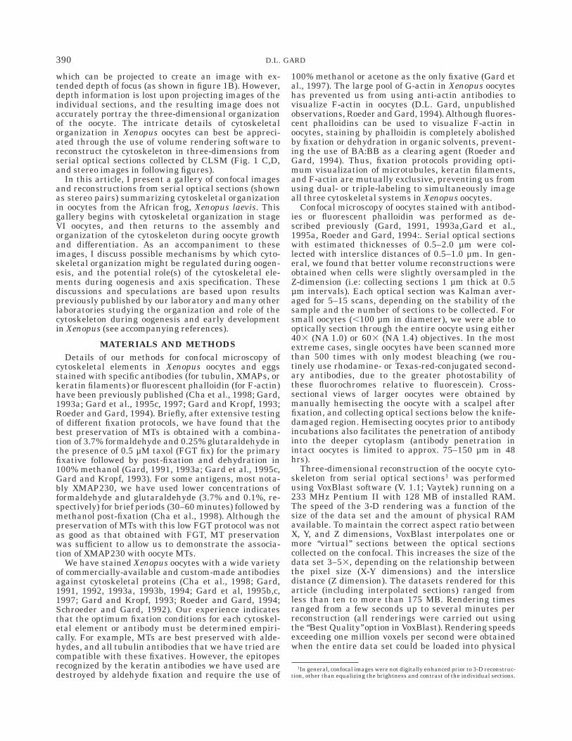
which can be projected to create an image with ex-tended depth of focus (as shown in figure 1B). However,depth information is lost upon projecting images of theindividual sections, and the resulting image does notaccurately portray the three-dimensional organizationof the oocyte. The intricate details of cytoskeletalorganization in Xenopus oocytes can best be appreci-ated through the use of volume rendering software toreconstruct the cytoskeleton in three-dimensions fromserial optical sections collected by CLSM (Fig. 1 C,D,and stereo images in following figures).
In this article, I present a gallery of confocal imagesand reconstructions from serial optical sections (shownas stereo pairs) summarizing cytoskeletal organizationin oocytes from the African frog, Xenopus laevis. Thisgallery begins with cytoskeletal organization in stageVI oocytes, and then returns to the assembly andorganization of the cytoskeleton during oocyte growthand differentiation. As an accompaniment to theseimages, I discuss possible mechanisms by which cyto-skeletal organization might be regulated during oogen-esis, and the potential role(s) of the cytoskeletal ele-ments during oogenesis and axis specification. Thesediscussions and speculations are based upon resultspreviously published by our laboratory and many otherlaboratories studying the organization and role of thecytoskeleton during oogenesis and early developmentin Xenopus (see accompanying references).
MATERIALS AND METHODSDetails of our methods for confocal microscopy of
cytoskeletal elements in Xenopus oocytes and eggsstained with specific antibodies (for tubulin, XMAPs, orkeratin filaments) or fluorescent phalloidin (for F-actin)have been previously published (Cha et al., 1998; Gard,1993a; Gard et al., 1995c, 1997; Gard and Kropf, 1993;Roeder and Gard, 1994). Briefly, after extensive testingof different fixation protocols, we have found that thebest preservation of MTs is obtained with a combina-tion of 3.7% formaldehyde and 0.25% glutaraldehyde inthe presence of 0.5 µM taxol (FGT fix) for the primaryfixative followed by post-fixation and dehydration in100% methanol (Gard, 1991, 1993a; Gard et al., 1995c,Gard and Kropf, 1993). For some antigens, most nota-bly XMAP230, we have used lower concentrations offormaldehyde and glutaraldehyde (3.7% and 0.1%, re-spectively) for brief periods (30–60 minutes) followed bymethanol post-fixation (Cha et al., 1998). Although thepreservation of MTs with this low FGT protocol was notas good as that obtained with FGT, MT preservationwas sufficient to allow us to demonstrate the associa-tion of XMAP230 with oocyte MTs.
We have stained Xenopus oocytes with a wide varietyof commercially-available and custom-made antibodiesagainst cytoskeletal proteins (Cha et al., 1998; Gard,1991, 1992, 1993a, 1993b, 1994; Gard et al, 1995b,c,1997; Gard and Kropf, 1993; Roeder and Gard, 1994;Schroeder and Gard, 1992). Our experience indicatesthat the optimum fixation conditions for each cytoskel-etal element or antibody must be determined empiri-cally. For example, MTs are best preserved with alde-hydes, and all tubulin antibodies that we have tried arecompatible with these fixatives. However, the epitopesrecognized by the keratin antibodies we have used aredestroyed by aldehyde fixation and require the use of
100% methanol or acetone as the only fixative (Gard etal., 1997). The large pool of G-actin in Xenopus oocyteshas prevented us from using anti-actin antibodies tovisualize F-actin in oocytes (D.L. Gard, unpublishedobservations, Roeder and Gard, 1994).Although fluores-cent phalloidins can be used to visualize F-actin inoocytes, staining by phalloidin is completely abolishedby fixation or dehydration in organic solvents, prevent-ing the use of BA:BB as a clearing agent (Roeder andGard, 1994). Thus, fixation protocols providing opti-mum visualization of microtubules, keratin filaments,and F-actin are mutually exclusive, preventing us fromusing dual- or triple-labeling to simultaneously imageall three cytoskeletal systems in Xenopus oocytes.
Confocal microscopy of oocytes stained with antibod-ies or fluorescent phalloidin was performed as de-scribed previously (Gard, 1991, 1993a,Gard et al.,1995a, Roeder and Gard, 1994:. Serial optical sectionswith estimated thicknesses of 0.5–2.0 µm were col-lected with interslice distances of 0.5–1.0 µm. In gen-eral, we found that better volume reconstructions wereobtained when cells were slightly oversampled in theZ-dimension (i.e: collecting sections 1 µm thick at 0.5µm intervals). Each optical section was Kalman aver-aged for 5–15 scans, depending on the stability of thesample and the number of sections to be collected. Forsmall oocytes (,100 µm in diameter), we were able tooptically section through the entire oocyte using either403 (NA 1.0) or 603 (NA 1.4) objectives. In the mostextreme cases, single oocytes have been scanned morethan 500 times with only modest bleaching (we rou-tinely use rhodamine- or Texas-red-conjugated second-ary antibodies, due to the greater photostability ofthese fluorochromes relative to fluorescein). Cross-sectional views of larger oocytes were obtained bymanually hemisecting the oocyte with a scalpel afterfixation, and collecting optical sections below the knife-damaged region. Hemisecting oocytes prior to antibodyincubations also facilitates the penetration of antibodyinto the deeper cytoplasm (antibody penetration inintact oocytes is limited to approx. 75–150 µm in 48hrs).
Three-dimensional reconstruction of the oocyte cyto-skeleton from serial optical sections1 was performedusing VoxBlast software (V. 1.1; Vaytek) running on a233 MHz Pentium II with 128 MB of installed RAM.The speed of the 3-D rendering was a function of thesize of the data set and the amount of physical RAMavailable. To maintain the correct aspect ratio betweenX, Y, and Z dimensions, VoxBlast interpolates one ormore ‘‘virtual’’ sections between the optical sectionscollected on the confocal. This increases the size of thedata set 3–53, depending on the relationship betweenthe pixel size (X-Y dimensions) and the interslicedistance (Z dimension). The datasets rendered for thisarticle (including interpolated sections) ranged fromless than ten to more than 175 MB. Rendering timesranged from a few seconds up to several minutes perreconstruction (all renderings were carried out usingthe ‘‘Best Quality’’ option in VoxBlast). Rendering speedsexceeding one million voxels per second were obtainedwhen the entire data set could be loaded into physical
1In general, confocal images were not digitally enhanced prior to 3-D reconstruc-tion, other than equalizing the brightness and contrast of the individual sections.
390 D.L. GARD

RAM. Rendering speeds dropped significantly if thesize of the data set required the use of the hard drive asvirtual memory.
3-D reconstructions made using linear color lookuptables (LUTs) and opacity functions ( a-function) ap-pear opaque (Fig. 1C), and provide little information onthe cytoskeletal organization deep within the sample.Therefore, non-linear LUTs and a-functions were usedto render the non-fluorescent background and cyto-plasm transparent, revealing the stained cytoskeletalelements within (figure 1D, which shows MTs in stage IXenopus oocyte). For each volume, the LUT and a-func-tion was determined empirically to provide optimumvisualization of cytoskeletal organization.
The stereo pairs presented in this article each consistof two views differing by six degrees in azimuth2.Substantial degradation of the 3-D reconstructionsbecomes apparent as the volumes are rotated morethan 15–20° around their X or Y axes (which can beseen to some degree in figure 1D). This loss of imagequality results from the reduced resolution of the Zdimension relative to the X and Y dimensions. AdobePhotoshop was used for simple image processing (minorsmoothing and conversion from RGB to grayscale) andassembly of the figures.
CYTOSKELETAL ORGANIZATION IN STAGE VIXENOPUS OOCYTES.
Stage VI Xenopus oocytes are arrested in prophaseprior to the first meiotic division (for reviews, see Gard,1995; Gerhart, 1980; Gerhart et al., 1983). The highlypolarized nature of these large (1.2–1.3 mm in diam-eter) cells is evident in their darkly-pigmented ‘‘animal’’hemisphere, and lightly pigmented ‘‘vegetal’’ hemi-sphere. Although it is established during oogenesis, theanimal-vegetal (A-V) axis plays an important role inthe specification of the three primary germ layers of thedeveloping embryo: cells derived from the animal hemi-sphere give rise to the ectodermal structures (includingthe central nervous system and epidermis), cells de-rived from the vegetal hemisphere give rise to endoder-mal structures (the primitive gut, etc), while cells of theequatorial (or marginal zone) are induced to becomemesoderm (Gimlich and Gerhart, 1980; Heasman et al.,1984b; Gerhart, 1980).
Closer inspection reveals that the animal-vegetalpolarity of stage VI Xenopus oocytes extends to manyfeatures of their internal organization (Danilchik andGerhart, 1987; Dumont, 1972; Gerhart, 1980; Gerhartet al., 1983). For example, the oocyte’s germinal vesicle(GV: the oocyte nucleus) is located in the animalcytoplasm, while large yolk-platelets are concentratedin the vegetal cytoplasm. Recent studies have demon-strated that a number of developmentally-importantmaternal RNAs are specifically localized to the vegetalcortex during oocyte differentiation (Elinson et al.,1993; Forristall et al., 1995; Kloc and Etkin, 1995; Klocet al., 1996; Melton, 1987; Mosquera et al., 1993; Mowry
and Melton, 1992; Weeks and Melton, 1987; Weeks etal., 1985; Yisraeli et al., 1989; Zhang and King, 1996;Zhou and King, 1996b), by mechanisms that are depen-dent upon both F-actin and MTs (Weeks and Melton,1987). One of these RNAs, Vg1, encodes a TGF- b-likegrowth factor that has been implicated in the inductionand specification of the embryonic mesoderm (Weeksand Melton, 1987). By implication, then, the assemblyand organization of the oocyte cytoskeleton plays acritical role in establishment of the embryonic bodyplan.
Like most eukaryotic cells, stage VI Xenopus oocytescontain an intricate cytoskeleton composed of threecytoplasmic filament systems: (1) microtubules (MTs)-(Gard 1991; Huchon et al., 1988; Palecek et al., 1982;reviewed in Gard et al., 1995c), composed of tubulin andMT-associated proteins (referred to as XMAPs(Andersen and Karsenti, 1997; Anderson et al., 1994;Gard and Kirschner, 1987b); (2) F-actin (Clark andMerriam, 1977, 1978; Clark and Rosenbaum, 1979;Franke et al., 1976; Merriam and Clark, 1978; Merriamet al., 1983; Roeder and Gard, 1994); and (3) intermedi-ate filaments, composed of keratins (keratin filaments,KFs)(Godsave et al., 1984b; Klymkowsky et al, 1987;Ryabova et al., 1993; Torpey et al., 1992b; reviewed inGard and Klymkowsky, 1998). However, the large sizeof the fully-grown Xenopus oocytes, eggs, and zygotes,along with the rapidity of the meiotic and mitotic cellcycles, place some interesting demands on the mecha-nisms that regulate cytoskeletal assembly and organi-zation. On the one hand, oocytes assemble an extensivecytoskeletal network during the prolonged diplotenestage of meiotic prophase. Although substantial remod-eling of the cytoskeleton occurs during this extendedperiod of oocyte growth and differentiation (Gard, 1991;Gard et al, 1995a; Gard et al., 1997; Roeder and Gard,1994), the oocyte cytoskeleton as a whole appears to berelatively stable (see below). Consistent with this view,substantial populations of acetylated, presumably stable(Schulze et al., 1987; Webster and Borisy, 1989), MTsare present throughout the post-mitotic stages of oocytedifferentiation (stages 0–VI)(Gard, 1992, 1993a). Onthe other hand, both the MT and KF networks of stageVI oocytes are rapidly disassembled within 30 minutesof GV breakdown during oocyte maturation (Gard,1992, 1993a; Gard and Klymkowsky, 1998; Klymkowskyet al., 1987) the fate of the cytoplasmic network of actincables during maturation has yet to be examined.During maturation, the extensive array of acetylatedMTs present in cytoplasm of stage VI oocytes is re-placed by an equally extensive array of dynamic (un-acetylated) cytoplasmic MTs surrounding the meioticspindles (Cha et al., 1998; Gard et al., 1995c). Thus,oocytes must have the capacity to rapidly switch from a‘‘stable’’ cytoskeleton during prophase arrest to thedynamic cytoskeleton of M-phase (meiosis).
Microtubule Organization in Stage VIXenopus Oocytes
Western blots reveal that a single stage VI Xenopusoocyte contains a pool of a- and b-tubulin sufficient toassemble more than 1.5 km of MTs (Gard and Kirsch-ner, 1987a; Jessus et al., 1987). Moreover, as much as20% of the cytoplasmic pool of tubulins in stage VIoocytes is present as sedimentable polymer (Gard,
2In most cases, a series of 20–60 views differing by 0.5 or 1 degree in azimuthwas generated using the ‘‘movie generation’’ tool in Voxblast. These images can beplayed back as an animated movie depicting rotation, or images differing by sixdegrees in azimuth can be used to generate stereo pairs (as done here).Compressed versions of many of these animations are available (in Video forWindows [AVI] or Quicktime [QT] formats) on our website: http://froglab.biology.utah.edu.
391CYTOSKELETON OF XENOPUS OOCYTES

1991; Jessus et al., 1987), indicating that a singleoocyte contains more than 300 meters of MTs! However,visualization of MTs in stage VI oocytes proved difficultin early studies using immunohistochemistry and/orelectron microscopy (Heidemann et al., 1985; Palecek etal., 1985). The apparent lack of MTs in stage VI oocytes,when combined with the observations that oocyte cyto-plasm inhibited MT assembly in vitro (Gard and Kirsch-ner, 1987a; Jessus et al., 1984), lead to speculation thatstage VI Xenopus oocytes were devoid of MTs (Gard andKirschner, 1987a). The sedimentable tubulin detectedby biochemical methods was then postulated to repre-sent a cytoplasmic pool of oligomeric tubulin, stored foruse later in development.
The inability to detect individual MTs in the earlystudies of Xenopus oocytes, however, probably resultedfrom the difficulties associated with fixing these verylarge, yolky cells. Results from later studies, in whichwhole-mount immunocytochemistry was used to exam-ine the distribution of tubulin in Xenopus oocytes(Yisraeli et al., 1989), suggested that oocytes did indeedcontain extensive MT arrays during later oogenesis,but did not provide the resolution needed to see indi-vidual MTs. Thus, the first visualization of individualMTs from stage VI Xenopus oocytes was provided byrapidly freezing squashed oocytes (Huchon et al., 1988),fixing the frozen cortices by freeze substitution, andexamining these cortical preparations by immunofluo-rescence microscopy using anti-tubulin. This techniquerevealed numerous individual MTs that were presumedto originate from the oocyte cortex (Huchon et al.,1988). Unfortunately, while confirming that Xenopusoocytes contain substantial numbers of MTs, littleinformation on the organization of these MTs in intactcells could be obtained.
Subsequently, we used confocal immunofluorescencemicroscopy to examine the organization of MTs through-out oogenesis (Gard 1991, 1992, 1994; Gard et al.,1995a). Results from our initial study, and others thathave followed, revealed that stage VI Xenopus oocytescontain a complex MT array, consisting of as many as0.5–1 million MTs, with a mean length estimated to be,600 µm. The density and complexity of the oocyte MTarray is best appreciated in 3-D images reconstructedfrom serial optical sections collected by CLSM, asshown in Figures 2–3.
In the animal hemisphere, loosely organized ‘‘bundles’’of MTs appear to radiate from the germinal vesicle (GV:the oocyte nucleus) to the animal cortex (Fig 2A) (Gard,1991). Many of the bundled MTs in the animal hemi-sphere are brightly stained with antibodies to acety-lated a-tubulin (Fig. 2A) (Gard 1991). Careful examina-tion at higher magnification reveals that many of theacetylated MTs in the animal hemisphere terminate at,or very near, the inner surface of the oocyte membrane(Fig. 2B). Previous studies have shown that acetylationof a-tubulin is correlated with MT stability (Schulze etal., 1987; Webster and Borisy, 1989). The extensivenetwork of acetylated MTs in stage VI oocytes (andpresent throughout oogenesis, see below) suggests thatMT stabilization plays an important role in oocytedifferentiation (discussed further below).
The GV, located in the animal cytoplasm of stage VIoocytes, is surrounded by an extensive network of MTs,many of which are acetylated (Fig. 3A). This peri-
nuclear MT network is more extensive near the basal(vegetal) surface of the GV, where MTs are concentratedin a perinuclear cap of yolk-free cytoplasm (Gard, 1991).
Numerous MTs are also apparent in the vegetalcytoplasm of stage VI oocytes. MTs in the vegetalhemisphere exhibit less bundling, appear less ordered,and are acetylated to a lesser degree than those in theanimal hemisphere (Gard, 1991); compare Figure 3Bwith Figure 2A.
MT inhibitors have been used to define several rolesfor the cytoplasmic MT arrays of stage VI Xenopusoocytes. For example, treating oocytes with colcemid,nocodazole, vinblastine, or cold results in the displace-ment of the GV from its normal position in the animalcytoplasm (Gard, 1991), indicating that MTs play amajor role in the anchoring of the GV within the cytoplasm.More recently, we have reported that MT depolymeriza-tion disrupts the A-V polarity of the cortical network ofKFs (Gard et al., 1997), suggesting that these twocytoskeletal systems interact (see below). Finally, MTshave been implicated in localization of maternal RNAsto the vegetal cortex during oocyte differentiation (For-ristall et al., 1995; Kloc and Etkin, 1995; Yisraeli et al.,1990). These results suggest that MTs play a number ofcritical roles in establishing or maintaining the cytoplas-mic polarity of stage VI Xenopus oocytes.
Regulation of MT Assembly and Organizationin Stage VI Oocytes
Most animal cells contain a discrete microtubule-organizing center (MTOC), known as the centrosome,that serves as the primary nucleation site for inter-phase MTs (Brinkley, 1985; McIntosh, 1983). Interest-ingly, the extensive MT networks present in stage VIXenopus oocytes are assembled in the absence of conven-tional centrosomes (Gard, 1991; Gerhart, 1980; Jessuset al., 1986). However, despite lacking centrosomes,Xenopus eggs have been shown contain an extensivepool of centrosomal components (Gard et al., 1990),including g-tubulin (Stearns et al., 1991; Stearns andKirschner, 1994). g-Tubulin binds predominantly to theminus-end of MTs in vitro and is concentrated at thecentrosomes and spindle poles of somatic cells (Joshi,1993, 1994; Joshi et al., 1992; Li and Joshi, 1995;Oakley, 1992; Stearns et al., 1991; Zheng et al., 1991),where it has been shown to play an important role innucleating MTs (Joshi et al., 1992; Stearns and Kirsch-ner, 1994). The stockpile of centrosomal components inXenopus eggs has been postulated to serve as a pool ofprecursors supplying centrosome duplication duringthe rapid cell cycles of early Xenopus development(Gard et al., 1990; Stearns et al., 1991; Stearns andKirschner, 1994). However, the same pool of centro-somal proteins might also play an important role inregulating the assembly and organization of MTs dur-ing oogenesis.
Several observations suggested that, in the absenceof a classical centrosome, the GV of stage VI oocytesmight serve as an MTOC (Gard, 1991). First, MTs inanimal hemisphere of stage VI oocytes appear to radi-ate from the GV towards the oocyte cortex. In addition,nucleation of MTs from the surface of the GV wasobserved during recovery from cold-induced MT disas-sembly. More recently, however, confocal immunofluo-rescence microscopy revealed that g-tubulin was concen-
392 D.L. GARD

trated in the cortex of stage VI oocytes (Figs. 3C–E)(Gard, 1994), raising questions regarding the relativecontributions of the GV and cortex in organizing theMT array of stage VI oocytes. Moreover, the distribu-tion of cortical g-tubulin was polarized along the A-Vaxis, suggesting that the cortex might play an impor-tant role in establishing the polarized MT array of stageVI oocytes.
The observation that g-tubulin was concentrated inthe oocyte cortex also led us to postulate that somefraction of the MTs in stage VI oocytes are oriented withtheir minus-ends in the cortex and plus-ends towardsthe GV (Gard, 1994; Gard et al., 1995b). This orienta-tion is opposite that found in most somatic cells (Eute-neuer and McIntosh, 1981; Schliwa, 1992). To addressthis issue, we have recently used tubulin hook-decoration and electron microscopy to confirm that themajority of MTs (.85%) in both the animal and vegetalcytoplasm of stage VI Xenopus oocytes are indeedoriented with their minus-ends towards the cortex
(Pfeiffer and Gard, manuscript in preparation), consis-tent with the localization of g-tubulin. This observationsuggests that the cell cortex does play a role in organiz-ing a large fraction of the cytoplasmic MTs in stage VIXenopus oocytes.
Numerous studies suggest that MT assembly anddynamics in somatic cells are regulated by a diversegroup of proteins know as microtubule-associated pro-teins, or MAPs (Avila et al, 1994; Bre and Karsenti,1990; Drechsel et al, 1992; Hirokawa, 1994; Mandelkowand Mandelkow, 1995; Marus, 1990; Pryer et al, 1992).At least three biochemically- and functionally-distinctMAPs have been identified in Xenopus oocytes andeggs: XMAP215, XMAP230, and XMAP3103 (Andersenand Karsenti, 1997, Anderson et al., 1994; Cha et al.,
3Using the nomenclature of Andersen et al., (1994), Xenopus MAPs are referredto by their apparent mass on SDS-PAGE. XMAP215 refers to the proteinoriginally called XMAP (Gard and Kirschner, 1987b). XMAP230 had previouslybeen referred to as p220 (Shiina et al., 1992), XMAP230 (Anderson et al., 1994),and MAP250 (Cha et al., 1994).
Fig. 2: Organization of acetylated MTs in the animal hemisphere of astage VI Xenopus oocyte. (A) A stereo pair (reconstructed from 10optical sections) showing the organization of acetylated MTs (stainedwith monoclonal antibodies to acetylated a-tubulin) into radially-oriented bundles extending from the GV to the animal cortex of a stageVI oocyte. The density and complexity of the MT network in stage VI
oocytes becomes more apparent when it is realized that the volumeshown in (A) represents less than 0.01% of the oocyte volume. (B) Athigher magnification, acetylated MTs can be seen to end at or verynear the oocyte surface (arrows). Reconstructed from 10 opticalsections. VE denotes a portion of the vitelline envelope. Scale bars are25 µm in A and 10 µm in B.
393CYTOSKELETON OF XENOPUS OOCYTES

1994; Faruki and Karsenti, 1994; Gard and Kirsch-ner,1987b; Jessus et al., 1985; Shiina et al, 1992).Results from in vitro studies have shown that XMAP230
(Anderson et al., 1994) and XMAP310 Anderson andKarsenti, 1997) stabilize MTs and suppress dynamicinstability in vitro, similar to the effects of MAPs
Fig. 3: MT organization in the perinuclear and vegetal cytoplasm ofstage VI Xenopus oocytes, and g-tubulin in the vegetal cortex (A) Astereo pair (reconstructed from 32 optical sections) of acetylated MTssurrounding the GV of a stage VI oocyte. Acetylated MTs appear moreconcentrated around the basal (vegetal) region of the GV (approximateorientation of the A-V axis is shown). (B) A stereo pair (reconstructedfrom 25 optical sections) showing MTs in the vegetal hemisphere of astage VI Xenopus oocyte (stained here anti- a-tubulin). MTs in the
vegetal cytoplasm appear more disordered in those of the animalhemisphere (compare with figure 2A). (C) Low magnification revealsthat g-tubulin (arrows) is concentrated in the cortex of stage VI oocytes(arrows). (D–E) At higher magnification, g-tubulin appears as smallfoci in cross- (D) and grazing (E) sections of the vegetal cortex (arrowsin D; E is a projection of three optical sections). Scale bars are 25 µm inA, B, and D; 250 µm in C; and 10 µm in E.
394 D.L. GARD

isolated from vertebrate brain (Drechsel et al., 1992;Pryer et al., 1992). Recently, we have reported thatmicroinjection of affinity-purified XMAP230 antibodiesdisrupts the organization and acetylation of MTs instage VI oocytes, without blocking MT assembly (Cha etal., 1998). From these results, we conclude thatXMAP230 primarily functions to stabilize and organizeMTs in stage VI oocytes, consistent with its effects onMT assembly in vitro.
XMAP215 was originally isolated from Xenopus eggsbased upon its ability to promote centrosome-nucleatedMT assembly in vitro (Gard and Kirschner, 1987b).Subsequent studies (Gard and Kirschner, 1987b;Vasquez et al., 1994) indicated that XMAP215 hasnovel effects on MT assembly. In contrast to the stabiliz-ing effects of other MAPs on both ends of MTs, XMAP215specifically enhances both the assembly and disassem-bly at the plus-ends of MTs (Vasquez et al., 1994), resultingin the assembly of long, but highly dynamic, MTs.
Recently, XMAP215 has been shown to be homolo-gous to ch-TOGp (Charisse et al., 1998), a human MAPthat is expressed in several normal adult tissues (includ-ing brain and testes) and is overexpressed in certainhuman tumors (Charasse et al., 1995, 1996). Compari-son of the predicted sequences of XMAP215 and ch-TOGp reveal that the frog and human proteins are,80% identical over their entire lengths (B. Becker andD. Gard, unpublished). Both vertebrate MAPs are alsodistantly related to MAPs identified in yeast (Stu-2 andp93dis1) (Nabeshima et al., 1995; Wang and Huffaker,1997)and C. elegans (Zyg-9) (Matthews et al., 1998).Although the role of XMAP215 in vivo has yet to bedetermined, its unique effects on MT assembly in vitrosuggest that XMAP215 may promote the rapid assem-bly of dynamic MTs, which are then stabilized bybinding other MAPs (such as XMAP230 or XMAP310).Consistent with this model, mutations in the Zyg-9gene of C. elegans exhibit reduced spindle asters andresult in misplacement of meiotic and mitotic spindles(Matthews et al., 1998). Interestingly, XMAP215, ch-TOGp, Stu-2p, p93dis1, and Zyg-9p have all been local-ized to MTOCs (centrosomes, spindle poles, or spindlepole bodies) (Gard et al., 1995c; Matthews et al., 1996;Nabeshima et al., 1995; Wang and Huffaker, 1997). Thefunctional relationship between the cytoplasmic local-ization of XMAP215 and its potential role in promotingMT elongation and dynamics remains uncertain.
Stage VI Oocytes Contain Cortical, Cytoplasmic,and Nuclear F-actin
F-actin is the most difficult of the three cytoskeletalelements to visualize in stage VI oocytes. On one hand,the large pool of actin in the cytoplasm of Xenopusoocytes, estimated to exceed 2 mg/ml (Clark and Mer-riam, 1978; Merriam and Clark, 1978), makes visualiza-tion of F-actin by immunofluorescence microscopy diffi-cult or impossible (Roeder and Gard, 1994; Roeder andGard, unpublished observations), even when confocalmicroscopy is available. On the other hand, staining ofF-actin using fluorescent phalloidins is not compatiblewith alcohol dehydration and clearing. Thus, we arelimited to viewing those portions of uncleared oocytesthat are sufficiently close to the surface to allow penetra-tion by the laser and detection of the resulting fluores-cent signal. In practice, we are able to collect images
from uncleared oocytes to a depth of only 8–12 µm(Roeder and Gard, 1994). Hemisection of oocytes priorto staining with phalloidin does allow collection ofcross-sectional views of uncleared oocytes. However, itis often not possible to avoid knife damage at the cutsurface of hemisected oocytes, which makes these im-ages less suitable for 3-D reconstruction.
When reconstructions of the cortex of phalloidin-stained oocytes are view from the outside, the brightly-stained microvilli on the surface of stage VI oocytesobscure details of actin organization in the cortical andsubcortical cytoplasm (not shown). However, when thesame set of optical sections are viewed from the insideof the oocyte, a complex network of F-actin cables can beseen against the bright background of the microvilli(seen from their basal end) (Fig. 4A). Cross-sectionalviews reveal that the cortical F-actin network extendsinto the deeper cytoplasm (Roeder and Gard, 1994),which is filled with a meshwork of actin cables. In theanimal hemisphere, actin cables are observed surround-ing the GV (see below) and in the yolk-free radii (Fig.4B), in which MTs and KFs are also found. In thevegetal hemisphere, actin cables form a complex, al-though apparently disordered, meshwork that extendsthroughout the vegetal cytoplasm (Fig. 4C).
Interestingly, optical cross-sections of stage VI oo-cytes stained with fluorescent phalloidin to visualizethe distribution of cytoplasmic F-actin also revealedthat the GV was brightly stained (Fig. 4D) (Roeder andGard, 1994). Although we were initially surprised bythis observation, previously published studies demon-strated that the GV of stage VI oocytes contains asubstantial pool of actin (Clark and Merriam, 1977,1978; Clark and Rosenbaum, 1979; Merriam and Clark,1978), some of which is present as polymer (F-actin).Confocal microscopy revealed that the nucleoplasm wasbrightly stained by fluorescent phalloidin, exhibiting adiffuse, granular appearance (Fig. 4E). In most cases,little substructure was evident, other than partialexclusion of staining from numerous inclusions thatprobably represent nucleoli (Fig. 4E inset). A network ofactin cables was also apparent surrounding the GV(Fig. 4E arrows) and in the cap of yolk-free cytoplasmapposed to the basal surface of the GV (Fig. 4F) (Roederand Gard, 1994).
To gain insight into the role of F-actin in stage VIoocytes, we examined the cytoplasmic organization ofoocytes treated with the actin inhibitor, cytochalasin B(CB) (Roeder and Gard, 1994). Confocal microscopyrevealed that incubation of oocytes in CB for 6–24 hourssubstantially disrupted actin organization. Althoughcortical F-actin was not completely disassembled, corti-cal organization was substantially disrupted (see be-low). Cytoplasmic actin cables were nearly completelyeliminated, leaving behind numerous brightly-stainedaggregates (which also contain keratins)(Gard et al.,1997). Extended treatment of stage VI oocytes with CBresulted in displacement and distortion of the GV(Coleman et al., 1981; Roeder and Gard, 1994). How-ever, displacement of the GV in CB-treated oocytes wasnot as immediate or dramatic as that observed in cold-or nocodazole-treated oocytes (Gard, 1991, 1993b), sug-gesting that F-actin plays a more secondary role inanchoring the nucleus in the animal cytoplasm, per-haps by anchoring MTs that extend from the cortex to
395CYTOSKELETON OF XENOPUS OOCYTES

the GV (see below). The distortion of the GV observed inCB-treated oocytes also suggests that nuclear F-actinmay play an important role in establishing or maintain-ing chromatin organization and/or the morphology ofthe GV itself. This conclusion is consistent with previ-ously published studies demonstrating that actin is
associated with the lampbrush chromosomes of amphib-ian oocytes (Karsenti et al., 1978), and with studies inwhich microinjection of actin antibodies or gelsolin intothe GV of Xenopus oocytes resulted in the disruption orcollapse of the lampbrush chromosomes (Rungger-Brandle, et al., 1979; Scheer et al., 1984).
Fig. 4: Actin organization in stage VI Xenopus oocytes. (A) A stereopair (reconstructed from 11 optical sections) showing the organizationof subcortical actin filaments in the animal cortex of a stage VIXenopus oocyte, viewed from inside the cell. (B) Short F-actin cables(arrows) are apparent in the yolk-free radii of the animal hemispherein a stage VI oocyte. (C) A dense meshwork of actin cables is apparentin the vegetal cytoplasm of this stage VI oocyte. (D) The GV of stage VI
Xenopus oocytes stains brightly with fluorescent phalloidin. (E) F-actincables (arrows) surround the brightly-stained GV (inset shows aless-brightly-stained inclusion, presumed to represent one of manyextrachromosomal nucleioli). (F) Numerous actin cables are apparentin the yolk-free cap of cytoplasm apposed to the basal side of the GV.Scale bars are 25 µm in A–C, E–F (10 µm in inset), and 250 µm in D.
396 D.L. GARD

Stage VI Xenopus Oocytes Contain an ElaborateNetwork of Keratin Filaments
Stage VI Xenopus oocytes have been reported tocontain two independent networks of intermediatefilaments, composed of vimentin (Godsave et al., 1984a;Tang et al., 1988; Torpey et al., 1990, 1992b) andkeratins (Fouquet et al., 1988; Franz et al., 1983; Gardet al., 1997; Godsave et al., 1984b; Klymkowsky et al.,1987; Ryabova et al., 1993; Torpey et al., 1992b).Although the presence of vimentin in Xenopus oocytesremains controversial (Dent et al., 1992; Dent andKlymkowsky, 1989; Dent et al., 1989; Franz et al., 1983;Herrmann et al., 1989), the presence of a complexnetwork of KFs is indisputable. Conventional micros-copy of whole-mounted or sectioned stage VI Xenopusoocytes stained with antibodies to keratins revealed acomplex, anastomosing network of KFs in the vegetalcortex, radial networks of cytoplasmic KFs, and anetwork of KFs surrounding the GV (Franz et al., 1983;Godsave et al., 1984b; Klymkowsky et al., 1987; Ry-abova et al., 1993; Torpey et al., 1992b).
More recently, we have used confocal immunofluores-cence microscopy to examine the organization of KFsthroughout oogenesis in Xenopus (Gard et al., 1997),providing a more complete view of KF organizationduring oocyte differentiation. Confocal microscopy re-vealed that the network of cortical KFs was not re-stricted to the vegetal hemisphere of stage VI oocytes,but rather surrounds the entire oocyte (Gard et al.,1997). In the vegetal cortex, KFs form a nearly planarnetwork of anastomosing filaments or filament bundles(Fig. 5A), as previously described by Klymkowsky et al.,(1987). However, an extensive network of KFs in thecortical and subcortical cytoplasm of the animal hemi-sphere was also apparent (Fig. 5B). This extensivenetwork was not readily seen by conventional micros-copy, although some suggestion of its existence wasobserved by Klymkowsky et al., (1987). When comparedto the vegetal KF network, the KF network in theanimal cortex exhibits a much finer mesh, and appearsmuch more three-dimensional, extending 8–12 µm intothe subcortical cytoplasm (compared to the 3–4 µmdepth of the vegetal KF network) (Gard et al., 1997).
The cortical KF network of stage VI oocytes isdirectly connected to a network of cytoplasmic KFs. Inthe animal hemisphere, these cytoplasmic KFs areorganized into a roughly radial array (Fig. 5C), withnumerous transverse connecting filaments or bundles.Less radial order is present in the vegetal hemisphere,probably due to the yolky nature of the vegetal cyto-plasm (not shown).
A complex KF network also surrounds the GV ofstage VI oocytes (Fig. 6). Numerous KFs are apparentin the perinuclear cap of yolk-free cytoplasm apposed tothe basal (vegetal) surface of the GV (seen in lateralview in Fig. 6A, and from inside the GV in Fig. 6B). Amore open meshwork of KFs is associated with theanimal and lateral surfaces of the GV (not shown). Theperinuclear KF network appears directly connected tothe cytoplasmic KFs in both the vegetal (Fig. 6A) andanimal hemispheres (not shown). As with MTs, then,KFs appear to directly link the GV to the oocyte cortex.However, although the cytoplasmic KF network ap-pears quite extensive, we estimate that a single stage
VI oocyte contains only 9–15,000 radially-oriented KFs(Gard et al., 1997). Cytoplasmic KFs are thus consider-ably less numerous than MTs (estimated to number0.5–1.0 million) (Gard, 1991).
The physiological role of the KFs in stage VI oocytesremains unclear. On the one hand, the dense meshworkof KFs in the oocyte cortex might provide mechanicalstrength and elasticity, and the cytoplasmic KF net-work might play a role in maintaining the position ofthe GV. Although the contribution of KFs to the me-chanical properties of the cortex has not been testeddirectly, disruption of the cytoplasmic KF network bythe microinjection of specific monoclonal antibodies intostage VI oocytes had no apparent effect on the positioningof the GV in the cytoplasm (Gard et al., 1997) or on otheraspects of oocyte structure or physiology (Gard andKlyhmkowsky, 1998). This result suggests that, unlikeMTs and actin, cytoplasmic KFs do not play a major rolein maintaining cytoplasmic organization.
Keratin filaments have also been proposed to play arole in the localization of developmentally-importantmaternal RNAs during the elaboration of the A-V axis.A number of maternal RNAs, including XCAT-2, Vg1,and others, have been identified and/or isolated fromkeratin-rich vegetal cortices peeled from stage VI oo-cytes (Elinson et al., 1993; Forristall et al., 1995; Rondeland King, 1988), leading to the suggestion that theseRNAs might be bound (directly or indirectly) to KFs inthe cortex (Pondel and King, 1988; Forristall et al.,1995). This hypothesis received additional support fromthe apparent temporal correlation between the releaseof Vg1 mRNA from the vegetal cortex and disassemblyof the cortical keratin filament array during oocytematuration (Klymkowsky et al., 1987). However, subse-quent studies revealed that disassembly of corticalkeratin filaments was not necessary for the release ofVg1 mRNA from the cortex, and that Vg1 could bereleased in the absence of keratin disassembly(Klymkowsky et al., 1991). A more detailed analysis willbe required to define the precise functional relation-ships, if any, between maternal RNAs and keratinfilaments in the oocyte cortex.
Finally, oocyte KFs might serve as a pool of compo-nents to be utilized later in development. Indeed, twostudies have shown that ablation of maternal KFsduring early development leads to gastrulation defects(Klymkowsky et al., 1992; Torpey et al., 1992a). How-ever, this does not preclude the likely possibility thatthe keratin filaments present in stage VI oocytes playsome, as yet undefined, role in the establishment ormaintenance of cytoplasmic organization.
Organization of the Cytoskeleton of Stage VIXenopus Oocytes is Dependent Upon a Hierarchyof Interactions Between F-Actin, Microtubules,
And Keratin FilamentsAlthough the incompatibility of the fixation condi-
tions required to preserve and label the three cytoskel-etal systems have prevented the direct colocalization ofthe different filament types (see Materials and Meth-ods), their similar distributions within the cytoplasmsuggest that they might interact, either directly orindirectly. Further evidence for interactions betweenall three filament systems (MTs, F-actin, and KFs) wasprovided by results obtained when each filament sys-
397CYTOSKELETON OF XENOPUS OOCYTES

Fig. 5: Keratin filament organization in stage VI Xenopus oocytes.(A) A stereo pair (reconstructed from eight optical sections) showingthe nearly planar network of KFs in the vegetal cortex of a stage VIXenopus oocyte, viewed from inside the cell. Radial KFs (arrows) canbe seen rising above the cortical KF network. (B) This stereo pair(reconstructed from ten optical sections) shows the KF network of the
animal cortex, seen from inside the cell. The cortical KF network in theanimal cortex is denser and more 3-dimensional than that of thevegetal cortex. (C) A stereo pair (reconstructed from ten opticalsections) showing the radial KF network in the animal cytoplasm.Arrows point to transverse filaments linking the radial KFs. Scalebars are 25 µm.
398 D.L. GARD

tem was disrupted in turn by inhibitors (F-actin andMTs) or by the microinjection of specific antibodies(KFs) (Gard et al., 1997). In these studies, disruption ofF-actin by treatment of stage VI oocytes with cytochala-sin B (CB) also disrupted the association of MTs (Fig.7A) and KFs (Fig. 7B) with the oocyte cortex, suggest-ing that cortical actin was required for anchoring bothof these filament systems. Similarly, disassembly ofMTs by treatment with nocodazole eliminated the A-Vpolarity of the cortical KF network (compare Fig. 7, C–Fto 5, A–B), suggesting that KF organization was depen-dent upon intact MTs. In contrast, disruption of theradial array of cytoplasmic KFs by the microinjection ofspecific monoclonal antibodies had no apparent effecton either MTs or F-actin.
From these results, we have proposed a model inwhich the cytoskeletal organization in stage VI Xeno-pus oocytes is governed by a hierarchy of interactionsbetween F-actin, MTs, and KFs (see Fig. 7, G–H) (Gardet al., 1997). Our results suggest that both MTs and
KFs are anchored to the oocyte cortex in a manner thatis dependent upon F-actin. In our model, we proposethat the assembly of oocyte MTs is nucleated fromcentrosomal proteins, such as g-tubulin, anchored (di-rectly or indirectly) to F-actin in the oocyte cortex. TheA-V polarization of the oocyte MT array, in which MTsare more numerous and exhibit a higher degree ofradial order in the animal hemisphere than in thevegetal hemisphere, results in part from the polarizeddistribution of nucleation sites in the oocyte cortex.However, a greater number of acetylated MTs are alsoobserved in the animal hemisphere, suggesting thatMTs in the animal hemisphere are also more stablethan those of the vegetal hemisphere. Thus, MT stabi-lizing factors might also be differentially localizedbetween the animal and vegetal hemispheres.
Continuing with our model, although KFs are an-chored to the cortex by F-actin, the A-V polarity of thecortical keratin network results from interactions be-tween the KF network with the polarized MT array.
Fig. 6: Organization of perinuclear KFs in stage VI Xenopus oocytes.(A) A dense network of KFs is apparent in the yolk-free cap ofcytoplasm apposed to the basal (vegetal) surface of the GV, seen instereo cross-section (reconstructed from fifteen optical sections). KFsfrom the perinuclear cap are continuous with a dense 3-D meshwork of
KFs in the vegetal cytoplasm (arrows). (B) This stereo pair (recon-structed from 25 optical sections) shows the KF network associatedwith the basal (vegetal) surface of the GV, as viewed from inside theGV. Scale bars are 25 µm.
399CYTOSKELETON OF XENOPUS OOCYTES

Fig. 7: Cytoskeletal organization in stage VI Xenopus oocytes isdependent upon a hierarchy of interactions. (A) Cytochalasin B (20µg/ml for 40 hours) disrupts the organization of acetylated MTs instage VI oocytes (compare to Fig. 2A). Note the loss of radially-orientedMT bundles and the appearance of broad cortical region containingdisordered MTs (arrows). (B) Stretching of the cortical KF networkinto the subcortical cytoplasm (arrows) is apparent in the animalhemisphere of an oocyte treated with 20 µg/ml CB for 20 hours (aprojection of five optical sections). Arrows denote the inner margin ofthe stretched KF network, and also point out aggregates of tangledkeratin filaments. (C–F) Nocodazole-induced depolymerization of MTseliminates the A-V polarity of cortical keratin filaments. Cross-sections (projections of five optical sections) of the vegetal (C) andanimal (D) cortex of nocodazole-treated oocytes reveal similar appear-ing cortical KF networks. Grazing views (projections of 2–3 opticalsections) of the vegetal (E) and animal (F) cortex of nocodazole-treated oocytes reveal similar KF networks (compare to Fig. 5 A,B).
(G) Cytoskeletal organization is dependent upon a hierarchy ofinteractions between F-actin, MTs, and KFs. (1) F-actin is required fororganization and polarity of both MTs and KFs; (2) MTs are requiredfor the polarity of the cortical network of KFs; but (3) the organizationof actin and MTs is independent of KFs. Candidates for the proteinmediators of these interactions are indicated (see text). (H) A model forthe organization of the cytoskeleton of stage VI Xenopus oocytes. MTs(grey lines) are anchored to cortical actin via the centrosomal proteing-tubulin (dots), and thus have their plus-ends extending inward.Keratin filaments (black lines) are anchored to F-actin in the corticaland perinuclear cytoplasm by an unidentified protein complex (*).Cortical arrows indicate ‘‘forces’’ acting to concentrate KFs in theanimal cortex and stretch the KF network in the vegetal cortex. Radialarrows indicate that KFs are also stretched inward along MTs. Thecombination of these ‘‘forces’’ gives rise to the observed polarization ofthe cortical KF network (see text).

Because MTs are more numerous in the animal hemi-sphere, binding (either direct of indirect) of the corticalKFs to MTs would cause them to concentrate, or ‘‘bunchup,’’ in the animal cortex (inward facing arrows in animalcortex in Fig. 7H). The A-V asymmetry of the corticalnetwork of KFs might be further intensified if theinteractions between KFs and MTs are restricted to thesubset of stable (acetylated) MTs, which are highly concen-trated in the animal hemisphere. In addition, we proposethat KFs in the animal cortex are stretched inward alongMTs (radial arrows in Fig. 7H) in the animal cytoplasm,perhaps through the action of MT-dependent motors.Together, these forces cause the KFs in the vegetalcortex to be stretched laterally (outward facing arrowsin vegetal cortex of figure 7H), resulting in the nearlyplanar, open meshwork observed in stage VI oocytes.
The proteins that mediate the interactions betweencytoskeletal elements in Xenopus oocytes remain largelyunknown. However, the search for cytoskeletal ‘‘linker’’proteins in Xenopus oocytes will undoubtedly benefitfrom similar studies in other model systems, includingsomatic cells, yeast, and other developmental systems.We have previously shown that the localization ofg-tubulin in the cortex of stage VI oocytes is disruptedby CB (Gard, 1994), and is therefore dependent uponF-actin. g-Tubulin might thus be responsible for anchor-ing the minus-ends of MTs in the cortex, and linkingthem to the cortical actin network. Alternatively, cyto-plasmic dynein and/or the dynactin complexes mightlink oocyte MTs to F-actin in the oocyte cortex, as hasbeen suggested in studies of spindle alignment inSaccharomyces cerevisiae (Carminati and Stearns, 1997;Li et al., 1993; muhua et al., 1994) and C. elegans (Hyman,1989; Hyman and White, 1987; Waddle et al., 1994).Recently, several intermediate filament associated pro-teins (IFAPs) of the plectin/BPAG family have beenreported to serve as linker molecules between interme-diate filaments and either F-actin or MTs in epithelialcells and neuronal cells (Foisner et al., 1995; Green etal., 1992; Seifert et al., 1992; Wiche et al., 1993), and otherreports have suggested that MAPs (Fujii et al., 1993;Leterrier et al., 1982; Pedrotti et al., 1994; Yamauchi andPurich, 1993) or MT motors (Gyoeva and Gelfand, 1991)mediate the linkage between intermediate filamentsand MTs in cultured somatic cells. Similar proteinsmight be responsible for mediating interactions betweenMTs, F-actin, and KFs in Xenopus oocytes, linking thesefilament systems into an integrated cytoskeleton.
CONSTRUCTING AND RECONSTRUCTINGTHE OOCYTE CYTOSKELETON DURING
OOGENESIS.Cytoskeletal Organization in Oogonia
and Stage 0 OocytesFrom time they are ‘‘born’’ by the final mitotic divi-
sion of a secondary oogonium to the onset of the meioticdivisions of maturation, Xenopus oocytes grow from aninitial diameter of approximately 12 µm to their finaldiameter of 1.2–1.3 mm. This corresponds to nearly amillion-fold increase in volume. Moreover, during growthand differentiation, oocytes specify, establish, and main-tain their A-V axis, which will play an important role inlater embryonic development (discussed earlier). Notsurprisingly, this tremendous growth and polarizationis accompanied by substantial changes in the organiza-tion of the oocyte cytoskeleton.
Oocyte differentiation ‘‘begins’’ with the completion ofthe last of four synchronous mitotic divisions by second-ary oogonia (Fig. 8A), giving rise to sixteen pear-shaped, post-mitotic oocytes. After replicating theirchromosomes during a final pre-meiotic S-phase, thesepost-mitotic oocytes, which we refer to as ‘‘stage 0oocytes,’’ enter the prolonged prophase of meiosis I(al-Mukhtar and Webb, 1971; Coggins, 1973).
Initially, stage 0 oocytes remain associated in acluster, or ‘‘nest,’’ surrounded by follicle cells. Theobserved synchrony of the oogonial divisions and earlydifferentiation of stage 0 oocytes within a nest sug-gested that oogonia and stage 0 oocytes remain linkedby cytoplasmic bridges resulting from incomplete cyto-kinesis (al-Mukhtar and Webb, 1971; Coggins, 1973),analogous to the ring canals observed in Drosophilaoocytes (Warn et al., 1985). Electron micrographs sup-porting the existence of such cytoplasmic bridges havebeen published (al-Mukhtar and Webbm, 1971; Cog-gins, 1973). However, confocal microscopy has failed toreveal any evidence of cytoskeletal specializations inXenopus oogonia or stage 0 oocytes (Gard et al., 1995a;Roeder and Gard, 1994) that are comparable to theactin-containing ring canals (Warn et al., 1985) or thepolarized MT arrays (Cooley and Theurkauf 1994;Theurkauf, 1994; Theurkauf et al., 1992, 1993) ob-served during oogenesis in Drosophila.
Like many somatic cells (Brinkley, 1985; McIntosh,1983; Schliwa, 1992), stage 0 oocytes are polarizedalong an axis defined by the locations of their nuclei andthe distribution of cytoplasmic organelles, especiallytheir centrosomes. Most of the cytoplasmic organelles,including numerous mitochondria, golgi, and the mater-nal centrosome (see below), are clustered in the narrowend of pear-shaped stage 0 oocytes (al-Mukhtar andWebb, 1971; Coggins, 1973; Gard et al., 1995a; Heas-man et al., 1984a), forming ‘‘cytoplasmic caps’’ whichare often oriented towards the center of the nest (as inFig. 8B). In the nucleus of stage 0 oocytes undergoingmeiotic recombination, the synapsed chromosomes arealso clustered into a ‘‘bouquet’’ apposed to nuclearenvelope abutting the cytoplasmic cap and centrosome(al-Mukhtar and Webb, 1971; Coggins 1973; Gard et al.,1995a), indicating that the nucleus of stage 0 oocytes isalso structurally polarized. It has been suggested thatthis nuclear-cytoplasmic axis (or, more specifically, thenuclear-centrosomal axis) of stage 0 oocytes mightpredict the A-V axis established later in oogenesis(Coggins, 1973; Gerhart et al., 1983; Heasman et al.,1984a). However, the evidence for such a functionalconnection is mostly circumstantial (see below).
Unlike stage VI oocytes, which lack classic centro-somes, several lines of evidence indicate that stage 0oocytes do indeed possess functional centrosomes. First,electron microscopy revealed that stage 0 oocytes con-tain a perinuclear pair of centrioles and pericentriolarmaterial (Coggins, 1973; Gard, 1995). In addition,confocal immunofluorescence microscopy revealed thatcentrosomal proteins found in somatic cells, includingg-tubulin (Fig. 8D)(Gard et al., 1995a) and pericentrin(Error, Doxsey, and Gard, unpublished observations;Doxsey et al., 1994), are localized to a single perinuclearspot in the cytoplasm of each stage 0 oocyte. Finally, asingle focus of MT nucleation in each cell was observedwhen stage 0 oocytes were returned to room tempera-ture after cold-induced disassembly of their cytoplas-
401CYTOSKELETON OF XENOPUS OOCYTES

mic MTs (Gard et al., 1995a). Dual immunofluorescencemicroscopy revealed that this functionally-identifiedMTOC coincided with the localization of g-tubulin,andthus corresponded to the centrosome (Gard et al., 1995).
Confocal immunofluorescence microscopy with anti-bodies to anti-tubulin revealed that stage 0 oocytescontain a dense network of microtubules surroundingthe maternal centrosome in their cytoplasmic cap (Fig.
Fig. 8: Cytoskeletal organization in oogonia and stage 0 Xenopusoocytes. (A) The final mitotic division of a cluster of oogonia is shownin this projection of 22 optical sections. Eight spindles are shown (twospindles are seen end on; *). (B) Acetylated MTs are concentrated inthe narrow, proximal end of these asymmetric, stage 0 oocytes.Portions of seven cells are visible in this single optical section. Arrowsdenote the approximate nuclear (N)-centrosomal axis of each cell,which point roughly towards the center of the nest. (C–D) A stage 0oocyte double stained with antibodies to a-tubulin (C) and g-tubulin
(D), reveal a typical centrosome (arrow in C-D). (E) Stage 0 oocytesviewed in cross section reveal that F-actin is predominantly restrictedto the cortex (arrows). The cortices of oocytes marked with an asterisk(*) are grazed by this single optical section. (F–G) A nest of stage 0oocytes stained with anti-keratin (F) and counterstained with thechromatin dye, Yo-Pro-1 (G). KFs appear restricted to the surroundingfollicle cells (the positions of the oocyte nuclei in are shown [*]). The‘‘bouquet’’ of chromatin in the stage 0 oocytes is apparent in G (*). Scalebars are 10 µm in A, B and E, 25 µm in A, and 5 µm in C-D.
402 D.L. GARD

8, B,C). Moreover, in contrast to the scarcity of acety-lated MTs in oogonia, many of the MTs of stage 0oocytes are brightly stained by antibodies to acetylateda-tubulin (Fig. 8, B,C) (Gard et al., 1995a). AcetylatedMTs are present throughout the later stages of oocytegrowth and differentiation (Gard, 1991; Gard et al.,1995a), from stage 0 through stage VI, suggesting thatstable MTs play an important role (or roles) duringoocyte differentiation and morphogenesis.
Confocal microscopy of cells stained with fluorescentphalloidin reveals that stage 0 oocytes also contain asubstantial amount of F-actin, which is concentrated inthe oocyte cortex (Fig. 8E) (Roeder and Gard, 1994). Incontrast, antibodies to keratins revealed that stage 0oocytes lack any evidence of a cytoplasmic KF network(Fig. 8F,G)(Gard et al., 1997). During this stage ofdifferentiation, KFs appear to be restricted to thesurrounding follicle tissue.
The Cytoskeleton is Reorganized DuringStage I of Oogenesis
As stage 0 oocytes complete the events of meioticrecombination, they enter a prolonged diplotene stagecharacterized by extensive growth, differentiation, andestablishment of the A-V axis. The diplotene stage ofoogenesis in Xenopus has been divided into six stagesthat are characterized by specific changes in oocyteorganization and/or appearance (Dumont, 1972).
Stage I is commonly used to refer to pre-vitellogenicoocytes between 50–300 µm in diameter (Dumont,1972). By early stage I, oocytes have lost much of thenuclear and cytoplasmic polarity that was so evidentduring stage 0: oocytes become rounder in shape, thegerminal vesicle (nucleus) becomes more centrally lo-cated in the cytoplasm, mitochondria and other cytoplas-mic organelles are distributed throughout the cyto-plasm, and the ‘‘bouquet’’ of condensed chromatinbecomes less apparent (Gard et al., 1995a). This transi-tion, from the highly polarized architecture of stage 0oocytes to the apparently unpolarized organization ofearly stage I, is also accompanied by two significantchanges in the organization of cytoplasmic MTs: (1)dispersal of MTs throughout the oocyte cytoplasm and(2) inactivation of the maternal microtubule-organizingcenter, or centrosome (Gard et al., 1995a).
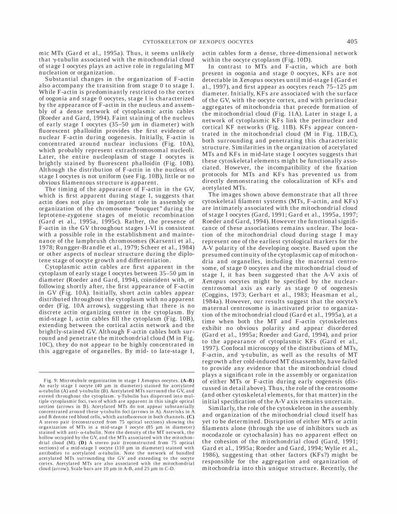
During early-mid stage I of oogenesis, the polarizedMT array of stage 0 Xenopus oocytes gives way to one inwhich MTs are more evenly distributed throughout theoocyte cytoplasm, with little or no evidence of order(Gard et al., 1995a). The complexity and apparentdisorder of the cytoplasmic MT array of stage I oocytesis best illustrated in three-dimensional reconstructions(shown in stereo in Fig. 9C). Individual MTs appear tonearly fill the cytoplasm between the GV (which ap-pears as an unstained hollow when figure 9C is viewedin stereo) and the oocyte cortex4.
Many of the MTs in stage I Xenopus oocytes stainbrightly with antibodies against acetylated a-tubulin(Fig. 9A and D) (Gard et al., 1995a), a marker for stable
MTs (Schulze et al., 1987; Webster and Borisy, 1989).During early stage I, acetylated MTs are concentratedin the perinuclear (Fig. 9A) and/or cortical cytoplasm(not shown). Later in stage I, many oocytes exhibitcomplex bundles of acetylated MTs that appear to linknetworks of acetylated MTs in the perinuclear andcortical cytoplasm (Fig. 9D). This organization lendscredence to the hypothesis that, as in stage VI oocytes,stable MTs play a role in the positioning and anchoringof the nucleus within the oocyte cytoplasm. The pres-ence of large numbers of acetylated (stable) MTsthroughout stage I also suggests that the dispersal ofMTs accompanying the transition from stage 0 to stageI occurs, at least in part, through a redistribution ofexisting MTs rather than by a process of disassemblyand re-assembly.
Numerous MTs, including a large number of acety-lated MTs, are closely associated with the mitochon-drial cloud of stage I oocytes (M in Fig. 9C) (Gard, 1991;Gard et al., 1995a). Optical sections reveal that MTsboth surround and penetrate this enigmatic aggregateof mitochondria and other membranous organelles pres-ent from mid- to late-stage I of oogenesis in Xenopus(al-Mukhtar and Webb, 1971; Billett and Adam, 1976;Coggins, 1973; Czolowska, 1969; Dumont, 1972; Heas-man et al., 1984a). However the, significance of thisassociation between MTs and the mitochondrial cloudremains unclear (see below).
The transition from stage 0 to stage I of oogenesis isalso accompanied by the functional inactivation of thematernal centrosome (Gard et al., 1995a). As discussedabove, stage 0 oocytes contain a typical centrosome,including a pair of centrioles and pericentriolar mate-rial containing g-tubulin (Fig. 8D; see above) andpericentrin. However, confocal immunofluorescence mi-croscopy revealed that g-tubulin was dispersed intomultiple cytoplasmic foci (ranging from 2–12 in num-ber) during early stage I of oogenesis (Fig. 9A,B). Thesenon-centrosomal foci of g-tubulin did not appear to betightly associated with MTs (Fig. 9A,B), and no longerfunctioned as nucleation sites during recovery fromcold-induced MT disassembly (Gard et al., 1995a).Although the fate of the maternal centrioles has notbeen established, these observations suggest that dis-persal of g-tubulin and inactivation of the maternalcentrosome occur during early stage I of oogenesis.
Recently, Kloc and Etkin (1998) have observed com-pact foci of g-tubulin associated with the mitochondrialmasses of sectioned mid-late stage I oocytes, and havesuggested that these structures were remnants of thematernal centrosome. We have been unable to confirmthe existence of these g-tubulin foci using confocalimmunofluorescence microscopy of intact stage I oo-cytes. Serial optical sectioning of dozens of oocytes fixedunder a variety of conditions failed to reveal anyconsistent association of g-tubulin with the mitochon-drial cloud during mid- to late-stage I (Gard et al.,1995a; Gard, unpublished observations). The reasonsunderlying the discrepancies between our results andthose of Kloc and Etkin are unknown. Possible explana-tions include differences in the protocols for fixationand preparation of oocytes, and/or the antibodies usedto identify g-tubulin. However, examination of thepatterns of MT regrowth after cold-induced disassem-bly revealed no evidence that the mitochondrial cloud ofstage I oocytes acts as an organizing center for cytoplas-
4However, it should be remembered that the apparent diameter of MTsobserved in an optical microscope (,0.5 µm, in this case) is significantlymagnified relative to their true diameter (,25 nm), due to the effects ofdiffraction. Despite the visual impact of the oocyte MT array, the calculatedvolume of the entire cytoplasmic MT array of a Xenopus oocyte occupies less than0.02–0.04% of the oocyte volume (based on estimates of the polymer content ofstage I and stage VI oocytes and the physical dimensions of MTs and oocytes).
403CYTOSKELETON OF XENOPUS OOCYTES

Fig. 9.
404 D.L. GARD
mic MTs (Gard et al., 1995a). Thus, it seems unlikelythat g-tubulin associated with the mitochondrial cloudof stage I oocytes plays an active role in regulating MTnucleation or organization.
Substantial changes in the organization of F-actinalso accompany the transition from stage 0 to stage I.While F-actin is predominantly restricted to the cortexof oogonia and stage 0 oocytes, stage I is characterizedby the appearance of F-actin in the nucleus and assem-bly of a dense network of cytoplasmic actin cables(Roeder and Gard, 1994). Faint staining of the nucleusof early stage I oocytes (35–50 µm in diameter) withfluorescent phalloidin provides the first evidence ofnuclear F-actin during oogenesis. Initially, F-actin isconcentrated around nuclear inclusions (Fig. 10A),which probably represent extrachromosomal nucleoli.Later, the entire nucleoplasm of stage I oocytes isbrightly stained by fluorescent phalloidin (Fig. 10B).Although the distribution of F-actin in the nucleus ofstage I oocytes is not uniform (see Fig. 10B), little or noobvious filamentous structure is apparent.
The timing of the appearance of F-actin in the GV,which is first apparent during stage I, suggests thatactin does not play an important role in assembly ororganization of the chromosome ‘‘bouquet’’ during theleptotene-zygotene stages of meiotic recombination(Gard et al., 1995a, 1995c). Rather, the presence ofF-actin in the GV throughout stages I-VI is consistentwith a possible role in the establishment and mainte-nance of the lampbrush chromosomes (Karsenti et al.,1978; Rungger-Brandle et al., 1979; Scheer et al., 1984)or other aspects of nuclear structure during the diplo-tene stage of oocyte growth and differentiation.
Cytoplasmic actin cables are first apparent in thecytoplasm of early stage I oocytes between 35–50 µm indiameter (Roeder and Gard, 1994), coincident with, orfollowing shortly after, the first appearance of F-actinin GV (Fig. 10A). Initially, short actin cables appeardistributed throughout the cytoplasm with no apparentorder (Fig. 10A arrows), suggesting that there is nodiscrete actin organizing center in the cytoplasm. Bymid-stage I, actin cables fill the cytoplasm (Fig. 10B),extending between the cortical actin network and thebrightly-stained GV. Although F-actin cables both sur-round and penetrate the mitochondrial cloud (M in Fig.10C), they do not appear to be highly concentrated inthis aggregate of organelles. By mid- to late-stage I,
actin cables form a dense, three-dimensional networkwithin the oocyte cytoplasm (Fig. 10D).
In contrast to MTs and F-actin, which are bothpresent in oogonia and stage 0 oocytes, KFs are notdetectable in Xenopus oocytes until mid-stage I (Gard etal., 1997), and first appear as oocytes reach 75–125 µmdiameter. Initially, KFs are associated with the surfaceof the GV, with the oocyte cortex, and with perinuclearaggregates of mitochondria that precede formation ofthe mitochondrial cloud (Fig. 11A). Later in stage I, anetwork of cytoplasmic KFs link the perinuclear andcortical KF networks (Fig. 11B). KFs appear concen-trated in the mitochondrial cloud (M in Fig. 11B,C),both surrounding and penetrating this characteristicstructure. Similarities in the organization of acetylatedMTs and KFs in mid-late stage I oocytes suggests thatthese cytoskeletal elements might be functionally asso-ciated. However, the incompatibility of the fixationprotocols for MTs and KFs has prevented us fromdirectly demonstrating the colocalization of KFs andacetylated MTs.
The images shown above demonstrate that all threecytoskeletal filament systems (MTs, F-actin, and KFs)are intimately associated with the mitochondrial cloudof stage I oocytes (Gard, 1991; Gard et al., 1995a, 1997;Roeder and Gard, 1994). However the functional signifi-cance of these associations remains unclear. The loca-tion of the mitochondrial cloud during stage I mayrepresent one of the earliest cytological markers for theA-V polarity of the developing oocyte. Based upon thepresumed continuity of the cytoplasmic cap of mitochon-dria and organelles, including the maternal centro-some, of stage 0 oocytes and the mitochondrial cloud ofstage I, it has been suggested that the A-V axis ofXenopus oocytes might be specified by the nuclear-centrosomal axis as early as stage 0 of oogenesis(Coggins, 1973; Gerhart et al., 1983; Heasman et al.,1984a). However, our results suggest that the oocyte’smaternal centrosome is inactivated prior to organiza-tion of the mitochondrial cloud (Gard et al., 1995a), at atime when both the MT and F-actin cytoskeletonsexhibit no obvious polarity and appear disordered(Gard et al., 1995a; Roeder and Gard, 1994), and priorto the appearance of cytoplasmic KFs (Gard et al.,1997). Confocal microscopy of the distributions of MTs,F-actin, and g-tubulin, as well as the results of MTregrowth after cold-induced MT disassembly, have failedto provide any evidence that the mitochondrial cloudplays a significant role in the assembly or organizationof either MTs or F-actin during early oogenesis (dis-cussed in detail above). Thus, the role of the centrosome(and other cytoskeletal elements, for that matter) in theinitial specification of the A-V axis remains uncertain.
Similarly, the role of the cytoskeleton in the assemblyand organization of the mitochondrial cloud itself hasyet to be determined. Disruption of either MTs or actinfilaments alone (through the use of inhibitors such asnocodazole or cytochalasin) has no apparent effect onthe cohesion of the mitochondrial cloud (Gard, 1991;Gard et al., 1995a; Roeder and Gard, 1994; Wylie et al.,1986), suggesting that other factors (KFs?) might beresponsible for the aggregation and organization ofmitochondria into this unique structure. Recently, the
Fig. 9: Microtubule organization in stage I Xenopus oocytes. (A–B)An early stage I oocyte (40 µm in diameter) stained for acetylateda-tubulin (A) and g-tubulin (B). Acetylated MTs surround the GV, andextend throughout the cytoplasm. g-Tubulin has dispersed into mul-tiple cytoplasmic foci, two of which are apparent in this single opticalsection (arrows in B). Acetylated MTs do not appear substantiallyconcentrated around these g-tubulin foci (arrows in A). Asterisks in Aand B denote red blood cells, which autofluoresce in both channels. (C)A stereo pair (reconstructed from 75 optical sections) showing theorganization of MTs in a mid-stage I oocyte (85 µm in diameter)stained with anti- a-tubulin. Note the density of the MT network, thehollow occupied by the GV, and the MTs associated with the mitochon-drial cloud (M). (D) A stereo pair (reconstructed from 75 opticalsections) of a mid-stage I oocyte (110 µm in diameter) stained withantibodies to acetylated a-tubulin. Note the network of bundledacetylated MTs surrounding the GV and extending to the oocytecortex. Acetylated MTs are also associated with the mitochondrialcloud (arrow). Scale bars are 10 µm in A-B, and 25 µm in C-D.
405CYTOSKELETON OF XENOPUS OOCYTES

Fig. 10: F-actin organization in stage I Xenopus oocytes. (A) An earlystage I oocyte (50 µm in diameter, a projection of two optical sections)stained with fluorescent phalloidin, showing the initial appearance ofF-actin in the GV and actin cables (arrows) in the cytoplasm. (B) Astereo pair (reconstructed from twenty optical sections) of a mid-stageI oocyte (100 µm in diameter) stained with fluorescent phalloidin toreveal F-actin distribution, showing the brightly-stained GV and
cortex, and cytoplasmic meshwork of actin cables. (C) Cables ofF-actin are associated with the mitochondrial cloud (M) of stage Ioocytes (a projection of two optical sections). (D) This stereo pair(reconstructed from twenty optical sections midway between thecortex and GV) shows the dense meshwork of cytoplasmic actin cablesin the cytoplasm of a late-stage I oocyte stained with fluorescentphalloidin. Scale bars are 10 µm in A, C, and D, and 25 µm in B.
406 D.L. GARD

mitochondrial cloud has been proposed to function inthe targeting of maternal RNAs to the cortex duringoogenesis (Kloc and Etkin, 1995, 1998; Kloc et al.,1996), and it is tempting to speculate that cytoskeletal
elements such as actin or MTs might function in suchan RNA transport pathway. However, further studieswill be required to define the precise roles of the oocytecytoskeleton in RNA targeting and transport.
Fig. 11: Keratin filament organization in stage I Xenopus oocytes.(A) A stereo view of a mid-stage I (125 µm dia.) oocyte stained withanti-keratin, showing the assembly of a perinuclear KF network andthe association of KFs with two mitochondrial aggregates (M). Notethe lack of cytoplasmic and cortical KFs. Reconstructed from 53 opticalsections. (B) A stereo view of a late stage I (185 µm diameter) oocytestained with anti-keratin showing a meshwork of KFs surrounding
the GV and linking the GV and mitochondrial mass (M) to the oocytecortex. Reconstructed from 76 optical sections. (C) A stereo pair of themitochondrial mass of a stage I oocyte (approx. 200 µm dia.) stainedwith anti-keratin (reconstructed from 25 optical sections). Note theextensive network of KFs surrounding and filling the mitochondrialmass. Scale bars are 25 µm.
407CYTOSKELETON OF XENOPUS OOCYTES

Remodeling of the Microtubule and KeratinCytoskeletons During Stages III-V
The difficulty of visualizing F-actin in vitellogenicXenopus oocytes (see Materials and Methods) has pre-vented us from obtaining a complete description ofF-actin organization during stages III–IV of oogenesis.On one hand, the opacity of the cytoplasm effectivelyprevents imaging more than a few micrometers be-neath the surface. One the other hand, their smallersize (300–500 µm compared to 1200–1300 µm for stageVI oocytes) makes handling and hemisecting oocytesduring the crucial initial stages of A-V axis formationquite difficult. Although F-actin is certainly present inthe GV (Fig. 12A) and actin cables are present in thesubcortical and deeper cytoplasm throughout stagesIII-V (not shown) (Roeder and Gard, 1994), the detailsof actin organization and reorganization during forma-tion of the A-V axis remain uncertain.
In contrast to the situation with F-actin, for whichvery little is know about its organization and redistribu-tion during A-V axis formation, confocal microscopyrevealed that remodeling of the MT and KF cytoskel-etons proceeds through at least two phases: 1) organiza-tion of radially-symmetric networks of MTs and KFsduring stage III; and 2) polarization of the MT and KFcytoskeletons during stages IV–V of oogenesis (Gard1991; Gard et al., 1997).
The disordered-appearing array of cytoplasmic MTspresent during stages I–II of oogenesis is dramaticallyremodeled during stage III (Gard, 1991). In stage IIIoocytes, a large, yolk-free region of cytoplasm is ob-served to surround the centrally-placed GV (Fig. 12B).Numerous MTs are apparent in this yolk-free peri-nuclear cytoplasm (Fig. 12C), which may represent theprecursor of the perinuclear cap of cytoplasm abuttingthe basal (vegetal) surface of the GV of stage VI oocytes.By the end of stage III, most cytoplasmic MTs appear tobe radially-oriented (Fig. 12D), extending from theperinuclear cytoplasm towards the oocyte cortex (Fig.12D). However, confocal microscopy revealed that thecentrosomal protein g-tubulin is concentrated in thecortex of stage III oocytes (Gard, 1994). Moreover,recent studies indicate that, as early as stage III, oocyteMTs are oriented with their minus-ends in the cortexand plus-ends extending inward towards the GV(Pfeiffer and Gard, in preparation). Together, theseobservations suggest that the cortex plays a major rolein establishing the radial organization of cytoplasmicMTs during the transition from stages I–II to stage III.
The transition from stage III to stage IV of oogenesisis marked by the concentration of pigment in theanimal cortex, and the reduction of pigment in thevegetal cortex (Dumont, 1972). Internally, the GV be-comes displaced towards the animal pole and large yolkplatelets become concentrated in the vegetal cytoplasm(Danilchik and Gerhart, 1987). By mid-stage IV, polar-ization of the cytoplasmic MT array also become appar-ent (Gard, 1991). Many of the MTs in the animalhemisphere exhibit a radial organization (Fig. 12E) andare highly acetylated. In contrast, MTs in the vegetalhemisphere appear less ordered, as they wind betweenthe large yolk-platelets of the vegetal cytoplasm (Fig.12F), and are acetylated to a lesser degree (Gard, 1991).The reorganization of the cytoplasmic MT array during
stage IV is accompanied by the redistribution of g-tubu-lin in the oocyte cortex (Gard, 1994), suggesting thatcortical g-tubulin might play a role in polarization ofthe MT array during stage IV of oogenesis.
The KF cytoskeleton of Xenopus oocytes also under-goes multiple phases of remodeling during the middlestages of oogenesis (Gard et al., 1997). During late stageI-stage II of oogenesis, KFs surround the GV, and adense network of KFs fills the cytoplasm, extending to adense meshwork of KFs in the cortical or subcorticalcytoplasm (Fig. 12G). This disordered-appearing KFnetwork of stages I and II is reorganized during stagesIII–IV of oogenesis, giving rise to a roughly radial arrayof KFs linking KF networks in the perinuclear andcortical cytoplasm (Fig. 13A,B). Numerous transversely-oriented KFs interconnect those of the radial array,giving the KF network the appearance of a complex,three-dimensional ‘‘Jacob’s ladder.’’
Interestingly, even as the A-V axis becomes exter-nally apparent due to the concentration of pigment inthe animal cortex, little polarity is evident in the KFnetwork of the animal and vegetal hemispheres (com-pare Fig. 13A,B, which show the animal and vegetal KFnetworks in the same stage IV oocyte) (Gard et al.,1997). Polarization of the KF network along the A-Vaxis does not become obvious until midway throughstage IV, as oocytes reach diameters of 600–800 µm(Fig. 13C,D). Surprisingly, the KF network in thevegetal cortex during mid-late stage IV is noticeablythicker than that in the animal cortex (Fig. 13C,D), asituation that is the reverse of that observed in fully-grown stage VI oocytes (recall Figs. 5 and 6). Thisindicates that the final reorganization of the oocyte KFnetwork in the animal hemisphere precedes that in thevegetal hemisphere, which is not completed until oo-cytes reach a diameter of 1.0–1.1 mm. The final organi-zation of the polarized KF network is thus not achieveduntil quite late in oogenesis, lagging behind the polar-ization of the cytoplasmic MT array. This is consistentwith our current model (shown in Fig. 7), in which thepolarity of the KF network in the cortex of stage VIXenopus oocytes is dependent upon A-V asymmetries inthe distribution and organization of cytoplasmic MTs.
Results from several studies have led to the sugges-tion that maternal RNAs, such as Vg1, might be targeted tothe vegetal cortex through interactions with the corticalKF network (Elinson et al., 1993; Forristall et al., 1995;Pondel and King, 1988)(see discussion above). However,the maternal RNAs examined to date become localized tothe vegetal cortex during stages II–IV of oogenesis (For-ristall et al., 1995; Kloc and Etkin, 1995; Yisraeli et al.,1990), prior to polarization of the cortical network of KFfilaments, which occurs during late stage IV-early stageV of oogenesis (Gard et al., 1997). Thus, it appearsunlikely that the polarized distribution of the corticalKFs could serve as a mechanism for targeting or directingRNAs (or other cytoplasmic components) to specific regionsof the cortex. However, once targeted to the cortex, thesubsequent polarization of the cortex, including the corticalKFs, could affect the final distribution of maternal RNAs orother developmentally-important molecules.
CONCLUSIONConfocal microscopy and 3-D reconstruction have
provided unprecedented views of cytoskeletal organiza-
408 D.L. GARD

tion in Xenopus oocytes, revealing intricate networks ofMTs, actin cables, and KFs that are assembled andcontinuously remodeled during oogenesis (summarizedin Fig. 14). The images provided by these techniquesalso underscore the difficulties faced by large cells, suchas oocytes, as they regulate, both spatially and tempo-
rally, the assembly, organization, and integration oftheir cytoskeleton. However, despite advances in ourunderstanding the cytoskeletal organization of Xeno-pus oocytes, much remains to be learned regarding theregulation of cytoskeletal assembly and organizationduring oogenesis, and the unique roles of the cytoskel-
Fig. 12: Cytoskeletal organization during stages II–IV of oogenesis.(A) A stage IV oocyte (700 µm diameter) stained with fluorescentphalloidin, revealing the brightly-stained GV. (B) A low magnificationview of this stage III oocyte (420 µm diameter) stained with anti-a-tubulin reveals a zone of yolk-free cytoplasm (*) surrounding the GV.(C) At higher magnification, numerous MTs are apparent in theperinuclear yolk-free cytoplasm of a stage III oocyte. (D) Mostcytoplasmic MTs in stage III oocytes are oriented radially. (E–F) By
stage IV, the MT array of Xenopus oocytes exhibits A-V polarity. MostMTs are radially oriented in the animal cytoplasm (E). MTs appearless ordered in the vegetal hemisphere (F). (G) A stereo view of a latestage II or early stage III (350–400 µm diameter) oocyte stained withanti-keratin. Little radial order is seen in the cytoplasmic KF network.Reconstructed from 39 optical sections. Scale bars are 250 µm in A, 100µm in B, 10 µm in C, 25 µm in D–G.
409CYTOSKELETON OF XENOPUS OOCYTES

eton during oocyte differentiation and polarization. Weare continuing our investigation of the regulation ofcytoskeletal assembly in Xenopus oocytes, focusing onthe identification and characterization of proteins orfactors that regulate the assembly, organization, andfunction of MTs during oogenesis. Many of these factorsare likely to be identical to those found in somatic cells,
such as g-tubulin. However, the large size of Xenopusoocytes and eggs, combined with their need for both MTstability and rapid remodeling of the MT array duringoocyte differentiation and maturation, may requireunique or novel regulatory proteins such as XMAP215.Investigations of the cytoskeleton of Xenopus oocytesmay thus provide interesting insights into the mecha-
Fig. 13: Organization and polarization of the KF network duringstages IV-V of oogenesis. (A–B) Stereo views (both reconstructed from15 optical sections) of the animal (A) and vegetal (B) regions of a singleearly stage IV oocyte (450–500 µm diameter) Stained with anti-keratin. No evidence of polarity is seen in the organization ofcytoplasmic or cortical KFs in these views of the same oocyte. Note thenumerous transverse connections between the radially-oriented KFsin both views. (C) A low magnification view of a mid-stage IV (600–650µm diameter) oocyte stained with anti-keratin. Note the difference inthickness of the cortical KF network in the animal and vegetal
hemispheres.(D) A composite of five images (each a projection of 5optical sections) spanning the equatorial region of a mid-stage IV(800–850 µm diameter) oocyte stained with anti-keratin. Note thepronounced difference in cytoplasmic and cortical keratin organizationin the animal (left) and vegetal (right) regions. KF organization in theanimal cortex and cytoplasm is similar to that of fully-grown stage VIoocytes. The KF network of the vegetal cortex remains much thickerthan that of fully-grown stage VI oocytes. Scale bars are 25 µm in A, B,and D, and 100 µm in C.
410 D.L. GARD

nisms regulating cytoskeletal assembly and organiza-tion in large eukaryotic cells.
ACKNOWLEDGMENTSI extend my thanks to all of the members of my lab
(past and present) who have contributed to the workand ideas presented in this commentary, to Dr. E. King,for his assistance with the confocal microscope facilityand many thought-provoking discussions, and to every-one who has contributed antibodies used in the studiesdescribed.
REFERENCESal-Mukhtar KA, Webb AC. 1971. An ultrastructural study of primor-
dial germ cells, oogonia and early oocytes in Xenopus laevis. JEmbryol Exp Morphol 26:195–217.
Andersen SS, Karsenti E. 1997. XMAP310: a Xenopus rescue-promoting factor localized to the mitotic spindle. J Cell Biol 139:975–83.
Anderson SS, Buendia B, Dominguez JE, Sawyer A, Karsenti E. 1994.Effect of microtubule dynamics of XMAP230, a microtubule-associated protein present in Xenopus laevis eggs and dividing cells.J Cell Biol 127:1289–1299.
Avila J, Dominguez J, Diaz-Nido J. 1994. Regulation of microtubuledynamics by microtubule-associated protein expression and phos-phorylation during neuronal development. Int J Dev Biol 38:13–25.
Billett FS, Adam E. 1976. The structure of the mitochondrial cloud ofXenopus laevis oocytes. J Embryol Exp Morphol 36:697–710.
Bre MH, Karsenti E. 1990. Effects of brain microtubule-associatedproteins on microtubule dynamics and the nucleating activity ofcentrosomes. Cell Motil Cytoskeleton 15:88–89.
Brinkley BR. 1985. Microtubule organizing centers. Annu Rev CellBiol 1:145–172.
Carminati J L, Stearns T. 1997. Microtubules orient the mitoticspindle in yeast through dynein- dependent interactions with thecell cortex. J Cell Biol 138:629–41.
Cha BJ, Error B, Gard DL. 1998. XMAP230 is required for theassembly and organization of acetylated microtubules and spindlesin Xenopus oocytes and eggs. J Cell Sci 111:2315–27.
Cha B-J, Error BM, Gard DL. 1994. The spatial and temporal
Fig. 14: A summary of cytoskeletal organization during the differen-tiation of Xenopus oocytes (see text for details). Microtubules: Duringstage 0 (,35 µm diameter), oocytes contain a dense network of MTs(thin black lines) and a functional centrosome (thick black lines). Byearly-stage I (35–75 µm diameter), the centrosome has been inacti-vated, and MTs extend throughout the cytoplasm. In mid-late-stage I(75–300 µm diameter), MTs extend throughout the cytoplasm, andsurround and penetrate the mitochondrial mass. By stage III (300–500µm diameter), MTs are reorganized, forming a radial appearing toextend from the GV to the cortex. The cytoplasmic MT array polarizesduring stages IV–VI (600–1300 µm). F-actin: In stage 0 oocytes,F-actin (black) is restricted to the oocyte cortex. By early stage I (35–75µm diameter), actin cables (black lines) are appearing in the cyto-plasm, and F-actin is located in the cortex and GV. A dense network ofactin cables are present by mid-stage I (75–150 µm), and actin cablesare associated with the mitochondrial cloud. F-actin is located in thecortex and GV throughout stages II–V, but technical obstacles hamperexamination of cytoplasmic actin cables in post-vitellogenic oocytes.
Thus, the organization and contribution of actin cables during forma-tion of the A-V axis remains uncertain. In stage VI oocytes (1200–1300µm), actin cables are present in the yolk-free radii of the animalcytoplasm, and an extensive meshwork of actin cables extends through-out the vegetal cytoplasm. F-actin is also present in the GV, and adense network of actin cables in the subcortical cytoplasm underliesthe surface microvilli of both the animal and vegetal hemispheres.Keratin filaments: KFs are not observed in stage 0 oocytes (, 35 µmdiameter) or early stage I oocytes (35–75 µm diameter), but arepresent in the surrounding follicle cells. By early- to mid-stage I(75–125 µm diameter), KFs are apparent around the GV, in the cortex,and associated with mitochondrial aggregates. By mid-stage I (125–250 µm diameter), a network of KFs links the GV and mitochondrialcloud to the cortex. The KF network becomes radially organized instage III–early stage IV (400–600 µm diameter). Polarization of the KFnetwork in the cortex and cytoplasm is not apparent until stages IV–V(600–1000 µm). Final organization of the polarized KF network is notcomplete until stages V–VI (1000–1300 µm diameter).
411CYTOSKELETON OF XENOPUS OOCYTES
distribution of a 250 KDmicrotubule-associated protein (MAP250.in Xenopus oocytes and embryos. Mol Biol Cell 5:169a.
Charrasse S, Coubes P, Arrancibia S, Larroque C. 1996. Expression ofthe tumor over-expressed ch-TOG gene in human and baboon brain.Neurosci Lett 212:119–22.
Charrasse S, Mazel M, Taviaux S, Berta P, Chow T, Larroque C. 1995.Characterization of the cDNA and pattern of expression of a newgene over-expressed in human hepatomas and colonic tumors. Eur JBiochem 234:406–13.
Charrasse S, Schroeder M, Gauthier-Rouviere C, Ango F, CassimerisL, Gard DL, Larroque C. 1998. The TOGp protein is a new humanmicrotubule-associated protein homologous to the XenopusXMAP215. J Cell Sci 111:1371–83.
Clark TG, Merriam RW. 1977. Diffusible and bound actin nuclei ofXenopus laevis oocytes. Cell 12:883–91.
Clark TG, Merriam RW. 1978. Actin in Xenopus oocytes. J Cell Biol77:427–38.
Clark TG, Rosenbaum JL. 1979. An actin filament matrix in hand-isolated nuclei of X. laevis oocytes. Cell 18:1101–8.
Coggins LW. 1973. An ultrastructural and radioautographic study ofearly oogenesis in the toad Xenopus laevis. J Cell Sci 12:71–93.
Colman A, Morser J, Lane C, Besley J, Wylie C, Valle G. 1981. Fate ofsecretory proteins trapped in oocytes of Xenopus laevis by disruptionof the cytoskeleton or by imbalanced subunit synthesis. J Cell Biol91:770–80.
Cooley L, Theurkauf WE. 1994. Cytoskeletal functions during Dro-sophila oogenesis. Science 266:590–6.
Czolowska R. 1969. Observations on the origin of the ’germinalcytoplasm’ in Xenopus laevis. J Embryol Exp Morphol 22:229–51.
Danilchik MV, Gerhart J C. 1987. Differentiation of the animal-vegetal axis in Xenopus laevis oocytes. I. Polarized intracellulartranslocation of platelets establishes the yolk gradient. Dev Biol122:101–12.
Dent JA, Cary RB, Bachant JB, Domingo A, Klymkowsky MW. 1992.Host cell factors controlling vimentin organization in the Xenopusoocyte. J Cell Biol 119:855–66.
Dent JA, Klymkowsky MW. 1989. Whole-mount analysis of cytoskel-etal reorganization and function during oogenesis and early embryo-genesis in Xenopus. In Schatten H, Schatten G, editors. The cellbiology of fertilization. pp 63–103. Orlando:Academic Press.
Dent JA, Polson AG, Klymkowsky MW. 1989. A whole-mount immuno-cytochemical analysis of the expression of the intermediate filamentprotein vimentin in Xenopus. Development 105:61–74.
Doxsey SJ, Stein P, Evans L, Calarco PD, Kirschner M. 1994.Pericentrin, a highly conserved centrosome protein involved inmicrotubule organization. Cell 76:639–650.
Drechsel DN, Hyman AA, Cobb MH, Kirschner MW. 1992. Modulationof the dynamic instability of tubulin assembly by the microtubule-associated protein tau. Molecular Biol Cell 3:1141–1154.
Dumont JN. 1972. Oogenesis in Xenopus laevis (Daudin) I. Stages ofoocyte development in laboratory maintained animals. J Morph136:153–180.
Elinson RP, King ML, Forristall C. 1993. Isolated vegetal cortex fromXenopus oocytes selectively retains localized mRNAs. Dev Biol160:554–62.
Euteneuer U, McIntosh JR. 1981. Polarity of some motility-relatedmicrotubules. Proc Natl Acad Sci USA 78:372–376.
Faruki S, Karsenti E. 1994. Purification of microtubule proteins fromXenopus eg. extracts: Identification of a 230KMAP4-like protein.Cell Motil Cytoskeleton 28:108–118.
Foisner R, Bohn W, Mannweiler K, Wiche G. 1995. Distribution andultrastructure of plectin arrays in subclones of rat glioma C6 cellsdiffering in intermediate filament protein (vimentin) expression. JStruct Biol 115:304–17.
Forristall C, Pondel M, Chen L, King ML. 1995. Patterns of localiza-tion and cytoskeletal association of two vegetally localized RNAsVg1 and Xcat–2. Development 121:201–8.
Fouquet B, Herrmann H, Franz JK, Franke WW. 1988. Expression ofintermediate filament proteins during development of Xenopuslaevis. III. Identification of mRNAs encoding cytokeratins typical ofcomplex epithelia. Development 104:533–548.
Franke WW, Rathke PC, Seib E, Trendelenburg MF, Osborn M, WeberK. 1976. Distribution and mode of arrangement of microfilamentousstructures and actin in the cortex of the amphibian oocyte. Cytobiolo-gie 14:111–130.
Franz JK, Gall L, Williams MA, Picheral B, Franke WW. 1983.Intermediate-size filaments in a germ cell: expression of cytokera-tins in oocytes and eggs of the frog Xenopus. Proc Natl Acad Sci USA80:6254–6258.
Fujii T, Watanabe M, Ogoma Y, Kondo Y, Arai T. 1993. Microtubule-
associated proteins MAP1A and MAP1B, interact with F-actin invitro. J Biochem (Tokyo) 114:827–929.
Gard DL. 1991. Organization, nucleation, and acetylation of microtu-bules in Xenopus laevis oocytes: a study by confocal immunofluores-cence microscopy. Dev Biol 143:346–362.
Gard DL. 1992. Microtubule organization during maturation of Xeno-pus oocytes: assembly and rotation of the meiotic spindles. Dev Biol151:516–340.
Gard DL. 1993a. Confocal immunofluorescence microscopy of microtu-bules in amphibian oocytes and eggs. Methods Cell Biol 38:241–264.
Gard DL. 1993b. Ectopic spindle assembly during maturation ofXenopus oocytes: evidence for functional polarization of the oocytecortex. Dev Biol 159:298–310.
Gard DL. 1994. g-tubulin is asymmetrically distributed in the cortex ofXenopus oocytes. Dev Biol 161:131–140.
Gard DL. 1995. Axis formation during amphibian oogenesis: reevaluat-ing the role of the cytoskeleton. Curr Top Dev Biol 30:215–252.
Gard DL, Affleck D, Error BM. 1995a. Microtubule organization,acetylation, and nucleation in Xenopus laevis oocytes: II. a develop-mental transition in microtubule organization during early diplo-tene. Dev Biol 168:189–201.
Gard DL, Cha BJ, King E. 1997. The organization and animal-vegetalasymmetry of cytokeratin filaments in stage VIXenopus oocytes isdependent upon F-actin and microtubules. Dev Biol 184:95–114.
Gard DL, Cha BJ, Roeder A D. 1995b. F-actin is required for spindleanchoring and rotation in Xenopus oocytes: a re-examination of theeffects of cytochalasin Bon oocyte maturation. Zygote 3:17–26.
Gard DL, Cha BJ, Schroeder MM. 1995c. Confocal immunofluores-cence microscopy of microtubules, microtubule- associated proteins,and microtubule-organizing centers during amphibian oogenesisand early development. Curr Top Dev Biol 31:383–431.
Gard DL, Hafezi S, Zhang T, Doxsey SJ. 1990. Centrosome duplicationcontinues in cycloheximide-treated Xenopus blastulae in the ab-sence of a detectable cell cycle. J Cell Biol 110:2033–2042.
Gard DL, Kirschner MW. 1987a. Microtubule assembly in cytoplasmicextracts of Xenopus oocytes and eggs. J Cell Biol 105:2191–2201.
Gard DL, Kirschner MW. 1987b. A microtubule-associated proteinfrom Xenopus eggs that specifically promotes assembly at theplus-end. J Cell Biol 105:2203–2215.
Gard DL, Klymkowsky MW. 1998. Intermediate filament organizationduring oogenesis and early development in the clawed frog Xenopuslaevis. In Herrmann H, Harris JR, editors. Subcellular Biochemis-try: Intermediate Filaments. pp. 35–69. Plenum Press.
Gard DL, Kropf DL. 1993. Confocal immunofluorescence microscopy ofmicrotubules in oocytes, eggs, and embryos of algae and amphib-ians. Methods Cell Biol 37:147–69.
Gerhart J, Black S, Gimlich R, Scharf S. 1983. Control of polarity inthe amphibian egg. In W. R. Jeffrey WR, Raff RA, editors, TimeSpace, and Pattern in Embryonic Development Vol. 2, pp. 261–286.New York: Alan R. Liss Inc.
Gerhart J C. 1980. Mechanisms regulating pattern formation in theamphibian egg and early embryo. In Goldberger R, editor, MolecularOrganization and Cell Function Vol. 2, pp. 133–316. New York:Plenum Press.
Gimlich RL, Gerhart J C. 1980. Early cellular interactions promoteembryonic axis formation in Xenopus laevis. Dev. Biol. 104:117–130.
Godsave SF, Anderton BH, Heasman J, Wylie CC. 1984a. Oocytes andearly embryos of Xenopus laevis contain intermediate filamentswhich react with anti-mammalian vimentin antibodies. J EmbryolExp Morphol 83:169–187.
Godsave SF, Wylie CC, Lane EB, Anderton BH. 1984b. Intermediatefilaments in the Xenopus oocyte: the appearance and distribution ofcytokeratin-containing filaments. J Embryol Exp Morphol 83:157–167.
Green KJ, Virata ML, Elgart GW, Stanley J R, Parry DA. 1992.Comparative structural analysis of desmoplakin, bullous pemphi-goid antigen, and plectin: members of a new gene family involved inthe organization of intermediate filaments. Int J Biol Macromol14:143–153.
Gyoeva FK, Gelfand VI. 1991. Coalignment of vimentin intermediatefilaments with microtubules depends on kinesin. Nature 353:445–448.
Heasman J, Quarmby J, Wylie CC. 1984a. The mitochondrial cloud ofXenopus oocytes: the source of germinal granule material. Dev Biol105:458–469.
Heasman J, Wylie CC, Hausen P, Smith J C. 1984b. Fates and states ofdetermination of single vegetal pole blastomeres of X. laevis. Cell37:185–194.
Heidemann SR, Hamborg MA, Balasz J E, Lindley S. 1985. Microtu-bules in immature oocytes of Xenopus laevis. J Cell Sci 77:129–141.
Herrmann H, Fouquet B, Franke WW. 1989. Expression of intermedi-
412 D.L. GARD

ate filament proteins during development of Xenopus laevis. I. cDNAclones encoding different forms of vimentin. Development 105:279–298.
Hirokawa N. 1994. Microtubule organization and dynamics dependenton microtubule-associated proteins. Curr Opin Cell Biol 6:74–81.
Huchon D, Jessus C, Thibier C, Ozon R. 1988. Presence of microtu-bules in isolated cortices of prophase I and metaphase II oocytes inXenopus laevis. Cell Tissue Res 254:415–420.
Huchon E, Crozet N, Cantenot N, Ozon R. 1981. Germinal vesiclebreakdown in the Xenopus laevis oocyte: description of a transientmicrotubular structure. Reprod Nutr Dev 21:135–148.
Hyman AA. 1989. Centrosome movement in the early divisions ofCaenorhabditis elegans: A cortical site determining centrosomeposition. J Cell Biol 109:1185–1193.
Hyman AA, White J G. 1987. Determination of cell division axes in theearly embryogenesis of Caenorhabditis elegans. J Cell Biol 105:2123–2135.
Jessus C, Friederich E, Francon J, Ozon R. 1984. In vitro inhibition oftubulin assembly by a ribonucleoprotein complex associated withthe free ribosome fraction isolated from Xenopus laevis oocytes:effect at the level of microtubule-associated proteins. Cell Differen-tiation 14:179–187.
Jessus C, Huchon D, Ozon R. 1986. Distribution of microtubulesduring the breakdown of the nuclear envelope of the Xenopus oocyte:an immunocytochemical study. Biol Cell 56:113–120.
Jessus C, Thibier C, Ozon R. 1985. Identification of microtubule-associated proteins (MAPs) in Xenopus oocyte. FEBSLett 192:135–140.
Jessus C, Thibier C, Ozon R. 1987. Levels of microtubules during themeiotic maturation of the Xenopus oocyte. J Cell Sci 87:705–712.
Joshi HC. 1993. g-tubulin: the hub of cellular microtubule assemblies.BioEssays 15:637–643.
Joshi HC. 1994. Microtubule organizing centers and g-tubulin. CurrOpin Cell Biol 6:55–62.
Joshi HC, Palacios MJ, McNamara L, Cleveland DW. 1992. g-tubulinis a centrosomal protein required for cell cycle-dependent microtu-bule nucleation. Nature 356:80–83.
Karsenti E, Gounon P, Bornens M. 1978. Immunocytochemical studyof lampbrush chromosomes: presence of tubulin and actin. Biol Cell31:219–224.
Keller RE. 1975. Vital dye mapping of the gastrula and neurula ofXenopus laevis. I. Prospective areas and morphogenetic movementsof the superficial layer. Dev Biol 42:222–241.
Kloc M, Etkin LD. 1995. Two distinct pathways for the localization ofRNAs at the vegetal cortex in Xenopus oocytes. Development121:287–297.
Kloc M, Etkin LD. 1998. Apparent continuity between the messengertransport organizer and late RNA localization pathways duringoogenesis in Xenopus. Mech Dev 73:95–106.
Kloc M, Larabell C, Etkin LD. 1996. Elaboration of the messengertransport organizer pathway for localization of RNA to the vegetalcortex of Xenopus oocytes. Dev Biol 180:119–130.
Klymkowsky MW, Maynell LA. 1989. MPF-induced breakdown ofcytokeratin filament organization in the maturing Xenopus oocytedepends upon the translation of maternal mRNAs. Dev Biol 134:479–485.
Klymkowsky MW, Maynell LA, Nislow C. 1991. Cytokeratin phosphor-ylation, cytokeratin filament severing and the solubilization of thematernal mRNA Vg1. J Cell Biol 114:787–797.
Klymkowsky MW, Maynell LA, Polson AG. 1987. Polar asymmetry inthe organization of the cortical cytokeratin system of Xenopus laevisoocytes and embryos. Development 100:543–557.
Klymkowsky MW, Shook DR, Maynell LA. 1992. Evidence that thedeep keratin filament systems of the Xenopus embryo act to ensurenormal gastrulation. Proc Natl Acad Sci USA 89:8736–8740.
Leterrier JF, Liem RK, Shelanski ML. 1982. Interactions betweenneurofilaments and microtubule-associated proteins: a possiblemechanism for intraorganellar bridging. J Cell Biol 95:982–986.
Li Q, Joshi HC. 1995. g-tubulin is a minus end-specific microtubulebinding protein. J Cell Biol 131:207–214.
Li YY, Yeh E, Hays T, Bloom K. 1993. Disruption of mitotic spindleorientation in a yeast dynein mutant. Proc Natl Acad Sci USA90:10096–10100.
Mandelkow E, Mandelkow EM. 1995. Microtubules and microtubule-associated proteins. Curr Opin Cell Biol 7:72–81.
Matthews LR, Carter P, Thierry-Mieg D, Kemphues K. 1998. ZYG–9, aCaenorhabditis elegans protein required for microtubule organiza-tion and function, is a component of meiotic and mitotic spindlepoles. J Cell Biol 141:1159–1168.
Matus A. 1990. Microtubule-associated proteins. Curr Opin Cell Biol2:10–14.
McIntosh JR. 1983. The centrosome as the organizer of the cytoskel-eton. Mod Cell Biol 2:115–142.
Melton DA. 1987. Translocation of a localized maternal mRNA to thevegetal pole of Xenopus oocytes. Nature 328:80–82.
Merriam RW, Clark TG. 1978. Actin in Xenopus oocytes. II. Intracellu-lar distribution and polymerizability. J Cell Biol 77:439–447.
Merriam RW, Sauterer RA, Christensen K. 1983. A subcortical,pigment-containing structure in Xenopus eggs with contractileproperties. Dev Biol 95:439–446.
Mosquera L, Forristall C, Zhou Y, King ML. 1993. A mRNA localized tothe vegetal cortex of Xenopus oocytes encodes a protein with ananos-like zinc finger domain. Development 117:377–386.
Mowry KL, Melton DA. 1992. Vegetal messenger RNA localizationdirected by a 340-nt RNA sequence element in Xenopus oocytes.Science 255:991–994.
Muhua L, Karpova TS, Cooper J A. 1994. A yeast actin-related proteinhomologous to that in vertebrate dynactin complex is important forspindle orientation and nuclear migration. Cell 78:669–679.
Nabeshima K, Kurooka H, Takeuchi M, Kinoshita K, Nakaseko Y,Yanagida M. 1995. p93dis1, which is required for sister chromatidseparation, is a novel microtubule and spindle pole body-associatingprotein phosphorylated at the Cdc2 target sites. Genes Dev 9:1572–1585.
Newport J, Kirschner M. 1982. A major developmental transition inearly Xenopus embryos: II. Control of the onset of transcription. Cell30:687–696.
Oakley BR. 1992. g-tubulin: the microtubule organizer? Trends CellBiol 2:1–5.
Palecek J, Habrova V, Nedvidek J, Romanovsky A. 1985. Dynamics oftubulin structures in Xenopus laevis oogenesis. J Embryol ExpMorphol 87:75–86.
Palecek J, Ubbels GA, Macha J. 1982. An immunocytochemicalmethod for the visualization of tubulin- containing structures in theegg of Xenopus laevis. Histochemistry 76:527–558.
Pedrotti B, Colombo R, Islam K. 1994. Microtubule associated proteinMAP1A is an actin-binding and crosslinking protein. Cell MotilCytoskeleton 29:110–116.
Pondel MD, King ML. 1988. Localized maternal mRNA related totransforming growth factor beta mRNAis concentrated in a cytokera-tin-enriched fraction from Xenopus oocytes. Proc Natl Acad Sci USA85:7612–7616.
Pryer NK, Walker RA, Skeen V. P, Bourns BD, Soboeiro MF, SalmonED. 1992. Brain microtubule-associated proteins modulate microtu-bule dynamic instability in vitro. Real-time observations using videomicroscopy. J Cell Sci 103:965–976.
Roeder A D, Gard DL. 1994. Confocal microscopy of F-actin distribu-tion in Xenopus oocytes. Zygote 2:111–124.
Rungger-Brandle E, Chaponnier C, Gabbiani G. 1979. Intranuclearinjection of anti-actin antibodies into Xenopus oocytes blocks chromo-some condensation. Nature 282:320–321.
Ryabova LV, Virtanen I, Lehtonin E, Wartiovaara J, Vasetzky SG.1993. Morphology of the keratin cytoskeleton of Xenopus oocytes asstudied using heterologous monoclonal antibodies. Russ. J Dev Biol24:364–372.
Scheer U, Hinssen H, Franke WW, Jockusch BM. 1984. Microinjectionof actin-binding proteins and actin antibodies demonstrates involve-ment of nuclear actin in transcription of lampbrush chromosomes.Cell 39:111–122.
Schliwa M. 1992. Cell polarity and centrosomes. In Kalnins VI, editor,The Centrosome, pp. 331–351. San Diego: Academic Press.
Schroeder MM, Gard DL. 1992. Organization and regulation ofcortical microtubules during the first cell cycle of Xenopus eggs.Development 114:699–709.
Schulze E, Asai DJ, Bulinski JC, Kirschner M. 1987. Posttranslationalmodification and microtubule stability. J Cell Biol 105:2167–2177.
Seifert GJ, Lawson D, Wiche G. 1992. Immunolocalization of theintermediate filament-associated protein plectin at focal contactsand actin stress fibers. Eur J Cell Biol 59:138–147.
Shiina N, Moriguchi T, Ohta K, Gotoh Y, Nishida S. 1992. Regulationof a major microtubule-associated protein by MPFand MAPkinase.EMBOJ 11:3977–3984.
Stearns T, Evans L, Kirschner M. 1991. g-tubulin is a highly conservedcomponent of the centrosome. Cell 65:825–836.
Stearns T, Kirschner M. 1994. In vitro reconstitution of centrosomeassembly and function: the central role of g-tubulin. Cell 76:623–637.
Tang P, Sharpe CR, Mohun TJ, Wylie CC. 1988. Vimentin expressionin oocytes, eggs and early embryos of Xenopus laevis. Development103:279–287.
Theurkauf WE. 1994. Immunofluorescence analysis of the cytoskel-
413CYTOSKELETON OF XENOPUS OOCYTES

eton during oogenesis and early embryogenesis. Methods Cell Biol44:489–505.
Theurkauf WE, Alberts BM, Jan YN, Jongens TA. 1993. A central rolefor microtubules in the differentiation of Drosophila oocytes. Devel-opment 118:1169–1180.
Theurkauf WE, Smiley S, Wong ML, Alberts BM. 1992. Reorganiza-tion of the cytoskeleton during Drosophila oogenesis: implicationsfor axis specification and intercellular transport. Development115:923–936.
Torpey N, Wylie CC, Heasman J. 1992a. Function of maternalcytokeratin in Xenopus development. Nature 357:413–415.
Torpey NP, Heasman J, Wylie CC. 1990. Identification of vimentin andnovel vimentin-related proteins in Xenopus oocytes and early em-bryos. Development 110:1185–1195.
Torpey NP, Heasman J, Wylie CC. 1992b. Distinct distribution ofvimentin and cytokeratin in Xenopus oocytes and early embryos. JCell Sci 101:151–160.
Vasquez RJ, Gard DL, Cassimeris L. 1994. XMAPfrom Xenopus eggspromotes rapid plus end assembly of microtubules and rapidmicrotubule polymer turnover. J Cell Biol 127:985–993.
Waddle JA, Cooper JA, Waterston RH. 1994. Transient localizedaccumulation of actin in Caenorhabditis elegans blastomeres withoriented asymmetric divisions. Development 120:2317–2328.
Wang PJ, Huffaker TC. 1997. Stu2p: A microtubule-binding proteinthat is an essential component of the yeast spindle pole body. J CellBiol 139:1271–1280.
Warn RM, Gutzeit HO, Smith L, Warn A. 1985. F-actin rings areassociated with the ring canals of the Drosophila egg chamber. ExpCell Res 157:355–363.
Webster DR, Borisy GG. 1989. Microtubules are acetylated in domainsthat turn over slowly. J Cell Sci 92:57–65.
Weeks DL, Melton DA. 1987. A maternal mRNA localized to thevegetal hemisphere in Xenopus eggs codes for a growth factorrelated to TGF-beta. Cell 51:861–867.
Weeks DL, Rebagliati MR, Harvey RP, Melton DA. 1985. Localized
maternal mRNAs in Xenopus laevis eggs. Cold Spring Harb SympQuant Biol 50:21–30.
Wiche G, Gromov D, Donovan A, Castanon MJ, Fuchs E. 1993.Expression of plectin mutant cDNA in cultured cells indicates a roleof COOH-terminal domain in intermediate filament association. JCell Biol 121:607–619.
Wylie CC, Brown D, Godsave SF, Quarmby J, Heasman J. 1985. Thecytoskeleton of Xenopus oocytes and its role in development. JEmbryol Exp Morphol 89 Suppl:1–15.
Wylie CC, Heasman J, Parke J M, Anderton B, Tang P1986. Cytoskel-etal changes during oogenesis and early development of Xenopuslaevis. J Cell Sci Suppl 5:329–341.
Yamauchi PS, Purich DL. 1993. Microtubule-associated protein inter-actions with actin filaments: evidence for differential behavior ofneuronal MAP–2 and tau in the presence of phosphatidyl-inositol.Biochem Biophys Res Commun 190:710–715.
Yisraeli JK, Sokol S, Melton DA. 1989. The process of localizing amaternal messenger RNA in Xenopus oocytes. Development 107:31–36.
Yisraeli JK, Sokol S, Melton DA. 1990. A two-step model for thelocalization of maternal mRNA in Xenopus oocytes: involvement ofmicrotubules and microfilaments in the translocation and anchoringof Vg1 mRNA. Development 108:289–298.
Zhang J, King ML. 1996. Xenopus VegTRNA is localized to the vegetalcortex during oogenesis and encodes a novel T-box transcriptionfactor involved in mesodermal patterning. Development 122:4119–4129.
Zheng Y, Jung MK, Oakley BR. 1991. g-tubulin is present in Dro-sophila melanogaster and Homo sapiens and is associated with thecentrosome. Cell 65:817–823.
Zhou Y, King ML. 1996a. Localization of Xcat–2 RNA, a putative germplasm component, to the mitochondrial cloud in Xenopus stageIoocytes. Development 122:2947–2953.
Zhou Y, King ML. 1996b. RNA transport to the vegetal cortex ofXenopus oocytes. Dev Biol 179:173–83.
414 D.L. GARD