Embed Size (px)
Citation preview
E6 CUTIS® WWW.CUTIS.COM
PHOTO CHALLENGE
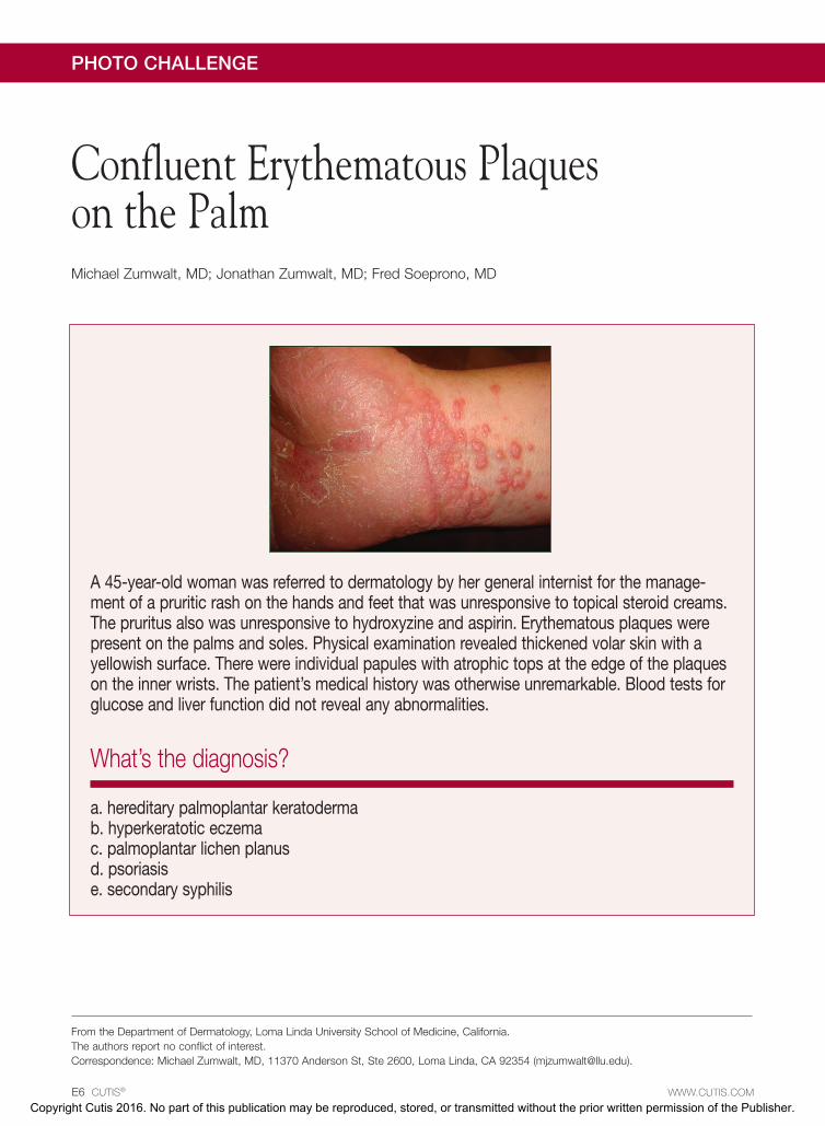
A 45-year-old woman was referred to dermatology by her general internist for the manage-ment of a pruritic rash on the hands and feet that was unresponsive to topical steroid creams. The pruritus also was unresponsive to hydroxyzine and aspirin. Erythematous plaques were present on the palms and soles. Physical examination revealed thickened volar skin with a yellowish surface. There were individual papules with atrophic tops at the edge of the plaques on the inner wrists. The patient’s medical history was otherwise unremarkable. Blood tests for glucose and liver function did not reveal any abnormalities.
What’s the diagnosis?
a. hereditary palmoplantar keratodermab. hyperkeratotic eczema c. palmoplantar lichen planusd. psoriasise. secondary syphilis
Confluent Erythematous Plaques on the Palm Michael Zumwalt, MD; Jonathan Zumwalt, MD; Fred Soeprono, MD
From the Department of Dermatology, Loma Linda University School of Medicine, California.The authors report no conflict of interest.Correspondence: Michael Zumwalt, MD, 11370 Anderson St, Ste 2600, Loma Linda, CA 92354 ([email protected]).
Copyright Cutis 2016. No part of this publication may be reproduced, stored, or transmitted without the prior written permission of the Publisher.
CUTIS D
o no
t cop
y
VOLUME 97, MARCH 2016 E7
Photo Challenge Discussion
WWW.CUTIS.COM
Palmoplantar Lichen Planus
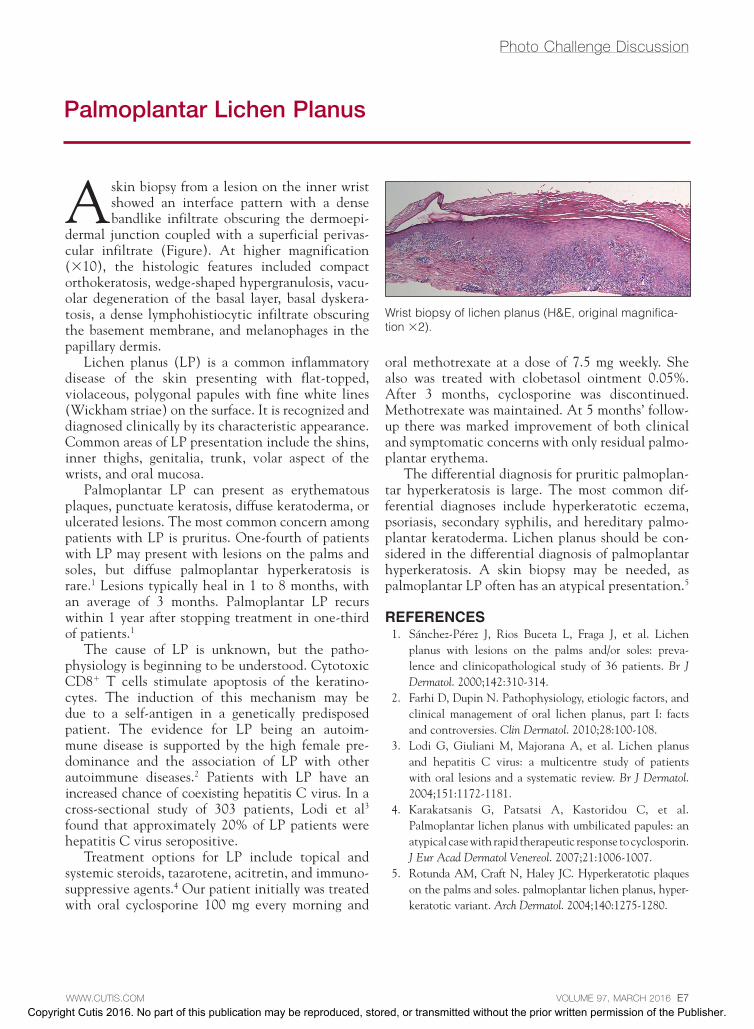
A skin biopsy from a lesion on the inner wrist showed an interface pattern with a dense bandlike infiltrate obscuring the dermoepi-
dermal junction coupled with a superficial perivas-cular infiltrate (Figure). At higher magnification (×10), the histologic features included compact orthokeratosis, wedge-shaped hypergranulosis, vacu-olar degeneration of the basal layer, basal dyskera-tosis, a dense lymphohistiocytic infiltrate obscuring the basement membrane, and melanophages in the papillary dermis.
Lichen planus (LP) is a common inflammatory disease of the skin presenting with flat-topped, violaceous, polygonal papules with fine white lines (Wickham striae) on the surface. It is recognized and diagnosed clinically by its characteristic appearance. Common areas of LP presentation include the shins, inner thighs, genitalia, trunk, volar aspect of the wrists, and oral mucosa.
Palmoplantar LP can present as erythematous plaques, punctuate keratosis, diffuse keratoderma, or ulcerated lesions. The most common concern among patients with LP is pruritus. One-fourth of patients with LP may present with lesions on the palms and soles, but diffuse palmoplantar hyperkeratosis is rare.1 Lesions typically heal in 1 to 8 months, with an average of 3 months. Palmoplantar LP recurs within 1 year after stopping treatment in one-third of patients.1
The cause of LP is unknown, but the patho-physiology is beginning to be understood. Cytotoxic CD8+ T cells stimulate apoptosis of the keratino-cytes. The induction of this mechanism may be due to a self-antigen in a genetically predisposed patient. The evidence for LP being an autoim-mune disease is supported by the high female pre-dominance and the association of LP with other autoimmune diseases.2 Patients with LP have an increased chance of coexisting hepatitis C virus. In a cross-sectional study of 303 patients, Lodi et al3 found that approximately 20% of LP patients were hepatitis C virus seropositive.
Treatment options for LP include topical and systemic steroids, tazarotene, acitretin, and immuno-suppressive agents.4 Our patient initially was treated with oral cyclosporine 100 mg every morning and
oral methotrexate at a dose of 7.5 mg weekly. She also was treated with clobetasol ointment 0.05%. After 3 months, cyclosporine was discontinued. Methotrexate was maintained. At 5 months’ follow-up there was marked improvement of both clinical and symptomatic concerns with only residual palmo-plantar erythema.
The differential diagnosis for pruritic palmoplan-tar hyperkeratosis is large. The most common dif-ferential diagnoses include hyperkeratotic eczema, psoriasis, secondary syphilis, and hereditary palmo-plantar keratoderma. Lichen planus should be con-sidered in the differential diagnosis of palmoplantar hyperkeratosis. A skin biopsy may be needed, as palmoplantar LP often has an atypical presentation.5
REFERENCES 1. Sánchez-Pérez J, Rios Buceta L, Fraga J, et al. Lichen
planus with lesions on the palms and/or soles: preva-lence and clinicopathological study of 36 patients. Br J Dermatol. 2000;142:310-314.
2. Farhi D, Dupin N. Pathophysiology, etiologic factors, and clinical management of oral lichen planus, part I: facts and controversies. Clin Dermatol. 2010;28:100-108.
3. Lodi G, Giuliani M, Majorana A, et al. Lichen planus and hepatitis C virus: a multicentre study of patients with oral lesions and a systematic review. Br J Dermatol. 2004;151:1172-1181.
4. Karakatsanis G, Patsatsi A, Kastoridou C, et al. Palmoplantar lichen planus with umbilicated papules: an atypical case with rapid therapeutic response to cyclosporin. J Eur Acad Dermatol Venereol. 2007;21:1006-1007.
5. Rotunda AM, Craft N, Haley JC. Hyperkeratotic plaques on the palms and soles. palmoplantar lichen planus, hyper-keratotic variant. Arch Dermatol. 2004;140:1275-1280.
Wrist biopsy of lichen planus (H&E, original magnifica-tion ×2).
Copyright Cutis 2016. No part of this publication may be reproduced, stored, or transmitted without the prior written permission of the Publisher.
CUTIS D
o no
t cop
y


![Palmoplantar Pustulosis: Recent Advances in ...ecent Adances in Palmoplantar Pustulosis 357 patientswithPPPrangesfrom10.2–49%indierentpopu-lations[2, 5, 25, 26]andisariskfactorforischemicheart](https://img.pdfslide.us/doc/110x75/602301c9840e343f9c5d167f/palmoplantar-pustulosis-recent-advances-in-ecent-adances-in-palmoplantar-pustulosis.jpg)