Embed Size (px)
Citation preview

CONDITIONING OF INTERICTAL BEHAVIOURS,
BUT NOT ICTAL BEHAVIOURS, SEIZURES,
OR AFTERDISCHARGE THRESHOLD,
BY KINDLING OF THE AMYGDALA IN RATS
A Thesis Submitted to the College of
Graduate Studies and Research
In Partial Fulfillment of the Requirements
For the Degree of Master of Arts
In the Department of Psychology
University of Saskatchewan
Saskatoon
By
JASON P. WAGNER
© Copyright Jason P. Wagner, January, 2007. All rights reserved.

i
Permission to Use
In presenting this thesis in partial fulfilment of the requirements for a
Postgraduate degree from the University of Saskatchewan, I agree that the Libraries of
this University may make it freely available for inspection. I further agree that
permission for copying of this thesis in any manner, in whole or in part, for scholarly
purposes may be granted by the professor or professors who supervised my thesis work
or, in their absence, by the Head of the Department or the Dean of the College in which
my thesis work was done. It is understood that any copying or publication or use of this
thesis or parts thereof for financial gain shall not be allowed without my written
permission. It is also understood that due recognition shall be given to me and to the
University of Saskatchewan in any scholarly use which may be made of any material in
my thesis.
Requests for permission to copy or to make other use of material in this
thesis in whole or part should be addressed to:
Head of the Department of Psychology
University of Saskatchewan
Saskatoon, Saskatchewan S7N 5A5
ii
Abstract
Repeated focal electrical stimulation of the brain results in kindling, the development of
generalized seizures that progress in length and severity as more seizures are elicited.
Barnes et al. (2001) paired one context (CS+) with kindling stimulation of the amygdala,
and another context (CS-) with sham stimulation. They found conditioned anticipatory
fear responses in the CS+, a conditioned place aversion to the CS+, and more intense
convulsions in the CS+ than in the CS- in a probe trial. The present experiment was an
attempt to replicate the findings, and to extend them by recording electroencephalographs
(EEG). As well, I tested for conditioned effects on afterdischarge threshold (ADT). Rats
received 45 pairings in each context before a conditioned place preference/aversion test,
to determine whether the stimulation and seizures were rewarding or punishing. After
more pairings, rats received suprathreshold stimulation in each context (switch test). Ictal
measures in this test included afterdischarge duration, clonus duration, latency to clonus,
class of convulsion, and falls. After more pairings, ADT was measured in each context. I
partially replicated the findings of Barnes et al., in that conditioned anticipatory responses
and conditioned place aversion were found. However, there were no conditioned effects
on any ictal measures, including ADT. I conclude that conditioning is unlikely to play a
major role in epileptogenesis.

iii
Acknowledgements
First I would like to thank my supervisor, Michael E. Corcoran. He has
contributed significantly to this thesis. His help has ranged from helping me start as a
kindling researcher, to providing motivation and valuable guidance, and to intelligent
critiques of earlier versions of this thesis. Perhaps most importantly, he has routinely
helped me see the bigger picture, and how science really works.
I also value the input of my committee members, Deb Saucier and Lisa
Kalynchuk, and wish to thank them. Their input has often provided me with fresh
thinking about my project and data.
My lab-mates have been very supportive and helpful, and so I thank them. Ken
Wolfe was instrumentally helpful in getting me started in the lab, and remained generous
in is help thereafter. Ann Lam, Bonita Ma, Naomi Whelan, Jolly De Guzman, and Joanne
Sitarski, have all been helpful, encouraging, and generally good company in my time in
the lab.
iv
Dedication
To my wife, Susan.
She has been profoundly supportive of me and my work.
I am amazed daily by her love and dedication.
v
TABLE OF CONTENTS
page
PERMISSION TO USE..................................................................................................... i
ABSTRACT...................................................................................................................... ii
ACKNOWLEDGEMENTS............................................................................................. iii
DEDICATION................................................................................................................. iv
LIST OF FIGURES ........................................................................................................ vii
INTRODUCTION ............................................................................................................ 1
Behavioural Treatments of the Epilepsies .................................................................... 1
Evidence from Experimental Preparations for a Role of Conditioning........................ 3
Conditioning and Kindling ........................................................................................... 6
METHOD ......................................................................................................................... 9
Subjects ......................................................................................................................... 9
Pre-Surgery Handling ................................................................................................. 10
Surgery........................................................................................................................ 10
Afterdischarge (AD) Threshold Testing ..................................................................... 10
Apparatus .................................................................................................................... 11
Groups......................................................................................................................... 11
Procedure .................................................................................................................... 12
Statistical Analyses ..................................................................................................... 14
RESULTS ....................................................................................................................... 14
Groups......................................................................................................................... 14
Kindling Rate .............................................................................................................. 15
vi
Prestimulation Tests.................................................................................................... 17
Freezing .................................................................................................................. 17
Locomotor Activity................................................................................................. 21
Rears ....................................................................................................................... 26
CPP Test...................................................................................................................... 34
Between-Subjects Switch Test.................................................................................... 37
Stage........................................................................................................................ 37
Latency.................................................................................................................... 37
Clonus Duration...................................................................................................... 37
AD Duration ........................................................................................................... 38
Falls......................................................................................................................... 38
WDS........................................................................................................................ 38
Within-Subjects Switch Test....................................................................................... 38
Stage........................................................................................................................ 38
Latency.................................................................................................................... 40
Clonus Duration...................................................................................................... 40
AD Duration ........................................................................................................... 43
Falls......................................................................................................................... 43
WDS........................................................................................................................ 46
ADT Switch Test ........................................................................................................ 46
DISCUSSION................................................................................................................. 48
REFERENCES ............................................................................................................... 61
APPENDIX 1: PERMISSION TO REPRINT COPYRIGHTED MATERIAL............. 70
vii
LIST OF FIGURES
Figure page
1. Electrode placements .................................................................................................. 16
2. Freezing in 16 prestimulation test trials...................................................................... 19
3. Freezing grand difference scores ................................................................................ 20
4. Freezing difference scores in four-trial blocks ........................................................... 22
5. Locomotor activity in 16 prestimulation test trials ..................................................... 24
6. Locomotor activity grand difference scores ............................................................... 25
7. Locomotor activity difference scores in four-trial blocks........................................... 27
8. Rearing by White rats in 16 prestimulation test trials................................................. 30
9. Rearing difference scores by White rats in four-trial blocks ...................................... 31
10. Rearing by Black rats in 16 prestimulation test trials ............................................... 33
11. Rearing difference scores by Black rats in four-trial blocks..................................... 35
12. Conditioned place preference test ............................................................................. 36
13. Convulsion stage in the within-subjects switch test ................................................. 39
14. Latency to stage 3 convulsion in the within-subjects switch test ............................. 41
15. Clonus duration in the within-subjects switch test ................................................... 42
16. Afterdischarge duration in the within-subjects switch test ....................................... 44
17. Falls in the within-subjects switch test ..................................................................... 45
18. Post-ictal wet dog shakes .......................................................................................... 47
19. Afterdischarge threshold in each context.................................................................. 49
20. Afterdischarge threshold by electrode placement..................................................... 50
1
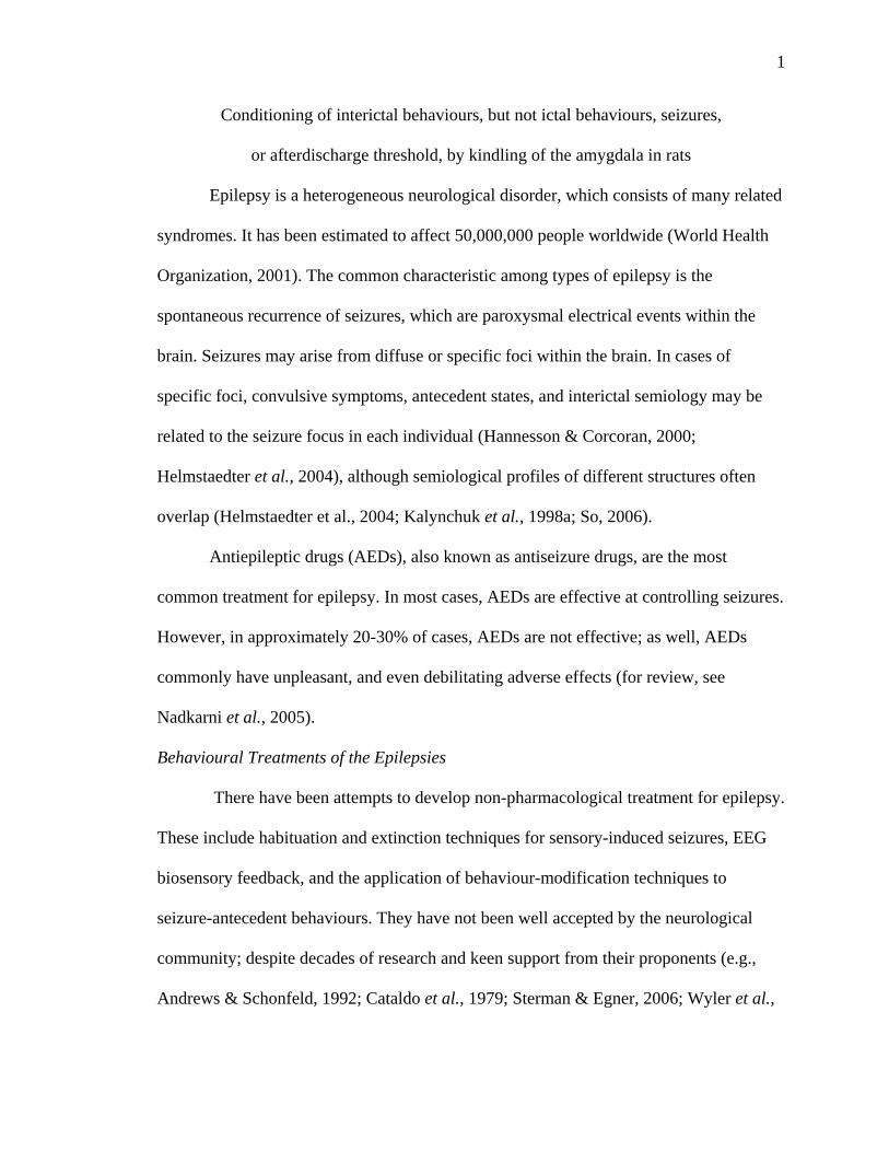
Conditioning of interictal behaviours, but not ictal behaviours, seizures,
or afterdischarge threshold, by kindling of the amygdala in rats
Epilepsy is a heterogeneous neurological disorder, which consists of many related
syndromes. It has been estimated to affect 50,000,000 people worldwide (World Health
Organization, 2001). The common characteristic among types of epilepsy is the
spontaneous recurrence of seizures, which are paroxysmal electrical events within the
brain. Seizures may arise from diffuse or specific foci within the brain. In cases of
specific foci, convulsive symptoms, antecedent states, and interictal semiology may be
related to the seizure focus in each individual (Hannesson & Corcoran, 2000;
Helmstaedter et al., 2004), although semiological profiles of different structures often
overlap (Helmstaedter et al., 2004; Kalynchuk et al., 1998a; So, 2006).
Antiepileptic drugs (AEDs), also known as antiseizure drugs, are the most
common treatment for epilepsy. In most cases, AEDs are effective at controlling seizures.
However, in approximately 20-30% of cases, AEDs are not effective; as well, AEDs
commonly have unpleasant, and even debilitating adverse effects (for review, see
Nadkarni et al., 2005).
Behavioural Treatments of the Epilepsies
There have been attempts to develop non-pharmacological treatment for epilepsy.
These include habituation and extinction techniques for sensory-induced seizures, EEG
biosensory feedback, and the application of behaviour-modification techniques to
seizure-antecedent behaviours. They have not been well accepted by the neurological
community; despite decades of research and keen support from their proponents (e.g.,
Andrews & Schonfeld, 1992; Cataldo et al., 1979; Sterman & Egner, 2006; Wyler et al.,
2
1976), such treatments rarely receive mention in authoritative descriptions of available
epilepsy treatments (Lowenstein, 2005; Nadkarni et al., 2005)
Forster (1969, 1972, 1967, 1964, 1965, 1964a, 1964b) successfully reduced the
frequency of sensory-induced seizures through conditioning. Other researchers have had
difficulty replicating his findings (Wolf, 2002). Most other early studies of behavioural
treatments for epilepsies had serious methodological problems (Krafft & Poling, 1982).
In some cases, seizures are reliably preceded by certain behaviours (Ng, 2002;
Zlutnick et al., 1975) or thoughts (Ritaccio et al., 2002). Some individuals intentionally
perform behaviours that elicit seizures (for review, see Ng, 2002). In cases of specific
preseizure behaviours, Zlutnick et al. (1975) reported reductions in seizures using
behaviour modification techniques targeting the preseizure behaviours. They argued that
those behaviours and the seizures formed behavioural chains that could be interrupted
(Skinner, 1934).
Another nonpharmaceutic approach to epilepsy treatment is EEG biofeedback,
which involves the provision of reinforcement for certain patterns of electrical activity as
measured on EEG (Blanchard & Young, 1974; Egner & Sterman, 2006; Sterman, 2000;
Sterman & Egner, 2006). Although its proponents argue that EEG biofeedback is a viable
treatment for epilepsy (Egner & Sterman, 2006; Sterman, 2000; Sterman & Egner, 2006),
biofeedback is controversial (Cott et al., 1979; Quy et al., 1979), and has not been fully
accepted by the medical community.
Insight into the potential role of the environment and conditioning (learning) in
epileptogenesis has come from studies of experimental epilepsy in animals. I will review

3
this evidence, with particular emphasis on kindling, the preparation I used in my own
research.
Evidence from Experimental Preparations for a Role of Conditioning
Repeated focal electrical stimulation of some discrete sites in the brain results in
kindling, the development of generalized seizures that progress in length and severity as
more seizures are elicited. Kindling was first reported and described by Goddard
(Goddard, 1967) and Racine (Racine, 1972a, 1972b). It is a good animal preparation of
partial complex epilepsy, in that the seizures originate from a discrete focus, and
eventually generalize to other parts of the brain. Additionally, seizures kindled from some
sites are accompanied by a loss of equilibrium, as evidenced by falling, and this is
considered to be analogous to an alteration of consciousness in a human complex seizure.
Kindling is also thought to be related to learning and memory. Whereas both
kindling and learning are thought to involve neuroplastic changes, those induced by
kindling are considered pathological, and not representative of normal learning and
memory (Saucier & Corcoran, 2003).
Many neuroanatomical structures have been kindled, but those in the limbic
system have been studied the most. This includes the amygdala, which was the structure
of interest in the first reports of kindling (Goddard, 1967; Racine, 1972a, 1972b).
Although originally used by Burdach to describe what we now call the Basolateral
Amygdala (BLA), the term amygdala is often used to describe that and several other
interconnected nearby nuclei (Sah et al., 2003). The amygdala receives input from a wide
variety of cortical and subcortical sources, including input from all sensory modalities.
4
Likewise, it projects to a wide variety of cortical and subcortical sites (for a thorough
review, see Sah et al., 2003).
The amygdala has long been recognized for its involvement in emotion. Initially,
this recognition came from studies showing deficits in fear and anxiety after experimental
lesions (e.g., Goldstein, 1965; e.g., Kluver & Bucy, 1939; Robinson, 1963). Stimulation
of the amygdala has been shown to result in increases in fear-related behaviour (e.g.,
Hunsperger, 1963; Maclean & Delgado, 1953). The amygdala is also involved in general
arousal, appetite, and sexual behaviour (Goddard, 1964).
For fear-related behaviours, there may be some functional differentiation between
the central and basolateral nuclei of the amygdala. Using experimental lesions, the
basolateral nucleus has been shown to mediate conditioned avoidance behaviour, whereas
the central nucleus has been shown to mediate fear-induced suppression of behaviour
(Killcross et al., 1997).
When limbic sites are kindled, the convulsions reliably progress through
characteristic stages, each associated with characteristic motor movements. Racine
(1972b) first described five stages (Stage 1: facial clonus; Stage 2: facial clonus with
head-nodding; Stage 3: unilateral forelimb clonus; Stage 4: rearing and bilateral forelimb
clonus; Stage 5: as with Stage 4, with one fall). This scale was later extended by Pinel
and Rovner (1978), to include three additional stages that occur with relatively extended
kindling of the amygdala (Stage 6: as with Stage 5, with multiple falls; Stage 7: as with
Stage 6, with running or jumping fits; Stage 8: a convulsion with one or more periods of
tonus).
5
It is generally believed that the kindled state is stable, that is, that animals kindled
to generalized seizures will predictably emit such seizures when later stimulated
(Goddard et al., 1969). As well, it has been believed that each animal’s ADT is indicative
of internal physiological states close to the electrode tip (Racine, 1972a), and not of
learned associations.
Kindling of some structures results in behavioural changes specific to those
structures (Barnes et al., 2005; Barnes et al., 2001; Barnes et al., 2003; Hannesson &
Corcoran, 2000; Kalynchuk et al., 1998a; D. C. McIntyre, 1979; D. C. McIntyre &
Molino, 1972; Wig et al., 2002). For example, kindling of the hippocampus results in
deficits in spatial cognition (Gilbert et al., 2000; Gilbert et al., 1996; Hannesson &
Corcoran, 2000; Hannesson et al., 2004; Leung et al., 1996; Lopes da Silva et al., 1986),
but not in other forms of cognition not thought to be mediated by the hippocampus
(Hannesson et al., 2001).
Kindling of the amygdala usually results in changes in anxiety-related behaviour
(R. Adamec & Shallow, 2000; R. E. Adamec & Morgan, 1994; Kalynchuk, 2000;
Kalynchuk et al., 1998a, 1999; Kalynchuk et al., 1998b; Kalynchuk et al., 1997; D. C.
McIntyre, 1970, 1979; Wintink et al., 2003). Kindling of the amygdala also disrupts
emotional learning (D. C. McIntyre, 1970, 1979). When rats are kindled extensively, with
up to 100 amygdaloid stimulations, they are more defensive in a resident-intruder test,
they are more resistant to capture by an unfamiliar experimenter, they display heightened
anxiety-like behaviour in the open field, and they display unusual anxiety like behaviour
in the elevated-plus maze, where they sometimes jump off the maze presumably to
escape it (Kalynchuk et al., 1998a, 1999; Kalynchuk et al., 1998b; Kalynchuk et al.,

6
1997; Wintink et al., 2003). These effects are reliably produced by extended amygdaloid
kindling, but they can also be produced by extended hippocampal kindling (Kalynchuk et
al., 1998a), suggesting that extensive kindling may decrease the focal specificity of
behavioural effects. Thus after many generalized seizures, behaviours mediated by
multiple related structures may be affected.
Conditioning and Kindling
In recent years, Steven J. Barnes and colleagues (Barnes et al., 2005; Barnes et
al., 2004; Barnes et al., 2001; Barnes et al., 2006; Barnes et al., 2003; Wig et al., 2002)
have used kindling, or some aspect thereof, as the unconditioned stimulus in a classical
conditioning paradigm. They have identified conditioned effects on interictal behaviours,
as well as conditioned effects on ictal measures. Their original paradigm (Barnes et al.,
2001) used a place conditioning procedure, in which rats were kindled by amygdaloid
stimulation in one context (CS+), and received sham stimulation in another (CS-). This
involved many (53 in total) pairings of each type. In the CS+, rats developed conditioned
anticipatory defensive responses, including decreased locomotion, decreased rearing, and
increased freezing, as compared to their baseline of behaviour in the CS-. As well, rats
showed a conditioned place aversion to the CS+ when given a choice between the two
contexts.
The place aversion described by Barnes et al. (2001) contrasts with the results of
Corcoran, Lanius, and Duren (1992), who used fewer pairings and did not find a
significant conditioned place preference or aversion. It also contrasts with the results of
Paredes, Muzzi, Aguirre, and Romero (2000), who found a conditioned place preference
for the context in which rats were placed after generalized kindled seizures. Paredes et al.
7
argued that the rewarding effects indicated by the place preference occur during the
postictal period. If the results of both Barnes et al. and Paredes et al. are correct, it would
seem that the aversive aspects of amygdaloid kindling or amygdaloid-kindled seizures are
more salient than the rewarding aspects of the postictal state produced thereby.
Given the role of the amygdala in fear, anxiety, and emotional memory, Barnes et
al.’s (2001) results may not seem surprising. However, they do contradict the long-held
view that generalized seizures have amnestic effects. For example, amygdaloid-kindled
generalized convulsions have been shown to produce amnestic effects on conditioned
emotional response (D. C. McIntyre, 1970).
The provocative results from Barnes et al.’s (2001) original paper and later papers
(Barnes et al., 2004; Barnes et al., 2001; Barnes et al., 2006; Barnes et al., 2003) are those
showing conditioned effects on ictal measures, such as convulsion stage and duration.
The researchers found that, with amydaloid-kindled convulsions, the convulsions were
less severe when elicited in the CS- than in the CS+. This was shown using “switch tests”
(Barnes et al., 2001), p1067), which came after many pairings of the CS+ with kindled
convulsions, and the CS- with sham stimulation. Barnes's results are in contrast to those
of Freeman and Mikulka (1986), who failed to find a contextually-conditioned effect on
AD duration with amygdaloid kindling. In any case, Barnes's results could be interpreted
to indicate that the environment and conditioning play a key role in kindling and the
intensity of kindled convulsions (Barnes et al., 2001).
The potentially important role of the environment and conditioning as a general
mechanism in kindling is challenged by Barnes's finding that when other forebrain
structures are kindled, different results are found (Barnes et al., 2005; Barnes et al.,
8
2003). When seizures were elicited by anterior neocortical kindling, there was no
evidence of conditioned defensiveness or place preference or aversion, and convulsions
were less severe in the CS+ than in the CS- (Barnes et al., 2003). Barnes et al. (2005) also
kindled three different sites in the hippocampal formation in a similar experiment.
Perirhinal kindling produced conditioned anticipatory freezing and decreased locomotion.
Unfortunately, rearing data were notably absent from that experiment. Ventral
hippocampal kindling only produced conditioned freezing, but not a conditioned decrease
in locomotion, when the experiment was extended to include many more pairings. Dorsal
hippocampal kindling did not produce any conditioned anticipatory defensiveness, even
with the extended experimental design. A conditioned place aversion was found with
perirhinal and ventral hippocampal kindling, but not with dorsal hippocampal kindling.
Barnes et al.'s (2005) reasonable explanation for the differential effects on
anticipatory defensive behaviours is that, because ventral and dorsal hippocampal
kindling require a large number of stimulations before generalized convulsions develop,
it is likely the generalized convulsions that serve as the relevant unconditioned stimulus –
that is, that conditioning does not occur until generalized convulsions have been kindled.
Alternatively, the hippocampus may not be sufficiently involved in anxiety and fear
relative to the amygdala to produce conditioned defensiveness. The perirhinal cortex is
also arguably anatomically and functionally dissociable from the hippocampal formation
(see Paxinos, 1995). Perhaps more important, there were no conditioned effects on ictal
measures. Ostensibly, this means that hippocampal seizures are not as easily conditioned.
However, it is possible that conditioning of ictal measures is not as reliable as some other

9
experiments have suggested (Barnes et al., 2004; Barnes et al., 2001; Barnes et al., 2006;
Barnes et al., 2003).
In the present experiment I attempted to replicate Barnes et al. (2001), and to
extend their findings by recording electroencephalographs (EEG), thereby permitting
testing for conditioned effects on afterdischarge (AD) and afterdischarge threshold
(ADT). Because triggering of AD is fundamental to amygdaloid kindling and kindled
convulsions, recording EEG seemed to be particularly relevant in this experiment. Note
that Barnes did not record EEG in his experiments. As well, I hypothesized that the
conditioned effects on ictal measures found by Barnes and colleagues may have been
related to conditioned differences in ADT, and I therefore included an ADT switch test in
the present experiment. I hypothesised that I would replicate the findings of Barnes et al.
(2001), and that I would find conditioned effects on AD and ADT.
Method
Subjects
Subjects were 41 male Long-Evans rats from Charles Rivers Laboratories (St.
Constant, Quebec, Canada), weighing 370g to 540g at the time of surgery. They were
housed in groups of two to four prior to surgery, and housed individually thereafter.
Food and water were freely available. Lights followed a 12h:12h light-dark schedule,
with lights on at 7am; experimental procedures were conducted during the light portion
of the cycle. Housing and experimental procedures were in accordance with the
guidelines of the Canadian Council on Animal Care.

10
Pre-surgery Handling
Starting 5 to 7 days after arrival, rats were handled for 1.5 to 3 minutes each, once
per day, for five days. Handling consisted of gentle stroking, picking up, holding, and
talking to each rat.
Surgery
Rats were anesthetized with isoflurane (IsoFlo) administered by inhalation (5%
initial, 2.5% thereafter) and placed in a stereotaxic apparatus. During surgery, Anafen ®
was injected s.c. (5mg/kg) for post-operative analgesia. Two bipolar electrodes, made
from 127µm-diameter enamel-insulated nichrome wire, were implanted at the following
coordinates: 2.6mm posterior, 4.5mm lateral, and 9.1mm ventral from bregma, with the
skull level. Coordinates were derived from Paxinos and Watson (1998). Four stainless
steel dental screws, to which a ground electrode was mounted, were fixed to the skull
surface. The electrode assembly was affixed to the skull using dental acrylic. Hibitaine
® was applied to the area of the incision to minimize infection. Starting 1 to 2 weeks
after surgery, rats were handled as before for five days.
Afterdischarge (AD) Threshold Testing
Prior to experimental pairings, AD threshold (ADT) was determined for each rat.
The rat was placed in a clear acrylic chamber that was different from the apparatus used
for the rest of the experiment. Electrical stimulation was generated on a Grass S8800
stimulator and consisted of a 1-s train of balanced biphasic square-wave pulses 1.0 ms in
duration and delivered at 60 pp/s to one of the two electrodes, starting at 20µΑ and
increasing by 10 µΑ every minute, to a maximum of 100 µΑ. If AD lasting at least 5sec
was not elicited, the schedule was repeated using the other electrode. If no AD was
11
elicited, the rat was returned to its home cage. The next day, the rat was returned to the
same chamber, and stimulation was applied in the same manner as before, but using the
following intensity schedule: 120 µA, 140 µA, 160 µA, 180 µA, 200 µA, 220 µA,
250 µA, 300 µA, 350 µA, 400 µA. The first electrode in each animal to elicit an AD
using this protocol was subsequently used as the stimulating electrode for kindling. This
was intended to select an electrode that was likely to be in the BLA, which tends to have
lower ADTs than adjacent nuclei (Mohapel et al., 1996).
Apparatus
The conditioned place preference apparatus was of a rectangular box shape, with
two distinct halves (contexts) separable by removable dividers. The apparatus measured
48cm (w) by 79cm (l) by 25cm (h) overall. With dividers in place, each context
measured 48cm (w) by 37cm (l) by 25cm (h). One context was constructed of white
acrylic panels, with recycled-paper bedding (CareFresh ®) on the floor. The other
context was constructed of black acrylic panels, and the floor was left bare. The entire
apparatus had two clear acrylic lids, mounted with hinges. When closed, the lids left a
3cm gap running the length of the apparatus, perpendicular to the dividers, which allowed
the stimulation and recording lead to reach the rat, without restricting the rat’s
movements.
Groups
Rats were randomly assigned to either the WHITE or BLACK group,
corresponding to the context that served as their CS+. WHITE rats received CS+ trials in
the white half of the apparatus and CS- trials in the black half. BLACK rats received
12
CS+ trials in the black half of the apparatus and CS- trials in the White half of the
apparatus.
Procedure
Rats received a total of 63 pairings of each of two types. In CS+ trials, each rat
was placed in its CS+. After 30sec in the context, the rat received a train of kindling
stimulation. In CS- trials, the rat was placed in its CS-, but received no stimulation.
Each trial lasted 180s, and there were two trials daily. The order of these trials was
pseudorandom, with the criterion that each rat received no more than three trials of one
type in a row. On every fourth day, each rat received one trial of each type, in
counterbalanced order. The purpose of these days was to examine the rats’ behaviour in
each context in the prestimulation interval, during the first 30sec after being placed in the
apparatus. The behaviours examined included the number of rears, the total duration of
freezing episodes longer than five sec, and locomotor activity as measured by gridline
crosses. Behavioural convulsions were scored using Pinel and Rovner’s (1978) eight-
point scale (Stage 1: facial clonus; Stage 2: facial clonus with head-nodding; Stage 3:
unilateral forelimb clonus; Stage 4: rearing and bilateral forelimb clonus; Stage 5: as with
Stage 4, with one fall; Stage 6: as with Stage 5, with multiple falls; Stage 7: as with Stage
6, with running or jumping fits; Stage 8: a convulsion one or more periods of tonus). As
well, latency to clonus, clonus duration, the number of falls, and AD duration were
recorded for each seizure.
After 45 pairings, rats were placed in the apparatus, with dividers removed. They
were thus able to move freely between the two contexts. This assessed their preference
13
between the two contexts, to test whether amygdaloid kindling is associated with
affective consequences, either punishing or rewarding.
After eight more pairings of each type, rats received a switch test (Barnes et al.,
2001). Half of the rats received stimulation in their CS+ context, and the other half
received stimulation in their CS- context. Comparison between the two groups
constituted the between-groups switch test. Assignment to the groups was
counterbalanced, in that it was not predicted by assignment to context (WHITE or
BLACK) or by the counterbalancing schedule used for prestimulation test days. Rats that
received a stimulation in the CS+ in that test received a stimulation in their CS-
approximately 4 to 5 hrs later. Comparison between each rat’s CS+ trial and CS- trial on
that day constituted the within-subjects switch test.
After eight more pairings of each type, the rats’ ADT was determined in each
context. Stimulation intensity was gradually increased until an AD of at least 5sec was
elicited, using the following schedule: 20 µΑ, 30 µΑ, 40µΑ, 50µΑ, 60µΑ, 70µΑ, 80µΑ,
90µΑ, 100µΑ, 120µΑ, 140µΑ, 160µΑ, 180µΑ, 200µΑ, 220µΑ, 250µΑ, 300µΑ, 350µΑ,
400µΑ. This procedure was completed in one session for each context, exclusively
employing the same electrode used for kindling. Half of the rats were first tested in their
CS+, and the other half were first tested in their CS-. After approximately 24hr, the rats
were tested in the other context. Assignment to this order was counterbalanced such that
it was not predicted by assignment to context (WHITE or BLACK), the counterbalancing
schedule used for prestimulation test days, or assignment to the switch test conditions.
Rats were euthanized with Euthanyl ®, and were then perfused transcardially with
phosphate-buffered saline (PBS) and 4% paraformaldehyde in PBS (PFA). Brains were

14
stored in PFA for 1 to 3 days. Each brain was then stored in 30% sucrose in PBS, until it
sunk (2 to 4 days). The brains were sectioned at 40 µm using a freezing microtome. The
sections were either stored in PBS at 4° C for less than 24hr or stored in cryoprotectant
solution at -20° C for up to 5 days. Every second section was then mounted on gelatin-
coated slides, and sections were stained with cresyl violet. Electrode placements were
verified using a light microscope and a reference atlas (Paxinos & Watson, 1998).
Statistical Analyses
Data were analyzed using univariate or mixed-model Analyses of Variance
(ANOVAs), where appropriate. Within groups of tests (e.g., Prestimulation Tests, or
Within-Subjects Switch Test), a Bonferroni correction was used. This was done to correct
for the increased probability of Type I error associated with multiple tests. The correction
divides the desired α level by the number of tests (e.g., three, for the Prestimulation
Tests), to obtain the α level to be used for the individual tests. I did not use a Multivariate
Analysis of Variance (MANOVA), because I was interested in effects on individual
dependent variables, rather than relations among the dependent variables. This approach
is supported by Huberty and Morris (1989). Finally, significant interactions from
individual ANOVAs were followed by t-tests. These tests were also adjusted using a
Bonferroni correction; the α level was adjusted for the number of t-tests that followed
each interaction.
Results
Groups
Of the 41 rats, only the data from the 28 rats with placements in the BLA (n = 20)
or central nucleus of the amygdala (n = 8) were used in this experiment (Figure 1
15
describes the placements). One rat died during surgery, and another died in recovery. One
rat was found, post-mortem, to have a trans-cranial infection causing a mass effect, and
its data were excluded from analyses. Another rat was found to have a cystic cavity,
approximately 2 mm3 at the tip of the kindling electrode, and was therefore excluded. As
well, 5 rats dislodged their electrode pedestals during the course of the experiment. There
were 4 rats with kindling-electrode tips located elsewhere in the amygdala. That group
was excluded from analyses, because its electrode placements were heterogeneously
distributed throughout the extended amygdala. Of the included rats, 17 had the white
context as their CS+ (White group), and 11 had the black context as their CS+ (Black
group).
Kindling Rate
Two measures were used to quantify each rat’s rate of kindling: the number of
kindling stimulations required to elicit the first stage 5 or greater seizure, and the number
of stimulations required to elicit five consecutive stage 5 seizures. Four rats did not have
five consecutive stage 5 seizures during the entire experiment, and were assigned values
of either 62 or 63, equal to the total number of stimulations they received. Each of these
dependent variables was analyzed using 2 (CS+) * 2 (Placement) Univariate ANOVAs,
with a Bonferroni correction requiring a p value of α/2 = 0.05/2 = 0.025. The p values are
reported uncorrected. The omnibus ANOVA analyzing the number of stimulations to the
first stage 5 did not reach significance; no main effects or interactions occurred, F(3, 24)
= 1.052, p = 0.388. The omnibus ANOVA analyzing the number of stimulations required
for five consecutive stage 5 seizures also did not reach significance; there were no main

16
Figure 1. Electrode placements from BLA rats (stars) and Central Amygdala rats
(circles). For rats in which both electrodes were used for stimulation (n = 3), both
placements are shown. Images were adapted from Paxinos and Watson (1997, used with
permission). Measurements are relative to Bregma.
17
effects or interactions, F < 1. Thus kindling was unaffected by the environment (White or
Black) in which stimulation was applied.
Prestimulation Tests
Freezing, Locomotor Activity, and Rears data from prestimulation test trials were
entered into separate 16 (Trial) * 2 (Context) * 2 (Placement) * 2 (CS+) mixed-model
ANOVAs. A Bonferroni correction was used to correct for an increased probability of
Type I error; this required a p value of α/3 = 0.05/3 = 0.0167. All p values are reported
uncorrected.
Freezing. Mauchly’s Test of Sphericity revealed that the assumption of sphericity
was violated for Trial, W(119) < 0.0005, p < 0.0005. As well, the assumption of
sphericity was found to be violated for the Trial * Context interaction term, W(119) <
0.0005, p < 0.0005. The Huynh-Feldt ε correction was therefore used for all tests
involving the Trial term (ε = 0.567), and for all tests involving the Trial * Context term (ε
= 0.469).
The main effect of Trial approached statistical significance, indicating there was
likely at least one difference that approached significance among the 16 trials, F(8.498,
203.960) = 2.147, p = 0.030.
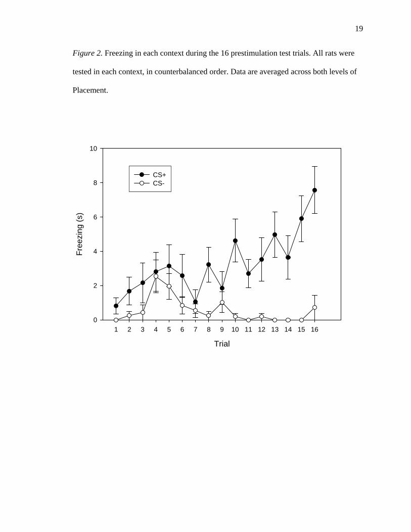
There was a significant main effect of Context, with rats freezing more in the CS+
(52.26 +/- 9.69 s) than in the CS- (8.91 +/- 2.74 s) during the prestimulation test trials,
F(1, 24) = 17.165, p < 0.0005. The main effect of Placement was not statistically
significant, and thus BLA rats (63.02 +/- 15.19 s) did not differ from Central Amygdala
rats (56.54 +/- 15.29 s) in time spent freezing during the prestimulation test trials, F < 1.
The main effect of CS+ was also not statistically significant, and thus White rats (74.26
18
+/- 15.03 s) did not differ significantly from Black rats (40.92 +/- 17.08 s) in time spent
freezing, F(1, 24) = 1.641, p = 0.212.
A variety of two-way interactions were statistically insignificant: Trial *
Placement, F < 1; Trial * CS+, F < 1;, Context * Placement, F < 1; and Placement * CS+,
F < 1. The Context * CS+ interaction approached significance, F(1, 24) = 4.017, p =
0.056. As illustrated in Figure 2, the Trial * Context interaction was significant, F(7.029,
1.8.687) = 2.787, p = 0.009.
A variety of three-way interactions were also statistically insignificant: Trial *
Placement * CS+, F < 1; Context * Placement * CS+, F < 1; Trial * Context *
Placement, F < 1, Trial * Context * CS+ interaction, F < 1; Trial * Context * Placement *
CS+, F < 1.
Although the Context * CS+ interaction did not reach significance, further
analysis was conducted to examine a possible differential effect of CS+ assignment to
contextually conditioned differences in freezing. Each rat’s total duration of freezing in
each context during prestimulation test trials was computed. These totals were then used
to compute a grand difference score, representing the total time spent freezing in the CS+
minus compared to that in the CS-. A positive score would therefore indicate a greater
total duration of freezing in the CS+ than in the CS-. An independent-samples t-test was
then computed to compare White rats to Black rats on the grand difference score (see
Figure 3). This t-test revealed that White rats showed a greater difference in freezing
between their CS+ and CS- (57.70 +/- 11.10 s) than Black rats (21.17 +/- 9.55 s), t(26) =
2.307, p = 0.029 (two-tailed). To follow up the Trial * Context interaction, the 16
prestimulation test trials were grouped into four blocks of four consecutive trials each. A
19
Figure 2. Freezing in each context during the 16 prestimulation test trials. All rats were
tested in each context, in counterbalanced order. Data are averaged across both levels of
Placement.
Trial
1 2 3 4 5 6 7 8 9 10 11 12 13 14 15 16
Free
zing
(s)
0
2
4
6
8
10
CS+CS-

20
Figure 3. Freezing grand difference scores, based on all prestimulation test trials, for rats
in each of the CS+ assignment groups. The difference scores were calculated by
subtracting the duration of all freezing in the CS- from the duration of all freezing in the
CS+. Data are averaged across both levels of Placement. White rats showed a
significantly larger difference in freezing between their CS+ and CS- than did Black rats,
p = 0.029.
CS+ Group
White Black
Free
zing
Gra
nd D
iffer
ence
Sco
re (s
)
0
20
40
60
80
21
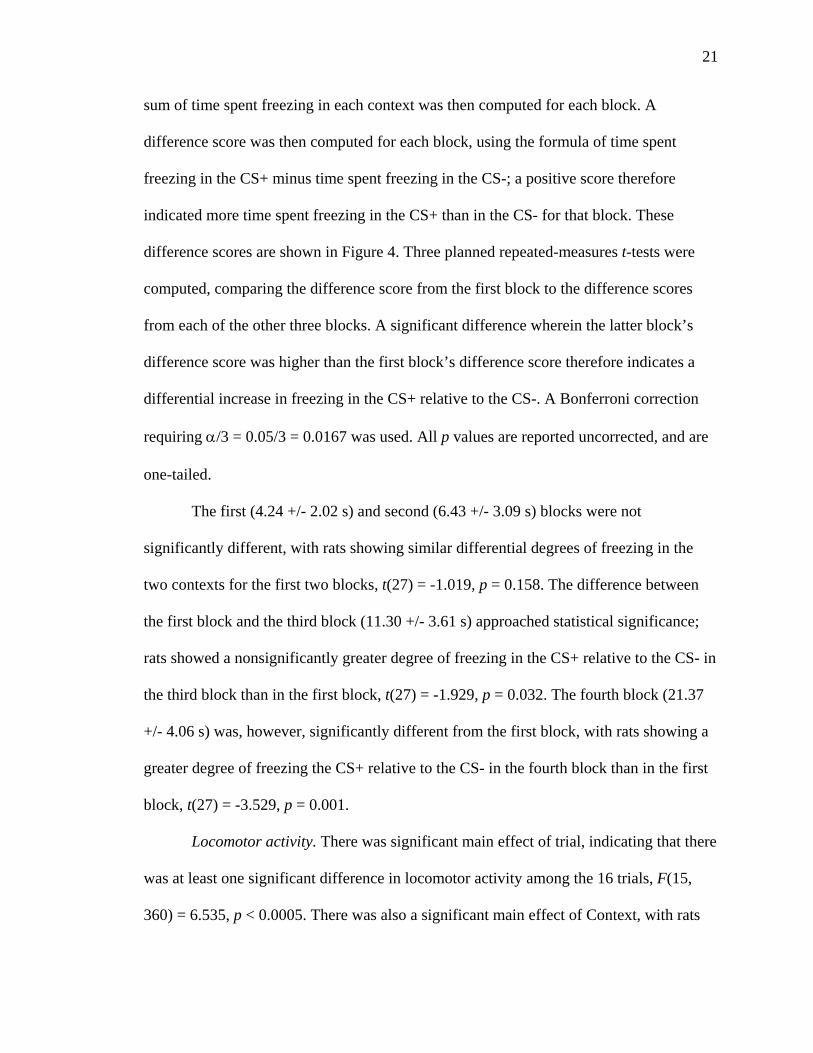
sum of time spent freezing in each context was then computed for each block. A
difference score was then computed for each block, using the formula of time spent
freezing in the CS+ minus time spent freezing in the CS-; a positive score therefore
indicated more time spent freezing in the CS+ than in the CS- for that block. These
difference scores are shown in Figure 4. Three planned repeated-measures t-tests were
computed, comparing the difference score from the first block to the difference scores
from each of the other three blocks. A significant difference wherein the latter block’s
difference score was higher than the first block’s difference score therefore indicates a
differential increase in freezing in the CS+ relative to the CS-. A Bonferroni correction
requiring α/3 = 0.05/3 = 0.0167 was used. All p values are reported uncorrected, and are
one-tailed.
The first (4.24 +/- 2.02 s) and second (6.43 +/- 3.09 s) blocks were not
significantly different, with rats showing similar differential degrees of freezing in the
two contexts for the first two blocks, t(27) = -1.019, p = 0.158. The difference between
the first block and the third block (11.30 +/- 3.61 s) approached statistical significance;
rats showed a nonsignificantly greater degree of freezing in the CS+ relative to the CS- in
the third block than in the first block, t(27) = -1.929, p = 0.032. The fourth block (21.37
+/- 4.06 s) was, however, significantly different from the first block, with rats showing a
greater degree of freezing the CS+ relative to the CS- in the fourth block than in the first
block, t(27) = -3.529, p = 0.001.
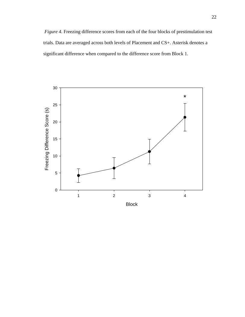
Locomotor activity. There was significant main effect of trial, indicating that there
was at least one significant difference in locomotor activity among the 16 trials, F(15,
360) = 6.535, p < 0.0005. There was also a significant main effect of Context, with rats
22
Figure 4. Freezing difference scores from each of the four blocks of prestimulation test
trials. Data are averaged across both levels of Placement and CS+. Asterisk denotes a
significant difference when compared to the difference score from Block 1.
Block
1 2 3 4
Free
zing
Diff
eren
ce S
core
(s)
0
5
10
15
20
25
30
*
23
making fewer line crosses in the CS+ (36.38 +/- 4.48) than in the CS- (103.54 +/- 7.92),
F(1, 24) = 69.076, p < 0.0005. There was no significant main effect of CS+, indicating
that White rats (138.40 +/- 11.39) did not differ from Black rats (142.27 +/- 20.96) in the
number of lines crossed, F < 1. There was also no significant main effect of Placement,
indicating that BLA rats (147.54 +/- 10.39) did not cross more or fewer lines than Central
Amygdala rats (120.88 +/- 26.27), F < 1.
The Trial * CS+ interaction was not significant, F(15, 360) = 1.093, p = 0.361.
There was no significant Trial * Placement interaction, F < 1. The Context * CS+
interaction approached significance, F(1, 24) = 5.710, p = 0.025. There was no significant
Context * Placement interaction, F < 1. There was a significant Trial * Context
interaction, illustrated in Figure 5, F(15, 360) = 8.319, p < 0.0005. The CS+ * Placement
interaction was not significant, F(1, 24) = 1.921, p = 0.178, nor was no the Trial * CS+ *
Placement interaction, F(15, 360) = 1.273, p = 0.216, the Context * CS+ * Placement
interaction, F < 1, the Trial * Context * CS+ interaction, F < 1, the Trial * Context *
Placement interaction, F < 1, or the Trial * Context * CS+ * Placement interaction, F < 1.
Although the Context * CS+ interaction did not reach significance, a further test
was performed to follow it up. A grand difference score was computed for each rat, by
subtracting the sum of all line crosses by that rat in the CS- from all line crosses by that
rat in the CS+. A lower value therefore indicates a greater degree of locomotor activity in
the CS- relative to the CS+, over the course of the experiment. Using the computed grand
difference score, an independent-samples t test was performed, with CS+ as the grouping
variable (Figure 6). This test revealed that White rats had a significantly lower difference
score (-83.25 +/- 7.88) than did Black rats (-42.27 +/- 10.96). That is, White rats showed
24
Figure 5. Locomotor activity, as indicated by line crosses, in each context during the 16
prestimulation test trials. All rats were tested in each context, in counterbalanced order.
Data are averaged across both levels of Placement and CS+.
Trial
1 2 3 4 5 6 7 8 9 10 11 12 13 14 15 16
Line
Cro
sses
0
2
4
6
8
10
12
CS+CS-
25
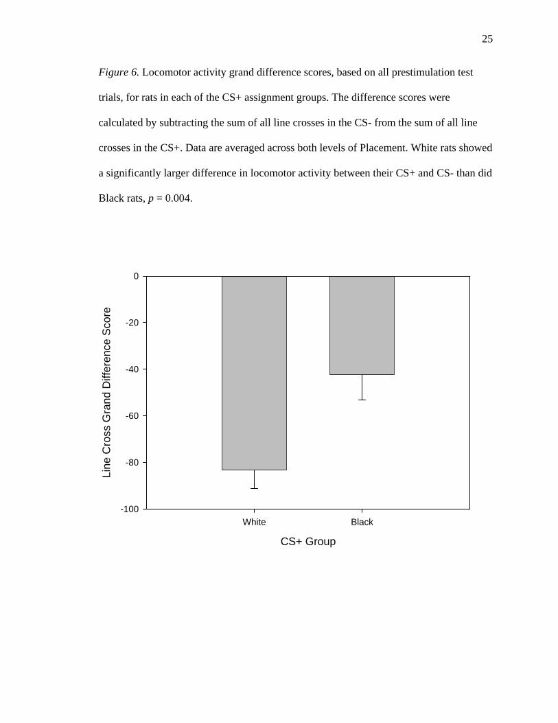
Figure 6. Locomotor activity grand difference scores, based on all prestimulation test
trials, for rats in each of the CS+ assignment groups. The difference scores were
calculated by subtracting the sum of all line crosses in the CS- from the sum of all line
crosses in the CS+. Data are averaged across both levels of Placement. White rats showed
a significantly larger difference in locomotor activity between their CS+ and CS- than did
Black rats, p = 0.004.
CS+ Group
White Black
Line
Cro
ss G
rand
Diff
eren
ce S
core
-100
-80
-60
-40
-20
0
26
a greater degree of locomotor activity in the CS- relative to the CS+ than did black rats,
t(26) = -3.112, p = 0.004 (two-tailed).
To follow up the Trial * Context interaction, the 16 prestimulation test trials were
grouped into four blocks of four consecutive trials each. A sum of line crosses in each
context was then computed for each block. A difference score was then computed for
each block, using the formula of line crosses in the CS+ minus line crosses in the CS-; a
positive score therefore indicates more line crosses in the CS+ than in the CS- for that
block. These difference scores are shown in Figure 7. Three planned repeated-measures t-
tests were computed, comparing the difference score from the first block to the difference
scores from each of the other three blocks. A significant difference wherein the latter
block’s difference score was lower than the first block’s difference score therefore
indicates a differential decrease in locomotor activity in the CS+ relative to the CS-. A
Bonferroni correction requiring α/3 = 0.05/3 = 0.0167 was used. All p values are reported
uncorrected, and are one-tailed.
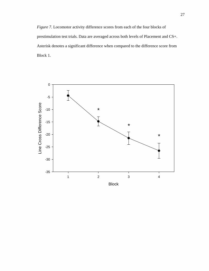
The second block had a significantly lower difference score (-14.75 +/- 1.84) than
the first block (-4.40 +/- 2.00), with rats showing a decrease in locomotor activity in the
CS+ relative to the CS- between the first and second blocks, t(27) = 5.089, p < 0.00025.
There was a similar difference between the first and the third (-21.46 +/- 2.52) blocks,
t(27) = 7.031, p < 0.00025. As well, the fourth block (-26.54 +/- 3.06) had a significantly
greater ratio of locomotor activity in the CS- to that in the CS+ than did the first block,
t(27) = 7.755, p < 0.00025.
Rears. There was a significant main effect of Trial, indicating that there was at
least one significant difference among the 16 trials, F(15, 360) = 9.006, p < 0.0005. There
27
Figure 7. Locomotor activity difference scores from each of the four blocks of
prestimulation test trials. Data are averaged across both levels of Placement and CS+.
Asterisk denotes a significant difference when compared to the difference score from
Block 1.
Block
1 2 3 4
Line
Cro
ss D
iffer
ence
Sco
re
-35
-30
-25
-20
-15
-10
-5
0
*
*
*
28
was a significant main effect of Context, with rats rearing fewer times in the CS+ (31.92
+/- 2.85) than in the CS- (44.54 +/- 3.08), F(1, 24) = 13.589, p = 0.001. There was no
significant main effect of CS+; thus White rats (75.05 +/- 6.60) did not differ
significantly from Black rats (78.64 +/- 9.04), F < 1. As well, there was no significant
main effect of Placement, in that BLA rats (78.44 +/- 5.74) did not differ significantly
from Central Amygdala rats (71.50 +/- 12.05), F < 1.
A number of interactions failed to reach statistical significance, including the
Trial * CS+ interaction, F(15, 360) = 1.450, p = 0.122; the Trial * Placement interaction,
F < 1, the Context * CS+ interaction, F(1, 24) = 4.126, p = 0.053; and the Context *
Placement interaction, F < 1. The Trial * Context interaction was significant, F(15, 360)
= 3.781, p < 0.0005. The CS+ * Placement interaction failed to reach statistical
significance, F(1, 24) = 1.180, p = 0.288, as did the Trial * CS+ * Placement interaction,
F < 1; the Context * CS+ * Placement interaction, F < 1; the Trial * Context * CS+
interaction, F(15, 360) = 1.893, p = 0.023;. the Trial * Context * Placement interaction, F
< 1; and the Trial * Context * CS+ * Placement interaction, F(15, 360) = 1.069, p =
0.384.
The Trial * Context * CS+ interaction, though not statistically significant at the α
= 0.0167 level, is taken as being sufficiently strong to warrant further analyses. This is
partially because of its hierarchically superior position to the Trial * Context 2-way
interaction. Two 16 (Trial) * 2 (Context) * 2 (Placement) mixed model ANOVAs were
therefore conducted, one for each CS+ group. A Bonferroni correction of α/2 = 0.05/2 =
0.025 was used. All p values are reported uncorrected.
29
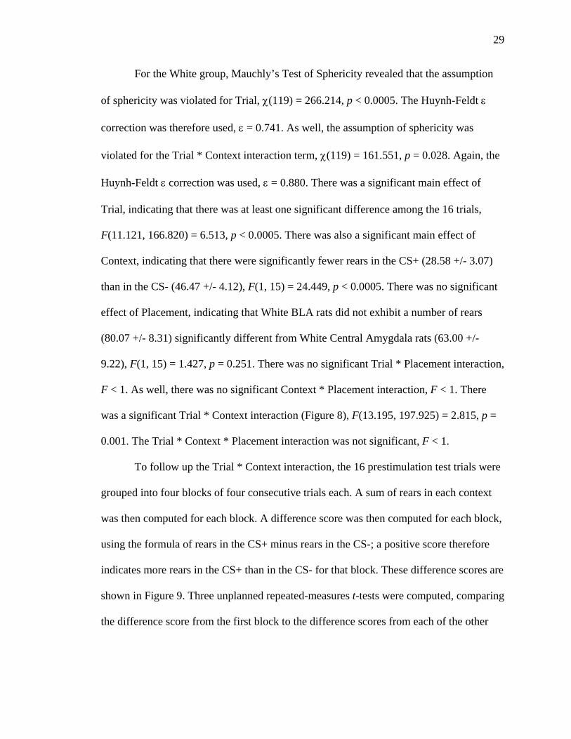
For the White group, Mauchly’s Test of Sphericity revealed that the assumption
of sphericity was violated for Trial, χ(119) = 266.214, p < 0.0005. The Huynh-Feldt ε
correction was therefore used, ε = 0.741. As well, the assumption of sphericity was
violated for the Trial * Context interaction term, χ(119) = 161.551, p = 0.028. Again, the
Huynh-Feldt ε correction was used, ε = 0.880. There was a significant main effect of
Trial, indicating that there was at least one significant difference among the 16 trials,
F(11.121, 166.820) = 6.513, p < 0.0005. There was also a significant main effect of
Context, indicating that there were significantly fewer rears in the CS+ (28.58 +/- 3.07)
than in the CS- (46.47 +/- 4.12), F(1, 15) = 24.449, p < 0.0005. There was no significant
effect of Placement, indicating that White BLA rats did not exhibit a number of rears
(80.07 +/- 8.31) significantly different from White Central Amygdala rats (63.00 +/-
9.22), F(1, 15) = 1.427, p = 0.251. There was no significant Trial * Placement interaction,
F < 1. As well, there was no significant Context * Placement interaction, F < 1. There
was a significant Trial * Context interaction (Figure 8), F(13.195, 197.925) = 2.815, p =
0.001. The Trial * Context * Placement interaction was not significant, F < 1.
To follow up the Trial * Context interaction, the 16 prestimulation test trials were
grouped into four blocks of four consecutive trials each. A sum of rears in each context
was then computed for each block. A difference score was then computed for each block,
using the formula of rears in the CS+ minus rears in the CS-; a positive score therefore
indicates more rears in the CS+ than in the CS- for that block. These difference scores are
shown in Figure 9. Three unplanned repeated-measures t-tests were computed, comparing
the difference score from the first block to the difference scores from each of the other

30
Figure 8. Rears by White rats in each context during the 16 prestimulation test trials. All
rats were tested in each context, in counterbalanced order. Data are averaged across both
levels of Placement.
Trial
1 2 3 4 5 6 7 8 9 10 11 12 13 14 15 16
Rea
rs
0
1
2
3
4
5
6
CS+CS-

31
Figure 9. Rearing difference scores from the White group, from each of the four blocks
of prestimulation test trials. Data are averaged across both levels of Placement and CS+.
Each of the latter three blocks was compared with the first block. No significant
differences were found at α = 0.0167. However, the difference of the fourth block from
the first block approached statistical significance, p = 0.025 (uncorrected).
Block
1 2 3 4
Rea
ring
Diff
eren
ce S
core
-12
-10
-8
-6
-4
-2
0
2
32
three blocks. A significant difference wherein the latter block’s difference score was
lower than the first block’s difference score therefore indicates a differential decrease in
rearing in the CS+ relative to the CS-. A Bonferroni correction requiring α/3 = 0.05/3 =
0.0167 was used. All p values are reported uncorrected, and are two-tailed. There was no
significant difference between the difference scores from the first block (-3.95 +/- 1.09)
and the second block (-1.29 +/- 1.34), t(16) = -1.737, p = 0.102. There was also no
significant difference between the first block and the third block (-4.18 +/- 0.94), t(16) =
0.159, p = 0.875. However, the difference between the first and fourth blocks approached
statistical significance; rats showed a higher ratio of rears in the CS- relative to the CS+
in the fourth block than in the first block, t(16) = 2.465, p = 0.025.
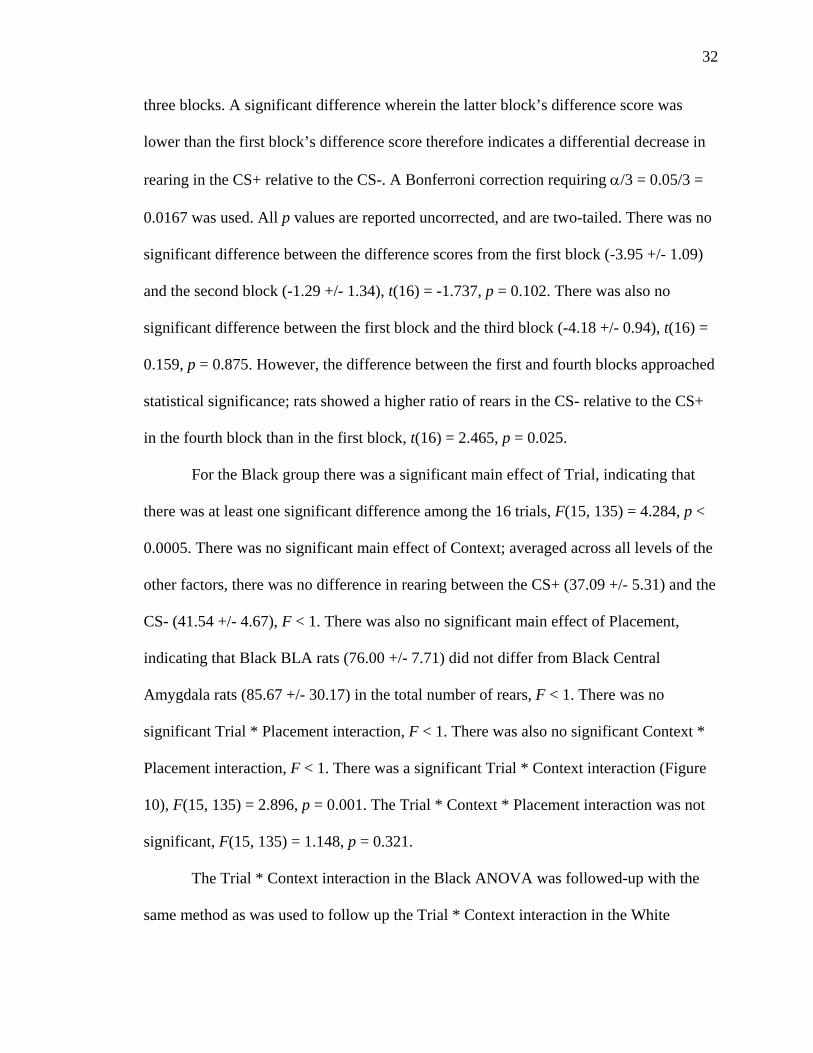
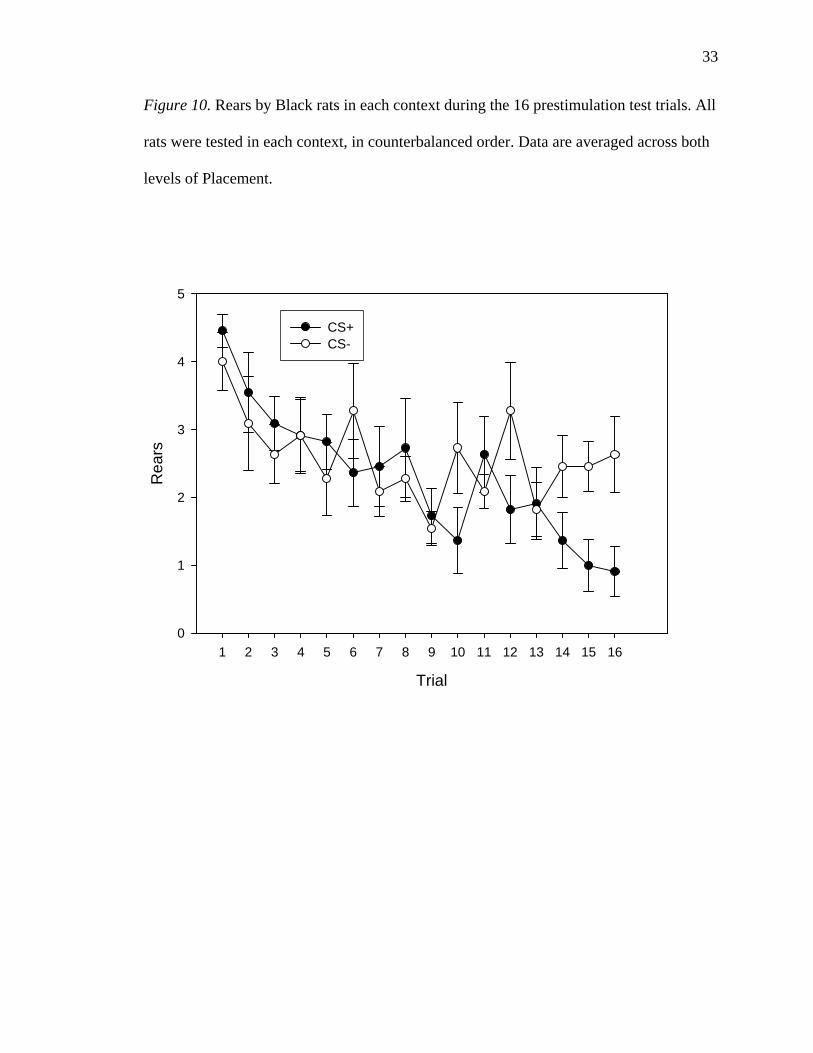
For the Black group there was a significant main effect of Trial, indicating that
there was at least one significant difference among the 16 trials, F(15, 135) = 4.284, p <
0.0005. There was no significant main effect of Context; averaged across all levels of the
other factors, there was no difference in rearing between the CS+ (37.09 +/- 5.31) and the
CS- (41.54 +/- 4.67), F < 1. There was also no significant main effect of Placement,
indicating that Black BLA rats (76.00 +/- 7.71) did not differ from Black Central
Amygdala rats (85.67 +/- 30.17) in the total number of rears, F < 1. There was no
significant Trial * Placement interaction, F < 1. There was also no significant Context *
Placement interaction, F < 1. There was a significant Trial * Context interaction (Figure
10), F(15, 135) = 2.896, p = 0.001. The Trial * Context * Placement interaction was not
significant, F(15, 135) = 1.148, p = 0.321.
The Trial * Context interaction in the Black ANOVA was followed-up with the
same method as was used to follow up the Trial * Context interaction in the White
33
Figure 10. Rears by Black rats in each context during the 16 prestimulation test trials. All
rats were tested in each context, in counterbalanced order. Data are averaged across both
levels of Placement.
Trial
1 2 3 4 5 6 7 8 9 10 11 12 13 14 15 16
Rea
rs
0
1
2
3
4
5
CS+CS-
34
ANOVA, by using difference scores in four blocks. Figure 11 illustrates these difference
scores. There was no significant difference between the difference scores from the first
block (1.36 +/- 0.80) and the second block (0.45 +/- 1.10), t(10) = 0.752, p = 0.470. The
difference between the difference scores from the third block (-2.09 +/- 1.76) and the first
block was not significant, p = 0.075. The difference score from the fourth block (-4.18 +/-
1.72), however, was significantly lower than that from the first block; Black rats showed
a significantly greater ratio of rears in the CS- relative to the CS+ in the fourth block than
in the first block, t(10) = 3.170, p = 0.010.
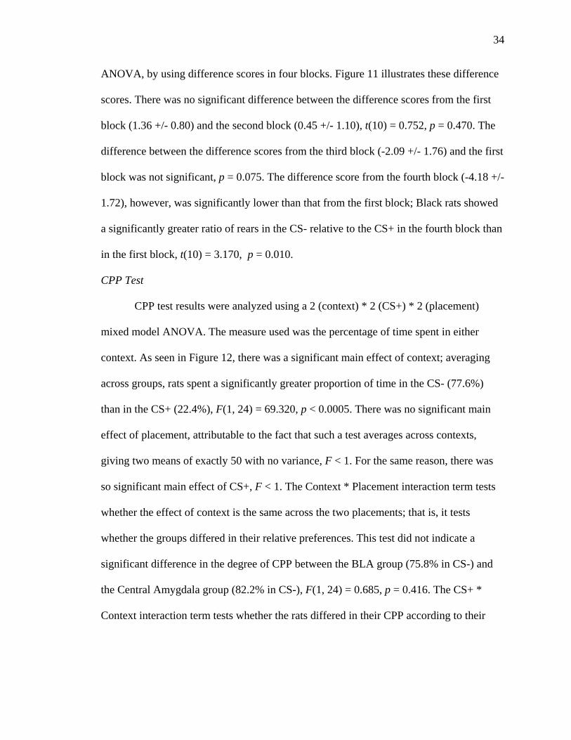
CPP Test
CPP test results were analyzed using a 2 (context) * 2 (CS+) * 2 (placement)
mixed model ANOVA. The measure used was the percentage of time spent in either
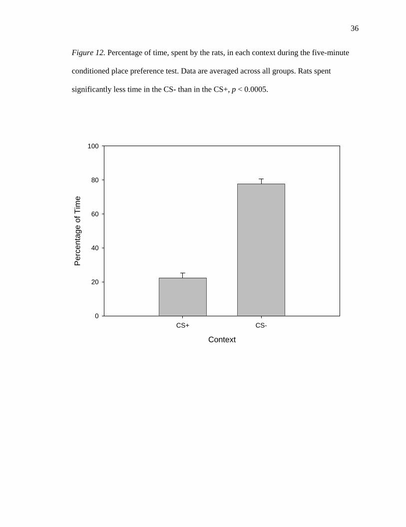
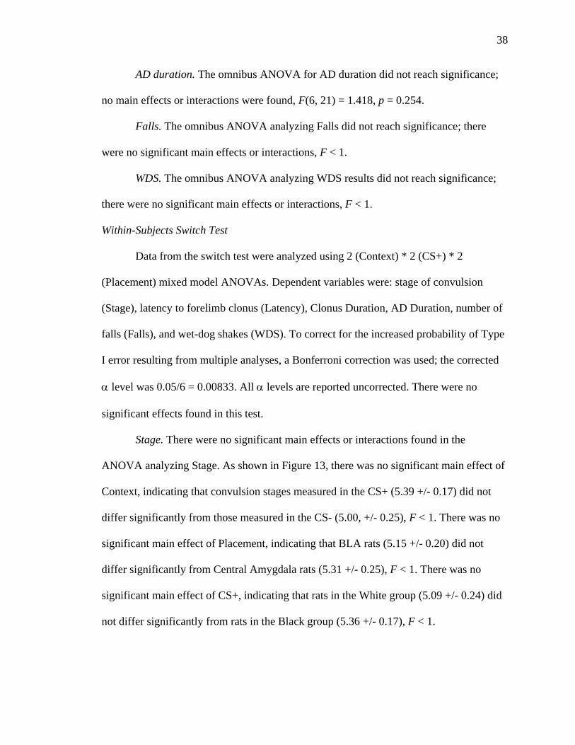
context. As seen in Figure 12, there was a significant main effect of context; averaging
across groups, rats spent a significantly greater proportion of time in the CS- (77.6%)
than in the CS+ (22.4%), F(1, 24) = 69.320, p < 0.0005. There was no significant main
effect of placement, attributable to the fact that such a test averages across contexts,
giving two means of exactly 50 with no variance, F < 1. For the same reason, there was
so significant main effect of CS+, F < 1. The Context * Placement interaction term tests
whether the effect of context is the same across the two placements; that is, it tests
whether the groups differed in their relative preferences. This test did not indicate a
significant difference in the degree of CPP between the BLA group (75.8% in CS-) and
the Central Amygdala group (82.2% in CS-), F(1, 24) = 0.685, p = 0.416. The CS+ *
Context interaction term tests whether the rats differed in their CPP according to their

35
Figure 11. Rearing difference scores from the Black group, from each of the four blocks
of prestimulation test trials. Data are averaged across both levels of Placement and CS+.
Asterisk denotes a significant difference when compared to the difference score from
Block 1.
Block
1 2 3 4
Rea
ring
Diff
eren
ce S
core
-8
-6
-4
-2
0
2
4
*
36
Figure 12. Percentage of time, spent by the rats, in each context during the five-minute
conditioned place preference test. Data are averaged across all groups. Rats spent
significantly less time in the CS- than in the CS+, p < 0.0005.
Context
CS+ CS-
Per
cent
age
of T
ime
0
20
40
60
80
100
37
assignment to CS+ condition. There was no significant CS+ * Context interaction;
assignment to the CS+ conditions did not affect CPP, F < 1. The CS+ * Placement
interaction term is meaningless, because it averages across the two contexts, which sum
to exactly 100% for each animal. This interaction was therefore not significant, F < 1.
There was no significant Context * CS+ * Placement interaction, F < 1.
Between-Subjects Switch Test
Data from the switch test were analyzed using 2 (Context) * 2 (CS+) * 2
(Placement) univariate ANOVAs. Dependent variables were: stage of convulsion (Stage),
latency to forelimb clonus (Latency), Clonus Duration, AD Duration, number of falls
(Falls), and wet-dog shakes (WDS). To correct for the increased probability of Type I
error resulting from multiple analyses, a Bonferroni correction was used; the corrected α
level was 0.05/6 = 0.00833. All α levels are reported uncorrected. There were no
significant effects found in this test.
Stage. The omnibus ANOVA analyzing convulsion stage results did not reach
significance; there were no significant main effects or interactions, F(6,21) = 1.512, p =
0.223.
Latency. One rat was excluded from the analysis of Latency data because it did
not display a Stage 3 or greater convulsion in the trial of interest. The omnibus ANOVA
using Latency data did not reach significance; there were no significant main effects or
interactions, F(6, 20) = 1.961, p = 0.120.
Clonus duration. The omnibus ANOVA analyzing Clonus Duration results did
not reach significance; no main effects or interactions were found, F < 1.
38
AD duration. The omnibus ANOVA for AD duration did not reach significance;
no main effects or interactions were found, F(6, 21) = 1.418, p = 0.254.
Falls. The omnibus ANOVA analyzing Falls did not reach significance; there
were no significant main effects or interactions, F < 1.
WDS. The omnibus ANOVA analyzing WDS results did not reach significance;
there were no significant main effects or interactions, F < 1.
Within-Subjects Switch Test
Data from the switch test were analyzed using 2 (Context) * 2 (CS+) * 2
(Placement) mixed model ANOVAs. Dependent variables were: stage of convulsion
(Stage), latency to forelimb clonus (Latency), Clonus Duration, AD Duration, number of
falls (Falls), and wet-dog shakes (WDS). To correct for the increased probability of Type
I error resulting from multiple analyses, a Bonferroni correction was used; the corrected
α level was 0.05/6 = 0.00833. All α levels are reported uncorrected. There were no
significant effects found in this test.
Stage. There were no significant main effects or interactions found in the
ANOVA analyzing Stage. As shown in Figure 13, there was no significant main effect of
Context, indicating that convulsion stages measured in the CS+ (5.39 +/- 0.17) did not
differ significantly from those measured in the CS- (5.00, +/- 0.25), F < 1. There was no
significant main effect of Placement, indicating that BLA rats (5.15 +/- 0.20) did not
differ significantly from Central Amygdala rats (5.31 +/- 0.25), F < 1. There was no
significant main effect of CS+, indicating that rats in the White group (5.09 +/- 0.24) did
not differ significantly from rats in the Black group (5.36 +/- 0.17), F < 1.

39
Figure 13. Convulsion stage, as observed in each context in the Within-Subjects Switch
Test. All rats were tested in each context, in counterbalanced order. Data are averaged
across groups. The difference between stage observed in the CS+ and in the CS- was not
significant, p = 0.727 (uncorrected).
Context
CS+ CS-
Con
vuls
ion
Stag
e
0
1
2
3
4
5
6
7
8

40
There was no significant Placement * CS+ interaction, F < 1. There was no
significant Context * Placement interaction, F(1, 24) = 1.177, p = 0.289. There was no
significant Context * CS+ interaction, F(1, 24) = 3.205, p = 0.086. There was no
significant Context * Placement * CS+ interaction, F < 1.
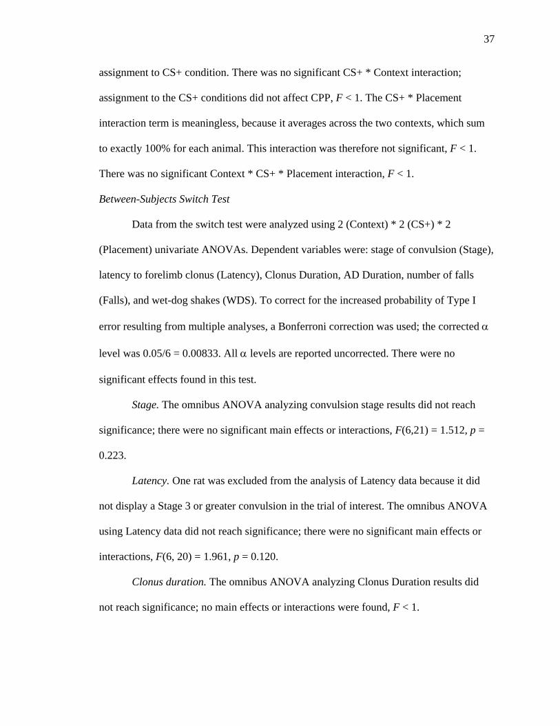
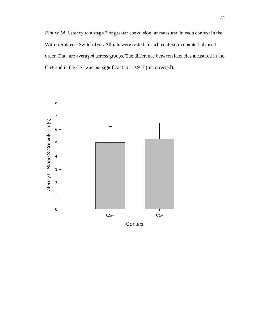
Latency. One rat was excluded from the analysis of Latency data because it did
not display a Stage 3 or greater convulsion in one of its Switch Test trials; latency could
therefore not be measured in that trial. The ANOVA analyzing Latency data did not yield
any significant main effects or interactions. As shown in Figure 14, there was no
significant main effect of Context; rats showed similar latencies in the CS+ (5.04 +/- 1.19
s) and in the CS- (5.26 +/- 1.25 s), F < 1.
There was no significant Placement * CS+ interaction, F < 1. There was no
significant Context * Placement interaction, F < 1. There was no significant Context *
CS+ interaction, F < 1. There was no significant Context * Placement * CS+ interaction,
F < 1.
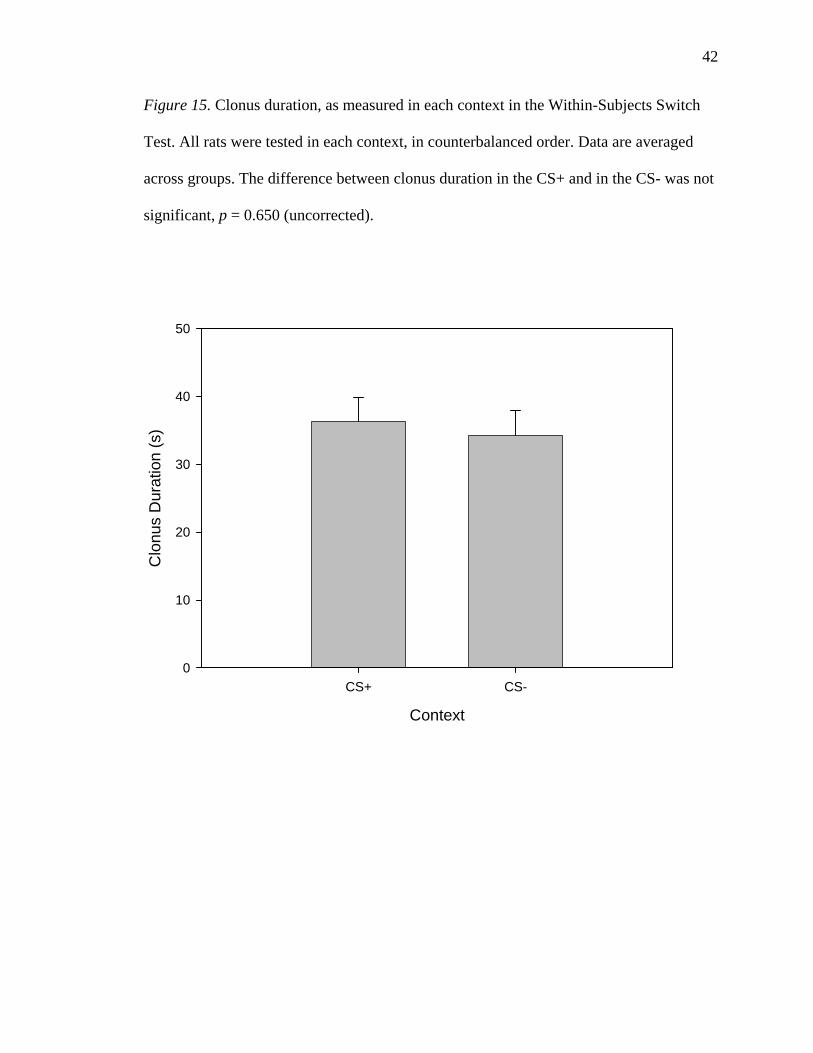
Clonus duration. No significant main effects or interactions were found in the
ANOVA analyzing Clonus Duration results. As shown in Figure 15, there was no
significant main effect of Context, indicating that clonus durations were similar in the
CS+ (36.25 +/- 3.67 s) and the CS- (34.18 +/- 3.70 s), F < 1. There was no significant
main effect of Placement, indicating that rats with BLA electrodes had durations of
clonus (34.88 +/- 2.96 s) similar to those of rats with Central Amygdala electrodes (36.06
+/- 4.22 s), F< 1. There was no significant main effect of CS+, indicating that rats in the
White group (33.26 +/-3.42 s) did not differ significantly from those in the Black group
(38.23 +/- 3.01 s), F(1, 24) = 1.048, p = 0.316.
41
Figure 14. Latency to a stage 3 or greater convulsion, as measured in each context in the
Within-Subjects Switch Test. All rats were tested in each context, in counterbalanced
order. Data are averaged across groups. The difference between latencies measured in the
CS+ and in the CS- was not significant, p = 0.917 (uncorrected).
Context
CS+ CS-
Late
ncy
to S
tage
3 C
onvu
lsio
n (s
)
0
1
2
3
4
5
6
7
8
42
Figure 15. Clonus duration, as measured in each context in the Within-Subjects Switch
Test. All rats were tested in each context, in counterbalanced order. Data are averaged
across groups. The difference between clonus duration in the CS+ and in the CS- was not
significant, p = 0.650 (uncorrected).
Context
CS+ CS-
Clo
nus
Dur
atio
n (s
)
0
10
20
30
40
50
43
There was no significant Placement * CS+ interaction, F < 1. There was no
significant Context * Placement interaction, F(1, 24) = 1.699, p = 0.205. There was no
significant Context * CS+ interaction, F(1, 24) = 1.639, p = 0.213. There was no
significant Context * Placement * CS+ interaction, F < 1.
AD duration. The ANOVA analyzing AD Duration results yielded no significant
main effects or interactions. As shown in Figure 16, there was no significant effect of
Context, indicating that rats had similarly long seizures in the CS+ (72.25 +/- 6.30 s) and
the CS- (68.07 +/- 6.82 s), F < 1. There was no significant main effect of Placement,
indicating that rats with electrodes in the BLA had long seizures (66.58 +/- 6.20 s) similar
to those of rats with electrodes in the central nucleus of the amygdala (79.12 +/- 11.69 s),
F < 1. There was no significant main effect of CS+, indicating that rats in the White
group had long seizures (66.26 +/- 7.55 s) similar to those of rats in the Black group
(76.18 +/- 7.97 s), F < 1.
There was no significant Placement * CS+ interaction, F(1, 24) = 1.628, p =
0.214. There was no significant Context * Placement interaction; F < 1. There was no
significant Context * CS+ interaction, F < 1. There was no significant Context *
Placement * CS+ interaction, F < 1.
Falls. The ANOVA analyzing Falls results yielded no significant main effects or
interactions. As shown in Figure 17, there was no significant main effect of Context,
indicating that rats had similar numbers of falls in the CS+ (2.68 +/- 0.44) as in the CS-
(1.93 +/- 0.36), F < 1. There was no significant main effect of Placement, indicating that
BLA rats (2.28 +/- 0.42) did not differ from Central Amygdala rats (2.38 +/- 0.64), F < 1.

44
Figure 16. AD duration as measured in each context in the Within-Subjects Switch Test.
All rats were tested in each context, in counterbalanced order. Data are averaged across
groups. The difference between AD durations measured in each context was not
significant, p = 0.800 (uncorrected).
Context
CS+ CS-
AD
Dur
atio
n (s
)
0
20
40
60
80
100

45
Figure 17. Falls by rats, as observed in each context in the Within-Subjects Switch Test.
All rats were tested in each context, in counterbalanced order. Data are averaged across
groups. The difference between falls observed in the CS+ and in the CS- was not
significant, p = 0.419 (uncorrected).
Context
CS+ CS-
Falls
0.0
0.5
1.0
1.5
2.0
2.5
3.0
3.5

46
There was no significant main effect of CS+, indicating that rats in the White group (2.24
+/- 0.43) did not differ from rats in the Black group (2.41+/- 0.59), F < 1.
There was no significant Placement * CS+ interaction, F < 1. There was no
significant Context * Placement interaction, F(1, 24) = 1.577, p = 0.221. There was no
significant Context * CS+ interaction, F(1, 24) = 1.861, p = 0.185. There was no
significant Context * Placement * CS+ interaction, F < 1.
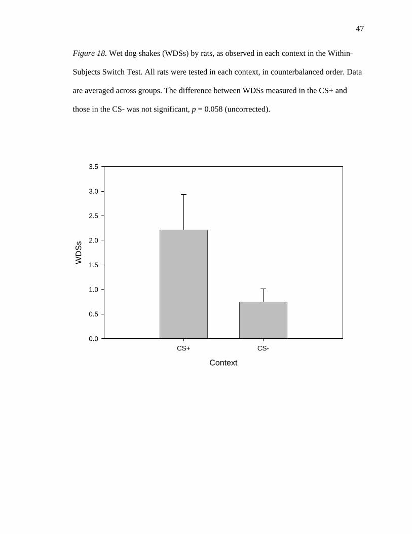
WDS. In the ANOVA analyzing WDS data, here were no significant main effects
or interactions. As shown in Figure 18, there was no significant main effect of Context,
indicating that the number of WDSs measured in the CS+ (2.21 +/- 0.72) did not differ
significantly from that measured in the CS- (0.75 +/- 0.26), F(1, 24) = 3.959, p = 0.058.
There was no significant main effect of Placement, indicating that BLA rats (1.48 +/-
0.43) did not differ significantly from Central Amygdala rats (1.50 +/- 0.95), F < 1. There
was no significant main effect of CS+, indicating that rats in the White group (1.47 +/-
0.52) did not differ significantly from rats in the Black group (1.50 +/- 0.66), F < 1.
There was no significant Placement * CS+ interaction, F(1, 24) = 1.89, p = 0.181.
There was no significant Context * Placement interaction, F < 1. There was no significant
Context * CS+ interaction, F < 1. There was no significant Context * Placement * CS+
interaction, F(1, 24) = 1.435, p = 0.243.
ADT Switch Test
Using the ADT measured in each context as the dependent variable, a 2 (Context)
* 2 (CS+) * 2 (Placement) mixed model ANOVA was computed. One rat, belonging to
the BLA and White groups, was excluded from this analysis because the EEG recorded
from its electrodes was too noisy for accurate identification of AD. As shown in Figure
47
Figure 18. Wet dog shakes (WDSs) by rats, as observed in each context in the Within-
Subjects Switch Test. All rats were tested in each context, in counterbalanced order. Data
are averaged across groups. The difference between WDSs measured in the CS+ and
those in the CS- was not significant, p = 0.058 (uncorrected).
Context
CS+ CS-
WD
Ss
0.0
0.5
1.0
1.5
2.0
2.5
3.0
3.5
48
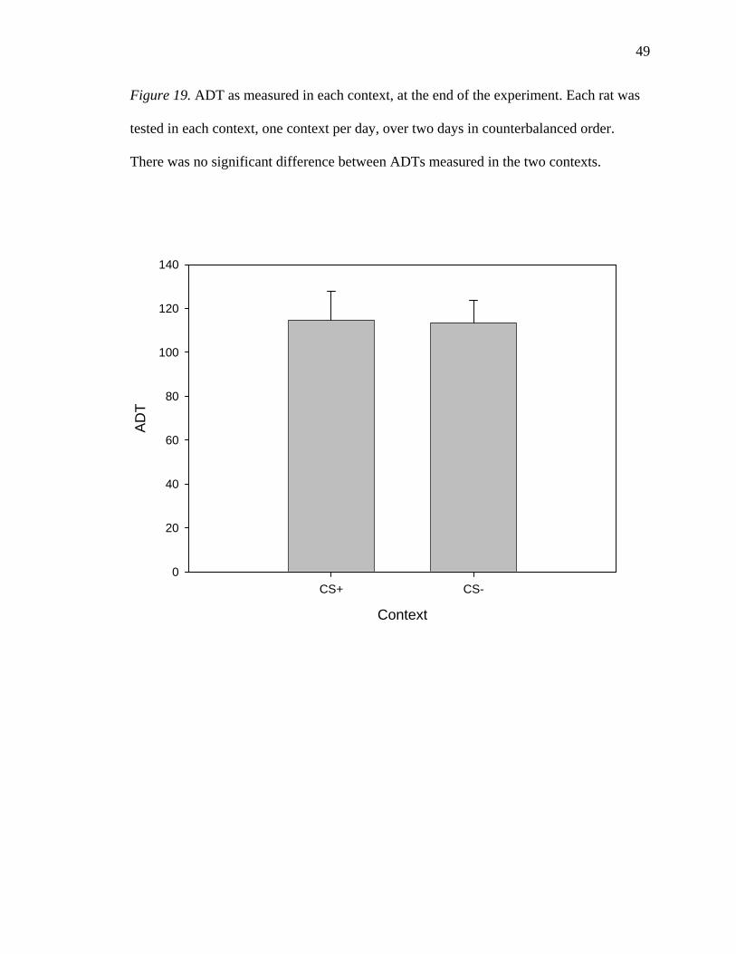
19, there was no significant main effect of Context; rats displayed similar ADTs in the
CS+ (114.44 +/- 13.41 µA) and the CS- (113.33 +/- 10.24 µA), F < 1. There was no
significant main effect of CS+, indicating that rats assigned to the White group had ADTs
(109.69 +/- 12.60 µA) similar to those of the Black group (120.00 +/- 19.85 µA). As seen
in Figure 20, there was no significant main effect of placement, indicating that rats with
electrodes in the central nucleus of the amygdala did not have significantly different
ADTs (85.62 +/- 6.44 µA) from those of rats with electrodes in the BLA (125.79 +/-
14.38 µA), F (1, 23) = 2.655, p = 0.117.
There was no significant Context * CS+ interaction; the effect of Context did not
vary according to assignment to CS+, F < 1. There was no significant Context *
Placement interaction, indicating that the effect of Context did not vary according to
electrode placement, F < 1. There was no significant CS+ * Placement interaction,
indicating that the effect of electrode placements did not vary according to assignment to
CS+, F < 1. There was no significant Context * CS+ * Placement interaction, F < 1.
Discussion
In the present experiment, seizures were kindled in rats by electrical stimulation
of the amygdala in one context (CS+), and the rats were connected to the equipment but
not stimulated in another context (CS-). Electrode tips were located in either the
basolateral amygdala (BLA) or the central nucleus of the amygdala (Central Amygdala),
and there were no differences in kindling rate, conditioned effects, or afterdischarge
threshold between those two groups. The rats developed conditioned anticipatory
defensive responses in the CS+; in that environment, they froze more, displayed reduced
locomotor activity, and reared less, relative to the CS-. These differences were enhanced
49
Figure 19. ADT as measured in each context, at the end of the experiment. Each rat was
tested in each context, one context per day, over two days in counterbalanced order.
There was no significant difference between ADTs measured in the two contexts.
Context
CS+ CS-
AD
T
0
20
40
60
80
100
120
140

50
Figure 20. Mean ADT, as measured in both contexts, for BLA and Central Amygdala
rats. The difference between the two groups was not significant, p = 0.117, uncorrected.
Electrode Placement
BLA Central Amygdala
Mea
n A
DT
0
20
40
60
80
100
120
140
160

51
by the White context, in that rats kindled in the White context showed the greatest
differences between the two contexts. There was no difference in the rate of kindling
between rats kindled in the White context and those kindled in the Black context. The rats
showed a conditioned place aversion for the CS+, indicating that amygdaloid kindling, or
some aspect thereof, is punishing. I failed, however, to find contextually conditioned
effects on convulsions, seizures, or afterdischarge threshold, using switch tests in which
rats were stimulated in each context. Because I found conditioned effects on interictal
behaviours but not on ictal measures, my results are therefore a partial replication of the
results of Barnes et al. (2001).
My results clearly indicate that the rats were able to form learned associations
between the contexts and the events that occurred therein, including kindling. Given the
results of Paredes et al. (2000), who used a place conditioning procedure to show that the
aftereffect of generalized amygdaloid seizures can be rewarding, this suggests that the
rats attended to the ictal events. That is, rats in the present experiment experienced both
kindled seizures and their aftereffects in the CS+. A conditioned aversive response
therefore seems to indicate that some aversive aspect of the ictal experience was more
salient than the rewarding aftereffect of the seizures.
Implicit in the argument above is the idea that some aspect of amygdaloid
kindling is aversive. But there are many aspects of kindling, and it is not immediately
clear which is aversive. Below I consider several possibilities.
It is possible, for example, that the 1-sec train of electrical stimulation used to
elicit a seizure is aversive. Using chemical stimulation of the BLA, Thielen and Shekhar
(2002) found conditioned place aversion to the stimulation-paired context, after only five
52
pairings of each type. Thus, stimulation of the BLA appears to be aversive, regardless of
how it is applied. Because I did not test for conditioned place aversion until 45 pairings
of each type had occurred, it is not known whether the rats would have shown a
conditioned place aversion after just five stimulations. It is therefore possible that an
aversion to the stimulation itself caused the conditioned place aversion. The conditioned
anticipatory defensive responses, however, did not become evident until many more than
five stimulations had occurred. The unconditioned stimulus for the conditioning of those
responses was likely not the stimulations themselves, but rather some aspect of the
resultant seizures. Further, as the anticipatory responses demonstrate that the seizures
have an aversive component that served as an unconditioned stimulus, it is unlikely that
the stimulation served uniquely as the unconditioned stimulus for the conditioned place
aversion. At a minimum, the seizures contributed to that role.
The forceful convulsions produced by generalized amygdaloid-kindled seizures
did not likely serve as an aversive US. Barnes et al. (2003) failed to demonstrate
conditioned place aversion or preference for a context paired with anterior-neocortex-
kindled convulsions, nor did they find conditioned anticipatory defensive responses. Such
convulsions are also forceful, but topologically different. A forceful convulsion, per se,
does not therefore seem to be sufficiently aversive to produce the conditioned effects I
observed.
Amygdaloid-kindled seizures could therefore be the relevant US. However,
conditioned anticipatory responses did not appear until rats were having generalized
seizures. If amygdaloid-kindled seizures, per se, were serving as the aversive US, those
53
effects should have appeared much earlier in the experiment. A plausible hypothesis is
thus that generalized amygdaloid-kindled seizures are the relevant aversive US.
However, generalized seizures produced by kindling of other limbic sites may be
equivalent for that purpose. Barnes et al. (2005) kindled perirhinal cortex, which kindles
at a rate similar to the amygdala, and found conditioned anticipatory defensive responses,
and a conditioned place aversion. Ventral hippocampal kindling, which progresses much
more slowly, failed to produce conditioned anticipatory defensive responses until rats had
experienced many more seizures than were necessary with perirhinal kindling. Dorsal
hippocampal kindling, which progresses even more slowly, failed to produce those
conditioned effects.
It is also possible that generalized seizures from non-limbic sites could serve as an
aversive US. Though Barnes et al. (2003) failed to find conditioned defensiveness or
place aversion when they kindled the anterior neocortex, they did not continue kindling
for a sufficient time for all of the rats to develop the second phase of neocortical kindling,
in which generalized seizures occur (Burnham, 1978; Seidel & Corcoran, 1986). Thus, it
remains to be shown whether generalized seizures produced by anterior neocortical
kindling can produce conditioned defensiveness in the present experimental paradigm.
If amygdaloid stimulations served as the US that produced conditioned place
aversion, and generalized seizures served as the US that produced conditioned
anticipatory responses, one would expect to find anticipatory responses, and not place
aversion, if many generalized anterior neocortically kindled seizures were evoked. After
receiving many stimulations to the anterior neocortex, rats did not show a conditioned
place aversion (Barnes et al., 2003). Anterior neocortical stimulation therefore does not

54
seem to be aversive in the same way that amygdaloid stimulation can be (Thielen &
Shekhar, 2002). However, it seems unlikely that each conditioned effect is produced by a
different US, because the conditioned anticipatory responses and the conditioned place
aversion are all defensive responses conditioned to the same environment.
Generalized seizures are likely the relevant aversive US for the conditioning of
anticipatory defensive responses. It is possible that amygdaloid stimulation itself is a
sufficiently aversive US to produce conditioned place aversion. At a minimum, however,
it seems likely that the generalized amygdaloid-kindled seizures that the rats experienced
also contributed to that role.
I suggest that the results contradict the interpretation that a generalized limbic-
kindled seizure results in a loss of consciousness. One possibility is that such a seizure
has a component wherein consciousness is intact that is sufficient to permit learning to
occur. Another possibility is that such a seizure alters consciousness to a sufficiently mild
degree that learning can occur. A third possibility is that a loss or alteration of
consciousness, in and of itself, is sufficient as an aversive US.
The first possibility is unlikely. A period of intact consciousness would likely
come very early in the seizure, when the rat is experiencing less than a stage 5
convulsion. If that early part of the seizure were a sufficient US, conditioned effects
should have been evident much earlier than they were, and perhaps been reduced as the
latency to a generalized convulsion decreased.
Most patients with temporal lobe epilepsy are at least partially aware of when
they have had a seizure (Inoue & Mihara, 1998; Kerling et al., 2006). Inoue and Mihara
(1998) found that none of the 88 patients with temporal lobe epilepsy they tested recalled

55
stimuli presented during partial seizures. However, the rats in my experiments did not
experience a seizure until they had been in the CS+ for 30 sec; that contingency alone
could account for conditioned effects, provided that the animals experienced some
aversive aspect of having a seizure. They would thus not need to attend to the
environment during a seizure, but would simply need to learn that the environment was
predictive of a seizure.
It remains possible that a loss of consciousness is the aversive US. However,
measurement of a rat’s state of consciousness is not at all direct. Among kindling
researchers, falling, or a loss of equilibrium, during a seizure is often taken as a sign of an
impairment or loss of consciousness. It is rather difficult to test whether the concept is
valid. I cannot therefore conclude whether a loss or impairment of consciousness per se
served as the relevant aversive US.
I did not find any differences between rats with electrode tips in the BLA and
those with electrode tips in the Central Nucleus of the amygdala. This could be because
these two highly interconnected nuclei (Sah et al., 2003) cannot be functionally
discriminated in the present paradigm. Indeed, both nuclei are involved in defensive
behaviours, even if those behaviours are different (Killcross et al., 1997). Another
possibility is that the stimulation activated approximately the same tissue, regardless of
subtle differences in electrode placements. Though some electrode tips were found in the
BLA and some in the Central Nucleus of the amygdala, the electrode tips were generally
quite closely distributed. That is, BLA placements tended to be in the medial extent of the
BLA, and Central Nucleus placements tended to be in the lateral extent of that nucleus.
Given the size of current density distribution likely produced by the intensity of high
56
stimulation (e.g., C. C. McIntyre & Grill, 2001), stimulation via the electrode placements
in the two groups may have activated amygdaloid nuclei similarly. Thus, the results
should not necessarily be taken to indicate functional similarity between the BLA and
Central Nucleus of the amygdala.
Rats kindled in the White context showed a greater contextual difference in
defensiveness than did rats kindled in the Black context. It seems that the White context
itself was anxiogenic. Indeed, bright light is understood to be anxiogenic for rats. It can
enhance fear-potentiated startle, and that effect has been shown to be dependent on the
BLA (Walker & Davis, 1997). An anxiogenic effect of the White context could therefore
have partially mitigated contextually conditioned defensiveness in the Black group, and
enhanced it in the White group.
In contrast to the results of Freeman and Mikulka (1986), I did not find a
difference in kindling rate between rats kindled in the White context and those kindled in
the Black context. However, their contexts differed more than mine with respect to
brightness, and I might have found such a contextual difference in kindling rate if my
contexts were more similar to theirs. In any case, my failure to replicate their effect
suggests that it is not easily produced.
Perhaps the most significant observation in my experiments is my failure to find
any conditioned effects on ictal measures, in contrast to others (e.g., Barnes et al., 2001).
One explanation is that subtle procedural differences mitigated the conditioning of the
effects. Though my procedure was designed to closely replicate that of Barnes et al.
(2001), there are some differences worth exploring.

57
My parameters of stimulation were different. Whereas Barnes et al. (2001) used
1-sec trains of 60 Hz sine wave stimulation at 400 µA RMS, I used a 1 sec train of 60
pp/s balanced biphasic square wave pulses, at 400 µA base-to-peak. This difference
should not be important, in my opinion. Both stimulation designs elicit AD, and it is well
established that kindling can occur using a variety of stimulation types as long as AD is
elicited. As well, both stimulation designs used intensities substantially above typical
ADTs for the amygdala.
On CS- trials, Barnes et al. (2001) triggered the switch on the stimulator, whereas
I did not do so. Because they did not record EEG, Barnes et al. were able to disconnect
the lead from their equipment for CS- trials, thus permitting them to trigger the
stimulation switch. In contrast, I recorded EEG on all trials, necessitating an intact
connection between the lead and the equipment. As triggering my, and likely their,
stimulation switch makes a slightly audible sound, it is possible that the sound served as a
conditioned stimulus predicting a seizure. In the switch tests, that sound was produced as
seizures were elicited in each context. If the sound, as opposed to the context, served as
the seizure-predicting CS, each rat’s contextual conditioning history would have been
irrelevant. However, my rats did apparently learn the predictive nature of the two
contexts, because they developed contextually conditioned defensiveness. The slight
clicking sound produced by triggering the switch may have served as an adjunct CS, but
likely did not serve as the main seizure-predictive CS.
Whereas I kept the stimulation/recording lead attached to each rat throughout all
trials, Barnes et al. (2001) removed the lead immediately after the (sham) stimulation was
delivered. This occurred on both CS+ and CS- trials during conditioning. A contextually
58
differential effect of removing the lead during the switch test would therefore be
contingent upon differential sensitivity to some seizure-modulating effect of removing
the lead. That is, removing the lead would have to have an anti-seizure effect to which the
rats gained contextually conditioned tolerance. In humans, startling auditory stimuli or
shaking by an experimenter near the onset of a seizure may have an anti-seizure effect
(Zlutnick et al., 1975). It therefore seems possible that removing the lead could have such
an effect on rats during kindling. If that effect is produced because of the surprising or
disruptive nature of lead removal, rats may have become habituated to it in the CS+ after
experiencing it many times. As such an anti-seizure effect would not have occurred in the
CS- prior to the switch test, contextually conditioned habituation may have occurred, and
the anti-seizure effect of removing the lead after stimulation would remain intact in the
CS-.
The place-conditioning apparatus I used was slightly different from that of Barnes
et al. (2001). It is unlikely that the lack of a central chamber would be relevant for
anything other than the precision of the place preference test. Of note, though, is that my
apparatus had bedding only in the White context. This likely increased the distinctiveness
of the contexts, perhaps adding olfactory cues. It should therefore have strengthened any
contextually conditioned effects. Ultimately, the rats in my experiments developed
contextually conditioned defensiveness; they were able to associate the contexts with the
events that occurred therein.
Contrary to the conclusions of Barnes et al. (2001), I suggest that conditioning is
not fundamental to kindling. At a minimum, the conditioned effects on ictal measures
found by Barnes et al. seem to be fragile. If my failure to replicate those effects can be
59
explained by procedural differences, the effects are not sufficiently robust to be present in
all kindling experiments or to have power as a potential mechanism of kindling.
In measuring AD and ADT, I explicitly tested the hypothesis that the conditioning
of convulsions found by Barnes et al. (e.g., 2001) was due to conditioned effects on AD
and/or ADT. I did not find any conditioned effects on AD or ADT. Thus, I did not find
support for that hypothesis. However, given that no conditioned effects on convulsions
were found in the present experiment, my rejection of that hypothesis is limited. Ideally
one would, while measuring EEG, replicate conditioned effects on convulsions, and test
for conditioned effects on AD and ADT. It remains ultimately unknown whether the
latter would be found if the former were found.
There have been attempts to modify seizures in humans using conditioning
techniques, which have been met with limited success and acceptance by the neurological
community. The present failure to condition ictal responses certainly does not support
such a use of conditioning techniques. Rather, interictal features of partial complex
epilepsy with temporal lobe foci may be produced and modified by conditioning. That is,
interictal emotionality, which may involve unusual affective value being assigned to
various stimuli (Bear & Fedio, 1977), may be produced by learned associations. Such
emotionality could then be modified by conditioning techniques such as extinction.
In summary, using amygdaloid kindling, or some aspect thereof, as an
unconditioned stimulus, I found evidence of conditioned interictal defensiveness, but not
conditioned effects on ictal measures. The generalized seizures elicited by kindling of the
amygdala were likely the relevant US in producing conditioned interictal defensiveness.
My failure to replicate the conditioned effects on ictal measures found by Barnes et al.

60
(2001) indicates that the effects are not as robust as previously thought. I conclude that
conditioning is not fundamental to kindling.
61
References
Adamec, R., & Shallow, T. (2000). Effects of baseline anxiety on response to kindling of
the right medial amygdala. Physiol Behav, 70(1-2), 67-80.
Adamec, R. E., & Morgan, H. D. (1994). The effect of kindling of different nuclei in the
left and right amygdala on anxiety in the rat. Physiol Behav, 55(1), 1-12.
Andrews, D. J., & Schonfeld, W. H. (1992). Predictive factors for controlling seizures
using a behavioural approach. Seizure, 1(2), 111-116.
Barnes, S. J., Hua, J. M., Pinel, J. P., Takahashi, A., & Wig, G. S. (2005). Conditioned
effects of kindling three different sites in the hippocampal complex of the rat.
Behav Neurosci, 119(6), 1572-1579.
Barnes, S. J., Magyar, O., Pinel, J. P., & Takahashi, A. (2004). Anticipating the attack:
Temporal conditioning during amygdala kindling in rats. Behav Neurosci, 118(1),
89-96.
Barnes, S. J., Pinel, J. P., Francis, L. H., & Wig, G. S. (2001). Conditioning of ictal and
interictal behaviors in rats by amygdala kindling: Context as the conditional
stimulus. Behav Neurosci, 115(5), 1065-1072.
Barnes, S. J., Pinel, J. P., Wee, E., Archambault, J., & Ailon, T. (2006). Effects of
expectation on amygdala-kindled convulsions and interictal behaviour in rats:
Confirmation of a pavlovian mechanism. Eur J Neurosci, 24(3), 829-840.
Barnes, S. J., Pinel, J. P., Wig, G. S., Stuettgen, M. C., & Holzel, C. H. (2003).
Stimulation site determines the conditioned effects of kindling in rats: Anterior
neocortex versus amygdala. Eur J Neurosci, 17(8), 1671-1679.

62
Bear, D. M., & Fedio, P. (1977). Quantitative analysis of interictal behavior in temporal
lobe epilepsy. Arch Neurol, 34(8), 454-467.
Blanchard, E. B., & Young, L. D. (1974). Clinical applications of biofeedback training. A
review of evidence. Arch Gen Psychiatry, 30(5), 573-589.
Cataldo, M. F., Russo, D. C., & Freeman, J. M. (1979). A behavior analysis approach to
high-rate myoclonic seizures. J Autism Dev Disord, 9(4), 413-427.
Corcoran, M. E., Lanius, R., & Duren, A. (1992). Reinforcing and punishing
consequences of kindling. Epilepsy Res, 13(3), 179-186.
Cott, A., Pavloski, R. P., & Black, A. H. (1979). Reducing epileptic seizures through
operant conditioning of central nervous system activity: Procedural variables.
Science, 203(4375), 73-75.
Egner, T., & Sterman, M. B. (2006). Neurofeedback treatment of epilepsy: From basic
rationale to practical application. Expert Rev Neurother, 6(2), 247-257.
Forster, F. M. (1969). Conditional reflexes and sensory-evoked epilepsy: The nature of
the therapeutic process. Cond Reflex, 4(2), 103-114.
Forster, F. M. (1972). The classification and conditioning treatment of the reflex
epilepsies. Int J Neurol, 9(1), 73-86.
Forster, F. M., Booker, H. E., & Gascon, G. (1967). Conditioning in musicogenic
epilepsy. Trans Am Neurol Assoc, 92, 236-237.
Forster, F. M., & Campos, G. B. (1964). Conditioning factors in stroboscopic-induced
seizures. Epilepsia, 59, 156-165.
Forster, F. M., Ptacek, L. J., & Peterson, W. G. (1965). Auditory clicks in extinction of
stroboscope-induced seizures. Epilepsia, 6(3), 217-225.
63
Forster, F. M., Ptacek, L. J., Peterson, W. G., Chun, R. W., Bengzon, A. R., & Campos,
G. B. (1964a). Stroboscopic-induced seizure discharges. Modification by
extinction techniques. Arch Neurol, 11, 603-608.
Forster, F. M., Ptacek, L. J., Peterson, W. G., Chun, R. W., & Campos, G. B. (1964b).
Stroboscopic induced seizures altered by extinction techniques. Trans Am Neurol
Assoc, 89, 136-139.
Freeman, F. G., & Mikulka, P. J. (1986). Differential conditioning of environmental cues
with amygdala kindling. Epilepsia, 27(3), 189-193.
Gilbert, T. H., Hannesson, D. K., & Corcoran, M. E. (2000). Hippocampal kindled
seizures impair spatial cognition in the morris water maze. Epilepsy Res, 38(2-3),
115-125.
Gilbert, T. H., McNamara, R. K., & Corcoran, M. E. (1996). Kindling of hippocampal
field ca1 impairs spatial learning and retention in the morris water maze. Behav
Brain Res, 82(1), 57-66.
Goddard, G. V. (1964). Functions of the amygdala. Psychol Bull, 62, 89-109.
Goddard, G. V. (1967). Development of epileptic seizures through brain stimulation at
low intensity. Nature, 214(92), 1020-1021.
Goddard, G. V., McIntyre, D. C., & Leech, C. K. (1969). A permanent change in brain
function resulting from daily electrical stimulation. Exp Neurol, 25(3), 295-330.
Goldstein, M. L. (1965). Effects of hippocampal, amygdala, hypothalamic and parietal
lesions on a classically conditioned fear response. Psychol Rep, 16, 211-219.
Hannesson, D. K., & Corcoran, M. E. (2000). The mnemonic effects of kindling.
Neurosci Biobehav Rev, 24(7), 725-751.

64
Hannesson, D. K., Howland, J., Pollock, M., Mohapel, P., Wallace, A. E., & Corcoran,
M. E. (2001). Dorsal hippocampal kindling produces a selective and enduring
disruption of hippocampally mediated behavior. J Neurosci, 21(12), 4443-4450.
Hannesson, D. K., Wallace, A. E., Pollock, M., Corley, S., Mohapel, P., & Corcoran, M.
E. (2004). The relation between extent of dorsal hippocampal kindling and
delayed-match-to-place performance in the morris water maze. Epilepsy Res,
58(2-3), 145-154.
Helmstaedter, C., Sonntag-Dillender, M., Hoppe, C., & Elger, C. E. (2004). Depressed
mood and memory impairment in temporal lobe epilepsy as a function of focus
lateralization and localization. Epilepsy Behav, 5(5), 696-701.
Huberty, C. J., & Morris, J. D. (1989). Multivariate analysis versus multiple univariate
analyses. PsycholBull, 105, 302-308.
Hunsperger, R. W. (1963). [affective behavior patterns elicited by electrical stimulation
of the brain stem and forebrain.]. J Physiol (Paris), 55, 45-98.
Inoue, Y., & Mihara, T. (1998). Awareness and responsiveness during partial seizures.
Epilepsia, 39 Suppl 5, 7-10.
Kalynchuk, L. E. (2000). Long-term amygdala kindling in rats as a model for the study of
interictal emotionality in temporal lobe epilepsy. Neurosci Biobehav Rev, 24(7),
691-704.
Kalynchuk, L. E., Pinel, J. P., & Treit, D. (1998a). Long-term kindling and interictal
emotionality in rats: Effect of stimulation site. Brain Res, 779(1-2), 149-157.
Kalynchuk, L. E., Pinel, J. P., & Treit, D. (1999). Characterization of the defensive nature
of kindling-induced emotionality. Behav Neurosci, 113(4), 766-775.
65
Kalynchuk, L. E., Pinel, J. P., Treit, D., Barnes, S. J., McEachern, J. C., & Kippin, T. E.
(1998b). Persistence of the interictal emotionality produced by long-term
amygdala kindling in rats. Neuroscience, 85(4), 1311-1319.
Kalynchuk, L. E., Pinel, J. P., Treit, D., & Kippin, T. E. (1997). Changes in emotional
behavior produced by long-term amygdala kindling in rats. Biol Psychiatry, 41(4),
438-451.
Lowenstein, D. H. (2005). Seizures and epilepsy. In D.L. Kasper, & T.R. Harrison.
Harrison's principles of internal medicine (16th ed.). New York: McGraw-Hill,
Medical Pub. Division.
Kerling, F., Mueller, S., Pauli, E., & Stefan, H. (2006). When do patients forget their
seizures? An electroclinical study. Epilepsy Behav, 9(2), 281-285.
Killcross, S., Robbins, T. W., & Everitt, B. J. (1997). Different types of fear-conditioned
behaviour mediated by separate nuclei within amygdala. Nature, 388(6640), 377-
380.
Kluver, H., & Bucy, P. (1939). Preliminary analysis of functions of the temporal lobes in
monkeys. Archives of Neurology & Psychiatry (Chicago) 42 1939, 979-1000.
Krafft, K. M., & Poling, A. D. (1982). Behavioral treatments of epilepsy: Methodological
characteristics and problems of published studies. Appl Res Ment Retard, 3(2),
151-162.
Leung, L. S., Brzozowski, D., & Shen, B. (1996). Partial hippocampal kindling affects
retention but not acquisition and place but not cue tasks on the radial arm maze.
Behav Neurosci, 110(5), 1017-1024.
66
Livingston, K. E., Hornykiewicz, O., & Society for Neuroscience. (1978). Limbic
mechanisms: The continuing evolution of the limbic system concept: [proceedings
of the limbic system symposium held at the university of toronto, ontario, canada,
november 5-6, 1976, as a satellite to the sixth annual meeting of the society of
neuroscience]. New York: Plenum Press.
Lopes da Silva, F. H., Gorter, J. A., & Wadman, W. J. (1986). Kindling of the
hippocampus induces spatial memory deficits in the rat. Neurosci Lett, 63(2), 115-
120.
Maclean, P. D., & Delgado, J. M. (1953). Electrical and chemical stimulation of
frontotemporal portion of limbic system in the waking animal.
Electroencephalogr Clin Neurophysiol Suppl, 5(1), 91-100.
McIntyre, C. C., & Grill, W. M. (2001). Finite element analysis of the current-density and
electric field generated by metal microelectrodes. Ann Biomed Eng, 29(3), 227-
235.
McIntyre, D. C. (1970). Differential amnestic effect of cortical vs. Amygdaloid elicited
convulsions in rats. Physiol Behav, 5(7), 747-753.
McIntyre, D. C. (1979). Effects of focal vs generalized kindled convulsions from anterior
neocortex or amygdala on cer acquisition in rats. Physiol Behav, 23(5), 855-859.
McIntyre, D. C., & Molino, A. (1972). Amygdala lesions and cer learning: Long term
effect of kindling. Physiol Behav, 8(6), 1055-1058.
Mohapel, P., Dufresne, C., Kelly, M. E., & McIntyre, D. C. (1996). Differential
sensitivity of various temporal lobe structures in the rat to kindling and status
epilepticus induction. Epilepsy Res, 23(3), 179-187.
67
Nadkarni, S., LaJoie, J., & Devinsky, O. (2005). Current treatments of epilepsy.
Neurology, 64(12 Suppl 3), S2-11.
Ng, B. Y. (2002). Psychiatric aspects of self-induced epileptic seizures. Aust N Z J
Psychiatry, 36(4), 534-543.
Paredes, R. G., Muzzi, G., Aguirre, E., & Romero, V. (2000). Can a generalized kindling
seizure induce a reward state? Epilepsy Res, 38(2-3), 249-257.
Paxinos, G. (1995). The rat nervous system (2nd ed.). San Diego: Academic Press.
Paxinos, G., & Watson, C. (1997). The Rat Brain in Stereotaxic Coordinates (Compact
3rd ed.). San Diego: Academic Press.
Figure 1 Reprinted from The Rat Brain in Stereotaxic Coordinates (Compact 3rd
ed.), Paxinos, G. & Watson, C., Figures 25-30, Copyright (1997), with permission
from Elsevier.
Paxinos, G., & Watson, C. (1998). The rat brain in stereotaxic coordinates (4th ed.). San
Diego: Academic Press.
Pinel, J. P., & Rovner, L. I. (1978). Experimental epileptogenesis: Kindling-induced
epilepsy in rats. Exp Neurol, 58(2), 190-202.
Quy, R. J., Hutt, S. J., & Forrest, S. (1979). Sensorimotor rhythm feedback training and
epilepsy: Some methodological and conceptual issues. Biol Psychol, 9(2), 129-
149.
Racine, R. J. (1972a). Modification of seizure activity by electrical stimulation. I. After-
discharge threshold. Electroencephalogr Clin Neurophysiol, 32(3), 269-279.
Racine, R. J. (1972b). Modification of seizure activity by electrical stimulation. Ii. Motor
seizure. Electroencephalogr Clin Neurophysiol, 32(3), 281-294.
68
Ritaccio, A. L., Singh, A., & Devinsky, O. (2002). Cognition-induced epilepsy. Epilepsy
Behav, 3(6), 496-501.
Robinson, E. (1963). Effect of amygdalectomy on fear-motivated behavior in rats. J
Comp Physiol Psychol, 56, 814-820.
Sah, P., Faber, E. S., Lopez De Armentia, M., & Power, J. (2003). The amygdaloid
complex: Anatomy and physiology. Physiol Rev, 83(3), 803-834.
Saucier, D. M., & Corcoran, M. E. (2003). Kindling. In L. Nadel (Ed.) Encyclopedia of
Cognitive Neuroscience. London: Nature Publishing Group. p660-666.
Seidel, W. T., & Corcoran, M. E. (1986). Relations between amygdaloid and anterior
neocortical kindling. Brain Res, 385(2), 375-378.
Skinner, B. F. (1934). The extinction of chained reflexes. Proc Natl Acad Sci U S A,
20(4), 234-237.
So, E. L. (2006). Value and limitations of seizure semiology in localizing seizure onset. J
Clin Neurophysiol, 23(4), 353-357.
Sterman, M. B. (2000). Basic concepts and clinical findings in the treatment of seizure
disorders with eeg operant conditioning. Clin Electroencephalogr, 31(1), 45-55.
Sterman, M. B., & Egner, T. (2006). Foundation and practice of neurofeedback for the
treatment of epilepsy. Appl Psychophysiol Biofeedback, 31(1), 21-35.
Thielen, S. K., & Shekhar, A. (2002). Amygdala priming results in conditioned place
avoidance. Pharmacol Biochem Behav, 71(3), 401-406.
Walker, D. L., & Davis, M. (1997). Double dissociation between the involvement of the
bed nucleus of the stria terminalis and the central nucleus of the amygdala in

69
startle increases produced by conditioned versus unconditioned fear. J Neurosci,
17(23), 9375-9383.
Wig, G. S., Barnes, S. J., & Pinel, J. P. (2002). Conditioning of a flavor aversion in rats
by amygdala kindling. Behav Neurosci, 116(2), 347-350.
Wintink, A. J., Young, N. A., Davis, A. C., Gregus, A., & Kalynchuk, L. E. (2003).
Kindling-induced emotional behavior in male and female rats. Behav Neurosci,
117(3), 632-640.
Wolf, P. (2002). The role of nonpharmaceutic conservative interventions in the treatment
and secondary prevention of epilepsy. Epilepsia, 43 Suppl 9, 2-5.
World Health Organization (2001). Epilepsy: aetiogy, epidemiology and prognosis.
Retrieved November 23, 2006, from
http://www.who.int/mediacentre/factsheets/fs165/en/
Wyler, A. R., Lockard, J. S., Ward, A. A., Jr., & Finch, C. A. (1976). Conditioned eeg
desynchronization and seizure occurrence in patients. Electroencephalogr Clin
Neurophysiol, 41(5), 501-512.
Zlutnick, S., Mayville, W. J., & Moffat, S. (1975). Modification of seizure disorders: The
interruption of behavioral chains. J Appl Behav Anal, 8(1), 1-12.

70
Appendix 1
Permission to Reprint Copyrighted Material
This electronic mail was received February 2, 2007, from Stephanie Smith
([email protected]), a representative of Elsevier Ltd.
We hereby grant you permission to reprint the aforementioned material at no charge in your thesis subject to the following conditions: 1. If any part of the material to be used (for example, figures) has appeared in our publication with credit or acknowledgement to another source, permission must also be sought from that source. If such permission is not obtained then that material may not be included in your publication/copies. 2. Suitable acknowledgment to the source must be made, either as a footnote or in a reference list at the end of your publication, as follows: "Reprinted from Publication title, Vol number, Author(s), Title of article, Pages No., Copyright (Year), with permission from Elsevier". 3. Your thesis may be submitted to your institution in either print or electronic form. 4. Reproduction of this material is confined to the purpose for which permission is hereby given. 5. This permission is granted for non-exclusive world English rights only. For other languages please reapply separately for each one required. Permission excludes use in an electronic form. Should you have a specific electronic project in mind please reapply for permission. 6. This includes permission for the Library and Archives of Canada to supply single copies, on demand, of the complete thesis. Should your thesis be published commercially, please reapply for permission. Yours sincerely Steph Smith Rights Assistant Elsevier Ltd The Boulevard Langford Lane Kidlington Oxford OX5 1GB

71
-----Original Message----- From: [email protected] [mailto:[email protected]] Sent: 01 February 2007 17:35 To: Rights and Permissions (ELS) Subject: Obtain Permission This Email was sent from the Elsevier Corporate Web Site and is related to Obtain Permission form: ---------------------------------------------------------------- Product: Customer Support Component: Obtain Permission Web server: http://www.elsevier.com IP address: 10.10.24.149 Client: Mozilla/5.0 (Windows; U; Windows NT 5.1; en-US; rv:1.8.1.1) Gecko/20061204 Firefox/2.0.0.1 Invoked from: http://www.elsevier.com/wps/find/obtainpermissionform.cws_home?isSubmitt ed=yes&navigateXmlFileName=/store/prod_webcache_act/framework_support/ob tainpermission.xml Request From: Jason P Wagner University of Saskatchewan Room 154- 9 Campus Drive S7N5A5 Saskatoon Canada Contact Details: Telephone: Fax: Email Address: [email protected] To use the following material: ISSN/ISBN: 0-12-547623-X Title: The rat brain in stereotaxic coordinates (3rd ed) Author(s): Paxinos, G., & Watson, C. Volume: (none) Issue: (none) Year: 1997 Pages: (n/a) - (n/a) Article title: (none) How much of the requested material is to be used: I wish to use portions (lower right quadrants) of figures 25-30 from the unlabeled thick line diagrams on the CD (Compact 3rd Edition). Are you the author: No Author at institute: No
72
How/where will the requested material be used: [how_used] Details: I wish to use this material in a single figure in my M.A. (Psychology) thesis, to show electrode tip placements. Additional Info: - end - For further info regarding this automatic email, please contact: WEB APPLICATIONS TEAM ( [email protected] )