Embed Size (px)
Citation preview

Computer Modelling in Molecular Biology
Edited by Julia M . Goodfellow OVCH Verlagsgesellschaft mbH. 1995
2 Modelling Protein Structures Tim J. I? Hubbard‘ and Arthur M . k s k 2
Centre for Protein Engineering and Department of Haematology. Medical Research Council Centre. Hills Road. Cambridge. CB2 2QH. England
Contents
2.1 Introduction . . . . . . . . . . . . . . . . . . . . . . . . . . . . . . . . . . . . . . . . . . . . . . . . . . . . . . 10 2.1.1 The Difficulty of Protein Structure Prediction ......................... 10
2.1.3 12 2.1.2 The Idea of Homology Between Proteins ............................. 11
A Summary of What Can and Cannot be Predicted ...................
2.2 Proving a Sequence/Structural Relationship ........................... 14
2.3 Modelling Starting from a Known Structure .......................... 17 2.3.1 Evolution of Protein Structures ...................................... 17 2.3.2 Techniques ........................................................ 22 2.3.2.1 Alignment and Division into SCR’s and SVR’s ........................ 22
2.3.2.3 28 2.3.2.2 Modelling Loop Regions ............................................ 23
2.3.3 Available Modelling Programs ....................................... 29 Side Chain Building and Optimisation of Side Chain Conformation . . . . .
2.4 Modelling de now: Structure Prediction .............................. 29
2.4.2 2.4.1 A Family of Similar Sequences ...................................... 29
A Lone Sequence or a Designed Sequence: no Multiple Sequence, no Known Relatives ................................................ 30
2.5 Future Possibilities . . . . . . . . . . . . . . . . . . . . . . . . . . . . . . . . . . . . . . . . . . . . . . . . . 31
2.6 Summary . . . . . . . . . . . . . . . . . . . . . . . . . . . . . . . . . . . . . . . . . . . . . . . . . . . . . . . . . 31
References ........................................................ 33
10 Tim J. I? Hubbard and Arthur M. Lesk
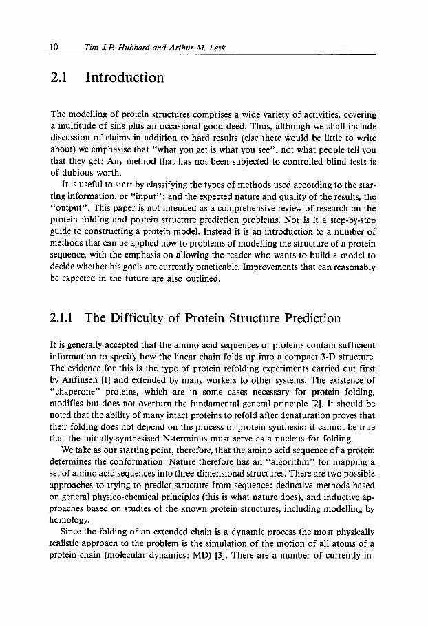
2.1 Introduction
The modelling of protein structures comprises a wide variety of activities, covering a multitude of sins plus an occasional good deed. Thus, although we shall include discussion of claims in addition to hard results (else there would be little to write about) we emphasise that “what you get is what you see”, not what people tell you that they get: Any method that has not been subjected to controlled blind tests is of dubious worth.
It is useful to start by classifying the types of methods used according to the star- ting information, or “input”; and the expected nature and quality of the results, the “output”. This paper is not intended as a comprehensive review of research on the protein folding and protein structure prediction problems. Nor is it a step-by-step guide to constructing a protein model. Instead it is an introduction to a number of methods that can be applied now to problems of modelling the structure of a protein sequence, with the emphasis on allowing the reader who wants to build a model to decide whether his goals are currently practicable. Improvements that can reasonably be expected in the future are also outlined.
2.1.1 The Difficulty of Protein Structure Prediction
It is generally accepted that the amino acid sequences of proteins contain sufficient information to specify how the linear chain folds up into a compact 3-D structure. The evidence for this is the type of protein refolding experiments carried out first by Anfinsen [l] and extended by many workers to other systems. The existence of “chaperone” proteins, which are in some cases necessary for protein folding, modifies but does not overturn the fundamental general principle [2]. It should be noted that the ability of many intact proteins to refold after denaturation proves that their folding does not depend on the process of protein synthesis: it cannot be true that the initially-synthesised N-terminus must serve as a nucleus for folding.
We take as our starting point, therefore, that the amino acid sequence of a protein determines the conformation. Nature therefore has an “algorithm” for mapping a set of amino acid sequences into three-dimensional structures. There are two possible approaches to trying to predict structure from sequence : deductive methods based on general physico-chemical principles (this is what nature does), and inductive ap- proaches based on studies of the known protein structures, including modelling by homology.
Since the folding of an extended chain is a dynamic process the most physically realistic approach to the problem is the simulation of the motion of all atoms of a protein chain (molecular dynamics: MD) [3]. There are a number of currently in-
2 Modellinn Protein Structures 11
surmountable problems with this approach that have deprived it of the success it should in principle someday achieve. The major problems are limitations on com- puter time (currently time intervals of at most nanoseconds can be simulated with today’s fastest computers, whereas protein folding is thought to occur over seconds); and the problem of incomplete and inexact representation of the thermodynamic in- teractions - particularly the problem of appropriately representing the protein- water interaction (proteins fold in an aqueous environment which must be represented explicitly and accurately) - and other uncertainties about the interac- tion potentials used between atoms in the system, particularly the electrostatic terms. MD can however be usefully applied to modelling problems where there are suffi- cient conformational restraints,
All other a priori prediction methods are less physically realistic than the above and involve either unproven assumptions or unlikely generalisations (or both). No method that might be expected to become generally successful is in sight.
As a priori prediction is unsuccessful, we are fortunate that - unlike a protein chain folding in vivo - modellers can make use of knowledge from all known pro- tein sequences and structures when predicting a protein fold. It is the extraction and application of this information that is the basis for all successful prediction methods. Central to such methods is the idea of homology.
2.1.2 The Idea of Homology Between Proteins
Two proteins are homologous if they are related by natural evolutionary processes. Many homologous sequences are sufficiently closely related that their amino acid se- quences can be aligned so that the number of similar or identical pairs of amino acids at aligned positions is greater than expected by chance. An alignment may con- tain gaps because as a protein sequence evolves both mutation and insertion/deletion events can occur in the encoding DNA.
Forming an accurate alignment of the amino acid sequences is absolutely essential for useful model building. When the divergence of the sequences has left no fewer than about 40% of the residues identical in an optimal alignment, it is likely that the standard sequence-alignment methods will provide a correct alignment [4]. In- deed, such an alignment is prima facie evidence for homology, which of course can only rarely be detected directly. (The exceptions are cases of obvious gene duplication and divergence. A classic example of this is the two domains of rhodanese [5 ] . ) When sequences have diverged substantially farther than this 40% threshold, it may be im- possible to determine the correct alignment from pairs of sequences only, but it may be possible to determine a correct alignment from a comparison of the structures, provided of course that they are available. Also, multiple sequence alignments are much more informative than alignments of only a single pair of sequences. In ex-
12 Tim J.P Hubbard and Arthur M. Lesk
treme cases, the sequences may have diverged so far that even the fact of a relation- ship cannot be detected from the sequences alone, and it is usually impossible to distinguish true homology from convergent evolution. We shall discuss this point in more detail below.
Close similarities among protein sequences allow proteins of different function or from different organisms to be clustered into families. This clustering provides evidence for homology, indicating that each member of a family is evolutionarily related and derived from a common ancestor. It is observed that homologous protein sequences also have similar 3-D structures and the relationship between sequence divergence and structure divergence has been quantified [6, 71. But structural similarity is often observed even if no sequence homology can be detected. A well- known example is the family containing hexokinase, heat shock protein 70 (hsp70) and actin: although many sequences were available, these were not known to be related until the structures of hsp70 and actin were solved and found to be very similar [B]. The converse - sequence similarity without structural similarity - has not been observed for anything other than very short peptides [9]. The conclusion is that protein structure is better conserved in evolution than protein sequence, and sufficient overall sequence similarity between proteins implies homology and a similarity of conformation. This idea underlies the methods of protein modelling we shall discuss.
2.1.3 A Summary of What Can and Cannot be Predicted
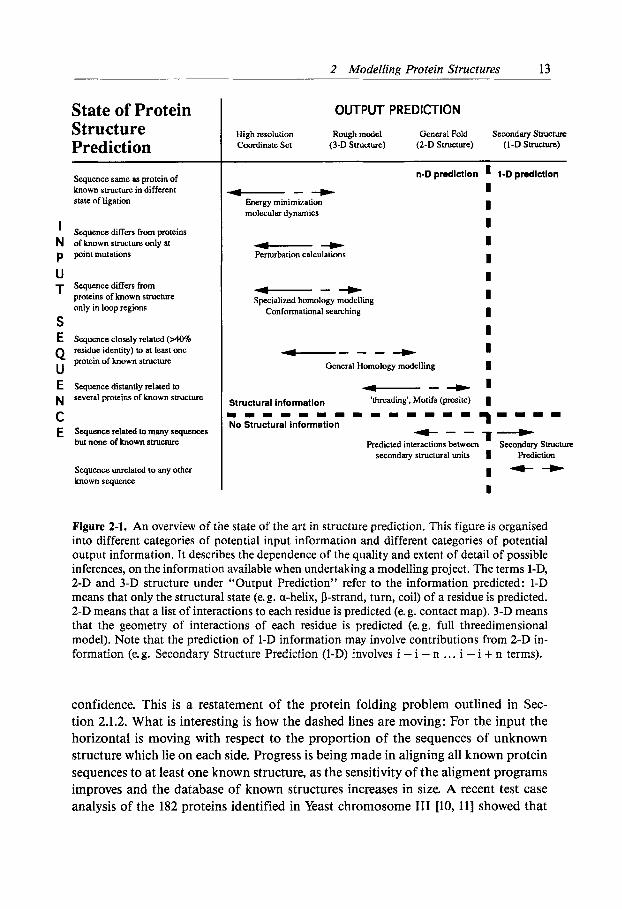
Figure 2-1 contains a representation of the current state of the art of protein struc- ture prediction. Vertically we characterise the input information: how much is known about proteins related to the target protein. Horizontally we characterise the output information: the more information we have about relatives of the target se- quence, the more - and the higher the quality - of the predictions we can make. Both input (unknown sequence) and output (structure prediction) axes can be divided into two distinct parts:
For an input above the dashed horizon line, the sequence to be modelled can be shown to be homologous to at least one other sequence the 3-D structure of which is known. An input below the horizon line is homologous only to other sequences the structure of which are unknown, or no homologies are known at all.
For an output to the left of the dashed vertical line 3-D structures are predictable with some degree of confidence and accuracy. Right of the vertical line a l-D struc- ture (i. e. secondary structure prediction), with perhaps hints of super-secondary structure, is the best result that is likely to be obtained.
The main message of this figure is that if the input sequence cannot be linked to any protein of known structure then no >l-D structure can be predicted with any
2 Modelling Protein Structures 13
State of Protein Structure Prediction Sequence same as protein of known sbucture in different state of ligation
’ Sequence differs from proteins N of known smcture only at p p in t mutations
U T Sequence differs From
proteins of known structure only in loop regions
S E Sequence closely related ( ~ 0 %
Q protein of known structure U E Sequence distantly relsled to
N
residue identity) to at least one
several proteins of known structure
C E Squence related to many sequences
but none of known structure
Sequence unrelated to any other known sequence
OUTPUT PREDICTION
High resolution Rough model General Fold Scoondary Structure Coordinate Set (3-D Structure) (2-D Structure) (I-D Structure)
--+ Energy minimization molecular dynamics
a--+ Specialized homology modelling
Conformational searching
n-D prediction I 1-D prediction
----+ I
--+ ’ General Homology modelling I
Structural information ‘threading’. Motifs (pmsite) I m m 1 1 1 m m m 1 1 m 1 1 = 7 = = = = No Structural Information +--T-
Predicted interactions between Secondary Structure secondary structural units I Prediction
I -- I
Figure 2-1. An overview of the state of the art in structure prediction. This figure is organised into different categories of potential input information and different categories of potential output information. It describes the dependence of the quality and extent of detail of possible inferences, on the information available when undertaking a modelling project. The terms 1 - 4 2-D and 3-D structure under “Output Prediction” refer to the information predicted: 1-D means that only the structural state (e. g. a-helix, P-strand, turn, coil) of a residue is predicted. 2-D means that a list of interactions to each residue is predicted (e. g. contact map). 3-D means that the geometry of interactions of each residue is predicted (e. g. full threedimensional model). Note that the prediction of 1-D information may involve contributions from 2-D in- formation (e. g. Secondary Structure Prediction (I-D) involves i - i - n . . . i - i + n terms).
confidence. This is a restatement of the protein folding problem outlined in Sec- tion 2.1.2. What is interesting is how the dashed lines are moving: For the input the horizontal is moving with respect to the proportion of the sequences of unknown structure which lie on each side. Progress is being made in aligning all known protein sequences to at least one known structure, as the sensitivity of the aligment programs improves and the database of known structures increases in size. A recent test case analysis of the 182 proteins identified in Yeast chromosome I11 [lo, 111 showed that

14 Tim Ll? Hubbard and Arthur M. Lesk
14 Yo of sequences could be associated with a protein of known structure using stan- dard sequence alignment methods.
For the output, the dividing line may soon become blurred if it becomes possible to predict more than just secondary structure (1-D) when families of homologous se- quences are considered together. The Yeast chromosome I11 analysis found that 24% of sequences could be associated with an existing sequence family that had no known structure. As more proteins are sequenced such families are coming to have increas- ingly large numbers of members with wider sequence diversity. Since related se- quences may all be expected to adopt the same fold, any prediction must be consis- tent with each sequence in such a family. This is a considerable restriction and has allowed significant improvements in 1-D secondary structure prediction [12- 141, the latter method being available to anyone with access to electronic mail (send “help” to Predictprotein @ embl-heidelberg-de). Since the number of natural folds is thought to be finite and may be as small as 1000 [I51 there will come a time when all new sequences can be associated with a known protein structure. There is therefore something of a race between various methods - fold recognition versus fold prediction - that seek to eliminate the current “unpredictable” region of se- quence space.
Figure 2-1 does not include all protein modelling exercises, as it omits designed sequences. It is important to realise that even if methods to predict a structure consis- tent with a large family of sequences are developed, this is not a solution of the folding problem. The assumptions that (1) any sequence folds and (2) folds are similar among homologous sequences are based on evolutionary reasoning, for se- quences that do not fold would be selected against and would not therefore be observed by chance, and significant sequence homologies are only likely to occur through divergent evolution, i. e. from a single fold. Designed sequences may not fold like the sequence to which they appear to be related and in many cases may not fold at all. In order to be able predict the structure of a designed sequence it will be necessary to predict structure from individual sequences, ignoring evolutionary rela- tions, i.e. to solve the a priori folding problem [16].
2.2 Proving a Sequence/Structural Relationship
The first stage in any modelling project should be to compare the sequence of the protein of interest with the contents of sequence databases. There are many sequence alignment programs available that can do this with varying speed and sensitivity. The objective is to find homologous sequences of known structure, but finding any homologous sequence is useful since it provides additional information about the protein to be modelled.

2 Modelling Protein Structures 15
The database of known structures, the PDB (Protein Data Bank) [17, 181 contains more than 3000 experimentally determined protein structures (Jan95 release) although by sequence homology these can be clustered into less than 400 distinct families [19], and by structural superposition into perhaps not more than 150 folds [20]. For each of these structures a file exists which contains all clearcut alignments between the sequence of that structure and all sequences in the protein sequence database Swissprot [21]. These “HSSP” files (homology-derived secondary structure of proteins) [22] are available by anonymous FTP over internet from ftp.emb1- heidelberg.de. If the sequence in question is not listed in any HSSP file it does not necessarily mean there is no relationship to any known structure: either the sequence is too new to be in the version of Swissprot used to generate the HSSP files or any homology is too weak be identified by such a method. Clearcut homology is con- sidered to exist where more than 40% of residues are identical in both sequences after alignment. HSSP files contain weaker homologies than this (down to around 30%) although such alignments should be evaluated carefully. Still weaker alignments may be detected by other methods: The detection of weak homologies by sequence methods alone is a science in itself. A wide number of methods are available but it requires experience to distinguish a real alignment from a false, ran- dom one [23, 241.
To try to detect structural similarities where sequence homologies are near the noise threshold, additional information must be included in the alignment pro- cedure. The two possible sources are multiple sequence information and structural information.
Rather than try to detect overall homologies, an alternative approach is to look for conserved sequence motifs. These are short regions of conserved sequence and can be found by examining a multiple sequence alignment. If a number of conserved motifs can be found, a search “template” can be constructed, being a series of motifs linked by variable lengths of connecting sequence. If an input sequence is a member of a sequence family, a template can be constructed for that family and used to search the sequence database looking for a match to a sequence of known struc- ture. There are also motif databases, collecting in Amos Bairoch’s Prosite [25] that can be scanned with appropriate software [26]. An example of the use of this techni- que to produce a successful fold recognition and subsequent modelling was the recognition that the HIV protease resembles half of an aspartic protease and as a dimer has the same active site [27]. In this case, although it was only the active site sequence Asp-Thr-Gly that was clearly a conserved motif, the protein chains turned out to have very similar folds.
Nevertheless, a conserved motif or motifs does not prove a global structural similarity and could even be a result of convergent rather than divergent evolution. For instance, the GTP binding site motif GxGxxG is common to a large number of protein families with folds of substantially different topology but which share a com- mon active site [25]. Therefore identification of a protein by a motif may permit in-
16 Tim J.P Hubbard and Arthur M. Lesk
ferences about and even a model of a binding site, but it may not be possible to ex- tend the model to the entire structure. In general, matching folds based on small fragments must be done with care.
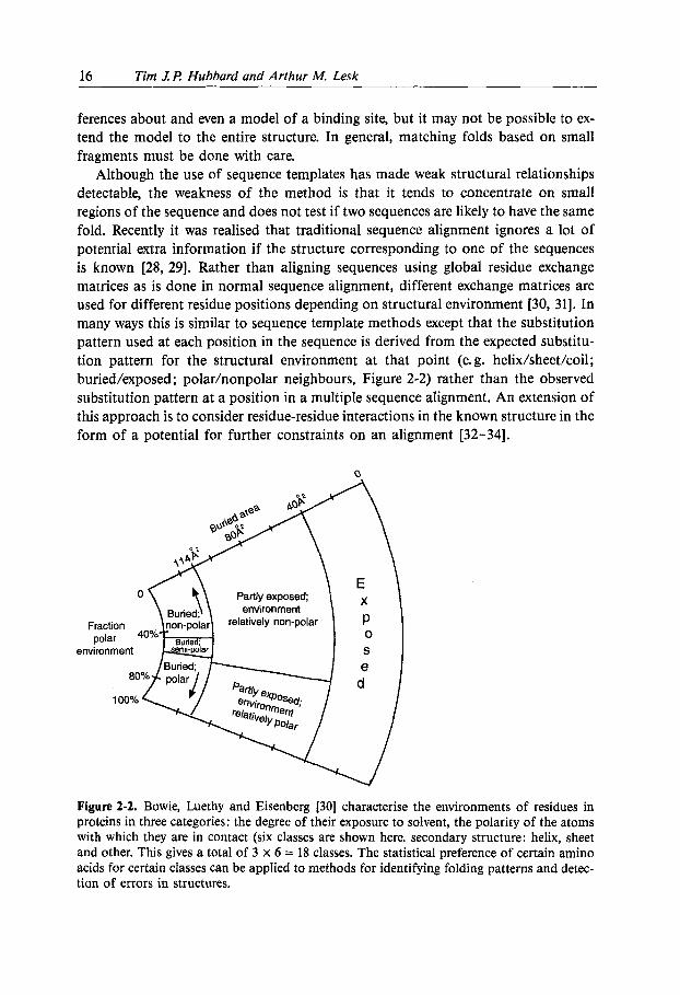
Although the use of sequence templates has made weak structural relationships detectable, the weakness of the method is that it tends to concentrate on small regions of the sequence and does not test if two sequences are likely to have the same fold. Recently it was realised that traditional sequence alignment ignores a lot of potential extra information if the structure corresponding to one of the sequences is known [28, 291. Rather than aligning sequences using global residue exchange matrices as is done in normal sequence alignment, different exchange matrices are used for different residue positions depending on structural environment [30, 311. In many ways this is similar to sequence template methods except that the substitution pattern used at each position in the sequence is derived from the expected substitu- tion pattern for the structural environment at that point (e. g. helix/sheet/coil; buried/exposed; polar/nonpolar neighbours, Figure 2-2) rather than the observed substitution pattern at a position in a multiple sequence alignment. An extension of this approach is to consider residue-residue interactions in the known structure in the form of a potential for further constraints on an alignment [32-341.
\ la,,
Fraction
environment polar 4 O % k emi-pda
Partly exposed;
buried: 'u I
Figure 2-2. Bowie, Luethy and Eisenberg [30] characterise the environments of residues in proteins in three categories: the degree of their exposure to solvent, the polarity of the atoms with which they are in contact (six classes are shown here. secondary structure: helix, sheet and other. This gives a total of 3 X 6 = 18 classes. The statistical preference of certain amino acids for certain classes can be applied to methods for identifying folding patterns and detec- tion of errors in structures.

2 Modelling Protein Structures 17
The use of structural information in sequence alignment can be thought of as “threading” a sequence of unknown structure onto the fold of the known structure and measuring the quality of the fit [35, 361. Such techniques are more sensitive than traditional sequence alignment techniques but it is not yet clear by how much. At present no method can cluster folds in the structural database as well as searches using structural superposition [37]. For example, a very common fold is that of the a/p (TIM) barrel. At least 20 examples of this fold exist, the relationship between which cannot be recognised by sequence alignment methods [38]. Although it seems likely that at least some barrels may have evolved by convergent evolution [39], a perfect fold recognition algorithm should able to cluster them all together in an unambiguous way due to the particular symmetry of this fold. Although there has been progress on this front [40] no method can currently achieve this without either missing some or including unrelated folds.
If after use of these methods an input sequence can be linked to a known struc- ture, prediction of the structure can proceed through homology modelling (Sec- tion 2.3). If not, pure structural prediction methods must be used (Section 2.4). In many cases it will be unclear whether a fold has been recognised or not. The only solution is to try to build a model based on the presumed structural similarity and then attempt to test its validity (in a similar way that a designed sequence is tested for compatibility with the fold it is meant to adopt (Section 2.4.2)). Even then the results may well be ambiguous. A current example of such uncertainty is the model of Hsp70 C-terminal domain based on the HLA binding site [41]. Despite con- siderable detailed analysis it remains difficult to decide, without an experimental structure determination, whether this model is right or wrong.
2.3 Modelling Starting from a Known Structure
2.3.1 Evolution of Protein Structures
In order to understand the possibilities and limitations of model-building by homology, it is necessary to appreciate the kinds of structural changes that occur as proteins diverge. Numerous studies analysing the structural relationships between related proteins [42], and the dependence of structural divergence on sequence divergence [6, 71, provide us with the ability to estimate quantitatively how successful a model-building exercise can be, knowing how closely the target protein and its homologues are related.
Natural variations in families of homologous proteins reveal how the structures accomodate changes in amino acid sequence. There are several accepted measures of

18 Tim JI? Hubbard and Arthur M. Lesk
divergence of sequences; we have used the percent identical residues in the alignment of the sequences. There is the corresponding problem of calibrating a measure of the similarity of two or more structures, or portions of structures. A useful mathe- matical technique is to determine the optimal “least-squares” superposition of a pair of structures or parts of structures. By this we mean the following: We fix the posi- tion and orientation of one of the structures, and vary the position and orientation of the other to find the minimum value of the sum of the squares of the distances between the corresponding atoms. The square root of the average value of the squared distances between corresponding atoms is the root-mean-square (r. m. s.) deviation. If the two objects were precisely congruent, it would be possible to superimpose them exactly, and the r. m. s. deviation would be zero. In real cases, the “fit” of two nonidentical structures is never exact, and the minimal r. m. s. deviation is a quantitative measure of the structural difference.
Included in the approximately 3000 protein structures now known are several members of families in which the molecules maintain the same basic folding pattern over ranges of sequence homology from near-identity down to below 20%. In both closely and distantly related proteins the general response to mutation is conforma- tional change. The maintenance of function in widely divergent sequences requires the integration of the response to mutations over all or at least a large portion of the molecule.
It is the ability of protein structures to accommodate mutations in nonfunctional residues that permits a large amount of apparently nonadaptive change to occur. Residues active in function, such as the proximal histidine of the globins or the catalytic serine, histidine and aspartate of the serine proteases, are resistant to muta- tion because changing them would interfere, explicitly and directly, with function. Most buried residues are in the well-packed interfaces between helices and sheets. During the course of evolution, the buried residues remain hydrophobic, but can change size. Mutations that change the volumes of buried residues generally do not change the conformations of individual helices or sheets, but produce distortions of their spatial assembly. These tend to take the form of rigid-body shifts and rota- tional, which may be as large as 7 A, but more typically are 3-5 A. Surface residues not involved in function are usually free to mutate. Loops on the surface can often accomodate changes by local refolding 1431.
The nature of the forces that stabilise protein structures sets general limita- tions on these conformational changes; other constraints derived from function vary from case to case. In some protein families large movements are coupled to conserve the structure of the active site (e.g., the globins); in others, active sites of alterna- tive structure are found (e. g., cytochromes c). In proteins that for functional reasons cannot tolerate conformational change - such as those with multiple binding sites that must maintain a relative spatial disposition, or those that must maintain a sur- face involved in complex formation - amino acid sequences are more highly con- served.

2 Modelling Protein Structures 19
Families of related proteins tend to retain similar folding patterns. If one examines sets of related proteins (see Figures 2-3 and 2-4) it is clear that although the general folding pattern is preserved, there are distortions which increase progressively as the amino acid sequences diverge. These distortions are not uniformly distributed throughout the structure. Instead, in any family of proteins there is a core of the structure that retains the same qualitative fold, and other parts of the structure that change conformation radically. To explain the idea of the common core of two struc- tures, consider the letters B and R. Considered as structures they have a common core which corresponds to the letter P. Outside the common core they differ: at the bottom right B has a loop and R has a diagonal stroke.
Figure 2-3. -0 closely-related proteins: (a) actinidin (crystal structure by E. N. Baker and E. J. Dodson [go]) and (b) papain (crystal structure by I. G. Kamphuis et al. [91]). The amino acid sequences of these molecules have about 50 070 identical residues.
Figure 2-4. W o distantly-related proteins: (a) poplar leaf plastocyanin (crystal structure by J. M. Guss and H. C. Freeman [92]) and (b) A. denitrificuns azurin (crystal structure by G. E. Norris, B. F. Anderson and E. N. Baker [93]). The circle near the top of the structure marks the position of the copper. In this case the double P-sheet portion of these molecules retains the same fold, but the long loop at the left changes its conformation completely.
20 Tim LJ? Hubbard and Arthur M. Lesk
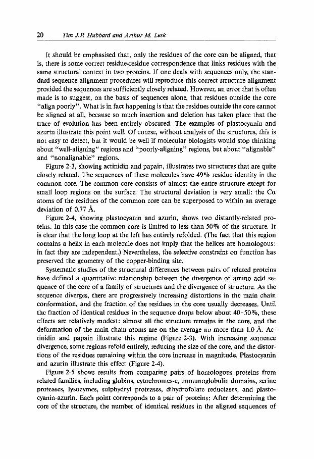
It should be emphasised that, only the residues of the core can be aligned, that is, there is some correct residue-residue correspondence that links residues with the same structural context in two proteins. If one deals with sequences only, the stan- dard sequence alignment procedures will reproduce this correct structure alignment provided the sequences are sufficiently closely related. However, an error that is often made is to suggest, on the basis of sequences alone, that residues outside the core “align poorly”. What is in fact happening is that the residues outside the core cannot be aligned at all, because so much insertion and deletion has taken place that the trace of evolution has been entirely obscured. The examples of plastocyanin and azurin illustrate this point well. Of course, without analysis of the structures, this is not easy to detect, but it would be well if molecular biologists would stop thinking about “well-aligning” regions and “poorly-aligning” regions, but about “alignable” and “nonalignable” regions.
Figure 2-3, showing actinidin and papain, illustrates two structures that are quite closely related. The sequences of these molecules have 49% residue identity in the common core. The common core consists of almost the entire structure except for small loop regions on the surface. The structural deviation is very small: the Ca atoms of the residues of the common core can be superposed to within an average deviation of 0.77 A.
Figure 2-4, showing plastocyanin and azurin, shows two distantly-related pro- teins. In this case the common core is limited to less than 50% of the structure. It is clear that the long loop at the left has entirely refolded. (The fact that this region contains a helix in each molecule does not imply that the helices are homologous: in fact they are independent.) Nevertheless, the selective constraint on function has preserved the geometry of the copper-binding site.
Systematic studies of the structural differences between pairs of related proteins have defined a quantitative relationship between the divergence of amino acid se- quence of the core of a family of structures and the divergence of structure. As the sequence diverges, there are progressively increasing distortions in the main chain conformation, and the fraction of the residues in the core usually decreases. Until the fraction of identical residues in the sequence drops below about 40-50%, these effects are relatively modest: almost all the structure remains in the core, and the deformation of the main chain atoms are on the average no more than 1.0 A. Ac- tinidin and papain illustrate this regime (Figure 2-3). With increasing sequence divergence, some regions refold entirely, reducing the size of the core, and the distor- tions of the residues remaining within the core increase in magnitude. Plastocyanin and azurin illustrate this effect (Figure 2-4).
Figure 2-5 shows results from comparing pairs of homologous proteins from related families, including globins, cytochromes-c, immunoglobulin domains, serine proteases, lysozymes, sulphydryl proteases, dihydrofolate reductases, and plasto- cyanin-azurin. Each point corresponds to a pair of proteins: After determining the core of the structure, the number of identical residues in the aligned sequences of

2 Modellinn Protein Structures 21
the core was counted, and the root-mean-square deviation of the main chain atoms of the core was calculated. (The points corresponding to 100% residue identity are proteins for which the structure was determined in two or more crystal environments, and the deviations show that crystal packing forces can modify slightly the confor- mation of the proteins.) Figure 2-6 shows the changes in the fraction of residues in
2
:: c 1.2-
m 3
m
c 0 2 0 . 6 -
'? 2.4- c 0 m > c
2
:: c 1.2-
m 3
m
c 0
8 0.0 1 I I I I 1 00 80 60 40 2 0 0
Percent residue identity
Figure 2-5. The relationship between the divergence of the amino-acid sequence of the core of related proteins and the divergence of the main chain conformation of the core.
c
%o I 8 0 ' 60 I 40 I 2b ' 0 ' Sequence identity ("YO)
Figure 2-6. The relationship between the divergence of related proteins and the relative size of the core.
of the amino-acid sequence of the core

22 Tim .l I! Hubbard and Arthur M. Lesk
the core as a function of sequence divergence. In pairs of distantly related proteins the size of the cores can vary: In some cases the fraction of residues in the core re- mains high, in others it can drop to below 50% of the structure.
2.3.2 Techniques
A general outline of the steps involved in model building by homology is as follows:
1. The sequence of unknown structure is aligned to the sequence(s) of known struc- ture and the sequence of unknown structure is divided into SCR’s and SVR’s: regions where the alignment has sufficient sequence conservation to be conserved structurally (alignable) are defined as SCR’s (Structurally Conserved Regions). The remaining regions (nonalignable - including but not restricted to loop regions) are defined as SVR’s (Structurally Variable Regions).
2. The main chain conformation and spatial relationship of the SCR’s are taken from the coordinates of the known structure to which they were aligned. Confor- mations are generated for each SVR in the sequence, with correct endpoint geometry and length, which do not clash sterically with the rest of the structure, either using a database search method [44] or any alternative approach. This creates a complete continuous main chain model.
3. Side chains are built onto the main chain model and their conformations op- timised.
2.3.2.1 Alignment and Division into SCR’s and SVR’s
Discovering a relationship between an input sequence and a structure does not necessarily give a full or accurate alignment. Frequently more sensitive alignment techniques not designed for fold recognition can give a more accurate alignment once the sequences to align have been identified. It is important to keep the lessons from evolution in mind (Section 2.3.1): There will be regions where there are several possible alternative alignments and there will be regions that cannot be aligned (nonalignable) because they are structurally different.
The first of these problems can be tackled by using as much information as possi- ble (multiple sequence alignments for both known and unknown) and by exploring significant alternative sub-optimal alignments [45, 461, and if necessary, by building multiple structural models using alternative alignments.
The second problem is to distinguish the regions in which the input sequence has the same fold as the model (SCR’s) and where it is different (SVR’s). Clearly where there are insertions and deletions the chain trace of the model must be different;

2 Modelling Protein Structures 23
however, regions on either side of any deletion may also have a changed conforma- tion. Secondary structure prediction (SSP) may be useful at this point to see whether there is a strong change in the prediction at any point where the alignment is weak. Regions that have been identified as SVR’s must be predicted by methods described in the next section, as “loops” connecting the two chain ends of the preceding and following SCR’s.
2.3.2.2 Modelling Loop Regions
The term “loops” refers to sections of the polypeptide chain that connect regions of secondary structure. Frequently, helices and strands of sheet run across a protein or domain from one surface to another, and loops are characterised by (a) appearing on the surfaces of proteins and (b) reversing the direction of the chain. A typical globular protein contains one third of its residues in loops.
In model-building by homology, loops often present special problems because they are often the sites of insertion and deletions. Frequently the residues cannot be aligned with those of the parent molecule because of this. Special techniques have therefore been developed to build loops, assuming that the core of the target protein has already been modelled.
Hairpin loops (those that connect successive strands of antiparallel P-sheet) have been studied extensively to classify them and to elucidate the determinants of their conformations [47-571. Most residues of proteins have their main chains in one of two sterically-favourable conformations (these correspond to the conformations of a-helices and P-sheets). However, in order for a short region of polypeptide chain 3-4 residues in length to reverse direction, and fold back on itself to form a loop, a residue that takes up a conformation outside these usual states is generally re- quired. The conformations of short loops therefore depend primarily on the position within the loop of special residues - usually Gly, Asn or Pro - that allow the chain to take up an unusual conformation. As pointed out by Sibanda and Thornton [55], the conformation of a short hairpin can often be deduced from the position in the sequence of such special residues.
These general rules are however of limited utility for the understanding and prediction of the conformations of many functionally important loop regions ; for instance the antigen-binding loops of immunoglobulins. Many loops are not short, or not hairpins, or neither; and the determinants of their conformations are not en- tirely intrinsic to the amino acid sequence of the loop itself, but involve tertiary in- teractions : hydrogen bonding and packing. Indeed, even for some short hairpins, ter- tiary interactions can override the predisposition of the sequence, to determine a con- formation of the loop that does not follow these sequence-structure correlations. An example important in immunoglobulin structure is the second hypervariable region

24 Tim 1 F! Hubbard and Arthur M. Lesk
of the VH domain (H2). The size of the residue at site 71, a site in the conserved 0-sheet of the VH domain, is a major determinant of the conformation and position of this loop [58].
Several general methods have been developed for prediction of the conformations of loops in proteins. The antigen-binding loops of antibodies have received special attention, and some special methods have been developed for them [52].
Prediction of Loop Conformations by Energy Calculations. The main chain confor- mation of a loop attached to a given framework must obey the constraint that the chain must connect two fixed endpoints using a specified number of residues. For loops of fewer than about six residues, it is possible to enumerate a fairly complete set of main chain and side chain conformations that bridge the given endpoints and do not make steric collisions within the loop or between the loop and the rest of the molecule. The search procedure can be fine enough to be sure to produce a loop close to the correct one.
However, there are in general many possible loops of different internal conforma- tions that bridge a given pair of endpoints. To choose one of them as the predicted conformation, it is possible to estimate conformational energies and evaluate the ac- cessible surface areas of each loop - in the context of the remainder of the protein - and set criteria for selecting the one that appears the most favourable. Typical conformational energy calculations include terms representing hydrogen bonding, van der Waals, and electrostatic interactions. Accessible surface area calculations give estimates of the interaction between the protein and the solvent. This is in princi- ple a completely general, automatic and objective procedure.
Procedures for conformation generation and evaluation have been implemented in a number of computer programs, of which the best known is CONGEN, by Bruc- coleri and Karplus [59]. (Other similar procedures have been developed by Fine et al. [60], and by Moult and James [61].) An application to predicting all six antigen- binding loops of McPC603 and HyHELS, based on the program CONGEN, has been described by Bruccoleri, Haber and Novotny [62]. The CONGEN procedure generates conformations for a single loop, and calculates energies of that loop in the context of the fixed portion of the structure. To apply this procedure to the predic- tion of several loops - for instance the six loops of an antigen-binding site - a pro- tocol must be used that involves a sequential prediction of the loops, starting with the loops that interact primarily with the known parts of the molecule, and then pro- ceeding to the loops that interact with each other.
Prediction of Loop Conformations by Data Base Screening. Jones and Thirup [44] developed a method of building loops, based on selecting from proteins in the database of known structures loops that span the given endpoints and overlap with peptides at the loop termini. Vpically, a user selects the endpoints and initiates a data base search. The results are displayed interactively at a graphics terminal.

2 Modelling Protein Structures 25
W o general possibilities may arise: All the main chains of the loops found lie within a narrow “sheaf” of trajectories between the fixed endpoints. Provided the common structure thus indicated does not have steric clashes with the rest of the protein, one can adopt the main chain with some confidence as an approximate model for the target loop. In many cases there is conservation of a special residue - such as Gly, Asn or Pro - that is responsible for the conformation of the loop.
Alternatively, the loops retrieved from the data base may “fan out” broadly. In this case, the selection of the model is more hazardous. One can look for the presence of special residues - again, Gly, Asn or Pro - at the same positions as in the target structure. Alternatively, it has been attempted to determine the conformational energies of the loops to select the best one.
Database searching is the most widely available method for loop building. It is a facility of a large number of computer graphics programs as a loop-building option and has been incorporated into many automatic model building programs such as Composer [63].
A particular problem that arises in building a model by grafting loops into a model of a set of SCR’s is the possibility of error at the junctions between loops and SCR’s. It is useful to apply programs that build a main chain with CP atoms directly from Ca coordinates alone, by automatic chain fitting procedures [37, 64, 651. One may test a model of the main chain of an entire structure for “self-consistency”, by extracting the Ca’s, rebuilding the complete backbone from them, and comparing with the starting model. A large number of main chain peptide “flips” between the original and automatically generated main chain may show errors in the original assumptions from which the model was built. (Such procedures are also useful in refinement of models during structure determinations.)
Special-Purpose Technique for Antigen-Building Loops of Immunoglobulins. Ana- lysis of the antigen-binding loops in known structures has shown that the main chain conformations are determined by a few particular residues and that only these residues, and the overall length of the loop, need to be conserved to maintain the conformation of the loop [52]. The conserved residues may be those that can adopt special main chain conformations - Gly, Asn or Pro - or that form special hydrogen-bonding or packing interactions. Other residues in the sequences of the loops are thus left free to vary, to modulate the surface topography and charge distribution of the antigen-binding site.
The ability to isolate the determinants of loop conformation in a few particular residues in the sequence makes it possible to analyse the distribution of loop confor- mations in the many known immunoglobulin sequences [66]. It appears that at least five of the hypervariable regions of antibodies have only a few main chain conforma- tions or “canonical structures”. Most sequence variations only modify the surface by altering the side chains on the same canonical main chain structure. Sequence

26 Tim .l I! Hubbard and Arthur M. Lesk
changes at a few specific sets of positions switch the main chain to a different canonical conformation.
As an example Figure 2-7 shows the L3 loop from VK McPC603. In this, the most common VK L3 conformation, there is a proline at position 95 in the loop, in a cis conformation. Hydrogen bonds between the side chain of the residue at position 90, just N-terminal to the loop, and the main chain atoms of residues in the loop, stabilise the conformation. The side chain is an Asn in McPC603; it can also be a Gln or His in other VK chains. The combination of the polar side chain at position 90 and the proline at position 95 constitute the “signature” of this conformation in this loop, from which it can be recognised in a sequence of an immunoglobulin of unknown structure.
b b Figure 2-7. An antigen-binding loop from the VK domain of the immunoglobulin McPC603. This loop contains a cis-proline, and is stabilised by hydrogen bonding between a polar side chain just N-terminal to the loop and inward-pointing main chain atoms in the loop.
The observed conformations are determined by the interactions of a few residues at specific sites in the hypervariable regions and, for certain loops, in the framework regions. Hypervariable regions that have the same conformations in different im- munoglobulins have the same or very similar residues at these sites. On the basis of the canonical structural model, it has been possible to create a detailed roster of the canonical conformations of each loop - with the possible exception of H3 which is more complicated and still uncertain - and the sets of “signature” residues that permit discrimination among them.
A procedure to predict the structures of the variable domains of immunoglobulins has been formulated based on the structures of solved immunoglobulins and the canonical structure model of the conformations of the hypervariable loops [65].

2 Modelling Protein Structures 27
1. Align the sequence of the VL and VH chains of the target immunoglobulin with the sequences of the corresponding domains in the known immunoglobulin struc- tures.
2. For each domain (VL and VH), select a parent domain from among the cor- responding domains of known structure. The percent residue identity with the target domain is usually in the range 45 070 and 85 070.
3. If the selected parent structures for VL and VH domains come from different im- munoglobulins, pack them together by a least-squares fit of the main chain atoms of residues conserved in the VLVH interface.
4, Identify the canonical structure of each loop by checking the sequence for the particular sets of residues that form the signature of each canonical structure. H3 is a special case, far more variable in length, sequence and structure; and must be modelled by other methods.
5. Graft a loop from a known immunoglobulin structure - preferably the domain from which the framework was built - into the framework model.
6 . If a canonical structure for any loop cannot be identified, the loop must be modelled by other means.
7. To build the side chains: At sites where the parent structure and the model have the same residue, retain the conformation of the parent structure. If the side chain is different, take its conformation, if possible, from an immunoglobulin having the same residue in the corresponding position; within hypervariable loops, take the side chain conformation only from a loop with the same canonical structure.
8. Subject the model to limited energy refinement, only to tidy up the stereo- chemistry.
How good a model can be expected from this procedure, assuming the hypothesis that for the three loops of the light chain and for the first two of the heavy chain a canonical structure present in the data base can be identified?
The first of several “blind” tests was made on the antilysozyme antibody D1.3 [6] . Comparison of this prediction with the best available crystal structure of D1.3 has shown that all six hypervariable regions had the predicted main chain conforma- tions. Other tests are described in Chothia et al. [66] . The general conclusion is that if the structures used as parent structures for the two domains and the loops are high resolution, well-refined structures, one can expect the backbone of the framework to be correct within 1.0 A r. m. s. deviation, and the backbone of the predicted loops, not including the special case of H3, to differ by about 0.7 A r.m.s. deviation on average, and by no more than 1.0-1.2 A in all cases. In addition, one can expect the positions of Ca atoms of residues in the loops to shift, relative to the frameworks of VL and VH domains, by 1.0-2.0 A typically and by up to 3 A in the worst cases.

28 Tim J l ? Hubbard and Arthur M. Lesk
2.3.2.3 Side Chain Building and Optimisation of Side Chain Conformation
Once a complete main chain model has been constructed side chains need to be built and their conformations determined.
For closely-related proteins, it is observed that most side chains tend to retain con- formation - even mutated ones. This is because each side chain, even those on the surface, is packed in a cage formed by its neighbours. In closely-related proteins, a mutated side chain is likely to find itself in a cage created largely by nonmutated neighbours, and must conform itself to it. Therefore the first approximation should be: for side chains that have not been changed from the parent structure, retain the same conformation; for mutated side chains, retain the same conformation as far as the stereochemical similarity will allow. Of course application of this rule will pro- duce some sterically impossible combinations.
An essential step therefore is to adjust the side chain conformations to achieve a low-energy conformation. There are now a large number of programs available to carry out such building and packing automatically [37, 64, 65, 671 which are pro- bably more accurate (and of course much quicker) than manual manipulation [68].
Currently available procedures for automatic side chain modelling and packing perform quite well when starting from experimental main chain atoms or Ca’s. It is not so clear what happens as the position of the main chain in the model becomes less and less accurate (as the closeness of the relationship between the input sequence and the sequence of known structure decreases). Exact Ca/main chain positions may force a unique side chain packing. In contrast, in a real modelling situation Cdmain chain positions will be inexact, and packing errors are more likely. In particular, the incorrect positioning of a large buried hydrophobic residue can result in serious packing errors within a whole region of the hydrophobic core of a protein.
Finally, once all atoms have been built, the model can be subjected to Energy Minimisation (EM) or Molecular Dynamics (MD). EM is a purely cosmetic opera- tion. It will remove some bad atom contacts in a model but will not significantly alter even side chain conformations. It can be useful however to “clean up” a model by small local adjustments. MD can be used to explore a much greater conformational space around the model structure than EM. If the changes in conformation are to be at all realistic, it is necessary to simulate in the presence of water. However, it is important to realise that if the starting model has substantial errors, even a very long MD run is very unlikely to improve it. Perhaps the most useful result of such simula- tions is to observe how a model moves with time. If the model is wrong it is likely to be more unstable when subjected to simulation.

2 Modelling Protein Structures 29
2.3.3 Available Modelling Programs
Homology modelling programs of various degrees of automation are now readily available. These include : Insight I1 [69] (commercial, graphics based, Homology modelling module: semi automatic); Quanta (commercial, graphics based, semi automatic module) ; What If [70] (academic, graphics based, semi automatic module); 0 [71] (academic, graphics based, essentially crystallographic modelling program but with database loop modelling features); Sybyl (commercial, graphics based, semi automatic module based on Composer [72, 731, also available as a non- graphical, academic program). All these packages contain essentially a sequence alignment program, a database loop searching program and features for optimising side chain conformations. For an assessment of the errors associated with such modelling procedures see Topham et al. [63]. Such semi-automatic modelling packages can very quickly produce models with no bad atom-atom contacts but which are partially or completely wrong due to the errors associated with alignment and loop building already discussed. Programs for evaluating homology models are not generally included in such packages: methods such as those described below (Section 2.4.2) should be used to look for errors. Regardless of the results of any tests, any user of models built in this way should always be mindful of the likely errors.
2.4 Modelling de novo: Structure Prediction
When no specific relationship can be found between a sequence of unknown struc- ture and any known structure only direct structure prediction methods remain an op- tion, and as shown in Figure 2-1, only the secondary structure can be predicted with any degree of accuracy at present.
2.4.1 A Family of Similar Sequences
Secondary structure prediction (SSP) can carried out on single sequences; however, where a family of homologous sequences exist more accurate results can be obtained. This has been known for some time [74, 751 but it is with the successful prediction [12] of the catalytic subunit of cyclic AMP dependent protein kinase [76] and the development of a neural-network based multiple sequence SSP method available over the internet by e-mail [13, 141 that use of such methods have become
30 Tim J . l ? Hubbard and Arthur M. Lesk
widespread. Such methods make use of multiple sequence information, looking for consistency between the predictions for different sequences. It should be noted that even given a correct secondary structure assignment, it is very difficult to determine how the units fit together in three dimensions. [77]. A table of aligned sequences may well contain derivable information about the 3-D structure of a protein but attempts to recover it have so far met with no more than sporadic success [12]. However, useful deductions about the most likely folded structure can be made in a systematic way from a combination of analysis of SSP results and the conservation patterns observed in a multiple sequence alignment. Successful predictions using such an ap- proach have been made for the annexin [78] and Src homology 2 (SH2) protein families [79].
2.4.2 A Lone Sequence or a Designed Sequence: no Multiple Sequence, no Known Relatives
This situation is the most unfavourable for model building; as one has no way of applying known sequence or structure information. In effect the problem can only be handled by a priori methods, Even secondary structure prediction is inaccurate for single sequences and therefore the likelihood of building a correct three-dimen- sional model is small.
If there is any suspicion (perhaps on functional grounds) that a natural sequence has a certain fold, or in the case of a designed sequence, built to fold in a particular way, the situation is slightly better since it is possible to test the likelihood that a se- quence can match a particular fold. Methods for doing this include checking polarity 1801 ; packing quality and residue-residue contact frequencies [81] ; various free energy functions incorporating solvation effects [82, 831, hydration and heat stability effects and more recently using threading techniques to establish if the sequence is compatible with the fold [MI.
The disadvantages of these methods are that (1) most provide only an assessment of the structure as a whole rather than of local regions (models are frequently only partially right e. g. [86]) (2) even at this level they are inaccurate, i. e. some experimen- tally determined structures are classified as incorrect whereas some misfolded models are classed as correct in blind tests and (3) that the results are essentially dependent on the quality of the model rather than the correctness of its fold. Moreover, these tests only look at the final state and do not assess if the sequence is compatible with any pathway to that state. For natural sequences it can be assumed that folding to a compact state can be achieved but this is more likely to fail to be the case for designed sequences. Current experimental [87] and theoretical work [88] on the folding pathways of proteins suggest that there are clear folding initiation sites

2 Modelling Protein Structures 31
specified in a protein chain. No method for identifying such sequences directly has yet been developed but it would seem clear that many sequences that are compatible with the desired final fold may contain no such folding signals. Until an understand- ing of the requirements for such sites is developed, de novo protein design [89] will remain very difficult, particularly for large proteins.
2.5 Future Possibilities
This review has tried to present a snapshot of what is possible now. What are the prospects for the near future?
Research is most active in the area of threading. The many groups developing potentials for fold recognition have taken a number of slightly different approaches, each with its own advantages. There will be more variations and those in the field anticipate substantial further improvements in the potentials. Fold recognition is however only the first stage in building a 3-D model.
Threading methods should ultimately be able to incorporate almost all the methods discussed for model building and evaluation so that a single sequence (Sec- tion 2.4.2) may be tested against all known folds. It is therefore anticipated that what will emerge will not only provide more accurate fold recognition, but the incorpora- tion of other, more detailed, model-building techniques to produce a specific three- dimensional structural prediction. It is by bringing together the various techniques for prediction and testing of structures that the interaction between them will ultimately generate the most satisfactory results.
2.6 Summary
The explosion of protein sequence and structural information has generated in its wake a number of significant advances in protein modelling methods. If a relation- ship can be demonstrated between the sequence to be modelled and some known structure, a 3-D model of predictable quality can be constructed. If no such relation- ship can be shown, models can still be constructed but with little quantification of the chance of their being correct.

32 Tim J I? Hubbard and Arthur M. Lesk
Note Added in Proof
There is a serious ‘catch 22’ like problem in evaluating the effectiveness of protein modelling: if you model a structure that is known, you cannot be sure how biased you were by that prior knowledge (since almost no modelling system is entirely a black box) whereas if you model a structure that is unknown you cannot assess the accuracy of your models. One way around this is to build models ‘just in time’, i. e. immediately before publication of an experimentally-determined structure, so it is possible to evaluate the accuracy of your model with the confidence that it was a blind prediction. When this chapter was written there had been isolated examples of this sort of arrangement between theoreticians and experimentalists but they were quite rare.
In the last month there has been a meeting to evaluate the first ever large scale protein structure prediction competition, which ran for most of 1994 [94]. - 3 5 groups made - 150 predictions about - 25 target proteins. The predictions were con- sidered in three categories : homology modelling, fold recognition and ab initio prediction. The results were instructive:
Homology modelling naturally gives the most reliable predictions, but despite the efforts made to automate the modelling process, it is clear that where the template structure used to build the model differs substantially from the experimental struc- ture the model is generally wrong: we are unable to model the variations that com- monly occur between homologous proteins (loops, man chain shift and the asso- ciated different side chain packing) with much greater accuracy than was possible by hand 10-15 years ago.
Although the accuracy of homology modelling was disappointing, the number of targets that could potentially be modelled based on a template structure is going to increase, since the meeting demonstrated that fold recognition techniques (using new methods such as ‘threading’) can already indentify the most similar fold in the struc- ture database in a substantial number of cases. Threading is still a very young techni- que and it is clear that many improvements can be made, so the accuracy and sen- sitivity can only increase.
Finally, it does appear that useful ab initio structure predictions can be made for targets where there are many homologous sequences. Secondary structure prediction by the PHD method [13, 141 in such cases is sufficiently reliable for predictors to con- sider how these secondary structural elements might be assembled (i. e. to attempt a full tertiary prediction) and new techniques are emerging to predict such long range interactions based on specialized potentials [95] and correlation information 1961.

2 Modellina Protein Structures 33
Acknowledgements
TJPH thanks the Medical Research Council and Zeneca Pharmaceuticals and AML thanks the Kay Kendall Foundation for generous support.
References
[l] Anfinsen, C. B., Science 1973, 181, 223-230. [2] Hubbard, T. J., Sander C., Protein Eng. 1991, 4, 711-717. [3] Karplus, M., Petsko, G. A., Nature 1990, 347, 631-639. [4] Barton, G. J., Sternberg, M . J. , Protein Eng. 1987, I, 89-94. [5] Ploegman, J. H. , et al., J. Mol. Biol. 1978, 123, 557-565. [6] Chothia, C. , Lesk, A. M . , EMBO J. 1986, 5, 823-826. [7] Hubbard, T. J., Blundell, T. L., Protein Eng. 1987, I, 159-171. [8] Flaherty, K. M. et al., Proc. Natl. Acad. Sci. USA 1991, 88, 5041-5045. [9] Kabsch, W., Sander, C., Proc. Natl. Acad. Sci. USA 1984, 81, 1075-1078.
[lo] Bork, P. et al., Protein Sci. 1992, 1, 1677-1690. [Ill Bork, P. et aI., Nature 1992, 358, 287. [12] Benner, S. A., Gerloff, D., Adv. Enz. Regul. 1990, 31, 121-181. [13] Rost, B., Sander, C., Nature 1992, 360. [14] Rost, B. et al., TZBS 1993, 18, 120-123. [15] Chothia, C., Nature 1992, 357, 543-544. [16] Pastore, A., Lesk, A. M., Curr. Opin. Biotech. 1991, 2, 592-598. [17] Bernstein, F. C., et al., .I Mol. Biol. 1977, 112, 535-542. [18] Abola, E. et al., in: Crystallographic Databases - Information Content, Software
Systems, Scientific Applications, Allen, F. H . , et al., (eds.), Data Commission of the In- ternational Union of Crystallography, Bonn/Cambridge/Chester, 1987, pp. 107- 132.
[19] Hobohm, U. et al, Protein Sci. 1992, 1, 409-417. [20] Holm, L. et al., Protein Sci. 1992, 1, 1691-1698. [21] Bairoch, A., Boeckmann, B., Nucl. Acids Res. 1991, 19, 2247-2250. [22] Sander, C., Schneider, R., Proteins 1991, 9, 56-68. [23] Argos, P., et al. Protein Eng. 1991, 4, 375-383. [24] von Heijne, G., Eur. J. Biochem. 1991, 199, 253-256. [25] Bairoch, A., Nucl. Acids Res. 1992, 20 (Suppl.), 2013-2018. [26] Sibbald, P. R., Argos, P., Comput. Appi. Biosci. 1990, 6, 279-288. [27] Pearl, L. H., Taylor, W. R., Nature 1987, 329, 351-354. [28] Overington, J. et al., Proc. R. SOC. London B. 1990, 241, 132-145. [29] Luethy, R. et al., Proteins 1991, 10, 229-239. [30] Bowie, J. U. et al., Science 1991, 253, 164-170. [31] Overington, J. et al., Protein Sci. 1992, I, 216-226. [32] Sippl, M. J., J. Mol. Biol. 1990, 213, 859-883. [33] Finkelstein, A. V., Reva, B. A., Nature 1991, 351, 497-499. [34] Sippl, M. J., Weitckus, S., Proteins 1992, 13, 258-271. [35] Jones, D. T., et al., Nature 1992, 358, 86-89. [36] Bryant, S. H., Lawrence, C. E., Proteins 1993, 16, 92-112.
34 Tim J. I! Hubbard and Arthur M. Lesk
[37] Holm, L., Sander, C., Proteins 1992, 14, 213-223. [38] Pickett, S. D. et al., J. Mol. Biol. 1992, 228, 170-187. [39] Lesk, A. M. et al., Proteins 1989, 5, 139-148. [40] Wilmanns, M., Eisenberg, D., Proc. Natl. Acad. Sci. USA 1993, 90, 1379-1383. [41] Rippmann, F. et al., EMBO J. 1991, 10, 1053-1059. [42] Lesk, A. M., Protein Architecture: A Practical Approach, IRL Press, Oxford, 1991. [43] Lesk, A. M., Chothia, C., Philos. Trans. R. SOC. (London) 1986, 317, 345-356. [44] Jones, T. A., Thirup, S., EMBO J. 1986, 5, 819-822. [45] Zuker, M., J. Mol. Biol. 1991, 221, 403-420. [46] Saqi, M. A. et al., Protein Eng. 1992, 5, 305-311. [47] Venkatachalam, C., Biopolymers 1968, 6, 1425 - 1436. [48] Rose, G. D. et al., Adv. Protein Chem. 1985, 37, 1-109. [49] Sibanda, B. L., Thornton, J. M., J. Mol. Biol. 1985, 316, 170-174. [50] Efimov, A. V., Mol. Biol. (USSR) 1986, 20, 208-216. [51] Leszczynski, J. F., Rose, G. D., Science 1986, 234, 849-855. [52] Chothia, C . , Lesk, A. M., J. Mol. Biol. 1987, 196, 901-917. [53] Wilmot, C. M., Thornton, J. M., J. Mol. Biol. 1988, 203, 221-232. [54] Milner-White, E. J. et al., J. Mol. Biol. 1988, 204, 777-782. [ 5 5 ] Sibanda, B. L. et al., J. Mol. Biol. 1989, 206, 759-777. [56] Sibanda, B. L., Thornton, J. M., J. Mol. Biol. 1993, 229, 428-447. [57] Tramontano, A. et al,, Proteins 1989, 6, 382-394. [58] Tramontano, A. et al., J. Mol. Biol. 1990, 215, 175-182. [59] Bruccoleri, R. E., Karplus, M., Biopolymers 1987, 26, 137-168. [60] Fine, R. M. et al., Proteins 1986, I, 342-362. [61] Moult, J., James, M. N., Proteins 1986, I, 146-163. [62] Bruccoleri, R. E. et al., Nature 1988, 335, 564-568. [63] Topham, C. M. et al., Biochem. SOC. Symp. 1990, 57, 1-9. [64] Summers, N. L., Karplus, M., Methods Enzymol. 1991, 202, 156-204. [65] Levitt, M., J. Mol. Biol. 1992, 226, 507-533. [66] Chothia, C. et al., Nature 1989, 342, 877-883. [67] Wilson, C. et al., J. Mol. Biol. 1993, 229, 996-1006. [68] Reid, L. S., Thornton, J. M., Proteins 1989, 5, 170-182. [69] Dayringer, H. E. et al., J. Mol. Graphics 1986, 4, 82-87. [70] Vriend, G., J. Mol. Graphics 1990, 8, 52-56. [71] Jones, T. A. et al., in: Crystallographic and Modelling Methods in Molecular Design,
199. Bugg, C. E. and Ealick, S. E. (eds.), New York, Springer-Verlag, 1990, pp. 189-199. [72] Sutcliffe, M. J. et al., Protein Eng. 1987a, 1, 377-384. [73] Sutcliffe, M. J. et al., Protein Eng. 1987b, I, 385-392. [74] Zvelebil, M. J. et al., J . Mol. Biol. 1987, 195, 957-961. [75] Niermann, T., Kirschner, K., Protein Eng. 1991, 4, 359-370. [76] Knighton, D. R. et al., Science 1991, 253, 407-414. [77] Fasman, G., in: Prediction of Protein Structure and the Principles of Protein Conforma-
[78] Barton, G. J. et al., Eur. J Biochem. 1991, 198, 749-760. [79] Russell, R. B. et al., FEBS Lett. 1992, 304, 15-20. [SO] Baumann, G. et al., Protein Eng. 1989, 2, 329-334. [81] Gregoret, L. M., Cohen, F. E., J. Mol. Biol. 1990, 211, 959-974. [82] Novotny, J. et al., Proteins 1988, 4, 19-30. [83] Chiche, L. et al., Proc. Natl. Acad. Sci. USA 1990, 87, 3240-3243. [84] Oobatake, M., Ooi, T., Prog. Biophys. Mol. Biol. 1993, 59, 237-284.
tion, Fasman, G. (ed.), New York, Plenum, 1989, pp. 193-316.

2 Modelling Protein Structures 35
[85] Luethy, R. et al., Nature 1992, 356, 83-85. [86] Bates, P. A. et al., Protein Eng. 1989, 3, 13-21. [87] Matouschek, A. et al,, Nature 1990, 346, 440-445. [88] Moult, J., Unger, R., Biochemistry 1991, 3 4 3816-3824. [89] Sander, C. et al., Proteins 1992, 12, 105-110. [90] Baker, E. N., Dodson, E. J., Acfa Crystallogr. Sect, A 1980, 36, 559. [91] Kamphuis, I. G. et ul., J. Mol. BioL 1984, 179, 233. [92] Guss, J. M., Freeman, H. C., J. Mol. Biol. 1983, 169, 521. [93] Norris, G. E. et al., J. Am. Chem. SOC. 1986, 108, 2184. [94] Conclusions of the Meeting for the Critical Assessment of Techniques for Protein Struc-
ture Prediction, Asilomar 1994, Proteins 1995, (in preparation). [95] Hubbard, T. J., in: Proceedings of the Biotechnology Computing Pack, Protein Struc-
ture Prediction MiniDack of the 27th HICSS, R. H . (ed.), IEEE Computer Society Press, 1994, pp. 336-354.
[96] Gobel, U., et al., 1994, Proteins 1994, 18, 309-317.