Embed Size (px)
Citation preview

COMPUTED RADIOGRAPHY TECHNIQUE FOR WELD INSPECTION: THE PROCESS OF
QUALIFICATION AND VALIDATION OF INSPECTION PROCEDURES IN BRAZIL
Carla Alves MARINHO1, José Maurício Barbosa RABELLO, Marcos AIUB de Mello, Eduardo
Tadami IGUCHI, Ricardo LOPES, Davi OLIVEIRA, Aline Saddock SILVA, Luis CASTRO, Luciano
FERREIRA, Silvana SANTOS, Hilton MILANI, Nudson Harley de FREITAS 1Petrobras Research Center Leopoldo A. Miguez de Melo, CENPES – PETROBRAS, Av. Horácio
Macedo, 950, Cidade Universitária, 21941-915 Rio de Janeiro, RJ, Brazil
ABSTRACT
Actually, until nowadays Computed Radiography (CR) testing procedures are still based on
experiments, trial and error, as a consolidated methodology to choose parameters, as in conventional
radiography, is not in place yet. PETROBRAS, the Brazilian Energy Company, has worked in order to
implant Computed Radiography for in-site weld inspection, running a big project in a partnership with
the Federal University of Rio de Janeiro (UFRJ), Carestream Health, GE Inspection Technologies, Dürr
NDT, and some Brazilian Inspection Companies: ArcTest – Technical Services in Maintenance and
Inspection and Qualitec – Engineering with Quality. This work presents the results delivered by the CR
systems found in the market, applied to welding inspections using the double wall double image
technique (DWDI). Radioactive sources and X-Ray equipment were used.
Key words: Computed Radiography, Weld Inspection, Qualification and Validation of Inspection
Procedures
INTRODUCTION
In order to qualify and validate procedures to inspect welded joints using computed radiography (CR),
PETROBRAS has coordinated a big project, where several uneven welded test specimens were
manufactured, and then invited CR System manufacturers, which sealed partnerships with Brazilian
inspection companies to begin the sample testing. The results delivered by each system used define
which system would be able to provide field services in PETROBRAS’ worksites. The project was
divided into three phases:
1- Preparation of inspection procedures according to PETROBRAS’ internal standards;
2- Procedure qualification and preparation of the NDT Procedure Qualification Records (RQPEND –
Registro da Qualificação de Procedimento de END);
3- Reproduction of lab conditions in field testing, using the documents previously prepared.
The following standards was used as standard criteria: ASME B31.3 – Process Piping , ASME Boiler
and Pressure Vessel Code, Section V and PETROBRAS N-2821 (Non-Destructive Testing – Computed
Radiography of Welded Joints), BS EN 14784 part 1 (Non-Destructive Testing – Industrial Computed
Radiography with Storage Phosphor Imaging Plates – Part 1: Classifications of Systems), ASTM E-
2445 (Standard Practice for Qualification and Long-Term Stability of Computed Radiology Systems)
and ASTM E-2446 (Standard Practice for Classification of Computed Radiology Systems). The

approved systems must meet the requirements of these Standards within the inspected diameter and
thickness range.
In the initial phase, radioactive sources (Ir-192 and Se-75) and X-Ray equipment were used. Only
satisfactory results acquired in a previous phase would allow the participation in the next step.
1. THE PROJECT– PHASES OF THE PROCESS
The PETROBRAS project was divided in three phases, as mentioned, and each one will be summarily
described.
1.1. Phase 1 - Preparation of Inspection Procedures According to PETROBRAS’ Internal Standards
In the first phase of the project, five service providing companies participated, as well as five
manufacturers of CR systems. Partnerships were established between manufacturers and contractors,
without interference of PETROBRAS. Table I shows the relation between companies, computed
radiography systems (scanners and imaging plates) and type of source (X or gamma rays) used. Table
II shows the essential characteristics of the scanners used.
Table I – Providers and Radiography Systems used in Phase 1
Provider CR System: scanner and IPs Supplier Source
DenOptix / IP Gendex HR Gendex ArcTest
ACR-2000i / IP KODAK HR KODAK
X-ray - Seifert
Mobile 160kV
Brasitest ACR-2000i / IP KODAK HR KODAK X-ray - COMET
NDT do Brasil DR-1400 / IP CIT SHR CIT X-ray - CIT / CP
160kV - 78R
X-ray - Seifert
Eresco MF3 series
Ir-192 Qualitec CR50P / IP GE IPS GE-IT
Se-75
X-ray - not
informed
Ir-192 Top Check HDCR35 Dürr / IP Dürr blue HD Dürr
Se-75
Table II – Essential features of the scanners used
Scanner Laser spot size
(ĩm) Pixel size (ĩm) Range bits
DenOptix Not informed 170 8
ACR2000i 87,5 73 12
DR-1400 12,5 20 16
CR50P 50 50 15
HDCR35 12,5 21 16
The images obtained by the service providers were evaluated by PETROBRAS and UFRJ, according to
the following parameters: radiographic sensitivity, by IQI contrast (wire or hole), basic spatial

resolution (SRb), through duplex wire IQI, and normalized signal to noise ratio (SNRN) through an
specific software called ISee!.
Each partnership received 5 test specimens in the form of circumferentially welded pipes. Table III
shows the dimensions of the pipes and the requirements of radiographic quality for approval.
Table III – Requirements of radiographic quality for the images in phase 1
Dimensions
(mm) Contrast Sensitivity
Sample
Ø
Thickness +
reinforcement
(ASME B31.3)
Essential
wire
(EN/DIN)
Essential
wire
(ASTM)
Essential
hole
SRb
(µm) SNRN
S1 48,5 5,08+1,5 W12 W6 15-2T 65 - 12D >70
S2 60,3 5,54+1,5 W12 W6 15-2T 65 - 12D >70
S3 60,3 11,07+3,0 W10 W8 20-2T 80 - 11D >60
S4 88,9 5,49+1,5 W12 W6 15-2T 65 - 12D >70
S5 88,9 7,62+3,0 W11 W7 17-2T 80 - 11D >60
Note: isotopes using as sources, for any sample, the SRb is required 160 µm-9D
Table IV shows the average of the results obtained for the 5 test specimens tested, in function of the
partnership and source. Results that do not achieve the conditions of the reference standards are
highlighted in red.
Table IV – Results of sensitivity, spatial resolution and SNRN in phase 1
Contrast
Sensitivity
Wire / hole
SRb (µm) SNRN
Company CR
System Source
S1 S2 S3 S4 S5 S1 S2 S3 S4 S5 S1 S2 S3 S4 S5
Gendex X-ray 12 12 -- 13 11 100 100 -- 100 100 41 48 -- 47 64
Arctest Kodak X-ray 12 12 -- 12 12 100
80-
100 -- 100 100 88 86 -- 90 101
X-ray 12 12 0-
10 12 13 100 100 130 100 100 132 150 173 194 138
Ir-192 10 0-
12
0-
10 10 11 200 200 200
200-
250 250 62 75 70 69 73 Qualitec GE
Se-75 13-
11 11 0 10
11-
10
160-
200 200 200 250 200 75 74 73 60 77
NDT
Brasil* CIT X-ray 6-7 6 7 6 7 65-80 80 80 80 80 100 95 84 83 84
Brasitest Kodak X-ray 5 5 6 5 6 100-
130
100-
130
100-
130
100-
130
100-
130 78 79 80 82 88
X-ray 15-
1T
15-
2T
17-
2T
15-
1T
17-
2T 65 50 65 50 65 97 130 105 123 104
Top
Check** Dürr***
Ir-192 15-
4T
15-
4T
20-
2T
15-
4T
15-
2T
17-
2T
160-
200
160-
200 200 160 160 40 35 34 40 50

Se-75
15-
4T
15-
2T
15-
4T
15-
2T
20-
4T
20-
2T
15-
2T
17-
2T
100-
130
160-
200
130-
200 160 160 48 42 54 43 40
Note: * The radiographs in this phase were performed in England by CIT itself.
** The radiographs in this phase were performed in Germany by Dürr itself.
***Dürr used the first HDIP available in the market during these tests.
Figure 1 shows images related to the S2, obtained with the Dürr CR System, using x-ray equipment (1
(a)), Se-75 (1 (b)) and Ir-192 (1 (c)). The system achieved all the requirements in 1 (a) did not reach the
SNRN in 1 (b) and failed in SRb and SNRN in 1 (c).
The first phase of the project aimed to select the CR systems that would be used in the subsequent
stage. The criteria used were based on values of sensitivity, resolution and normalized signal to noise
ratio. Systems that were approved in at least two of these three quality requirements were classified to
stage 2.
The Gendex system was not adequate to inspect welds for being applied to the odontological segment,
presenting characteristics that impede the compliance with requirements of resolution and SNRN. The
major limiter of this system is just the low signal to noise ratio attainable. The SNRN parameter is
strictly related to the sensitivity of detection of defects and interferes significantly in the detectability
(1).
The use of isotopes has presented limitations in function of the source size. It was found that although
the system used by Dürr has completely met the normative requirements with the use of X-rays, it
Figure 1 – Effect of the source used: (a) X-rays; (b) Se-75; (c) Ir-192.

obtained partial approval (in two requirements) in only 35% of attempts with gamma rays. A single
attempt with Se-75 was completely successful. However, the GE CR system (CR50P scanner and high-
quality IPs) obtained partial approval in tests with X-rays and in only 30% of the cases with gamma
rays. For this system there was no case of full compliance with the normative requirements, and with
gamma rays the performance was inferior and unsatisfactory.
It was still found that only Computed Radiography systems which made use of X-ray sources, high-
quality phosphor plates and scanners with the maximum size of the player of 12.5 µm have achieved
full approval in all of the normative requirements. This was the case of CIT / NDT and Dürr / Top
Check. This approval, however, has not happened in 100% of the cases, which shows that it takes more
experience with the technique.
1.2. Phase 2 - Procedure Qualification and Preparation of the NDT Procedure Qualification Registers
Top Check and NDT do Brasil have declined the project, the use of isotopes was not allowed and the
Gendex system was not approved. Due to these changes, new connections were established among the
participants, as shown in Table V.
Table V – Providers and Radiography Systems used in Phase 2
Provider CR System: Supplier
ACR-2000i / IP KODAK HR KODAK
HDCR35 Dürr / IP Dürr blue
HD Dürr ArcTest
DR-1400 CIT
Brasitest ACR-2000i / IP KODAK HR KODAK
Qualitec CR50P / IP GE IPS GE-IT
In this phase, 4 sets of 21 spools were made, each one with 9 joints, shown in Figure 2, considering that
the suppliers should radiograph 2 joints of each spool (one pipe x pipe joint and one pipe x connection
joint). Table VI shows the dimensions of these new test specimens and radiographic quality
requirements for approval. Only the ranges of diameter and thickness in which all requirements were
met would be inspected in Phase 3, of field tests.
The evaluation of the images was performed in a similar manner to what was done in phase 1,
involving even the same individuals.
Figure 2 – Spool set.

Table VI - Requirements of radiographic quality for the images in phase 2
Dimensions Contrast Sensitivity
Ø
(in) sch
Thickness +
reinforcement
(ASME
B31.3)
(mm)
Essential
Wire
(EN/DIN)
Essential
Wire
(ASTM)
Essential
Hole
SRb
(µm) SNRN
½" 80 3,73+1,5 W13 W5 12-2T 65 – 12D >70
½" 160 4,76+1,5 W13 W5 12-2T 65 – 12D >70
¾" 40 2,87+1,5 W13 W5 12-2T 65 – 12D >70
¾" 80 3,9+1,5 W13 W5 12-2T 65 – 12D >70
¾" 160 5,56+1,5 W12 W6 15-2T 65 – 12D >70
¾" XXS 7,82+3,0 W11 W7 17-2T 80 – 11D >60
1" 80 4,55+1,5 W13 W5 12-2T 65 – 12D >70
1" 160 6,35+3,0 W12 W6 15-2T 80 – 11D >70
1" XXS 9,09+3,0 W11 W7 17-2T 80 – 11D >60
1 ½" 80 5,08+1,5 W12 W6 15-2T 65 – 12D >70
1 ½" 160 7,14+3,0 W11 W7 17-2T 80 – 11D >60
1 ½" XXS 10,15+3,0 W10 W8 20-2T 80 – 11D >60
2" 40 3,91+1,5 W13 W5 12-2T 65 – 12D >70
2" 80 5,54+1,5 W12 W6 15-2T 65 – 12D >70
2" 160 8,74+3,0 W11 W7 17-2T 80 – 11D >60
2" XXS 11,07+3,0 W10 W8 20-2T 80 – 11D >60
2 ½" 40 5,16+1,5 W12 W6 15-2T 65 – 12D >70
2 ½" 80 7,01+3,0 W11 W7 17-2T 80 – 11D >60
3" 40 5,49+1,5 W12 W6 15-2T 65 – 12D >70
3" 80 7,62+3,0 W11 W7 17-2T 80 – 11D >60
3" 160 11,13+3,0 W10 W8 20-2T 80 – 11D >60
The results obtained in phase 2 are presented in partnership (CR system – contractor) in Table VII. The
values of contrast sensitivity, SRb and SNRN achieved for all spools are shown. The X-ray equipments
were the same of the previous phase. The CIT-ArcTest partnership did not have conditions to test all
samples by lack of availability of the CR system. Thus, its results will not be described. Disapproved
results are indicated in red.
The second phase of the project aimed to select the ranges of diameter and thickness inspectable by
each CR system participating in the project. In this phase, comparisons with the conventional
radiography, in terms of detectability, were not made either, and the criterion of approval was only due
to the standard requirements.
All partnerships created the Procedure Qualification Records related to each spool inspected, and were
subjected to performance evaluation. This evaluation was carried out through follow-up of
PETROBRAS and UFRJ representatives directly at the facilities of the service providers, with witness
of the execution of the tests under the conditions expressed in the registers.

Table VII shows that the best results in terms of resolution were obtained with use of the Dürr system,
but the Kodak system, also operated by ArcTest presented the best overall performance. The company
had less time to become familiar with the Dürr system, and it was reflected in the final results.
Although Brasitest used the same Kodak system, it presented a much lower performance due to the size
of the focal source it has worked with. During the performance of the tests in phase 2, the X-ray
equipment presented problems, with only the possibility of working with the coarse focus (4.5 mm). As
a result, its results were highly jeopardized.
The results of the GEIT-Qualitec partnership failed enough in terms of resolution, a variable that has
been proving to be the major obstacle for most of the CR systems in the market.
According to the previous experience of PETROBRAS (2), the partnerships were enabled to work with
the ranges of diameter and thickness indicated in Table VIII, even if the resolution requirements had
not been achieved in phase 2 (if the difference was only one pair). The spools were separated by classes
of thickness, according to requirement of PETROBRAS Standard N-2821, for purpose of validation
with the conventional radiography.
Table VII - Results of sensitivity, SR b and SNRN for each partnership
Samples Essential wire (EN/DIN) SRb (µm) SNRN
1 2 3 4 Ø
(in) sch
t
(mm) (DIN) (DIN) (ASTM) (DIN) 1 2 3 4 1 2 3 4
½” 80 3,73 W13 W13 W5 –
W6 W11 80 65 100
80–
100 95 72 110 65
½” 160 4,76 W13 W13 W5 W11 80 65
100
–
130
100 103 69 131 77
¾” 40 2,87 W13 W12 W4 –
W5 W11 80 65
80
–
160
80–
65 85 87 122 130
¾” 80 3,9 W13 W13 W5 –
W6 W11 80 100 100 100 97 59 113 79
¾” 160 5,56 W13 W12 W6 –
W7 W11 80 80 100 100 104 82 117 107
¾” XXS 7,82 W11 0 0 W10
– 0 80 80 100 100 124 87 92 96
1” 80 4,55 W13 W13 W5 –
W6
W12
–W11 80 65
80–
100 100 125 92 115 91
1” 160 6,35 W13 W12 W6 –
W7
W11
– 0 80 80 100 100 126 83 139 124
1” XXS 9,09 0 – W7 W10
– 0
100
–
130
100 94 90
1
½” 80 5,08 W13 W12 W6
W13
–
W12
80 100 100 80–
65 116 70 80 135
1
½” 160 7,14
W13-
W12 W11 W6 W12 80 80 100 100 123 85 107 109

1
½” XXS 10,15
W7 –
W8 W10 100 100 83 115
2” 40 3,91 W13 W13 W4 –
W5 W13 80 80
100
–
130
65 110 78 101 210
2” 80 5,54 W12 W12 W6 –
W7
W13
–W12 80 80 100 80 125 89 120 141
2” 160 8,74 W7 W12
100
–
130
100–
130 87 121
2” XXS 11,07 W7 –
W8
W11
– W9
100
–
130
100–
130 94 114
2
½” 40 5,16 W13 W12
W6 –
W7 W13 80 100 100 100 123 70 118 143
2
½” 80 7,01
W12-
W11 W11
W6 –
W7 W12 80 100
100
–
130
100 130 77 114 126
3” 40 5,49 W13 W12 W6 –
W7
W13
–
W12
80 100
100
–
130
80 124 79 93 187
3” 80 7,62 W12 W11 W6 –
W7 W12 80 130 100 100 139 68 110 143
3” 160 11,13 W7 –
W9 W10 160
100–
130 65 122
Notes: Partnerships - 1 = Kodak and ArcTest, 2 = Dürr and ArcTest, 3 = Kodak and Brasitest, 4 = GEIT and Qualitec
Table VIII - Ranges of diameter and thickness approved for each CR system
Spools
½” ¾” ¾” 2” ½” ¾” ¾” 1” 1” 1 ½” 1 ½” 1 ½” 2” 2 ½” 2 ½” 3” 3”
80 40 80 40 160 160 XXS 80 160 80 160 XXS 80 40 80 40 80
Kodak System +ArcTest
Dürr System +ArcTest
GEIT System +Qualitec
Kodak System +Brasitest
Due to internal order matters, Brasitest declined to participate in the third phase.
1.3. Phase 3 - Reproduction of Lab Conditions in Field Testing

In this phase, the companies have carried out field tests at a site where a scenario that reproduced
unfavorable conditions of scattered radiation was purposely set up. This procedure aimed to simulate
critical field conditions for the computed radiography, and was repeated for all partnerships. Figure 3
shows the test site.
Among the 4 sets of 21 spools inspected in phase 2, only one of them was chosen for each range of
diameter and thickness to be tested in phase 3. All joints of these samples were radiographed, through
the conventional technique and with the use of X-rays and gamma ray sources.
For tests with CR, the contractors followed the same parameters defined in the registers. PETROBRAS
team determined which would be the first joint to be radiographed and the result was compared, in
terms of detectability, with the corresponding radiographic film. Simultaneously, the parameters of
basic spatial resolution (SRb), contrast sensitivity and normalized signal to noise ratio (SNRN) were
evaluated. If there was full compliance, the contractor continued with the inspection of the joints in that
spool, otherwise, new exposures, with parameter adjustments, were performed until the detectability
equivalent to the conventional technique was achieved. This procedure was repeated for the each range
of diameter/thickness tested.
It is noteworthy that the detectability was not evaluated in phases 1 and 2 of the project, and that the
adjustments made in phase 3 to ensure the validation of results were based on the previous experience
(2), and performance of the technique observed for each system during the field tests.
Due to the good repeatability conditions achieved, it was decided that the duplex wire IQI, essential for
the determination of the SRb would be used only in the first joint of each spool. In this stage, the use of
hole IQI was not allowed. The companies used wire IQI according to the EN/DIN standard. The images
were evaluated by PETROBRAS.
The validation process followed the provisions of PETROBRAS Standard N-2821, which provided that
for each combination of diameter and thickness, a number of 30 joints are inspected by CR, with
complete equivalence in detectability with the conventional radiography. Met this condition, the
inspection procedure is considered validated for the dimensional range tested. For the DWDI
technique, the thickness ranges requiring 30 joints are: up to 6.0 mm, between 6.0 and 12.0 mm
(including), between 12.0 and 20.0 mm (including), between 20.0 and 25.0 mm (including) and greater
than 25.0 mm. The joints to be inspected were chosen by PETROBRAS team with basis on the results
of the conventional radiography and in a manner to comply with the required sampling.
Figure 3 – Test performance site

1.3.1. Results
Table IX shows the parameters that allowed the validation of the procedures in comparison with the
conditions adopted in conventional radiography.
Table IX - Validation of the inspection procedures: test parameters
Tables X, XI and XII show the comparison between the average values of resolution, contrast and
normalized signal-to-noise ratio and intensity (gray level) achieved by the images approved in phase 2
and after exposure corrections in phase 3
Table X - Kodak CR System: comparisons between the results of phases 2 and 3
Ø (in) Sch t (mm) SRb phase 2 SRb phase
3
Contrast
phase 2
Contrast
phase 3
SNRN(*)
phase 3
I(*)
phase 3
½" 80 3,73 11D 10D W13 W13 71 9800
¾" 40 2,87 11D 10D W13 W13 70 12000
¾" 80 3,9 11D 10D W13 W13 106 6000
2" 40 3,91 11D 10D W13 W13 107 4700
½" 160 4,76 11D 10D W13 W13 67 6500
¾" 160 5,56 11D 10D W13 W12 78 7400
¾" XXS 7,82 11D 10D W11 W11 91 24000
1" 80 4,55 11D 10D W13 W13 67 5500
1 ½" 80 5,08 11D 10D W13 W12 97 6500
Conventional
radiography Computed Radiography (CR)
Kodak+ArcTest Dürr+ArcTest GEIT+Qualitec Ø
(in) Sch
t
(mm) kV
Exp
(mA.min) kV Exp
(mA.min) kV
Exp
(mA.min) kV
Exp
(mA.min)
½" 80 3,73 120 19,8 130 9,0 140 12,0 -- --
¾" 40 2,87 110 19,3 130 9,0 140 15,0 -- --
¾" 80 3,9 120 19,8 130 9,0 140 15,0 -- --
2" 40 3,91 120 21,0 130 9,0 140 15,0 -- --
½" 160 4,76 130 19,3 140 10,0 140 16,7 -- --
¾" 160 5,56 130 21,0 140 10,0 160 16,7 -- --
¾" XXS 7,82 150 26,8 180 14,4 180 10,7 -- --
1" 80 4,55 130 19,8 140 7,0 140 16,7 -- --
1 ½" 80 5,08 130 19,8 140 7,0 -- -- 140 11,1
1 ½" 160 7,14 150 23,1 180 16,0 180 10,7 150 14,8
2" 80 5,54 130 21,0 140 13,5 160 20,8 140 11,1
2 ½" 40 5,16 130 17,5 140 7,0 -- -- NA NA
2 ½" 80 7,01 150 23,3 190 12,0 190 12,0 150 11,1
3" 40 5,49 130 21,0 140 7,0 -- -- 140 14,8
3" 80 7,62 150 26,8 200 12,0 -- -- 160 14,8

1 ½" 160 7,14 11D 10D W13 W12 109 25000
2" 80 5,54 11D 10D W12 W13-W12 93 5000
2 ½" 40 5,16 11D 9D W13 W12-W11 88 4500
2 ½" 80 7,01 11D 10D W12 W12 137 27000
3" 40 5,49 11D 10-9D W13 W12 102 5700
3" 80 7,62 11D 10D W12 W11 125 20000 Notes: (*) Values obtained after linearization of the images with LUT 12bitdelog – Scanner gain STD 120
Table XI - Dürr CR System: comparisons between the results of phases 2 and 3
Ø (in) Sch t (mm) SRb phase 2 SRb phase
3
Contrast
phase 2
Contrast
phase 3
SNRN
phase 3
I
phase 3
½" 80 3,73 12D 13D W13 W13 135 11000
¾" 40 2,87 12D 12D W12 W13 150 20000
¾" 80 3,9 10D 12D W13 W13 145 16500
2" 40 3,91 11D 11-10D W13 W13 140 13500
½" 160 4,76 12D 13D W13 W13 165 19000
¾" 160 5,56 11D 12D W12 W13-W12 160 29000
¾" XXS 7,82 11D 11D W11 W11 170 40000
1" 80 4,55 12D 12D W13 W14-W13 150 16000
1 ½" 160 7,14 11D 10D W11 W12 190 38000
2" 80 5,54 11D 11D W12 W13 170 20500
2 ½" 80 7,01 10D 13D W11 W12-W11 175 25000 Note: Scanner gain of 620V
Table XII-GEIT CR System: comparisons between the results of phases 2 and 3
Ø (in) Sch t (mm) SRb phase 2 SR phase
3
Contrast
phase 2
Contrast
phase 3
SNRN
phase 3
I
phase 3
1½" 80 5,08 11D 11D W13-W12 W13-W12 160 30000
1½"(*) 160 7,14 10D 11D W12 W12 200 35000
2" 80 5,54 11D 11D W13-W12 W13-W12 170 25000
2 ½" 80 7,01 10D 11D W12 W12 160 25000
3" 40 5,49 11D 11D W13-W12 W13 180 25000
3" 80 7,62 10D 11D W12 W12 180 35000 Note: (*) Scanner gain of 650V. The other cases, 500V
Figures 4 to 6 show images obtained by each of the three systems for the same joint. Figure 7 shows
the scanned radiographs related to these joints.

The radiographs shown in figure 7 were scanned with FS50 GEIT scanner, pixel size of 50µm and
dynamic range of 16bits.
Figure 4 – Spool Ø1½" sch160, joint 09 position C: (a) Kodak System; (b) Dürr System; (c)
GEIT System – in this case, the sample was slightly dislocated from de correct position. High-
Pass Filter.
Figure 5 – Spool Ø2" sch80, joint 08 position B: (a) Kodak System; (b)Dürr System; (c) GEIT
System. High-pass Filter
Figure 6 – Spool Ø2½" sch80, joint 03 position B: (a) Kodak System; (b)Dürr System; (c) GEIT
System. High Pass Filter
(a) (b) (c)
(a) (b) (c)

2. DISCUSSION OF RESULTS
The Kodak system, among the three systems tested in this stage, is the most efficient in terms of
radiation absorption and extraction of the latent image from phosphor plates, therefore it achieved
detectability equivalent for relatively low values of SNRN (threshold about 95, average). On the one
hand, there are, in general, more optimized conditions of exposure, but on the other hand the images
are noisier. It is also reflected in the fact that the basic spatial resolution was jeopardized in 1 pair of
wires in the field conditions (Table X), showing the negative influence of low energy radiations
(scattering) that easily impress the IPs. The contrast, however, remained unchanged in most cases, but
there were 6 situations in which one wire was lost (see Table X).
The linearised signal intensity is also a useful parameter for evaluation, although it is extremely
dependent on factors such as the PMT (photomultiplier tube) gain. For the minor thickness ranges, the
average values from 4700 to 12000 levels. In the range of 4-5,5 mm, the variation was minor, ranging
from 4500 to 7400. For thicknesses up to 7mm, these values remained between 20000 and 27000. One
factor that contributed to this less uniform behavior among the thinner spools, is related to the large
variations of SNRN in the samples of small diameter (below 1 "). This behavior has been shown to be
typical of the Kodak system tested.
Table XI shows that Dürr system’s behavior was more uniform than the Kodak’s one. Spools in the
range of the thinner thicknesses, including samples even with thickness about 4.7 mm, required
energies of 140kV. Thicknesses in the range of 5.5 mm required energy of 160kV. The exposures
Figure 7 – Scanned Films: (a) Ø1½" sch160, J09C; (b) Ø2" sch80,
J08B; (c) Ø2½" sch80, J03B. High-Pass Filter.

ranged from 12 to 15mA.min, for the thinner spools, and stood at 16.7 mA.min for the range of 4-5,5
mm (except for the sample of Ø2” sch80, which required 20,8 mA. min). The thicker samples again
required greater increase of energy, 180-190kV, and exposure between 10.7 and 12mA.min. The Table
still shows that few parameter corrections have been made in relation to the laboratory test phase, with
only exposure adjustments in most cases.
The Dürr system, with the best resolution among the three systems tested, requires more energy and
exposure, on average, and generates images with minor contrast than the others. However, the defects
are better defined and the final quality of the radiographs is a very strong point in favor of the system.
Compared to the Kodak system, in the case of thicker samples, the system presented better overall
performance, requiring more optimized energy and exposure values. The quality of the images, even in
terms of visual contrast on the computer screen was superior.
In field conditions, the system gained 1 pair of wires in the SRb or it has remained the same. In two
cases, however, one pair was lost (Ø1 ½ "SCH160 and Ø2" sch40 - one image). Probably the problem
was caused by the bad positioning of the arrangement and/or double wire IQI on the sample. In terms
of contrast, 1 more wire was gained or it remained unchanged. The SNRN threshold was of about 160
(Table XI). The reasons for this better performance in the field test were basically two: ArcTest team
was more familiar with the system and new phosphor plates were used, only in this phase (HD IP
PLUS).
It is also highlighted that this system worked with focal size of source greater than Kodak's (5,5mm vs
1mm), but despite that, the performance was satisfactory.
With the GEIT system only the spools with thickness from 5mm were inspected. Table XII shows that
the in the range of 5-5.5 mm of thickness, the energy required was 140kV, whereas from 7mm this
value was 150kV; except for the sample of Ø3”, which required 160kV. In all cases the exposure
ranged between 11.1 and 14.8 mA.min. It is noted that this system worked under more optimized
conditions for the thicker samples, in terms of energy, compared to the other two. There were few
corrections, basically in relation to the exposure.
Qualitec was advised to modify its procedure, with basis on the conclusions obtained at the beginning
of the project, and changed the PMT gain for 500V (before this value was 450V) as well as and no
longer used lead front screens. Thus, besides the reduction of exposure achieved in the case of the
spool of Ø3" sch80, it obtained better resolution values in some cases (see Table XII). The contrast
remained the same.
The SNRN threshold was of 170 and the signal intensity remained between 25000 and 35000. Figures 4
to 6 show that the system generated intermediate definition images between the two previous cases, but
with good contrast to noise ratio.
The general trend of CR followed what had already been observed by PETROBRAS (2), in other
words, the values of energy required are greater (Table IX) than for conventional radiography.
Regarding exposure, the comparison would be made possible only if, for the conventional technique,
the KV values were corrected for the same levels used in the CR in each case.
In the comparison between the systems, in general, the Dürr one required more energy and exposure.
For thinner samples this statement is completely valid. In the range of 4-5,5 mm, in some cases, Dürr
and Kodak required the same energy levels, and the exposures for the Dürr system were always greater.
GEIT system required energy values comparable with those of Kodak, with higher exposures in
general. The thicker samples constituted a change in the trend of behavior in the three systems, and in
this case, Kodak presented the worst performance. GEIT stood out presenting the most optimized
conditions.
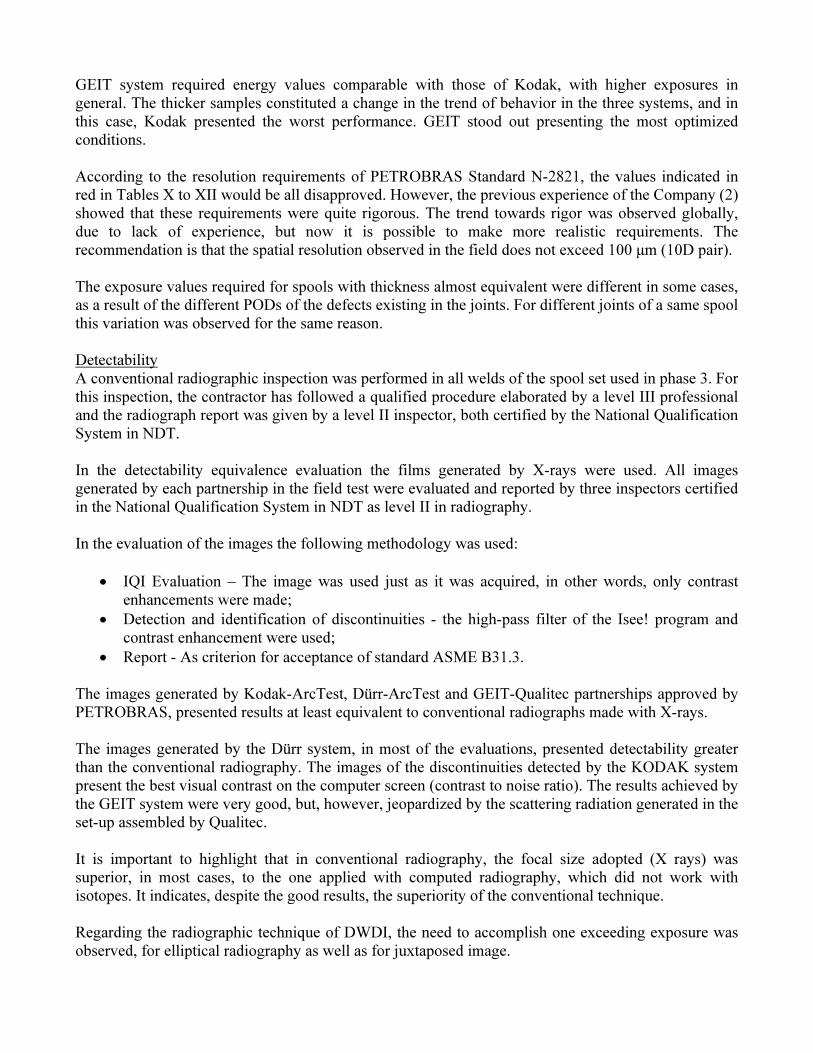
According to the resolution requirements of PETROBRAS Standard N-2821, the values indicated in
red in Tables X to XII would be all disapproved. However, the previous experience of the Company (2)
showed that these requirements were quite rigorous. The trend towards rigor was observed globally,
due to lack of experience, but now it is possible to make more realistic requirements. The
recommendation is that the spatial resolution observed in the field does not exceed 100 µm (10D pair).
The exposure values required for spools with thickness almost equivalent were different in some cases,
as a result of the different PODs of the defects existing in the joints. For different joints of a same spool
this variation was observed for the same reason.
Detectability
A conventional radiographic inspection was performed in all welds of the spool set used in phase 3. For
this inspection, the contractor has followed a qualified procedure elaborated by a level III professional
and the radiograph report was given by a level II inspector, both certified by the National Qualification
System in NDT.
In the detectability equivalence evaluation the films generated by X-rays were used. All images
generated by each partnership in the field test were evaluated and reported by three inspectors certified
in the National Qualification System in NDT as level II in radiography.
In the evaluation of the images the following methodology was used:
• IQI Evaluation – The image was used just as it was acquired, in other words, only contrast
enhancements were made;
• Detection and identification of discontinuities - the high-pass filter of the Isee! program and
contrast enhancement were used;
• Report - As criterion for acceptance of standard ASME B31.3.
The images generated by Kodak-ArcTest, Dürr-ArcTest and GEIT-Qualitec partnerships approved by
PETROBRAS, presented results at least equivalent to conventional radiographs made with X-rays.
The images generated by the Dürr system, in most of the evaluations, presented detectability greater
than the conventional radiography. The images of the discontinuities detected by the KODAK system
present the best visual contrast on the computer screen (contrast to noise ratio). The results achieved by
the GEIT system were very good, but, however, jeopardized by the scattering radiation generated in the
set-up assembled by Qualitec.
It is important to highlight that in conventional radiography, the focal size adopted (X rays) was
superior, in most cases, to the one applied with computed radiography, which did not work with
isotopes. It indicates, despite the good results, the superiority of the conventional technique.
Regarding the radiographic technique of DWDI, the need to accomplish one exceeding exposure was
observed, for elliptical radiography as well as for juxtaposed image.

3. CONCLUSIONS
Due to the advantages inherent to computed radiography (CR), PETROBRAS has sought to replace the
conventional radiography in its works by the referred technique. However, this process requires a lot of
study and experimental work.
The project ends with the qualification of 13 ranges of diameter and thickness for the Kodak-ArcTest
partnership, 11 ranges for the Dürr-ArcTest partnership and 6 ranges for the GEIT-Qualitec
partnership.
The partnerships could have qualified more comprehensive ranges, especially with the Dürr and GEIT
systems, but they have failed in yield due to lack of technical bases in the understanding of computed
radiography or familiarity with the systems. Certainly the results can be improved, but it depends on
the contractor’s efforts to become more qualified. PETROBRAS intends to invest in the CR technique
and qualify more ranges of diameter and thickness, even in the scope of double wall single image
techniques (DWSI) and single wall single image (SWSI).
The criteria of PETROBRAS Standard N-2821, as well as the international standards, need to be
revised. For inspection of welds with computed radiography, a limit value of acceptable resolution for
most of the situations found in the oil industry would be equal to 100 µm. The required values of SNRN
also need to be adjusted. Different thresholds of SNRN for each system were observed. These
thresholds ensure detectability equivalent with the conventional radiography within the bypass
conditions tested in this project.
The CR detectability continuously improves as the radiation dose increases, and equivalence was
achieved for energy values superior to those used for conventional radiography.
The images generated by the Kodak-ArcTest, Dürr-ArcTest and GEIT-Qualitec partnerships in all
situations, presented results at least equivalent to conventional radiographs made with X-rays. Dürr
System stood out in most of the evaluations, with detectability greater than in conventional
radiography.
However, it is important to highlight that, in the conventional radiography, the focal size adopted (X-
rays) was superior, in most cases, to the one applied with computed radiography, which did not work
with isotopes. It indicates, despite the good results, the superiority of the conventional technique.
The replacement of the conventional radiography by computed radiography in weld inspection is
perfectly feasible, although the new technique still needs improvements. Due to the lack of experience
in the country and worldwide, the replacement process should be conducted cautiously.
4. ACKNOWLEDGMENTS
The authors acknowledge the support of PETROBRAS, the Brazilian Energy Company, and wish to
thank their colleagues at Federal University of Rio de Janeiro, Laboratório de Instrumentação Nuclear
(LIN/UFRJ) that contributed for the performance of this project. We also thank the Bundesanstalt für
Materialforschung und –Prüfung (BAM) for making the software ISee! available.
Acknowledgments to CIT, NDT do Brasil and Brasitest for their participation in the project.

5. BIBLIOGRAPHIC REFERENCES
1 - EWERT U., ZSCHERPEL U., Bavendiek K., "Strategies for Film Replacement in Radiography -
Films and Digital Detectors in Comparison", In: Proceedings of 17th World Conference on
Nondestructive Testing, Shanghai, China, Oct 2008.
2 - MARINHO C.A., Lopes R.T., Rebello J.M.A., "Film Replacement at Digital Techniques in Weld
Inspection", Materials Evaluation, July 2008, pp529-539.