Embed Size (px)
Citation preview

Computational Analysis of Methyl Transfer Reactions in DengueVirus MethyltransferaseTobias Schmidt,† Torsten Schwede,† and Markus Meuwly*,‡
‡Department of Chemistry, University of Basel, Klingelbergstrasse 80, 4056 Basel, Switzerland†SIB Swiss Institute of Bioinformatics, Basel, Switzerland Biozentrum, University of Basel, Klingelbergstrasse 50/70, 4056 Basel,Switzerland
ABSTRACT: S-Adenosyl-L-methionine (SAM) dependent methyltransferases(MTases) play crucial roles in many biological processes. The MTase of the denguevirus is of particular interest for the development of antiviral drugs against flaviviruses. Itcatalyzes two distinct methylation reactions at the N7 and the 2′O position of the viralRNA cap structure. Based on density functional theory (DFT) electronic structurecalculations, the molecular basis of the underlying chemical reactions involved in the N7and the 2′O methyl transfer reactions of this enzyme were investigated using modelsystems. Calculations in the condensed phase show that both reactions are exergonicwith significant activation barriers of 13.7 and 17.6 kcal/mol and stable product states,stabilized by 23.5 and 16.9 kcal/mol compared to the reactant states for the N7 and the2′O reaction, respectively. We find that the reaction rate for the 2′O reaction issignificantly enhanced in the presence of the native proton acceptor group, which lowersthe activation barrier in the catalyzed reaction by 3.8 kcal/mol compared to theuncatalyzed reaction in aqueous solution. Furthermore, the 2′O reaction involves amethyl and a proton transfer reaction. Our results suggest that these two reactions occur in a concerted fashion in which themethyl group and the proton are transferred simultaneously. From a therapeutic viewpoint, SAM analogues stable underphysiological conditions are particularly relevant. One such compound in MeAzaSAM, an isostructural mimic of SAM, for whichthe present calculations suggest that the methyl transfer reaction is unlikely to occur under biologically relevant conditions.
■ INTRODUCTION
Methyl transfer reactions play crucial roles in many biologicalprocesses including metabolism, signaling, regulation, andmolecular recognition. The transfer of methyl groups ontoparticular substrates is catalyzed by methyltransferases(MTases). Overall, more than 150 different reactions arecatalyzed by various MTases with diverse methylation targetsincluding small molecules, proteins, nucleic acids, and glycans.Methyl group acceptor atoms can be nitrogen, oxygen, carbon,sulfur, or halides.1
Although several MTase classes are known, most reactionsare catalyzed by S-adenosyl-L-methionine- (SAM) dependentMTases where SAM acts as a methyl group donor to methylatethe substrate.2 Other methyl donors include tetrahydrofolicacid and betaine, but biologically, SAM is largely preferred dueto the associated highly favorable reaction energetics.3
SAM is a high energy compound in which the methyl group,activated by a sulfonium center,4 induces a partial positivecharge on the neighboring methyl group.1 Although the methyltransfer reaction is thermodynamically highly favorable with afree energy gain of ∼17 kcal/mol,2 the reaction is oftenkinetically inhibited and thus enzymatic catalysis is oftenrequired. Generally, the methylation reaction is assumed tooccur in an SN2-type reaction as shown in Figure 1.Enzymatic catalysis largely facilitates this reaction through
two different effects: first, by correctly arranging the two
substrates in close proximity and thereby increasing theeffective concentration of the reaction partners;5 second,through activation of the attacking nucleophile which canoccur by abstraction of a proton or by alignment of anucleophile lone pair, which is of particular importance in caseof a protonated nucleophile where the overall reactionrepresents a substitution of a hydrogen atom by a methylgroup.6,7
Since MTases play key roles in many biological processes,they are potential drug targets for a wide variety of diseases,with applications against cancer,8 neurodegeneration,9 viralinfections,10 and many others. One SAM-dependent MTase ofparticular interest for the development of antiviral drugs againstflaviviruses is the MTase located at the N-terminal domain ofthe nonstructural protein 5 (NS5) of the dengue virus.11 ThisMTase is essential in the maturation process of the viralgenome in which a type 1 cap structure is added at the 5′-endof the viral RNA.12 This cap structure is vital for viralreplication, since it ensures RNA stability by protecting againstRNases and it enhances recognition by the ribosomes.13,14 Thecapping process includes two methylation reactions: first, at thecap guanosine amino group in the 7 position (N7) and second,
Received: March 22, 2014Revised: May 7, 2014Published: May 9, 2014
Article
pubs.acs.org/JPCB
© 2014 American Chemical Society 5882 dx.doi.org/10.1021/jp5028564 | J. Phys. Chem. B 2014, 118, 5882−5890
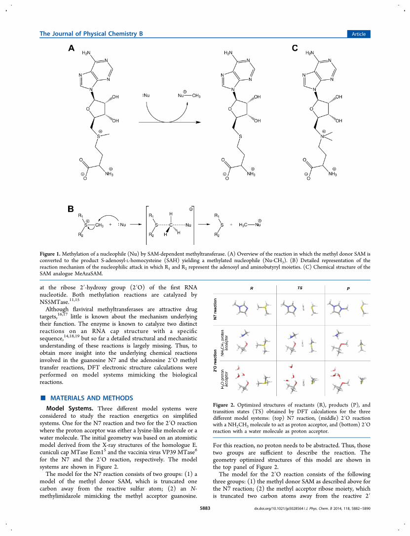
at the ribose 2′-hydroxy group (2′O) of the first RNAnucleotide. Both methylation reactions are catalyzed byNS5MTase.11,15
Although flaviviral methyltransferases are attractive drugtargets,16,17 little is known about the mechanism underlyingtheir function. The enzyme is known to catalyze two distinctreactions on an RNA cap structure with a specificsequence,14,18,19 but so far a detailed structural and mechanisticunderstanding of these reactions is largely missing. Thus, toobtain more insight into the underlying chemical reactionsinvolved in the guanosine N7 and the adenosine 2′O methyltransfer reactions, DFT electronic structure calculations wereperformed on model systems mimicking the biologicalreactions.
■ MATERIALS AND METHODS
Model Systems. Three different model systems wereconsidered to study the reaction energetics on simplifiedsystems. One for the N7 reaction and two for the 2′O reactionwhere the proton acceptor was either a lysine-like molecule or awater molecule. The initial geometry was based on an atomisticmodel derived from the X-ray structures of the homologue E.cuniculi cap MTase Ecm15 and the vaccinia virus VP39 MTase6
for the N7 and the 2′O reaction, respectively. The modelsystems are shown in Figure 2.The model for the N7 reaction consists of two groups: (1) a
model of the methyl donor SAM, which is truncated onecarbon away from the reactive sulfur atom; (2) an N-methylimidazole mimicking the methyl acceptor guanosine.
For this reaction, no proton needs to be abstracted. Thus, thosetwo groups are sufficient to describe the reaction. Thegeometry optimized structures of this model are shown inthe top panel of Figure 2.The model for the 2′O reaction consists of the following
three groups: (1) the methyl donor SAM as described above forthe N7 reaction; (2) the methyl acceptor ribose moiety, whichis truncated two carbon atoms away from the reactive 2′
Figure 1.Methylation of a nucleophile (Nu) by SAM-dependent methyltransferase. (A) Overview of the reaction in which the methyl donor SAM isconverted to the product S-adenosyl-L-homocysteine (SAH) yielding a methylated nucleophile (Nu-CH3). (B) Detailed representation of thereaction mechanism of the nucleophilic attack in which R1 and R2 represent the adenosyl and aminobutyryl moieties. (C) Chemical structure of theSAM analogue MeAzaSAM.
Figure 2. Optimized structures of reactants (R), products (P), andtransition states (TS) obtained by DFT calculations for the threedifferent model systems: (top) N7 reaction, (middle) 2′O reactionwith a NH2CH3 molecule to act as proton acceptor, and (bottom) 2′Oreaction with a water molecule as proton acceptor.
The Journal of Physical Chemistry B Article
dx.doi.org/10.1021/jp5028564 | J. Phys. Chem. B 2014, 118, 5882−58905883
hydroxy group; (3) the side chain of Lys181, truncated onecarbon atom away from the Nζ, which acts as a protonacceptor. The geometry optimized structures of this model areshown in the middle panel of Figure 2.A third model was designed to investigate the uncatalyzed
2′O reaction. It is identical to that for the catalyzed 2′Oreaction, except that the proton acceptor moiety is replaced bya water molecule that accepts the transferred proton. Tostabilize this water molecule, two additional water moleculeswere added, which are hydrogen bonded to the active water butdo not directly participate in the reaction. The geometryoptimized structures of this model are shown in the bottompanel of Figure 2.Model systems for the SAM analogue MeAzaSAM were
generated from the optimized geometries of the SAM modelsystems in the product and the reactant state by manualreplacement of the sulfur atom by a nitrogen atom and additionof a methyl group. MeAzaSAM systems were subsequentlyenergy minimized as described below.Electronic Structure Calculations. All electronic structure
calculations were performed with Gaussian0320 using theB3LYP21 density functional theory method and the 6-311++G(d,p) basis set.22 A similar level of theory was alreadyemployed in studying the Menshutkin reaction.23 Solvationeffects were included during both optimization and energycalculations based on the C-PCM implicit solvent model.24 Forall model systems, the reactant and the product complexes werefully optimized in implicit solvent. Subsequently, using theobtained structures, transition state optimization with QST325
was carried out. During all optimizations, all atoms were free tomove. For all fully optimized structures, vibrational analyseswere performed at the same level of theory in order to confirmthe nature of the stationary points and to compute thermalcorrections to the Gibbs free energy which include transla-tional, rotational, and vibrational contributions. From theobtained energies, a free energy profile for the reaction wascalculated and point charges were computed based on a naturalbond orbital (NBO) analysis.26
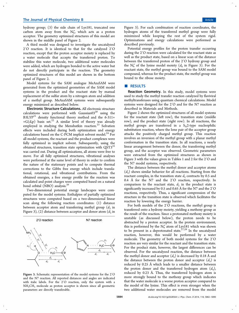
Two-dimensional potential energy landscapes were com-puted for the model systems. Enthalpies of partially optimizedstructures were computed based on a two-dimensional linearscan along the following reaction coordinates: (1) distancebetween acceptor atom and transferring methyl group (d3 inFigure 3), (2) distance between acceptor and donor atom (d2 in
Figure 3). For each combination of reaction coordinates, thehydrogen atoms of the transferred methyl group were fullyminimized while keeping the rest of the system rigid.Optimizations and energy calculations were performed asdescribed previously.Potential energy profiles for the proton transfer occurring
during the 2′O reaction were calculated for the reactant state aswell as the product state, based on a linear scan of the distancebetween the transferred proton of the 2′O hydroxy group andthe Nζ of the lysine model moiety (d6 in Figure 3). For thereactant state, the methyl group was bound to the SAM modelcompound, whereas for the product state, the methyl group wasbound to the ribose moiety.
■ RESULTSReaction Geometry. In this study, model systems were
used to study the methyl transfer reaction catalyzed by flaviviralmethyltransferases using quantum chemical calculations. Modelsystems were designed for the 2′O and for the N7 reaction asdescribed in Materials and Methods.Figure 2 shows the optimized structures of all model systems
for the reactant state (left row), the transition state (middlerow), and the product state (right row). In all reactions, themethyl groups are transferred in a SN2-type nucleophilicsubstitution reaction, where the lone pair of the acceptor groupattacks the positively charged methyl group. This reactioninvolves an inversion of the methyl group with a planar methylconformation in the transition state. In all reactions, a nearlylinear arrangement between the donor, the transferring methylgroup, and the acceptor was observed. Geometric parameterswere extracted from the optimized structures as shown inFigure 3 with the values given in Tables 1 and 2 for the 2′O andthe N7 model systems, respectively.The distance between the methyl donor and acceptor atoms
(d2) shows similar behavior for all reactions. Starting from thereactant complex, in the transition state d2 contracts by 0.5 and0.4 Å for the N7 and the 2′O reaction, respectively. Incomparison to the reactant state, d2 in the product state issignificantly increased by 0.5 and 0.65 Å for the N7 and the 2′Oreactions, respectively. Thus, a significant compression of thestructure in the transition state is observed which facilitates thereaction by lowering the energy barrier.For both models of the 2′O reactions, the methyl group is
transferred onto a hydroxy moiety, yielding a methoxy group asthe result of the reaction. Since a protonated methoxy moiety isunstable (as discussed below), the proton needs to beabstracted by a proton acceptor. In the protein environment,this is performed by the Nζ atom of Lys181 which was shownto be present in a deprotonated state.27,28 In the uncatalyzedreaction, however, this would be performed by a watermolecule. The geometry of both model systems for the 2′Oreaction are very similar for the reactant and the transition state.For the product state, however, the largest differences can beobserved. For the uncatalyzed reaction, the distance betweenthe methyl donor and acceptor (d2) is decreased by 0.18 Å andthe distance between the proton donor and acceptor (d5) isreduced by 0.25 Å which leads to a smaller distance betweenthe proton donor and the transferred hydrogen atom (d4),reduced by 0.23 Å. Thus, the transferred hydrogen atom ismore strongly bound to the methoxy group which indicatesthat the water molecule is a worse proton acceptor compared tothe model of the lysine. This effect is even stronger when thetwo additional water molecules are removed from the model
Figure 3. Schematic representation of the model systems for the 2′Oand the N7 reaction. All reported distances and angles are indicatedwith italic labels. For the 2′O reaction, only the system with aNH2CH3 molecule as proton acceptor is shown since all geometricparameters are directly transferable.
The Journal of Physical Chemistry B Article
dx.doi.org/10.1021/jp5028564 | J. Phys. Chem. B 2014, 118, 5882−58905884

system (i.e., leaving only the reactive water molecule). In thatcase, no proton transfer is observed yielding a protonatedmethoxy group in the product state (data not shown).Energy Profiles. The Gibbs free energies were calculated
for all geometries of the optimized structures of all modelsystems for reactant (left), transition (middle), and productstate (right) and are illustrated in Figure 4. Table 3 summarizesall free energies.
For both the 2′O as well as the N7 system, the calculationsreveal that the reactions are exergonic processes where theproduct state is energetically significantly lowered compared tothe reactant state with an energetically unfavorable transitionstate in between. The Gibbs free energy difference betweenproduct and reactant is −23.5 kcal/mol for the N7 reaction and−16.9 kcal/mol for the catalyzed 2′O reaction. For thetransition state, an energy barrier of 13.7 and 17.6 kcal/mol
is observed for N7 and catalyzed 2′O reactions, respectively.From experimentally determined turnover numbers (kcat) forthe 2′O reaction, the activation barrier of this reaction can beestimated with transition state theory29 using the formula kcat =(kBT)/h·exp(−ΔG‡/(RT)) where ΔG‡ is the activation barrier,T the temperature, R the universal gas constant, kB theBoltzmann constant, and h the Planck constant. kcat values havebeen reported between 2.47 × 10⁻³ s−1 and 3.25 × 10⁻⁴ s−1
which yield estimated activation barriers in the range 21.0−22.2kcal/mol.30,31 Thus, the calculated activation energy barriers arein a similar range indicating that the postulated SN2-typemethyl transfer reaction is energetically feasible. It should benoted that DFT methods are known to generally underestimatebarrier heights.32
When the uncatalyzed 2′O methyl transfer reaction iscompared to the catalyzed one (Table 3 columns 1 and 2), asignificant reduction of the product state free energy can beobserved in the catalyzed reaction. The product state energy islowered by 10.6 kcal/mol and the reaction energy barrier islowered by 3.8 kcal/mol which, based on transition statetheory, corresponds to a reaction rate enhancement of ∼600times.In summary, for the 2′O reaction, the data suggests the
importance of the correct proton acceptor moiety. In thecatalyzed reaction, a lysine residue, representing Lys181 in theactive site of the protein, acts as a proton acceptor andsignificantly stabilizes the product state and reduces theactivation barrier compared to the reaction in aqueous solution.The N7 methyl transfer reaction, on the other hand, has asignificantly lower energy barrier to overcome in aqueoussolution and is thus more likely to occur without direct proteininteractions. In conclusion, the data agrees well with themechanistic hypothesis, where the 2′O reaction needs directinvolvement of protein residues, whereas the N7 methylation
Table 1. Geometric parameters obtained from minimized structures for model systems of the 2′O reaction (Figure 3)a
NH2CH3 proton acceptor H2O proton acceptor
reactant complex transition state product complex reactant complex transition state product complex
d1 (Å) 1.82 2.39 4.00 1.82 2.39 3.82d2 (Å) 4.78 4.36 5.43 4.82 4.36 5.25d3 (Å) 2.96 1.97 1.43 2.99 1.97 1.44d4 (Å) 0.99 1.04 1.72 0.98 1.00 1.49d5 (Å) 2.86 2.66 2.77 2.80 2.62 2.52d6 (Å) 1.87 1.63 1.05 1.83 1.63 1.03α1 (deg) 176.9 177.0 176.2 177.7 177.1 175.7α2 (deg) 174.3 175.0 174.9 173.5 171.2 174.1
aEither NH3CH3, mimicking Lys181 (left columns), or a water molecule (right columns), is present in the model system to act as a proton acceptor.
Table 2. Geometric Parameters Obtained from MinimizedStructures for Model Systems of the N7 Reaction (Figure 3)
reactant complex transition state product complex
d1 (Å) 1.82 2.30 3.98d2 (Å) 4.94 4.40 5.44d3 (Å) 3.12 2.11 1.47α1 (deg) 177.4 180.0 176.6
Figure 4. Gibbs free energy profile for reactants, product, andtransition state, obtained from DFT calculations for the N7 and the2′O methyl transfer reaction. For the 2′O reaction, either NH3CH3,mimicking Lys181, or a water molecule is present in the model systemto act as a proton acceptor.
Table 3. Summary of the Gibbs Free Energies for Reactant,Product, and Transition State, Obtained from Ab InitioModel System Calculations for the N7 and the 2′O MethylTransfer Reactiona
2′O reaction (NH2CH3proton acceptor)
2′O reaction (H2Oproton acceptor)
N7reaction
reactantcomplex
0.0 0.0 0.0
transitionstate
17.6 21.4 13.7
productcomplex
−16.9 −6.3 −23.5
aFor the 2′O reaction, either NH3CH3, mimicking Lys181, or a watermolecule is present in the model system to act as a proton acceptor.
The Journal of Physical Chemistry B Article
dx.doi.org/10.1021/jp5028564 | J. Phys. Chem. B 2014, 118, 5882−58905885
does not need direct contact with the protein as long as the tworeactants are in close proximity.5
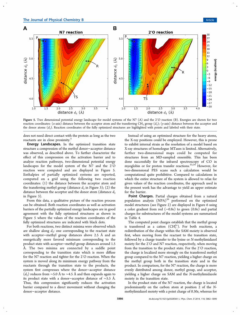
Energy Landscapes. In the optimized transition statestructure a compression of the methyl donor−acceptor distancewas observed, as described above. To further characterize theeffect of this compression on the activation barrier and toanalyze reaction pathways, two-dimensional potential energylandscapes for the model system of the N7 and the 2′Oreaction were computed and are displayed in Figure 5.Enthalpies of partially optimized systems are reported,computed on a grid using the following two reactioncoordinates: (1) the distance between the acceptor atom andthe transferring methyl group (distance d3 in Figure 3); (2) thedistance between the acceptor and the donor atom (distance d2in Figure 3).From this data, a qualitative picture of the reaction process
can be obtained. Both reaction coordinates as well as activationbarriers of the partially optimized energy landscapes are in goodagreement with the fully optimized structures as shown inFigure 5 where the values of the reaction coordinates of thefully optimized structures are indicated with black dots.For both reactions, two distinct minima were observed which
are shallow along d3: one corresponding to the reactant statewith acceptor−methyl group distances above 2.5 Å and anenergetically more favored minimum corresponding to theproduct state with acceptor−methyl group distances around 1.5Å. The two minima are connected by a saddle pointcorresponding to the transition state which is more diffusefor the N7 reaction and tighter for the 2′O reaction. When thesystem is moved along its minimum energy pathway from thereactants through the transition state to the products, thesystem first compresses where the donor−acceptor distance(d2) reduces from ∼5.0 Å to ∼4.5 Å and then expands again toits product state with a donor−acceptor distance of ∼5.5 Å.Thus, this compression significantly reduces the activationbarrier compared to a direct movement without changing thedonor−acceptor distance.
Instead of using an optimized structure for the heavy atoms,the X-ray positions could be employed. However, this is proneto exhibit internal strain as the resolution of a model based onX-ray structures of homologue MTases is limited. Alternatively,further two-dimensional maps could be computed forstructures from an MD-sampled ensemble. This has beendone successfully for the infrared spectroscopy of CO inmyoglobin or for proton transfer reactions.33,34 However, fortwo-dimensional PES scans such a calculation would becomputational quite prohibitive. Compared to calculations inwhich the entire structure of ths system is allowed to relax forgiven values of the reaction coordinates, the approach used inthe present work has the advantage to yield an upper estimatefor the barrier.
Point Charges. Partial charges obtained from a naturalpopulation analysis (NPA)26 performed on the optimizedmodel structures (see Figure 2) are displayed in Figure 6 usinga color gradient from red (−0.8e) to green (0.8e) and partialcharges for substructures of the model systems are summarizedin Table 4.The computed point charges establish that the methyl group
is transferred as a cation (CH3δ+). For both reactions, a
redistribution of the charge within the SAM moiety is observedfirst, when moving from the reactant to the transition state,followed by a charge transfer to the lysine or N-methylimidazolmoiety for the 2′O and N7 reaction, respectively, when movingfrom the transition to the product state. For the 2′O reaction,the charge is localized more strongly on the transferred methylgroup compared to the N7 reaction, yielding a higher charge onthe methyl group both in the transition state and in theproduct. In comparison, for the N7 reaction, the charge is moreevenly distributed among donor, methyl group, and acceptor,yielding a higher charge on SAM and the N-methylimidazolemoiety in the transition state.In the product state of the N7 reaction, the charge is located
predominantly on the carbon atom at position 2 of the N-methylimidazol moiety with a point charge of 0.30e, whereas for
Figure 5. Two dimensional potential energy landscape for model systems of the N7 (A) and the 2′O reaction (B). Energies are shown for tworeaction coordinates: (x-axis) distance between the acceptor atom and the transferring CH3 group (d3); (y-axis) distance between the acceptor andthe donor atoms (d2). Reaction coordinates of the fully optimized structures are highlighted with points and labeled with their state.
The Journal of Physical Chemistry B Article
dx.doi.org/10.1021/jp5028564 | J. Phys. Chem. B 2014, 118, 5882−58905886

the 2′O reaction, the charge is located to a large extent on theproton which was transferred to the model lysine moiety, with acharge of 0.46e.Evidence for a Concerted Mechanism. Methyl transfer
reactions to aromatic nitrogen acceptors typically do not needgeneral base catalysis since orienting the nitrogen lone pairtoward the SAM methyl group is sufficient. On the contrary, inthe case of hydroxyl oxygen acceptors a general base mediatesthe substitution of a proton by a methyl group where theproton can be abstracted before, during, or after the methyltransfer reaction.1 For the DENV MTase 2′O reaction thecatalytic lysine residue acts as a proton acceptor. However, sofar, it is unknown if the methyl transfer and the proton transferoccur as two individual steps or as a single concerted reaction.In a two-step mechanism the catalytic lysine residuedeprotonates the 2′-hydroxy group initially. This leads to theformation of a 2′-oxyanion onto which the methyl group istransferred in a subsequent step. In a concerted mechanism onthe other hand, the proton is transferred to the catalytic lysinesimultaneously to the methyl transfer. Thus, prior to the
reaction, the proton acceptor does not deprotonate the 2′-hydroxy group but steers its orientation. NMR experimentsindicate that the latter mechanism is operative in the enzymaticreaction of vaccinia virus mRNA cap specific 2′O MTaseVP39.27
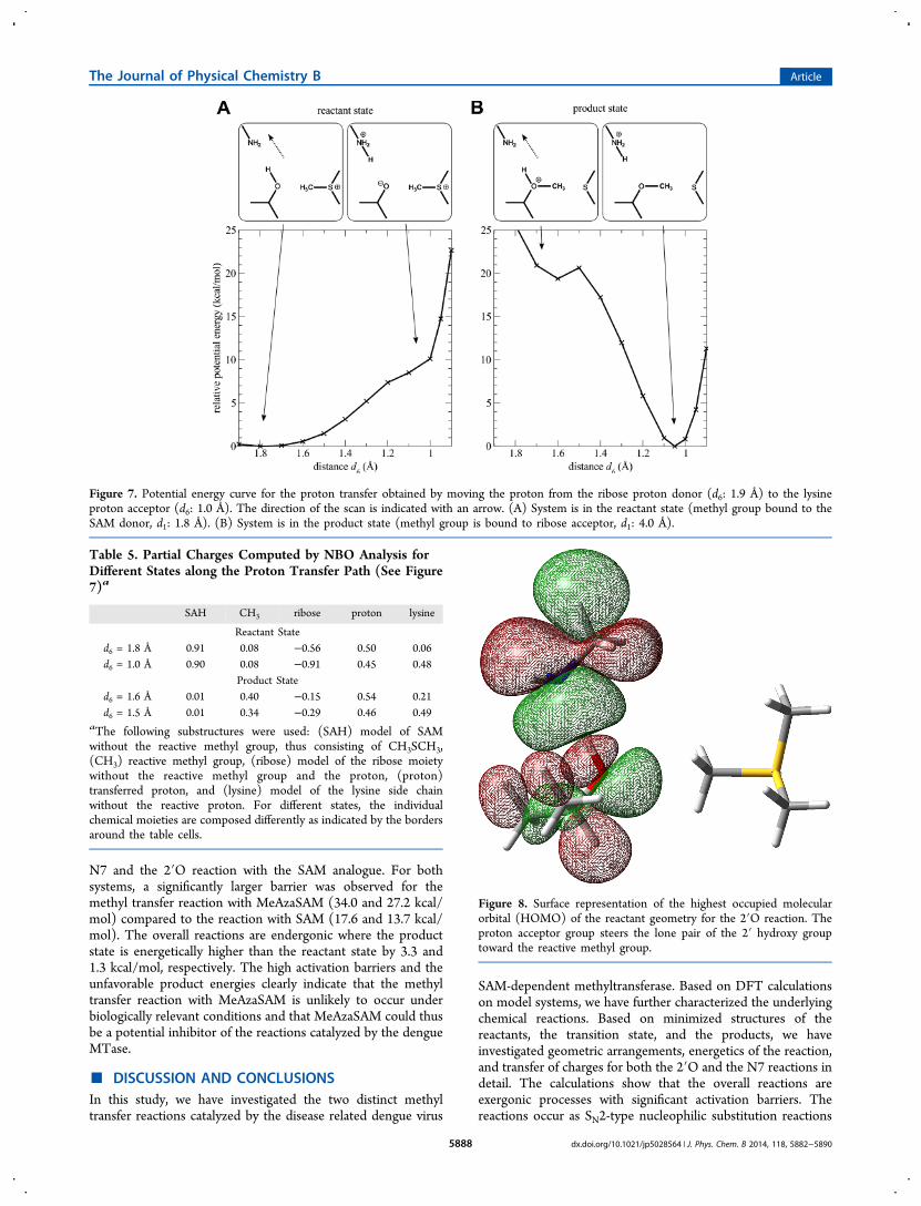
Geometry optimized model structures support a concertedmechanism, as in the reactant and the transition state theproton is still bound to the ribose model compound and onlytransferred to the proton acceptor in the product state. Thedistance between the 2′-oxygen atom and the proton (d4) is0.99, 1.04, and 1.72 Å for reactants, transition state, andproducts, respectively.To confirm this, a linear scan was performed by moving the
proton from the 2′O hydroxy group to the Nζ of the lysinemodel moiety (distance d6 in Figure 3). During the scan, themethyl group was fixed in the reactant state and thus bound tothe SAM model compound. Therefore, the end point of thescan yields a 2′-oxyanion. The energy profile is shown in Figure7A and the partial charges are summarized in Table 5. Thecalculations estimate the cost for proton transfer prior to themethyl transfer to ∼10 kcal/mol. The profile increasesmonotonically and no significant minimum was observed.These findings suggest a concerted mechanism as the oxyanionconformation is not stabilized.It should be noted that, when the methyl group is moved to
its product state, i.e., bound to the ribose moiety, minimizationof the structure yields a spontaneous proton transfer with noobservable barrier in between. This was confirmed by a linearscan of the proton position when the methyl group is in itsproduct state (Figure 7B). This scan estimates the energy gainfor the proton transfer in the product state to ∼20 kcal/mol.To summarize, these calculations show that the two modeled
intermediate states are unstable and thus suggest that the tworeactions occur in a concerted fashion where no oxyanion holeis generated but the orientation of the proton acceptor steersthe 2′O hydroxy lone pair into the direction of the nucleophilicattack. This is also indicated by the electron densitydistributions as shown in Figure 8.
Inhibition by SAM Analogues. Analogues of SAM thatare stable under physiological conditions and still retain theirbinding affinity to their target protein have been described inthe literature.35−37 To investigate the effect of such analogueson the methyl transfer reaction and the mechanism ofinhibition, we have extended our study to the stable quaternarydimethylammonium analogue of SAM (MeAzaSAM), anisostructural mimic which also carries a positive charge (Figure1C).Table 6 shows the Gibbs free energy profiles for optimized
structures of the reactant, transition, and product state for the
Figure 6. NBO point charges from ab initio calculations mapped ontothe model systems for the N7 (left) and the 2′O reaction (right). Thecolor gradient ranges from −0.8 (red) to 0.8e (green). Numericalvalues for substructures are given in Table 4.
Table 4. Partial Charges Computed by Natural Bond Orbital Analysis for Substructures of the Model Systems for the 2′O andthe N7 Reactiona
2′O reaction N7 reaction
SAH CH3 ribose proton lysine SAH CH3 guanine
reactant 0.92 0.07 −0.54 0.49 0.05 0.90 0.08 0.01TS 0.41 0.32 −0.36 0.51 0.12 0.48 0.25 0.27product 0.01 0.34 −0.29 0.46 0.49 0.01 0.19 0.80
aThe following substructures were used: (SAH) model of SAM without the reactive methyl group, thus consisting of CH3SCH3, (CH3) reactivemethyl group, (ribose) model of the ribose moiety without the reactive methyl group and the proton, (proton) transferred proton, (lysine) model ofthe lysine side chain without the transferred proton for the 2′O reaction, (guanine) model of the guanine acceptor for the N7 reaction. For differentstates, the individual chemical moieties are composed differently as indicated by the borders around the table cells.
The Journal of Physical Chemistry B Article
dx.doi.org/10.1021/jp5028564 | J. Phys. Chem. B 2014, 118, 5882−58905887
N7 and the 2′O reaction with the SAM analogue. For bothsystems, a significantly larger barrier was observed for themethyl transfer reaction with MeAzaSAM (34.0 and 27.2 kcal/mol) compared to the reaction with SAM (17.6 and 13.7 kcal/mol). The overall reactions are endergonic where the productstate is energetically higher than the reactant state by 3.3 and1.3 kcal/mol, respectively. The high activation barriers and theunfavorable product energies clearly indicate that the methyltransfer reaction with MeAzaSAM is unlikely to occur underbiologically relevant conditions and that MeAzaSAM could thusbe a potential inhibitor of the reactions catalyzed by the dengueMTase.
■ DISCUSSION AND CONCLUSIONSIn this study, we have investigated the two distinct methyltransfer reactions catalyzed by the disease related dengue virus
SAM-dependent methyltransferase. Based on DFT calculationson model systems, we have further characterized the underlyingchemical reactions. Based on minimized structures of thereactants, the transition state, and the products, we haveinvestigated geometric arrangements, energetics of the reaction,and transfer of charges for both the 2′O and the N7 reactions indetail. The calculations show that the overall reactions areexergonic processes with significant activation barriers. Thereactions occur as SN2-type nucleophilic substitution reactions
Figure 7. Potential energy curve for the proton transfer obtained by moving the proton from the ribose proton donor (d6: 1.9 Å) to the lysineproton acceptor (d6: 1.0 Å). The direction of the scan is indicated with an arrow. (A) System is in the reactant state (methyl group bound to theSAM donor, d1: 1.8 Å). (B) System is in the product state (methyl group is bound to ribose acceptor, d1: 4.0 Å).
Table 5. Partial Charges Computed by NBO Analysis forDifferent States along the Proton Transfer Path (See Figure7)a
SAH CH3 ribose proton lysine
Reactant Stated6 = 1.8 Å 0.91 0.08 −0.56 0.50 0.06d6 = 1.0 Å 0.90 0.08 −0.91 0.45 0.48
Product Stated6 = 1.6 Å 0.01 0.40 −0.15 0.54 0.21d6 = 1.5 Å 0.01 0.34 −0.29 0.46 0.49
aThe following substructures were used: (SAH) model of SAMwithout the reactive methyl group, thus consisting of CH3SCH3,(CH3) reactive methyl group, (ribose) model of the ribose moietywithout the reactive methyl group and the proton, (proton)transferred proton, and (lysine) model of the lysine side chainwithout the reactive proton. For different states, the individualchemical moieties are composed differently as indicated by the bordersaround the table cells.
Figure 8. Surface representation of the highest occupied molecularorbital (HOMO) of the reactant geometry for the 2′O reaction. Theproton acceptor group steers the lone pair of the 2′ hydroxy grouptoward the reactive methyl group.
The Journal of Physical Chemistry B Article
dx.doi.org/10.1021/jp5028564 | J. Phys. Chem. B 2014, 118, 5882−58905888

with linear arrangements, compressed transition states, and atransfer of the methyl group as a cationic species.For the 2′O reaction, we found that the “protein”
environment substantially lowers the activation barrier,mediated by an active site lysine residue, representing Lys181in the active site of the protein. This lysine residue acts as aproton acceptor during the methyl transfer reaction andstabilizes the transition state as well as the product statecompared to the uncatalyzed reaction in aqueous solution.The overall 2′O reaction is composed of two distinct
reactions: a methyl transfer and a proton transfer reaction.Based on geometry optimizations and potential energy scans,we have shown that these two reactions occur in a concertedfashion where during the methyl transfer, the proton istransferred to the proton acceptor. Thus, prior to the reaction,the proton acceptor does not deprotonate the 2′-hydroxy group(no oxyanion hole) but rather steers the orientation of theproton acceptor lone pair.Furthermore, we have investigated the effect on the reaction
and the mechanism of inhibition of stable nitrogen analogues ofSAM which have been described in the literature and whichwere proposed as inhibitors of methyl transfer reactions.Calculations on MeAzaSAM, an isostructural mimic of SAM,show that the overall reactions are endergonic processes withvery large activation barriers. These high activation barriers andthe unfavorable product energies clearly indicated that themethyl transfer reaction with MeAzaSAM is unlikely to occurunder biologically relevant conditions and that this analoguemight thus be a viable inhibitor.Throughout this study the role of dynamics was not included
explicitly. One possibility to include such effects would be atthe level of mixed quantum mechanics/molecular mechanicssimulations.38,39 Such simulations are, however, computation-ally very demanding. Alternatively, the quantum mechanicalpart could be treated at the semiempirical level (e.g., SCC-DFTB40,41) or by resorting to adiabatic reactive moleculardynamics simulations.42−46 This would need to be coupled toenhanced sampling techniques such as umbrella samplingbecause the barriers involved are high.47
■ AUTHOR INFORMATIONCorresponding Author*E-mail: [email protected]. Phone: +41 (0)61 267 38 21.Fax: +41 (0)61 267 38 55.NotesThe authors declare no competing financial interest.
■ ACKNOWLEDGMENTSThe authors would like to thank Dr. Juergen Haas fornumerous fruitful discussions. Financial support from theSwiss National Science Foundation (SNF) through grants
200021-117810 (to MM) and the NCCR-MUST (to MM) isgratefully acknowledged.
■ REFERENCES(1) Klimasauskas, S.; Lukinavicius, G. Wiley Encyclopedia of ChemicalBiology; John Wiley and Sons, Inc., 2007.(2) Cantoni, G. L. Biological Methylation - Selected Aspects. Annu.Rev. Biochem. 1975, 44, 435−451.(3) Schubert, H. L.; Blumenthal, R. M.; Cheng, X. D. Many Paths toMethyltransfer: A Chronicle of Convergence. Trends Biochem. Sci.2003, 28, 329−335.(4) Cantoni, G. L. The Nature of the Active Methyl Donor FormedEnzymatically from L-Methionine and Adenosinetriphosphate. J. Am.Chem. Soc. 1952, 74, 2942−2943.(5) Fabrega, C.; Hausmann, S.; Shen, V.; Shuman, S.; Lima, C. D.Structure and Mechanism of mRNA cap (guanine-N7) Methyltrans-ferase. Mol. Cell 2004, 13, 77−89.(6) Hodel, A. E.; Gershon, P. D.; Quiocho, F. A. Structural Basis forSequence-Nonspecific Recognition of 5′-Capped mRNA by a Cap-Modifying Enzyme. Mol. Cell 1998, 1, 443−447.(7) Hager, J.; Staker, B. L.; Bugl, H.; Jakob, U. Active Site in RrmJ, aHeat Shock-Induced Methyltransferase. J. Biol. Chem. 2002, 277,41978−41986.(8) Copeland, R. A.; Solomon, M. E.; Richon, V. M. ProteinMethyltransferases As a Target Class for Drug Discovery. Nat. Rev.Drug Discuss. 2009, 8, 724−732.(9) Mannisto, P. T.; Kaakkola, S. Catechol-O-methyltransferase(COMT): Biochemistry, Molecular Biology, Pharmacology, AndClinical Efficacy of the New Selective COMT Inhibitors. Pharmacol.Rev. 1999, 51, 593−628.(10) Ferron, F.; Decroly, E.; Selisko, B.; Canard, B. The Viral RNACapping Machinery As a Target for Antiviral Drugs. Antivir. Res. 2012,96, 21−31.(11) Egloff, M. P.; Benarroch, D.; Selisko, B.; Romette, J. L.; Canard,B. An RNA Cap (Nucleoside-2′-O-)-methyltransferase in theFlavivirus RNA Polymerase NS5: Crystal Structure and FunctionalCharacterization. EMBO J. 2002, 21, 2757−2768.(12) Cleaves, G.; Dubin, D. Methylation Stratus of IntracellularDengue Type 2 40 S RNA. Virology 1979, 96, 159−165.(13) Furuichi, Y.; Shatkin, A. J. Viral and Cellular mRNA Capping:Past and Prospects. Adv. Virol. Res. 2000, 55, 135−184.(14) Ray, D.; Shah, A.; Tilgner, M.; Guo, Y.; Zhao, Y. W.; Dong, H.P.; Deas, T. S.; Zhou, Y. S.; Li, H. M.; Shi, P. Y. West Nile Virus 5′-Cap Structure Is Formed by Sequential Guanine N-7 and Ribose 2′-OMethylations by Nonstructural Protein 5. J. Virol. 2006, 80, 8362−8370.(15) Dong, H. P.; Ren, S. P.; Zhang, B.; Zhou, Y. S.; Puig-Basagoiti,F.; Li, H. M.; Shi, P. Y. West Nile Virus Methyltransferase CatalyzesTwo Methylations of the Viral Rna Cap through a Substrate-Repositioning Mechanism. J. Virol. 2008, 82, 4295−4307.(16) Dong, H. P.; Zhang, B.; Shi, P. Y. Flavivirus Methyltransferase:A Novel Antiviral Target. Antivir. Res. 2008, 80, 1−10.(17) Bollati, M.; et al. Structure and Functionality in Flavivirus NS-Proteins: Perspectives for Drug Design. Antivir. Res. 2010, 87, 125−148.(18) Zhou, Y. S.; Ray, D.; Zhao, Y. W.; Dong, H. P.; Ren, S. P.; Li, Z.;Guo, Y.; Bernard, K. A.; Shi, P. Y.; Li, H. M. Structure and Function ofFlavivirus NS5 Methyltransferase. J. Virol. 2007, 81, 3891−3903.(19) Egloff, M. P.; Decroly, E.; Malet, H.; Selisko, B.; Benarroch, D.;Ferron, F.; Canard, B. Structural and Functional Analysis ofMethylation and 5′-RNA Sequence Requirements of Short CappedRNAs by the Methyltransferase Domain of Dengue Virus NS5. J. Mol.Biol. 2007, 372, 723−736.(20) Frisch, M. J. et al. Gaussian 03, rev D.02; Gaussian, Inc.,Wallingford, CT, 2004,.(21) Becke, A. Density-Functional Thermochemistry. III. The RoleOf Exact Exchange. J. Chem. Phys. 1993, 7, 5648−5652.
Table 6. Gibbs Free Energy Profile for Reactants, Product,and Transition State, Obtained from Ab Initio Model SystemCalculations for the N7 (Orange) and the 2′O MethylTransfer Reaction with the Stable SAM AnalogueMeAzaSAM
2′O reaction (NH2CH3 proton acceptor) N7 reaction
reactant complex 0.0 0.0transition state 34.0 27.2product complex 3.3 1.3
The Journal of Physical Chemistry B Article
dx.doi.org/10.1021/jp5028564 | J. Phys. Chem. B 2014, 118, 5882−58905889

(22) Ochterski, J.; Petersson, G.; Montgomery, J. A Complete BasisSet Model Chemistry. V. Extensions to Six or More Heavy Atoms. J.Chem. Phys. 1996, 104, 2598−2619.(23) Castejon, H.; Wiberg, K. Solvent Effects on Methyl TransferReactions. 1. The Menshutkin Reaction. J. Am. Chem. Soc. 1999, 121,2139−2146.(24) Cossi, M.; Rega, N.; Scalmani, G.; Barone, V. Energies,Structures, and Electronic Properties of Molecules in Solution with theC-PCM solvation Model. J. Comput. Chem. 2003, 24, 669−681.(25) Peng, C. Y.; Schlegel, H. B. Combining Synchronous Transitand Quasi-Newton Methods to Find Transition-States. Isr. J. Chem.1993, 33, 449−454.(26) Reed, A. E.; Weinstock, R. B.; Weinhold, F. Natural PopulationAnalysis. J. Chem. Phys. 1985, 83, 735−746.(27) Li, C.; Xia, Y.; Gao, X.; Gershon, P. D. Mechanism of RNA 2′-O-Methylation: Evidence that the Catalytic Lysine Acts to SteerRather than Deprotonate the Target Nucleophile. Biochemistry 2004,43, 5680−5687.(28) Li, C.; Gershon, P. D. pK(a) of the mRNA Cap-Specific 2′-O-Methyltransferase Catalytic Lysine by HSQC NMR Detection of aTwo-Carbon Probe. Biochemistry 2006, 45, 907−917.(29) Eyring, H. The Activated Complex in Chemical Reactions. J.Chem. Phys. 1935, 3, 107−115.(30) Lim, S. P.; Wen, D. Y.; Yap, T. L.; Yan, C. K.; Lescar, J.;Vasudevan, S. G. A Scintillation Proximity Assay for Dengue VirusNS5 2′-O-methyltransferase-kinetic and Inhibition Analyses. Antivir.Res. 2008, 80, 360−369.(31) Selisko, B.; Peyrane, F. F.; Canard, B.; Alvarez, K.; Decroly, E.Biochemical Characterization of the (Nucleoside-2′O)-methyltransfer-ase Activity of Dengue Virus Protein NS5 Using Purified Capped RNAOligonucleotides (7Me)GpppAC(n) and GpppAC(n). J. Gen. Vir.2010, 91, 112−121.(32) Zhao, Y.; Gonzalez-Garcia, N.; Truhlar, D. G. BenchmarkDatabase of Barrier Heights for Heavy Atom Transfer, NucleophilicSubstitution, Association, and Unimolecular Reactions and Its Use toTest Theoretical Methods. J. Phys. Chem. A 2005, 109, 2012−2018.(33) Meuwly, M. ChemPhysChem 2006, 7, 2061.(34) Lutz, S.; Tubert-Brohman, I.; Yang, Y.; Meuwly, M. Water-Assisted Proton Transfer in Ferredoxin I. J. Biol. Chem. 2011, 286,23679−23687.(35) Thompson, M. J.; Mekhalfia, A.; Jakeman, D. L.; Phillips, S. E.V.; Phillips, K.; Porter, J.; Blackburn, G. M. Homochiral Synthesis ofan Aza Analogue of S-Adenosyl-L-methionine (AdoMet) and ItsBinding to the E-Coli Methionine Repressor Protein (MetJ). Chem.Commun. 1996, 791−792.(36) Thompson, M. J.; Mekhalfia, A.; Hornby, D. P.; Blackburn, G.M. Synthesis of Two Stable Nitrogen Analogues of S-adenosyl-L-methionine. J. Org. Chem. 1999, 64, 7467−7473.(37) Joce, C.; Caryl, J.; Stockley, P. G.; Warriner, S.; Nelson, A.Identification of Stable S-Adenosylmethionine (SAM) AnaloguesDerivatised with Bioorthogonal Tags: Effect of Ligands on the Affinityof the E. coli Methionine Repressor, MetJ, for Its Operator DNA. Org.Biomol. Chem. 2009, 7, 635−638.(38) Warshel, A.; Levitt, M. Theoretical Studies Of EnzymicReactions - Dielectric, Electrostatic And Steric Stabilization OfCarbonium-Ion In Reaction Of Lysozyme. J. Mol. Biol. 1976, 103,227−249.(39) Field, M.; Bash, P.; Karplus, M. A Combined Quantum-Mechanical And Molecular Mechanical Potential For Molecular-Dynamics Simulations. J. Comput. Chem. 1990, 11, 700−733.(40) Elstner, M.; Porezag, D.; Jungnickel, G.; Elsner, J.; Haugk, M.;Frauenheim, T.; Suhai, S.; Seifert, G. Self-Consistent-Charge Density-Functional Tight-Binding Method for Simulations of ComplexMaterials Properties. Phys. Rev. B 1998, 58, 7260−7268.(41) Cui, Q.; Elstner, M.; Kaxiras, E.; Frauenheim, T.; Karplus, M. AQM/MM Implementation of the Self-Consistent Charge DensityFunctional Tight Binding (SCC-DFTB) Method. J. Phys. Chem. B2001, 105, 569−585.
(42) Nutt, D. R.; Meuwly, M. Studying Reactive Processes withClassical Dynamics: Rebinding Dynamics in MbNO. Biophys. J. 2006,90, 1191−1201.(43) Danielsson, J.; Meuwly, M. Atomistic Simulation of AdiabaticReactive Processes Based on Multi-State Potential Energy Surfaces. J.Chem. Theor. Comput. 2008, 4, 1083−1093.(44) Mishra, S.; Meuwly, M. Atomistic Simulation of NODioxygenation in Group I Truncated Hemoglobin. J. Am. Chem. Soc.2010, 132, 2968−2982.(45) Nienhaus, K.; Lutz, S.; Meuwly, M.; Nienhaus, G. U. Reaction-Pathway Selection in the Structural Dynamics of a Heme Protein.Chem. Commun. 2013, 19, 3558−3562.(46) Nagy, T.; Yosa, J.; Meuwly, M. Multi-State Adiabatic ReactiveMolecular Dynamics. J. Chem. Theor. Comput. 2014, 10 (4), 1366−1375.(47) Torrie, G. M.; Valleau, J. Non-Physical Sampling DistributionsIn Monte-Carlo Free-Energy Estimation - Umbrella Sampling. J.Comput. Phys. 1977, 23, 187−199.
The Journal of Physical Chemistry B Article
dx.doi.org/10.1021/jp5028564 | J. Phys. Chem. B 2014, 118, 5882−58905890