Embed Size (px)
Citation preview

Compromised Neurocircuitry in ChronicBlast-Related Mild Traumatic Brain Injury
Ping-Hong Yeh,1,2* Cheng Guan Koay,2 Binquan Wang,1 John Morissette,2
Elyssa Sham,2 Justin Senseney,2 David Joy,2 Alex Kubli,2 Chen-Haur Yeh,2
Victora Eskay,2 Wei Liu,2 Louis M. French,2,3 Terrence R. Oakes,2
Gerard Riedy,2,3 and John Ollinger2
1Henry Jackson Foundation for the Advancement of Military Medicine, Rockledge, Maryland2National Intrepid Center of Excellence (NICoE), Walter Reed National Military Medical
Center, Bethesda, Maryland3Center for Neuroscience and Regenerative Medicine (CNRM), Uniformed Services University
of the Health Sciences (USUHS), Bethesda, Maryland
r r
Abstract: The aim of this study was to apply recently developed automated fiber segmentation and quantifica-tion methods using diffusion tensor imaging (DTI) and DTI-based deterministic and probabilistic tractographyto access local and global diffusion changes in blast-induced mild traumatic brain injury (bmTBI). Two hun-dred and two (202) male active US service members who reported persistent post-concussion symptoms formore than 6 months after injury were recruited. An additional forty (40) male military controls were includedfor comparison. DTI results were examined in relation to post-concussion and post-traumatic stress disorder(PTSD) symptoms. No significant group difference in DTI metrics was found using voxel-wise analysis. How-ever, group comparison using tract profile analysis and tract specific analysis, as well as single subject analysisusing tract profile analysis revealed the most prominent white matter microstructural injury in chronic bmTBIpatients over the frontal fiber tracts, that is, the front-limbic projection fibers (cingulum bundle, uncinate fascic-ulus), the fronto-parieto-temporal association fibers (superior longitudinal fasciculus), and the fronto-striatalpathways (anterior thalamic radiation). Effects were noted to be sensitive to the number of previous blast expo-sures, with a negative association between fractional anisotropy (FA) and time since most severe blast exposurein a subset of the multiple blast-exposed group. However, these patterns were not observed in the subgroupsclassified using macrostructural changes (T2 white matter hyperintensities). Moreover, post-concussion symp-toms and PTSD symptoms, as well as neuropsychological function were associated with low FA in the majornodes of compromised neurocircuitry. Hum Brain Mapp 38:352–369, 2017. VC 2016 Wiley Periodicals, Inc.
Key words: mild traumatic brain injury; blast injury; cognition; concussion; post-traumatic stress disor-der; diffusion tensor imaging; tractography; neurocircuitry
r r
Additional Supporting Information may be found in the onlineversion of this article.
Contract grant sponsors: CDMRP to USUHS (CNRM) GrantPT074437 (G.R.) and NARSAD, Brain Behavior Research FundGrant 18317 (P-H. Y.).
*Correspondence to: Ping-Hong Yeh, PhD; 8901 Wisconsin Ave,Building 51, Room 1128, Walter Reed National Military MedicalCenter, Bethesda, MD 20889. E-mail: [email protected]
Received for publication 29 February 2016; Revised 16 August2016; Accepted 23 August 2016.
DOI: 10.1002/hbm.23365Published online 15 September 2016 in Wiley Online Library(wileyonlinelibrary.com).
r Human Brain Mapping 38:352–369 (2017) r
VC 2016 Wiley Periodicals, Inc.

INTRODUCTION
Traumatic brain injury (TBI), frequently through blast, isa common combat injury in those serving in OperationEnduring Freedom in Afghanistan and Operation IraqiFreedom (OEF/OIF), with mild TBI (mTBI) being the mostcommon among this group [Bhattacharjee, 2008; Linget al., 2009; Taber et al., 2006; Warden, 2006]. This injurymechanism is in contrast to the civilian population wherethe majority of traumatic brain injuries are due to automo-bile accidents and falls [Langlois et al., 2006]. Blast TBI(bTBI) is categorized by injuries resulting from the differ-ent physical aspects of the blast phenomena [Cernak, 2010;Chen et al., 2009], such as the shockwaves generated by anexplosion blast, secondary injury by displaced objectsaccelerated by the energy of explosion, blast wind injuryand the thermal properties of the blast [Chen et al., 2009].Blast-induced TBI can manifest at all levels of severity andtypes of injury, with the level of bTBI severity based pri-marily on the duration of altered mental status.
Traumatic axonal injury [Kumar et al., 2009; Langloiset al., 2006; Parizel et al., 1998], or axonal stretch [Chatelinet al., 2011; Garman et al., 2011], is a hallmark of moresevere TBI and conventionally refers to white matter(WM) damage arising from torsional forces generated bythe sudden rotational acceleration/deceleration forces(“bobblehead” effects) of an impact head injury [Parizelet al., 1998, 2005; Shaw, 2002]. However, results fromexperimental studies using animal models of blast expo-sure have demonstrated that direct blast shockwave iscapable of penetrating the calvarium [Chavko et al., 2007]and can induce high strain rates leading to structural defi-cits such as axonal membrane disruption [Connell et al.,2011], myelin disruption, and neuronal death [Cernaket al., 2001; Saljo et al., 2002], as well as altered brain func-tion [Robinson et al., 2015].
Diffusion tensor imaging (DTI) measures the diffusionbehavior of water molecules and is sensitive to differencesrelated to the microstructure of brain nerve tissues. DTIyields estimates of the main direction of axon fibers withreasonably good spatial resolution [Basser et al., 1994;Basser and Jones, 2002]. It also provides a unique insightinto the microstructure of various tissues. Within thebrain, DTI can be used to quantify white matter integrityand extract white matter features for visualization such astractography [Basser et al., 2000]. Neuronal tissue is afibrillar structure consisting of highly oriented and tightlypacked axons that are surrounded by glial cells. Thus, theorganized bundles of neuronal tissue restrict the move-ment of water molecules on a micrometric scale to a great-er extent in the direction perpendicular [radial diffusivity(RD)] than parallel [axial diffusivity (AD)] to the axonalorientation. Several recent studies have investigated therole of diffusion MR and shown promising results indetecting microstructural changes in mild TBI [Kasaharaet al., 2012; Matsushita et al., 2011; Mayer et al., 2010].
Our previous study shows that central and superi-or–inferiorly oriented fibers such as the projection fibersinterconnecting cortico-subcortical regions, for example,centrum semiovale, superior corona radiata, cerebralpeduncles/corticospinal tracts, cingulum bundles, precu-neal white matter, fornix, cerebellar peduncles, and brain-stem fibers, are more vulnerable to blast injury [Yeh et al.,2014]. These fibers form the substrate for informationtransfer between brain regions and are central to ourunderstanding of brain function. However, one recentstudy failed to find a significant group difference of DTImetrics between mild/moderate bTBI patients and con-trols [Levin et al., 2010]. This discrepancy might be due tothe timing post-injury and heterogeneous mechanisms ofinjury in military TBI. Regional quantitative tractographyhas been used to reveal microstructural white matterchanges over the long association fibers in civilian mildTBI patients without associated findings in routine struc-tural MRI exams [Brandstack et al., 2013]. Although diffu-sion MRI offers a non-invasive quantitative measurerelated to WM microstructure, there is no consensus onhow DTI should be analyzed, and it is unclear how post-processing would affect the results. Diffusion MRI tractog-raphy has an inherent limitation in estimating local fiberorientation, particularly long-range anatomical pathways.Additionally, anatomical accuracy in fiber tracking is high-ly dependent on the parameters of tractography algo-rithms [Thomas et al., 2014]. Deterministic tractographybased on a single tensor model has a better specificity (itis less likely to identify spurious pathways), but is subop-timal in estimating local fiber orientation in locationswhere fibers cross. On the contrary, probabilistic tractogra-phy, which uses a more liberal visual threshold, would bemore appropriate if the goal is to reduce the likelihood ofmissing salient pathways [Thomas et al., 2014]. Neverthe-less, when constructing the known pathways, using a priorianatomical knowledge to constrain tractography canreduce the occurrence of false-positive trajectories of WMtracts.
On the other hand, white matter hyperintensities(WMHs) have been noted to occur with increased frequen-cy in TBI cases [Riedy et al., 2016]. WMHs-associated tis-sue changes (see for [Gouw et al., 2011] for review) maytake place long before the manifestation of cognitive dys-function [Hoge et al., 2008b]. Nevertheless, the impact ofprevious military-related blast exposure(s) and the effectof time since injury on white matter structural changes inchronic mTBI; and the relationship between disruptedwhite matter tracts and affected domains of cognitivefunction is unclear.
The objectives of this study are to: (1) assess regionaland global WM microstructural changes in chronic mildbTBI service members using different DTI reconstructionmethods and tractography algorithms; (2) evaluate theeffect of time since injury on WM microstructural changesin chronic mTBI; and (3) evaluate their associations with
r Compromised Neurocircuitry r
r 353 r
the number of previous blast exposures, clinical symptomsand neuropsychological function. We hypothesize that theparasagittal white matter fibers, such as fronto-parieto-temporal, thalamo-(sub)cortico-thalamic, and inter-hemispheric pathways, are particularly vulnerable to blastinjury in military-related operations with a faster agingtrajectory of WM integrity in high blast-exposed groupthan low blast-exposed group. Additionally, the compro-mised neurocircuitry has significant effects on the reportedsymptoms and neuropsychological functions of militaryTBI patients.
MATERIALS AND METHODS
This study was approved by the Institutional ReviewBoard of The Walter Reed National Military Medical Cen-ter (WRNMMC), Bethesda, Maryland. Written informedconsent was obtained from all the subjects beforeparticipation.
Experimental Design
Study participants were not randomized or blinded inthis observational study.
Participants
Two hundred and two (202) male blast-related mild TBIparticipants (mean age 6 standard deviation 5 31.9 6 7.3years), who were US military personnel and had beenexposed to military-related blast(s) and reported persistentpostconcussion symptoms (Silverberg, Lange et al. 2013)that interfered with full military functioning, were treatedat National Intrepid Center of Excellence (NICoE),WRNMMC and recruited into the study. In general,admission to the NICoE intensive treatment programrequires the presence of persistent symptoms for at least 6months and a history of TBI. Prior to admission, all sub-jects have received a careful review of their medical histo-ry in the military electronic medical record to documentongoing difficulties and relevant exposure history. Subjectsincluded here were even more remote from point of injury(576.0 6 238.9 days). Forty (40) male nonTBI controls werealso recruited (mean age 6 standard deviation 5 33.5 6 8.6years), who were military personnel on active duty buthad not been previously deployed. Handedness informa-tion was only available for 100 TBI participants (87 rightdominance, 11 left dominance, and 2 ambidextrous) and30 nonTBI (29 right handed and 1 ambidextrous). The con-trol subjects were military health care beneficiaries whowere recruited through advertisements at the hospital.Control subjects recruited for this study were free fromTBI, previous diagnosis of psychiatric condition such aspsychosis, head injuries or neurological injuries or condi-tions (e.g., stroke, multiple sclerosis, spinal cord injury)and between the ages of 18 and 60. Subjects were asked to
report relevant health conditions and queried for TBI his-tory. As needed, previous health care contacts werereviewed in the military electronic medical record by oneof the study physicians (G. R.). The control subjects wereselected from a larger group of 67 controls. Eighteen ofthose controls were rejected based on their sex, two hadincomplete data, four were rejected because of incidentalfindings on the scans (e.g., Chiari malformation), and threewere rejected because of evidence of prior TBI or signifi-cant blast exposure.
All patients had sustained a blast injury and agreed tothe use of their clinical and neuroimaging data forresearch purposes. The presence of a “blast-related” injurywas defined as a TBI that occurred in which a blast wasthe cause of the injury (e.g., an explosion causing a vehiclecrash; a blast causing one to fall over and strike his head).Although blast was an important component of injury inall of the “blast” cases, these were not primary blast cases;usually a mechanical mechanism (e.g., fall) was alsoinvolved.
Post-hoc diagnosis of TBI was based on a routine com-prehensive clinical screening evaluation undertaken bymedical/health-care professionals at the NICoE,WRNMMC. Diagnosis of TBI is based on the presence andduration of loss of consciousness (LOC), presence andduration of post-traumatic amnesia (PTA), duration ofalteration of consciousness, and neuroradiological scans.Self-reported symptoms, such as post-concussion andPTSD symptoms, are routinely obtained during the TBIevaluation but are not used for diagnostic or classificationpurposes.
TBI patients were diagnosed as mild TBI based on dura-tion of LOC, duration of PTA, alteration in mental statusat the time of injury, and presence of focal neurologicaldeficit, and consistent with DoD/VA standards [Cifuet al., 2009], namely that mild TBI was defined as alter-ation of consciousness and (if present) LOC less than 30minutes or PTA less than 24 hours and no radiologicabnormalities were noted on conventional imaging. All ofthe patients had blast injuries, caused by either improvisedexplosive device (IED) or rocket attacks. One hundred andtwelve (112) TBI patients had multiple episodes of blastinjury with a median number of two blast exposuresamong all TBI participants. The days elapsed since theinjury ranged from 185 to 1074 days (mean days 6
standard deviation 5 576.0 6 238.9 days, median 5 560days). One hundred and sixty (160) mTBI patients werediagnosed with comorbid PTSD, anxiety and/or depres-sion based on medical records (Supporting InformationI.A). Two nonTBI participants had previously been diag-nosed depression and/or anxiety based on the self-reported screening and medical records.
Military blast exposure in mTBI
To investigate the potential effects of white mattermicrostructural changes due to blast exposure, TBI
r Yeh et al. r
r 354 r

participants were further divided into two groups, highblast exposure group having three or more previous blastevents, and low blast exposure group having one or twoblast exposures.
Post-concussion and PTSD symptoms
TBI patients were administered the NeurobehavioralSymptom Inventory (NSI) [Cicerone and Kalmar, 1995]and PTSD Check List-Civilian Version (PCLC) [Blanchardet al., 1996] at the time of enrollment/MR scan in thestudy. The NSI [Cicerone and Kalmar, 1995] is a 22-itemself-report of post-concussion symptoms, including theseverity/presence of headache, balance problems, nausea,fatigue, sensitivity to noise, irritability, sadness, nervous-ness, visual problems, and difficulty concentrating andremembering. The PCLC [Blanchard et al., 1996] is a 17-item self-report measure of PTSD symptoms, designedspecifically to address Category B, C, and D symptom cri-teria of the DSM-IV [APA, 1994] for PTSD (SupportingInformation I.A.1). A cutoff PCLC score of 50 [Mcdonaldand Calhoun, 2010] was used to classify whether the par-ticipants had PTSD or not. The Validity-10 scale was usedfor validity assessment of NSI. For those participants withnon-valid response, data of self-report symptoms and neu-ropsychological testing were discarded; but neuroimagingdata were still used for further analyses.
Table I lists demographics of the participants, and injuryseverity characteristics and descriptive summary of NSIand PCLC scores.
MR Acquisition
MR Imaging was performed on a 3T 750 MRI scanner(General Electric, Milwaukee, WI) with a 32-channel headcoil (MR Instruments, Minnetonka, MN). In addition todiffusion-weighted imaging (DWI), a series of MRI data,including structural MRI, dynamic contrast susceptibility,fMRI, and MR spectroscopic imaging, were acquired in a
ninety minute session for all of the participants. DWI wasacquired with a single shot dual spin-echo echo planarimaging sequence with slice-selection gradient reversaland peripheral cardiac-gating [TR/TE � 10,000/90 ms,FOV 5 256 3 256 mm2, matrix 5 128 3 128, voxel size 2 3
2 3 2 mm3, b 5 1,000 s/mm2, 48 noncollinear diffusiongradient directions plus 7 volumes of non-diffusionweighted (b0) evenly distributed among diffusion-weighted volumes]. Fluid attenuated inversion recovery(FLAIR) imaging was used to assess presence of T2WMHs.
DTI Processing and Analysis
Preprocessing
Preprocessing of DTI data included correction of EPIgeometric distortion using B0 fieldmap [Jezzard and Bala-ban, 1995], correction of motion and eddy current artifactsand digital brain extraction (skull stripping) using soft-ware from the FSL toolkit [Smith et al., 2004]. DTI scalarimages, for example, fractional anisotropy (FA) and meandiffusivity (MD), were created using either a simple leastsquares fit for single tensor reconstruction method or arobust estimation of tensors by outlier rejection through aniterative re-weighting process [Chang et al., 2005] (Sup-porting Information I.B.1).
Voxel-wise analysis of diffusion metrics
Spatial normalization of whole brain DTI metrics wascarried out by using high-dimensional tensor-based imageregistration [Zhang et al., 2005] (Supporting InformationI.B.2).
Tract specific analysis
To perform tract specific statistical mapping analysis,deformable geometric medial models were used to modelthe continuous medial representations (cm-reps) of
TABLE I. Demographics of all participants
Controls (40 males) mTBI (202 males) Statistical inference
Years of age 33.5 6 8.6; [19, 50] 31.9 6 7.3; [20, 50] t 5 1.21, P 5 0.29Years of education 16.8 6 3.5; [12, 24] 13.8 6 2.1; [11, 20] t 5 5.12, P< 0.0001Days since the major blast event – 576.0 6 238.9; [185, 1074] –Percentage right-handed 100% 89.19% Pearson chi-square 5 2.3,
P 5 0.31Number of blast exposures – 2.6 6 2.3; [1, 19] –Percentage of Microhemorrhages 0% 1.98% (4 TBI patients) –Number of WMHs 1.3 6 2.9; [0, 14]
(14 (34.14%) of controls)5.1 6 14.5; [0, 99]
(98 (48.51%) of mTBI participants)t 5 3.42, P 5 0.007
NSI total 5.1 6 7.8; [0, 32] 31.2 6 13.7; [3, 68] t 5 14.33, P< 0.0001PCLC total 20.9 6 7.8; [17, 57] 51.7 6 16.2; [19, 85] t 5 10.90, P< 0.0001
Numbers are shown as mean 6 SD, and [] is the range.NSI, Neurobehavioral Symptom Inventory; PCLC, PTSD Check List-Civilian Version; WMHs, white matter hyperintensities.
r Compromised Neurocircuitry r
r 355 r

individual sheet-like white matter structures [Yushkevichet al., 2008]. Under the framework of modeling averagetensor-based features along directions perpendicular to thetracts using cm-reps, a generic atlas of six major white mat-ter tracts [Yushkevich et al., 2008] was used for the spatialnormalization of white matter tracts and tract specific sta-tistical analysis across all participants. The six major fibertracts include the corpus callosum, bilateral corticospinaltracts (CST), inferior longitudinal fasciculi (ILF), superiorlongitudinal fasciculi (SLF), inferior fronto-occipital fascic-uli (IFOF), and uncinate fasciculi (UNC).
For whole brain spatial analysis, voxel-wise analyses ofdiffusion metrics across the group of study participantswere carried out only on the voxels with FA� 0.20, inorder to avoid gray matter which typically has FAbetween 0.1 and 0.2 [Mori and van Zijl, 2002] and to mini-mize mis-registration caused by large ventricles and/orbrain atrophy. The images of spatially normalized wholebrain DTI metrics reconstructed using high-dimensionaltensor warping before statistical analysis.
Fiber tracking and segmentation
Two automated fiber segmentation and quantificationmethods, Automated Fiber Quantification (AFQ) uses thedeterministic streamlines algorithm [Basser et al., 2000;Mori et al., 1999] while TRActs Constrained by UnderLyingAnatomy (TRACULA) uses ball-and-stick probabilistictracking [Jbabdi et al., 2007] were implemented to assessthe integrity of major white matter tracts.
Automated fiber quantification (AFQ) [Yeatman et al.,
2012]. AFQ uses a deterministic streamlines trackingalgorithm [Basser et al., 2000; Mori et al., 1999] and way-point ROI-based fiber tact segmentation [Wakana et al.,2007]. Tract refinement based on a standard fiber tractprobability map [Wakana et al., 2007] was then used toidentify twenty major tracts. The 1D profiles of weightedmean FA of each fiber tract along the 1D path were calcu-lated for tract profile analysis (Supporting InformationI.B.3).
TRActs constrained by underlying anatomy (TRACULA)
[Yendiki, 2011]. TRACULA is based on the Bayesianframework for global probabilistic tractography [Jbabdiet al., 2007]. It utilizes prior information on the anatomy ofthe white matter pathways from a set of training subjects.The average weighted mean DTI metrics of the wholewhite matter tracts were calculated for ROI analysis (Sup-porting Information I.B.4).
Segmented and labeled fiber tracts using AFQ or TRA-CULA included anterior thalamic radiation (ATR), cingu-lum–cingulate gyrus (supracallosal) bundle (CCG),cingulum–angular (infracallosal, parahippocampal) bundle(CAB), corpus callosum–forceps major (FMAJ), and corpuscallosum–forceps minor (FMIN), CST, superior longitudi-nal fasciculus–parietal bundle (SLFp), superior
longitudinal fasciculus–temporal bundle (SLFt, TRACULAmethod)/arcuate fasciculus (ARC) (AFQ method), ILF,UNC, and IFOF (only for AFQ method) (Supporting Infor-mation Fig. 1).
Neuropsychological Testing
Selected neuropsychological tests were administered tomTBI participants depending on clinical judgments. Neu-ropsychological measures included a comprehensive bat-tery of tasks that were conceptually grouped intoneuropsychological domains in order to facilitate datareduction. The neuropsychological domains and their asso-ciated measures included seven major neuropsychologicaldomains (overall intelligence, fine-motor skills, attention,language, visual-spatial, memory, and executive function)(Supporting Information I.C). Age-based standard scoreswere calculated for all of the tests for data analyses.
Statistical Analyses
Statistical analyses of whole brain voxel-wise analyseswere performed on 1 mm3 isotropic voxels using standardneuroimaging analysis packages (FSL, AFNI, andMRTRIX3). The summary statistics (Table I) and the ROIanalysis were carried out using SAS (Cary, NC, version 9.2).
Group comparisons of FA
Whole brain voxel-wise analysis. For both whole brainvoxel-wise and following tract specific analysis, generallinear model (GLM) analyses evaluated the local groupdifferences, that is, the level of two groups (nonTBI vs.TBI) of measured FA within the white matter tracts afterregressing out the effects of age. Education level was notincluded as a covariate in GLM due to missing data ofsome participants, and the incapability of handling miss-ing data by GLM. For voxel-wise analysis, statistical infer-ence P-values were corrected for multiple comparisonswith controlled family-wise error (FWE) rate set at 5%based on the calculated t-statistic images, with false dis-covery rate (FDR) set at rates of 5%, 6%, 7%, 8%, 9%, and10%, based on the uncorrected P-values [Benjamini andHochberg, 1995]. For controlling FWE, permutation meth-ods [Nichols and Holmes, 2002] followed by threshold-freecluster enhancement (TFCE) approaches [Smith and Nich-ols, 2008] with corrected P� 0.05 considered as significant-ly different between groups.
Tractography-based analysis. Tract specific analysis.
GLM analysis followed by permutation test was used toevaluate the local group differences of measured FA acrossthe white matter tracts after regressing out the effects ofage. Significance was set at P� 0.05.Tract profile analysis. For AFQ, generalized linear mod-els [fitglm in 2014 MATLABVR (www.mathworks.com) Sta-tistics and Machine Learning Toolbox] with age as a
r Yeh et al. r
r 356 r

covariate were used to compare average FA values along1D tract profiles between TBI and control groups. Falsediscovery rate (FDR) [Benjamini and Hochberg, 1995] atrates of 5%, 6%, 7%, 8%, 9%, and 10%, and permutationtest at a 5% of FWE were used to control Type I error bycorrecting for multiple comparisons.
Additional subgroup, including 37 nonTBI controls(age 5 33.16 6 8.73 years, education 5 16.8 6 3.6 years) and 163mTBI patients (age 5 33.90 6 7.33 years, education 5 13.8 6 2.0years) who had the information of education level, was analyzeseparately by adding education in years as a covariate, in addi-tion to age, in the linear model. Another subgroup withmatched years of education and age but a smaller size (19 non-TBI controls vs. 162 mTBI patients: age 5 29.20 6 7.30 vs.31.96 6 7.31 years, education 5 14.3 6 1.7 vs. 13.8 6 2.0 years; t
test, P 5 0.13 and 0.45, respectively) was also analyzed to con-sider education level as a confounder in white matter integrity[Teipel et al., 2009].
Region of interest (ROI) analysis
The mean FA calculated from TRACULA was used forROI analysis. To account for correlated responses betweenand within subjects and white matter tracts, linear mixedmodel (LME) (MIXED procedures, SAS institute) usingunstructured multivariate correlated errors model basedon repeated measures analysis of covariance was appliedto test group mean difference, with covariates of age andyears of education. Education level was included as acovariate in the LME because MIXED is capable of han-dling missing data. While comparing the least squaresmeans of group fixed effects, Bonferroni correction wasused to adjust P values for multiple comparisons of FAdifferences of white matter tracts between group (cor-rected P� 0.05).
Associations between DTI metrics and post-
concussive symptom, PTSD symptom, time post-
injury, WMHs, and neuropsychological function
Pearson partial correlation accounting for age effect wasused to assess associations between regional white matterintegrity (FA and MD of the tracts segmented using TRA-CULA) and post-concussive symptom (NSI score), PTSDsymptoms (PCLC score), number of whole brain WMHs,days since most severe blast injury and NP test scores inmTBI patients. Interaction terms including blast group 3
days since blast injury and WMHs group 3 days sinceblast injury were added in the MIXED modeling to testwhether the association was significantly different betweenblast groups and between WMHs group, respectively.Among them 165 mTBI patients had completed self-reportquestionnaires for post-concussive symptoms (NSI), and163 mTBI patients for PCLC. The other fourteen mTBI par-ticipants had finished partial PCLC questionnaire; howev-er, if the sum of the completed PCLC items had a score of
50 or above, the participants was classified as mTBI withPTSD.
Bonferroni method was used to correct multiple com-parisons with adjusted P� 0.05.
Single Subject Analysis
Tract profile analysis using automated fiber quantifi-
cation (AFQ)
For individual level inference using AFQ, standardizedtract profiles were created by calculating the mean andstandard deviation of FA values at each node of each tractin the control samples. Confidence intervals of 5th and95th percentile for each tract of each mTBI patient werethen quantified. FA values larger than 95th or smaller than5th percentiles with more than three consecutive nodeswere considered abnormal.
RESULTS
Participant Demographics
Age did not differ significantly between groups (nonTBIvs. TBI). The TBI group had a lower mean education level(in years) than the nonTBI group (13.8 6 2.0 vs. 16.8 6 3.6years, P< 0.0001). However, NSI and PCLC scores signifi-cantly differed between TBI and HC groups with meantotal NSI and PCLC scores significantly greater in TBIpatients (NSI: 31.2 6 13.7 in the range of 3–68 for TBI vs.5.1 6 7.8 in the range of 0–32 for nonTBI; PCLC: 51.7 6 16.2in the range of 19–85 for TBI vs. 20.9 6 7.8 in the range of17–57 for nonTBI). Based on the cutoff PCLC score of 50,ninety-six (96) mTBI participants were classified as mTBIwith PTSD and eighty-one (81) mTBI participants as mTBIwithout PTSD. One nonTBI participant with a PCLC scoreof 57 and the other nonTBI participant with a PCLC of 41were those who had a past history of depression and/oranxiety. NSI total score and PCLC total score were highlycorrelated with each other (r 5 0.83, P< 0.0001). Four ofmTBI participants had microhemorrhages revealed bysusceptibility-weighted imaging. Ninety-eight (98) of 202mTBI patients and fourteen (14) of 40 nonTBI participantshad one or more foci of T2 WMHs demonstrated onFLAIR ((both evaluated by G. R.) (Table I).
To examine the effect of blast exposure on white mattermicrostructural changes, TBI patients were further dividedinto two subgroups, high blast-exposed group (three ormore blasts, 48 participants) and low blast-exposed group(fewer than three blasts, 154 participants).
To examine the possibility of interaction effect of WMHson white matter microstructural changes, TBI patientswere split into two groups, high WMHs (four or moretotal WMHs, 53 mTBI participants) and low WMHs group(fewer than four WMHs, 149 mTBI participants).
r Compromised Neurocircuitry r
r 357 r
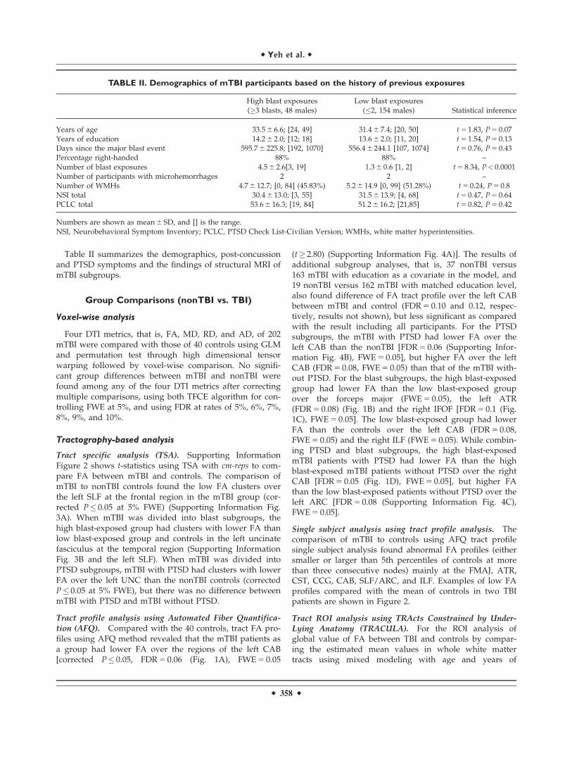
Table II summarizes the demographics, post-concussionand PTSD symptoms and the findings of structural MRI ofmTBI subgroups.
Group Comparisons (nonTBI vs. TBI)
Voxel-wise analysis
Four DTI metrics, that is, FA, MD, RD, and AD, of 202mTBI were compared with those of 40 controls using GLMand permutation test through high dimensional tensorwarping followed by voxel-wise comparison. No signifi-cant group differences between mTBI and nonTBI werefound among any of the four DTI metrics after correctingmultiple comparisons, using both TFCE algorithm for con-trolling FWE at 5%, and using FDR at rates of 5%, 6%, 7%,8%, 9%, and 10%.
Tractography-based analysis
Tract specific analysis (TSA). Supporting InformationFigure 2 shows t-statistics using TSA with cm-reps to com-pare FA between mTBI and controls. The comparison ofmTBI to nonTBI controls found the low FA clusters overthe left SLF at the frontal region in the mTBI group (cor-rected P� 0.05 at 5% FWE) (Supporting Information Fig.3A). When mTBI was divided into blast subgroups, thehigh blast-exposed group had clusters with lower FA thanlow blast-exposed group and controls in the left uncinatefasciculus at the temporal region (Supporting InformationFig. 3B and the left SLF). When mTBI was divided intoPTSD subgroups, mTBI with PTSD had clusters with lowerFA over the left UNC than the nonTBI controls (correctedP� 0.05 at 5% FWE), but there was no difference betweenmTBI with PTSD and mTBI without PTSD.
Tract profile analysis using Automated Fiber Quantifica-tion (AFQ). Compared with the 40 controls, tract FA pro-files using AFQ method revealed that the mTBI patients asa group had lower FA over the regions of the left CAB[corrected P� 0.05, FDR 5 0.06 (Fig. 1A), FWE 5 0.05
(t� 2.80) (Supporting Information Fig. 4A)]. The results ofadditional subgroup analyses, that is, 37 nonTBI versus163 mTBI with education as a covariate in the model, and19 nonTBI versus 162 mTBI with matched education level,also found difference of FA tract profile over the left CABbetween mTBI and control (FDR 5 0.10 and 0.12, respec-tively, results not shown), but less significant as comparedwith the result including all participants. For the PTSDsubgroups, the mTBI with PTSD had lower FA over theleft CAB than the nonTBI [FDR 5 0.06 (Supporting Infor-mation Fig. 4B), FWE 5 0.05], but higher FA over the leftCAB (FDR 5 0.08, FWE 5 0.05) than that of the mTBI with-out PTSD. For the blast subgroups, the high blast-exposedgroup had lower FA than the low blast-exposed groupover the forceps major (FWE 5 0.05), the left ATR(FDR 5 0.08) (Fig. 1B) and the right IFOF [FDR 5 0.1 (Fig.1C), FWE 5 0.05]. The low blast-exposed group had lowerFA than the controls over the left CAB (FDR 5 0.08,FWE 5 0.05) and the right ILF (FWE 5 0.05). While combin-ing PTSD and blast subgroups, the high blast-exposedmTBI patients with PTSD had lower FA than the highblast-exposed mTBI patients without PTSD over the rightCAB [FDR 5 0.05 (Fig. 1D), FWE 5 0.05], but higher FAthan the low blast-exposed patients without PTSD over theleft ARC [FDR 5 0.08 (Supporting Information Fig. 4C),FWE 5 0.05].
Single subject analysis using tract profile analysis. Thecomparison of mTBI to controls using AFQ tract profilesingle subject analysis found abnormal FA profiles (eithersmaller or larger than 5th percentiles of controls at morethan three consecutive nodes) mainly at the FMAJ, ATR,CST, CCG, CAB, SLF/ARC, and ILF. Examples of low FAprofiles compared with the mean of controls in two TBIpatients are shown in Figure 2.
Tract ROI analysis using TRActs Constrained by Under-Lying Anatomy (TRACULA). For the ROI analysis ofglobal value of FA between TBI and controls by compar-ing the estimated mean values in whole white mattertracts using mixed modeling with age and years of
TABLE II. Demographics of mTBI participants based on the history of previous exposures
High blast exposures(�3 blasts, 48 males)
Low blast exposures(�2, 154 males) Statistical inference
Years of age 33.5 6 6.6; [24, 49] 31.4 6 7.4; [20, 50] t 5 1.83, P 5 0.07Years of education 14.2 6 2.0; [12; 18] 13.6 6 2.0; [11, 20] t 5 1.54, P 5 0.13Days since the major blast event 595.7 6 225.8; [192, 1070] 556.4 6 244.1 [107, 1074] t 5 0.76, P 5 0.43Percentage right-handed 88% 88% –Number of blast exposures 4.5 6 2.6[3, 19] 1.3 6 0.6 [1, 2] t 5 8.34, P< 0.0001Number of participants with microhemorrhages 2 2 –Number of WMHs 4.7 6 12.7; [0, 84] (45.83%) 5.2 6 14.9 [0, 99] (51.28%) t 5 0.24, P 5 0.8NSI total 30.4 6 13.0; [3, 55] 31.5 6 13.9; [4, 68] t 5 0.47, P 5 0.64PCLC total 53.6 6 16.3; [19, 84] 51.2 6 16.2; [21,85] t 5 0.82, P 5 0.42
Numbers are shown as mean 6 SD, and [] is the range.NSI, Neurobehavioral Symptom Inventory; PCLC, PTSD Check List-Civilian Version; WMHs, white matter hyperintensities.
r Yeh et al. r
r 358 r

education as covariates, there is no significant group dif-ference among the DTI metrics (FA, MD, AD, and RD)between TBI and nonTBI after correcting for multiple com-parisons, but TBI had lower FA than nonTBI over the rightCAB at the margin of statistical significance (Bonferroniand FDR corrected P 5 0.0526). However, this significancedid not survive the multiple comparisons correction whenmTBI was divided into groups with or without PTSD. Forthe blast subgroups, the high blast-exposed group hadlower FA than the controls over the right CAB (meanFA 6 standard error 5 0.275 6 0.004 vs. 0.300 6 0.006, cor-rected P 5 0.0051, both Bonferroni and FDR). When mTBIpatients was further grouped according to the presence ofPTSD and the severity of previous blast exposures, themTBI patients with PTSD and high blast exposures tended
to have the lowest FA of right CAB (uncorrectedP 5 0.0016, corrected P 5 0.1167).
Interaction between blast exposure, WMHs, and aging onDTI measures. To further contrast the interaction effect ofblast exposures and aging on white matter integrity, theblast 3 age interaction was added to the model. Signifi-cant (P� 0.05) blast exposure and age interaction effectson FA measure were observed over the cingulate part ofbilateral cingulum bundles with the high blast-exposedgroup having a faster regional aging trajectory, that is,higher negative slope of the group and age interactionterm (left: 22.60E-4 6 5.97E-4, right: 22.63E-4 6 5.73E-4)toward reduced white matter integrity (lower FA) than thelow blast-exposed group (left: 24.20E-4 6 5.73E-4, right:
Figure 1.
AFQ tract profile analyses of FA showing significant group differ-
ence, with lower FA in mTBI group over the parahippocampal
part of the left cingulum bundle (corrected P� 0.05,
FDR 5 0.05) than non-TBI controls (A); lower FA in high blast-
exposed group than low blast-exposed over the left anterior
thalamic radiation (FDR 5 0.08) (B), and the left inferior fronto-
occipital fasciculus (FDR 5 0.10) (C); lower FA over the right
CAB (FDR 5 0.08) in the high blast-exposed mTBI with PTSD
than the high blast-exposed mTBI without PTSD (D).
r Compromised Neurocircuitry r
r 359 r

28.20E-4 6 2.92E-4), and the control group (left: 2.36E-4 6 5.94E-4, right: 3.67E-4 6 6.27E-4) (high blast vs. controlin the left CCG and the right CCG: t 5 3.37, 3.53, P 5 0.02,0.03, and d 5 0.74, 0.77, respectively) (Fig. 3A).
After correcting for multiple comparisons, there was noregional FA difference of any TRACULA segmented tractsamong the mTBI subgroups, for example, high incidenceversus low incidence of blast exposures or high numberversus low number of WMHs groups. There was also noMD difference, except that the high WMHs group as agroup had a higher MD (mean 6 standard error 5 88.89E-5 6 0.48E-5) over the FMAJ than the low MWHs group(86.97E-5 6 0.29E-5) and the control group (87.75E-5 6 0.65E-5) (t 5 7.08 and 4.93, and d 5 0.41 and 0.31,respectively, adjusted P< 0.05).
In addition, MD of the right ATR in the high WMHsgroup was significantly correlated with the number of
WMHs (r 5 0.51, adjusted P 5 0.004, Fig. 3B), but no signif-icant correlation between FA and the number WMHs wasfound among the 18 segmented tracts. Furthermore, therewas no significant correlation between the number ofblasts and the number of WMHs, nor was there significanttwo-way interaction effect of WMHs and age on FA orMD when the WMHs * age interaction was included inthe model.
Post-Concussion and PTSD Symptoms, and Time
Post-Injury and FA
Using ROI analysis of the segmented major white mattertracts (TRACULA), a higher score in the NSI sensorydomain (more reported sensory symptoms) was associatedwith lower FA of the left SLFt (r 5 20.25, adjustedP 5 0.02) after correcting multiple comparisons. Higher
Figure 2.
Examples of single subject tract profile analysis using AFQ, iden-
tified by FA values smaller than the 5th percentiles of control
participants, with larger than 3 consecutive nodes. A mild blast
TBI patient (male, 30 years old, 710 days post-injury) injured by
grenade and mortar blast with complaints of headaches, tinnitus,
vision, sleep, attention, irritability and cognition. Low FA were
found at forceps major (A) and superior longitudinal fasciculus–
temporal/arcuate fasciculus (B). A mild blast TBI patient (male,
52 years old, 2,454 days post-injury) injured by mortar blast and
fall with complaints of hearing loss, depression, anxiety, memory
loss, and poor cognition. Low FA were found at left superior
longitudinal fasciculus (C) and left uncinate fasciculus (D). [Color
figure can be viewed at wileyonlinelibrary.com.]
r Yeh et al. r
r 360 r
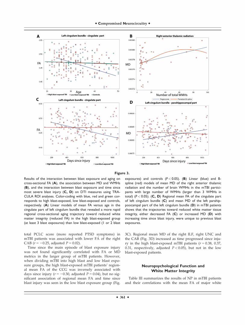
total PCLC score (more reported PTSD symptoms) inmTBI patients was associated with lower FA of the rightCAB (r 5 20.25, adjusted P 5 0.02).
Time since the main episode of blast exposure injurywas not found significantly correlated with FA or MDmetrics in the larger group of mTBI patients. However,when dividing mTBI into high blast and low blast expo-sure groups, the high blast-exposed mTBI patients’ region-al mean FA of the CCG was inversely associated withdays since injury (r 5 20.30, adjusted P 5 0.04), but no sig-nificant association of regional mean FA and time sinceblast injury was seen in the low blast exposure group (Fig.
3C). Regional mean MD of the right ILF, right UNC andthe CAB (Fig. 3D) increased as time progressed since inju-ry in the high blast-exposed mTBI patients (r 5 0.38, 0.37,0.31, respectively, adjusted P< 0.05), but not in the lowblast-exposed patients.
Neuropsychological Function and
White Matter Integrity
Table III summarizes the results of NP in mTBI patientsand their correlations with the mean FA of major white
Figure 3.
Results of the interaction between blast exposure and aging on
cross-sectional FA (A), the association between MD and WMHs
(B), and the interaction between blast exposure and time since
most severe blast injury (C, D) on DTI measures using TRA-
CULA ROI analyses. Color-coding with blue, red and green cor-
responds to high blast-exposed, low blast-exposed and controls,
respectively. (A) Linear models of mean FA versus age in the
cingulate part of left cingulum bundle that revealed a more rapid
regional cross-sectional aging trajectory toward reduced white
matter integrity (reduced FA) in the high blast-exposed group
(at least 3 blast exposures) than low blast-exposed (1 or 2 blast
exposures) and controls (P< 0.05). (B) Linear (blue) and B-
spline (red) models of mean MD of the right anterior thalamic
radiation and the number of brain WMHs in the mTBI partici-
pants with large number of WMHs (larger than 3 WMHs in
total) (P< 0.05). (C, D) Regional mean FA of the cingulate part
of left cingulum bundle (C) and mean MD of the left parahip-
poocampal part of the left cingulum bundle (D) in mTBI patients
shows that the trajectories toward reduced white matter tissue
integrity, either decreased FA (C) or increased MD (D) with
increasing time since blast injury, were unique to previous blast
exposures.
r Compromised Neurocircuitry r
r 361 r
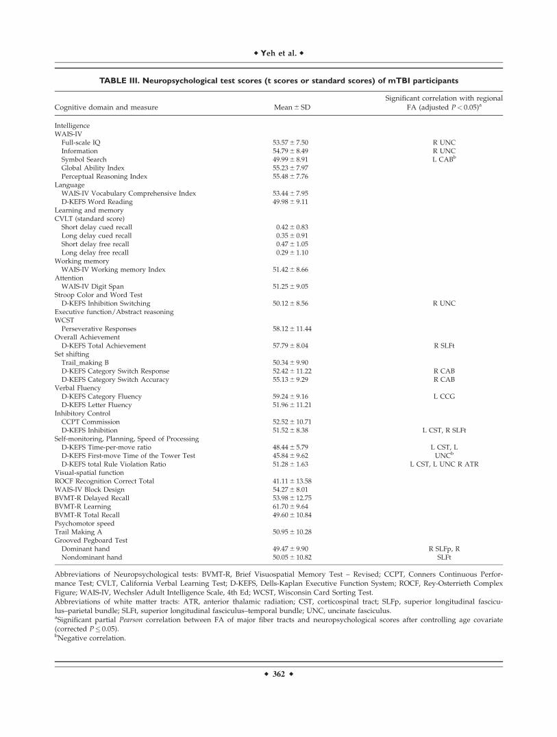
TABLE III. Neuropsychological test scores (t scores or standard scores) of mTBI participants
Cognitive domain and measure Mean 6 SDSignificant correlation with regional
FA (adjusted P< 0.05)a
IntelligenceWAIS-IV
Full-scale IQ 53.57 6 7.50 R UNCInformation 54.79 6 8.49 R UNCSymbol Search 49.99 6 8.91 L CABb
Global Ability Index 55.23 6 7.97Perceptual Reasoning Index 55.48 6 7.76
LanguageWAIS-IV Vocabulary Comprehensive Index 53.44 6 7.95D-KEFS Word Reading 49.98 6 9.11
Learning and memoryCVLT (standard score)
Short delay cued recall 0.42 6 0.83Long delay cued recall 0.35 6 0.91Short delay free recall 0.47 6 1.05Long delay free recall 0.29 6 1.10
Working memoryWAIS-IV Working memory Index 51.42 6 8.66
AttentionWAIS-IV Digit Span 51.25 6 9.05
Stroop Color and Word TestD-KEFS Inhibition Switching 50.12 6 8.56 R UNC
Executive function/Abstract reasoningWCST
Perseverative Responses 58.12 6 11.44Overall Achievement
D-KEFS Total Achievement 57.79 6 8.04 R SLFtSet shifting
Trail_making B 50.34 6 9.90D-KEFS Category Switch Response 52.42 6 11.22 R CABD-KEFS Category Switch Accuracy 55.13 6 9.29 R CAB
Verbal FluencyD-KEFS Category Fluency 59.24 6 9.16 L CCGD-KEFS Letter Fluency 51.96 6 11.21
Inhibitory ControlCCPT Commission 52.52 6 10.71D-KEFS Inhibition 51.52 6 8.38 L CST, R SLFt
Self-monitoring, Planning, Speed of ProcessingD-KEFS Time-per-move ratio 48.44 6 5.79 L CST, LD-KEFS First-move Time of the Tower Test 45.84 6 9.62 UNCb
D-KEFS total Rule Violation Ratio 51.28 6 1.63 L CST, L UNC R ATRVisual-spatial functionROCF Recognition Correct Total 41.11 6 13.58WAIS-IV Block Design 54.27 6 8.01BVMT-R Delayed Recall 53.98 6 12.75BVMT-R Learning 61.70 6 9.64BVMT-R Total Recall 49.60 6 10.84Psychomotor speedTrail Making A 50.95 6 10.28Grooved Pegboard Test
Dominant hand 49.47 6 9.90 R SLFp, RNondominant hand 50.05 6 10.82 SLFt
Abbreviations of Neuropsychological tests: BVMT-R, Brief Visuospatial Memory Test – Revised; CCPT, Conners Continuous Perfor-mance Test; CVLT, California Verbal Learning Test; D-KEFS, Dells-Kaplan Executive Function System; ROCF, Rey-Osterrieth ComplexFigure; WAIS-IV, Wechsler Adult Intelligence Scale, 4th Ed; WCST, Wisconsin Card Sorting Test.Abbreviations of white matter tracts: ATR, anterior thalamic radiation; CST, corticospinal tract; SLFp, superior longitudinal fascicu-lus–parietal bundle; SLFt, superior longitudinal fasciculus–temporal bundle; UNC, uncinate fasciculus.aSignificant partial Pearson correlation between FA of major fiber tracts and neuropsychological scores after controlling age covariate(corrected P� 0.05).bNegative correlation.
r Yeh et al. r
r 362 r

matter tracts. All of the P-values of significant correlationswere corrected using Bonferroni method (correctedP� 0.05). Significance showed that integrity of UNC wasimportant for memory function, CCG for attentional con-trol and cognitive speed, CAB for set-shifting, UNC andCST for response inhibition control; and SLF was criticalfor overall executive function, inhibition control and fine-motor skills (Supporting Information II.A). For example,Category Fluency of the Dellis–Kaplan Executive FunctionSystem (D-KEFS) Verbal Fluency score positively correlat-ed with FA of the left CCG (r 5 0.63, P 5 0.008; Fig. 4A);FA values of right UNC correlated positively with theInformation score (r 5 0.56, P 5 0.0019) and Full scale IQ ofthe Wechsler Adult Intelligence Scale, 4th Ed (WAIS-IV)(r 5 0.50, P 5 0.026; Fig. 4B); and Total Achievement scoreof the D-KEFS correlated with the FA of temporal branchof the right SLF (r 5 0.58, P 5 0.037).
DISCUSSION
Using tract profile analysis and tract specific analysis, wehave revealed evidence of white matter injury in chronicblast TBI patients. Due to the heterogeneity of injury mecha-nisms from blast TBI, it is not surprising to observe hetero-geneous and spatially diverse white matter abnormalities[Davenport et al., 2012] reported in different studies [MacDonald et al., 2011; Petrie et al., 2014; Taber et al., 2014].
Our results show that white matter damages indicatedby reduced FA were most prominent in the pathwayswithin the fronto-limbic and the fronto-striatal circuits,particularly the fiber tracts in the parasagittal white matterof the cerebral cortex such as the CAB, ATR, SLF and
IFOF. These results are similar to our previous report [Yehet al., 2014] and other reports of chronic changes inmilitary-related blast mTBI [Mac Donald et al., 2011] usingvoxel-wise analysis for group comparison. In addition,newer approaches using tract profile analyses were usefulin group comparison and single subject analysis, whichpinpoint specific damaged fiber tracts and affectedneurocircuitry.
Moreover, the compromised fiber tracts (reduced FA) inthe nodes of fronto-striatal and fronto-limbic circuits wereassociated with greater post-concussion and PTSD symp-toms. These findings suggest the networks of the fronto-parieto-temporal circuit, fronto-striatal circuit, and thefrontal-limbic circuit are most vulnerable to military relat-ed blast injury, which may also play an important role inthe development of neuropsychological symptoms fre-quently seen in military TBI patients.
Interpretation of Microstructural Changes in
Chronic Blast TBI
Group analysis
Blast TBI as described in this work refers to “blast plusimpact.” That is, while components of primary blast maybe involved, most injuries also involve secondary or tertia-ry effects, which have features also seen with mechanicallyinduced TBI [Warden, 2006].
Through simulation, it has been shown the dynamicdeformation of white matter arising from shock wavepropagation can result in axonal stretch and disruption[Besenski, 2002; Chafi et al., 2010]. From a mechanical
Figure 4.
Examples of the results of significant associations between FA in
two distinct white matter tracts and cognitive functions in mTBI
service members with military-related blast injury. Cognition func-
tions were displayed as t scores. (A) Correlation of Dells-Kaplan
Executive Function System Category Fluency of Verbal Fluency
performance and average FA of the cingulate part of the left cingu-
lum bundle (r 5 0.63, P 5 0.00). (B) Correlation of Wechsler Adult
Intelligence Scale, 4th Ed Full Scale IQ score and average FA of
the right uncinate fasciculus (r 5 0.50, P 5 0.026). [Color figure
can be viewed at wileyonlinelibrary.com.]
r Compromised Neurocircuitry r
r 363 r
standpoint, macro and micro interfaces between structureswith distinct differences in density and elasticity are par-ticularly vulnerable to damage caused by shock wavepropagation observed in blast-related brain injury. Theanatomical locations of low FA in blast-related mTBI dem-onstrate that parasagittal frontal white matter, particularlythe fronto-limbic and the fronto-striatal projection fiberssuch as CCG/CAB, ATR and FMAJ (Figs. 1 and 2), as wellas long association fibers interconnecting the medial fron-tal, temporal, parietal and occipital lobes (SupportingInformation Figs. 3 and 4) are vulnerable to blast injury.These results were consistent with our previous DTI andtractography report in subacute TBI patients [Yeh et al.,2014] that revealed subcortical superior-inferiorly orientedtracts were particularly vulnerable to blast injury, as wellas other reports of chronic changes in mTBI, either mili-tary personnel [Mac Donald et al., 2011] or civilian popula-tions [Inglese et al., 2005; Kraus et al., 2007; Lipton et al.,2009]. The anatomical locations of low FA using tract spe-cific analysis and tract profile analysis in this study arealso consistent with the results from using mechanical sim-ulation and finite element analysis of brain exposure toblasts, showing that the highest level of axonal shear/strain effects developed in the regions of corpus callosumand corona radiata [Chatelin et al., 2011] in mTBI model.
Secondary brain injury and repair mechanisms such aschronic inflammation and hypermetabolism may sensitizethe brain to the subsequent injury [Calabrese et al., 2014],leading to axonal repair, reactive gliosis [Glushakovaet al., 2014; Johnson et al., 2013; Kiraly and Kiraly, 2007] orirreversible axonal damage [Gupta and Przekwas, 2013].Our findings of the inverse relationship between post-injury duration and FA over the cingulum bundles (Fig.3C) in TBI patients with previous high blast exposure fre-quency raise the possibility of Wallerian degenerationwhich may contribute to the long-lasting impairment ofcognitive performance seen in many chronic TBI patients.
Diffusion MRI tractography-based analysis, either ROI-based, tract specific or tract profile analysis, can be verytime-consuming. With larger ROIs, there is a risk ofincluding voxels containing signals from adjacent tissuessuch as CSF and other tracts and this contamination canbe even more pronounced in studying small white mattertracts [Vos et al., 2011]. In addition, ROI-based approachescan result in loss of information regarding local variationsin diffusion parameters [Kvickstr€om et al., 2011], whichmay explain why no significant group difference betweenthe mTBI group and controls was found using ROI-basedTRACULA. To the best of our knowledge, this is the firstreport evaluating DTI metrics along white mater tracts inchronic blast mTBI patients as a function of distance fromspecific anatomical landmarks. It is anticipated that theexact spatial locations of low FA clusters would be slightlydifferent among different tractography-based analysismethods. Nevertheless, the identified abnormal tracts,such as, the CCG and the SLF in TBI patients were
consistent between tract specific analysis and tract profileanalysis in our results.
Single subject analysis
In contrast to the results using voxel-wise analysis insingle subject analysis, the results using tract profile analy-sis specified the injured tracts by revealing the low FAprofile within the whole tract, either the entire tract (Fig.2D) or segment(s) within the tract (Fig. 2A–C). FA profilevalues quantified by tract profile analysis varied substan-tially within a tract but the shape of the tract FA profilewas consistent across subjects (Fig. 2), demonstrating theprecision of tract profile analysis for quantifying whitematter properties at specific spatial locations on a fibertract across subjects. There is minimal contamination ofFA measurements from crossing or kissing fibers withinthe central portion of the tract, thus the measured FA val-ues within the main fascicles are more indicative of tractintegrity than those from peripheral locations.
Post-concussion and PTSD symptoms and neurocir-
cuitry affected in TBI
Our findings of significant associations between WMintegrity and post-concussion, and WM integrity and PTSDsymptoms were in the affected regions of the neural net-works in which the somatic sequelae (SLF sensory/motorpathways) and affective symptoms (cingulum bundle oflimbic pathways) have intriguing clinical-anatomical corre-lates. The cingulum bundle is one of the locations found tobe associated with PTSD [Daniels et al., 2013], which is con-sistent with the findings of our group comparisons usingAFQ tract profile analysis, showing lower FA over the leftCAB in mTBI with PTSD than nonTBI controls (SupportingInformation Fig. 4B), and lower FA over the right CAB inthe high blast-exposed mTBI patients with PTSD than thehigh blast-exposed mTBI patients without PTSD (Fig. 1D).In addition, the finding of lower FA over the left UNC inmTBI with PTSD than the nonTBI revealed by tract specificanalysis suggests disrupted WM networks in limbic system,for example, cingulum bundle and uncinate fasciculus, areassociated with the PTSD symptoms in comorbid mTBI-PTSD patients.
The frequent comorbidity of PTSD and TBI is welldescribed in military TBI patients [Belanger et al., 2010;Hoge et al., 2008a; Ruff et al., 2010; Warden, 2006]; and therisk of PTSD might be increased by cognitive dysfunctionfollowing injury. Therefore, the compromised integrity ofwhite matter fiber connections of this study can be thecombination of comorbid PTSD and TBI, chronic TBI spec-trum, as these two separate and distinct diseases sharecommon clinical symptoms. It should be noted as a limita-tion that self-report measures were used for post-concussive and post-traumatic stress disorder symptoms.Self-report measures have been shown to correlate weaklywith objectively measured deficits in various domains in
r Yeh et al. r
r 364 r
multiple studies [Drag et al., 2012; French et al., 2014;Spencer et al., 2010]. However, those individuals with evi-dence of symptom exaggeration on the NSI were excludedfrom these analyses.
Frequency of blast exposures, WMHs, and aging on
white matter integrity
Our findings of significant blast exposure 3 age interac-tion on FA of the bilateral cingulum bundles (Fig. 3A) areconsistent with the findings of a prior work [Trotter et al.,2015] using voxel-wise analysis followed by ROI analysesto study chronic TBI veterans, including moderate andsevere TBI patients besides mTBI patients of the recentconflicts. In addition, the cingulum bundle in high blast-exposed individuals, but not in age-matched low blast-exposed individuals, had rapid trajectory (Fig. 3C,D)toward reduced WM tissue integrity (FA) with increasingdays since most severe blast injury. The findings of highblast-exposed service members exhibiting both a more rap-id cross-sectional age trajectory and time since major blasttrajectories toward reduced white matter tissue integritythan those of low blast-exposed individuals and controlssuggest repeated blast exposures can initiate detrimentalprocess on white matter integrity prior to normal aging[Trotter et al., 2015] and may lead to Wallerian degenera-tion [Pierpaoli et al., 2001].
The radiological appearances of T2 FLAIR WMHs inmTBI participants were small, round with relatively homo-geneous in size (around 2–3 mm3 for each WMH), andmainly located in the deep WM region but not in the peri-ventricular region (not shown). Thus, the number of WMHswould be proportional to the total volume of WMHs,though total WMHs size was not quantified in this study.The prevalence of WMHs of this cohort was around 30% forcontrols, which is relatively higher than the previous reportsfor neurologically non-diseased adults under age 50 [Hop-kins et al., 2006] or 15% at the aged of 60 [Ylikoski et al.,1995]; and 50% for mTBI participants, which is consistentwith other TBI studies [Bigler et al., 2013; Marquez de laPlata et al., 2007]. Our findings of significant associationbetween the number of total WMHs and MD, but not FA, ofthe ATR, and difference of MD of the FMAJ between mTBIsubgroups of high and low WMHs suggest that underlyingpathological changes of WMHs of mTBI is likely due toenlarged perivascular spaces with increased interstitialspace, scarring or gliosis. However, no significant associa-tion between the number of WMHs and the number of blastexposures might suggest that the WMHs can be unrelated toblast injury directly.
Disrupted subcortical and cortical projections and
neuropsychological implications in mTBI
The most common residual deficits in mTBI patients arecognitive speed, attention, memory abilities, and visuocon-struction [Millis et al., 2001], which can cause adverse
long-term neuropsychological outcomes [Vanderploeget al., 2005]. Reduced memory and attention correspondedwith disrupted WM integrity mainly over the regions ofthe ventral prefrontal WM such as UNC, CAB [Wu et al.,2010], ILF, and genu and splenium of the CC [Bigler, 2013;Niogi et al., 2008], indicating that cognitive deficits frommTBI may be region and task specific [Niogi et al., 2008].Furthermore, disrupted dorsal prefrontal WM, such asCCG and SLF, impairs frontal top-down control, verbalworking memory [Charlton et al., 2010; Kennedy and Raz,2009; Palacios et al., 2011] and overall executive functionperformance [Ashley, 2004].
However, by taking error variance of measurement intoaccount [Bendlin et al., 2008], traditional neuropsychologi-cal testing is not sensitive and has limited ability to docu-ment ongoing brain function impairment in mTBI (ifpresent) [Heitger et al., 2009]. Therefore, the relationshipbetween neuroimaging biomarkers in mTBI and neuropsy-chological outcome is not conclusive (see [Bigler, 2013] forreview).
Our findings of the association between microstructuralvariation of the cingulum bundles (CCG, CAB) and thesubdomains of D-KEFS support previous literature reportsregarding the importance of anterior cingulum integrity inattentional control and cognitive speed [Kubicki et al.,2009; Nestor et al., 2004], which is essential for executivefunction, decision-making and emotion control [Heilbron-ner and Haber, 2014]. The findings of the associations ofthe UNC FA with subdomains of WAIS-IV and inhibitionswitching performance suggest that damage in the UNCmay link to memory dysfunction [von der Heide et al.,2013] and executive dysfunction [Widjaja et al., 2013] suchas response inhibition in mTBI. Our findings of the corre-lation between the SLF FA and the overall executive func-tion and the inhibition response domains of D-KEFS, aswell as Grooved pegboard motor speed support theimportance of the integrity of the SLF in the visual-motorcoordination [Skranes et al., 2007], visuospatial processing,speed and manual dexterity.
Limitation
The blast injuries seen in our population reflect “blast-plus” in which components of the blast wave (primaryblast) are combined with traditional mechanical and rota-tional components. Self-report measures were used forpost-concussive and PTSD symptoms, and mTBI partici-pants were on the average of 2 years from point of injury;thus, the neuroimaging findings may not be directly relat-ed to symptoms caused by blast injury itself. The micro-structural alterations in white matter integrity revealed inthe study may not be specific to blast mTBI only, but pos-sibly caused by comorbidities such as PTSD. Furthermore,we did not administer neuropsychological testing to thecontrols and were not able to assess the difference of thecorrelation between NP testing and DTI measures among
r Compromised Neurocircuitry r
r 365 r

TBI and controls. In addition, we are aware of inherentlimitation using diffusion tensor tractography to determinetrajectories of WM pathways. However, priori anatomicalinformation was applied in this study to constrain tractog-raphy, which can greatly reduce the occurrence of false-positive trajectories of fiber pathways. Future study usingadvanced diffusion MRI techniques such as high angularresolution diffusion imaging is needed to verify theseresults. Nevertheless, the results of this study suggest thatdiffusion MRI tractography can be used as noninvasivebiomarkers in assessing affected networks and for a betterunderstanding of neuropathology of blast mTBI.
CONCLUSION
Our findings suggest that the association and projectionfibers interconnecting fronto-parieto-temporal region, forexample, CCG/CAB, SLF, and UNC; and fronto-subcortical regions, for example, ATR, are particularly vul-nerable to military-related blast injury, where the compro-mised circuits have significant effects on the functionaloutcome of chronic mTBI patients. Furthermore, high fre-quency of blast exposures may deflect negatively normalaging trajectories of white matter integrity. However, lon-gitudinal study with follow-up scans is needed to validatethese findings. Nevertheless, our results suggest the use-fulness of diffusion MRI and tractography in assessingwhite matter changes in chronic blast-related mTBI.
Disclaimer
The views expressed in this article are those of theauthor and do not reflect the official policy of the Depart-ment of Army/Navy/Air Force, Department of Defense,or U.S. Government.
ACKNOWLEDGMENTS
We thank Rachel Wolfowitz and Jamie Harper for partici-pant recruitment and project coordination.
REFERENCES
APA (1994): Diagnostic and Statistical Manual of Mental Disor-
ders Fourth Edition. Washington, DC: American Psychiatric
Association.Ashley MJ (2004): Traumatic Brain Injury: Rehabilitative Treat-
ment and Case Management, 2nd ed. Boca Raton: CRC Press.Basser PJ, Jones DK (2002): Diffusion-tensor MRI: Theory, experi-
mental design and data analysis - a technical review. NMR
Biomed 15:456–467.Basser PJ, Mattiello J, LeBihan D (1994): Estimation of the effective
self-diffusion tensor from the NMR spin echo. J Magn Reson B
103:247–254.Basser PJ, Pajevic S, Pierpaoli C, Duda J, Aldroubi A (2000): In
vivo fiber tractography using DT-MRI data. Magn Reson Med
44:625–632.
Belanger HG, Kretzmer T, Vanderploeg RD, French LM (2010):
Symptom complaints following combat-related traumatic brain
injury: Relationship to traumatic brain injury severity and post-
traumatic stress disorder. J Int Neuropsychol Soc 16:194–199.Bendlin BB, Ries ML, Lazar M, Alexander AL, Dempsey RJ,
Rowley HA, Sherman JE, Johnson SC (2008): Longitudinal
changes in patients with traumatic brain injury assessed with
diffusion-tensor and volumetric imaging. Neuroimage 42:
503–514.Benjamini Y, Hochberg Y (1995): Controlling the false discovery
rate: A practical and powerful approach to multiple testing.
J R Stat Soc Ser B 57:289–300.Besenski N (2002): Traumatic injuries: Imaging of head injuries.
Eur Radiol 12:1237–1252.Bhattacharjee Y (2008): Neuroscience. Shell shock revisited: Solv-
ing the puzzle of blast trauma. Science (New York, N.Y.) 319:
406–408.Bigler ED (2013): Neuroimaging biomarkers in mild traumatic
brain injury (mTBI). Neuropsychol Rev 23:169–209.Bigler ED, Abildskov TJ, Petrie J, Farrer TJ, Dennis M, Simic N,
Taylor HG, Rubin KH, Vannatta K, Gerhardt CA, Stancin T,
Owen Yeates K (2013): Heterogeneity of brain lesions in pedi-
atric traumatic brain injury. Neuropsychology 27:438–451.Blanchard EB, Jones-Alexander J, Buckley TC, Forneris CA (1996):
Psychometric properties of the PTSD Checklist (PCL). Behav
Res Ther 34:669–673.Brandstack N, Kurki T, Tenovuo O (2013): Quantitative diffusion-
tensor tractography of long association tracts in patients with
traumatic brain injury without associated findings at routine
MR imaging. Radiology 267:231–239.Calabrese E, Du F, Garman RH, Johnson GA, Riccio C, Tong LC,
Long JB (2014): Diffusion tensor imaging reveals white matter
injury in a rat model of repetitive blast-induced traumatic
brain injury. J Neurotrauma 31:938–950.Cernak I (2010): The importance of systemic response in the
pathobiology of blast-induced neurotrauma. Front Neurol 1:
151.Cernak I, Wang Z, Jiang J, Bian X, Savic J (2001): Ultrastructural
and functional characteristics of blast injury-induced neuro-
trauma. J Trauma 50:695–706.Chafi MS, Karami G, Ziejewski M (2010): Biomechanical assess-
ment of brain dynamic responses due to blast pressure waves.
Ann Biomed Eng 38:490–504.Chang LC, Jones DK, Pierpaoli C (2005): RESTORE: Robust esti-
mation of tensors by outlier rejection. Magn Reson Med 53:
1088–1095.Charlton RA, Barrick TR, Lawes INC, Markus HS, Morris RG
(2010): White matter pathways associated with working memo-
ry in normal aging. Cortex 46:474–489.Chatelin S, Deck C, Renard F, Kremer S, Heinrich C, Armspach
JP, Willinger R (2011): Computation of axonal elongation in
head trauma finite element simulation. J Mech Behav Biomed
Mater 4:1905–1919.Chavko M, Koller WA, Prusaczyk WK, McCarron RM (2007):
Measurement of blast wave by a miniature fiber optic pressure
transducer in the rat brain. J Neurosci Methods 159:277–281.Chen YC, Smith DH, Meaney DF (2009): In-vitro approaches for
studying blast-induced traumatic brain injury. J Neurotrauma
26:861–876.Cicerone KD, Kalmar K (1995): Persistent postconcussion syn-
drome: The structure of subjective complaints after mild trau-
matic brain injury. J Head Trauma Rehabil 10:1–7.
r Yeh et al. r
r 366 r

Cifu D, Hurley R, Peterson M, Cornis-Pop M, Rikli PA, Ruff RL,Scott SG, Sigford BJ, Silva KA, Tortorice K, Vanderploeg RD,
Withlock W, Bowles A, Cooper D, Drake A, Engel C (2009):
Clinical practice guideline: Management of concussion/mild
traumatic brain injury. J Rehabil Res Dev 46:CP1.Connell S, Gao J, Chen J, Shi R (2011): Novel model to investigate
blast injury in the central nervous system. J Neurotrauma 28:
1229–1236.Daniels JK, Lamke J-P, Gaebler M, Walter H, Scheel M (2013):
White matter integrity and its relationship to PTSD and child-
hood trauma: A systematic review and meta-analysis. Depress
Anxiety 30:207–216.Davenport ND, Lim KO, Armstrong MT, Sponheim SR (2012):
Diffuse and spatially variable white matter disruptions areassociated with blast-related mild traumatic brain injury. Neu-
roimage 59:2017–2024.Drag LL, Spencer RJ, Walker SJ, Pangilinan PH, Bieliauskas LA
(2012): The contributions of self-reported injury characteristics
and psychiatric symptoms to cognitive functioning in oef/oif
veterans with mild traumatic brain injury. J Int Neuropsychol
Soc 18:576–584.French LM, Lange RT, Brickell TA (2014): Subjective cognitive
complaints and neuropsychological test performance following
military-related traumatic brain injury. J Rehabil Res Dev 51:933–950.
Garman RH, Jenkins LW, Switzer RC, Bauman RA, Tong LC,
Swauger PV, Parks SA, Ritzel DV, Dixon CE, Clark RSB, BayirH, Kagan V, Jackson EK, Kochanek PM (2011): Blast exposure
in rats with body shielding is characterized primarily by dif-
fuse axonal injury. J Neurotrauma 28:947–959.Glushakova OY, Johnson D, Hayes RL (2014): Delayed increases
in microvascular pathology after experimental traumatic brain
injury are associated with prolonged inflammation, blood-
brain barrier disruption, and progressive white matter damage.
J Neurotrauma 31:1180–1193.Gouw AA, Seewann A, van der Flier WM, Barkhof F,
Rozemuller AM, Scheltens P, Geurts JJG (2011): Heterogenei-ty of small vessel disease: A systematic review of MRI and
histopathology correlations. J Neurol Neurosurg Psychiatry
82:126–135.Gupta RK, Przekwas A (2013): Mathematical models of blast-
induced TBI: Current status, challenges, and prospects. Front
Neurol 4:59.von der Heide RJ, Skipper LM, Klobusicky E, Olson IR (2013):
Dissecting the uncinate fasciculus: Disorders, controversies
and a hypothesis. Brain 136:1692–1707.Heilbronner SR, Haber SN (2014): Frontal cortical and subcortical
projections provide a basis for segmenting the cingulum bun-
dle: Implications for neuroimaging and psychiatric disorders.
J Neurosci 34:10041–10054.Heitger MH, Jones RD, MacLeod AD, Snell DL, Frampton CM,
Anderson TJ (2009): Impaired eye movements in post-
concussion syndrome indicate suboptimal brain functionbeyond the influence of depression, malingering or intellectual
ability. Brain 132:2850–2870.Hoge CW, Castro CA, Messer SC, McGurk D, Cotting DI,
Koffman RL (2008a): Combat duty in Iraq and Afghanistan,
mental health problems and barriers to care. US Army Med
Dep J 7–17.Hoge CW, McHurk D, Thomas JL, Cox AL, Engel CC, Castro CA
(2008b): Mild traumatic brain injury in u.s. soldiers returning
from Iraw. N Engl J Med 358:453–463.
Hopkins RO, Beck CJ, Burnett DL, Weaver LK, Victoroff J,
Bigler ED (2006): Prevalence of white matter hyperinten-sities in a young healthy population. J Neuroimaging 16:
243–251.Inglese M, Makani S, Johnson G, Cohen BA, Silver JA, Gonen O,
Grossman RI (2005): Diffuse axonal injury in mild traumatic
brain injury: A diffusion tensor imaging study. J Neurosurg
103:298–303.Jbabdi S, Woolrich MW, Andersson JL, Behrens TE (2007): A
Bayesian framework for global tractography. Neuroimage 37:
116–129.Jezzard P, Balaban RS (1995): Correction for geometric distortion
in echo planar images from B0 field variations. Magn Reson
Med 34:65–73.Johnson VE, Stewart W, Smith DH (2013): Axonal pathology in
traumatic brain injury. Exp Neurol 246:35–43.Kasahara K, Hashimoto K, Abo M, Senoo A (2012): Voxel- and
atlas-based analysis of diffusion tensor imaging may revealfocal axonal injuries in mild traumatic brain injury - compari-
son with diffuse axonal injury. Magn Reson Imaging 30:
496–505.Kennedy KM, Raz N (2009): Aging white matter and cognition:
Differential effects of regional variations in diffusion properties
on memory, executive functions, and speed. Neuropsychologia
47:916–927.Kiraly M, Kiraly SJ (2007): Traumatic brain injury and delayed
sequelae: A review–traumatic brain injury and mild traumatic
brain injury (concussion) are precursors to later-onset brain
disorders, including early-onset dementia. ScientificWorldJour-
nal 7:1768–1776.Kraus MF, Susmaras T, Caughlin BP, Walker CJ, Sweeney JA,
Little DM (2007): White matter integrity and cognition in
chronic traumatic brain injury: A diffusion tensor imaging
study. Brain 130:2508–2519.Kubicki M, Niznikiewicz M, Connor E, Ungar L, Nestor PG,
Bouix S, Dreusicke M, Kikinis R, McCarley RW, Shenton ME
(2009): Relationship between white matter integrity, attention,and memory in schizophrenia: A diffusion tensor imaging
study. Brain Imaging Behav 3:191–201.Kumar R, Husain M, Gupta RK, Hasan KM, Haris M, Agarwal
AK, Pandey CM, Narayana PA (2009): Serial changes in thewhite matter diffusion tensor imaging metrics in moderate
traumatic brain injury and correlation with neuro-cognitive
function. J Neurotrauma 26:481–495.Kvickstr€om P, Eriksson B, van Westen D, L€att J, Elfgren C,
Nilsson C (2011): Selective frontal neurodegeneration of the
inferior fronto-occipital fasciculus in progressive supranuclear
palsy (PSP) demonstrated by diffusion tensor tractography.
BMC Neurol 11:13.Langlois JA, Rutland-Brown W, Wald MM (2006): The epidemiol-
ogy and impact of traumatic brain injury: A brief overview.
J Head Trauma Rehabil 21:375–378.Levin HS, Wilde E, Troyanskaya M, Petersen NJ, Scheibel R,
Newsome M, Radaideh M, Wu T, Yallampalli R, Chu Z, Li X
(2010): Diffusion tensor imaging of mild to moderate blast-
related traumatic brain injury and its sequelae. J Neurotrauma27:683–694.
Ling G, Bandak F, Armonda R, Grant G, Ecklund J (2009): Explo-
sive blast neurotrauma. J Neurotrauma 26:815–825.Lipton ML, Gulko E, Zimmerman ME, Friedman BW, Kim M,
Gellella E, Gold T, Shifteh K, Ardekani BA, Branch CA (2009):
Diffusion-tensor imaging implicates prefrontal axonal injury in
r Compromised Neurocircuitry r
r 367 r

executive function impairment following very mild traumatic
brain injury. Radiology 252:816–824.Mac Donald CL, Johnson AM, Cooper D, Nelson EC, Werner NJ,
Shimony JS, Snyder AZ, Raichle ME, Witherow JR, Fang R,
Flaherty SF, Brody DL (2011): Detection of blast-related trau-matic brain injury in U.S. military personnel. N Engl J Med
364:2091–2100.Marquez de la Plata C, Ardelean A, Koovakkattu D, Srinivasan P,
Miller A, Phuong V, Harper C, Moore C, Whittemore A,
Madden C, Diaz-Arrastia R, Devous M Sr. (2007): Magnetic reso-
nance imaging of diffuse axonal injury: Quantitative assessment
of white matter lesion volume. J Neurotrauma 24:591–598.Matsushita M, Hosoda K, Naitoh Y, Yamashita H, Kohmura E
(2011): Utility of diffusion tensor imaging in the acute stage of
mild to moderate traumatic brain injury for detecting white
matter lesions and predicting long-term cognitive function in
adults. J Neurosurg 115:130–139.Mayer AR, Ling J, Mannell MV, Gasparovic C, Phillips JP,
Doezema D, Reichard R, Yeo RA (2010): A prospective diffu-
sion tensor imaging study in mild traumatic brain injury. Neu-
rology 74:643–650.Mcdonald SD, Calhoun PS (2010): Clinical Psychology Review
The diagnostic accuracy of the PTSD Checklist: A critical
review. Clin Psychol Rev 30:976–987.Millis SR, Rosenthal M, Novack TA, Sherer M, Nick TG, Kreutzer
JS, High WM, Ricker JH (2001): Long-term neuropsychological
outcome after traumatic brain injury. J Head Trauma Rehabil16:343–355.
Mori S, van Zijl PC (2002): Fiber tracking: Principles and strategies
- a technical review. NMR Biomed 15:468–480.Mori S, Crain BJ, Chacko VP, van Zijl PC (1999): Three-dimension-
al tracking of axonal projections in the brain by magnetic reso-
nance imaging. Ann Neurol 45:265–269.Nestor PG, Kubicki M, Gurrera RJ, Niznikiewicz M, Frumin M,
McCarley RW, Shenton ME (2004): Neuropsychological corre-
lates of diffusion tensor imaging in schizophrenia. Neuropsy-
chology 18:629–637.Nichols TE, Holmes AP (2002): Nonparametric permutation tests
for functional neuroimaging: A primer with examples. Hum
Brain Mapp 15:1–25.Niogi SN, Mukherjee P, Ghajar J, Johnson CE, Kolster R, Lee H,
Suh M, Zimmerman RD, Manley GT, McCandliss BD (2008):
Structural dissociation of attentional control and memory in
adults with and without mild traumatic brain injury. Brain131:3209–3221.
Palacios EM, Fernandez-Espejo D, Junque C, Sanchez-Carrion R,
Roig T, Tormos JM, Bargallo N, Vendrell P (2011): Diffusiontensor imaging differences relate to memory deficits in diffuse
traumatic brain injury. BMC Neurol 11:24.Parizel PM, Ozsarlak, Van Goethem JW, van den Hauwe L, Dillen
C, Verlooy J, Cosyns P, De Schepper AM (1998): Imaging find-
ings in diffuse axonal injury after closed head trauma. Eur
Radiol 8:960–965.Parizel PM, Van Goethem JW, Ozsarlak O, Maes M, Phillips CD
(2005): New developments in the neuroradiological diagnosis
of craniocerebral trauma. Eur Radiol 15:569–581.Petrie EC, Cross DJ, Yarnykh VL, Richards T, Martin NM,
Pagulayan K, Hoff D, Hart K, Mayer C, Tarabochia M,
Raskind MA, Minoshima S, Peskind ER (2014): Neuroimaging,
behavioral, and psychological sequelae of repetitive combined
blast/impact mild traumatic brain injury in Iraq and Afghani-
stan war veterans. J Neurotrauma 31:425–436.
Pierpaoli C, Barnett A, Pajevic S, Chen R, Penix LR, Virta A,
Basser P (2001): Water diffusion changes in Wallerian degener-ation and their dependence on white matter architecture. Neu-
roimage 13:1174–1185.Riedy G, Senseney JS, Liu W, Ollinger J, Sham E, Krapiva P, Patel
JB, Smith A, Yeh P-H, Graner J, Nathan D, Caban J, French
LM, Harper J, Eskay V, Morissette J, Oakes TR (2016): Findings
from structural mr imaging in military traumatic brain injury.
Radiology 279:207–215.Robinson ME, Lindemer ER, Fonda JR, Milberg WP, Mcglinchey
RE, Salat DH (2015): Close-range blast exposure is associated
with altered functional connectivity in veterans independent of
concussion symptoms at time of exposure. Hum Brain Mapp
36:911–922.Ruff RL, Riechers RG, Ruff SS (2010): Relationships between mild
traumatic brain injury sustained in combat and post-traumatic
stress disorder. F1000 Med Rep 2:64.Saljo A, Bao F, Shi J, Hamberger A, Hansson HA, Haglid KG
(2002): Expression of c-Fos and c-Myc and deposition of beta-
APP in neurons in the adult rat brain as a result of exposure
to short-lasting impulse noise. J Neurotrauma 19:379–385.Shaw NA (2002): The neurophysiology of concussion. Prog Neuro-
biol 67:281–344.Skranes J, Vangberg TR, Kulseng S, Indredavik MS, Evensen KAI,
Martinussen M, Dale AM, Haraldseth O, Brubakk AM (2007):
Clinical findings and white matter abnormalities seen on diffu-
sion tensor imaging in adolescents with very low birth weight.Brain 130:654–666.
Smith SM, Nichols TE (2008): Threshold-free cluster enhancement:
Addressing problems of smoothing, threshold dependence and
localisation in cluster inference. Neuroimage 44:83–98.Smith SM, Jenkinson M, Woolrich MW, Beckmann CF, Behrens
TE, Johansen-Berg H, Bannister PR, De Luca M, Drobnjak I,
Flitney DE, Niazy RK, Saunders J, Vickers J, Zhang Y, DeStefano N, Brady JM, Matthews PM (2004): Advances in func-
tional and structural MR image analysis and implementation
as FSL. Neuroimage 23 Suppl 1:S208–S219.Spencer RJ, Drag LL, Walker SJ, Bieliauskas LA (2010): Self-
reported cognitive symptoms following mild traumatic brain
injury are poorly associated with neuropsychological perfor-
mance in OIF/OEF veterans. J Rehabil Res Dev 47:521–530.Taber KH, Warden DL, Hurley RA (2006): Blast-related traumatic
brain injury: What is known? J Neuropsychiatry Clin Neurosci
18:141–145.Taber KH, Hurley RA, Haswell CC, Rowland JA, Hurt SD, Lamar
CD, Morey RA (2014): White matter compromise in veterans
exposed to primary blast forces. J Head Trauma Rehabil 30:
E15–E25.Teipel SJ, Meindl T, Wagner M, Kohl T, B€urger K, Reiser MF,
Herpertz S, M€oller H-J, Hampel H (2009): White matter micro-
structure in relation to education in aging and Alzheimer’s dis-
ease. J Alzheimers Dis 17:571–583.Thomas C, Ye FQ, Irfanoglu MO, Modi P, Saleem KS, Leopold
DA (2014): Anatomical accuracy of brain connections derived
from diffusion MRI tractography is inherently limited. Proc
Natl Acad Sci U S A 111:16574–16579.Trotter BB, Robinson ME, Milberg WP, McGlinchey RE, Salat DH
(2015): Military blast exposure, ageing and white matter integ-
rity. Brain 138:2278–2292.Vanderploeg RD, Curtiss G, Belanger HG (2005): Long-term
neuropsychological outcomes following mild traumatic brain
injury. J Int Neuropsychol Soc 11:228–236.
r Yeh et al. r
r 368 r

Vos SB, Jones DK, Viergever MA, Leemans A (2011): Partial vol-ume effect as a hidden covariate in DTI analyses. Neuroimage55:1566–1576.
Wakana S, Caprihan A, Panzenboeck MM, Fallon JH, Perry M,Gollub RL, Hua K, Zhang J, Jiang H, Dubey P, Blitz A, vanZijl P, Mori S (2007): Reproducibility of quantitative tractogra-phy methods applied to cerebral white matter. Neuroimage 36:630–644.
Warden D (2006): Military TBI during the Iraq and Afghanistanwars. J Head Trauma Rehabil 21:398–402.
Widjaja E, Skocic J, Go C, Snead OC, Mabbott D, Smith ML(2013): Abnormal white matter correlates with neuropsycho-logical impairment in children with localization-related epilep-sy. Epilepsia 54:1065–1073.
Wu TC, Wilde EA, Bigler ED, Yallampalli R, McCauley SR,Troyanskaya M, Chu Z, Li X, Hanten G, Hunter JV, Levin HS(2010): Evaluating the relationship between memory function-ing and cingulum bundles in acute mild traumatic brain injuryusing diffusion tensor imaging. J Neurotrauma 27:303–307.
Yeatman JD, Dougherty RF, Myall NJ, Wandell BA, Feldman HM(2012): Tract profiles of white matter properties: Automatingfiber-tract quantification. PLoS One 7:e49790.
Yeh PH, Wang B, Oakes TR, French LM, Pan H, Graner J, Liu W,Riedy G (2014): Postconcussional disorder and PTSD symp-toms of military-related traumatic brain injury associated withcompromised neurocircuitry. Hum Brain Mapp 35:2652–2673.
Yendiki A (2011): Automated probabilistic reconstruction ofwhite-matter pathways in health and disease using an atlas ofthe underlying anatomy. Front Neuroinform 5:23.
Ylikoski A, Erkinjuntti T, Raininko R, Sarna S, Sulkava R, Tilvis R(1995): White matter hyperintensities on MRI in the neurologi-cally nondiseased elderly. Analysis of cohorts of consecutivesubjects aged 55 to 85 years living at home. Stroke 26:1171–1177.
Yushkevich PA, Zhang H, Simon TJ, Gee JC (2008): Structure-specificstatistical mapping of white matter tracts. Neuroimage 41:448–461.
Zhang H, Yushkevich PA, Gee JC (2005): Deformable registrationof diffusion tensor MR images with explicit orientation optimi-zation. Med Image Comput Comput Assist Interv 8:172–179.
r Compromised Neurocircuitry r
r 369 r