Embed Size (px)
Citation preview
BJR © 2015 The Authors. Published by the British Institute of Radiology
Received:15 June 2015
Revised:26 August 2015
Accepted:23 September 2015
doi: 10.1259/bjr.20150487
Cite this article as:Jaspan ON, Fleysher R, Lipton ML. Compressed sensing MRI: a review of the clinical literature. Br J Radiol 2015; 88: 20150487.
REVIEW ARTICLE
Compressed sensing MRI: a review of the clinical literature
1OREN N JASPAN, BS, 2ROMAN FLEYSHER, PhD and 3MICHAEL L LIPTON, MD, PhD
1Albert Einstein College of Medicine, The Bronx, NY, USA2The Gruss Magnetic Resonance Research Center, Department of Radiology, Albert Einstein College of Medicine, The Bronx, NY, USA3The Gruss Magnetic Resonance Research Center, Departments of Radiology, Psychiatry and Behavioral Sciences and The Dominick P.Purpura Department of Neuroscience, Albert Einstein College of Medicine, The Bronx, NY, USA
Address correspondence to: Dr Michael L LiptonE-mail: [email protected]
ABSTRACT
MRI is one of the most dynamic and safe imaging techniques available in the clinic today. However, MRI acquisitions tend
to be slow, limiting patient throughput and limiting potential indications for use while driving up costs. Compressed
sensing (CS) is a method for accelerating MRI acquisition by acquiring less data through undersampling of k-space. This
has the potential to mitigate the time-intensiveness of MRI. The limited body of research evaluating the effects of CS on
MR images has been mostly positive with regards to its potential as a clinical tool. Studies have successfully accelerated
MRI with this technology, with varying degrees of success. However, more must be performed before its diagnostic
efficacy and benefits are clear. Studies involving a greater number radiologists and images must be completed, rating CS
based on its diagnostic efficacy. Also, standardized methods for determining optimal imaging parameters must be
developed.
INTRODUCTIONMRI is one of the pre-eminent imaging modalities used inclinical practice today. Its ability to provide soft-tissuecontrast is unmatched by most, if not all other imagingmodalities. MRI can survey and quantify metabolic andphysiological features of tissue, yielding valuable in-formation about pathological processes that would other-wise be difficult to assess non-invasively. Not to beoverlooked, MRI does not expose patients to dangerousionizing radiation, making it safer than CT, the modalitymost likely to replace MRI, when MRI is not available orcontraindicated. These properties of MRI contribute to itspotential to be the most versatile imaging tool available tophysicians.
However, a major obstacle to realizing this potential inmany applications is limited imaging speed. MR images arethus susceptible to motion-related artefacts, which mayeven necessitate sedation or anaesthesia. Relatively lowtemporal resolution limits MRI of body parts that movewith respiration, such as in abdominal1 and cardiac im-aging.2 Long scan times increase costs and limit thenumber of patients for whom MRI is available.3 For thesereasons, physicians often seek alternate paths to diagnosepatients and are routinely compelled to use CT instead ofMRI, despite the added risk of exposure to ionizingradiation.4
The issue of long acquisition times in MRI stemming fromsegmented sampling of the k-space was recognized soonafter its consideration as a clinical tool; attempts to accel-erate MRI date to the late 1970s, before MRI was generallyavailable for clinical use.3 Motivations for increasing im-aging speed included the limitations described above andespecially the demands of physiological measurements thatrequire higher spatial and/or spectral resolutions.5 Becausefor a given sequence structure, the number of segmentsin k-space is a direct determinant of image acquisitiontime, methods for accelerating MRI commonly involvereducing their number, i.e. undersampling k-space. Theseapproaches capitalize on the inherent redundancy in MRimages, where individual points in k-space do not arisefrom distinct spatial locations.6 Examples of this strategyinclude partial Fourier reconstruction7 and parallel imag-ing (PI).8,9 PI has made its way into the mainstream ofclinical imaging. This approach exploits the fact that con-currently collected signals from coils with different spatialsensitivities carry distinct information about spatial local-ization complementing conventional spatial encoding andallowing skipping acquisition of k-space points.10 However,PI acceleration factors .2 are not reliably achievable in theclinic, without unacceptable image degradation.1
Within the past decade, another technique has been de-veloped and has been applied to accelerate MRI acquisition.
Compressed sensing (CS) is also founded on the premise ofreconstructing an image from an incompletely filled k-space.6,11,12 Unlike PI, however, in CS, no complementary in-formation is collected. The inspiration for CS came fromattempts to solve a somewhat related imaging problem: storageand transmission of increasingly large imaging data sets. Thesolution to this problem was lossy image compression, a processthat reduces file size by permanently discarding certain dataelements. Using a variety of algorithms, medical images can besuccessfully compressed while preserving diagnostic efficacy,even at compression ratios from 9 : 1 to 25 : 1.13 Advances inimage compression prompted Candes et al11 and Donoho12 toraise a series of questions which Lustig et al6 summarized in thecontext of MRI: “Since the images we intend to acquire will becompressible, with most transform coefficients negligible orunimportant, is it really necessary to acquire all that data in thefirst place? Can we not simply measure the compressed in-formation directly from a small number of measurements, andstill reconstruct the same image, which would arise from thefully sampled set? Furthermore, since MRI measures Fouriercoefficients …, the question is whether it is possible to do theabove by measuring only a subset of k-space.”
Affirmative answers to these questions form the basis of CS foraccelerating MRI acquisition. Operationally, the reconstructedimage must be consistent with the Fourier transform of the ac-quired k-space data and have few large-valued coefficients whensparsely transformed. Mathematically, this entails solving an opti-mization problem, i.e. finding the image that minimizes this form:6
minx
ð‖S2 Fx‖22 1l‖Cx‖1Þ (1)
where x is the reconstructed image, S is the measured k-spacedata, F is Fourier transform, C is a sparsifying transform and lis a regularization parameter weighting relative importance ofthe two terms. The symbols ‖…‖1 and ‖…‖22 denote sums ofabsolute values and their squares, respectively.
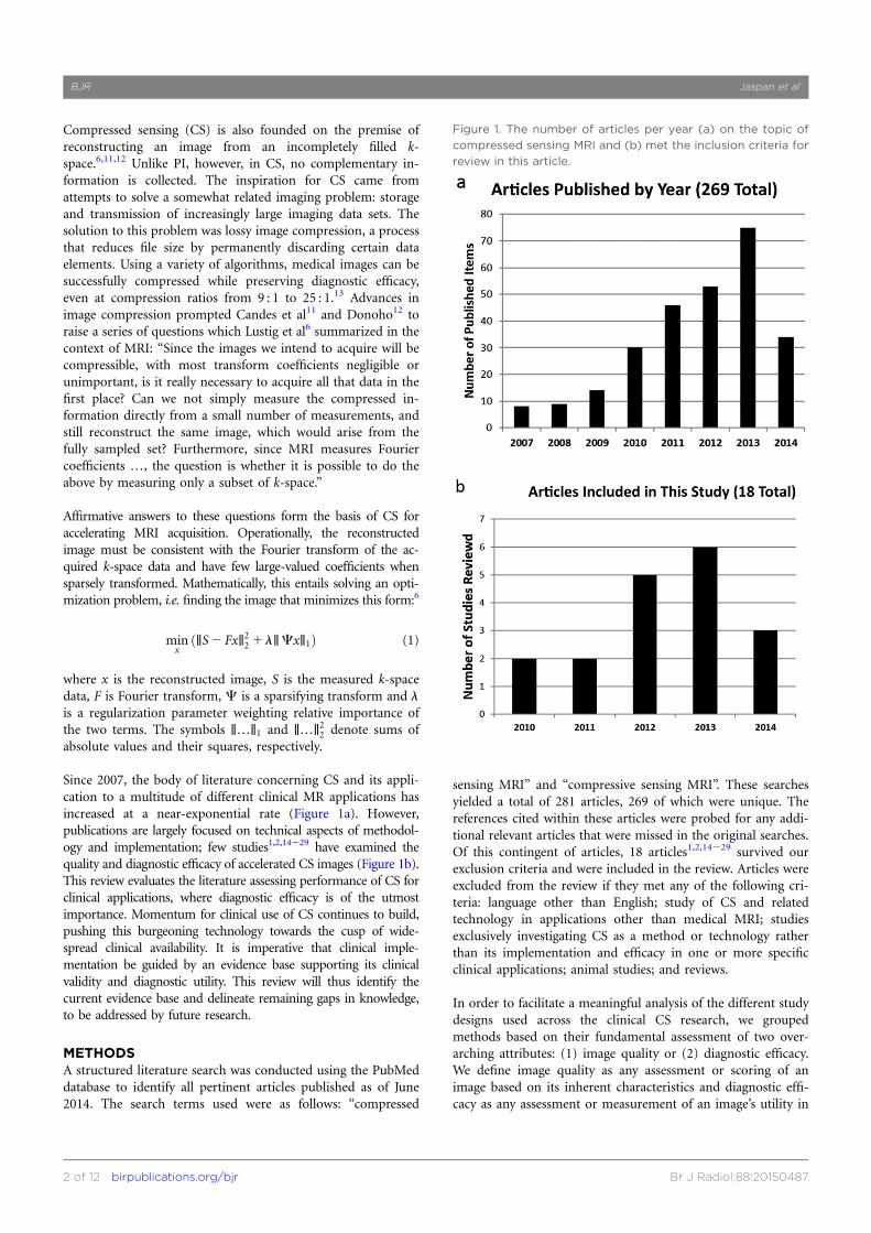
Since 2007, the body of literature concerning CS and its appli-cation to a multitude of different clinical MR applications hasincreased at a near-exponential rate (Figure 1a). However,publications are largely focused on technical aspects of methodol-ogy and implementation; few studies1,2,14229 have examined thequality and diagnostic efficacy of accelerated CS images (Figure 1b).This review evaluates the literature assessing performance of CS forclinical applications, where diagnostic efficacy is of the utmostimportance. Momentum for clinical use of CS continues to build,pushing this burgeoning technology towards the cusp of wide-spread clinical availability. It is imperative that clinical imple-mentation be guided by an evidence base supporting its clinicalvalidity and diagnostic utility. This review will thus identify thecurrent evidence base and delineate remaining gaps in knowledge,to be addressed by future research.
METHODSA structured literature search was conducted using the PubMeddatabase to identify all pertinent articles published as of June2014. The search terms used were as follows: “compressed
sensing MRI” and “compressive sensing MRI”. These searchesyielded a total of 281 articles, 269 of which were unique. Thereferences cited within these articles were probed for any addi-tional relevant articles that were missed in the original searches.Of this contingent of articles, 18 articles1,2,14229 survived ourexclusion criteria and were included in the review. Articles wereexcluded from the review if they met any of the following cri-teria: language other than English; study of CS and relatedtechnology in applications other than medical MRI; studiesexclusively investigating CS as a method or technology ratherthan its implementation and efficacy in one or more specificclinical applications; animal studies; and reviews.
In order to facilitate a meaningful analysis of the different studydesigns used across the clinical CS research, we groupedmethods based on their fundamental assessment of two over-arching attributes: (1) image quality or (2) diagnostic efficacy.We define image quality as any assessment or scoring of animage based on its inherent characteristics and diagnostic effi-cacy as any assessment or measurement of an image’s utility in
Figure 1. The number of articles per year (a) on the topic of
compressed sensing MRI and (b) met the inclusion criteria for
review in this article.
BJR Jaspan et al
2 of 12 birpublications.org/bjr Br J Radiol;88:20150487

making an accurate diagnosis, in comparison with an externalstandard. Each of these two characteristics can be assessedqualitatively or quantitatively, as shown in Figure 2. All of theassessment criteria used by each of the studies is available in theSupplementary Table A.
RESULTSClinical applications of compressed sensingDecades of research studying the effects of compression ondifferent types of medical images has provided evidence that the“tolerance” of an image to compression is dependent on the
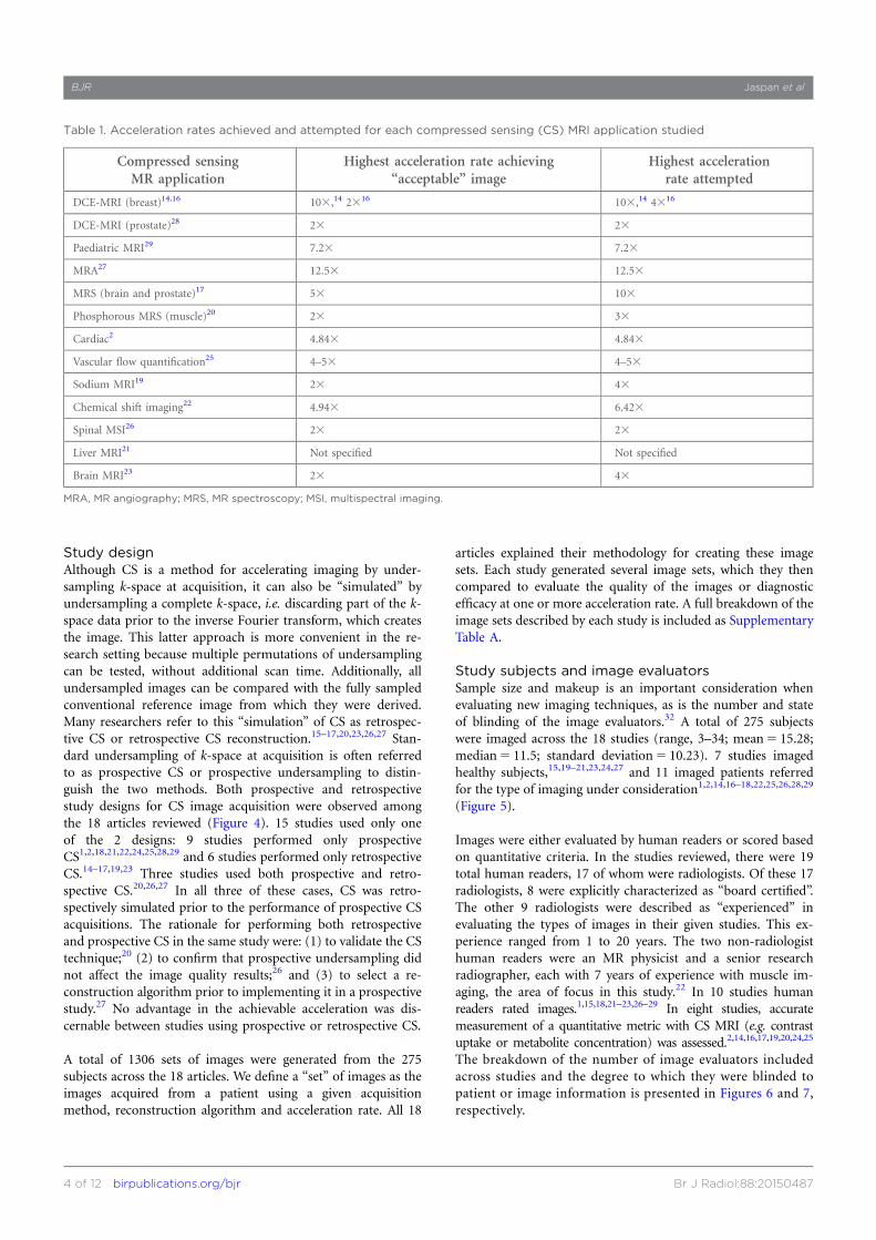
modality and the nature of the anatomical and pathologicalcontent of the image.30 Therefore, the degree of compressionimplemented in the clinical setting is dependent upon thesesame factors.31 It stands to reason that CS, a technology relatedto image compression, should similarly be evaluated across thebreadth of MRI applications and anatomical regions. The 18articles included in this review evaluated the effect of CS ona wide range of MR applications (Figure 3). These studiesassessed image quality at different acceleration rates and repor-ted to what extent remained acceptable in the face of accelera-tion (Table 1).
Figure 2. Scheme for categorizing the study design of each article. Studies were first grouped as to whether they evaluated image
quality or diagnostic efficacy (some assessed both). These studies were then further classified as to whether they employed
qualitative or quantitative assessment methods.
Figure 3. The number of studies that have evaluated compressed sensing with each MR application: dynamic contrast-enhanced
MRI (DCE-MRI);14,16,28 paediatric MRI;1,29 MR angiography;15,27 MR spectroscopy of the brain and prostate17 and for the measurement
of phosphocreatine regeneration in muscle;20 phase-contrast MRI for cardiac imaging2,18 and vascular flow quantification;24,25
sodium MRI for early detection of osteoarthritis;19 chemical shift imaging for fat fraction quantification in Becker’s muscular
dystrophy;22 multispectral imaging (MSI) of the spine;26 contrast-enhanced multiphase MRI of the liver;21 and brain MRI.23 Subjects
imaged in the paediatric MRI studies were referred for abdominal, cardiac, knee or cholangiopancreatography.
Review article: A review of clinical compressed sensing MRI BJR
3 of 12 birpublications.org/bjr Br J Radiol;88:20150487
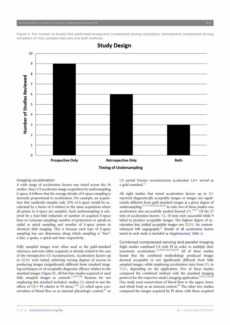
Study designAlthough CS is a method for accelerating imaging by under-sampling k-space at acquisition, it can also be “simulated” byundersampling a complete k-space, i.e. discarding part of the k-space data prior to the inverse Fourier transform, which createsthe image. This latter approach is more convenient in the re-search setting because multiple permutations of undersamplingcan be tested, without additional scan time. Additionally, allundersampled images can be compared with the fully sampledconventional reference image from which they were derived.Many researchers refer to this “simulation” of CS as retrospec-tive CS or retrospective CS reconstruction.15–17,20,23,26,27 Stan-dard undersampling of k-space at acquisition is often referredto as prospective CS or prospective undersampling to distin-guish the two methods. Both prospective and retrospectivestudy designs for CS image acquisition were observed amongthe 18 articles reviewed (Figure 4). 15 studies used only oneof the 2 designs: 9 studies performed only prospectiveCS1,2,18,21,22,24,25,28,29 and 6 studies performed only retrospectiveCS.14–17,19,23 Three studies used both prospective and retro-spective CS.20,26,27 In all three of these cases, CS was retro-spectively simulated prior to the performance of prospective CSacquisitions. The rationale for performing both retrospectiveand prospective CS in the same study were: (1) to validate the CStechnique;20 (2) to confirm that prospective undersampling didnot affect the image quality results;26 and (3) to select a re-construction algorithm prior to implementing it in a prospectivestudy.27 No advantage in the achievable acceleration was dis-cernable between studies using prospective or retrospective CS.
A total of 1306 sets of images were generated from the 275subjects across the 18 articles. We define a “set” of images as theimages acquired from a patient using a given acquisitionmethod, reconstruction algorithm and acceleration rate. All 18
articles explained their methodology for creating these imagesets. Each study generated several image sets, which they thencompared to evaluate the quality of the images or diagnosticefficacy at one or more acceleration rate. A full breakdown of theimage sets described by each study is included as SupplementaryTable A.
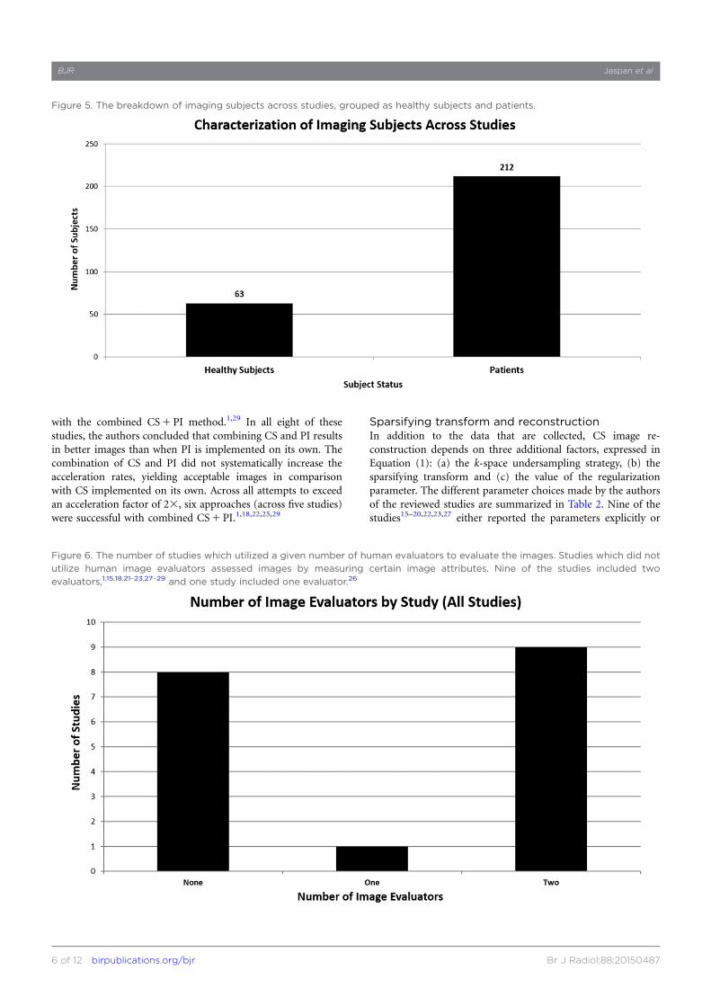
Study subjects and image evaluatorsSample size and makeup is an important consideration whenevaluating new imaging techniques, as is the number and stateof blinding of the image evaluators.32 A total of 275 subjectswere imaged across the 18 studies (range, 3–34; mean5 15.28;median5 11.5; standard deviation5 10.23). 7 studies imagedhealthy subjects,15,19–21,23,24,27 and 11 imaged patients referredfor the type of imaging under consideration1,2,14,16–18,22,25,26,28,29
(Figure 5).
Images were either evaluated by human readers or scored basedon quantitative criteria. In the studies reviewed, there were 19total human readers, 17 of whom were radiologists. Of these 17radiologists, 8 were explicitly characterized as “board certified”.The other 9 radiologists were described as “experienced” inevaluating the types of images in their given studies. This ex-perience ranged from 1 to 20 years. The two non-radiologisthuman readers were an MR physicist and a senior researchradiographer, each with 7 years of experience with muscle im-aging, the area of focus in this study.22 In 10 studies humanreaders rated images.1,15,18,21–23,26–29 In eight studies, accuratemeasurement of a quantitative metric with CS MRI (e.g. contrastuptake or metabolite concentration) was assessed.2,14,16,17,19,20,24,25
The breakdown of the number of image evaluators includedacross studies and the degree to which they were blinded topatient or image information is presented in Figures 6 and 7,respectively.
Table 1. Acceleration rates achieved and attempted for each compressed sensing (CS) MRI application studied
Compressed sensingMR application
Highest acceleration rate achieving“acceptable” image
Highest accelerationrate attempted
DCE-MRI (breast)14,16 103,14 2316 103,14 4316
DCE-MRI (prostate)28 23 23
Paediatric MRI29 7.23 7.23
MRA27 12.53 12.53
MRS (brain and prostate)17 53 103
Phosphorous MRS (muscle)20 23 33
Cardiac2 4.843 4.843
Vascular flow quantification25 4–53 4–53
Sodium MRI19 23 43
Chemical shift imaging22 4.943 6.423
Spinal MSI26 23 23
Liver MRI21 Not specified Not specified
Brain MRI23 23 43
MRA, MR angiography; MRS, MR spectroscopy; MSI, multispectral imaging.
BJR Jaspan et al
4 of 12 birpublications.org/bjr Br J Radiol;88:20150487
Imaging accelerationA wide range of acceleration factors was tested across the 18studies. Since CS accelerates image acquisition by undersamplingk-space, it follows that the average density of k-space sampling isinversely proportional to acceleration. For example, an acquisi-tion that randomly samples only 25% of k-space would be ac-celerated by a factor of 4 relative to the same acquisition whereall points in k-space are sampled. Such undersampling is ach-ieved by a four-fold reduction of number of acquired k-spacelines in Cartesian sampling; number of projections or spirals inradial or spiral sampling and number of k-space points inchemical shift imaging. This is because each type of k-spacesampling has one dimension along which sampling is “free”:a line, a spoke, a spiral and time respectively.
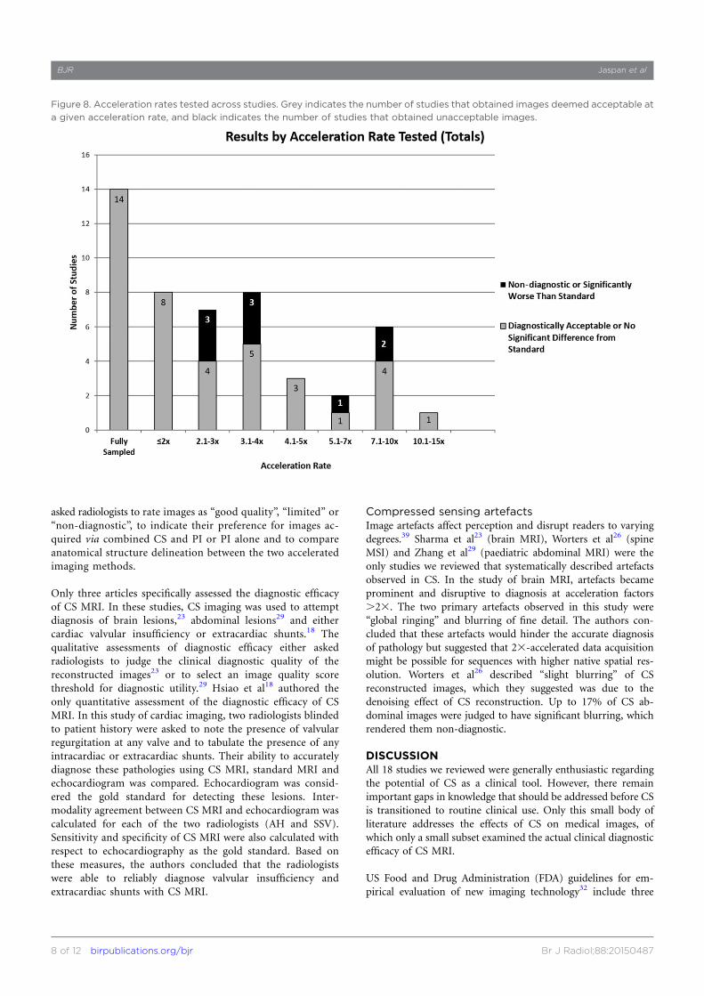
Fully sampled images were often used as the gold-standardreference, and were either acquired, or already existed in the caseof the retrospective CS reconstructions. Acceleration factors upto 12.53 were tested, achieving varying degrees of success inproducing images insignificantly different from standard imag-ing techniques or of acceptable diagnostic efficacy relative to thestandard images (Figure 8). All but four studies acquired or usedfully sampled images as controls.1,25,27,29 Reasons for notemploying this standard included: studies (1) aimed to test theeffects of CS1 PI relative to PI alone,1,29 (2) relied upon con-servation of blood flow as an internal physiologic control,25 or
(3) partial Fourier reconstruction accelerated 1.63 served asa gold standard.27
All eight studies that tested acceleration factors up to 23reported diagnostically acceptable images or images not signif-icantly different from gold standard images at a given degree ofundersampling.15–17,19,20,23,26,28 In only two of these studies wasacceleration also successfully pushed beyond 23.15,17 Of the 27tests of acceleration factors .2, 18 tests were successful while 9failed to produce acceptable images. The highest degree of ac-celeration that yielded acceptable images was 12.53, for contrast-enhanced MR angiography.27 Results of all acceleration factorstested in each study is included as Supplementary Table A.
Combined compressed sensing and parallel imagingEight studies combined CS with PI in order to multiply theirmaximum acceleration.1,2,18,21,22,25,28,29 All of these studiesfound that the combined methodology produced imagesdeemed acceptable or not significantly different from fullysampled images, while employing acceleration rates from 23 to7.23, depending on the application. Five of these studiescompared the combined method with the standard imagingprotocol for the respective study’s imaging application.2,18,21,22,28
One study used conservation of blood flow to the upper, lowerand whole body as an internal control.25 The other two studiescompared the images acquired by PI alone with those acquired
Figure 4. The number of studies that performed prospective compressed sensing acquisition, retrospective compressed sensing
simulation on fully sampled data sets and both methods.
Review article: A review of clinical compressed sensing MRI BJR
5 of 12 birpublications.org/bjr Br J Radiol;88:20150487
with the combined CS1PI method.1,29 In all eight of thesestudies, the authors concluded that combining CS and PI resultsin better images than when PI is implemented on its own. Thecombination of CS and PI did not systematically increase theacceleration rates, yielding acceptable images in comparisonwith CS implemented on its own. Across all attempts to exceedan acceleration factor of 23, six approaches (across five studies)were successful with combined CS1PI.1,18,22,25,29
Sparsifying transform and reconstructionIn addition to the data that are collected, CS image re-construction depends on three additional factors, expressed inEquation (1): (a) the k-space undersampling strategy, (b) thesparsifying transform and (c) the value of the regularizationparameter. The different parameter choices made by the authorsof the reviewed studies are summarized in Table 2. Nine of thestudies15–20,22,23,27 either reported the parameters explicitly or
Figure 5. The breakdown of imaging subjects across studies, grouped as healthy subjects and patients.
Figure 6. The number of studies which utilized a given number of human evaluators to evaluate the images. Studies which did not
utilize human image evaluators assessed images by measuring certain image attributes. Nine of the studies included two
evaluators,1,15,18,21–23,27–29 and one study included one evaluator.26
BJR Jaspan et al
6 of 12 birpublications.org/bjr Br J Radiol;88:20150487
cited correspondingly in Greengard et al,33 Lustig and Pauly,34
Feng et al,35 Uecker et al,36 Zhang et al37 where each could befound, though the available information may have been in-complete. Simply describing the sparsifying transform asa wavelet type, for example, still leaves many details unclear asthere are many types of wavelets, each with different proper-ties.38 All of the studies utilized undersampling strategies, whicheither fully sampled the centre of k-space or sampled it moredensely than the periphery of k-space. When Poisson disc orGaussian sampling was used, the parameters of these dis-tributions (minimum distance between points and its de-pendence on location in k-space for Poisson-disc sampling orwidth of the Gaussian sampling) were not always specified.While intuitive or semianalytic substantiation of the choice ofsparsifying transform and sampling strategy can be found, thegreatest challenge is presented by the regularization parameter.As seen in Table 2, the regularization parameter appears to de-pend on image type, acceleration factor, sparsifying transform,sampling scheme etc. Importantly, it also depends on how thedata are scaled. Indeed, Equation (1) verifies that scaling themeasured data S and the reconstructed image x by a factor of 2also requires corresponding scaling of the regularization pa-rameter. Thus, for the value of the regularization parameter toconvey useful information, the scaling of the data needs to bedescribed. Only one study, by Akcakaya et al,15 reported how thedata were scaled. All studies noted that the value of the regu-larization parameter impacts perceived image quality, artefactsor synthetic appearance.
Another relevant aspect of image reconstruction is its speed.Given non-linear and iterative nature of the available algorithms,
the reconstruction is inherently slow. Tabulating their perfor-mance requires standardization of the task: type of acquisition,k-space undersampling strategy, sparsifying transform etc. andtherefore is more appropriate for a technical review of CS.Nevertheless, long delay between image acquisition and itsavailability for review by a radiologist presents a barrier for thewidespread clinical use of CS. Depending on the task and al-gorithm, the delay may be between 30 and 210min.1,14,19
However, we remain optimistic that through a combination ofincreased central processing unit speeds, parallel computing,graphics processing unit implementation and maturation ofalgorithms, this impediment will be resolved over time.
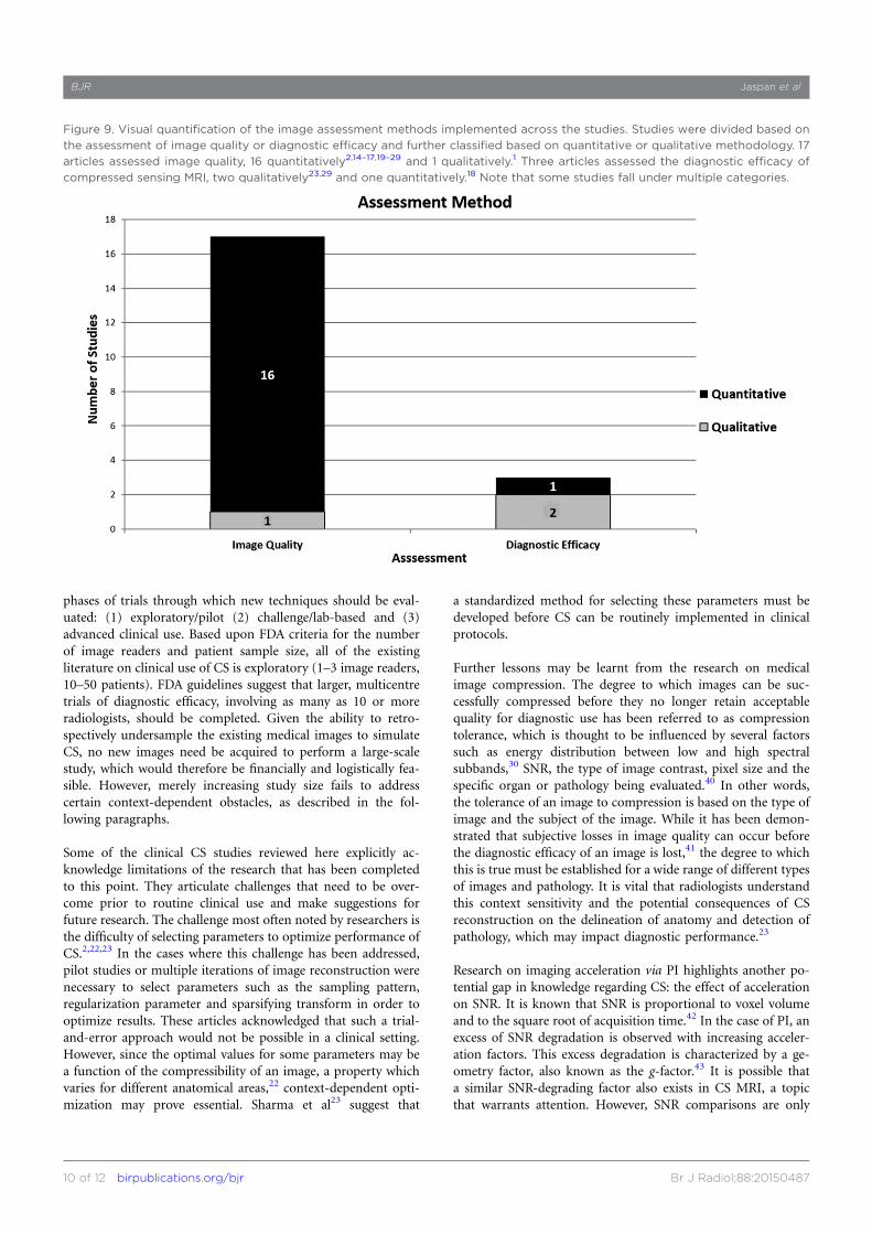
Image assessmentThe 18 articles varied substantially in their methods for evalu-ating images. Figure 9 presents the breakdown of the number ofstudies whose assessment methods fit each of the above classi-fications. 8 of the 16 quantitative assessments of image qualityused radiologists as readers.15,21–23,26–29 Each reader ratedimages on a numerical scale, based on the delineation or clarityof a given set of anatomical landmarks or lesion borders, inaddition to other criteria. The other eight articles that quanti-tatively evaluated image quality rated a CS image by comparingit to a measurement made with standard imaging. These com-parisons included correlation between contrast uptake curves;14
voxel intensities;16 metabolite maps and intensities;17 ven-tricular volume, ejection fraction and various vascular flowrates;2 signal-to-noise ratio (SNR) and tissue sodium con-centration;19 muscle phosphocreatine resynthesis rate;20 andvarious vascular velocity and flow rates.24,25 In the only articlethat qualitatively assessed image quality, Vasanawala et al1
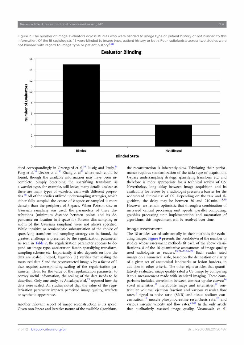
Figure 7. The number of image evaluators across studies who were blinded to image type or patient history or not blinded to this
information. Of the 19 radiologists, 15 were blinded to image type, patient history or both. Four radiologists across two studies were
not blinded with regard to image type or patient history.1,28
Review article: A review of clinical compressed sensing MRI BJR
7 of 12 birpublications.org/bjr Br J Radiol;88:20150487
asked radiologists to rate images as “good quality”, “limited” or“non-diagnostic”, to indicate their preference for images ac-quired via combined CS and PI or PI alone and to compareanatomical structure delineation between the two acceleratedimaging methods.
Only three articles specifically assessed the diagnostic efficacyof CS MRI. In these studies, CS imaging was used to attemptdiagnosis of brain lesions,23 abdominal lesions29 and eithercardiac valvular insufficiency or extracardiac shunts.18 Thequalitative assessments of diagnostic efficacy either askedradiologists to judge the clinical diagnostic quality of thereconstructed images23 or to select an image quality scorethreshold for diagnostic utility.29 Hsiao et al18 authored theonly quantitative assessment of the diagnostic efficacy of CSMRI. In this study of cardiac imaging, two radiologists blindedto patient history were asked to note the presence of valvularregurgitation at any valve and to tabulate the presence of anyintracardiac or extracardiac shunts. Their ability to accuratelydiagnose these pathologies using CS MRI, standard MRI andechocardiogram was compared. Echocardiogram was consid-ered the gold standard for detecting these lesions. Inter-modality agreement between CS MRI and echocardiogram wascalculated for each of the two radiologists (AH and SSV).Sensitivity and specificity of CS MRI were also calculated withrespect to echocardiography as the gold standard. Based onthese measures, the authors concluded that the radiologistswere able to reliably diagnose valvular insufficiency andextracardiac shunts with CS MRI.
Compressed sensing artefactsImage artefacts affect perception and disrupt readers to varyingdegrees.39 Sharma et al23 (brain MRI), Worters et al26 (spineMSI) and Zhang et al29 (paediatric abdominal MRI) were theonly studies we reviewed that systematically described artefactsobserved in CS. In the study of brain MRI, artefacts becameprominent and disruptive to diagnosis at acceleration factors.23. The two primary artefacts observed in this study were“global ringing” and blurring of fine detail. The authors con-cluded that these artefacts would hinder the accurate diagnosisof pathology but suggested that 23-accelerated data acquisitionmight be possible for sequences with higher native spatial res-olution. Worters et al26 described “slight blurring” of CSreconstructed images, which they suggested was due to thedenoising effect of CS reconstruction. Up to 17% of CS ab-dominal images were judged to have significant blurring, whichrendered them non-diagnostic.
DISCUSSIONAll 18 studies we reviewed were generally enthusiastic regardingthe potential of CS as a clinical tool. However, there remainimportant gaps in knowledge that should be addressed before CSis transitioned to routine clinical use. Only this small body ofliterature addresses the effects of CS on medical images, ofwhich only a small subset examined the actual clinical diagnosticefficacy of CS MRI.
US Food and Drug Administration (FDA) guidelines for em-pirical evaluation of new imaging technology32 include three
Figure 8. Acceleration rates tested across studies. Grey indicates the number of studies that obtained images deemed acceptable at
a given acceleration rate, and black indicates the number of studies that obtained unacceptable images.
BJR Jaspan et al
8 of 12 birpublications.org/bjr Br J Radiol;88:20150487
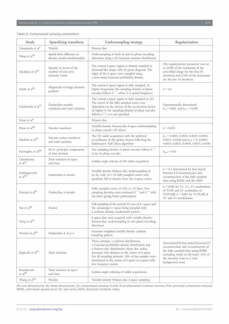
Table 2. Compressed sensing parameters
Study Sparsifying transform Undersampling strategy Regularization
Vasanawala et al1 Wavelet Poisson disc
Wang et al14Spatial finite difference ordiscrete wavelet transformation
Undersampling in both ky and kz phase-encodingdirections using a 2D Gaussian random distribution
Akcakaya et al15Sparsity in terms of thenumber of non-zerointensity voxels
The central k-space region is densely sampled indiamond-like shape with 24-point diagonal. Theedges of the k-space were sampled usinga zero-mean Gaussian-probability density
The regularization parameter was setto 2/500 of the maximum of thezero-filled image for the first 50iterations and 2/100 of the maximumfor the last 10 iterations
Smith et al16Magnitude of image intensitygradient
The central k-space region is fully sampled. Athigher frequencies the sampling density of phaseencodes follows k21, where k is spatial frequency
l5 0.1
Geethanath et al17Daubechies wavelettransform and total variation
The central k-space region is fully sampled in 2D.The extent of the fully sampled centre wasdependent on the inverse of the acceleration factor.At higher k, the sampling density of phase encodesfollows k2n, n is not specified
Experimentally determinedlw5 0.001 and lTV5 0.005
Hsiao et al2 Poisson disc
Hsiao et al18 Wavelet transformVariable density Poisson-disc k-space undersamplingin phase encode (2D slices)
l5 0.015
Madelin et al19Discrete cosine transformand total variation
The 3D radial acquisition with the sphericalco-ordinates of the spokes chosen following theRakhmanov–Saff–Zhou algorithm
lw5 0.0005, 0.0010, 0.0025, 0.0050,0.0075, 0.0100 and lTV5 0, 0.0005,0.0010, 0.0025, 0.0050, 0.0075, 0.0100
Parasoglou et al20PCA—principal componentsin time domain
The sampling density of phase encodes follows k23
in ky–kz phase encodeslpca5 0.01
Chandaranaet al21
Total variation in spaceand time
Golden-angle scheme of 2D radial acquisition
Hollingsworthet al22
Daubechies 4 waveletVariable density Poisson-disc undersampling inky–kz, with 243 24 fully sampled centre withquadratic fall in density from the k-space centre
l5 0.1 determined by best matchbetween CS reconstruction andreconstruction of the fully sampleddata using RMSE and the SSIM
Sharma et al23 Daubechies 4 waveletFully sampled centre of 10% i.e. 25 lines. Twosampling densities were evaluated k22 and k26 withthe latter giving better performance
l50.002 for 23, 33, 43 accelerationsof T2-SE and 23 acceleration ofT2-FLAIR; l50.001 for T2-FLAIR at33 and 43 accelerations
Tao et al24 FourierFull sampling of the central 20 rows of k-t space andthe remaining k-t space being sampled witha uniform density, randomised pattern
Tariq et al25k-space data were acquired with variable densityPoisson-disc undersampling in two-phase-encodingdirections
Worters et al26 Daubechies 4, in y–zGaussian-weighted variable density randomsampling pattern
Rapacchi et al27 Total variation
Three schemes: a uniform distribution,a Gaussian-probability density distribution anda Poisson-disc distribution where disc radiusincreases with distance to the centre of k-space.For all sampling patterns, 10% of the samples weredistributed in the centre of k-space to acquire fullylow frequency points
Determined by best match between CSreconstruction and reconstruction ofthe fully sampled data using RMSEexcluding voxels in the lower 10% ofthe intensity scale to a voidbackground noise
Rosenkrantzet al28
Total variation in spaceand time
Golden-angle ordering of radial acquisitions
Zhang et al29 Wavelet Variable density Poisson-disc k-space sampling
2D, two-dimensional; 3D, three-dimensional; CS, compressed sensing; FLAIR, fluid-attenuated inversion recover; PCA, principal component analysis;RMSE, root mean square error; SE, spin echo; SSIM, structural similarity index.
Review article: A review of clinical compressed sensing MRI BJR
9 of 12 birpublications.org/bjr Br J Radiol;88:20150487
phases of trials through which new techniques should be eval-uated: (1) exploratory/pilot (2) challenge/lab-based and (3)advanced clinical use. Based upon FDA criteria for the numberof image readers and patient sample size, all of the existingliterature on clinical use of CS is exploratory (1–3 image readers,10–50 patients). FDA guidelines suggest that larger, multicentretrials of diagnostic efficacy, involving as many as 10 or moreradiologists, should be completed. Given the ability to retro-spectively undersample the existing medical images to simulateCS, no new images need be acquired to perform a large-scalestudy, which would therefore be financially and logistically fea-sible. However, merely increasing study size fails to addresscertain context-dependent obstacles, as described in the fol-lowing paragraphs.
Some of the clinical CS studies reviewed here explicitly ac-knowledge limitations of the research that has been completedto this point. They articulate challenges that need to be over-come prior to routine clinical use and make suggestions forfuture research. The challenge most often noted by researchers isthe difficulty of selecting parameters to optimize performance ofCS.2,22,23 In the cases where this challenge has been addressed,pilot studies or multiple iterations of image reconstruction werenecessary to select parameters such as the sampling pattern,regularization parameter and sparsifying transform in order tooptimize results. These articles acknowledged that such a trial-and-error approach would not be possible in a clinical setting.However, since the optimal values for some parameters may bea function of the compressibility of an image, a property whichvaries for different anatomical areas,22 context-dependent opti-mization may prove essential. Sharma et al23 suggest that
a standardized method for selecting these parameters must bedeveloped before CS can be routinely implemented in clinicalprotocols.
Further lessons may be learnt from the research on medicalimage compression. The degree to which images can be suc-cessfully compressed before they no longer retain acceptablequality for diagnostic use has been referred to as compressiontolerance, which is thought to be influenced by several factorssuch as energy distribution between low and high spectralsubbands,30 SNR, the type of image contrast, pixel size and thespecific organ or pathology being evaluated.40 In other words,the tolerance of an image to compression is based on the type ofimage and the subject of the image. While it has been demon-strated that subjective losses in image quality can occur beforethe diagnostic efficacy of an image is lost,41 the degree to whichthis is true must be established for a wide range of different typesof images and pathology. It is vital that radiologists understandthis context sensitivity and the potential consequences of CSreconstruction on the delineation of anatomy and detection ofpathology, which may impact diagnostic performance.23
Research on imaging acceleration via PI highlights another po-tential gap in knowledge regarding CS: the effect of accelerationon SNR. It is known that SNR is proportional to voxel volumeand to the square root of acquisition time.42 In the case of PI, anexcess of SNR degradation is observed with increasing acceler-ation factors. This excess degradation is characterized by a ge-ometry factor, also known as the g-factor.43 It is possible thata similar SNR-degrading factor also exists in CS MRI, a topicthat warrants attention. However, SNR comparisons are only
Figure 9. Visual quantification of the image assessment methods implemented across the studies. Studies were divided based on
the assessment of image quality or diagnostic efficacy and further classified based on quantitative or qualitative methodology. 17
articles assessed image quality, 16 quantitatively2,14–17,19–29 and 1 qualitatively.1 Three articles assessed the diagnostic efficacy of
compressed sensing MRI, two qualitatively23,29 and one quantitatively.18 Note that some studies fall under multiple categories.
BJR Jaspan et al
10 of 12 birpublications.org/bjr Br J Radiol;88:20150487
valid when voxel dimensions are fixed. Unlike PI or conventionalCartesian imaging where voxel size is given by the ratio of fieldof view to number of phase-encode lines (adjusted for parallelacceleration factor), this definition does not apply in CS, as isevident by the blurring artefact seen in CS images, as discussedin the section entitled “Compressed sensing artefacts”. Moreover,SNR is a function of the regularization parameter and can bemade arbitrarily high: Equation (1) verifies that increase ofregularization reduces the consistency of the reconstructed im-age with the noisy data leading to the reduction of noise in theimage. Such trade of accuracy of image reconstruction for SNRvia regularization has been explored in the context of PI;44,45
however, it is not essential to the PI method. Therefore, non-linear and denoising properties of CS reconstruction, which mayalso remove important diagnostic information, present a chal-lenge for SNR characterization. A more promising approach is
a recently developed framework for task-based assessment ofimage quality in CS which models clinical uses of MRI, suchas identification and localization of abnormalities.46
The potential for CS to accelerate MRI acquisition with minimaleffects on image quality is an exciting development for the futureof radiology, and medicine as a whole. In this day of increasedcognizance of healthcare costs, it is apparent that efficiency of allmedical services is at a premium. This is especially true in MRI,a relatively costly and time-intensive imaging modality. Longacquisition times also limit the number of patients for whom theservice is available and decrease the utility of MRI for manyapplications requiring high imaging speed. Thus, optimizing CSfor various clinical MRI contexts is an important goal withpotential to transform diagnostic imaging with respect to effi-ciency, cost-effectiveness and ultimately clinical utility.
REFERENCES
1. Vasanawala SS, Alley MT, Hargreaves BA,
Barth RA, Pauly JM, Lustig M. Improved
pediatric MR imaging with compressed
sensing. Radiology 2010; 256: 607–16. doi:
10.1148/radiol.10091218
2. Hsiao A, Lustig M, Alley MT, Murphy M,
Chan FP, Herfkens RJ, et al. Rapid pediatric
cardiac assessment of flow and ventricular
volume with compressed sensing parallel
imaging volumetric cine phase-contrast
MRI. AJR Am J Roentgenol 2012;
198: W250–9.
3. Cohen MS, Weisskoff RM. Ultra-fast imag-
ing. Magn Reson Imaging 1991; 9: 1–37.
4. Olsen OE. Imaging of abdominal tumours:
CTor MRI? Pediatr Radiol 2008; 38(Suppl 3):
S452–8. doi: 10.1007/s00247-008-0846-5
5. Geethanath S, Reddy R, Konar AS, Imam S,
Sundaresan R, D R RB, et al. Compressed
sensing MRI: a review. Crit Rev Biomed Eng
2013; 41: 183–204.
6. Lustig M, Donoho D, Pauly JM. Sparse MRI:
the application of compressed sensing for
rapid MR imaging. Magn Reson Med 2007;
58: 1182–95. doi: 10.1002/mrm.21391
7. McGibney G, Smith MR, Nichols ST,
Crawley A. Quantitative evaluation of several
partial Fourier reconstruction algorithms
used in MRI. Magn Reson Med 1993; 30:
51–9. doi: 10.1002/mrm.1910300109
8. Pruessmann KP, Weiger M, Scheidegger MB,
Boesiger P. SENSE: sensitivity encoding for
fast MRI. Magn Reson Med 1999;
42: 952–62.
9. Sodickson DK, Manning WJ. Simultaneous
acquisition of spatial harmonics (SMASH):
fast imaging with radiofrequency coil arrays.
Magn Reson Med 1997; 38: 591–603. doi:
10.1002/mrm.1910380414
10. Hutchinson M, Raff U. Fast MRI data
acquisition using multiple detectors. Magn
Reson Med 1988; 6: 87–91.
11. Candes EJ, Romberg J, Tao T. Robust
uncertainty principles: exact signal recon-
struction from highly incomplete frequency
information. IEEE Trans Inf Theory 2006;
52: 489–509.
12. Donoho DL. Compressed sensing. IEEE
Trans Inf Theory 2006; 52: 1289–306. doi:
10.1109/TIT.2006.871582
13. Koff DA, Shulman H. An overview of digital
compression of medical images: can we use
lossy image compression in radiology? Can
Assoc Radiol J 2006; 57: 211–17.
14. Wang H, Miao Y, Zhou K, Yu Y, Bao S, He Q,
et al. Feasibility of high temporal resolution
breast DCE-MRI using compressed sensing
theory. Med Phys 2010; 37: 4971–81. doi:
10.1118/1.3483094
15. Akcakaya M, Hu P, Chuang ML, Hauser TH,
Ngo LH, Manning WJ, et al. Accelerated
noncontrast-enhanced pulmonary vein MRA
with distributed compressed sensing. J Magn
Reson Imaging 2011; 33: 1248–55.
16. Smith DS, Welch EB, Li X, Arlinghaus LR,
Loveless ME, Koyama T, et al. Quantitative
effects of using compressed sensing in
dynamic contrast enhanced MRI. Phys Med
Biol 2011; 56: 4933–46. doi: 10.1088/
0031-9155/56/15/018
17. Geethanath S, Baek HM, Ganji SK, Ding Y,
Maher EA, Sims RD, et al. Compressive
sensing could accelerate 1H MR metabolic
imaging in the clinic. Radiology 2012; 262:
985–94. doi: 10.1148/radiol.11111098
18. Hsiao A, Lustig M, Alley MT, Murphy MJ,
Vasanawala SS. Evaluation of valvular in-
sufficiency and shunts with parallel-imaging
compressed-sensing 4D phase-contrast MR
imaging with stereoscopic 3D velocity-fusion
volume-rendered visualization. Radiology 2012;
265: 87–95. doi: 10.1148/radiol.12120055
19. Madelin G, Chang G, Otazo R, Jerschow A,
Regatte RR. Compressed sensing sodium
MRI of cartilage at 7T: preliminary study.
J Magn Reson 2012; 214: 360–5. doi: 10.1016/
j.jmr.2011.12.005
20. Parasoglou P, Feng L, Xia D, Otazo R, Regatte RR.
Rapid 3D-imaging of phosphocreatine recovery
kinetics in the human lower leg muscles with
compressed sensing.Magn Reson Med 2012; 68:
1738–46. doi: 10.1002/mrm.24484
21. Chandarana H, Feng L, Block TK,
Rosenkrantz AB, Lim RP, Babb JS, et al. Free-
breathing contrast-enhanced multiphase MRI
of the liver using a combination of compressed
sensing, parallel imaging, and golden-angle
radial sampling. Invest Radiol 2013; 48: 10–16.
doi: 10.1097/RLI.0b013e318271869c
22. Hollingsworth KG, Higgins DM, McCallum M,
Ward L, Coombs A, Straub V. Investigating
the quantitative fidelity of prospectively
undersampled chemical shift imaging in
muscular dystrophy with compressed
sensing and parallel imaging reconstruction.
Magn Reson Med 2013; 72: 1610–19. doi:
10.1002/mrm.25077.
23. Sharma SD, Fong CL, Tzung BS, Law M, Nayak
KS. Clinical image quality assessment of accel-
erated magnetic resonance neuroimaging using
compressed sensing. Invest Radiol 2013; 48:
638–45. doi: 10.1097/RLI.0b013e31828a012d
24. Tao Y, Rilling G, Davies M, Marshall I.
Carotid blood flow measurement accelerated
by compressed sensing: validation in healthy
volunteers. Magn Reson Imaging 2013; 31:
1485–91. doi: 10.1016/j.mri.2013.05.009
Review article: A review of clinical compressed sensing MRI BJR
11 of 12 birpublications.org/bjr Br J Radiol;88:20150487
25. Tariq U, Hsiao A, Alley M, Zhang T, Lustig M,
Vasanawala SS. Venous and arterial flow
quantification are equally accurate and pre-
cise with parallel imaging compressed sens-
ing 4D phase contrast MRI. J Magn Reson
Imaging 2013; 37: 1419–26. doi: 10.1002/
jmri.23936
26. Worters PW, Sung K, Stevens KJ, Koch KM,
Hargreaves BA. Compressed-sensing multi-
spectral imaging of the postoperative spine. J
Magn Reson Imaging 2013; 37: 243–8. doi:
10.1002/jmri.23750
27. Rapacchi S, Han F, Natsuaki Y, Kroeker R,
Plotnik A, Lehrman E, et al. High spatial and
temporal resolution dynamic contrast-
enhanced magnetic resonance angiography
using compressed sensing with magnitude
image subtraction. Magn Reson Med 2014; 71:
1771–83. doi: 10.1002/mrm.24842
28. Rosenkrantz AB, Geppert C, Grimm R,
Block TK, Glielmi C, Feng L, et al. Dynamic
contrast-enhanced MRI of the prostate with
high spatiotemporal resolution using com-
pressed sensing, parallel imaging, and con-
tinuous golden-angle radial sampling:
preliminary experience. J Magn Reson Imag-
ing 2014; 41: 1365–73. doi: 10.1002/
jmri.24661
29. Zhang T, Chowdhury S, Lustig M, Barth RA,
Alley MT, Grafendorfer T, et al. Clinical
performance of contrast enhanced abdominal
pediatric MRI with fast combined parallel
imaging compressed sensing reconstruction.
J Magn Reson Imaging 2014; 40: 13–25. doi:
10.1002/jmri.24333
30. Persons K, Palisson P, Manduca A, Erickson BJ,
Savcenko V. An analytical look at the effects of
compression on medical images. J Digit Imaging
1997; 10(3 Suppl 1): 60–6. doi: 10.1007/
BF03168659
31. Koff D, Bak P, Brownrigg P, Hosseinzadeh D,
Khademi A, Kiss A, et al. Pan-Canadian
evaluation of irreversible compression ratios
(“lossy” compression) for development of
national guidelines. J Digit Imaging 2009;
22: 569–78.
32. Gallas BD, Chan HP, D’Orsi CJ, Dodd LE,
Giger ML, Gur D, et al. Evaluating imaging
and computer-aided detection and diagnosis
devices at the FDA. Acad Radiol 2012; 19:
463–77. doi: 10.1016/j.acra.2011.12.016
33. Greengard L, Lee J-Y, Inati S. The fast sinc
transform and image reconstruction from
non-uniform samples in k-space. Comm Appl
Math Comp Sci 2006; 1: 121–32.
34. Lustig M, Pauly JM. SPIRiT: iterative
self-consistent parallel imaging
reconstruction from arbitrary k-space.
Magn Reson Med 2010; 64: 457–71. doi:
10.1002/mrm.22428
35. Feng L, Srichai MB, Lim RP, Harrison A,
King W, Adluru G, et al. Highly accelerated
real-time cardiac cine MRI using k–t
SPARSE-SENSE. Magn Reson Med 2013; 70:
64–74. doi: 10.1002/mrm.24440
36. Uecker M, Lai P, Murphy MJ, Virtue P, Elad
M, Pauly JM, et al. ESPIRiT—an eigenvalue
approach to autocalibrating parallel MRI:
where SENSE meets GRAPPA. Magn Reson
Med 2014; 71: 990–1001.
37. Zhang T, Pauly JM, Levesque IR. Accelerating
parameter mapping with a locally low rank
constraint. Magn Reson Med 2015; 73:
655–61. doi: 10.1002/mrm.25161
38. Antonini M, Barlaud M, Mathieu P,
Daubechies I. Image coding using wavelet
transform. IEEE Trans Image Process 1992;
1: 205–20. doi: 10.1109/83.136597
39. Miao J, Huang F, Narayan S, Wilson DL. A
new perceptual difference model for di-
agnostically relevant quantitative image
quality evaluation: a preliminary study. Magn
Reson Imaging 2013; 31: 596–603.
40. Chan KK, Lou SL, Huang HK. Full-frame
transform compression of CT and MR
images. Radiology 1989; 171: 847–51. doi:
10.1148/radiology.171.3.2717763
41. Terae S, Miyasaka K, Kudoh K, Nambu T,
Shimizu T, Kaneko K, et al. Wavelet com-
pression on detection of brain lesions with
magnetic resonance imaging. J Digit Imaging
2000; 13: 178–90. doi: 10.1007/BF03168393
42. Macovski A. Noise in MRI. Magn Reson Med
1996; 36: 494–7.
43. Sodickson DK, Hardy CJ, Zhu Y, Giaquinto
RO, Gross P, Kenwood G, et al. Rapid
volumetric MRI using parallel imaging with
order-of-magnitude accelerations and a 32-
element RF coil array: feasibility and impli-
cations. Acad Radiol 2005; 12: 626–35. doi:
10.1016/j.acra.2005.01.012
44. Lin F-H, Kwong KK, Belliveau JW, Wald LL.
Parallel imaging reconstruction using auto-
matic regularization. Magn Reson Med 2004;
51: 559–67. doi: 10.1002/mrm.10718
45. Sanchez-Gonzalez J, Tsao J, Dydak U,
Desco M, Boesiger P, Paul Pruessmann K.
Minimum-norm reconstruction for
sensitivity-encoded magnetic resonance
spectroscopic imaging. Magn Reson Med
2006; 55: 287–95. doi: 10.1002/mrm.20758
46. Graff CG. Framework for task-based assess-
ment of MR image quality. Proc Intl Soc Mag
Reson Med 2013; 21: 701.
BJR Jaspan et al
12 of 12 birpublications.org/bjr Br J Radiol;88:20150487