Embed Size (px)
Citation preview
Comprehensive Review of Human Sapoviruses
Tomoichiro Oka,a,b Qiuhong Wang,b Kazuhiko Katayama,a Linda J. Saifb
Department of Virology II, National Institute of Infectious Diseases, Musashi-murayama, Tokyo, Japana; Food Animal Health Research Program, Ohio Agricultural Researchand Development Center, Department of Veterinary Preventive Medicine, The Ohio State University, Wooster, Ohio, USAb
SUMMARY . . . . . . . . . . . . . . . . . . . . . . . . . . . . . . . . . . . . . . . . . . . . . . . . . . . . . . . . . . . . . . . . . . . . . . . . . . . . . . . . . . . . . . . . . . . . . . . . . . . . . . . . . . . . . . . . . . . . . . . . . . . . . . . . . . . . . . . . . . . . . . . . . . .32INTRODUCTION . . . . . . . . . . . . . . . . . . . . . . . . . . . . . . . . . . . . . . . . . . . . . . . . . . . . . . . . . . . . . . . . . . . . . . . . . . . . . . . . . . . . . . . . . . . . . . . . . . . . . . . . . . . . . . . . . . . . . . . . . . . . . . . . . . . . . . . . . . . . . .32HISTORY. . . . . . . . . . . . . . . . . . . . . . . . . . . . . . . . . . . . . . . . . . . . . . . . . . . . . . . . . . . . . . . . . . . . . . . . . . . . . . . . . . . . . . . . . . . . . . . . . . . . . . . . . . . . . . . . . . . . . . . . . . . . . . . . . . . . . . . . . . . . . . . . . . . . . .32TAXONOMY . . . . . . . . . . . . . . . . . . . . . . . . . . . . . . . . . . . . . . . . . . . . . . . . . . . . . . . . . . . . . . . . . . . . . . . . . . . . . . . . . . . . . . . . . . . . . . . . . . . . . . . . . . . . . . . . . . . . . . . . . . . . . . . . . . . . . . . . . . . . . . . . . .33CELL CULTURE AND ANIMAL INFECTION TRIALS . . . . . . . . . . . . . . . . . . . . . . . . . . . . . . . . . . . . . . . . . . . . . . . . . . . . . . . . . . . . . . . . . . . . . . . . . . . . . . . . . . . . . . . . . . . . . . . . . . . . . . . . . . .33PHYSICAL CHARACTERISTICS AND STABILITY . . . . . . . . . . . . . . . . . . . . . . . . . . . . . . . . . . . . . . . . . . . . . . . . . . . . . . . . . . . . . . . . . . . . . . . . . . . . . . . . . . . . . . . . . . . . . . . . . . . . . . . . . . . . . .33GENOMIC ORGANIZATION . . . . . . . . . . . . . . . . . . . . . . . . . . . . . . . . . . . . . . . . . . . . . . . . . . . . . . . . . . . . . . . . . . . . . . . . . . . . . . . . . . . . . . . . . . . . . . . . . . . . . . . . . . . . . . . . . . . . . . . . . . . . . . . . . .33GENOMIC SEQUENCE AND ANTIGENICITY . . . . . . . . . . . . . . . . . . . . . . . . . . . . . . . . . . . . . . . . . . . . . . . . . . . . . . . . . . . . . . . . . . . . . . . . . . . . . . . . . . . . . . . . . . . . . . . . . . . . . . . . . . . . . . . . . .35MOLECULAR CHARACTERIZATION . . . . . . . . . . . . . . . . . . . . . . . . . . . . . . . . . . . . . . . . . . . . . . . . . . . . . . . . . . . . . . . . . . . . . . . . . . . . . . . . . . . . . . . . . . . . . . . . . . . . . . . . . . . . . . . . . . . . . . . . .35
Genogroups and Genotypes. . . . . . . . . . . . . . . . . . . . . . . . . . . . . . . . . . . . . . . . . . . . . . . . . . . . . . . . . . . . . . . . . . . . . . . . . . . . . . . . . . . . . . . . . . . . . . . . . . . . . . . . . . . . . . . . . . . . . . . . . . . . . . .35Evolution and Emergence of Predominant Sapovirus Strains. . . . . . . . . . . . . . . . . . . . . . . . . . . . . . . . . . . . . . . . . . . . . . . . . . . . . . . . . . . . . . . . . . . . . . . . . . . . . . . . . . . . . . . . . . . . . .37Recombinant Strains. . . . . . . . . . . . . . . . . . . . . . . . . . . . . . . . . . . . . . . . . . . . . . . . . . . . . . . . . . . . . . . . . . . . . . . . . . . . . . . . . . . . . . . . . . . . . . . . . . . . . . . . . . . . . . . . . . . . . . . . . . . . . . . . . . . . . . .37
LABORATORY DIAGNOSIS . . . . . . . . . . . . . . . . . . . . . . . . . . . . . . . . . . . . . . . . . . . . . . . . . . . . . . . . . . . . . . . . . . . . . . . . . . . . . . . . . . . . . . . . . . . . . . . . . . . . . . . . . . . . . . . . . . . . . . . . . . . . . . . . . .37Virus Particle Detection . . . . . . . . . . . . . . . . . . . . . . . . . . . . . . . . . . . . . . . . . . . . . . . . . . . . . . . . . . . . . . . . . . . . . . . . . . . . . . . . . . . . . . . . . . . . . . . . . . . . . . . . . . . . . . . . . . . . . . . . . . . . . . . . . . . .37Antigen Detection Methods . . . . . . . . . . . . . . . . . . . . . . . . . . . . . . . . . . . . . . . . . . . . . . . . . . . . . . . . . . . . . . . . . . . . . . . . . . . . . . . . . . . . . . . . . . . . . . . . . . . . . . . . . . . . . . . . . . . . . . . . . . . . . . .37Nucleic Acid Detection Methods . . . . . . . . . . . . . . . . . . . . . . . . . . . . . . . . . . . . . . . . . . . . . . . . . . . . . . . . . . . . . . . . . . . . . . . . . . . . . . . . . . . . . . . . . . . . . . . . . . . . . . . . . . . . . . . . . . . . . . . . . .37Selection of Detection Methods . . . . . . . . . . . . . . . . . . . . . . . . . . . . . . . . . . . . . . . . . . . . . . . . . . . . . . . . . . . . . . . . . . . . . . . . . . . . . . . . . . . . . . . . . . . . . . . . . . . . . . . . . . . . . . . . . . . . . . . . . . .42Full-Genome Sequencing Approaches . . . . . . . . . . . . . . . . . . . . . . . . . . . . . . . . . . . . . . . . . . . . . . . . . . . . . . . . . . . . . . . . . . . . . . . . . . . . . . . . . . . . . . . . . . . . . . . . . . . . . . . . . . . . . . . . . . . .42
CLINICAL AND EPIDEMIOLOGICAL OBSERVATIONS . . . . . . . . . . . . . . . . . . . . . . . . . . . . . . . . . . . . . . . . . . . . . . . . . . . . . . . . . . . . . . . . . . . . . . . . . . . . . . . . . . . . . . . . . . . . . . . . . . . . . . .42Symptoms and Severity of Disease . . . . . . . . . . . . . . . . . . . . . . . . . . . . . . . . . . . . . . . . . . . . . . . . . . . . . . . . . . . . . . . . . . . . . . . . . . . . . . . . . . . . . . . . . . . . . . . . . . . . . . . . . . . . . . . . . . . . . . . .42Shedding Levels and Patterns in Feces . . . . . . . . . . . . . . . . . . . . . . . . . . . . . . . . . . . . . . . . . . . . . . . . . . . . . . . . . . . . . . . . . . . . . . . . . . . . . . . . . . . . . . . . . . . . . . . . . . . . . . . . . . . . . . . . . . . .42Sporadic Cases . . . . . . . . . . . . . . . . . . . . . . . . . . . . . . . . . . . . . . . . . . . . . . . . . . . . . . . . . . . . . . . . . . . . . . . . . . . . . . . . . . . . . . . . . . . . . . . . . . . . . . . . . . . . . . . . . . . . . . . . . . . . . . . . . . . . . . . . . . . . .42Outbreaks . . . . . . . . . . . . . . . . . . . . . . . . . . . . . . . . . . . . . . . . . . . . . . . . . . . . . . . . . . . . . . . . . . . . . . . . . . . . . . . . . . . . . . . . . . . . . . . . . . . . . . . . . . . . . . . . . . . . . . . . . . . . . . . . . . . . . . . . . . . . . . . . . .43Sapoviruses in Seafood, Environmental Water, and Animals. . . . . . . . . . . . . . . . . . . . . . . . . . . . . . . . . . . . . . . . . . . . . . . . . . . . . . . . . . . . . . . . . . . . . . . . . . . . . . . . . . . . . . . . . . . . . . .43Transmission Route and Host Susceptibility . . . . . . . . . . . . . . . . . . . . . . . . . . . . . . . . . . . . . . . . . . . . . . . . . . . . . . . . . . . . . . . . . . . . . . . . . . . . . . . . . . . . . . . . . . . . . . . . . . . . . . . . . . . . . . .44Immunity . . . . . . . . . . . . . . . . . . . . . . . . . . . . . . . . . . . . . . . . . . . . . . . . . . . . . . . . . . . . . . . . . . . . . . . . . . . . . . . . . . . . . . . . . . . . . . . . . . . . . . . . . . . . . . . . . . . . . . . . . . . . . . . . . . . . . . . . . . . . . . . . . .44
CONCLUSIONS AND FUTURE DIRECTIONS. . . . . . . . . . . . . . . . . . . . . . . . . . . . . . . . . . . . . . . . . . . . . . . . . . . . . . . . . . . . . . . . . . . . . . . . . . . . . . . . . . . . . . . . . . . . . . . . . . . . . . . . . . . . . . . . . .45ACKNOWLEDGMENTS . . . . . . . . . . . . . . . . . . . . . . . . . . . . . . . . . . . . . . . . . . . . . . . . . . . . . . . . . . . . . . . . . . . . . . . . . . . . . . . . . . . . . . . . . . . . . . . . . . . . . . . . . . . . . . . . . . . . . . . . . . . . . . . . . . . . . . .45REFERENCES . . . . . . . . . . . . . . . . . . . . . . . . . . . . . . . . . . . . . . . . . . . . . . . . . . . . . . . . . . . . . . . . . . . . . . . . . . . . . . . . . . . . . . . . . . . . . . . . . . . . . . . . . . . . . . . . . . . . . . . . . . . . . . . . . . . . . . . . . . . . . . . . .45AUTHOR BIOS . . . . . . . . . . . . . . . . . . . . . . . . . . . . . . . . . . . . . . . . . . . . . . . . . . . . . . . . . . . . . . . . . . . . . . . . . . . . . . . . . . . . . . . . . . . . . . . . . . . . . . . . . . . . . . . . . . . . . . . . . . . . . . . . . . . . . . . . . . . . . . . .53
SUMMARY
Sapoviruses cause acute gastroenteritis in humans and animals. Theybelong to the genus Sapovirus within the family Caliciviridae. Theyinfect and cause disease in humans of all ages, in both sporadiccases and outbreaks. The clinical symptoms of sapovirus gastro-enteritis are indistinguishable from those caused by noroviruses,so laboratory diagnosis is essential to identify the pathogen. Sapo-viruses are highly diverse genetically and antigenically. Currently,reverse transcription-PCR (RT-PCR) assays are widely used forsapovirus detection from clinical specimens due to their high sen-sitivity and broad reactivity as well as the lack of sensitive assays forantigen detection or cell culture systems for the detection of infec-tious viruses. Sapoviruses were first discovered in 1976 by electronmicroscopy in diarrheic samples of humans. To date, sapoviruseshave also been detected from several animals: pigs, mink, dogs, sealions, and bats. In this review, we focus on genomic and antigenicfeatures, molecular typing/classification, detection methods, andclinical and epidemiological profiles of human sapoviruses.
INTRODUCTION
Sapoviruses cause acute gastroenteritis in humans and animals.They belong to the genus Sapovirus within the family Calici-
viridae. Sapovirus infections are a public health problem because
they cause acute gastroenteritis in people of all ages in both out-breaks and sporadic cases worldwide. Outbreaks often occur insemiclosed settings. Outbreaks caused by foodborne transmissionof sapovirus have also been reported. In this comprehensive re-view, we focus mainly on human sapoviruses.
HISTORY
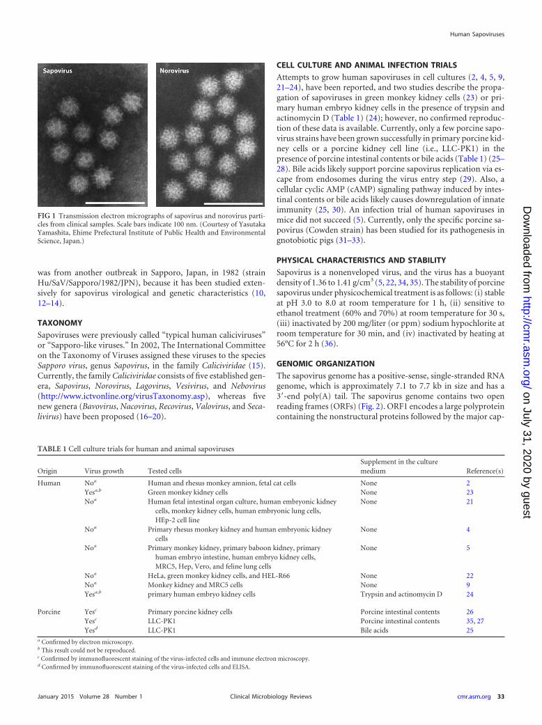
Sapovirus particles are small (about 30 to 38 nm in diameter) andicosahedral and have cup-shaped depressions on the surface,which is a typical calicivirus morphology (Fig. 1) (1). Sapovirusparticles were first detected in human diarrheic stool samples in1976 in the United Kingdom using electron microscopy (EM) (2),and the virus was soon recognized as a new gastroenteritis patho-gen (3–11). However, the prototype strain of the Sapovirus genus
Citation Oka T, Wang Q, Katayama K, Saif LJ. 2015. Comprehensive review ofhuman sapoviruses. Clin Microbiol Rev 28:32–53. doi:10.1128/CMR.00011-14.
Address correspondence to Tomoichiro Oka, [email protected], or Qiuhong Wang,[email protected].
Copyright © 2015, American Society for Microbiology. All Rights Reserved.
doi:10.1128/CMR.00011-14
32 cmr.asm.org January 2015 Volume 28 Number 1Clinical Microbiology Reviews
on July 31, 2020 by guesthttp://cm
r.asm.org/
Dow
nloaded from
was from another outbreak in Sapporo, Japan, in 1982 (strainHu/SaV/Sapporo/1982/JPN), because it has been studied exten-sively for sapovirus virological and genetic characteristics (10,12–14).
TAXONOMY
Sapoviruses were previously called “typical human caliciviruses”or “Sapporo-like viruses.” In 2002, The International Committeeon the Taxonomy of Viruses assigned these viruses to the speciesSapporo virus, genus Sapovirus, in the family Caliciviridae (15).Currently, the family Caliciviridae consists of five established gen-era, Sapovirus, Norovirus, Lagovirus, Vesivirus, and Nebovirus(http://www.ictvonline.org/virusTaxonomy.asp), whereas fivenew genera (Bavovirus, Nacovirus, Recovirus, Valovirus, and Seca-livirus) have been proposed (16–20).
CELL CULTURE AND ANIMAL INFECTION TRIALS
Attempts to grow human sapoviruses in cell cultures (2, 4, 5, 9,21–24), have been reported, and two studies describe the propa-gation of sapoviruses in green monkey kidney cells (23) or pri-mary human embryo kidney cells in the presence of trypsin andactinomycin D (Table 1) (24); however, no confirmed reproduc-tion of these data is available. Currently, only a few porcine sapo-virus strains have been grown successfully in primary porcine kid-ney cells or a porcine kidney cell line (i.e., LLC-PK1) in thepresence of porcine intestinal contents or bile acids (Table 1) (25–28). Bile acids likely support porcine sapovirus replication via es-cape from endosomes during the virus entry step (29). Also, acellular cyclic AMP (cAMP) signaling pathway induced by intes-tinal contents or bile acids likely causes downregulation of innateimmunity (25, 30). An infection trial of human sapoviruses inmice did not succeed (5). Currently, only the specific porcine sa-povirus (Cowden strain) has been studied for its pathogenesis ingnotobiotic pigs (31–33).
PHYSICAL CHARACTERISTICS AND STABILITY
Sapovirus is a nonenveloped virus, and the virus has a buoyantdensity of 1.36 to 1.41 g/cm3 (5, 22, 34, 35). The stability of porcinesapovirus under physicochemical treatment is as follows: (i) stableat pH 3.0 to 8.0 at room temperature for 1 h, (ii) sensitive toethanol treatment (60% and 70%) at room temperature for 30 s,(iii) inactivated by 200 mg/liter (or ppm) sodium hypochlorite atroom temperature for 30 min, and (iv) inactivated by heating at56°C for 2 h (36).
GENOMIC ORGANIZATION
The sapovirus genome has a positive-sense, single-stranded RNAgenome, which is approximately 7.1 to 7.7 kb in size and has a3=-end poly(A) tail. The sapovirus genome contains two openreading frames (ORFs) (Fig. 2). ORF1 encodes a large polyproteincontaining the nonstructural proteins followed by the major cap-
FIG 1 Transmission electron micrographs of sapovirus and norovirus parti-cles from clinical samples. Scale bars indicate 100 nm. (Courtesy of YasutakaYamashita, Ehime Prefectural Institute of Public Health and EnvironmentalScience, Japan.)
TABLE 1 Cell culture trials for human and animal sapoviruses
Origin Virus growth Tested cellsSupplement in the culturemedium Reference(s)
Human Noa Human and rhesus monkey amnion, fetal cat cells None 2Yesa,b Green monkey kidney cells None 23Noa Human fetal intestinal organ culture, human embryonic kidney
cells, monkey kidney cells, human embryonic lung cells,HEp-2 cell line
None 21
Noa Primary rhesus monkey kidney and human embryonic kidneycells
None 4
Noa Primary monkey kidney, primary baboon kidney, primaryhuman embryo intestine, human embryo kidney cells,MRC5, Hep, Vero, and feline lung cells
None 5
Noa HeLa, green monkey kidney cells, and HEL-R66 None 22Noa Monkey kidney and MRC5 cells None 9Yesa,b primary human embryo kidney cells Trypsin and actinomycin D 24
Porcine Yesc Primary porcine kidney cells Porcine intestinal contents 26Yesc LLC-PK1 Porcine intestinal contents 35, 27Yesd LLC-PK1 Bile acids 25
a Confirmed by electron microscopy.b This result could not be reproduced.c Confirmed by immunofluorescent staining of the virus-infected cells and immune electron microscopy.d Confirmed by immunofluorescent staining of the virus-infected cells and ELISA.
Human Sapoviruses
January 2015 Volume 28 Number 1 cmr.asm.org 33Clinical Microbiology Reviews
on July 31, 2020 by guesthttp://cm
r.asm.org/
Dow
nloaded from
sid protein, VP1 (Fig. 2). ORF2 is predicted to encode the minorstructural protein VP2 (Fig. 2) (28, 37). A similar genomic orga-nization (i.e., two ORFs, with the first ORF encoding the non-structural proteins and VP1) is found in other calicivirus genera,such as Lagovirus, Nebovirus, and the newly proposed genera Valo-virus, Bavovirus, and Nacovirus (17, 19, 20, 38, 39). The genomicorganization of Norovirus, Vesivirus, and Recovirus differs fromthat of Sapovirus: ORF1 encodes nonstructural proteins, andORF2 and ORF3 encode structural proteins VP1 and VP2, respec-tively (18, 37, 40). A third ORF (ORF3) has been predicted inseveral human (12, 41–47) and bat (48) sapovirus strains; how-ever, its function is unknown.
The ORF1-encoded polyprotein is expressed and processedinto at least six nonstructural (NS) proteins (NS1, NS2, NS3, NS4,NS5, and NS6-NS7) and a structural protein (VP1) by virus-en-coded protease (Fig. 2) (49–54). In vitro studies failed to showcleavage of the NS6-NS7 protein by the viral protease (28, 49, 50,52, 55, 56), although both the NS6 and NS7 proteins can carry outtheir respective functions (proteolytic and polymerase) when ex-pressed individually in vitro (56–58). The NS6-NS7 protein wasalso detected in porcine sapovirus-infected cells (28). Similar to
the case for sapoviruses, vesivirus also produces the NS6-NS7 pro-tein (fused protease-polymerase) (52, 53, 59–61), whereas noro-viruses and lagoviruses produce an individual protease and poly-merase, NS6 and NS7, respectively (51, 53, 62–67). The biologicalfunctions of the other sapovirus NS proteins have not been exper-imentally determined; however, NS3 and NS5 have a typical cali-civirus NTPase motif (GAPGIGKT) and VPg motifs (KGKTK andDDEYDE), respectively (Fig. 2) (37, 49, 68, 69). VPg is linked tothe 5= end of the viral RNA and is critical for calicivirus genomereplication, transcription, and translation (37, 70).
VP1, an approximately 60-kDa protein, is a major componentof the complete virus (34, 35). Two mechanisms can be consideredin the production of sapovirus VP1. One is that VP1 is cleavedfrom the ORF1-encoded polyprotein, and the other is that VP1 istranslated from a subgenomic RNA (from the 3=-coterminal RNAcorresponding to VP1 to the genome end region) (Fig. 2) (71, 72).A subgenomic RNA was confirmed for the sapovirus Cowdenstrain during replication (25). The VP2 protein has not yet beenidentified in sapovirus virions; however, the expression of thisprotein was detected in the in vitro translation products of a por-cine sapovirus full-length genomic cDNA construct and from
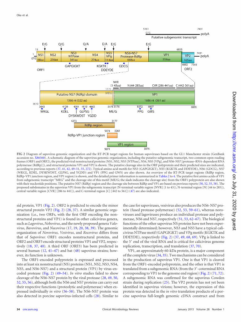
FIG 2 Diagram of sapovirus genomic organization and the RT-PCR target regions for human sapoviruses based on the GI.1 Manchester strain (GenBankaccession no. X86560). A schematic diagram of the sapovirus genomic organization, including the putative subgenomic transcript, two common open readingframes (ORF1 and ORF2), the predicted viral nonstructural proteins (NS1, NS2, NS3 [NTPase], NS4, NS5 [VPg], and NS6-NS7 [protease–RNA-dependent RNApolymerase {RdRp}]), and structural proteins VP1 and VP2 is shown. The putative cleavage sites in the ORF polyprotein and their predicted sizes are indicated,according to previous reports (37, 41, 42, 49–51, 55, 272). Typical amino acid motifs for NS3 (GAPGIGKT), NS5 (KGKTK and DDEYDE), NS6 (GDCG), NS7(WKGL, KDEL, DYSKWDST, GLPSG, and YGDD) and VP1 (PPG and GWS) are also shown. An overview of the RT-PCR target regions (RdRp region,RdRp-VP1 junction region, and VP1 region) is shown, and the detailed primer information is summarized in Tables 2 to 4. The putative first amino acids of VP1from subgenomic transcript “MEG” and the cleavage site of this motif (ME/G; the slash indicates the cleavage site) from the ORF1 polyprotein are also shownwith their nucleotide positions. The putative NS7 (RdRp) region and the cleavage site between RdRp and VP1 are based on previous reports (50, 52, 55, 58). Theproposed subdomains in the sapovirus VP1 from the subgenomic transcript (N-terminal variable region [NVR] [1 to 43]), N-terminal region [N] [44 to 285]),central variable region [CVR] [286 to 441]), and C-terminal region [C] [442 to 561]) (47) are also indicated.
Oka et al.
34 cmr.asm.org January 2015 Volume 28 Number 1Clinical Microbiology Reviews
on July 31, 2020 by guesthttp://cm
r.asm.org/
Dow
nloaded from
porcine sapovirus-infected cells (28). VP2 is predicted to be astrong basic protein and is identified as an interior component ofthe norovirus particles (73).
The expression of VP1 in insect or mammalian cells resulted inspontaneously assembled virus-like particles (VLPs) (12, 71, 72,74–81). The sapovirus VLPs are morphologically and antigeni-cally indistinguishable from those of the native sapovirus virionsfound in clinical specimens (12, 74). Digitized electron cryomi-crographs of the human sapovirus VLPs revealed that the icosa-hedral capsid is formed from 180 molecules of VP1, the same as innorovirus (76). Sapovirus VP1 could be separated into severaldomains: the N-terminal variable region (NVR), N-terminal re-gion (N), central variable region (CVR), and C-terminal region(C) (Fig. 2) (47). The conserved amino acid motif “GWS” wasfound in the predicted N and CVR junction (Fig. 2). The “G” inthis motif is strictly conserved among caliciviruses (76). Norovi-rus VP1 has also been separated into several domains, the N-ter-minal domain, shell domain, and protruding (P) domain, which isfurther divided into P1 and P2 subdomains (76, 82, 83). The sa-povirus VP1 CVR region likely corresponds to the highly variableP2 domain of norovirus VP1 (47, 76).
GENOMIC SEQUENCE AND ANTIGENICITY
The first complete genome of a sapovirus was determined for theManchester strain detected in the United Kingdom in 1993 (Hu/Manchester/93/UK; GenBank accession no. X86560) (41, 42),which is closely related genetically to the prototype Sapporo strain(14). Thus far, 26 (21 from humans and five from animals [por-cine and bat]) complete sapovirus genomes are available inGenBank (as of 1 September 2013). The VP1-encoding region isthe most diverse region in the genome (84–86), and sapovirusesare divided into multiple genogroups based on complete VP1 se-quences. Five genogroups (GI to GV) are recognized (46, 87), andnine additional genogroups (GVI to GXIV) were recently pro-posed (88). To date, human sapoviruses have been classified intofour genogroups (GI, GII, GIV, and GV).
Distinct antigenicity among sapovirus strains has been demon-strated by using clinical specimens (9, 43, 89–91), recombinantVP1 proteins (77, 92), or virus-like particles (VLPs) (74, 77, 80, 81,
93). Antigenicity differs among GI, GII, GIV, and GV strains (93,94) and is also distinct among different genotypes within GI andGII (80, 81, 94). These experimental results also support that VP1determines sapovirus antigenicity. The antigenic differences be-tween human and animal sapoviruses have not yet been deter-mined.
MOLECULAR CHARACTERIZATION
Genogroups and Genotypes
The partial RNA-dependent RNA polymerase (RdRp) or partialVP1 region (Fig. 2) or both of these regions can be used to partiallycharacterize detected sapoviruses, as well as to investigate the sim-ilarity of the detected sapovirus for epidemiological surveys. Incontrast, the RdRp-VP1 junction region (Fig. 2) is too short forsuch sequence analysis.
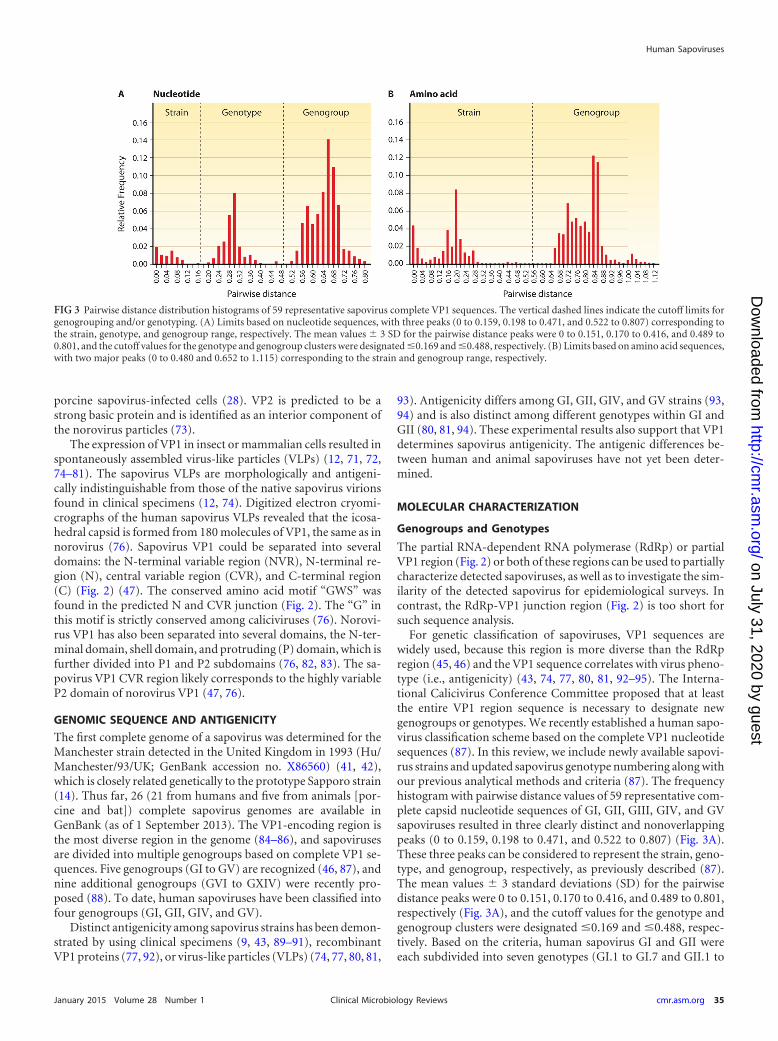
For genetic classification of sapoviruses, VP1 sequences arewidely used, because this region is more diverse than the RdRpregion (45, 46) and the VP1 sequence correlates with virus pheno-type (i.e., antigenicity) (43, 74, 77, 80, 81, 92–95). The Interna-tional Calicivirus Conference Committee proposed that at leastthe entire VP1 region sequence is necessary to designate newgenogroups or genotypes. We recently established a human sapo-virus classification scheme based on the complete VP1 nucleotidesequences (87). In this review, we include newly available sapovi-rus strains and updated sapovirus genotype numbering along withour previous analytical methods and criteria (87). The frequencyhistogram with pairwise distance values of 59 representative com-plete capsid nucleotide sequences of GI, GII, GIII, GIV, and GVsapoviruses resulted in three clearly distinct and nonoverlappingpeaks (0 to 0.159, 0.198 to 0.471, and 0.522 to 0.807) (Fig. 3A).These three peaks can be considered to represent the strain, geno-type, and genogroup, respectively, as previously described (87).The mean values � 3 standard deviations (SD) for the pairwisedistance peaks were 0 to 0.151, 0.170 to 0.416, and 0.489 to 0.801,respectively (Fig. 3A), and the cutoff values for the genotype andgenogroup clusters were designated �0.169 and �0.488, respec-tively. Based on the criteria, human sapovirus GI and GII wereeach subdivided into seven genotypes (GI.1 to GI.7 and GII.1 to
FIG 3 Pairwise distance distribution histograms of 59 representative sapovirus complete VP1 sequences. The vertical dashed lines indicate the cutoff limits forgenogrouping and/or genotyping. (A) Limits based on nucleotide sequences, with three peaks (0 to 0.159, 0.198 to 0.471, and 0.522 to 0.807) corresponding tothe strain, genotype, and genogroup range, respectively. The mean values � 3 SD for the pairwise distance peaks were 0 to 0.151, 0.170 to 0.416, and 0.489 to0.801, and the cutoff values for the genotype and genogroup clusters were designated �0.169 and �0.488, respectively. (B) Limits based on amino acid sequences,with two major peaks (0 to 0.480 and 0.652 to 1.115) corresponding to the strain and genogroup range, respectively.
Human Sapoviruses
January 2015 Volume 28 Number 1 cmr.asm.org 35Clinical Microbiology Reviews
on July 31, 2020 by guesthttp://cm
r.asm.org/
Dow
nloaded from
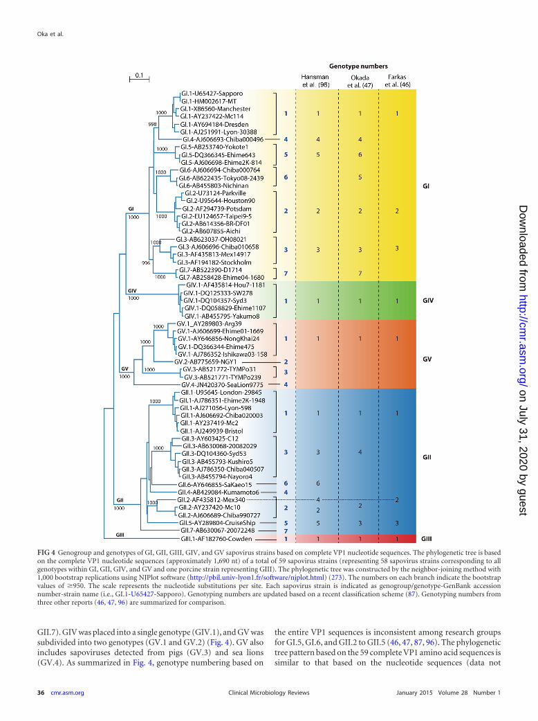
GII.7). GIV was placed into a single genotype (GIV.1), and GV wassubdivided into two genotypes (GV.1 and GV.2) (Fig. 4). GV alsoincludes sapoviruses detected from pigs (GV.3) and sea lions(GV.4). As summarized in Fig. 4, genotype numbering based on
the entire VP1 sequences is inconsistent among research groupsfor GI.5, GI.6, and GII.2 to GII.5 (46, 47, 87, 96). The phylogenetictree pattern based on the 59 complete VP1 amino acid sequences issimilar to that based on the nucleotide sequences (data not
FIG 4 Genogroup and genotypes of GI, GII, GIII, GIV, and GV sapovirus strains based on complete VP1 nucleotide sequences. The phylogenetic tree is basedon the complete VP1 nucleotide sequences (approximately 1,690 nt) of a total of 59 sapovirus strains (representing 58 sapovirus strains corresponding to allgenotypes within GI, GII, GIV, and GV and one porcine strain representing GIII). The phylogenetic tree was constructed by the neighbor-joining method with1,000 bootstrap replications using NJPlot software (http://pbil.univ-lyon1.fr/software/njplot.html) (273). The numbers on each branch indicate the bootstrapvalues of �950. The scale represents the nucleotide substitutions per site. Each sapovirus strain is indicated as genogroup/genotype-GenBank accessionnumber-strain name (i.e., GI.1-U65427-Sapporo). Genotyping numbers are updated based on a recent classification scheme (87). Genotyping numbers fromthree other reports (46, 47, 96) are summarized for comparison.
Oka et al.
36 cmr.asm.org January 2015 Volume 28 Number 1Clinical Microbiology Reviews
on July 31, 2020 by guesthttp://cm
r.asm.org/
Dow
nloaded from
shown); however, the pairwise distance histogram showed onlytwo major peaks (0 to 0.480 and 0.652 to 1.115), and we cannotdefine the genotype range statistically (Fig. 3B). This differs fromthe case for noroviruses, because genotypes could be defined sta-tistically by both VP1 nucleotide and amino acid sequences (97).
Evolution and Emergence of Predominant Sapovirus Strains
Genogroup and genotype analysis is important to characterize thecurrently circulating sapoviruses in the population. Emergence ofgenetically similar sapoviruses in multiple countries in Europe(98) and dynamic changes of genogroups and genotypes in differ-ent years among gastroenteritis patients in the same geographicalarea in Japan have been reported (99–101). Interestingly, GIV.1strains were detected predominantly in Japan, Canada, the UnitedStates, and Europe around 2007 (98, 101–104). The dynamicchange of the detected sapovirus genogroups in 2007 was alsoidentified by national surveillance through regional diagnosticlabs network in Japan (http://www.nih.go.jp/niid/en/iasr-table/2784-iasrtve.html; see “IASR Tables Virus” “By Season” “Gastroin-testinal Pathogens” “PDF”). This is a distinct trend compared tonoroviruses, in which a specific genogroup and genotype (i.e.,genogroup II and genotype 4 [GII.4]) have been predominant inthe past decade in Japan (http://www.nih.go.jp/niid/en/iasr-table/2784-iasrtve.html) and in multiple other countries (105–108). Inthe case of norovirus GII.4, time-ordered genetic and antigenicchange of VP1 was identified (109, 110). Recent studies from Eu-rope reported similar time-ordered genetic change in the VP1region of the sapovirus GI.2 strains (98), as reported for norovirusGII.4 strains (111–115).
Due to the inconsistent genotype numbering systems used bydifferent research groups for GI.5, GI.6, and GII.2 to GII.5 (46, 47,87, 96) (Fig. 4), it is important to indicate which numbering sys-tem was used for genotyping, and a harmonized genotype num-bering system will facilitate comparison and exchange of informa-tion from sapovirus surveillance at national and internationallevels.
Recombinant Strains
Sapoviruses with inconsistent grouping between the nonstruc-tural protein-encoding region (including the RdRp region) andthe VP1 encoding region have been designated “recombinant” or“chimeric” strains. Both intra- and intergenogroup recombinantstrains have been reported (Fig. 5). All reported intergenogrouprecombinant strains were GIV (based on VP1 sequence), whereasthey were clustered together with GII strains in the RdRp region(46, 85, 102, 116, 117). Intragenogroup recombinant strainswithin GI (118–120), GII (84, 121, 122), and GIII (123) have beenidentified.
Recently, a norovirus classification scheme has been reported(97). The authors used nucleotide sequences of nearly completeRdRp (1,300 nucleotides [nt]) and both amino acid and nucleo-tide sequences of VP1 for classification (97). Although sapovirusRdRp (NS7) is fused with protease (NS6) (Fig. 2), we defined theputative complete RdRp-encoding region (1,566 nt) for sapovi-ruses (Fig. 2) based on our previous in vitro studies (52, 58).Among GI, GII, GIV, and GV sapoviruses, the nucleotide se-quences spanning the putative complete RdRp- and VP1-en-coding regions (Fig. 2) of 26 strains (10 GI, 5 GII, 5 GIV, and 6GV based on VP1) were available in GenBank as of 1 September2013. All of these strains and a representative GIII Cowden
strain of pig origin were used for phylogenetic analysis basedon the RdRp and VP1 regions. We found conserved amino acidmotif “WKGL” (Fig. 2) at amino acid positions 12 to 15 in theputative compete RdRp (NS7) region among these sapovirusstrains. As shown in Fig. 5, several strains clustered differentlyon the phylogenetic trees based on RdRp- and VP1-encodingregions. For example, based on the RdRp region, GII and GIVstrains are not well separated, as discussed previously (85). TheGII.2 Mc10 and GII.3 C12 strains also cluster together in theRdRp region and were previously reported as intragenogrouprecombinant strains (84). The GV.4 strain clustered togetherwith other GV strains (GV.1, GV.2, and GV.3) in the VP1 re-gion, but it is separated from other GV strains in the RdRpregion. However, the RdRp sequence-based classification is lessreliable due to the fewer available sequences compared to thecomplete VP1 sequences. Further accumulation of sufficientsequence data spanning the complete or sufficient length of theRdRp- to the VP1-encoding regions for all the genogroups andgenotypes are critical to provide a better understanding of “re-combinant” or “chimeric” strains and to establish a reliableclassification scheme for the sapovirus RdRp region in the fu-ture, because the putative complete RdRp sequence data forGI.3, GI.4, GI.6, GI.7, GII.4, GII.5, and GII.7 sapoviruses arenot yet available (Fig. 5). In addition, it is also critical to am-plify a single PCR fragment covering the partial RdRp- andVP1-encoding region for recombination analysis to avoid thepossibility of coinfection of different genogroups and/or geno-types of sapovirus strains, as discussed previously (124).
LABORATORY DIAGNOSIS
Virus Particle Detection
Sapoviruses are morphologically distinguishable from other gas-troenteritis pathogens (e.g., norovirus, rotavirus, astrovirus, oradenovirus) by their typical “Star of David” surface morphologyunder the electron microscope (1, 33, 90, 125) (Fig. 1). However,this has low sensitivity compared to nucleic acid detection meth-ods (116, 126–128, 130, 131).
Antigen Detection Methods
Enzyme-linked immunosorbent assays (ELISAs) have been devel-oped for the detection of human sapovirus antigens (91, 93, 132,190) and have been used for the detection of sapoviruses fromclinical samples (43, 91, 132–135, 190). However, these assays arenot widely used for diagnosis due to the difficulty in detection ofantigenically diverse sapovirus strains, low sensitivity comparedto nucleic acid detection methods (43, 91, 93), and current lack ofcommercial availability. The development of a broadly reactiveELISA or immunochromatography system for the detection ofsapovirus antigens depends on the combination of a panel ofgenogroup/genotype-specific antisera and/or using broadly reac-tive monoclonal antibodies. These approaches may be feasible,because a common epitope(s) likely exists among GI, GII, GIV,and GV sapovirus strains (94). Broadly reactive norovirus-specificmonoclonal antibodies that recognize VP1s of different geno-groups of noroviruses were also reported (136–140).
Nucleic Acid Detection Methods
Reverse transcription-PCR (RT-PCR), especially real-time RT-PCR, has become a major and routine method for sapovirus de-
Human Sapoviruses
January 2015 Volume 28 Number 1 cmr.asm.org 37Clinical Microbiology Reviews
on July 31, 2020 by guesthttp://cm
r.asm.org/
Dow
nloaded from
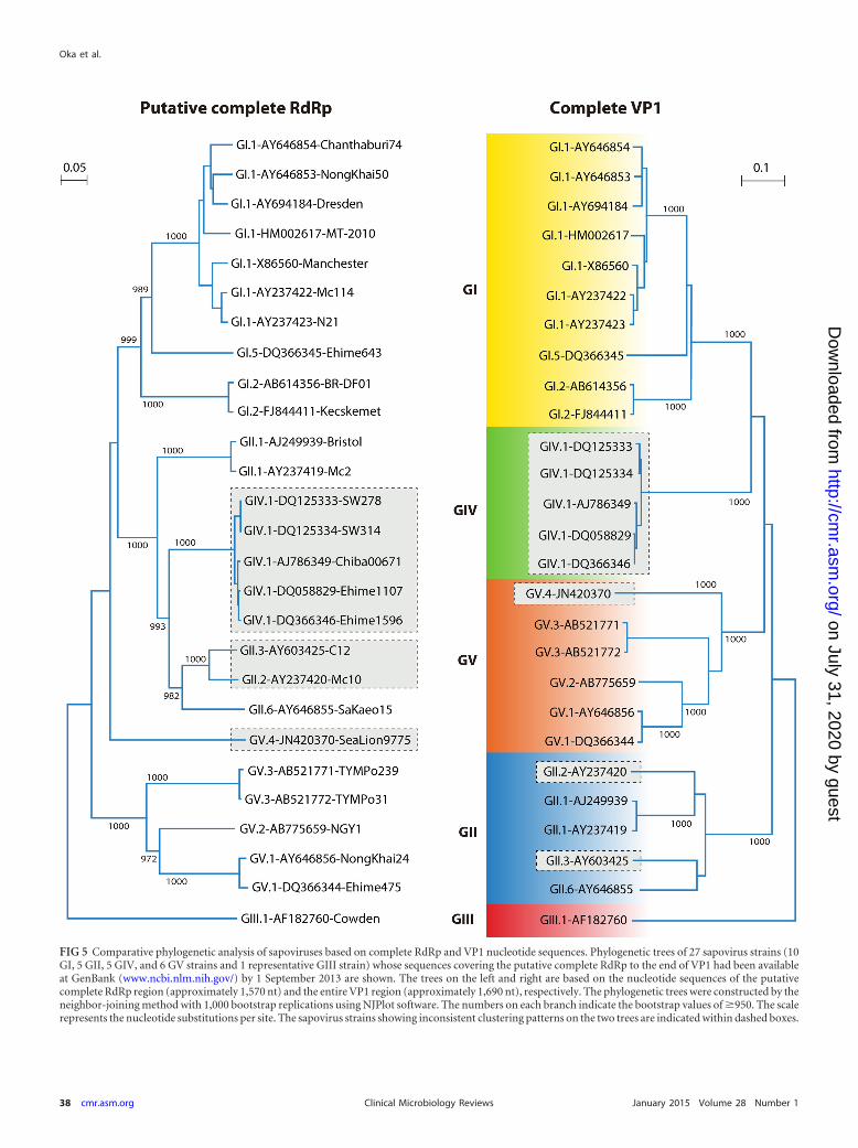
FIG 5 Comparative phylogenetic analysis of sapoviruses based on complete RdRp and VP1 nucleotide sequences. Phylogenetic trees of 27 sapovirus strains (10GI, 5 GII, 5 GIV, and 6 GV strains and 1 representative GIII strain) whose sequences covering the putative complete RdRp to the end of VP1 had been availableat GenBank (www.ncbi.nlm.nih.gov/) by 1 September 2013 are shown. The trees on the left and right are based on the nucleotide sequences of the putativecomplete RdRp region (approximately 1,570 nt) and the entire VP1 region (approximately 1,690 nt), respectively. The phylogenetic trees were constructed by theneighbor-joining method with 1,000 bootstrap replications using NJPlot software. The numbers on each branch indicate the bootstrap values of �950. The scalerepresents the nucleotide substitutions per site. The sapovirus strains showing inconsistent clustering patterns on the two trees are indicated within dashed boxes.
Oka et al.
38 cmr.asm.org January 2015 Volume 28 Number 1Clinical Microbiology Reviews
on July 31, 2020 by guesthttp://cm
r.asm.org/
Dow
nloaded from
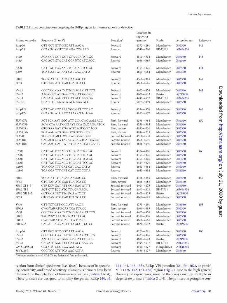
tection from clinical specimens (i.e., feces), because of its specific-ity, sensitivity, and broad reactivity. Numerous primers have beendesigned for the detection of human sapoviruses (Tables 2 to 4).These primers are designed to amplify the partial RdRp (44, 46,
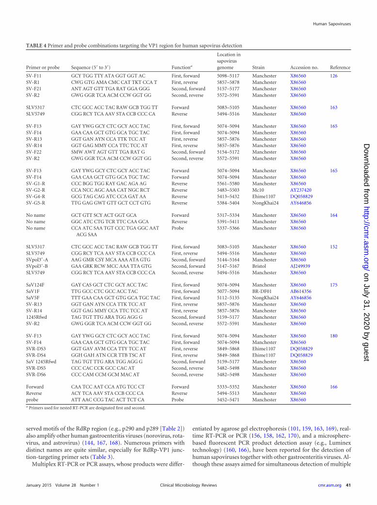
141–144, 146–153), RdRp-VP1 junction (86, 154–162), or partialVP1 (126, 152, 163–166) region (Fig. 2). Due to the high geneticdiversity of sapoviruses, most of the assays include multiple ordegenerate primers (Tables 2 to 4). The primers targeting the con-
TABLE 2 Primer combinations targeting the RdRp region for human sapovirus detection
Primer or probe Sequence (5= to 3=) Functiona
Location insapovirusgenome Strain Accession no. Reference
Sapp36 GTT GCT GTT GGC ATT AAC A Forward 4273–4291 Manchester X86560 141Sapp35 GCA GTG GGT TTG AGA CCA AAG Reverse 4740–4760 BR-DF01 AB614356
4490 ACA CGT GGT GGT CTA CCA TCT GG Forward 4510–4532 Manchester X86560 1434485 CAC ACT GTA CAT GCA RTC ATC ACC Reverse 4666–4689 Manchester X86560
p290 GAT TAC TCC AAG TGG GAC TCC AC Forward 4354–4376 Manchester X86560 144p289 TGA CAA TGT AAT CAT CAC CAT A Reverse 4663–4684 Manchester X86560
SR80 TGG GAT TCT ACA CAA AAC CC Forward 4366–4385 Manchester X86560 147JV33 GTG TAN ATG CAR TCA TCA CC Reverse 4666–4685 Manchester X86560
SV-s1 CCC TGG CAA TAT TGG AGA GAT TTG Forward 4403–4426 Manchester X86560 148SV-s2 AAG GCC TAT GAA CCA CAT GGG GC Forward 4601–4623 Bristol AJ249939SV-s3 GAC ATC AAG TTT GAT ACC AAG GA Forward 4495–4517 BR-DF01 AB614356SV-r-c GCA TTG TAG GTG GCG AGA GCC Reverse 5079–5099 Manchester X86560
Sapp128 GAT TAC ACC AAA TGG GAT TCC AC Forward 4354–4376 Manchester X86560 149Sapp129 GCA GTC ATC ACC ATA CGT GTG AA Reverse 4655–4677 Manchester X86560
SLV-OFa ACT SCA AAT GGG ATT CCA CWC AAM ACC First, forward 4358–4384 Manchester X86560 150SLV-OFb ACW CTA AAT GGG AYT CCA CAC AGA ATC C First, forward 4358–4385 Manchester X86560SLV-ORa GTG RAA GAT KGA WGC RGT GGC AGG First, reverse 4693–4716 Manchester X86560SLV-ORb TCA AAG ATG GAA GCG GTT GCC G First, reverse 4694–4715 Manchester X86560SLV-IF TGG HCT MCC WTC WGG SAT GCC Second, forward 4518–4538 Manchester X86560SLV-IRa CAC ACR CTG TAS ATG CAG TCA TCA CC Second, reverse 4666–4691 Manchester X86560SLV-IRb CAC AAG GAG TAT ATG CAA TCA TCA CC Second, reverse 4666–4691 Manchester X86560
p290h GAT TAC TCC AGG TGG GAC TCC AC Forward 4354–4376 Manchester X86560 46p290i GAT TAC TCC AGG TGG GAC TCA AC Forward 4354–4376 Manchester X86560p290j GAT TAC TCC AGG TGG GAT TCA AC Forward 4354–4376 Manchester X86560p290k GAT TAC TCC AGG TGG GAT TCC AC Forward 4354–4376 Manchester X86560p289h TGA CGA TTT CAT CAT CAC CAT A Reverse 4663–4684 Manchester X86560p289i TGA CGA TTT CAT CAT CCC CGT A Reverse 4663–4684 Manchester X86560
SR80 TGG GAT TCT ACA CAA AAC CC First, forward 4366–4385 Manchester X86560 152JV33 GTG TAN ATG CAR TCA TCA CC First, reverse 4666–4685 Manchester X86560SR80 GI 1–3 CTR KCV GAT ATT GGA RAG ATT T Second, forward 4404–4425 Manchester X86560SR80 GI 2 AGT CTY TCC ATC TTA GAG AGA Second, forward 4402–4422 BR-DF01 AB614356SR80 GII 1–2 GCT GCR TCY TTG KCA ATC CT Second, forward 4400–4419 Bristol AJ249939JV33 GTG TAN ATG CAR TCA TCA CC Second, reverse 4666–4685 Manchester X86560
SV36 GTT TCT GTT GGC ATT AAC A First, forward 4273–4291 Manchester X86560 153SRGA CWG TAB ATG CAR TCA TCA CC First, reverse 4666–4685 Manchester X86560SVS1 CCC TGG CAA TAT TGG AGA GAT TTG Second, forward 4403–4426 Manchester X86560SRGE TAC WGY AAA TGG GAT TCCAC Second, forward 4357–4376 Manchester X86560SRGA CWG TAB ATG CAR TCA TCA CC Second, reverse 4666–4685 Manchester X86560SRGS2A CAC ATT ACC AGT GTA AGG TGC CC Second, reverse 4620–4642 Bristol AJ249939
Sapp36 GTT GCT GTT GGC ATT AAC A Forward 4273–4291 Manchester X86560 100SV-s1 CCC TGG CAA TAT TGG AGA GAT TTG Forward 4403–4426 Manchester X86560SV-s2 AAG GCC TAT GAA CCA CAT GGG GC Forward 4601–4623 Bristol AJ249939SV-s3 GAC ATC AAG TTT GAT ACC AAG GA Forward 4495–4517 BR-DF01 AB614356GV-GLPSGM GGT CTC CCC TCG GGC ATG Forward 4560–4577 NongKhai24 AY646856SaV1245R CCC TCC ATY TCA AAC ACT A Reverse 5159–5177 Manchester X86560a Primers used for nested RT-PCR are designated first and second.
Human Sapoviruses
January 2015 Volume 28 Number 1 cmr.asm.org 39Clinical Microbiology Reviews
on July 31, 2020 by guesthttp://cm
r.asm.org/
Dow
nloaded from
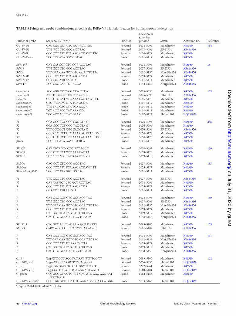
TABLE 3 Primer and probe combinations targeting the RdRp-VP1 junction region for human sapovirus detection
Primer or probe Sequence (5= to 3=)a Function
Location insapovirusgenome Strain Accession no. Reference
CU-SV-F1 GAC CAG GCT CTC GCY ACC TAC Forward 5074–5094 Manchester X86560 154CU-SV-F2 TTG GCC CTC GCC ACC TAC Forward 5077–5094 BR-DF01 AB614356CU-SV-R CCC TCC ATY TCA AAC ACT AWT TTG Reverse 5154–5177 Manchester X86560CU-SV-Probe TGG TTY ATA GGY GGT AC Probe 5101–5117 Manchester X86560
SaV124F GAY CAS GCT CTC GCY ACC TAC Forward 5074–5094 Manchester X86560 86SaV1F TTG GCC CTC GCC ACC TAC Forward 5077–5094 BR-DF01 AB614356SaV5F TTT GAA CAA GCT GTG GCA TGC TAC Forward 5112–5135 NongKhai24 AY646856SaV1245R CCC TCC ATY TCA AAC ACT A Reverse 5159–5177 Manchester X86560SaV124TP CCR CCT ATR AAC CA Probe 5101–5114 Manchester X86560SaV5TP TGC CAC CAA TGT ACC A Probe 5142–5157 NongKhai24 AY646856
sapo.fwdA ACC AGG CTC TCG CCA CCT A Forward 5075–5093 Manchester X86560 155sapo.fwdB ATT TGG CCC TCG CCA CCT A Forward 5075–5093 BR-DF01 AB614356sapo.rev GCC CTC CAT YTC AAA CAC TAW TTT Reverse 5155–5178 Manchester X86560sapo.probeA CTG TAC CAC CTA TGA ACC A Probe 5101–5119 Manchester X86560sapo.probeB TTG TAC CAC CTA TGA ACC A Probe 5101–5119 Manchester X86560sapo.probeC TGT ACC ACC TAT AAA CCA Probe 5101–5118 Manchester X86560sapo.probeD TGC ACC ACC TAT GAA C Probe 5107–5122 Ehime1107 DQ058829
F1 CCA GGC TCT CGC CAC CTA C Forward 5076–5094 Manchester X86560 248F2 CCA GGC TCT CGC TAC CTA C Forward 5076–5094 Manchester X86560F3 TTT GGC CCT CGC CAC CTA C Forward 5076–5094 BR-DF01 AB614356R1 GCC CTC CAT CTC AAA CAC TAT TTT G Reverse 5154–5178 Manchester X86560R2 GCC CTC CAT TTC AAA CAC TAA TTT G Reverse 5154–5178 Manchester X86560probe TGG TTY ATA GGY GGT RCA Probe 5101–5118 Manchester X86560
SLVCF GAY CWG GCY CTC GCC ACC T Forward 5074–5092 Manchester X86560 156SLVCR GCC CTC CAT YTC AAA CAC TA Reverse 5159–5178 Manchester X86560SVLCP TGY ACC ACC TAT RAA CCA VG Probe 5099–5118 Manchester X86560
SAPOs CAG GCT CTC GCC ACC TAC Forward 5077–5094 Manchester X86560 157SAPOas CCC TCC ATY TCA AAC ACT AWT TT Reverse 5155–5177 Manchester X86560SAPO-XS-QS705 TGG TTC ATA GGT GGT RC Probe 5101–5117 Manchester X86560
F1 TTG GCC CTC GCC ACC TAC Forward 5077–5094 BR-DF01 AB614356 158F2 GAY CAS GCT CTC GCY ACC TAC Forward 5074–5094 Manchester X86560R CCC TCC ATY TCA AAC ACT A Reverse 5159–5177 Manchester X86560P CCR CCT ATR AAC CA Probe 5101–5114 Manchester X86560
F GAY CAG GCT CTC GCY ACC TAC Forward 5074–5094 Manchester X86560 160F TTG GCC CTC GCC ACC TAC Forward 5077–5094 BR-DF01 AB614356F TTT GAA CAA GCT GTG GCA TGC TAC Forward 5112–5135 NongKhai24 AY646856R CCC TCC ATY TCA AAC ACT A Reverse 5159–5177 Manchester X86560P CYT GGT TCA TAG GTG GTR CAG Probe 5099–5119 Manchester X86560P CAG CTG GTA CAT TGG TGG CAC Probe 5138–5158 NongKhai24 AY646856
SLV5317 CTC GCC ACC TAC RAW GCB TGG TT Forward 5083–5105 Manchester X86560 159SMP-R CMW WCC CCT CCA TYT CAA ACA C Reverse 5161–5182 BR-DF01 AB614356
F GAY CAG GCT CTC GCY ACC TAC Forward 5074–5094 Manchester X86560 161F TTT GAA CAA GCT GTG GCA TGC TAC Forward 5112–5135 NongKhai24 AY646856R CCC TCC ATY TC AAA CAC TA Reverse 5159–5177 Manchester X86560P CYT GGT TCA TAG GTG GTR CAG Probe 5099–5119 Manchester X86560P CAG CTG GTA CAT TGG TGG CAC Probe 5138–5158 NongKhai24 AY646856
GI-F Tag-CTC GCC ACC TAC AAT GCY TGG TT Forward 5083–5105 Manchester X86560 162GII, GIV, V-F Tag-ACR GCC AAR GCT GAG GGG Forward 5036–5053 Ehime1107 DQ058829GI-R Tag-TGG GAT GTG GTC GGV CCA GT Reverse 5242–5261 Manchester X86560GII, GIV, V-R Tag-CCC TCC ATT TCA AAC ACT AAT T Reverse 5160–5181 Ehime1107 DQ058829GI probe CCG AGC CTA GTG TTT GAG ATG GAG GGC AAT
GGC TCG GProbe 5152–5188 Manchester X86560
GII, GIV, V-Probe CCC TGG GCC CCA GTG AAG AGA CCA CCA GGG Probe 5133–5162 Ehime1107 DQ058829a Tag, GCAAGCCCTCACGTAGCGAA.
Oka et al.
40 cmr.asm.org January 2015 Volume 28 Number 1Clinical Microbiology Reviews
on July 31, 2020 by guesthttp://cm
r.asm.org/
Dow
nloaded from
served motifs of the RdRp region (e.g., p290 and p289 [Table 2])also amplify other human gastroenteritis viruses (norovirus, rota-virus, and astrovirus) (144, 167, 168). Numerous primers withdistinct names are quite similar, especially for RdRp-VP1 junc-tion-targeting primer sets (Table 3).
Multiplex RT-PCR or PCR assays, whose products were differ-
entiated by agarose gel electrophoresis (101, 159, 163, 169), real-time RT-PCR or PCR (156, 158, 162, 170), and a microsphere-based fluorescent PCR product detection assay (e.g., Luminextechnology) (160, 166), have been reported for the detection ofhuman sapoviruses together with other gastroenteritis viruses. Al-though these assays aimed for simultaneous detection of multiple
TABLE 4 Primer and probe combinations targeting the VP1 region for human sapovirus detection
Primer or probe Sequence (5= to 3=) Functiona
Location insapovirusgenome Strain Accession no. Reference
SV-F11 GCY TGG TTY ATA GGT GGT AC First, forward 5098–5117 Manchester X86560 126SV-R1 CWG GTG AMA CMC CAT TKT CCA T First, reverse 5857–5878 Manchester X86560SV-F21 ANT AGT GTT TGA RAT GGA GGG Second, forward 5157–5177 Manchester X86560SV-R2 GWG GGR TCA ACM CCW GGT GG Second, reverse 5572–5591 Manchester X86560
SLV5317 CTC GCC ACC TAC RAW GCB TGG TT Forward 5083–5105 Manchester X86560 163SLV5749 CGG RCY TCA AAV STA CCB CCC CA Reverse 5494–5516 Manchester X86560
SV-F13 GAY YWG GCY CTC GCY ACC TAC First, forward 5074–5094 Manchester X86560 165SV-F14 GAA CAA GCT GTG GCA TGC TAC First, forward 5074–5094 Manchester X86560SV-R13 GGT GAN AYN CCA TTK TCC AT First, reverse 5857–5876 Manchester X86560SV-R14 GGT GAG MMY CCA TTC TCC AT First, reverse 5857–5876 Manchester X86560SV-F22 SMW AWT AGT GTT TGA RAT G Second, forward 5154–5172 Manchester X86560SV-R2 GWG GGR TCA ACM CCW GGT GG Second, reverse 5572–5591 Manchester X86560
SV-F13 GAY YWG GCY CTC GCY ACC TAC Forward 5074–5094 Manchester X86560 165SV-F14 GAA CAA GCT GTG GCA TGC TAC Forward 5074–5094 Manchester X86560SV-G1-R CCC BGG TGG KAY GAC AGA AG Reverse 5561–5580 Manchester X86560SV-G2-R CCA NCC AGC AAA CAT NGC RCT Reverse 5483–5503 Mc10 AY237420SV-G4-R GCG TAG CAG ATC CCA GAT AA Reverse 5413–5432 Ehime1107 DQ058829SV-G5-R TTG GAG GWT GTT GCT CCT GTG Reverse 5384–5404 NongKhai24 AY646856
No name GCT GTT SCY ACT GGT GCA Forward 5317–5334 Manchester X86560 164No name GGC ATC CTG TCR TTC CAA GCA Reverse 5391–5411 Manchester X86560No name CCA ATC SAA TGT CCC TGA GGC AAT
ACG SAAProbe 5337–5366 Manchester X86560
SLV5317 CTC GCC ACC TAC RAW GCB TGG TT First, forward 5083–5105 Manchester X86560 152SLV5749 CGG RCY TCA AAV STA CCB CCC CA First, reverse 5494–5516 Manchester X86560SVpol3=-A AAG GMR CSY MCA AAA ATA GTG Second, forward 5144–5164 Manchester X86560SVpol3=-B GAA GRK RCW MCC AAA TTA GTG Second, forward 5147–5167 Bristol AJ249939SLV5749 CGG RCY TCA AAV STA CCB CCC CA Second, reverse 5494–5516 Manchester X86560
SaV124F GAY CAS GCT CTC GCY ACC TAC First, forward 5074–5094 Manchester X86560 175SaV1F TTG GCC CTC GCC ACC TAC First, forward 5077–5094 BR-DF01 AB614356SaV5F TTT GAA CAA GCT GTG GCA TGC TAC First, forward 5112–5135 NongKhai24 AY646856SV-R13 GGT GAN AYN CCA TTK TCC AT First, reverse 5857–5876 Manchester X86560SV-R14 GGT GAG MMY CCA TTC TCC AT First, reverse 5857–5876 Manchester X865601245Rfwd TAG TGT TTG ARA TGG AGG G Second, forward 5159–5177 Manchester X86560SV-R2 GWG GGR TCA ACM CCW GGT GG Second, reverse 5572–5591 Manchester X86560
SV-F13 GAY YWG GCY CTC GCY ACC TAC First, forward 5074–5094 Manchester X86560 180SV-F14 GAA CAA GCT GTG GCA TGC TAC First, forward 5074–5094 Manchester X86560SVR-DS3 GGT GAV AVM CCA TTY TCC AT First, reverse 5849–5868 Ehime1107 DQ058829SVR-DS4 GGH GAH ATN CCR TTB TSC AT First, reverse 5849–5868 Ehime1107 DQ058829SaV 1245Rfwd TAG TGT TTG ARA TGG AGG G Second, forward 5159–5177 Manchester X86560SVR-DS5 CCC CAC CCK GCC CAC AT Second, reverse 5482–5498 Manchester X86560SVR-DS6 CCC CAM CCM GCM MAC AT Second, reverse 5482–5498 Manchester X86560
Forward CAA TCC AAT CCA ATG TCC CT Forward 5333–5352 Manchester X86560 166Reverse ACY TCA AAV STA CCB CCC CA Reverse 5494–5513 Manchester X86560probe ATT AAC CCG TAC ACT TCT CA Probe 5452–5471 Manchester X86560a Primers used for nested RT-PCR are designated first and second.
Human Sapoviruses
January 2015 Volume 28 Number 1 cmr.asm.org 41Clinical Microbiology Reviews
on July 31, 2020 by guesthttp://cm
r.asm.org/
Dow
nloaded from
viruses, it is unclear whether these assays can detect all genogroupsof human sapoviruses.
Sapoviruses were also detected by specific primer-indepen-dent techniques (i.e., the metagenomic sequence approach)from untreated sewage (16), sewage sludge (171), and fecesfrom California sea lions (172), dogs (173), and humans. Theseapproaches are not widely used for diagnosis but may be appli-cable for routine clinical diagnosis in the future, when the costof such assays and data analysis is comparable to that of tradi-tional assays.
Selection of Detection Methods
The nucleic acid detection method is more sensitive than EM(116, 126–128, 130, 131) or ELISA (91). Different detection ratesamong different PCR assays using the same panel of specimens(clinical specimens, environmental water, and shellfish) were re-ported (99–101, 174–177). Assays targeting the RdRp-VP1 junc-tion region have the highest detection rate and can be used as thefirst choice for sapovirus screening from clinical specimens (101,174). The VP1-targeting RT-PCR is preferred because the prod-ucts can be sequenced for reliable genotyping (99–101). Similarresults were reported for environmental water samples (i.e., riverwater) (175). RdRp-VP1 junction-targeting real-time RT-PCRwas also used for the detection of sapoviruses from shellfish (178,179): however, the nested RT-PCR targeting the partial VP1 re-gion is superior to the real-time RT-PCR and single-round RT-PCR because of the low level of viral RNA in shellfish compared toclinical specimens (177, 178). Currently, limited primer sets (47,86, 100, 165, 175, 180) have demonstrated the ability to detect allgenogroups of human sapoviruses.
Full-Genome Sequencing Approaches
Full genomic sequence analysis is still not practical for routinediagnosis. A long single-round or nested RT-PCR to amplify a2- to 2.5-kb PCR fragment to determine the complete VP1sequences of various sapovirus strains from clinical specimensis feasible by using forward primers targeting the RdRp and/orRdRp-VP1 junction region (Tables 2 to 4) and a reverse primerhybridized to the 3=-end poly(A) tail (Fig. 2) (46, 47, 87, 130,174, 181, 182). In contrast, the amplification of the 5=-end 5- to5.5-kb fragment corresponding to the beginning of the genometo the VP1 upstream region is variable because of the lack ofuniversal primers. As a new technology, the specific primer-independent metagenomic sequencing approach (i.e., next-generation sequencing techniques) can be used to determinethe nearly complete genome sequences (lacking the 5= end orboth the 5= and 3= ends) from fecal specimens (172, 173). 5=rapid amplification of cDNA ends (RACE) techniques (14, 41,42) are still necessary to determine 5= ends to obtain the com-plete sapovirus genomic sequences.
CLINICAL AND EPIDEMIOLOGICAL OBSERVATIONS
Symptoms and Severity of Disease
Based on the epidemiological data from patients with sapovirusgastroenteritis, the incubation period ranges from less than 1 dayto 4 days (5, 8, 44, 130, 135, 178, 183, 184). Major clinical symp-toms include diarrhea and vomiting; however, additional consti-tutional symptoms (i.e., nausea, stomach/abdominal cramps,chills, headache, myalgia, or malaise) are also frequently reported.
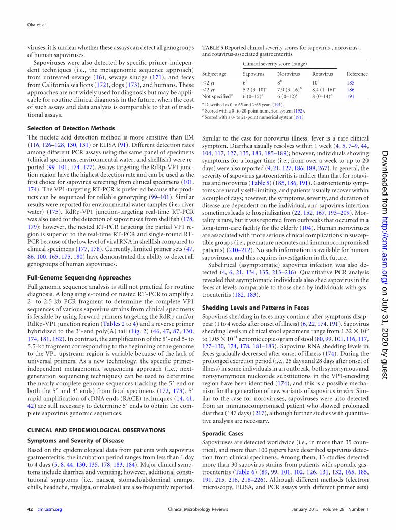
Similar to the case for norovirus illness, fever is a rare clinicalsymptom. Diarrhea usually resolves within 1 week (4, 5, 7–9, 44,104, 117, 127, 135, 183, 185–189); however, individuals showingsymptoms for a longer time (i.e., from over a week to up to 20days) were also reported (9, 21, 127, 186, 188, 267). In general, theseverity of sapovirus gastroenteritis is milder than that for rotavi-rus and norovirus (Table 5) (185, 186, 191). Gastroenteritis symp-toms are usually self-limiting, and patients usually recover withina couple of days; however, the symptoms, severity, and duration ofdisease are dependent on the individual, and sapovirus infectionsometimes leads to hospitalization (22, 152, 167, 193–209). Mor-tality is rare, but it was reported from outbreaks that occurred in along-term-care facility for the elderly (104). Human norovirusesare associated with more serious clinical complications in suscep-tible groups (i.e., premature neonates and immunocompromisedpatients) (210–212). No such information is available for humansapoviruses, and this requires investigation in the future.
Subclinical (asymptomatic) sapovirus infection was also de-tected (4, 6, 21, 134, 135, 213–216). Quantitative PCR analysisrevealed that asymptomatic individuals also shed sapovirus in thefeces at levels comparable to those shed by individuals with gas-troenteritis (182, 183).
Shedding Levels and Patterns in Feces
Sapovirus shedding in feces may continue after symptoms disap-pear (1 to 4 weeks after onset of illness) (6, 22, 174, 191). Sapovirusshedding levels in clinical stool specimens range from 1.32 � 105
to 1.05 � 1011 genomic copies/gram of stool (80, 99, 101, 116, 117,127–130, 174, 178, 181–183). Sapovirus RNA shedding levels infeces gradually decreased after onset of illness (174). During theprolonged excretion period (i.e., 25 days and 28 days after onset ofillness) in some individuals in an outbreak, both synonymous andnonsynonymous nucleotide substitutions in the VP1-encodingregion have been identified (174), and this is a possible mecha-nism for the generation of new variants of sapovirus in vivo. Sim-ilar to the case for noroviruses, sapoviruses were also detectedfrom an immunocompromised patient who showed prolongeddiarrhea (147 days) (217), although further studies with quantita-tive analysis are necessary.
Sporadic Cases
Sapoviruses are detected worldwide (i.e., in more than 35 coun-tries), and more than 100 papers have described sapovirus detec-tion from clinical specimens. Among them, 13 studies detectedmore than 30 sapovirus strains from patients with sporadic gas-troenteritis (Table 6) (89, 99, 101, 102, 126, 131, 132, 165, 185,191, 215, 216, 218–226). Although different methods (electronmicroscopy, ELISA, and PCR assays with different primer sets)
TABLE 5 Reported clinical severity scores for sapovirus-, norovirus-,and rotavirus-associated gastroenteritis
Subject age
Clinical severity score (range)
ReferenceSapovirus Norovirus Rotavirus
�2 yr 6b 8b 10b 185�2 yr 5.2 (3–10)b 7.9 (3–16)b 8.4 (1–16)b 186Not specifieda 6 (0–15)c 6 (0–12)c 8 (0–14)c 191a Described as 0 to 65 and �65 years (191).b Scored with a 0- to 20-point numerical system (192).c Scored with a 0- to 21-point numerical system (191).
Oka et al.
42 cmr.asm.org January 2015 Volume 28 Number 1Clinical Microbiology Reviews
on July 31, 2020 by guesthttp://cm
r.asm.org/
Dow
nloaded from
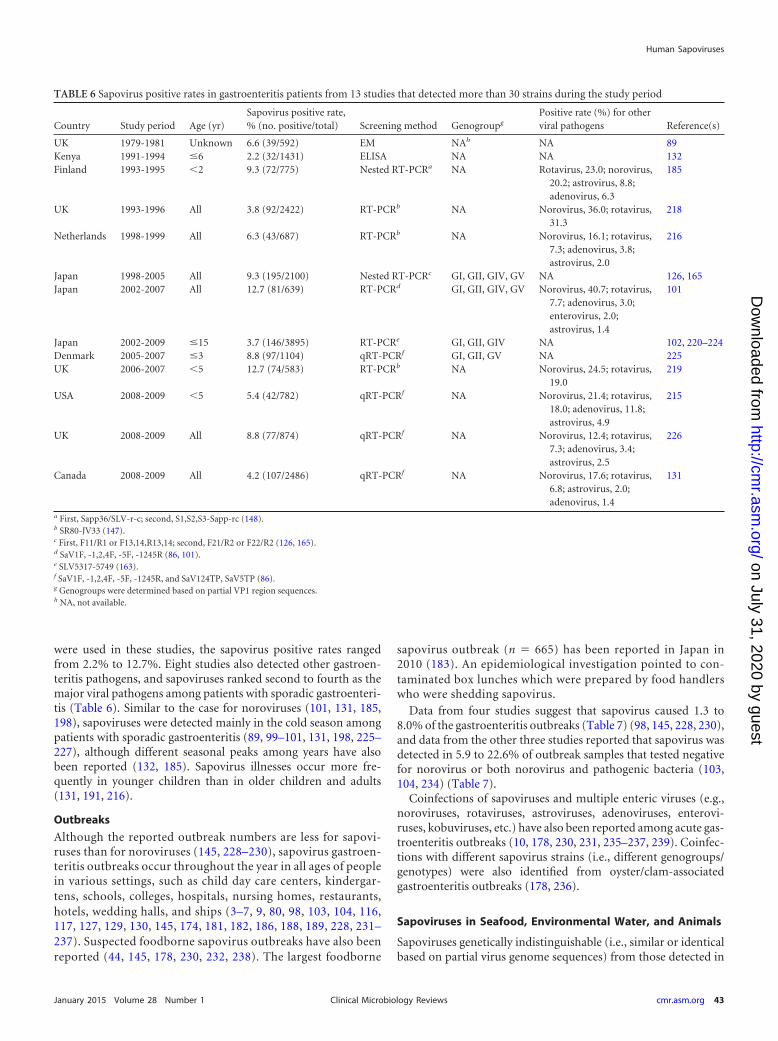
were used in these studies, the sapovirus positive rates rangedfrom 2.2% to 12.7%. Eight studies also detected other gastroen-teritis pathogens, and sapoviruses ranked second to fourth as themajor viral pathogens among patients with sporadic gastroenteri-tis (Table 6). Similar to the case for noroviruses (101, 131, 185,198), sapoviruses were detected mainly in the cold season amongpatients with sporadic gastroenteritis (89, 99–101, 131, 198, 225–227), although different seasonal peaks among years have alsobeen reported (132, 185). Sapovirus illnesses occur more fre-quently in younger children than in older children and adults(131, 191, 216).
Outbreaks
Although the reported outbreak numbers are less for sapovi-ruses than for noroviruses (145, 228–230), sapovirus gastroen-teritis outbreaks occur throughout the year in all ages of peoplein various settings, such as child day care centers, kindergar-tens, schools, colleges, hospitals, nursing homes, restaurants,hotels, wedding halls, and ships (3–7, 9, 80, 98, 103, 104, 116,117, 127, 129, 130, 145, 174, 181, 182, 186, 188, 189, 228, 231–237). Suspected foodborne sapovirus outbreaks have also beenreported (44, 145, 178, 230, 232, 238). The largest foodborne
sapovirus outbreak (n � 665) has been reported in Japan in2010 (183). An epidemiological investigation pointed to con-taminated box lunches which were prepared by food handlerswho were shedding sapovirus.
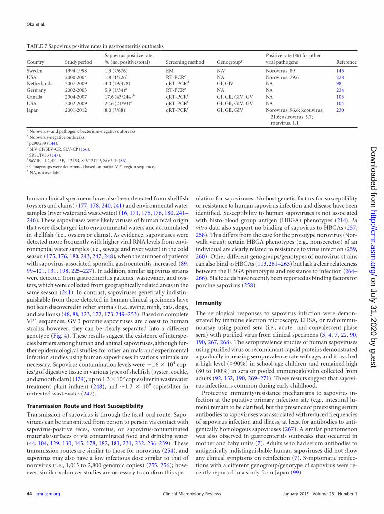
Data from four studies suggest that sapovirus caused 1.3 to8.0% of the gastroenteritis outbreaks (Table 7) (98, 145, 228, 230),and data from the other three studies reported that sapovirus wasdetected in 5.9 to 22.6% of outbreak samples that tested negativefor norovirus or both norovirus and pathogenic bacteria (103,104, 234) (Table 7).
Coinfections of sapoviruses and multiple enteric viruses (e.g.,noroviruses, rotaviruses, astroviruses, adenoviruses, enterovi-ruses, kobuviruses, etc.) have also been reported among acute gas-troenteritis outbreaks (10, 178, 230, 231, 235–237, 239). Coinfec-tions with different sapovirus strains (i.e., different genogroups/genotypes) were also identified from oyster/clam-associatedgastroenteritis outbreaks (178, 236).
Sapoviruses in Seafood, Environmental Water, and Animals
Sapoviruses genetically indistinguishable (i.e., similar or identicalbased on partial virus genome sequences) from those detected in
TABLE 6 Sapovirus positive rates in gastroenteritis patients from 13 studies that detected more than 30 strains during the study period
Country Study period Age (yr)Sapovirus positive rate,% (no. positive/total) Screening method Genogroupg
Positive rate (%) for otherviral pathogens Reference(s)
UK 1979-1981 Unknown 6.6 (39/592) EM NAh NA 89Kenya 1991-1994 �6 2.2 (32/1431) ELISA NA NA 132Finland 1993-1995 �2 9.3 (72/775) Nested RT-PCRa NA Rotavirus, 23.0; norovirus,
20.2; astrovirus, 8.8;adenovirus, 6.3
185
UK 1993-1996 All 3.8 (92/2422) RT-PCRb NA Norovirus, 36.0; rotavirus,31.3
218
Netherlands 1998-1999 All 6.3 (43/687) RT-PCRb NA Norovirus, 16.1; rotavirus,7.3; adenovirus, 3.8;astrovirus, 2.0
216
Japan 1998-2005 All 9.3 (195/2100) Nested RT-PCRc GI, GII, GIV, GV NA 126, 165Japan 2002-2007 All 12.7 (81/639) RT-PCRd GI, GII, GIV, GV Norovirus, 40.7; rotavirus,
7.7; adenovirus, 3.0;enterovirus, 2.0;astrovirus, 1.4
101
Japan 2002-2009 �15 3.7 (146/3895) RT-PCRe GI, GII, GIV NA 102, 220–224Denmark 2005-2007 �3 8.8 (97/1104) qRT-PCRf GI, GII, GV NA 225UK 2006-2007 �5 12.7 (74/583) RT-PCRb NA Norovirus, 24.5; rotavirus,
19.0219
USA 2008-2009 �5 5.4 (42/782) qRT-PCRf NA Norovirus, 21.4; rotavirus,18.0; adenovirus, 11.8;astrovirus, 4.9
215
UK 2008-2009 All 8.8 (77/874) qRT-PCRf NA Norovirus, 12.4; rotavirus,7.3; adenovirus, 3.4;astrovirus, 2.5
226
Canada 2008-2009 All 4.2 (107/2486) qRT-PCRf NA Norovirus, 17.6; rotavirus,6.8; astrovirus, 2.0;adenovirus, 1.4
131
a First, Sapp36/SLV-r-c; second, S1,S2,S3-Sapp-rc (148).b SR80-JV33 (147).c First, F11/R1 or F13,14,R13,14; second, F21/R2 or F22/R2 (126, 165).d SaV1F, -1,2,4F, -5F, -1245R (86, 101).e SLV5317-5749 (163).f SaV1F, -1,2,4F, -5F, -1245R, and SaV124TP, SaV5TP (86).g Genogroups were determined based on partial VP1 region sequences.h NA, not available.
Human Sapoviruses
January 2015 Volume 28 Number 1 cmr.asm.org 43Clinical Microbiology Reviews
on July 31, 2020 by guesthttp://cm
r.asm.org/
Dow
nloaded from
human clinical specimens have also been detected from shellfish(oysters and clams) (177, 178, 240, 241) and environmental watersamples (river water and wastewater) (16, 171, 175, 176, 180, 241–246). These sapoviruses were likely viruses of human fecal originthat were discharged into environmental waters and accumulatedin shellfish (i.e., oysters or clams). As evidence, sapoviruses weredetected more frequently with higher viral RNA levels from envi-ronmental water samples (i.e., sewage and river water) in the coldseason (175, 176, 180, 243, 247, 248), when the number of patientswith sapovirus-associated sporadic gastroenteritis increased (89,99–101, 131, 198, 225–227). In addition, similar sapovirus strainswere detected from gastroenteritis patients, wastewater, and oys-ters, which were collected from geographically related areas in thesame season (241). In contrast, sapoviruses genetically indistin-guishable from those detected in human clinical specimens havenot been discovered in other animals (i.e., swine, mink, bats, dogs,and sea lions) (48, 88, 123, 172, 173, 249–253). Based on completeVP1 sequences, GV.3 porcine sapoviruses are closest to humanstrains; however, they can be clearly separated into a differentgenotype (Fig. 4). These results suggest the existence of interspe-cies barriers among human and animal sapoviruses, although fur-ther epidemiological studies for other animals and experimentalinfection studies using human sapoviruses in various animals arenecessary. Sapovirus contamination levels were �1.6 � 104 cop-ies/g of digestive tissue in various types of shellfish (oyster, cockle,and smooth clam) (179), up to 1.3 � 105 copies/liter in wastewatertreatment plant influent (248), and �1.3 � 109 copies/liter inuntreated wastewater (247).
Transmission Route and Host Susceptibility
Transmission of sapovirus is through the fecal-oral route. Sapo-viruses can be transmitted from person to person via contact withsapovirus-positive feces, vomitus, or sapovirus-contaminatedmaterials/surfaces or via contaminated food and drinking water(44, 104, 129, 130, 145, 178, 182, 183, 231, 232, 236–239). Thesetransmission routes are similar to those for norovirus (254), andsapovirus may also have a low infectious dose similar to that ofnorovirus (i.e., 1,015 to 2,800 genomic copies) (255, 256); how-ever, similar volunteer studies are necessary to confirm this spec-
ulation for sapoviruses. No host genetic factors for susceptibilityor resistance to human sapovirus infection and disease have beenidentified. Susceptibility to human sapoviruses is not associatedwith histo-blood group antigen (HBGA) phenotypes (214). Invitro data also support no binding of sapovirus to HBGAs (257,258). This differs from the case for the prototype norovirus (Nor-walk virus): certain HBGA phenotypes (e.g., nonsecretor) of anindividual are clearly related to resistance to virus infection (259,260). Other different genogroups/genotypes of norovirus strainscan also bind to HBGAs (113, 261–263) but lack a clear relatednessbetween the HBGA phenotypes and resistance to infection (264–266). Sialic acids have recently been reported as binding factors forporcine sapovirus (258).
Immunity
The serological responses to sapovirus infection were demon-strated by immune electron microscopy, ELISA, or radioimmu-noassay using paired sera (i.e., acute- and convalescent-phasesera) with purified virus from clinical specimens (3, 4, 7, 22, 90,190, 267, 268). The seroprevalence studies of human sapovirusesusing purified virus or recombinant capsid proteins demonstrateda gradually increasing seroprevalence rate with age, and it reacheda high level (�90%) in school-age children, and remained high(80 to 100%) in sera or pooled immunoglobulin collected fromadults (92, 132, 190, 269–271). These results suggest that sapovi-rus infection is common during early childhood.
Protective immunity/resistance mechanisms to sapovirus in-fection at the putative primary infection site (e.g., intestinal lu-men) remain to be clarified, but the presence of preexisting serumantibodies to sapoviruses was associated with reduced frequenciesof sapovirus infection and illness, at least for antibodies to anti-genically homologous sapoviruses (267). A similar phenomenonwas also observed in gastroenteritis outbreaks that occurred inmother and baby units (7). Adults who had serum antibodies toantigenically indistinguishable human sapoviruses did not showany clinical symptoms on reinfection (7). Symptomatic reinfec-tions with a different genogroup/genotype of sapovirus were re-cently reported in a study from Japan (99).
TABLE 7 Sapovirus positive rates in gastroenteritis outbreaks
Country Study periodSapovirus positive rate,% (no. positive/total) Screening method Genogroupg
Positive rate (%) for otherviral pathogens Reference
Sweden 1994-1998 1.3 (9/676) EM NAh Norovirus, 89 145USA 2000-2004 1.8 (4/226) RT-PCRc NA Norovirus, 79.6 228Netherlands 2007-2009 4.0 (19/478) qRT-PCRd GI, GIV NA 98Germany 2002-2003 5.9 (2/34)a RT-PCRe NA NA 234Canada 2004-2007 17.6 (43/244)b qRT-PCRf GI, GII, GIV, GV NA 103USA 2002-2009 22.6 (21/93)b qRT-PCRf GI, GII, GIV, GV NA 104Japan 2001-2012 8.0 (7/88) qRT-PCRf GI, GII, GIV Norovirus, 96.6; kobuvirus,
21.6; astrovirus, 5.7;rotavirus, 1.1
230
a Norovirus- and pathogenic bacterium-negative outbreaks.b Norovirus-negative outbreaks.c p290/289 (144).d SLV-CF/SLV-CR, SLV-CP (156).e SR80/JV33 (147).f SaV1F, -1,2,4F, -5F, -1245R, SaV124TP, SaV5TP (86).g Genogroups were determined based on partial VP1 region sequences.h NA, not available.
Oka et al.
44 cmr.asm.org January 2015 Volume 28 Number 1Clinical Microbiology Reviews
on July 31, 2020 by guesthttp://cm
r.asm.org/
Dow
nloaded from
CONCLUSIONS AND FUTURE DIRECTIONS
Recent epidemiological studies with improved diagnostic assayshave highlighted the impact of sapovirus-associated gastroenteri-tis. Genetically highly diverse sapovirus strains were identifiedthrough epidemiological surveillance studies. Continuous sur-veillance with a broadly reactive detection system(s) and molecu-lar characterization will permit the identification of changes inmajor strains as well as the emergence of new strains and an un-derstanding of the evolution of sapoviruses among humans andanimals. However, in contrast to the significant improvement insapovirus detection methods, the basic understanding of infec-tion/replication sites, pathological changes in infected persons,immunological responses and protective immunity to sapovirusinfections in humans, infectious dose, and stability in the environ-ment remain unknown. To date, no vaccines or antiviral drugs areavailable for the control and prevention of human sapovirus in-fections. The mechanisms of virus binding and entry into targetcells and viral RNA replication and translation are undefined, par-tially due to the lack of a cell culture system. Extensive studies ofhuman sapoviruses in clinical cases, the use of the cell culture-adapted porcine sapovirus strain as a model, and establishment ofa human sapovirus cell culture system will improve our knowl-edge of sapoviruses and may lead to more targeted control mea-sures for prevention of sapovirus gastroenteritis in the future.
ACKNOWLEDGMENTS
We thank Yasutaka Yamashita, who kindly provided transmission EMpictures for human sapovirus and norovirus, and Kelly A. Scheuer andSusan Sommer-Wagner for their editing of the manuscript.
REFERENCES1. Madeley CR. 1979. Comparison of the features of astroviruses and cali-
civiruses seen in samples of feces by electron microscopy. J Infect Dis139:519 –523. http://dx.doi.org/10.1093/infdis/139.5.519.
2. Madeley CR, Cosgrove BP. 1976. Caliciviruses in man. Lancet i:199 –200. (Letter.) http://dx.doi.org/10.1016/S0140-6736(76)91309-X.
3. McSwiggan DA, Cubitt D, Moore W. 1978. Calicivirus associated withwinter vomiting disease. Lancet i:1215. http://dx.doi.org/10.1016/S0140-6736(78)91012-7.
4. Chiba S, Sakuma Y, Kogasaka R, Akihara M, Horino K, Nakao T,Fukui S. 1979. An outbreak of gastroenteritis associated with calicivirusin an infant home. J Med Virol 4:249 –254. http://dx.doi.org/10.1002/jmv.1890040402.
5. Cubitt WD, McSwiggan DA, Moore W. 1979. Winter vomiting diseasecaused by calicivirus. J Clin Pathol 32:786 –793. http://dx.doi.org/10.1136/jcp.32.8.786.
6. Chiba S, Sakuma Y, Kogasaka R, Akihara M, Terashima H, Horino K,Nakao T. 1980. Fecal shedding of virus in relation to the days of illness ininfantile gastroenteritis due to calicivirus. J Infect Dis 142:247–249. http://dx.doi.org/10.1093/infdis/142.2.247.
7. Cubitt WD, McSwiggan DA, Arstall S. 1980. An outbreak of calicivirusinfection in a mother and baby unit. J Clin Pathol 33:1095–1098. http://dx.doi.org/10.1136/jcp.33.11.1095.
8. Humphrey TJ, Cruickshank JG, Cubitt WD. 1984. An outbreak ofcalicivirus associated gastroenteritis in an elderly persons home. A pos-sible zoonosis? J Hyg (Lond) 93:293–299. http://dx.doi.org/10.1017/S0022172400064822.
9. Cubitt WD, Pead PJ, Saeed AA. 1981. A new serotype of calicivirusassociated with an outbreak of gastroenteritis in a residential home forthe elderly. J Clin Pathol 34:924 –926. http://dx.doi.org/10.1136/jcp.34.8.924.
10. Nakata S, Honma S, Numata KK, Kogawa K, Ukae S, Morita Y, AdachiN, Chiba S. 2000. Members of the family caliciviridae (Norwalk virusand Sapporo virus) are the most prevalent cause of gastroenteritis out-breaks among infants in Japan. J Infect Dis 181:2029 –2032. http://dx.doi.org/10.1086/315500.
11. Chiba S, Nakata S, Numata-Kinoshita K, Honma S. 2000. Sapporovirus: history and recent findings. J Infect Dis 181(Suppl 2):S303–S308.http://dx.doi.org/10.1086/315574.
12. Numata K, Hardy ME, Nakata S, Chiba S, Estes MK. 1997. Molecularcharacterization of morphologically typical human calicivirus Sapporo.Arch Virol 142:1537–1552. http://dx.doi.org/10.1007/s007050050178.
13. Matson DO, Zhong WM, Nakata S, Numata K, Jiang X, Pickering LK,Chiba S, Estes MK. 1995. Molecular characterization of a human cali-civirus with sequence relationships closer to animal caliciviruses thanother known human caliciviruses. J Med Virol 45:215–222. http://dx.doi.org/10.1002/jmv.1890450218.
14. Nakanishi K, Tatsumi M, Kinoshita-Numata K, Tsugawa T, Nakata S,Tsutsumi H. 2011. Full sequence analysis of the original Sapporo virus.Microbiol Immunol 55:657– 660. http://dx.doi.org/10.1111/j.1348-0421.2011.00358.x.
15. Mayo MA. 2002. A summary of taxonomic changes recently approved byICTV. Arch Virol 147:1655–1663. http://dx.doi.org/10.1007/s007050200039.
16. Ng TF, Marine R, Wang C, Simmonds P, Kapusinszky B, Bodhidatta L,Oderinde BS, Wommack KE, Delwart E. 2012. High variety of known andnew RNA and DNA viruses of diverse origins in untreated sewage. J Virol86:12161–12175. http://dx.doi.org/10.1128/JVI.00869-12.
17. Wolf S, Reetz J, Hoffmann K, Grundel A, Schwarz BA, Hanel I, OttoPH. 2012. Discovery and genetic characterization of novel calicivirusesin German and Dutch poultry. Arch Virol 157:1499 –1507. http://dx.doi.org/10.1007/s00705-012-1326-7.
18. Farkas T, Sestak K, Wei C, Jiang X. 2008. Characterization of a rhesusmonkey calicivirus representing a new genus of Caliciviridae. J Virol82:5408 –5416. http://dx.doi.org/10.1128/JVI.00070-08.
19. L’Homme Y, Sansregret R, Plante-Fortier E, Lamontagne AM, Ouar-dani M, Lacroix G, Simard C. 2009. Genomic characterization of swinecaliciviruses representing a new genus of Caliciviridae. Virus Genes 39:66 –75. http://dx.doi.org/10.1007/s11262-009-0360-3.
20. Wolf S, Reetz J, Otto P. 2011. Genetic characterization of a novelcalicivirus from a chicken. Arch Virol 156:1143–1150. http://dx.doi.org/10.1007/s00705-011-0964-5.
21. Spratt HC, Marks MI, Gomersall M, Gill P, Pai CH. 1978. Nosocomialinfantile gastroenteritis associated with minirotavirus and calicivirus. JPediatr 93:922–926. http://dx.doi.org/10.1016/S0022-3476(78)81212-8.
22. Suzuki H, Konno T, Kutsuzawa T, Imai A, Tazawa F, Ishida N,Katsushima N, Sakamoto M. 1979. The occurrence of calicivirus ininfants with acute gastroenteritis. J Med Virol 4:321–326. http://dx.doi.org/10.1002/jmv.1890040410.
23. Kjeldsberg E. 1977. Small spherical viruses in faeces from gastroenteritispatients. Acta Pathol Microbiol Scand B 85B:351–354.
24. Cubitt WD, Barrett AD. 1984. Propagation of human candidate calici-virus in cell culture. J Gen Virol 65:1123–1126. http://dx.doi.org/10.1099/0022-1317-65-6-1123.
25. Chang KO, Sosnovtsev SV, Belliot G, Kim Y, Saif LJ, Green KY. 2004.Bile acids are essential for porcine enteric calicivirus replication in asso-ciation with down-regulation of signal transducer and activator of tran-scription 1. Proc Natl Acad Sci U S A 101:8733– 8738. http://dx.doi.org/10.1073/pnas.0401126101.
26. Flynn WT, Saif LJ. 1988. Serial propagation of porcine enteric calicivi-rus-like virus in primary porcine kidney cell cultures. J Clin Microbiol26:206 –212.
27. Parwani AV, Flynn WT, Gadfield KL, Saif LJ. 1991. Serial propagationof porcine enteric calicivirus in a continuous cell line. Effect of mediumsupplementation with intestinal contents or enzymes. Arch Virol 120:115–122. http://dx.doi.org/10.1007/BF01310954.
28. Chang KO, Sosnovtsev SS, Belliot G, Wang Q, Saif LJ, Green KY. 2005.Reverse genetics system for porcine enteric calicivirus, a prototype sapo-virus in the Caliciviridae. J Virol 79:1409 –1416. http://dx.doi.org/10.1128/JVI.79.3.1409-1416.2005.
29. Shivanna V, Kim Y, Chang KO. 2014. The crucial role of bile acids in theentry of porcine enteric calicivirus. Virology 456:268 –278. http://dx.doi.org/10.1016/j.virol.2014.04.002.
30. Chang KO, Kim Y, Green KY, Saif LJ. 2002. Cell-culture propagationof porcine enteric calicivirus mediated by intestinal contents is depen-dent on the cyclic AMP signaling pathway. Virology 304:302–310. http://dx.doi.org/10.1006/viro.2002.1665.
31. Flynn WT, Saif LJ, Moorhead PD. 1988. Pathogenesis of porcine enteric
Human Sapoviruses
January 2015 Volume 28 Number 1 cmr.asm.org 45Clinical Microbiology Reviews
on July 31, 2020 by guesthttp://cm
r.asm.org/
Dow
nloaded from
calicivirus-like virus in four-day-old gnotobiotic pigs. Am J Vet Res 49:819 – 825.
32. Guo M, Hayes J, Cho KO, Parwani AV, Lucas LM, Saif LJ. 2001.Comparative pathogenesis of tissue culture-adapted and wild-typeCowden porcine enteric calicivirus (PEC) in gnotobiotic pigs and induc-tion of diarrhea by intravenous inoculation of wild-type PEC. J Virol75:9239 –9251. http://dx.doi.org/10.1128/JVI.75.19.9239-9251.2001.
33. Saif LJ, Bohl EH, Theil KW, Cross RF, House JA. 1980. Rotavirus-like,calicivirus-like, and 23-nm virus-like particles associated with diarrheain young pigs. J Clin Microbiol 12:105–111.
34. Terashima H, Chiba S, Sakuma Y, Kogasaka R, Nakata S, Minami R,Horino K, Nakao T. 1983. The polypeptide of a human calicivirus. ArchVirol 78:1–7. http://dx.doi.org/10.1007/BF01310853.
35. Parwani AV, Saif LJ, Kang SY. 1990. Biochemical characterization ofporcine enteric calicivirus: analysis of structural and nonstructural viralproteins. Arch Virol 112:41–53. http://dx.doi.org/10.1007/BF01348984.
36. Wang Q, Zhang Z, Saif LJ. 2012. Stability of and attachment tolettuce by a culturable porcine sapovirus surrogate for human calici-viruses. Appl Environ Microbiol 78:3932–3940. http://dx.doi.org/10.1128/AEM.06600-11.
37. Green KY. 2007. Caliciviridae: the noroviruses: specific virus families, p949 –979. In Knipe DM, Howley PM, Griffin DE, Lamb RA, Martin MA,Roizman B, Straus SE (ed), Fields virology, 5th ed. Lippincott Williams &Wilkins, Philadelphia, PA.
38. Smiley JR, Chang KO, Hayes J, Vinje J, Saif LJ. 2002. Characterizationof an enteropathogenic bovine calicivirus representing a potentially newcalicivirus genus. J Virol 76:10089 –10098. http://dx.doi.org/10.1128/JVI.76.20.10089-10098.2002.
39. Meyers G, Wirblich C, Thiel HJ. 1991. Rabbit hemorrhagic diseasevirus—molecular cloning and nucleotide sequencing of a calicivirus ge-nome. Virology 184:664 – 676. http://dx.doi.org/10.1016/0042-6822(91)90436-F.
40. Clarke IN, Lambden PR. 2000. Organization and expression of calici-virus genes. J Infect Dis 181(Suppl)2:S309 –S316. http://dx.doi.org/10.1086/315575.
41. Liu BL, Clarke IN, Caul EO, Lambden PR. 1995. Human entericcaliciviruses have a unique genome structure and are distinct from theNorwalk-like viruses. Arch Virol 140:1345–1356. http://dx.doi.org/10.1007/BF01322662.
42. Liu B, Clarke IN, Caul EO, Lambden PR. 1997. The genomic 5=terminus of Manchester calicivirus. Virus Genes 15:25–28. http://dx.doi.org/10.1023/A:1007946628253.
43. Jiang X, Cubitt WD, Berke T, Zhong W, Dai X, Nakata S, PickeringLK, Matson DO. 1997. Sapporo-like human caliciviruses are geneticallyand antigenically diverse. Arch Virol 142:1813–1827. http://dx.doi.org/10.1007/s007050050199.
44. Noel JS, Liu BL, Humphrey CD, Rodriguez EM, Lambden PR,Clarke IN, Dwyer DM, Ando T, Glass RI, Monroe SS. 1997.Parkville virus: a novel genetic variant of human calicivirus in theSapporo virus clade, associated with an outbreak of gastroenteritis inadults. J Med Virol 52:173–178. http://dx.doi.org/10.1002/(SICI)1096-9071(199706)52:2�173::AID-JMV10�3.0.CO;2-M.
45. Schuffenecker I, Ando T, Thouvenot D, Lina B, Aymard M. 2001.Genetic classification of “Sapporo-like viruses.” Arch Virol 146:2115–2132. http://dx.doi.org/10.1007/s007050170024.
46. Farkas T, Zhong WM, Jing Y, Huang PW, Espinosa SM, Martinez N,Morrow AL, Ruiz-Palacios GM, Pickering LK, Jiang X. 2004. Geneticdiversity among sapoviruses. Arch Virol 149:1309 –1323. http://dx.doi.org/10.1007/s00705-004-0296-9.
47. Okada M, Yamashita Y, Oseto M, Ogawa T, Kaiho I, Shinozaki K.2006. Genetic variability in the sapovirus capsid protein. Virus Genes33:157–161. http://dx.doi.org/10.1007/s11262-005-0051-7.
48. Tse H, Chan WM, Li KS, Lau SK, Woo PC, Yuen KY. 2012. Discoveryand genomic characterization of a novel bat sapovirus with unusualgenomic features and phylogenetic position. PLoS One 7:e34987. http://dx.doi.org/10.1371/journal.pone.0034987.
49. Oka T, Katayama K, Ogawa S, Hansman GS, Kageyama T, UshijimaH, Miyamura T, Takeda N. 2005. Proteolytic processing of sapovirusORF1 polyprotein. J Virol 79:7283–7290. http://dx.doi.org/10.1128/JVI.79.12.7283-7290.2005.
50. Oka T, Yamamoto M, Katayama K, Hansman GS, Ogawa S, MiyamuraT, Takeda N. 2006. Identification of the cleavage sites of sapovirus open
reading frame 1 polyprotein. J Gen Virol 87:3329 –3338. http://dx.doi.org/10.1099/vir.0.81799-0.
51. Oka T, Yokoyama M, Katayama K, Tsunemitsu H, Yamamoto M,Miyashita K, Ogawa S, Motomura K, Mori H, Nakamura H, Wakita T,Takeda N, Sato H. 2009. Structural and biological constraints on diver-sity of regions immediately upstream of cleavage sites in calicivirus pre-cursor proteins. Virology 394:119 –129. http://dx.doi.org/10.1016/j.virol.2009.08.018.
52. Oka T, Yamamoto M, Yokoyama M, Ogawa S, Hansman GS, Katay-ama K, Miyashita K, Takagi H, Tohya Y, Sato H, Takeda N. 2007.Highly conserved configuration of catalytic amino acid residues amongcalicivirus-encoded proteases. J Virol 81:6798 – 6806. http://dx.doi.org/10.1128/JVI.02840-06.
53. Oka T, Murakami K, Wakita T, Katayama K. 2011. Comparativesite-directed mutagenesis in the catalytic amino acid triad in calicivirusproteases. Microbiol Immunol 55:108 –114. http://dx.doi.org/10.1111/j.1348-0421.2010.00295.x.
54. Yokoyama M, Oka T, Kojima H, Nagano T, Okabe T, Katayama K,Wakita T, Kanda T, Sato H. 2012. Structural basis for specific recogni-tion of substrates by sapovirus protease. Front Microbiol 3:312. http://dx.doi.org/10.3389/fmicb.2012.00312.
55. Oka T, Katayama K, Ogawa S, Hansman GS, Kageyama T, MiyamuraT, Takeda N. 2005. Cleavage activity of the sapovirus 3C-like protease inEscherichia coli. Arch Virol 150:2539 –2548. http://dx.doi.org/10.1007/s00705-005-0591-0.
56. Robel I, Gebhardt J, Mesters JR, Gorbalenya A, Coutard B, Canard B,Hilgenfeld R, Rohayem J. 2008. Functional characterization of thecleavage specificity of the sapovirus chymotrypsin-like protease. J Virol82:8085– 8093. http://dx.doi.org/10.1128/JVI.00693-08.
57. Fullerton SW, Blaschke M, Coutard B, Gebhardt J, Gorbalenya A,Canard B, Tucker PA, Rohayem J. 2007. Structural and functionalcharacterization of sapovirus RNA-dependent RNA polymerase. J Virol81:1858 –1871. http://dx.doi.org/10.1128/JVI.01462-06.
58. Bull RA, Hyde J, Mackenzie JM, Hansman GS, Oka T, Takeda N,White PA. 2011. Comparison of the replication properties of murineand human calicivirus RNA-dependent RNA polymerases. Virus Genes42:16 –27. http://dx.doi.org/10.1007/s11262-010-0535-y.
59. Green KY, Mory A, Fogg MH, Weisberg A, Belliot G, Wagner M,Mitra T, Ehrenfeld E, Cameron CE, Sosnovtsev SV. 2002. Isolation ofenzymatically active replication complexes from feline calicivirus-infected cells. J Virol 76:8582– 8595. http://dx.doi.org/10.1128/JVI.76.17.8582-8595.2002.
60. Sosnovtsev SV, Garfield M, Green KY. 2002. Processing map andessential cleavage sites of the nonstructural polyprotein encoded byORF1 of the feline calicivirus genome. J Virol 76:7060 –7072. http://dx.doi.org/10.1128/JVI.76.14.7060-7072.2002.
61. Oehmig A, Buttner M, Weiland F, Werz W, Bergemann K, Pfaff E.2003. Identification of a calicivirus isolate of unknown origin. J Gen Virol84:2837–2845. http://dx.doi.org/10.1099/vir.0.19042-0.
62. Konig M, Thiel HJ, Meyers G. 1998. Detection of viral proteins afterinfection of cultured hepatocytes with rabbit hemorrhagic disease virus.J Virol 72:4492– 4497.
63. Belliot G, Sosnovtsev SV, Mitra T, Hammer C, Garfield M, Green KY.2003. In vitro proteolytic processing of the MD145 norovirus ORF1 non-structural polyprotein yields stable precursors and products similar tothose detected in calicivirus-infected cells. J Virol 77:10957–10974. http://dx.doi.org/10.1128/JVI.77.20.10957-10974.2003.
64. Chang KO, Sosnovtsev SV, Belliot G, King AD, Green KY. 2006. Stableexpression of a Norwalk virus RNA replicon in a human hepatoma cellline. Virology 353:463– 473. http://dx.doi.org/10.1016/j.virol.2006.06.006.
65. Sosnovtsev SV, Belliot G, Chang KO, Prikhodko VG, Thackray LB,Wobus CE, Karst SM, Virgin HW, Green KY. 2006. Cleavage map andproteolytic processing of the murine norovirus nonstructural polypro-tein in infected cells. J Virol 80:7816 –7831. http://dx.doi.org/10.1128/JVI.00532-06.
66. Katayama K, Hansman GS, Oka T, Ogawa S, Takeda N. 2006. Inves-tigation of norovirus replication in a human cell line. Arch Virol 151:1291–1308. http://dx.doi.org/10.1007/s00705-005-0720-9.
67. Meyers G, Wirblich C, Thiel HJ, Thumfart JO. 2000. Rabbit hemor-rhagic disease virus: genome organization and polyprotein processing ofa calicivirus studied after transient expression of cDNA constructs. Vi-rology 276:349 –363. http://dx.doi.org/10.1006/viro.2000.0545.
Oka et al.
46 cmr.asm.org January 2015 Volume 28 Number 1Clinical Microbiology Reviews
on July 31, 2020 by guesthttp://cm
r.asm.org/
Dow
nloaded from
68. Pfister T, Wimmer E. 2001. Polypeptide p41 of a Norwalk-like virus is anucleic acid-independent nucleoside triphosphatase. J Virol 75:1611–1619. http://dx.doi.org/10.1128/JVI.75.4.1611-1619.2001.
69. Marin MS, Casais R, Alonso JM, Parra F. 2000. ATP binding andATPase activities associated with recombinant rabbit hemorrhagic dis-ease virus 2C-like polypeptide. J Virol 74:10846 –10851. http://dx.doi.org/10.1128/JVI.74.22.10846-10851.2000.
70. Leen EN, Kwok KY, Birtley JR, Simpson PJ, Subba-Reddy CV,Chaudhry Y, Sosnovtsev SV, Green KY, Prater SN, Tong M, Young JC,Chung LM, Marchant J, Roberts LO, Kao CC, Matthews S, GoodfellowIG, Curry S. 2013. Structures of the compact helical core domains offeline calicivirus and murine norovirus VPg proteins. J Virol 87:5318 –5330. http://dx.doi.org/10.1128/JVI.03151-12.
71. Oka T, Yamamoto M, Miyashita K, Ogawa S, Katayama K, Wakita T,Takeda N. 2009. Self-assembly of sapovirus recombinant virus-like par-ticles from polyprotein in mammalian cells. Microbiol Immunol 53:49 –52. http://dx.doi.org/10.1111/j.1348-0421.2008.00086.x.
72. Hansman GS, Oka T, Takeda N. 2008. Sapovirus-like particles derivedfrom polyprotein. Virus Res 137:261–265. http://dx.doi.org/10.1016/j.virusres.2008.07.002.
73. Vongpunsawad S, Venkataram Prasad BV, Estes MK. 2013. Norwalkvirus minor capsid protein VP2 associates within the VP1 shell domain.J Virol 87:4818 – 4825. http://dx.doi.org/10.1128/JVI.03508-12.
74. Jiang X, Zhong W, Kaplan M, Pickering LK, Matson DO. 1999.Expression and characterization of Sapporo-like human calicivirus cap-sid proteins in baculovirus. J Virol Methods 78:81–91. http://dx.doi.org/10.1016/S0166-0934(98)00169-4.
75. Guo M, Qian Y, Chang KO, Saif LJ. 2001. Expression and self-assemblyin baculovirus of porcine enteric calicivirus capsids into virus-like parti-cles and their use in an enzyme-linked immunosorbent assay for anti-body detection in swine. J Clin Microbiol 39:1487–1493. http://dx.doi.org/10.1128/JCM.39.4.1487-1493.2001.
76. Chen R, Neill JD, Noel JS, Hutson AM, Glass RI, Estes MK, PrasadBV. 2004. Inter- and intragenus structural variations in caliciviruses andtheir functional implications. J Virol 78:6469 – 6479. http://dx.doi.org/10.1128/JVI.78.12.6469-6479.2004.
77. Hansman GS, Natori K, Oka T, Ogawa S, Tanaka K, Nagata N,Ushijima H, Takeda N, Katayama K. 2005. Cross-reactivity amongsapovirus recombinant capsid proteins. Arch Virol 150:21–36. http://dx.doi.org/10.1007/s00705-004-0406-8.
78. Hansman GS, Oka T, Katayama K, Takeda N. 2006. Enhancement ofsapovirus recombinant capsid protein expression in insect cells. FEBSLett 580:4047– 4050. http://dx.doi.org/10.1016/j.febslet.2006.06.040.
79. Oka T, Hansman GS, Katayama K, Ogawa S, Nagata N, Miyamura T,Takeda N. 2006. Expression of sapovirus virus-like particles in mamma-lian cells. Arch Virol 151:399 – 404. http://dx.doi.org/10.1007/s00705-005-0613-y.
80. Hansman GS, Saito H, Shibata C, Ishizuka S, Oseto M, Oka T, TakedaN. 2007. Outbreak of gastroenteritis due to sapovirus. J Clin Microbiol45:1347–1349. http://dx.doi.org/10.1128/JCM.01854-06.
81. Oka T, Miyashita K, Katayama K, Wakita T, Takeda N. 2009.Distinct genotype and antigenicity among genogroup II sapoviruses.Microbiol Immunol 53:417– 420. http://dx.doi.org/10.1111/j.1348-0421.2009.00133.x.
82. Prasad BV, Hardy ME, Dokland T, Bella J, Rossmann MG, Estes MK.1999. X-ray crystallographic structure of the Norwalk virus capsid. Sci-ence 286:287–290. http://dx.doi.org/10.1126/science.286.5438.287.
83. Hardy ME. 2005. Norovirus protein structure and function. FEMS Mi-crobiol Lett 253:1– 8. http://dx.doi.org/10.1016/j.femsle.2005.08.031.
84. Katayama K, Miyoshi T, Uchino K, Oka T, Tanaka T, Takeda N,Hansman GS. 2004. Novel recombinant sapovirus. Emerg Infect Dis10:1874 –1876. http://dx.doi.org/10.3201/eid1010.040395.
85. Hansman GS, Takeda N, Oka T, Oseto M, Hedlund KO, Katayama K.2005. Intergenogroup recombination in sapoviruses. Emerg Infect Dis11:1916 –1920. http://dx.doi.org/10.3201/eid1112.050722.
86. Oka T, Katayama K, Hansman GS, Kageyama T, Ogawa S, Wu FT,White PA, Takeda N. 2006. Detection of human sapovirus by real-timereverse transcription-polymerase chain reaction. J Med Virol 78:1347–1353. http://dx.doi.org/10.1002/jmv.20699.
87. Oka T, Mori K, Iritani N, Harada S, Ueki Y, Iizuka S, Mise K,Murakami K, Wakita T, Katayama K. 2012. Human sapovirus classifi-cation based on complete capsid nucleotide sequences. Arch Virol 157:349 –352. http://dx.doi.org/10.1007/s00705-011-1161-2.
88. Scheuer KA, Oka T, Hoet AE, Gebreyes WA, Molla BZ, Saif LJ, WangQ. 2013. Prevalence of porcine noroviruses, molecular characterizationof emerging porcine sapoviruses from finisher swine in the United States,and unified classification scheme for sapoviruses. J Clin Microbiol 51:2344 –2353. http://dx.doi.org/10.1128/JCM.00865-13.
89. Cubitt WD, McSwiggan DA. 1981. Calicivirus gastroenteritis in NorthWest London. Lancet ii:975–977. http://dx.doi.org/10.1016/S0140-6736(81)91167-3.
90. Cubitt WD, Blacklow NR, Herrmann JE, Nowak NA, Nakata S, ChibaS. 1987. Antigenic relationships between human caliciviruses and Nor-walk virus. J Infect Dis 156:806 – 814. http://dx.doi.org/10.1093/infdis/156.5.806.
91. Hansman GS, Guntapong R, Pongsuwanna Y, Natori K, Katayama K,Takeda N. 2006. Development of an antigen ELISA to detect sapovirus inclinical stool specimens. Arch Virol 151:551–561. http://dx.doi.org/10.1007/s00705-005-0630-x.
92. Farkas T, Deng X, Ruiz-Palacios G, Morrow A, Jiang X. 2006. Develop-ment of an enzyme immunoassay for detection of sapovirus-specific anti-bodies and its application in a study of seroprevalence in children. J ClinMicrobiol 44:3674–3679. http://dx.doi.org/10.1128/JCM.01087-06.
93. Hansman GS, Oka T, Sakon N, Takeda N. 2007. Antigenic diversity ofhuman sapoviruses. Emerg Infect Dis 13:1519 –1525. http://dx.doi.org/10.3201/eid1310.070402.
94. Kitamoto N, Oka T, Katayama K, Li TC, Takeda N, Kato Y, MiyoshiT, Tanaka T. 2012. Novel monoclonal antibodies broadly reactive tohuman recombinant sapovirus-like particles. Microbiol Immunol 56:760 –770. http://dx.doi.org/10.1111/j.1348-0421.2012.00499.x.
95. Hansman GS, Natori K, Ushijima H, Katayama K, Takeda N. 2005.Characterization of polyclonal antibodies raised against sapovirus geno-group five virus-like particles. Arch Virol 150:1433–1437. http://dx.doi.org/10.1007/s00705-005-0506-0.
96. Hansman GS, Oka T, Katayama K, Takeda N. 2007. Human sapoviruses:genetic diversity, recombination, and classification. Rev Med Virol 17:133–141. http://dx.doi.org/10.1002/rmv.533. (Erratum, 17:2364, http://dx.doi.org/10.1002/rmv.552.)
97. Kroneman A, Vega E, Vennema H, Vinje J, White PA, Hansman G,Green K, Martella V, Katayama K, Koopmans M. 2013. Proposal for aunified norovirus nomenclature and genotyping. Arch Virol 158:2059 –2068. http://dx.doi.org/10.1007/s00705-013-1708-5.
98. Svraka S, Vennema H, van der Veer B, Hedlund KO, Thorhagen M,Siebenga J, Duizer E, Koopmans M. 2010. Epidemiology and genotypeanalysis of emerging sapovirus-associated infections across Europe. JClin Microbiol 48:2191–2198. http://dx.doi.org/10.1128/JCM.02427-09.
99. Harada S, Oka T, Tokuoka E, Kiyota N, Nishimura K, Shimada Y,Ueno T, Ikezawa S, Wakita T, Wang Q, Saif LJ, Katayama K. 2012. Aconfirmation of sapovirus re-infection gastroenteritis cases with differ-ent genogroups and genetic shifts in the evolving sapovirus genotypes,2002-2011. Arch Virol 157:1999 –2003. http://dx.doi.org/10.1007/s00705-012-1387-7. (Erratum, 158:2641–2642, 2013, http://dx.doi.org/10.1007/s00705-013-1757-9.)
100. Harada S, Tokuoka E, Kiyota N, Katayama K, Oka T. 2013. Phyloge-netic analysis of the nonstructural and structural protein encoding re-gion sequences, indicating successive appearance of genomically diversesapovirus strains from gastroenteritis patients. Jpn J Infect Dis 66:454 –457.
101. Harada S, Okada M, Yahiro S, Nishimura K, Matsuo S, Miyasaka J,Nakashima R, Shimada Y, Ueno T, Ikezawa S, Shinozaki K, Katayama K,Wakita T, Takeda N, Oka T. 2009. Surveillance of pathogens in outpatientswith gastroenteritis and characterization of sapovirus strains between 2002and 2007 in Kumamoto Prefecture, Japan. J Med Virol 81:1117–1127. http://dx.doi.org/10.1002/jmv.21454. (Erratum, 85:1685–1686, 2013, http://dx.doi.org/10.1002/jmv.23677.)
102. Chanit W, Thongprachum A, Khamrin P, Okitsu S, Mizuguchi M,Ushijima H. 2009. Intergenogroup recombinant sapovirus in Japan,2007–2008. Emerg Infect Dis 15:1084 –1087. http://dx.doi.org/10.3201/eid1507.090153.
103. Pang XL, Lee BE, Tyrrell GJ, Preiksaitis JK. 2009. Epidemiology andgenotype analysis of sapovirus associated with gastroenteritis outbreaksin Alberta, Canada: 2004-2007. J Infect Dis 199:547–551. http://dx.doi.org/10.1086/596210.
104. Lee LE, Cebelinski EA, Fuller C, Keene WE, Smith K, Vinje J, BesserJM. 2012. Sapovirus outbreaks in long-term care facilities, Oregon and
Human Sapoviruses
January 2015 Volume 28 Number 1 cmr.asm.org 47Clinical Microbiology Reviews
on July 31, 2020 by guesthttp://cm
r.asm.org/
Dow
nloaded from

Minnesota, USA, 2002-2009. Emerg Infect Dis 18:873– 876. http://dx.doi.org/10.3201/eid1805.111843.
105. Siebenga JJ, Vennema H, Duizer E, Koopmans MP. 2007. Gastroen-teritis caused by norovirus GGII.4, The Netherlands, 1994-2005. EmergInfect Dis 13:144 –146. http://dx.doi.org/10.3201/eid1301.060800.
106. Glass RI, Parashar UD, Estes MK. 2009. Norovirus gastroenteritis. NEngl J Med 361:1776 –1785. http://dx.doi.org/10.1056/NEJMra0804575.
107. Siebenga JJ, Vennema H, Zheng DP, Vinje J, Lee BE, Pang XL, Ho EC,Lim W, Choudekar A, Broor S, Halperin T, Rasool NB, Hewitt J,Greening GE, Jin M, Duan ZJ, Lucero Y, O’Ryan M, Hoehne M,Schreier E, Ratcliff RM, White PA, Iritani N, Reuter G, Koopmans M.2009. Norovirus illness is a global problem: emergence and spread ofnorovirus GII.4 variants, 2001-2007. J Infect Dis 200:802– 812. http://dx.doi.org/10.1086/605127.
108. Vega E, Barclay L, Gregoricus N, Shirley SH, Lee D, Vinje J. 2014.Genotypic and epidemiologic trends of norovirus outbreaks in theUnited States, 2009 to 2013. J Clin Microbiol 52:147–155. http://dx.doi.org/10.1128/JCM.02680-13.
109. Lindesmith LC, Beltramello M, Donaldson EF, Corti D, Swanstrom J,Debbink K, Lanzavecchia A, Baric RS. 2012. Immunogenetic mecha-nisms driving norovirus GII. 4 antigenic variation. PLoS Pathog8:e1002705. http://dx.doi.org/10.1371/journal.ppat.1002705.
110. Lindesmith LC, Donaldson EF, Baric RS. 2011. Norovirus GII.4 strainantigenic variation. J Virol 85:231–242. http://dx.doi.org/10.1128/JVI.01364-10.
111. Siebenga JJ, Vennema H, Renckens B, de Bruin E, van der Veer B,Siezen RJ, Koopmans M. 2007. Epochal evolution of GGII.4 noroviruscapsid proteins from 1995 to 2006. J Virol 81:9932–9941. http://dx.doi.org/10.1128/JVI.00674-07.
112. Motomura K, Oka T, Yokoyama M, Nakamura H, Mori H, Ode H,Hansman GS, Katayama K, Kanda T, Tanaka T, Takeda N, Sato H.2008. Identification of monomorphic and divergent haplotypes in the2006-2007 norovirus GII/4 epidemic population by genomewide tracingof evolutionary history. J Virol 82:11247–11262. http://dx.doi.org/10.1128/JVI.00897-08.
113. Bok K, Abente EJ, Realpe-Quintero M, Mitra T, Sosnovtsev SV,Kapikian AZ, Green KY. 2009. Evolutionary dynamics of GII.4 norovi-ruses over a 34-year period. J Virol 83:11890 –11901. http://dx.doi.org/10.1128/JVI.00864-09.
114. Motomura K, Yokoyama M, Ode H, Nakamura H, Mori H, Kanda T,Oka T, Katayama K, Noda M, Tanaka T, Takeda N, Sato H. 2010.Divergent evolution of norovirus GII/4 by genome recombination fromMay 2006 to February 2009 in Japan. J Virol 84:8085– 8097. http://dx.doi.org/10.1128/JVI.02125-09.
115. van Beek J, Ambert-Balay K, Botteldoorn N, Eden JS, Fonager J,Hewitt J, Iritani N, Kroneman A, Vennema H, Vinje J, White PA,Koopmans M. 2013. Indications for worldwide increased norovirus ac-tivity associated with emergence of a new variant of genotype II.4, late2012. Euro Surveill 18(1):pii�20345. http://www.eurosurveillance.org/ViewArticle.aspx?ArticleId�20345.
116. Hansman GS, Ishida S, Yoshizumi S, Miyoshi M, Ikeda T, Oka T,Takeda N. 2007. Recombinant sapovirus gastroenteritis, Japan. EmergInfect Dis 13:786 –788. http://dx.doi.org/10.3201/eid1305.070049.
117. Mikula C, Springer B, Reichart S, Bierbacher K, Lichtenschopf A,Hoehne M. 2010. Sapovirus in adults in rehabilitation center, upperAustria. Emerg Infect Dis 16:1186 –1187. http://dx.doi.org/10.3201/eid1607.091789.
118. Phan TG, Okitsu S, Muller WE, Kohno H, Ushijima H. 2006. Novelrecombinant sapovirus, Japan. Emerg Infect Dis 12:865– 867. http://dx.doi.org/10.3201/eid1205.051608.
119. Phan TG, Trinh QD, Yagyu F, Sugita K, Okitsu S, Muller WE,Ushijima H. 2006. Outbreak of sapovirus infection among infants andchildren with acute gastroenteritis in Osaka City, Japan during 2004-2005. J Med Virol 78:839 – 846. http://dx.doi.org/10.1002/jmv.20632.
120. Dey SK, Mizuguchi M, Okitsu S, Ushijima H. 2011. Novel recombinantsapovirus in Bangladesh. Clin Lab 57:91–94.
121. Hansman GS, Takeda N, Katayama K, Tu ET, McIver CJ, RawlinsonWD, White PA. 2006. Genetic diversity of sapovirus in children, Aus-tralia. Emerg Infect Dis 12:141–143. http://dx.doi.org/10.3201/eid1201.050846.
122. Nguyen TA, Hoang L, Pham le D, Hoang KT, Okitsu S, Mizuguchi M,Ushijima H. 2008. Norovirus and sapovirus infections among children
with acute gastroenteritis in Ho Chi Minh City during 2005-2006. J TropPediatr 54:102–113. http://dx.doi.org/10.1093/tropej/fmm096.
123. Wang QH, Han MG, Funk JA, Bowman G, Janies DA, Saif LJ. 2005.Genetic diversity and recombination of porcine sapoviruses. J Clin Mi-crobiol 43:5963–5972. http://dx.doi.org/10.1128/JCM.43.12.5963-5972.2005.
124. Gallimore CI, Iturriza-Gomara M, Lewis D, Cubitt D, Cotterill H,Gray JJ. 2006. Characterization of sapoviruses collected in the UnitedKingdom from 1989 to 2004. J Med Virol 78:673– 682. http://dx.doi.org/10.1002/jmv.20592.
125. Roingeard P. 2008. Viral detection by electron microscopy: past,present and future. Biol Cell 100:491–501. http://dx.doi.org/10.1042/BC20070173.
126. Okada M, Shinozaki K, Ogawa T, Kaiho I. 2002. Molecular epidemi-ology and phylogenetic analysis of Sapporo-like viruses. Arch Virol 147:1445–1451. http://dx.doi.org/10.1007/s00705-002-0821-7.
127. Wu FT, Oka T, Takeda N, Katayama K, Hansman GS, Muo CH, LiangSY, Hung CH, Dah-Shyong Jiang D, Hsin Chang J, Yang JY, Wu HS,Yang CF. 2008. Acute gastroenteritis caused by GI/2 sapovirus, Taiwan,2007. Emerg Infect Dis 14:1169 –1171. http://dx.doi.org/10.3201/eid1407.071531.
128. Ootsuka Y, Yamashita Y, Ichikawa T, Kondo R, Oseto M, Katayama K,Takeda N, Oka T. 2009. Molecular characterization of sapoviruses de-tected in sporadic gastroenteritis cases in 2007 in Ehime Prefecture, Ja-pan. Jpn J Infect Dis 62:246 –248.
129. Miyoshi M, Yoshizumi S, Kanda N, Karino T, Nagano H, Kudo S,Okano M, Ishida S. 2010. Different genotypic sapoviruses detected intwo simultaneous outbreaks of gastroenteritis among schoolchildren inthe same school district in Hokkaido, Japan. Jpn J Infect Dis 63:75–78.
130. Yamashita Y, Ootsuka Y, Kondo R, Oseto M, Doi M, Miyamoto T,Ueda T, Kondo H, Tanaka T, Wakita T, Katayama K, Takeda N, OkaT. 2010. Molecular characterization of Sapovirus detected in a gastroen-teritis outbreak at a wedding hall. J Med Virol 82:720 –726. http://dx.doi.org/10.1002/jmv.21646.
131. Pang XL, Preiksaitis JK, Lee BE. 2014. Enhanced enteric virus detectionin sporadic gastroenteritis using a multi-target real-time PCR panel: aone-year study. J Med Virol 86:1594 –1601. http://dx.doi.org/10.1002/jmv.23851.
132. Nakata S, Honma S, Numata K, Kogawa K, Ukae S, Adachi N, JiangX, Estes MK, Gatheru Z, Tukei PM, Chiba S. 1998. Prevalence ofhuman calicivirus infections in Kenya as determined by enzyme immu-noassays for three genogroups of the virus. J Clin Microbiol 36:3160 –3163.
133. Matson DO, Estes MK, Glass RI, Bartlett AV, Penaranda M, CalomeniE, Tanaka T, Nakata S, Chiba S. 1989. Human calicivirus-associateddiarrhea in children attending day care centers. J Infect Dis 159:71–78.http://dx.doi.org/10.1093/infdis/159.1.71.
134. Matson DO, Estes MK, Tanaka T, Bartlett AV, Pickering LK. 1990.Asymptomatic human calicivirus infection in a day care center. PediatrInfect Dis J 9:190 –196. http://dx.doi.org/10.1097/00006454-199003000-00009.
135. Grohmann G, Glass RI, Gold J, James M, Edwards P, Borg T, Stine SE,Goldsmith C, Monroe SS. 1991. Outbreak of human calicivirus gastro-enteritis in a day-care center in Sydney, Australia. J Clin Microbiol 29:544 –550.
136. Kitamoto N, Tanaka T, Natori K, Takeda N, Nakata S, Jiang X, EstesMK. 2002. Cross-reactivity among several recombinant calicivirus virus-like particles (VLPs) with monoclonal antibodies obtained from miceimmunized orally with one type of VLP. J Clin Microbiol 40:2459 –2465.http://dx.doi.org/10.1128/JCM.40.7.2459-2465.2002.
137. Parker TD, Kitamoto N, Tanaka T, Hutson AM, Estes MK. 2005.Identification of genogroup I and genogroup II broadly reactive epitopeson the norovirus capsid. J Virol 79:7402–7409. http://dx.doi.org/10.1128/JVI.79.12.7402-7409.2005.
138. Yoda T, Suzuki Y, Terano Y, Yamazaki K, Sakon N, Kuzuguchi T, OdaH, Tsukamoto T. 2003. Precise characterization of norovirus (Norwalk-like virus)-specific monoclonal antibodies with broad reactivity. J ClinMicrobiol 41:2367–2371. http://dx.doi.org/10.1128/JCM.41.6.2367-2371.2003.
139. Shiota T, Okame M, Takanashi S, Khamrin P, Takagi M, Satou K,Masuoka Y, Yagyu F, Shimizu Y, Kohno H, Mizuguchi M, Okitsu S,Ushijima H. 2007. Characterization of a broadly reactive monoclonalantibody against norovirus genogroups I and II: recognition of a novel
Oka et al.
48 cmr.asm.org January 2015 Volume 28 Number 1Clinical Microbiology Reviews
on July 31, 2020 by guesthttp://cm
r.asm.org/
Dow
nloaded from
conformational epitope. J Virol 81:12298 –12306. http://dx.doi.org/10.1128/JVI.00891-07.
140. Parra GI, Azure J, Fischer R, Bok K, Sandoval-Jaime C, Sosnovtsev SV,Sander P, Green KY. 2013. Identification of a broadly cross-reactiveepitope in the inner shell of the norovirus capsid. PLoS One 8:e67592.http://dx.doi.org/10.1371/journal.pone.0067592.
141. Berke T, Golding B, Jiang X, Cubitt DW, Wolfaardt M, Smith AW,Matson DO. 1997. Phylogenetic analysis of the caliciviruses. J Med Virol52:419 – 424. http://dx.doi.org/10.1002/(SICI)1096-9071(199708)52:4�419::AID-JMV13�3.0.CO;2-B.
142. Wolfaardt M, Taylor MB, Booysen HF, Engelbrecht L, Grabow WO,Jiang X. 1997. Incidence of human calicivirus and rotavirus infection inpatients with gastroenteritis in South Africa. J Med Virol 51:290 –296.http://dx.doi.org/10.1002/(SICI)1096-9071(199704)51:4�290::AID-JMV6�3.0.CO;2-0.
143. Wright PJ, Gunesekere IC, Doultree JC, Marshall JA. 1998. Smallround-structured (Norwalk-like) viruses and classical human calicivi-ruses in southeastern Australia, 1980-1996. J Med Virol 55:312–320.http://dx.doi.org/10.1002/(SICI)1096-9071(199808)55:4�312::AID-JMV10�3.0.CO;2-G.
144. Jiang X, Huang PW, Zhong WM, Farkas T, Cubitt DW, Matson DO.1999. Design and evaluation of a primer pair that detects both Norwalk-and Sapporo-like caliciviruses by RT-PCR. J Virol Methods 83:145–154.http://dx.doi.org/10.1016/S0166-0934(99)00114-7.
145. Hedlund KO, Rubilar-Abreu E, Svensson L. 2000. Epidemiology ofcalicivirus infections in Sweden, 1994-1998. J Infect Dis 181(Suppl 2):S275–S280. http://dx.doi.org/10.1086/315585.
146. Honma S, Nakata S, Kinoshita-Numata K, Kogawa K, Chiba S. 2000.Evaluation of nine sets of PCR primers in the RNA dependent RNApolymerase region for detection and differentiation of members of thefamily Caliciviridae, Norwalk virus and Sapporo virus. Microbiol Immu-nol 44:411– 419. http://dx.doi.org/10.1111/j.1348-0421.2000.tb02515.x.
147. Vinje J, Deijl H, van der Heide R, Lewis D, Hedlund KO, Svensson L,Koopmans MP. 2000. Molecular detection and epidemiology of Sappo-ro-like viruses. J Clin Microbiol 38:530 –536.
148. Honma S, Nakata S, Sakai Y, Tatsumi M, Numata-Kinoshita K, ChibaS. 2001. Sensitive detection and differentiation of Sapporo virus, a mem-ber of the family Caliciviridae, by standard and booster nested polymer-ase chain reaction. J Med Virol 65:413– 417. http://dx.doi.org/10.1002/jmv.2050.
149. Martinez N, Espul C, Cuello H, Zhong W, Jiang X, Matson DO, BerkeT. 2002. Sequence diversity of human caliciviruses recovered from chil-dren with diarrhea in Mendoza, Argentina, 1995-1998. J Med Virol 67:289 –298. http://dx.doi.org/10.1002/jmv.2220.
150. Ratcliff RM, Doherty JC, Higgins GD. 2002. Sensitive detection of RNAviruses associated with gastroenteritis by a hanging-drop single-tubenested reverse transcription-PCR method. J Clin Microbiol 40:4091–4099. http://dx.doi.org/10.1128/JCM.40.11.4091-4099.2002.
151. Bon F, Giraudon H, Sancey C, Barranger C, Joannes M, Pothier P, KohliE. 2004. Development and evaluation of a new commercial test allowing thesimultaneous detection of noroviruses and sapoviruses by reverse transcrip-tion-PCR and microplate hybridization. J Clin Microbiol 42:2218–2220.http://dx.doi.org/10.1128/JCM.42.5.2218-2220.2004.
152. Monica B, Ramani S, Banerjee I, Primrose B, Iturriza-Gomara M,Gallimore CI, Brown DW, Fathima M, Moses PD, Gray JJ, Kang G.2007. Human caliciviruses in symptomatic and asymptomatic infectionsin children in Vellore, South India. J Med Virol 79:544 –551. http://dx.doi.org/10.1002/jmv.20862.
153. Rachakonda G, Choudekar A, Parveen S, Bhatnagar S, Patwari A,Broor S. 2008. Genetic diversity of noroviruses and sapoviruses in chil-dren with acute sporadic gastroenteritis in New Delhi, India. J Clin Virol43:42– 48. http://dx.doi.org/10.1016/j.jcv.2008.05.006.
154. Chan MC, Sung JJ, Lam RK, Chan PK, Lai RW, Leung WK. 2006.Sapovirus detection by quantitative real-time RT-PCR in clinical stoolspecimens. J Virol Methods 134:146 –153. http://dx.doi.org/10.1016/j.jviromet.2005.12.013.
155. Logan C, O’Leary JJ, O’Sullivan N. 2007. Real-time reverse transcrip-tion PCR detection of norovirus, sapovirus and astrovirus as causativeagents of acute viral gastroenteritis. J Virol Methods 146:36 – 44. http://dx.doi.org/10.1016/j.jviromet.2007.05.031.
156. Svraka S, van der Veer B, Duizer E, Dekkers J, Koopmans M, Ven-nema H. 2009. Novel approach for detection of enteric viruses to enable
syndrome surveillance of acute viral gastroenteritis. J Clin Microbiol 47:1674 –1679. http://dx.doi.org/10.1128/JCM.00307-09.
157. van Maarseveen NM, Wessels E, de Brouwer CS, Vossen AC, Claas EC.2010. Diagnosis of viral gastroenteritis by simultaneous detection of ad-enovirus group F, astrovirus, rotavirus group A, norovirus genogroups Iand II, and sapovirus in two internally controlled multiplex real-timePCR assays. J Clin Virol 49:205–210. http://dx.doi.org/10.1016/j.jcv.2010.07.019.
158. Gustavsson L, Westin J, Andersson LM, Lindh M. 2011. Rectal swabscan be used for diagnosis of viral gastroenteritis with a multiple real-timePCR assay. J Clin Virol 51:279 –282. http://dx.doi.org/10.1016/j.jcv.2011.05.025.
159. Khamrin P, Okame M, Thongprachum A, Nantachit N, Nishimura S,Okitsu S, Maneekarn N, Ushijima H. 2011. A single-tube multiplex PCRfor rapid detection in feces of 10 viruses causing diarrhea. J Virol Methods173:390–393. http://dx.doi.org/10.1016/j.jviromet.2011.02.012.
160. Liu J, Kibiki G, Maro V, Maro A, Kumburu H, Swai N, Taniuchi M,Gratz J, Toney D, Kang G, Houpt E. 2011. Multiplex reverse transcrip-tion PCR Luminex assay for detection and quantitation of viral agents ofgastroenteritis. J Clin Virol 50:308 –313. http://dx.doi.org/10.1016/j.jcv.2010.12.009.
161. Liu J, Gratz J, Amour C, Kibiki G, Becker S, Janaki L, Verweij JJ,Taniuchi M, Sobuz SU, Haque R, Haverstick DM, Houpt ER. 2013. Alaboratory-developed TaqMan Array Card for simultaneous detection of19 enteropathogens. J Clin Microbiol 51:472– 480. http://dx.doi.org/10.1128/JCM.02658-12.
162. Jiang Y, Fang L, Shi X, Zhang H, Li Y, Lin Y, Qiu Y, Chen Q, Li H,Zhou L, Hu Q. 2014. Simultaneous detection of five enteric virusesassociated with gastroenteritis by use of a PCR assay: a single real-timemultiplex reaction and its clinical application. J Clin Microbiol 52:1266 –1268. http://dx.doi.org/10.1128/JCM.00245-14.
163. Yan H, Yagyu F, Okitsu S, Nishio O, Ushijima H. 2003. Detection ofnorovirus (GI, GII), sapovirus and astrovirus in fecal samples using re-verse transcription single-round multiplex PCR. J Virol Methods 114:37– 44. http://dx.doi.org/10.1016/j.jviromet.2003.08.009.
164. Gunson RN, Collins TC, Carman WF. 2006. The real-time detection ofsapovirus. J Clin Virol 35:321–322. http://dx.doi.org/10.1016/j.jcv.2005.11.001.
165. Okada M, Yamashita Y, Oseto M, Shinozaki K. 2006. The detection ofhuman sapoviruses with universal and genogroup-specific primers. ArchVirol 151:2503–2509. http://dx.doi.org/10.1007/s00705-006-0820-1.
166. Liu Y, Xu ZQ, Zhang Q, Jin M, Yu JM, Li JS, Liu N, Cui SX, Kong XY,Wang H, Li HY, Cheng WX, Duan ZJ. 2012. Simultaneous detection ofseven enteric viruses associated with acute gastroenteritis by a multi-plexed Luminex-based assay. J Clin Microbiol 50:2384 –2389. http://dx.doi.org/10.1128/JCM.06790-11.
167. Zintz C, Bok K, Parada E, Barnes-Eley M, Berke T, Staat MA, AzimiP, Jiang X, Matson DO. 2005. Prevalence and genetic characterization ofcaliciviruses among children hospitalized for acute gastroenteritis in theUnited States. Infect Genet Evol 5:281–290. http://dx.doi.org/10.1016/j.meegid.2004.06.010.
168. Ludert JE, Alcala AC, Liprandi F. 2004. Primer pair p289-p290, de-signed to detect both noroviruses and sapoviruses by reverse transcrip-tion-PCR, also detects rotaviruses by cross-reactivity. J Clin Microbiol42:835– 836. http://dx.doi.org/10.1128/JCM.42.2.835-836.2004.
169. Shigemoto N, Fukuda S, Tanizawa Y, Kuwayama M, Ohara S, Seno M.2011. Detection of norovirus, sapovirus, and human astrovirus in fecalspecimens using a multiplex reverse transcription-PCR with fluorescentdye-labeled primers. Microbiol Immunol 55:369 –372. http://dx.doi.org/10.1111/j.1348-0421.2011.00325.x.
170. Niwa S, Tsukagoshi H, Ishioka T, Sasaki Y, Yoshizumi M, Morita Y,Kimura H, Kozawa K. 2014. Triplex real-time PCR assay for detectionand quantification of norovirus (GI and GII) and sapovirus. MicrobiolImmunol 58:68 –71. http://dx.doi.org/10.1111/1348-0421.12107.
171. Bibby K, Peccia J. 2013. Identification of viral pathogen diversity insewage sludge by metagenome analysis. Environ Sci Technol 47:1945–1951. http://dx.doi.org/10.1021/es305181x.
172. Li L, Shan T, Wang C, Cote C, Kolman J, Onions D, Gulland FM,Delwart E. 2011. The fecal viral flora of California sea lions. J Virol85:9909 –9917. http://dx.doi.org/10.1128/JVI.05026-11.
173. Li L, Pesavento PA, Shan T, Leutenegger CM, Wang C, Delwart E.2011. Viruses in diarrhoeic dogs include novel kobuviruses and sapo-
Human Sapoviruses
January 2015 Volume 28 Number 1 cmr.asm.org 49Clinical Microbiology Reviews
on July 31, 2020 by guesthttp://cm
r.asm.org/
Dow
nloaded from
viruses. J Gen Virol 92:2534 –2541. http://dx.doi.org/10.1099/vir.0.034611-0.
174. Iwakiri A, Ganmyo H, Yamamoto S, Otao K, Mikasa M, Kizoe S,Katayama K, Wakita T, Takeda N, Oka T. 2009. Quantitative analysisof fecal sapovirus shedding: identification of nucleotide substitutions inthe capsid protein during prolonged excretion. Arch Virol 154:689 – 693.http://dx.doi.org/10.1007/s00705-009-0358-0.
175. Kitajima M, Oka T, Haramoto E, Katayama H, Takeda N, KatayamaK, Ohgaki S. 2010. Detection and genetic analysis of human sapovirusesin river water in Japan. Appl Environ Microbiol 76:2461–2467. http://dx.doi.org/10.1128/AEM.02739-09.
176. Kitajima M, Haramoto E, Phanuwan C, Katayama H. 2011. Genotypedistribution of human sapoviruses in wastewater in Japan. Appl EnvironMicrobiol 77:4226 – 4229. http://dx.doi.org/10.1128/AEM.00088-11.
177. Iizuka S, Takai-Todaka R, Ohshiro H, Kitajima M, Wang Q, Saif LJ,Wakita T, Noda M, Katayama K, Oka T. 2013. Detection of multiplehuman sapoviruses from imported frozen individual clams. Food Envi-ron Virol 5:119 –125. http://dx.doi.org/10.1007/s12560-013-9109-1.
178. Iizuka S, Oka T, Tabara K, Omura T, Katayama K, Takeda N, Noda M.2010. Detection of sapoviruses and noroviruses in an outbreak of gastro-enteritis linked genetically to shellfish. J Med Virol 82:1247–1254. http://dx.doi.org/10.1002/jmv.21791.
179. Benabbes L, Ollivier J, Schaeffer J, Parnaudeau S, Rhaissi H, Nourlil J,Le Guyader FS. 2013. Norovirus and other human enteric viruses inmoroccan shellfish. Food Environ Virol 5:35– 40. http://dx.doi.org/10.1007/s12560-012-9095-8.
180. Sano D, Perez-Sautu U, Guix S, Pinto RM, Miura T, Okabe S, BoschA. 2011. Quantification and genotyping of human sapoviruses in theLlobregat river catchment, Spain. Appl Environ Microbiol 77:1111–1114. http://dx.doi.org/10.1128/AEM.01721-10.
181. Ishida S, Yoshizumi S, Miyoshi M, Ikeda T, Okui T, Katayama K,Takeda N, Oka T. 2008. Characterization of sapoviruses detected inHokkaido, Japan. Jpn J Infect Dis 61:504 –506.
182. Yoshida T, Kasuo S, Azegami Y, Uchiyama Y, Satsumabayashi K,Shiraishi T, Katayama K, Wakita T, Takeda N, Oka T. 2009. Charac-terization of sapoviruses detected in gastroenteritis outbreaks and iden-tification of asymptomatic adults with high viral load. J Clin Virol 45:67–71. http://dx.doi.org/10.1016/j.jcv.2009.03.003.
183. Kobayashi S, Fujiwara N, Yasui Y, Yamashita T, Hiramatsu R, Mina-gawa H. 2012. A foodborne outbreak of sapovirus linked to catered boxlunches in Japan. Arch Virol 157:1995–1997. http://dx.doi.org/10.1007/s00705-012-1394-8.
184. Lee RM, Lessler J, Lee RA, Rudolph KE, Reich NG, Perl TM, Cum-mings DA. 2013. Incubation periods of viral gastroenteritis: a systematicreview. BMC Infect Dis 13:446. http://dx.doi.org/10.1186/1471-2334-13-446.
185. Pang XL, Honma S, Nakata S, Vesikari T. 2000. Human caliciviruses inacute gastroenteritis of young children in the community. J Infect Dis181(Suppl 2):S288 –S294. http://dx.doi.org/10.1086/315590.
186. Sakai Y, Nakata S, Honma S, Tatsumi M, Numata-Kinoshita K, ChibaS. 2001. Clinical severity of Norwalk virus and Sapporo virus gastroen-teritis in children in Hokkaido, Japan. Pediatr Infect Dis J 20:849 – 853.http://dx.doi.org/10.1097/00006454-200109000-00005.
187. Akihara S, Phan TG, Nguyen TA, Hansman G, Okitsu S, Ushijima H.2005. Existence of multiple outbreaks of viral gastroenteritis among in-fants in a day care center in Japan. Arch Virol 150:2061–2075. http://dx.doi.org/10.1007/s00705-005-0540-y.
188. Johansson PJ, Bergentoft K, Larsson PA, Magnusson G, Widell A,Thorhagen M, Hedlund KO. 2005. A nosocomial sapovirus-associatedoutbreak of gastroenteritis in adults. Scand J Infect Dis 37:200 –204. http://dx.doi.org/10.1080/00365540410020974.
189. Yan H, Abe T, Phan TG, Nguyen TA, Iso T, Ikezawa Y, Ishii K, OkitsuS, Ushijima H. 2005. Outbreak of acute gastroenteritis associated withgroup A rotavirus and genogroup I sapovirus among adults in a mentalhealth care facility in Japan. J Med Virol 75:475– 481. http://dx.doi.org/10.1002/jmv.20292.
190. Nakata S, Estes MK, Chiba S. 1988. Detection of human calicivirusantigen and antibody by enzyme-linked immunosorbent assays. J ClinMicrobiol 26:2001–2005.
191. Rockx B, De Wit M, Vennema H, Vinje J, De Bruin E, Van Duyn-hoven Y, Koopmans M. 2002. Natural history of human calicivirusinfection: a prospective cohort study. Clin Infect Dis 35:246 –253. http://dx.doi.org/10.1086/341408.
192. Ruuska T, Vesikari T. 1990. Rotavirus disease in Finnish children: use ofnumerical scores for clinical severity of diarrhoeal episodes. Scand J In-fect Dis 22:259 –267. http://dx.doi.org/10.3109/00365549009027046.
193. Kirkwood CD, Bishop RF. 2001. Molecular detection of human calici-virus in young children hospitalized with acute gastroenteritis in Mel-bourne, Australia, during 1999. J Clin Microbiol 39:2722–2724. http://dx.doi.org/10.1128/JCM.39.7.2722-2724.2001.
194. Dove W, Cunliffe NA, Gondwe JS, Broadhead RL, Molyneux ME,Nakagomi O, Hart CA. 2005. Detection and characterization of humancaliciviruses in hospitalized children with acute gastroenteritis in Blan-tyre, Malawi. J Med Virol 77:522–527. http://dx.doi.org/10.1002/jmv.20488.
195. Guntapong R, Hansman GS, Oka T, Ogawa S, Kageyama T, Pongsu-wanna Y, Katayama K. 2004. Norovirus and sapovirus infections inThailand. Jpn J Infect Dis 57:276 –278.
196. Hansman GS, Katayama K, Maneekarn N, Peerakome S, Khamrin P,Tonusin S, Okitsu S, Nishio O, Takeda N, Ushijima H. 2004. Geneticdiversity of norovirus and sapovirus in hospitalized infants with sporadiccases of acute gastroenteritis in Chiang Mai, Thailand. J Clin Microbiol42:1305–1307. http://dx.doi.org/10.1128/JCM.42.3.1305-1307.2004.
197. Kirkwood CD, Clark R, Bogdanovic-Sakran N, Bishop RF. 2005. A5-year study of the prevalence and genetic diversity of human calicivi-ruses associated with sporadic cases of acute gastroenteritis in youngchildren admitted to hospital in Melbourne, Australia (1998-2002). JMed Virol 77:96 –101. http://dx.doi.org/10.1002/jmv.20419.
198. Phan TG, Okame M, Nguyen TA, Maneekarn N, Nishio O, Okitsu S,Ushijima H. 2004. Human astrovirus, norovirus (GI, GII), and sapovi-rus infections in Pakistani children with diarrhea. J Med Virol 73:256 –261. http://dx.doi.org/10.1002/jmv.20084.
199. Khamrin P, Maneekarn N, Peerakome S, Tonusin S, Malasao R,Mizuguchi M, Okitsu S, Ushijima H. 2007. Genetic diversity of noro-viruses and sapoviruses in children hospitalized with acute gastroenteri-tis in Chiang Mai, Thailand. J Med Virol 79:1921–1926. http://dx.doi.org/10.1002/jmv.21004.
200. Nguyen TA, Yagyu F, Okame M, Phan TG, Trinh QD, Yan H, HoangKT, Cao AT, Le Hoang P, Okitsu S, Ushijima H. 2007. Diversity ofviruses associated with acute gastroenteritis in children hospitalized withdiarrhea in Ho Chi Minh City, Vietnam. J Med Virol 79:582–590. http://dx.doi.org/10.1002/jmv.20857.
201. Phan TG, Okame M, Nguyen TA, Nishio O, Okitsu S, Ushijima H.2005. Genetic diversity of sapovirus in fecal specimens from infants andchildren with acute gastroenteritis in Pakistan. Arch Virol 150:371–377.http://dx.doi.org/10.1007/s00705-004-0401-0.
202. Gutierrez-Escolano AL, Velazquez FR, Escobar-Herrera J, LopezSaucedo C, Torres J, Estrada-Garcia T. 2010. Human calicivirusesdetected in Mexican children admitted to hospital during 1998-2000,with severe acute gastroenteritis not due to other enteropathogens. J MedVirol 82:632– 637. http://dx.doi.org/10.1002/jmv.21743.
203. Jin Y, Cheng WX, Yang XM, Jin M, Zhang Q, Xu ZQ, Yu JM, Zhu L,Yang SH, Liu N, Cui SX, Fang ZY, Duan ZJ. 2009. Viral agentsassociated with acute gastroenteritis in children hospitalized with diar-rhea in Lanzhou, China. J Clin Virol 44:238 –241. http://dx.doi.org/10.1016/j.jcv.2008.12.010.
204. Li CS, Chan PK, Tang JW. 2009. Prevalence of diarrhea viruses inhospitalized children in Hong Kong in 2008. J Med Virol 81:1903–1911.http://dx.doi.org/10.1002/jmv.21611.
205. Lorrot M, Bon F, El Hajje MJ, Aho S, Wolfer M, Giraudon H, KaplonJ, Marc E, Raymond J, Lebon P, Pothier P, Gendrel D. 2011. Epide-miology and clinical features of gastroenteritis in hospitalised children:prospective survey during a 2-year period in a Parisian hospital, France.Eur J Clin Microbiol Infect Dis 30:361–368. http://dx.doi.org/10.1007/s10096-010-1094-9.
206. Medici MC, Tummolo F, Albonetti V, Abelli LA, Chezzi C, CalderaroA. 2012. Molecular detection and epidemiology of astrovirus, bocavirus,and sapovirus in Italian children admitted to hospital with acute gastro-enteritis, 2008-2009. J Med Virol 84:643– 650. http://dx.doi.org/10.1002/jmv.23231.
207. Rimoldi SG, Stefani F, Pagani C, Chenal LL, Zanchetta N, Di BartoloI, Lombardi A, Ruggeri FM, Di Lillo D, Zuccotti GV, Gismondo MR.2011. Epidemiological and clinical characteristics of pediatric gastroen-teritis associated with new viral agents. Arch Virol 156:1583–1589. http://dx.doi.org/10.1007/s00705-011-1037-5.
208. Trang NV, Luan le T, Kim-Anh le T, Hau VT, Nhung le TH, Phasuk
Oka et al.
50 cmr.asm.org January 2015 Volume 28 Number 1Clinical Microbiology Reviews
on July 31, 2020 by guesthttp://cm
r.asm.org/
Dow
nloaded from
P, Setrabutr O, Shirley H, Vinje J, Anh DD, Mason CJ. 2012. Detectionand molecular characterization of noroviruses and sapoviruses in chil-dren admitted to hospital with acute gastroenteritis in Vietnam. J MedVirol 84:290 –297. http://dx.doi.org/10.1002/jmv.23185.
209. Park S, Oh S, Cho S, Lee J, Ryu S, Song M, Jung H, Park G, Choi S,Chae Y, Kim H. 2012. Genetic characterization of sapovirus detected inhospitalized children with acute gastroenteritis in Korea. Clin Lab 58:1219 –1224. http://dx.doi.org/10.7754/Clin.Lab.2012.120118.
210. Bok K, Green KY. 2012. Norovirus gastroenteritis in immunocompro-mised patients. N Engl J Med 367:2126 –2132. http://dx.doi.org/10.1056/NEJMra1207742.
211. Frange P, Touzot F, Debre M, Heritier S, Leruez-Ville M, Cros G,Rouzioux C, Blanche S, Fischer A, Avettand-Fenoel V. 2012. Preva-lence and clinical impact of norovirus fecal shedding in children withinherited immune deficiencies. J Infect Dis 206:1269 –1274. http://dx.doi.org/10.1093/infdis/jis498.
212. Naing Z, Rayner B, Killikulangara A, Vunnam K, Leach S, McIver CJ,Scott GM, Craig ME, Lui K, Rawlinson WD. 2013. Prevalence of virusesin stool of premature neonates at a neonatal intensive care unit. J PaediatrChild Health 49:E221–E226. http://dx.doi.org/10.1111/jpc.12113.
213. Akihara S, Phan TG, Nguyen TA, Yagyu F, Okitsu S, Muller WE,Ushijima H. 2005. Identification of sapovirus infection among Japaneseinfants in a day care center. J Med Virol 77:595– 601. http://dx.doi.org/10.1002/jmv.20497.
214. Bucardo F, Carlsson B, Nordgren J, Larson G, Blandon P, Vilchez S,Svensson L. 2012. Susceptibility of children to sapovirus infections, Ni-caragua, 2005-2006. Emerg Infect Dis 18:1875–1878. http://dx.doi.org/10.3201/eid1811.111581.
215. Chhabra P, Payne DC, Szilagyi PG, Edwards KM, Staat MA, ShirleySH, Wikswo M, Nix WA, Lu X, Parashar UD, Vinje J. 2013. Etiologyof viral gastroenteritis in children �5 years of age in the United States,2008-2009. J Infect Dis 208:790 – 800. http://dx.doi.org/10.1093/infdis/jit254.
216. de Wit MA, Koopmans MP, Kortbeek LM, Wannet WJ, Vinje J, vanLeusden F, Bartelds AI, van Duynhoven YT. 2001. Sensor, a popula-tion-based cohort study on gastroenteritis in the Netherlands: incidenceand etiology. Am J Epidemiol 154:666 – 674. http://dx.doi.org/10.1093/aje/154.7.666.
217. Roos-Weil D, Ambert-Balay K, Lanternier F, Mamzer-Bruneel MF,Nochy D, Pothier P, Avettand-Fenoel V, Anglicheau D, Snanoudj R,Bererhi L, Thervet E, Lecuit M, Legendre C, Lortholary O, Zuber J.2011. Impact of norovirus/sapovirus-related diarrhea in renal transplantrecipients hospitalized for diarrhea. Transplantation 92:61– 69. http://dx.doi.org/10.1097/TP.0b013e31821c9392.
218. Amar CF, East CL, Gray J, Iturriza-Gomara M, Maclure EA, McLauchlinJ. 2007. Detection by PCR of eight groups of enteric pathogens in 4,627 faecalsamples: re-examination of the English case-control Infectious IntestinalDisease Study (1993-1996). Eur J Clin Microbiol Infect Dis 26:311–323. http://dx.doi.org/10.1007/s10096-007-0290-8.
219. Iturriza-Gomara M, Elliot AJ, Dockery C, Fleming DM, Gray JJ. 2009.Structured surveillance of infectious intestinal disease in pre-school chil-dren in the community: ‘The Nappy Study.’ Epidemiol Infect 137:922–931. http://dx.doi.org/10.1017/S0950268808001556.
220. Phan TG, Nguyen TA, Nishimura S, Nishimura T, Yamamoto A,Okitsu S, Ushijima H. 2005. Etiologic agents of acute gastroenteritisamong Japanese infants and children: virus diversity and genetic analysisof sapovirus. Arch Virol 150:1415–1424. http://dx.doi.org/10.1007/s00705-005-0514-0.
221. Phan TG, Khamrin P, Quang TD, Dey SK, Takanashi S, Okitsu S,Maneekarn N, Ushijima H. 2007. Emergence of intragenotype recom-binant sapovirus in Japan. Infect Genet Evol 7:542–546. http://dx.doi.org/10.1016/j.meegid.2007.02.004.
222. Phan TG, Trinh QD, Yagyu F, Okitsu S, Ushijima H. 2007. Emergenceof rare sapovirus genotype among infants and children with acute gas-troenteritis in Japan. Eur J Clin Microbiol Infect Dis 26:21–27. http://dx.doi.org/10.1007/s10096-006-0235-7.
223. Chan-it W, Thongprachum A, Okitsu S, Mizuguchi M, Ushijima H.2010. Epidemiology and molecular characterization of sapovirus andastrovirus in Japan, 2008-2009. Jpn J Infect Dis 63:302–303.
224. Dey SK, Phan GT, Nishimura S, Mizuguchi M, Okitsu S, Ushijima H.2010. Molecular and epidemiological trend of sapovirus, and astrovirusinfection in Japan. J Trop Pediatr 56:205–207. http://dx.doi.org/10.1093/tropej/fmp082.
225. Johnsen CK, Midgley S, Bottiger B. 2009. Genetic diversity of sapovirusinfections in Danish children 2005-2007. J Clin Virol 46:265–269. http://dx.doi.org/10.1016/j.jcv.2009.07.008.
226. Tam CC, O’Brien SJ, Tompkins DS, Bolton FJ, Berry L, Dodds J,Choudhury D, Halstead F, Iturriza-Gomara M, Mather K, Rait G,Ridge A, Rodrigues LC, Wain J, Wood B, Gray JJ. 2012. Changes incauses of acute gastroenteritis in the United Kingdom over 15 years:microbiologic findings from 2 prospective, population-based studies ofinfectious intestinal disease. Clin Infect Dis 54:1275–1286. http://dx.doi.org/10.1093/cid/cis028.
227. Dey SK, Phathammavong O, Nguyen TD, Thongprachum A, Chan-ItW, Okitsu S, Mizuguchi M, Ushijima H. 2012. Seasonal pattern andgenotype distribution of sapovirus infection in Japan, 2003-2009. Epide-miol Infect 140:74 –77. http://dx.doi.org/10.1017/S0950268811000240.
228. Blanton LH, Adams SM, Beard RS, Wei G, Bulens SN, WiddowsonMA, Glass RI, Monroe SS. 2006. Molecular and epidemiologic trends ofcaliciviruses associated with outbreaks of acute gastroenteritis in theUnited States, 2000-2004. J Infect Dis 193:413– 421. http://dx.doi.org/10.1086/499315.
229. Hall AJ, Wikswo ME, Manikonda K, Roberts VA, Yoder JS, Gould LH.2013. Acute gastroenteritis surveillance through the National OutbreakReporting System, United States. Emerg Infect Dis 19:1305–1309. http://dx.doi.org/10.3201/eid1908.130482.
230. Iritani N, Kaida A, Abe N, Kubo H, Sekiguchi JI, Yamamoto SP, GotoK, Tanaka T, Noda M. 2014. Detection and genetic characterization ofhuman enteric viruses in oyster-associated gastroenteritis outbreaks be-tween 2001 and 2012 in Osaka City, Japan. J Med Virol 86:2019 –2025.http://dx.doi.org/10.1002/jmv.23883.
231. Bon F, Ambert-Balay K, Giraudon H, Kaplon J, Le Guyader S, Pom-mepuy M, Gallay A, Vaillant V, de Valk H, Chikhi-Brachet R, FlahautA, Pothier P, Kohli E. 2005. Molecular epidemiology of calicivirusesdetected in sporadic and outbreak cases of gastroenteritis in France fromDecember 1998 to February 2004. J Clin Microbiol 43:4659 – 4664. http://dx.doi.org/10.1128/JCM.43.9.4659-4664.2005.
232. Gallimore CI, Pipkin C, Shrimpton H, Green AD, Pickford Y, Mc-Cartney C, Sutherland G, Brown DW, Gray JJ. 2005. Detection ofmultiple enteric virus strains within a foodborne outbreak of gastroen-teritis: an indication of the source of contamination. Epidemiol Infect133:41– 47. http://dx.doi.org/10.1017/S0950268804003218.
233. Gomes KA, Stupka JA, Gomez J, Parra GI. 2007. Molecular character-ization of calicivirus strains detected in outbreaks of gastroenteritis inArgentina. J Med Virol 79:1703–1709. http://dx.doi.org/10.1002/jmv.20989.
234. Ike AC, Hartelt K, Oehme RM, Brockmann SO. 2008. Detection andcharacterization of sapoviruses in outbreaks of gastroenteritis in south-west Germany. J Clin Virol 43:37– 41. http://dx.doi.org/10.1016/j.jcv.2008.04.003.
235. Lyman WH, Walsh JF, Kotch JB, Weber DJ, Gunn E, Vinje J. 2009.Prospective study of etiologic agents of acute gastroenteritis outbreaks inchild care centers. J Pediatr 154:253–257. http://dx.doi.org/10.1016/j.jpeds.2008.07.057.
236. Nakagawa-Okamoto R, Arita-Nishida T, Toda S, Kato H, Iwata H,Akiyama M, Nishio O, Kimura H, Noda M, Takeda N, Oka T. 2009.Detection of multiple sapovirus genotypes and genogroups in oyster-associated outbreaks. Jpn J Infect Dis 62:63– 66.
237. Le Guyader FS, Krol J, Ambert-Balay K, Ruvoen-Clouet N, Desaub-liaux B, Parnaudeau S, Le Saux JC, Ponge A, Pothier P, Atmar RL, LePendu J. 2010. Comprehensive analysis of a norovirus-associated gas-troenteritis outbreak, from the environment to the consumer. J ClinMicrobiol 48:915–920. http://dx.doi.org/10.1128/JCM.01664-09.
238. Usuku S, Kumazaki M, Kitamura K, Tochikubo O, Noguchi Y. 2008.An outbreak of food-borne gastroenteritis due to sapovirus among ju-nior high school students. Jpn J Infect Dis 61:438 – 441.
239. Rasanen S, Lappalainen S, Kaikkonen S, Hamalainen M, Salminen M,Vesikari T. 2010. Mixed viral infections causing acute gastroenteritis inchildren in a waterborne outbreak. Epidemiol Infect 138:1227–1234.http://dx.doi.org/10.1017/S0950268809991671.
240. Hansman GS, Oka T, Okamoto R, Nishida T, Toda S, Noda M, SanoD, Ueki Y, Imai T, Omura T, Nishio O, Kimura H, Takeda N. 2007.Human sapovirus in clams, Japan. Emerg Infect Dis 13:620 – 622. http://dx.doi.org/10.3201/eid1304.061390.
241. Ueki Y, Shoji M, Okimura Y, Miyota Y, Masago Y, Oka T, KatayamaK, Takeda N, Noda M, Miura T, Sano D, Omura T. 2010. Detection of
Human Sapoviruses
January 2015 Volume 28 Number 1 cmr.asm.org 51Clinical Microbiology Reviews
on July 31, 2020 by guesthttp://cm
r.asm.org/
Dow
nloaded from
SApovirus in oysters. Microbiol Immunol 54:483– 486. http://dx.doi.org/10.1111/j.1348-0421.2010.00239.x.
242. Hansman GS, Sano D, Ueki Y, Imai T, Oka T, Katayama K, Takeda N,Omura T. 2007. Sapovirus in water, Japan. Emerg Infect Dis 13:133–135.http://dx.doi.org/10.3201/eid1301.061047.
243. Iwai M, Hasegawa S, Obara M, Nakamura K, Horimoto E, TakizawaT, Kurata T, Sogen S, Shiraki K. 2009. Continuous presence of noro-viruses and sapoviruses in raw sewage reflects infections among inhabit-ants of Toyama, Japan (2006 to 2008). Appl Environ Microbiol 75:1264 –1270. http://dx.doi.org/10.1128/AEM.01166-08.
244. Di Bartolo I, Ponterio E, Battistone A, Bonomo P, Cicala A, MercurioP, Triassi M, Pennino F, Fiore L, Ruggeri FM. 2013. Identification andgenotyping of human sapoviruses collected from sewage water in Naplesand Palermo, Italy, in 2011. Food Environ Virol 5:236 –240. http://dx.doi.org/10.1007/s12560-013-9124-2.
245. Murray TY, Mans J, Taylor MB. 2013. First detection of human sapo-viruses in river water in South Africa. Water Sci Technol 67:2776 –2783.http://dx.doi.org/10.2166/wst.2013.203.
246. Murray TY, Mans J, Taylor MB. 2013. Human calicivirus diversity inwastewater in South Africa. J Appl Microbiol 114:1843–1853. http://dx.doi.org/10.1111/jam.12167.
247. Murray TY, Mans J, van Zyl WB, Taylor MB. 2013. Application of acompetitive internal amplification control for the detection of sapovi-ruses in wastewater. Food Environ Virol 5:61– 68. http://dx.doi.org/10.1007/s12560-012-9101-1.
248. Haramoto E, Katayama H, Phanuwan C, Ohgaki S. 2008. Quantitativedetection of sapoviruses in wastewater and river water in Japan. Lett ApplMicrobiol 46:408 – 413. http://dx.doi.org/10.1111/j.1472-765X.2008.02330.x.
249. Wang QH, Costantini V, Saif LJ. 2007. Porcine enteric caliciviruses:genetic and antigenic relatedness to human caliciviruses, diagnosisand epidemiology. Vaccine 25:5453–5466. http://dx.doi.org/10.1016/j.vaccine.2006.12.032.
250. Martella V, Lorusso E, Banyai K, Decaro N, Corrente M, Elia G,Cavalli A, Radogna A, Costantini V, Saif LJ, Lavazza A, Di Trani L,Buonavoglia C. 2008. Identification of a porcine calicivirus related ge-netically to human sapoviruses. J Clin Microbiol 46:1907–1913. http://dx.doi.org/10.1128/JCM.00341-08.
251. Nakamura K, Saga Y, Iwai M, Obara M, Horimoto E, Hasegawa S,Kurata T, Okumura H, Nagoshi M, Takizawa T. 2010. Frequent de-tection of noroviruses and sapoviruses in swine and high genetic diversityof porcine sapovirus in Japan during fiscal year 2008. J Clin Microbiol48:1215–1222. http://dx.doi.org/10.1128/JCM.02130-09.
252. Sisay Z, Wang Q, Oka T, Saif L. 2013. Prevalence and molecularcharacterization of porcine enteric caliciviruses and first detection ofporcine kobuviruses in US swine. Arch Virol 158:1583–1588. http://dx.doi.org/10.1007/s00705-013-1619-5.
253. Guo M, Evermann JF, Saif LJ. 2001. Detection and molecular charac-terization of cultivable caliciviruses from clinically normal mink andenteric caliciviruses associated with diarrhea in mink. Arch Virol 146:479 – 493. http://dx.doi.org/10.1007/s007050170157.
254. CDC. 2011. Updated norovirus outbreak management and disease pre-vention guidelines. MMWR Recommend Rep 60:1–18.
255. Atmar RL, Opekun AR, Gilger MA, Estes MK, Crawford SE, Neill FH,Ramani S, Hill H, Ferreira J, Graham DY. 2014. Determination of the50% human infectious dose for norwalk virus. J Infect Dis 209:1016 –1022. http://dx.doi.org/10.1093/infdis/jit620.
256. Teunis PF, Moe CL, Liu P, Miller SE, Lindesmith L, Baric RS, LePendu J, Calderon RL. 2008. Norwalk virus: how infectious is it? J MedVirol 80:1468 –1476. http://dx.doi.org/10.1002/jmv.21237.
257. Shirato-Horikoshi H, Ogawa S, Wakita T, Takeda N, Hansman GS.2007. Binding activity of norovirus and sapovirus to histo-blood groupantigens. Arch Virol 152:457– 461. http://dx.doi.org/10.1007/s00705-006-0883-z.
258. Kim DS, Hosmillo M, Alfajaro MM, Kim JY, Park JG, Son KY, Ryu
EH, Sorgeloos F, Kwon HJ, Park SJ, Lee WS, Cho D, Kwon J, Choi JS,Kang MI, Goodfellow I, Cho KO. 2014. Both alpha2,3- and alpha2,6-linked sialic acids on O-linked glycoproteins act as functional receptorsfor porcine sapovirus. PLoS Pathog 10:e1004172. http://dx.doi.org/10.1371/journal.ppat.1004172.
259. Hutson AM, Atmar RL, Graham DY, Estes MK. 2002. Norwalk virusinfection and disease is associated with ABO histo-blood group type. JInfect Dis 185:1335–1337. http://dx.doi.org/10.1086/339883.
260. Lindesmith L, Moe C, Marionneau S, Ruvoen N, Jiang X, Lindblad L,Stewart P, LePendu J, Baric R. 2003. Human susceptibility and resis-tance to Norwalk virus infection. Nat Med 9:548 –553. http://dx.doi.org/10.1038/nm860.
261. Shirato H, Ogawa S, Ito H, Sato T, Kameyama A, Narimatsu H,Xiaofan Z, Miyamura T, Wakita T, Ishii K, Takeda N. 2008. Norovi-ruses distinguish between type 1 and type 2 histo-blood group antigensfor binding. J Virol 82:10756 –10767. http://dx.doi.org/10.1128/JVI.00802-08.
262. Donaldson EF, Lindesmith LC, Lobue AD, Baric RS. 2010. Viralshape-shifting: norovirus evasion of the human immune system. Nat RevMicrobiol 8:231–241. http://dx.doi.org/10.1038/nrmicro2296.
263. Huang P, Farkas T, Zhong W, Tan M, Thornton S, Morrow AL, JiangX. 2005. Norovirus and histo-blood group antigens: demonstration of awide spectrum of strain specificities and classification of two major bind-ing groups among multiple binding patterns. J Virol 79:6714 – 6722. http://dx.doi.org/10.1128/JVI.79.11.6714-6722.2005.
264. Takanashi S, Wang Q, Chen N, Shen Q, Jung K, Zhang Z, YokoyamaM, Lindesmith LC, Baric RS, Saif LJ. 2011. Characterization of emerg-ing GII.g/GII.12 noroviruses from a gastroenteritis outbreak in theUnited States in 2010. J Clin Microbiol 49:3234 –3244. http://dx.doi.org/10.1128/JCM.00305-11.
265. Larsson MM, Rydell GE, Grahn A, Rodriguez-Diaz J, Akerlind B,Hutson AM, Estes MK, Larson G, Svensson L. 2006. Antibody preva-lence and titer to norovirus (genogroup II) correlate with secretor(FUT2) but not with ABO phenotype or Lewis (FUT3) genotype. J InfectDis 194:1422–1427. http://dx.doi.org/10.1086/508430.
266. Lindesmith L, Moe C, Lependu J, Frelinger JA, Treanor J, Baric RS.2005. Cellular and humoral immunity following Snow Mountain viruschallenge. J Virol 79:2900 –2909. http://dx.doi.org/10.1128/JVI.79.5.2900-2909.2005.
267. Nakata S, Chiba S, Terashima H, Yokoyama T, Nakao T. 1985.Humoral immunity in infants with gastroenteritis caused by human cali-civirus. J Infect Dis 152:274 –279. http://dx.doi.org/10.1093/infdis/152.2.274.
268. Kjeldsberg E. 1986. Demonstration of calicivirus in human faeces byimmunosorbent and immunogold-labelling electron microscopy meth-ods. J Virol Methods 14:321–333. http://dx.doi.org/10.1016/0166-0934(86)90034-0.
269. Cubitt WD, McSwiggan DA. 1987. Seroepidemiological survey of theprevalence of antibodies to a strain of human calicivirus. J Med Virol21:361–368. http://dx.doi.org/10.1002/jmv.1890210408.
270. Sakuma Y, Chiba S, Kogasaka R, Terashima H, Nakamura S, HorinoK, Nakao T. 1981. Prevalence of antibody to human calicivirus in generalpopulation of northern Japan. J Med Virol 7:221–225. http://dx.doi.org/10.1002/jmv.1890070306.
271. Nakata S, Chiba S, Terashima H, Nakao T. 1985. Prevalence of anti-body to human calicivirus in Japan and Southeast Asia determined byradioimmunoassay. J Clin Microbiol 22:519 –521.
272. Simmonds P, Karakasiliotis I, Bailey D, Chaudhry Y, Evans DJ,Goodfellow IG. 2008. Bioinformatic and functional analysis of RNAsecondary structure elements among different genera of human and an-imal caliciviruses. Nucleic Acids Res 36:2530 –2546. http://dx.doi.org/10.1093/nar/gkn096.
273. Perriere G, Gouy M. 1996. WWW-query: an on-line retrieval system forbiological sequence banks. Biochimie 78:364 –369. http://dx.doi.org/10.1016/0300-9084(96)84768-7.
Oka et al.
52 cmr.asm.org January 2015 Volume 28 Number 1Clinical Microbiology Reviews
on July 31, 2020 by guesthttp://cm
r.asm.org/
Dow
nloaded from

Tomoichiro Oka is a senior researcher in theDepartment of Virology II of the National In-stitute of Infectious Diseases (NIID) in Japan.He received his B.S. degree in 1993, an M.S. de-gree in 1995, and a Ph.D. degree in 2002 inpharmaceutics from Showa PharmaceuticalUniversity in Japan. From 1995 to 2002, he wasa researcher in the Research and DevelopmentCenter at Bio Medical Laboratories (BML),Inc., in Japan. In 2002, he joined the Laboratoryof Gastroenteritis Viruses in the Department ofVirology II of the NIID. Since then, his research has focused mainly onnoroviruses and sapoviruses. From September 2011 to August 2014, he stud-ied at Dr. Linda J. Saif’s lab and at Dr. Qiuhong Wang’s lab in the FoodAnimal Health Research Program, Department of Veterinary PreventiveMedicine, Ohio Agricultural Research and Development Center, The OhioState University, as a visiting scholar and worked on both human and animalenteric viruses.
Qiuhong Wang earned her Bachelor of Medi-cine degree from Beijing Medical University,China, in 1994, her M.S. from the University ofTokyo, Japan, in 2000, and her Ph.D. from theOhio State University in 2005. During 2006 to2007, she was a postdoctoral fellow at the Med-ical College of Wisconsin. Since 1994, her inter-ests have focused on enteric viruses. Currently,she is Assistant Professor in the Food AnimalHealth Research Program, Department of Vet-erinary Preventive Medicine, Ohio AgriculturalResearch and Development Center, The Ohio State University. Her researchfocus is on enteric caliciviruses and coronaviruses, including diagnosis ofviral infections, molecular epidemiology, molecular characterization of newviruses, propagation of enteric viruses in cell culture, molecular mechanismsof cell culture adaptation and attenuation, interspecies transmission of vi-ruses between humans and animals, the mechanism of enteric virus trans-mission through leafy greens, and the development of vaccines.
Kazuhiko Katayama is Chief of the Laboratoryof Gastroenteritis Viruses, Department of Vi-rology II, of the National Institute of InfectiousDiseases (NIID) in Japan. He graduated fromthe Tokyo University of Agriculture and Tech-nology, Japan, in 1985. He obtained his Ph.D. atthe University of Tokyo in 2000. After gradua-tion, he worked as a research scientist in infec-tious diseases and diagnostics at Bio MedicalLaboratories, Inc., for 16 years and then movedto Virology II, NIID. His main focus is basicresearch on noroviruses, sapoviruses, and rotaviruses. He developed RT-PCR, real-time RT-PCR, and genotyping systems for human noroviruses.From 2005 to 2007, he studied a norovirus reverse genetics system in Dr.Mary K. Estes’s lab at Baylor College of Medicine. Recently, he successfullydeveloped a reverse genetics system for murine and human noroviruses.
Linda J. Saif is a Distinguished University Pro-fessor in the Food Animal Health Research Pro-gram (OARDC), Department of VeterinaryPreventive Medicine, Ohio Agricultural Re-search and Development Center, The OhioState University. She graduated from the Col-lege of Wooster, Wooster, OH, in 1969 andearned her M.S. in 1971 and her Ph.D. in 1976 atThe Ohio State University, Columbus, OH. Herresearch focuses on enteric and respiratory viralinfections (coronaviruses, rotaviruses, and cali-civiruses) of food animals and humans, zoonotic infection, mucosal andneonatal immunity, and vaccine development. Her lab identified new entericviruses (group C rotavirus and caliciviruses) and characterized their patho-genesis. Dr. Saif has authored or coauthored over 300 journal publicationsand 57 book chapters pertaining to her research. She has served as a memberof advisory teams for various organizations (USAID, CDC, WHO, etc.). Sheis a member of the U.S. National Academy of Sciences.
Human Sapoviruses
January 2015 Volume 28 Number 1 cmr.asm.org 53Clinical Microbiology Reviews
on July 31, 2020 by guesthttp://cm
r.asm.org/
Dow
nloaded from
![Preparationand pentaammineruthenium ...the [a5RuHis]C13 solution (Fig. 1D), whichis equimolarwith the native protein solution. Thebandmaximaat 299and302 nmin Fig. 1 CandD, respectively,](https://img.pdfslide.us/doc/110x75/5f3edd9d1a68bc5ceb427f65/preparationand-pentaammineruthenium-the-a5ruhisc13-solution-fig-1d-whichis.jpg)


![The 60-Kilodalton Protein Encoded by orf2 in the cry19A ...operon in pHT315 (A. Delécluse, Institut Pasteur, Paris, France [19]). Cloned genes and recombinant plasmid constructs were](https://img.pdfslide.us/doc/110x75/5e3399390bb9fc1ac711d68d/the-60-kilodalton-protein-encoded-by-orf2-in-the-cry19a-operon-in-pht315-a.jpg)