Embed Size (px)
Citation preview

INTERNATIONAL ATOMIC ENERGY AGENCYVIENNA
ISBN 978–92–0–112009–0 ISSN 2075–3772
Currently there is great interest in quality assurance processes and quality improvement in diagnostic radiology. This publication is a guide for the organization and conduct of a comprehensive clinical audit. It includes a structured set of standards appropriate for diagnostic radiology and an audit guide to their clinical review. The data collection sheets included, which have already been successfully tested in pilot studies, have been designed for the rapid production of reports.
IAEA HumAn HEAltH SErIES
Com
prehensive Clinical A
udits of Diagnostic R
adiology Practices:
A Tool for Q
uality Improvem
entIAEA Hum
An HEAltH SErIES no. 4
IAEA HumAn HEAltH SErIESno. 4
Comprehensive Clinical Audits of Diagnostic Radiology
Practices: A Tool for Quality Improvement
Quality Assurance Audit for Diagnostic Radiology Improvement and Learning (QUAADRIL)
IAEA Hum
09-43621_P1425_cover.indd 1 2010-01-22 10:59:08
IAEA HUMAN HEALTH SERIES PUBLICATIONS
The mandate of the IAEA human health programme originates from Article II of its Statute, which states that the “Agency shall seek to accelerate and enlarge the contribution of atomic energy to peace, health and prosperity throughout the world”. The main objective of the human health programme is to enhance the capabilities of IAEA Member States in addressing issues related to the prevention, diagnosis and treatment of health problems through the development and application of nuclear techniques, within a framework of quality assurance.
Publications in the IAEA Human Health Series provide information in the areas of: radiation medicine, including diagnostic radiology, diagnostic and therapeutic nuclear medicine, and radiation therapy; dosimetry and medical radiation physics; and stable isotope techniques and other nuclear applications in nutrition. The publications have a broad readership and are aimed at medical practitioners, researchers and other professionals. International experts assist the IAEA Secretariat in drafting and reviewing these publications. Some of the publications in this series may also be endorsed or co-sponsored by international organizations and professional societies active in the relevant fields. There are two categories of publications in this series:
IAEA HUMAN HEALTH SERIESPublications in this category present analyses or provide information of an
advisory nature, for example guidelines, codes and standards of practice, and quality assurance manuals. Monographs and high level educational material, such as graduate texts, are also published in this series.
IAEA HUMAN HEALTH REPORTSHuman Health Reports complement information published in the IAEA Human
Health Series in areas of radiation medicine, dosimetry and medical radiation physics, and nutrition. These publications include reports of technical meetings, the results of IAEA coordinated research projects, interim reports on IAEA projects, and educational material compiled for IAEA training courses dealing with human health related subjects. In some cases, these reports may provide supporting material relating to publications issued in the IAEA Human Health Series.
All of these publications can be downloaded cost free from the IAEA web site:http://www.iaea.org/Publications/index.html
Further information is available from:Marketing and Sales UnitInternational Atomic Energy AgencyVienna International CentrePO Box 1001400 Vienna, Austria
Readers are invited to provide their impressions on these publications. Information may be provided via the IAEA web site, by mail at the address given above, or by email to:
09-43621_P1425_cover.indd 2 2010-01-22 10:59:08
COMPREHENSIVE CLINICALAUDITS OF DIAGNOSTICRADIOLOGY PRACTICES:
A TOOL FOR QUALITYIMPROVEMENT

The following States are Members of the International Atomic Energy Agency:
AFGHANISTANALBANIAALGERIAANGOLAARGENTINAARMENIAAUSTRALIAAUSTRIAAZERBAIJANBAHRAINBANGLADESHBELARUSBELGIUMBELIZEBENINBOLIVIABOSNIA AND HERZEGOVINABOTSWANABRAZILBULGARIABURKINA FASOBURUNDICAMBODIACAMEROONCANADACENTRAL AFRICAN
REPUBLICCHADCHILECHINACOLOMBIACONGOCOSTA RICACÔTE D’IVOIRECROATIACUBACYPRUSCZECH REPUBLICDEMOCRATIC REPUBLIC
OF THE CONGODENMARKDOMINICAN REPUBLICECUADOREGYPTEL SALVADOR
GHANAGREECEGUATEMALAHAITIHOLY SEEHONDURASHUNGARYICELANDINDIAINDONESIAIRAN, ISLAMIC REPUBLIC OF IRAQIRELANDISRAELITALYJAMAICAJAPANJORDANKAZAKHSTANKENYAKOREA, REPUBLIC OFKUWAITKYRGYZSTANLATVIALEBANONLESOTHOLIBERIALIBYAN ARAB JAMAHIRIYALIECHTENSTEINLITHUANIALUXEMBOURGMADAGASCARMALAWIMALAYSIAMALIMALTAMARSHALL ISLANDSMAURITANIAMAURITIUSMEXICOMONACOMONGOLIAMONTENEGROMOROCCOMOZAMBIQUE
NORWAYOMANPAKISTANPALAUPANAMAPARAGUAYPERUPHILIPPINESPOLANDPORTUGALQATARREPUBLIC OF MOLDOVAROMANIARUSSIAN FEDERATIONSAUDI ARABIASENEGALSERBIASEYCHELLESSIERRA LEONESINGAPORESLOVAKIASLOVENIASOUTH AFRICASPAINSRI LANKASUDANSWEDENSWITZERLANDSYRIAN ARAB REPUBLICTAJIKISTANTHAILANDTHE FORMER YUGOSLAV
REPUBLIC OF MACEDONIATUNISIATURKEYUGANDAUKRAINEUNITED ARAB EMIRATESUNITED KINGDOM OF
GREAT BRITAIN AND NORTHERN IRELAND
UNITED REPUBLIC OF TANZANIA
The Agency’s Statute was approved on 23 October 1956 by the Conference on the Statute of theIAEA held at United Nations Headquarters, New York; it entered into force on 29 July 1957. TheHeadquarters of the Agency are situated in Vienna. Its principal objective is “to accelerate and enlarge thecontribution of atomic energy to peace, health and prosperity throughout the world’’.
ERITREAESTONIAETHIOPIAFINLANDFRANCEGABONGEORGIAGERMANY
MYANMARNAMIBIANEPAL NETHERLANDSNEW ZEALANDNICARAGUANIGERNIGERIA
UNITED STATES OF AMERICAURUGUAYUZBEKISTANVENEZUELAVIETNAMYEMENZAMBIAZIMBABWE
IAEA HUMAN HEALTH SERIES No. 4
COMPREHENSIVE CLINICAL AUDITS OF DIAGNOSTIC RADIOLOGY PRACTICES:
A TOOL FOR QUALITY IMPROVEMENT
QUALITY ASSURANCE AUDIT FOR DIAGNOSTIC RADIOLOGY IMPROVEMENT
AND LEARNING (QUAADRIL)
INTERNATIONAL ATOMIC ENERGY AGENCYVIENNA, 2010

IAEA Library Cataloguing in Publication Data
Comprehensive clinical audits of diagnostic radiology practices : a tool for quality improvement. — Vienna : International Atomic Energy Agency, 2010.
p. ; 24 cm. — (IAEA human health series, ISSN 2075–3772 ; no. 4)
COPYRIGHT NOTICE
All IAEA scientific and technical publications are protected by the terms of the Universal Copyright Convention as adopted in 1952 (Berne) and as revised in 1972 (Paris). The copyright has since been extended by the World Intellectual Property Organization (Geneva) to include electronic and virtual intellectual property. Permission to use whole or parts of texts contained in IAEA publications in printed or electronic form must be obtained and is usually subject to royalty agreements. Proposals for non-commercial reproductions and translations are welcomed and considered on a case-by-case basis. Enquiries should be addressed to the IAEA Publishing Section at:
Marketing and Sales Unit, Publishing SectionInternational Atomic Energy AgencyVienna International CentrePO Box 1001400 Vienna, Austriafax: +43 1 2600 29302tel.: +43 1 2600 22417email: [email protected] http://www.iaea.org/books
© IAEA, 2010
Printed by the IAEA in AustriaJanuary 2010
STI/PUB/1425
STI/PUB/1425ISBN 978–92–0–112009–0Includes bibliographical references.
1. Radiography, Medical — Quality control. 2. Medical audit. I. Interna-tional Atomic Energy Agency. II. Series.
IAEAL 09–00611
FOREWORD
The application of radiation to human health, for both diagnosis and treatment of disease, is an important component of the work of the IAEA. While much of this work has typically centred on components of radiation medicine such as quality assurance (QA), dosimetry and calibration, more recently it has become apparent that a comprehensive review of all aspects of radiation medicine, or comprehensive clinical audits, will deliver many benefits to patients that cannot otherwise be realized. This publication is intended to cover the practice of diagnostic radiology.
Currently, there is much interest in QA processes and quality improvement in diagnostic radiology, which is driven by a number of factors. These include the high cost of radiological equipment, the ever increasing complexity of examination equipment and examination procedures due to technical advances, the acknowledgement of the possibility of increasing doses to patients, and the importance of radiological diagnosis to patient management within the health care environment. The importance of these matters has been acknowledged within Europe through a European Council directive (No. 97/43/Euratom), and is also under consideration in other regional areas. However, it has not been possible to find within the published literature any existing guidelines for comprehensive audit that could be adopted by Member States.
Development of this publication was started in 2007 with the appointment of a drafting committee of international experts. The committee was informed by an earlier IAEA book, Comprehensive Audits of Radiotherapy Practices: A Tool for Quality Improvement, which was published in 2007. Two pilot audits have been carried out using the current publication, one in Asia and one in Europe. The current publication has been endorsed by the European Federation of Organisations in Medical Physics and the Asia–Oceania Federation of Organizations for Medical Physics.
The IAEA acknowledges the special contribution of the drafting committee chaired by H. Järvinen (Finland), with B. Abdullah (Malaysia), P. Butler (United States of America), K. Faulkner (United Kingdom), M. Rickard (Australia). The American College of Radiology, which sponsored the participation of M. Pentecost, is also thanked. The IAEA officer responsible for this publication was I.D. McLean of the Division of Human Health.

EDITORIAL NOTE
Although great care has been taken to maintain the accuracy of information contained in this publication, neither the IAEA nor its Member States assume any responsibility for consequences which may arise from its use.
The use of particular designations of countries or territories does not imply any judgement by the publisher, the IAEA, as to the legal status of such countries or territories, of their authorities and institutions or of the delimitation of their boundaries.
The mention of names of specific companies or products (whether or not indicated as registered) does not imply any intention to infringe proprietary rights, nor should it be construed as an endorsement or recommendation on the part of the IAEA.

CONTENTS
1. INTRODUCTION . . . . . . . . . . . . . . . . . . . . . . . . . . . . . . . . . . . . . . . . 1
1.1. Clinical audit: A tool for quality improvement . . . . . . . . . . . . . 11.1.1. Basic objectives . . . . . . . . . . . . . . . . . . . . . . . . . . . . . . . 11.1.2. Internal or external audits? . . . . . . . . . . . . . . . . . . . . . . . 21.1.3. Confidentiality of audits . . . . . . . . . . . . . . . . . . . . . . . . . 31.1.4. Borderline between clinical audit and other related
activities . . . . . . . . . . . . . . . . . . . . . . . . . . . . . . . . . . . . . 31.2. Purpose and scope . . . . . . . . . . . . . . . . . . . . . . . . . . . . . . . . . . . 4
2. AUDIT STRUCTURE FOR QUAADRIL MISSIONS . . . . . . . . . . . 6
2.1. Request for an audit . . . . . . . . . . . . . . . . . . . . . . . . . . . . . . . . . . 62.2. Composition of the audit team . . . . . . . . . . . . . . . . . . . . . . . . . . 72.3. Preparation for the audit . . . . . . . . . . . . . . . . . . . . . . . . . . . . . . . 8
2.3.1. Auditing body . . . . . . . . . . . . . . . . . . . . . . . . . . . . . . . . 82.3.2. Facilities/institutions . . . . . . . . . . . . . . . . . . . . . . . . . . . 82.3.3. Audit team . . . . . . . . . . . . . . . . . . . . . . . . . . . . . . . . . . . 9
2.4. Audit site visit . . . . . . . . . . . . . . . . . . . . . . . . . . . . . . . . . . . . . . 102.4.1. Entrance briefing . . . . . . . . . . . . . . . . . . . . . . . . . . . . . . 102.4.2. Review . . . . . . . . . . . . . . . . . . . . . . . . . . . . . . . . . . . . . . 112.4.3. Exit briefing . . . . . . . . . . . . . . . . . . . . . . . . . . . . . . . . . . 12
2.5. The audit report . . . . . . . . . . . . . . . . . . . . . . . . . . . . . . . . . . . . . 122.5.1. Title page and contents page . . . . . . . . . . . . . . . . . . . . . 132.5.2. Executive summary . . . . . . . . . . . . . . . . . . . . . . . . . . . . 132.5.3. Recommendations . . . . . . . . . . . . . . . . . . . . . . . . . . . . . 132.5.4. Report on findings . . . . . . . . . . . . . . . . . . . . . . . . . . . . . 142.5.5. Conclusions . . . . . . . . . . . . . . . . . . . . . . . . . . . . . . . . . . 142.5.6. Annexes . . . . . . . . . . . . . . . . . . . . . . . . . . . . . . . . . . . . . 14
2.6. Dissemination of the report . . . . . . . . . . . . . . . . . . . . . . . . . . . . 152.7. Evaluation and follow-up of the audit process . . . . . . . . . . . . . 15
3. QUALITY MANAGEMENT PROCEDURES AND
INFRASTRUCTURE . . . . . . . . . . . . . . . . . . . . . . . . . . . . . . . . . . . . . 153.1. Principles and criteria for good practice . . . . . . . . . . . . . . . . . . 153.1.1. Mission and vision of the diagnostic radiology
facility . . . . . . . . . . . . . . . . . . . . . . . . . . . . . . . . . . . . . . 15
3.1.2. Quality management system . . . . . . . . . . . . . . . . . . . . . 163.1.3. Structure of the diagnostic radiology facility . . . . . . . . . 173.1.4. Equipment . . . . . . . . . . . . . . . . . . . . . . . . . . . . . . . . . . . 203.1.5. Documentation control . . . . . . . . . . . . . . . . . . . . . . . . . . 213.1.6. Patient confidentiality, feedback and complaints . . . . . . 213.1.7. Communication . . . . . . . . . . . . . . . . . . . . . . . . . . . . . . . 22
3.2. The audit programme . . . . . . . . . . . . . . . . . . . . . . . . . . . . . . . . . 223.2.1. Mission and vision of the diagnostic radiology facility . . . 223.2.2. Quality management system . . . . . . . . . . . . . . . . . . . . . 233.2.3. Structure of the diagnostic radiology facility . . . . . . . . . 243.2.4. Equipment . . . . . . . . . . . . . . . . . . . . . . . . . . . . . . . . . . . 253.2.5. Documentation control . . . . . . . . . . . . . . . . . . . . . . . . . . 263.2.6. Patient confidentiality, feedback and complaints . . . . . 273.2.7. Communications . . . . . . . . . . . . . . . . . . . . . . . . . . . . . . 27
4. PATIENT RELATED PROCEDURES . . . . . . . . . . . . . . . . . . . . . . . 27
4.1. Principles and criteria for good practice . . . . . . . . . . . . . . . . . . 274.1.1. Referral of the patient for examination . . . . . . . . . . . . . 284.1.2. Identification of the patient . . . . . . . . . . . . . . . . . . . . . . 314.1.3. Examinations . . . . . . . . . . . . . . . . . . . . . . . . . . . . . . . . . 314.1.4. The imaging report . . . . . . . . . . . . . . . . . . . . . . . . . . . . . 354.1.5. Report communication . . . . . . . . . . . . . . . . . . . . . . . . . . 364.1.6. Continuity of clinical care . . . . . . . . . . . . . . . . . . . . . . . 364.1.7. Accident and incident reporting . . . . . . . . . . . . . . . . . . . 364.1.8. Retention of records and images . . . . . . . . . . . . . . . . . . 37
4.2. The audit programme . . . . . . . . . . . . . . . . . . . . . . . . . . . . . . . . . 374.2.1. Referral of the patient for examination . . . . . . . . . . . . . 374.2.2. Identification of the patient . . . . . . . . . . . . . . . . . . . . . . 394.2.3. Examination . . . . . . . . . . . . . . . . . . . . . . . . . . . . . . . . . . 404.2.4. The imaging report . . . . . . . . . . . . . . . . . . . . . . . . . . . . . 424.2.5. Report communication . . . . . . . . . . . . . . . . . . . . . . . . . . 434.2.6. Continuity of clinical care . . . . . . . . . . . . . . . . . . . . . . . 434.2.7. Accident and incident reporting . . . . . . . . . . . . . . . . . . . 434.2.8. Record and image retention . . . . . . . . . . . . . . . . . . . . . . 44
5. TECHNICAL PROCEDURES . . . . . . . . . . . . . . . . . . . . . . . . . . . . . . 44
5.1. Principles and criteria for good practice . . . . . . . . . . . . . . . . . . 445.1.1. Infrastructure . . . . . . . . . . . . . . . . . . . . . . . . . . . . . . . . . 455.1.2. Radiation protection and safety . . . . . . . . . . . . . . . . . . . 48
5.1.3. Quality assurance processes for imaging equipment . . . 495.1.4. Optimization in clinical practice . . . . . . . . . . . . . . . . . . 515.1.5. Dosimetry . . . . . . . . . . . . . . . . . . . . . . . . . . . . . . . . . . . . 525.1.6. Instrumentation and calibration . . . . . . . . . . . . . . . . . . . 53
5.2. The audit programme . . . . . . . . . . . . . . . . . . . . . . . . . . . . . . . . 535.2.1. Infrastructure . . . . . . . . . . . . . . . . . . . . . . . . . . . . . . . . . 545.2.2. Radiation protection and safety . . . . . . . . . . . . . . . . . . . 555.2.3. Imaging equipment QA processes . . . . . . . . . . . . . . . . . 565.2.4. Optimization in clinical practice . . . . . . . . . . . . . . . . . . 585.2.5. Dosimetry . . . . . . . . . . . . . . . . . . . . . . . . . . . . . . . . . . . . 585.2.6. Instrumentation and calibration . . . . . . . . . . . . . . . . . . 59
6. EDUCATION, TRAINING AND RESEARCH PROGRAMMES . . . 60
6.1. Principles and criteria for good practice . . . . . . . . . . . . . . . . . . 606.1.1. Education and training programmes . . . . . . . . . . . . . . . 606.1.2. Research . . . . . . . . . . . . . . . . . . . . . . . . . . . . . . . . . . . . . 62
6.2. The audit programme . . . . . . . . . . . . . . . . . . . . . . . . . . . . . . . . . 636.2.1. Education and training programmes . . . . . . . . . . . . . . . 636.2.2. Research . . . . . . . . . . . . . . . . . . . . . . . . . . . . . . . . . . . . . 64
APPENDIX I: AUDIT FLOW CHARTS . . . . . . . . . . . . . . . . . . . . . . . . 67
APPENDIX II: SUGGESTED SCHEDULE FOR AN AUDIT VISIT . . 69
APPENDIX III: AUDIT REPORT FORMAT: SUMMARY . . . . . . . . . . . 72
APPENDIX IV: AUDIT REPORT FORMS . . . . . . . . . . . . . . . . . . . . . . . 74
APPENDIX V: NOTES ON PHYSICIST MEASUREMENTS . . . . . . . . 132
REFERENCES . . . . . . . . . . . . . . . . . . . . . . . . . . . . . . . . . . . . . . . . . . . . . . 135
ANNEX I : APPLICATION FORM . . . . . . . . . . . . . . . . . . . . . . . . . . 139
ANNEX II: LIST OF ITEMS REQUESTED TO BE AVAILABLE
ON-SITE . . . . . . . . . . . . . . . . . . . . . . . . . . . . . . . . . . . . . 145ANNEX III: EQUIPMENT-SPECIFIC CHECKLIST FORMS FORSECTIONS 3–5 . . . . . . . . . . . . . . . . . . . . . . . . . . . . . . . . 147
ANNEX IV: WORKSHEET FORMS FOR PHYSICSPROCEDURES . . . . . . . . . . . . . . . . . . . . . . . . . . . . . . . . 176
ANNEX V: CONTENTS OF THE ATTACHED CD . . . . . . . . . . . . . 192
CONTRIBUTORS TO DRAFTING AND REVIEW . . . . . . . . . . . . . . . . . 193
1. INTRODUCTION
1.1. CLINICAL AUDIT: A TOOL FOR QUALITY IMPROVEMENT
1.1.1. Basic objectives
For a variety of reasons — professional, public, financial and political — most countries seek to establish visible systems for managing quality in health care. One of the key elements in this development is the establishment of clinical audit. This may be defined as:
“the systematic and critical analysis of the quality of clinical care. This includes the procedures used for diagnosis and treatment, the associated use of resources and the effect of care on the outcome and quality of life for the patient.” (see Ref. [1]).
The primary goal of this form of quality assurance (QA) is to improve patient care with the intention of maximizing the effect of clinical care and minimizing its harm to the individual and to society as a whole. An alternative definition as given by the European Council directive [2], is:
“a systematic examination or review of medical RADIOLOGICAL procedures which seeks to improve the quality and the outcome of patient care, through structured review whereby RADIOLOGICAL practices, procedures, and results are examined against agreed standards for good medical RADIOLOGICAL procedures, with modifications of the practices where indicated and the application of new standards if necessary.”
Clinical audit involves evaluation of data, documents and resources to check performance against standards. It is essentially a process of fact finding and interpretation and, as such, provides an efficient tool for improvement of quality. The purpose of a multidisciplinary clinical audit can be generally summarized as:
1
—To improve the quality of patient care;—To promote the effective use of resources;—To enhance the provision and organization of clinical services;—To further professional education and training.
The last purpose highlights the fact that many clinicians accept clinical audit also as an educational activity, led by the profession but reported in general terms to managers.
1.1.2. Internal or external audits?
Multidisciplinary clinical audit concerns not only the clinical practice within individual professions but also demonstrates the contributions made by each and the organizational links between them. Clinical audit thus reflects the clinical directorate and health care team setting, as well as their relationship to the overall management structure.
The general principle of audit imposes the requirement that the auditor has to be independent of the service or process to be audited. Clinical audits are often internal, i.e. carried out within a certain health care institution, when the principle of independence is implemented by nominating auditors from subunits or departments of the institution different from the subunit to be audited. Internal audits should be routine activities within a good quality system.
Emphasis has recently been placed on external audits, in which the auditors are external to the institution to be audited and thus totally independent of this institution [3]. The value of external audits lies mainly in providing the audit with more universal and broader perspectives, removing the possible inability of internal auditors to recognize, in their own environment, the weaknesses and limitations which may involve long-standing or routine practices. The external auditors may be able to better judge the consistency of procedures from one health care service to another and from one user to another. Recognition of substantial variations of a medical procedure among health care services and among clinicians can encourage a more systematic approach to this procedure and lead to subsequent improvement of the agreed practices. The systematic undertaking of clinical audits in a local or national health care area, and the sharing of the knowledge of good practices with a wider audience, will also contribute to further quality improvement of the practices, for the benefit of the medical services, patients and staff.
By comparing the practice of the service against the standards of good practice, clinical audits can inform the staff of the health care service, as well as all other stakeholders, about the essential elements of quality and the weak points
2
of the overall clinical service. The audits will indicate areas for improvement and provide reassurance on issues such as safety and efficacy, all of which are essential to creating an environment of continuous development.
1.1.3. Confidentiality of audits
Confidentiality is a critical issue in relation to clinical audits. It is essential that all parties, those being audited and those carrying out an audit, respect the confidentiality of patient data, interviews/discussions with staff and audit checklist/performance data. Patient confidentiality should be respected, and, whenever possible, anonymized patient information should be provided to the audit team.
Confidentiality will facilitate the discussion of important QA issues. The information obtained and evaluated as part of clinical audit should therefore be regarded as peer review information and hence not be discoverable by third parties. The clinical audit report is the formal public document describing the visit and its findings.
1.1.4. Borderline between clinical audit and other related activities
Owing to their wide general purpose and multidisciplinary nature (see Section 1.1.1), external clinical audits in diagnostic radiology bear a close relationship to other quality assessment systems, such as certification of quality assessment systems and accreditations, and also to regulatory inspections of radiation protection and safety. However, it is of high importance to understand that clinical audit is different from these other quality assessment systems. While the practical procedures can be partly similar, there are clear differences in the focus of the evaluation, as well as in the consequences of the results of the observations. While the exact borderline is dependent on the national infrastructure and provisions for quality assessments, clinical audit should be considered as supplementing and not duplicating the other efforts.
The focus in clinical audits is, as a peer review activity, always on the clinical issues of the service, where comparisons with clinical good practice are relevant, and the results are recommendations with no inherent obligation on their implementation. Regulatory inspections address the legal requirements and can lead to enforcement actions if practices do not comply with requirements. Quality audits for the certification of quality systems are directed towards checking the conformity of the local quality system to the given quality standard, and do not directly ensure the good quality of the practices in terms of clinical judgements.
3
Audits carried out for accreditation may in certain respects come closest to the objectives of clinical auditing, but they are often limited to standard procedures where definite standards are available.
Legal requirements and international basic safety standards in relation to radiological procedures require QA programmes for medical exposure, which in turn impose requirements on audits of aspects of these programmes [4]. Such
audits are part of clinical audit but cover only a limited part of a comprehensive clinical audit. The results of these audits should be reviewed and utilized in carrying out a comprehensive clinical audit in the context of these IAEA guidelines.
Further information on the relationship of clinical audit with regulatory inspections and other quality assessment systems is given in the clinical audit guidelines published by the Commission of the European Communities (CEC) [5] for the improved implementation of the requirement on clinical audits given in CEC directive 97/43/Euratom [2]. The CEC guidelines also contain information on many practical aspects of establishing a sustainable system of clinical audits to cover all three disciplines of the medical uses of radiation (diagnostic radiology, nuclear medicine and radiotherapy), and can be used to supplement the guidance given in these IAEA guidelines.
1.2. PURPOSE AND SCOPE
Advice about quality practices has formed an integral part of the advice provided by the IAEA [6]. Such practices have a basis in statistical sampling [7] and have long been an integral part of industrial processes, and more recently also in the assessment of clinical practice [8]. Donabedian [8] has described three approaches to clinical practice assessment, which can be classified under three categories: structure, process and outcome:
(1) Structure denotes the attributes of the settings in which care occurs. This includes the attributes of material resources (such as facilities, equipment and finance), of human resources (such as the number and qualifications of personnel), and of organizational structures (such as organization of medical staff, methods of peer review and methods of reimbursement).
(2) Process denotes what is actually done in giving and receiving care. It includes the patient’s activities in seeking care and carrying it out, as well as the practitioner’s activities in making a diagnosis and recommending or implementing treatment.
(3) Outcome denotes the effects of care on the health status of patients and populations. Improvements in the patient’s knowledge and salutary changes
4
in the patient’s behaviour are included under a broad definition of health status, as is the degree of the patient’s satisfaction with care received.
Clinical audit should ideally cover the three categories above. In external clinical audits, structure and process can be well covered; however, assessment of outcome is more difficult in diagnostic imaging. For effective assessment of the outcome,
evidence based medical research is usually needed. Research is ongoing to develop appropriate indicators of clinical outcome. Any methods of outcome follow-up used by the diagnostic radiological facility1 should be assessed during external audit.
A comprehensive clinical audit of diagnostic radiology practices consists of a review and evaluation of the quality of all elements involved in the practices, including staff, equipment and procedures, patient protection and safety, and overall performance of the diagnostic radiology facility, as well as its interaction with external service providers. Any gaps in technology, human resources and procedures should be identified so that the institution will be able to plan for improvement.
These IAEA guidelines have been prepared for the various applications of ionizing radiation in diagnostic radiology services, whether in public facilities or in private facilities. For the evaluation of other diagnostic modalities (e.g. ultrasound and magnetic resonance imaging (MRI)), the general audit structure (Section 2) and the principles, criteria and audit programmes for the various components of the clinical service (see Sections 3–6) can either be directly applied or be used as a basis for appropriate modification.
As is evident from Section 1.1, it is important to recognize that clinical audits are not designed for:
(a) Regulatory purposes. The audit teams are not convened as an enforcing tool but solely as an impartial source of advice on quality improvement.
(b) Investigation of accidents or reportable medical events. In the event of an investigation specifically into adverse events, a more focused audit is required.
(c) Assessment for entry into cooperative clinical research studies. These assessments are conducted by peers within the group involved in the study and are focused on the strict adherence of an institute to a single specified clinical protocol in a selected group of patients.
Clinical audits are intended as an independent assessment of how actual clinical practice compares with good practice, and of how well the systems in place are achieving the set quality standards, with the primary aim of improving patient care.
The primary purpose of these guidelines is to give advice on standards and processes used for comprehensive clinical audit of diagnostic radiology services,
5
both to IAEA Member States requesting audits of diagnostic facilities and to the audit teams convened by the IAEA to carry out the requested audits. This
1 Within this publication, a diagnostic radiological facility is taken to mean the department or clinic where radiological procedures are carried out.

publication can also be used for clinical audits with a limited scope, where only selected aspects of radiological practice are evaluated.
The guidelines given here outline the principles and criteria for good practice of the various components of the clinical service, followed by advice for the conduct of the audit programme, and provide corresponding audit checklists for items undergoing evaluation. More exactly, Sections 3.1, 4.1, 5.1 and 6.1 define the principles of good practice, while Sections 3.2, 4.2, 5.2 and 6.2 introduce the corresponding audit programmes, i.e. the practical methods of assessment. Appendices IV and V introduce the corresponding forms for recording of observations. Further information on the basis of setting the criteria for good practice can be found in Refs [9–24]. The guidelines also include guides to audit applications, preparations, site visits and audit reports.
It is the ultimate objective of the IAEA that Member States will establish their own appropriate sustainable systems for clinical auditing of diagnostic radiology practices, covering their own local or national needs. The principles, methods and procedures presented in these guidelines can serve as a model for developing such Member State systems.
2. AUDIT STRUCTURE FOR QUAADRIL MISSIONS
2.1. REQUEST FOR AN AUDIT
IAEA comprehensive clinical audits in diagnostic radiology are voluntary. The request for an audit normally originates from the diagnostic radiology facility to be audited. In some cases, the administration of the institution or another body may request an audit. In cases where radiological procedures are performed outside of the diagnostic radiology facility, in the cardiology department, for example, the request for the audit should include authorizations from all relevant facilities for participation in the audit. The head of the radiology facility is responsible for coordination of the audit process, in order to ensure optimum cooperation and adherence to time-frames, and to maximize the benefits of the audit.
6
It is expected that the responsible staff at any facility/institution participating in an audit should have read these audit guidelines before the request is made.
It is assumed that the requesting facility/institution will have at least the basic infrastructure and quality processes in place to deliver good quality diagnostic imaging or interventional procedures.

As a result of the comprehensive clinical audit, the facility/institution may expect:
—Assistance in improving clinical practice;—Strengthening of their QA programme;—Assistance towards ensuring that the requirements for patient protection are met;—Assistance in the development of local clinical audit programmes;—Guidance for further development of the facility/institution.
The vision and immediate objectives of the facility/institution should be clearly stated in the request. The audit process may assist in the achievement of goals such as:
—Becoming a training centre for a region;—Soliciting funding from national authorities or other funding bodies
including the IAEA.
The request should be prepared in accordance with given application instructions (the application form is given in Annex I).
The auditing body, the IAEA, will communicate the result of the audit request to the requesting facility/institution. If the request is approved, a proposed time-line for the audit process will be agreed upon between the facility/institution and the audit body, subject to the availability of the audit team.
2.2. COMPOSITION OF THE AUDIT TEAM
The audit team will consist of a multidisciplinary peer review panel with expertise in diagnostic radiology and familiarity with clinical audit methodology. At least one member of the audit team must be able to interview members of the audited department in a language they understand. One member of the audit team will act as the team leader.
The composition of the audit team will depend on the nature of the audit visit, but will usually include as a minimum:
2
7
—A radiologist ;—A diagnostic radiology medical physicist; —A radiographer or diagnostic radiology facility manager.
2 Also referred to as a radiological medical practitioner in the text.

As appropriate, a service engineer or other person with special competencies (e.g. radiation protection or medical specialization) may be included.
2.3. PREPARATION FOR THE AUDIT
The success of an audit depends strongly on thorough preparation by all the parties involved, including the auditing body, the audit team and the participating facility/institution.
2.3.1. Auditing body
The auditing body, the IAEA, is responsible for:
(a) Forming an appropriate audit team and appointing a team leader; (b) Informing the facility/institution about the composition of the audit team;(c) Agreeing, in conjunction with the facility/institution and the auditors, on
suitable dates for the audit visit;(d) Confirming the contact persons for radiology, medical physics and
radiography at the facility/institution;(e) Confirming the delivery address for equipment with the medical physics
contact person at the facility/institution;(f) Providing the audit team with the audit guidelines, copies of the application
and responses to data requests and the proposed audit time-line;(g) Providing the audit team with copies of any prior interactions between the
facility/institution and the auditing body, for example, reports from earlier audits and subsequent correspondence;
(h) Briefing the audit team, outlining any issues specific to the facility/institution, and emphasizing the importance of the completeness and timeliness of their final report;
(i) Arranging for all necessary equipment to be delivered to the audit team on-site;
(j) Making arrangements for travel and accommodation for the audit team.
2.3.2. Facilities/institutions
8
In this context, the term ‘facility’ includes the radiology service/department and any other relevant services/departments (e.g. the cardiology department) that have requested to be audited.
The facility/institution is responsible for:
(a) Agreeing to a schedule for the audit visit in consultation with the audit team leader. Such agreement is particularly important for large facilities and institutions with a number of participating facilities (Appendix IV provides a typical audit schedule).
(b) Informing all relevant facilities and institution managements of the audit time-frame and of the schedule for the visit.
(c) Distributing copies of the audit guidelines and a completed application to all relevant facilities and to institution management.
(d) Identifying the contact persons for radiology, medical physics and radiography at the facility/institution, for liaison with the audit team prior to and during the audit visit.
(e) Supplying in advance of the audit any supplementary requested information in addition to the complete and correct application information.
(f) Identifying and ensuring the participation of those individuals at facilities and institutions who are needed at the time of the audit visit. However, the audit team should be free to interview any staff member they deem appropriate. For example, the audit team should have access to a local radiation protection practice expert, nominated by the facility/institution.
(g) Arranging for a private room to be available for the auditors to discuss or work on audit matters during the site visit, along with access to a printer and paper for the draft report.
(h) Preparing data and relevant documentation and making these available on-site at the time of the audit visit (see Annex II).
(i) Providing the images and reports requested by the audit team and making these available on-site at the time of the audit visit.
2.3.3. Audit team
The team leader is required to:
(a) Be in contact with the other members of the team, discuss their approach to the audit and allocate the responsibilities to the various team members;
(b) Confirm the contact persons for radiology, medical physics, radiography
9
and administration at the facility/institution;(c) Develop a system to ensure coordination of requests from team members
for additional information;(d) Be aware of the audit report requirements and discuss these with other team
members;
(e) Confirm with the facility/institution that all needed equipment is in place and that all required preparations have been made for the audit;
(f) Offer and coordinate educational lectures and tutorials by the audit team to the facility/institution.
Each member of the audit team is required to:
(a) Be familiar with the audit principles and procedures as outlined and referenced in this publication;
(b) Review the information provided by the facility/institution with the application and that by the auditing body;
(c) Identify if any additional information is required;(d) Request additional information either from the corresponding contact
person at the facility/institution or from the auditing body;(e) Familiarize themselves with all the audit schedule report forms — the
report format summary, checklists and worksheets (see Appendices II and III and Annexes III and IV);
(f) Prepare for educational lectures/tutorials as applicable.
2.4. AUDIT SITE VISIT
The clinical audit site visit will include an entrance briefing, a review and an exit briefing.
2.4.1. Entrance briefing
A meeting, the entrance briefing, will be held at the commencement of the audit site visit. The audit team members and all relevant institution and facility representatives should attend. The entrance briefing should encompass:
(a) An introduction of the auditors to the facility/institution officials and staff members;
(b) A presentation by the facility/institution of its organization, governance and mission, and other background information. This should include
10
information about the vision of the facility/institution and the perceived benefits of clinical audit.
(c) A presentation by the audit team leader on the goals of the audit and on the audit methodology. The principle of respect for the facility/institution and patient confidentiality will be reinforced.
(d) Confirmation of the audit schedule.
2.4.2. Review
The review process will utilize interviews, observations, documentation review and measurements, and will be carried out in line with the audit schedule. Sometimes the interviews, observations of work and documentary reviews provide sufficient evidence of the local practice meeting ‘good’ practice standards. Often, however, it is desirable to support these observations with the results of suitable measurements or tests. These measurements and tests should be carried out using appropriate methods and equipment independent of the host facility/institution. The purpose of the tests and measurements is to verify the technique and accuracy of the local methods, as well as the competence of the local staff.
Some aspects of the audit process will be carried out by the whole audit team; others will be carried out by relevant members of the audit team. The audit report forms in Appendix IV, equipment-specific checklists in Annex III and worksheets for physics procedures in Annex IV should be used by the team members to guide and record their evaluations during the audit review. It should be expected that all aspects of the diagnostic service will be reviewed in some way.
The review will cover all of the elements outlined in Sections 3–5, under the following headings:
—Quality management procedures and infrastructure;—Patient related procedures;—Technical procedures.
The review process and schedule of the audit visit are outlined in Appendix II.At the end of the review, the audit team will prepare a draft report to be
presented at the exit briefing, using the supplied format (see Appendix III).The auditors are expected to comment on the extent to which the facility/
institution has met the criteria for good practice as outlined in the guidelines. Any significant issues should be verified prior to documentation in the draft report. It is also appropriate to document positive findings regarding areas of good practice.
The draft report should also include recommendations at two levels:
11
(1) Recommendations in regard to minor or major problems potentially resolvable by the facility/institution;
(2) Recommendations in regard to major problems that may require intervention from outside the facility/institution for resolution.
The report should summarize the auditors’ assessment of the overall quality of patient care at the facility/institution and their vision for the future.
2.4.3. Exit briefing
At the completion of the audit visit, a meeting will be convened of facility/institution officials, appropriate members from all relevant facilities and the audit team, to conduct an interactive exit briefing. It is the prerogative of the facility/institution to determine which staff members attend the meeting. This meeting should allow enough time for the following activities:
(a) Presentation by the auditors of the draft audit report, including the preliminary findings and recommendations.
(b) Discussion of the report. The institution and facility members should be strongly encouraged to clarify any issues they consider may have been misunderstood by the auditors.
(c) Discussion by the audit team of the action plans for any recommendations, particularly those that may be urgent in nature.
(d) Confirmation by the audit team of the time-frame for receiving the final report.
A written summary of the draft report may be left with the facility/institution. Copies of completed forms, calculations performed and the results of essential measurements carried out as part of the audit should be left with the facility/institution.
2.5. THE AUDIT REPORT
The audit team will prepare the finalized draft report discussed during the exit briefing with the facility/institution. At all times the audit report will be confidential and available only to clearly designated recipients and the auditing body.
The report will be reviewed by the auditing body, which will discuss the contents of the report with the auditors and, if required, with the facility/
12
institution. Once the report has been completed and approved by the audit team and auditing body, it will be disseminated. The document template for the audit report should follow the headings of Sections 2.5.1–2.5.6.

2.5.1. Title page and contents page
The title page should include the name and address of the facility/institution audited, the names of the members of the audit team (including identification of the leader), the date of the audit and any identification required by the auditing facility/institution. A contents page is essential, and this is generated automatically by the document template.
2.5.2. Executive summary
The summary should describe the reasons for a comprehensive clinical audit, and comment on the extent to which the facility/institution has met the criteria for good practice as outlined in these guidelines, along with the vision of the facility/institution for the future, concluding with a summary of recommendations to the clinical facility and the external organizations. This section should not exceed two pages in length.
2.5.3. Recommendations
Recommendations will be prepared for two recipients:
(1) The audited facility/institution. These recommendations should address minor or major issues potentially resolvable by the facility/institution. They should be grouped using the sections in this guide to allow the audited institution to rapidly reference the appropriate sections. A summary of these recommendations is included in the executive summary as mentioned above.
(2) External organizations (e.g. government agencies). These recommendations should address major issues that may require intervention from outside the facility/institution for resolution, and other recommendations that should be brought to the attention of the government. These recommendations are usually included in the executive summary; however, if they are detailed and lengthy they could form a distinct section.
All recommendations should be reviewed, with those of major importance as
13
determined by the audit team being highlighted. A short summary of such a recommendation should appear in the executive summary. Any issues involving serious safety concerns should be appropriately indicated.
Recommendations that deal with issues within the authority of the auditing body, for example, the need for additional training of auditors, will not be part of
the audit report but will be made separately to the auditing body and will not be distributed outside of the auditing body.
While it is the role of the audit team to identify areas for quality improvement of the services provided by the facility/institution, it is not the responsibility of the auditing body to rectify any deficiencies identified. Instead, the role of the auditing body is to identify the need for improvements and thus to initiate their implementation and to facilitate learning.
2.5.4. Report on findings
This section of the report will include detailed data and observations as follows:
(a) The names of key individuals involved with the audit: (i) Those requesting the audit; (ii) Heads of audited facilities;(iii) Contact persons for the audit.
(b) A vision of the facility/institution for future development.(c) A description of audit activities and findings as recorded on the audit report
forms (see Appendix IV), and will cover the following areas: (i) Quality management procedures and infrastructure; (ii) Patient related procedures;(iii) Technical procedures;(iv) Education, training and research programmes.
2.5.5. Conclusions
The conclusions should include some mention of an agreed action plan with the audited facility in response to the audit recommendations (see Section 2.7), and may also include a statement from each individual audit team member (guideline length for each member: one page).
2.5.6. Annexes
Annexes should include:
14
—A full list of the individuals interviewed during the audit;—The application form completed by the facility/institution;—Any other documents relevant to the audit.
2.6. DISSEMINATION OF THE REPORT
The entire final audit report (not including the recommendations to the auditing body) will be sent to the requestor of the application and the designated contact persons on the initial application.
In most circumstances, an abbreviated report will be sent to the government body of the facility/institution. This report would typically consist of the summary and recommendations to the government.
2.7. EVALUATION AND FOLLOW-UP OF THE AUDIT PROCESS
As the purpose of clinical audit is quality improvement, the facility/institution should develop an action plan in response to the audit recommendations. Ideally, this action plan would be required by the auditing body and be used to monitor the response of the facility/institution and might include provision for a follow-up review or partial audit.
Any issues of serious safety concern should be addressed by the facility/institution as soon as possible. If, after an agreed time interval, the auditing body is aware that the facility/institution has failed to address significant recommendations relating to serious safety concerns, the facility/institution will be informed that they have the responsibility of notifying the appropriate regulatory authorities.
3. QUALITY MANAGEMENT PROCEDURES AND INFRASTRUCTURE
3.1. PRINCIPLES AND CRITERIA FOR GOOD PRACTICE
3.1.1. Mission and vision of the diagnostic radiology facility
15
3.1.1.1. Objectives of the facility
The role of the diagnostic radiology facility within its parent institution and the role of the institution within the national health care system, or its mission to provide radiological services, should be described in the institution’s quality manual (see Section 3.1.2). It is the responsibility of the head of the diagnostic
radiology facility to provide an environment to foster safe and good quality imaging services. It is important that the facility’s relationship with associated services and other specialties within the institution are recognized and taken into consideration in the planning and organizing of its practices.
The mission statement of the facility should describe the nature and extent of its services and also specify its objectives for teaching and research activities. This should include the utilization of available resources, for example, to act as a national or local training centre. The financial structure of the organization to meet the specified objectives should also be described.
The facility should have an annual plan of activities, and this should include vision statements and long term objectives.
3.1.1.2. Patient demographic data and workload
The existing infrastructure and resources should be sufficient to meet the specified objectives of the institution for radiological services, for the typical number of examinations or procedures encountered, and also when working under pressure with maximum patient throughput.
The demand for imaging services, as indicated by the number and range of examinations performed annually, and the facility staffing levels, should be clearly documented. Patient demographic and annual workload data trends should be monitored to permit informed planning of facilities and personnel levels. Ideally, there should be no socioeconomic confounding factors that might have an adverse impact on providing the specified radiological services.
3.1.2. Quality management system
A quality management system is a framework to support the operation of a facility/service, with the objective of continuous quality improvement.
A quality management system involves:
—The objectives and policies of the organization;—Documented procedures consistent with these objectives and policies;—Written practice instructions for staff;—Monitoring, recording and auditing of practice.
16
A diagnostic imaging service should have a person or persons in the role of quality manager with the responsibility of implementing and maintaining the quality management system. This should be a facility-wide QA programme, covering all activities not only at the permanent site but also elsewhere (e.g., mobile services).

A QA committee should be established to provide periodic review and evaluation of the facility’s QA programme. The committee should consist of physicians, radiographers, medical physicists, nurses, and administrative and other staff, as appropriate. Quality assurance of technical procedures is described in Section 5.1.
The quality management system should be documented, preferably in a quality manual.
The scope of responsibilities of the quality manager(s) should be defined and should include maintenance of the quality manual and associated documentation, and identification of persons responsible for practical implementation of QA and quality control (QC).
The quality manual and the policy and procedure manuals should be realistic and they should be regularly reviewed for relevance to existing practices. Records should be kept of the results of the reviews.
The results of QA activities should be assessed. Results that do not meet tolerance levels should lead to appropriate and timely remedial actions, and all such actions should be recorded. A QA committee should review the QA results, as appropriate.
Internal and external audits to assess the quality of practices should be regularly organized, and quality improvement activities implemented in response. A planned audit programme should be in place.
3.1.3. Structure of the diagnostic radiology facility
3.1.3.1. Personnel
The personnel of a radiology facility form a multidisciplinary team that typically includes: radiological medical practitioners3, radiographers, technical assistants, sonographers, nurses, medical physicists, service engineers, information technology (IT) specialists and administrative staff. The facility staffing levels and the professional competence of the staff should be sufficient to provide safe imaging examinations of good quality, and to meet the specified objectives of the institution for radiological services. Facilities should implement
17
3 A radiological medical practitioner is an individual who: (a) has been accredited through appropriate national procedures as a health professional; (b) fulfils the national requirements on training and experience for performing or overseeing procedures involving medical exposure; (c) is entitled in accordance with the relevant authorization to perform or oversee procedures involving medical exposures.

processes to ensure that all members of staff work in a collaborative relationship as part of a team.
It is assumed that the minimum qualifications and continuing education of all staff involved in delivery, supervision, support and management of diagnostic imaging services are consistent with clinical requirements, and meet appropriate national and local regulatory requirements.
All staff should have adequate training for their roles, and the introduction of any new techniques should be accompanied by information and training for the users of the new techniques. Where tasks are delegated, professional supervision should be readily available.
Processes should be documented, preferably in the quality manual, and followed in regard to all aspects of staff management including:
—Recruitment;—Individual job descriptions;—Orientation programmes for new staff;—Appropriate supervision and training by senior staff; —Evaluation of staff performance;—Continuing professional development (CPD).
The institution should provide an opportunity for staff development by participation in:
(a) Regular facility, institutional or professional meetings and audits. These should be scheduled as regular activities within staff job descriptions.
(b) Access to library materials, including computer resources.(c) Internal teaching programmes (such as facility seminars).(d) External educational programmes (such as conferences).
These activities should be encouraged and supported.Individual personnel training records should be maintained.If teaching, training and/or research are undertaken within the facility, the
roles and responsibilities of staff involved in these activities need to be documented.
18
3.1.3.2. Facility organization and management
Appropriate organizational structures and management systems should be in place so as to maximize the quality of service delivery and make efficient use of all resources. The commitment of senior management to good practice and quality improvement should be documented in the quality manual.
Lines of authority should be reflected in the institution’s and facility’s organizational charts. The share of responsibilities among different professionals should be clearly and unambiguously defined through the job descriptions. The responsibilities and authorities of visiting workers should also be clearly defined. As appropriate, the organizational chart should identify subspecialty services (e.g., computed tomography (CT) and emergency radiological services).
The organization of the facility’s work processes should be consistent with the demand for services based on the specified objectives of the institution and on the patient demographic data. The operating hours of the institution’s radiological services and the working hours and rosters of different professionals should meet patient requirements. The staffing level should be sufficient to allow the equipment to be operated efficiently.
If teaching, training and/or research are undertaken within the facility, the organization of these activities should be documented (see Section 6.1).
3.1.3.3. Premises
The premises of the radiological facility should be adequate to safely meet the specified objectives and operations of the institution. The premises should be clean and designed to optimize patient access, comfort, privacy and special needs.
Radiation protection of the patient, staff and general public should be addressed. For a detailed review of the processes in radiation protection see Section 5.1. Note that this audit process is intended to avoid overlap with regulatory requirements (see Section 1.1.4).
The location of the facilities should take into consideration the other services necessary for good patient care, as well as effective patient movement and access.
Appropriate space should be available for:
—Imaging examination rooms;—Control rooms;—Processing rooms;—Image interpretation rooms;—Patient changing rooms;
19
—Recovery/post-procedural areas;—Waiting areas;—Patient movement within the facility;—Administration;—Storage; —Record filing;

—Engineering services;—Staff accommodation.
Equipment needed to ensure that environmental conditions are appropriate should be maintained, for example, heating and air-conditioning equipment.
The need for access to clinical services and physician admitting privileges must be available if interventional procedures are carried out.
When the specified objectives include teaching and research activities, the proximity of, or access to, other necessary facilities (such as the Internet, libraries or laboratories) should be considered.
3.1.4. Equipment
3.1.4.1. Equipment policy
The types and numbers of items of equipment should correspond to the objectives and scope of the facility’s operations as specified in the institution’s quality manual. Policies and procedures should be documented and monitored with regard to equipment, as follows:
—Purchase, usage and replacement4; —An inventory;—Appropriate checks before use;—Quality control;—Maintenance, particularly with respect to safety and infection control;—Data protection and backup.
The protection and maintenance of data and equipment should be ensured by measures such as power fluctuation control devices, computer network maintenance and use of backup facilities.
Equipment should only be used by authorized trained personnel. The types of equipment to be documented include:
—Imaging equipment and modalities;—Software and hardware for digital imaging and teleradiology;
20
—Auxiliary imaging equipment such as viewing devices and contrast pumps;—Quality assurance phantoms and dosimetry equipment;
4 The replacement of imaging equipment shall be consistent with the appropriate regulatory requirements for radiation safety (see Section 5.1).

—Medical support equipment such as wheelchairs and trolleys;—Medical equipment for resuscitation, anaesthesia and sedation, and
monitoring;—Administrative equipment such as computers, printers and software.
3.1.4.2. Equipment inventory
All types of facility equipment should be recorded in a comprehensive equipment inventory. Recorded information for each piece of equipment should include (as applicable):
—Name, manufacturer and serial number or other identifier;—Dates of acquisition and installation;—Instruction manual;—Acceptance performance or validation documentation;—Maintenance contract, and maintenance and safety testing records;—Quality control, calibration and corrective action records;—Service records;—Manufacturer’s specification and any modifications.
Inventories for contrast agents, drugs and gases (for resuscitation, anaesthesia, etc.) should be maintained. Procedures for tracking implanted medical devices (e.g. stents and coils) should be documented.
3.1.5. Documentation control
All facility documentation, such as policy and procedure manuals and inventories, requires proper control to ensure that it is current, regularly updated and distributed. A master list of controlled documents should be maintained.
Document control should include unique identification (e.g., date, version number, page numbering and total number of pages) and issuing authority.
Only current documents should be available to staff, and obsolete documents should be removed from circulation.
Documents (in printed or computer form) should be regularly reviewed and amended as appropriate.
21
3.1.6. Patient confidentiality, feedback and complaints
To ensure that patient personal information is protected, the confidentiality policies and procedures should be documented, and each staff member in contact

with patient data should have agreed to abide by the facility and institution rules in regard to confidentiality (see Section 4.1.3.1).
As a measure of how well the service provided meets the expectations and needs of patients, the facility should actively seek patient feedback. There should be policies and procedures in place to address complaints from patients. Records should be maintained of patient complaints, the results of their investigation, and actions taken to rectify problems identified.
3.1.7. Communication
Good communication is essential for the effective conduct of a diagnostic radiology service. Communication systems throughout the diagnostic radiology facility and with related facilities should ensure reliable, unambiguous, confidential and timely transfer and recording of information.
There should be no gaps in the flow of information among staff members, regardless of job responsibilities or working hours.
The facility should have its policies and procedures documented, and these should be communicated to, and be available to, personnel. Personnel should be aware of the requirement to know and implement these policies and procedures.
3.2. THE AUDIT PROGRAMME
3.2.1. Mission and vision of the diagnostic radiology facility
3.2.1.1. Objectives of the facility
The audit team should:
(a) Review the mission statement of the facility in the quality manual;(b) Check the statement in regard to the nature and range of the service
objectives; (c) Check the facility’s relationship with other specialties and related services;(d) Discuss with senior management the role of the facility in teaching and
research;
22
(e) Discuss with senior management the financial structure in place to support these objectives;
(f) Discuss with senior management the extent to which objectives are being met, and any impediments to achieving them;
(g) Make an assessment of the role of the facility and the adequacy of its objectives in the context of the national health care system;
(h) Discuss the facility’s annual plan of activities;(i) Review the vision statement and the long term objectives of the facility.
3.2.1.2. Patient demographic data and facility workload
The audit team should:
(a) Review the current demand for imaging services, the number and range of examinations performed annually, and the trend data;
(b) Examine the age range of patients examined;(c) Examine the routine hours of operation and the emergency/after-hour
services provided; (d) Review the facility staffing levels (current and planned); (e) Review the funding mechanism for the facility and patient payments;(f) Discuss with management future plans for development of the facility,
including socioeconomic concerns.
3.2.2. Quality management system
The audit team should:
(a) Identify the person(s) who has the role of quality manager and discuss with them their roles and responsibilities;
(b) Review the membership, role and records of the QA committee;(c) Check that all aspects of the facility’s activities are subject to quality
management, including those at other sites;(d) Identify staff responsible for various QA and QC activities across the
facility;(e) Review the quality manual, and the policy and procedure manuals;(f) Check the dates and records of reviews of the quality manual;(g) Assess the audit programme of the facility and review the documentation of
past internal and external audits, and the responses to those audit reports.
23

3.2.3. Structure of the diagnostic radiology facility
3.2.3.1. Personnel
The audit team should:
(a) Review the range of staff employed or contracted by the facility, for example, radiological medical practitioners, radiographers, technical assistants, sonographers, medical physicists, nurses, administrators, clerks, engineers, computer technicians and IT support staff;
(b) Review the number of trainee and supervisory staff if applicable;(c) Discuss with management the local/national qualification requirements for
staff positions, and how these are met and documented;(d) Review evidence of training policies, and discuss with staff their experience
with training for the use of routine and new equipment/technology;(e) Discuss with staff the supervision that they receive;(f) Review documentation with regard to staff management, including
recruitment, individual job descriptions and orientation programmes for new staff;
(g) Review evidence of staff performance appraisals;(h) Confirm documented CPD for all staff members;(i) Examine records of the contents of, and attendance at, facility/institutional
meetings and audits;(j) Confirm with staff their access to a library and other teaching materials;(k) Confirm with staff their participation in CPD, through internal or external
programmes;(l) Confirm that individual personnel records are kept;(m) If applicable, discuss with staff involved in teaching and/or research their
roles and responsibilities, and the mechanisms used to meet professional supervision.
3.2.3.2. Facility organization and management
The audit team should:
24
(a) Discuss with senior management their commitment to QA and quality improvement for the facility, and their planning for use of resources;
(b) Review the quality manual for documentation of commitment to quality management;
(c) Examine the facility’s organizational chart and lines of authority, including those for subspecialty services (e.g. CT and emergency radiological services);
(d) Examine the share of responsibilities through job descriptions and the rules for visiting workers;
(e) Discuss with management the relationship of the facility with services in other parts of the institution or other institutions (if any);
(f) Discuss with management the demand for services, operating hours for radiological services, and staff working hours and rosters.
3.2.3.3. Premises
The audit team should:
(a) Check the cleanliness of the premises;(b) Review the design of the premises with respect to patient comfort, privacy
and special needs;(c) Review the radiation protection policies for patients, staff and the general
public;(d) Discuss the location of the facility and other services involved in patient
care and patient movement;(e) Check the location of other facility off-site services; (f) Review floor plans for checks on appropriateness/adequacy of areas
(including those off-site) for: image examination, control, processing, image interpretation, patients changing their clothes, recovery/post-procedural care, waiting, administration, storage, record filing, engineering services, staff accommodation and amenities;
(g) Check environmental control (e.g. heating and air-conditioning);(h) Check access to the Internet and the proximity to libraries and laboratories,
as applicable;(i) Confirm physician admitting privileges or access to appropriate clinical
services for interventional cases as applicable.
3.2.4. Equipment
25
3.2.4.1. Equipment policy
The audit team should:
(a) Check that the equipment is appropriate in types and numbers for the services offered;
(b) Review the facility policies and procedures in regard to QA of equipment (equipment purchase, usage and replacement, inventory, appropriate checks before usage, QC, maintenance, particularly with respect to safety and infection control, data protection and backup);
(c) Check the measures for protection and maintenance of data and equipment, and review documentary evidence of preventive maintenance programme;
(d) Confirm that equipment usage is by trained and authorized personnel;(e) Examine storage facilities for consumables.
3.2.4.2. Equipment inventory
The audit team should:
(a) Check that all types of the facility’s equipment are recorded in the equipment inventory, including: imaging equipment/modalities, software and hardware for digital imaging and teleradiology, auxiliary imaging equipment such as viewing devices and contrast pumps, phantoms and dosimetry equipment, medical support equipment such as wheelchairs and trolleys, medical equipment for resuscitation, anaesthesia, sedation and monitoring, and administrative equipment such as computers, printers and software;
(b) Check that sufficient information has been recorded for each piece of equipment (see Section 3.1.4.2);
(c) Review the inventories for contrast agents and drugs; (d) Check for control of medical gases;(e) Check procedures for tracking of implanted medical devices (e.g. stents and
coils);(f) Check for protection against power supply fluctuations;(g) Check for adequacy and maintenance of computer IT network, storage and
backup facilities.
3.2.5. Documentation control
The audit team should:
26
(a) Check the master list of controlled documents and its contents for evidence of unique identification of documents (e.g. date, version number, page numbering and total number of pages) and issuing authority;
(b) Check the availability of up to date documents in offices, imaging rooms, etc.;
(c) Consider policy in regard to regular reviews and amendments to documents;
(d) Check documents for evidence of past reviews.
3.2.6. Patient confidentiality, feedback and complaints
The audit team should:
(a) Check for documented confidentiality policies and procedures; (b) View signed confidentiality agreements from each staff member in contact
with patient data;(c) Review policies and procedures to address patient complaints;(d) Review records of patient complaints, the results of investigations into
them, and the actions taken in response.
3.2.7. Communications
The audit team should:
(a) Check mechanisms of information flow (e.g. manuals and the radiology information system (RIS)) among staff members, including those with various job responsibilities and working hours;
(b) Discuss with staff the availability of documentation about facility policies and procedures;
(c) Discuss with staff their access to telephones, computers, emails and faxes;(d) Discuss with personnel their knowledge of the contents of documentation,
and how these policies and procedures have been implemented;(e) Discuss with staff the communication facilities between the radiology
facility and the other facilities at the institution.
4. PATIENT RELATED PROCEDURES
27
4.1. PRINCIPLES AND CRITERIA FOR GOOD PRACTICE
The radiological medical practitioner performing or supervising a radiological examination is responsible for the protection, safety and care of the patient, including justification of the examination and optimization of radiation

protection and clinical outcome. The other health professionals involved in the preparation for, and delivery of, the examination also have specific responsibilities in regard to patient protection, safety and care [4].
4.1.1. Referral of the patient for examination
4.1.1.1. Appropriateness of examination/justification
The radiology consultation begins with the critical task of selection of the examination required.
In health screening programmes of asymptomatic populations, specific justification for a radiological examination should be established by the relevant health authorities.
An examination intended for the health screening of an individual, but not as part of an approved health screening programme, requires justification by the radiological medical practitioner and the referring medical practitioner5, in accordance with the guidelines of relevant professional bodies.
Justification of the specific examination requested for any individual patient is informed by clinical assessment of the patient, existing guidelines/criteria for referral and examination availability.
Justification of an examination requires evidence that the diagnostic benefits of the examination outweigh the risks for the patient, particularly if the patient is pregnant or potentially pregnant, breastfeeding or paediatric, and is based on a knowledge of the:
—Indications for available examinations;—Advantages and limitations of examination options;—Complementary nature of other examinations;—Results of prior examinations;—Risk–benefit considerations including adverse effects;—Contraindications.
Appropriate clinical information is essential for good quality radiology practice. While it is the responsibility of the referring medical practitioner to ensure that the request contains the necessary information, the facility should
28
5 A referring medical practitioner is defined as a health professional who, in accordance with national requirements, may refer individuals for medical exposure to a radiological medical practitioner.
require a written policy and procedure on the verification of requested data and a justification of examination selection.
A radiological medical practitioner (or delegate) should review the request and determine if the examination requested is appropriate given the clinical information provided, and, as appropriate, contact the referring medical practitioner for further discussion of the clinical findings and imaging examination options.
4.1.1.2. Quality of the referral
Except for approved health screening programmes, all patients must be referred for an examination by a physician or appropriate health care practitioner.
There should be a mechanism in place to confirm given information prior to the commencement of the examination.
Department processes should include a review of referrals for accuracy and completeness, with a mechanism to correct errors as required.
The minimal information required is the following:
(a) Patient’s name, date of birth, address and contact details, such as hospital ward or phone number;
(b) Study requested;(c) Clinical indication for examination;(d) Date of the request;(e) Referring medical practitioner’s signature, printed name and contact
details;(f) Pregnancy status.
4.1.1.3. Referrer education
There should be a process in place to ensure that information regarding examinations — indications, advantages–benefits and limitations–risks — is readily available to the referring medical practitioners to allow appropriate selection and justification of an examination. The process should include regular updating of available information. Information concerning radiation exposure and associated risks is essential, particularly in regard to infants, children and
29
pregnant or potentially pregnant patients.
4.1.1.4. Patient education and consent
Information regarding the potential benefits and risks (such as contrast agent and radiation risks) associated with the relevant examination/s should be

made available to the patient prior to the examination. The patient should be given the opportunity and adequate time to ask questions about the examination and its risks, including radiation exposure in pregnancy, and what other examinations are available. The patient should be aware that they have the option to refuse the examination or to withdraw their consent at any time.
The consent of the patient to undergo examination should be obtained and documented as appropriate.
4.1.1.5. Pre-procedure screening and preparation
Policies and procedures should be in place to identify the clinical conditions relevant to the hazards of specific radiological examinations, such as:
—Contrast media contraindications (e.g. netforium);—Latex and food allergies;—Renal impairment;—Pacemakers and aneurysmal clips;—Anti-coagulant therapy;—Pregnancy status.
Policies and procedures should also be in place to identify patient conditions that may affect safe conduct of the examination, such as:
(a) Age;(b) Infections, particularly with regard to cross-patient contamination, for
example, with multiresistant Staphylococcus aureus (MRSA) bacteria; (c) Mobility and transport issues;(d) Sedation and anaesthesia support.
Scheduling and patient preparation should be modified in response to these clinical conditions.
There should also be processes in place to ensure that examination-specific preparation processes (e.g. fasting) are communicated accurately to patients and/or their carers, and that the facility has procedures for managing patients who are inappropriately prepared.
30
4.1.1.6. Scheduling
Timely scheduling is the next step. Staff with appropriate clinical training should be responsible for prioritizing examinations.
Once examination scheduling has been confirmed, there should be a mechanism to ensure recall of prior imaging examination images and reports, which should be available to the reporting radiological medical practitioner as soon as practicable.
The monitoring of scheduling efficiency permits optimization of access, throughput and resource allocation.
4.1.2. Identification of the patient
It is crucial that fail-safe mechanisms be in place to ensure that the patient is correctly identified, that the correct examination is performed and that the correct anatomical region is studied. There should be a documented protocol to ensure accurate identification of the patient and the examination performed.
In view of the complexity of patient communications, multiple checks and balances should be in place. Constant vigilance is vital to protect patient safety.
4.1.3. Examinations
4.1.3.1. Patient confidentiality and physical privacy
It is the responsibility of the facility to have policies and procedures in place to ensure that security and confidentiality of patient information and patient physical privacy are respected throughout the patient’s stay in the radiology facility. All staff should be aware of their responsibilities and obligations with regard to patient information and privacy.
Patient information includes:
—Biographical data;—Clinical information;—Medical images.
The physical environment encompasses:
—Waiting rooms;—Changing rooms;
31
—Examination rooms;—Post-procedure observation areas;—Counselling rooms.
4.1.3.2. Imaging technique
Protocols and procedures for all imaging examinations should be documented and regularly updated, and they should be readily accessible to imaging staff at all times. All examination protocols should be optimized for image quality and dose, with input from the relevant imaging team members: the radiological medical practitioner, radiographer and medical physicist. The recommended techniques and parameters should be based on the principle of exposing the patient to the least radiation dose necessary to achieve the appropriate diagnostic information (see Section 5). In special populations, such as paediatrics, specific documentation relevant to these populations should be in place.
Recommended radiographic technique factors should be used for preprogramming of equipment, or they should be readily available for use by the radiographer.
Recommended practice for secure storage of image data should be observed, including backup of electronic data and image data management through the Picture Archiving and Communication System (PACS).
It should be ensured that members of staff are aware of current protocols and procedures, and immediately notified when changes are made.
Protocols and procedures should be in place for studies such as:
—Plain radiography;—Fluoroscopy; —Mammography;—Computed tomography;—Ultrasonography;—Magnetic resonance imaging;—PET/CT;—Image guided procedures;—Dental radiography.
There should be a documented process to indicate the circumstances in which radiographers or radiological medical practitioners/physicians who are in training should seek further guidance from the supervising radiographer or
32
radiological medical practitioner/physician, respectively, such as:
(a) Unexpected imaging findings;(b) Difficulties in obtaining the image required because of patient or equipment
problems.

Documentation of the details of the examination should include information such as:
(a) Examination demographics:—Location, time, equipment, etc.;—Identification of radiographer.
(b) Film labelling, including patient identification.(c) Plain radiography:
—Laterality;—Projections;—Exposure factors;—Collimation;—Field size.
(d) Sectional imaging:—Scan parameters;—Radiation dose.
(e) Contrast medium:—Type;—Dosage.
(f) Patient limitations affecting examination quality.(g) Clinical notes:
—Adverse reactions and events;—Conscious sedation;—Type and dosage of medications.
Any deviations from the established protocols should also be recorded. Infection control involves both patients and staff. Documented policies and
procedures should be in place to cover:
(a) Room cleanliness;(b) Contaminated waste disposal;(c) General aseptic techniques in radiology;(d) Sharps protection;(e) Sterile techniques for interventional procedures;(f) Sterilization of reused equipment;
33
(g) Other specific situations, for example, protection of central line sterility, care of immune system compromised patients.
4.1.3.3. Clinical care, patient sedation/anaesthesia and contrast agents
All elements of routine clinical care must be provided for during the patient’s stay in the radiology facility including:
—Monitoring of vital clinical signs;—Routine nursing care, such as maintenance of intravenous (IV) equipment
and drainage catheters;—Safe, comfortable and private physical environment.
The safe use of sedation and anaesthesia requires policies and procedures to be documented with regard to drugs, equipment, personnel and training. There should be a suitably trained and designated staff member (e.g. a nurse), or members, responsible for maintenance and storage of drug and gas supplies and related equipment, and their inventories. Only authorized persons should administer sedation medication and/or anaesthesia. Appropriate monitoring personnel and equipment should be available.
There should be policies and procedures in place giving details of the storage and administration of contrast agents, including who is authorized to administer the contrast agent and the dosages. Prior to contrast media usage, it should be confirmed that the patient has been screened for a history of allergies or risk factors, and that the relevant findings have been recorded. Records of contrast media usage should be kept.
Untoward events must be anticipated and appropriate resources be made available to enable prompt attention to patients and resuscitation if necessary.
Management protocols for critical scenarios, such as contrast medium reactions and cardiopulmonary collapse, should be documented and be readily available, preferably displayed in strategic locations. A suitably trained and designated staff member(s) (e.g. a nurse) should be responsible for the storage and maintenance of resuscitation drug and gas supplies and trolleys/equipment, and their inventories. Staff appropriately trained in resuscitation techniques and equipment usage should be available at all times.
4.1.3.4. Image quality
34
Good image quality contributes positively to the value of the imaging procedure to patient care, but there is a relationship between image quality and dose, which should be optimized. The quality of the final image is a result of many factors, particularly accurate positioning and appropriate use of technology. Radiography staff should be aware of the positioning and equipment usage factors that contribute to image quality, and of the criteria used to assess image quality.
There should be a radiographer responsible for a radiography image QA programme, with feedback to radiography staff. Regular image reviews and audits for all modalities should be performed and include:
—Repeat rate and analysis of rejected films;—Peer reviewed critical evaluation of individual image quality by
radiographers;—Evaluation of image quality by radiological medical practitioners.
Critical evaluation can employ criteria such as the:
(a) Image quality criteria for plain radiography, proposed by the CEC [13];(b) United Kingdom PGMI (perfect, good, moderate, inadequate) method of
evaluation of clinical image quality in mammography [25].
The reporting radiological medical practitioner is ultimately responsible for the radiological interpretation, and should be consulted if there are difficulties in obtaining the appropriate image quality.
4.1.4. The imaging report
The imaging report is integral with radiology practice, and all examinations should be reported by qualified and trained radiological medical practitioners/physicians, or trainee radiological medical practitioners/physicians under appropriate supervision.
The report should include:
—The referring medical practitioner’s name;—The patient’s name and unique identifier;—The clinical indications;—The title of the examination and the date of performance;—The body part examined and, as appropriate, any laterality;—A description of the examination technique and views taken;—A description of the imaging findings; —A comparison with previous examinations, as appropriate;
35
—A correlation with complementary examinations;—The significance of the imaging findings;—Any correlation with clinical symptoms and findings, if present;—Conclusions and guidance to further patient management;—Untoward effects of the examination;—The name of the reporter and facility, and the date.
4.1.5. Report communication
Communication of the imaging report to the referring medical practitioners is also integral to radiology practice. For this to be effective, mechanisms need to be in place to ensure that there is:
—Reporting and authentication of reports for all completed examinations;—Completion of reporting within agreed time-frames;—Communication of the final report to the referring medical practitioner.
Policies and procedures should be in place in regard to communication of reports and specifically of urgent results. A method for identifying urgent results and for ensuring an appropriate response is required. Policies and procedures should be regularly monitored.
All these issues should have documented QC protocols with records of audit.
4.1.6. Continuity of clinical care
The responsibility of the radiological medical practitioner does not end when the patient leaves the radiology facility.
The contributions of the radiological medical practitioner to continued clinical management of patients should include involvement in documented:
—Multidisciplinary clinico-pathologic correlation meetings;—Tumour boards;—Morbidity and mortality conferences.
It is strongly encouraged that patient outcomes be regularly and consistently evaluated through internal and external audits. These results should be recorded and used for quality improvement and education.
4.1.7. Accident and incident reporting
Untoward incidents involving patients should be documented promptly.In accordance with written policies, serious patient safety incidents6 and
36
patterns of events should be:
6 Critical event reporting is encouraged on the IAEA’s Radiation Protection of Patients web site for interventional radiology [26].

—Recorded;—Systematically evaluated;—Acted upon.
Critical incidents (e.g. sentinel events such as a wrong sided interventional procedure) should, in addition, be reported to the institution and the regulatory authorities.
Lessons learned from analysis of these events should be documented, communicated and used for quality improvement.
4.1.8. Retention of records and images
All records and images from imaging examinations should be retained for a period in accordance with local/national regulatory requirements.
The radiology facility should have documented policies and procedures on retention of records and images.
Retained material should be clearly identifiable, stored securely and accessible. A process should be in place for the tracking of records and image files.
The radiology facility should audit compliance with its retention policies and procedures.
4.2. THE AUDIT PROGRAMME
4.2.1. Referral of the patient for examination
4.2.1.1. Appropriateness of examination/justification
The audit team should:
(a) Check for documented guidelines in regard to any screening examinations, both those that are part of and those that are not part of an approved health screening programme;
(b) Check for documented referral guidelines in regard to examination selection/justification, and reference to pregnant, breastfeeding and
37
paediatric patients;(c) Identify radiological medical practitioners (or delegates) involved in
reviewing examination requests;(d) Check facility processes to contact referring medical practitioners as
required;
(e) Check facility processes to change orders as required;(f) Review policies and procedure documentation in regard to specific
examination contraindications.
4.2.1.2. Quality of the referral
The audit team should:
(a) Review a sample of requests for appropriateness of authorizations;(b) Review a sample of requests for completeness of general and clinical
information;(c) Review a sample of requests for completeness of order accuracy, for
example, body part and laterality;(d) Check that the department has a policy and procedure with regard to
confirming accuracy and completeness of request information prior to commencement of the examination.
4.2.1.3. Referrer education
The audit team should:
(a) Review information about justification — the depth and extent of content — prepared for referrers;
(b) Review information on radiation risks;(c) Check processes for information updates and distribution.
4.2.1.4. Patient education/consent
The audit team should:
(a) Check for the information provided for patient education regarding examinations;
(b) Check for patient consent forms;(c) Observe the consent process;(d) Check for compliance with patient consent policies;
38
(e) Check for consent documentation.
4.2.1.5. Pre-procedure screening and preparation
The audit team should:
(a) Check for documentation on policies and procedures in regard to identifying the clinical conditions relevant to the hazards of specific radiological examinations (e.g. contrast media contraindications);
(b) Interview staff to assess compliance with documentation about policies and procedures on hazards;
(c) Check for documentation on policies and procedures, to identify conditions that may affect the safe conduct of examinations;
(d) Interview staff to assess compliance with documentation on policies and procedures about safe conduct;
(e) Check policies and procedures for examination-specific preparation requirements;
(f) Interview staff to assess compliance with documentation on policies and procedures about examination-specific preparations.
4.2.1.6. Scheduling
The audit team should:
(a) Assess the clinical training of scheduling staff;(b) Evaluate the response time to requests for emergency and urgent
examinations;(c) Review image/file storage facilities and assess their capacity and
efficiency;(d) Request retrieval of a random sample of filed images and reports;(e) Establish that examination images and reports from previous imaging are
routinely made available to radiography and radiology staff prior to commencement of examinations;
(f) Check for processes for monitoring the efficiency of scheduling.
4.2.2. Identification of the patient
39
The audit team should:
(a) Check policies and procedures for verifying patient identification and examination order accuracy;
(b) Evaluate the methodology used to establish accurate patient identification prior to examination (by orderly and receptionist);
(c) Assess that radiography/radiology staff reliably confirm the accuracy of patient identification;
(d) Assess that radiography/radiology staff reliably confirm the accuracy of the examination and anatomical area to be imaged;
(e) Establish evidence that there is a continuous review of staff compliance with the above procedures.
4.2.3. Examination
4.2.3.1. Patient confidentiality and physical privacy
The audit team should:
(a) Check the policies and procedures of the radiology facility regarding security and confidentiality of patient information;
(b) Check the policies and procedures of the radiology facility regarding care of the physical privacy of patients;
(c) Observe handling of patient request forms, file notes and records, to confirm compliance with policies;
(d) Inspect the physical environment of areas for waiting, changing clothes, examination, post-procedure observation and counselling;
(e) Discuss confidentiality and privacy protocols with staff;(f) Observe patient care in the facility for compliance with personal privacy
policies.
4.2.3.2. Imaging technique
The audit team should:
(a) Review the protocols for all imaging examinations, including those for special populations such as paediatric patients, and check for updating of documentation;
(b) Check protocols for optimization of image quality and dose;(c) Determine the location and availability of documented examination
protocols;
40
(d) Check for availability of printed and electronic copies of radiography technique information and parameters;
(e) Interview staff regarding awareness of, and adherence to, examination protocols;

(f) Ask radiological medical practitioners to provide examples of images of examinations that are tailored to address patient problems (routine and specific);
(g) Interview radiographers or radiological medical practitioners/physicians who are in-training, to discuss protocols for supervision and handling of unexpected imaging findings or for technical difficulties;
(h) Examine images for completeness of labelling and technique details;(i) Check records for patient- and examination-specific documentation of, for
example, contrast usage and scan parameters;(j) Check records for notes regarding administration of medication for, for
example, sedation and resuscitation;(k) Review policies and procedures for infection control;(l) Check room cleanliness and facilities for disposal of general and
contaminated waste;(m) Interview staff regarding their awareness of, and adherence to, policies and
procedures related to infection control, including sterilization protocols;(n) Check for provision of sharps protection;(o) Observe the conduct of relevant examinations for compliance with operator
technique, adherence to sterile techniques, patient safety, etc.
4.2.3.3. Clinical care, patient sedation/anaesthesia and contrast agents
The audit team should:
(a) Check that all elements of clinical care are provided for, with written records of monitoring of vital clinical signs, maintenance of IV equipment, drainage catheters, etc.;
(b) Check the physical environment for patient safety and privacy;(c) Check the policy for use of sedatives and anaesthetics;(d) Identify the designated staff members and discuss their role in the
maintenance of drugs, gases and equipment for sedation and anaesthesia, and their inventories;
(e) Check policies and procedures for storage and administration of contrast agents, as well as staff authorization, dose protocols, etc.;
(f) Discuss contrast protocols with staff;
41
(g) Check for policies and procedures for the management of untoward events such as contrast medium reactions and cardiopulmonary collapse;
(h) Identify the designated staff members and discuss their role in the maintenance of resuscitation drugs, gases and equipment, and their inventories;

(i) Examine the availability, accessibility and contents of resuscitation resources;
(j) Discuss with staff and examine documentation regarding staff training and competencies for resuscitation;
(k) Check the availability of trained staff for resuscitation.
4.2.3.4. Image quality
The audit team should:
(a) Review a sample of images from different examination procedures and assess the image quality;
(b) Interview staff regarding their awareness of factors contributing to image quality;
(c) Identify the radiographer responsible for the radiographic QA programme, and discuss their roles and responsibilities;
(d) Interview staff regarding their awareness of image quality evaluation criteria for various examination procedures;
(e) Review records of image quality audits, and records of analyses of rejected and repeat images;
(f) Discuss with radiography staff the feedback given to them about their individual performance;
(g) Check for the process regarding provision of feedback by radiological medical practitioners regarding suboptimal images;
(h) Check for the process for discussing image quality difficulties with the reporting radiological medical practitioner.
4.2.4. The imaging report
The audit team should:
(a) Observe the reporting environment to confirm the presence of prior studies and reports, and reviews of them;
(b) Analyse a random sample of examination reports to assess their contents, including clinical indications, a description of the technique used, imaging
42
findings and their correlation, and guidance for further management;(c) Review a random sample of reports to confirm the presence of final
signatures;(d) Request a sample of reports in which untoward effects have been
documented.

4.2.5. Report communication
The audit team should:
(a) Review departmental process documentation for ensuring that all completed examinations are reported and that reports are distributed effectively;
(b) Review a sample of reports to assess whether reports are thorough and timely;(c) Discuss with staff the methods of report communication;(d) Check medical records for documentation that urgent and emergency
results are communicated appropriately;(e) Interview referring medical practitioners for their satisfaction with the
timeliness, appropriateness and completeness of reporting;(f) Review audit records of report communication.
4.2.6. Continuity of clinical care
The audit team should:
(a) Examine departmental rosters for multidisciplinary clinico-pathologic correlation meetings, tumour boards and morbidity/mortality conferences, and results recording;
(b) Assess documentary evidence of attendance of radiological medical practitioners and other radiology staff at multidisciplinary clinico-pathologic correlation meetings, tumour boards and morbidity/mortality conferences;
(c) Review examples of tracking of patient outcomes;(d) Assess documentary evidence of internal and external audits of
involvement of individual radiological medical practitioners in reviews of patient outcomes.
4.2.7. Accident and incident reporting
The audit team should:
43
(a) Examine documented protocols for handling of patient safety incidents and sentinel events;
(b) Review the log book of critical incidents, for example, sentinel reports, their analysis and resulting actions;
(c) Review the log book of serious patient safety incidents for the manner in which the incidents are reported, analysed and responded to;
(d) Request evidence of quality improvement initiatives derived from accident and incident analysis.
4.2.8. Record and image retention
The auditors should:
(a) Review policies and procedures for record and image retention, and confirm that these meet regulatory requirements;
(b) View the location of retained records for security of storage and accessibility;
(c) Check a sample of records for clarity of patient identification;(d) Check for evidence of tracking and compliance audit records.
5. TECHNICAL PROCEDURES
5.1. PRINCIPLES AND CRITERIA FOR GOOD PRACTICE
The principles and criteria for good practice of the technical aspects of radiology involve the available facility infrastructure, radiation protection and safety, imaging equipment QA processes, optimization in clinical practice, dosimetry, and instrumentation and calibration. All policies and procedures should be documented and regularly updated, and be available to staff at all times.
The imaging modalities and auxiliary support equipment used in radiology include the following:
—Plain radiography;—Fluoroscopy; —Image guided interventional procedures;—Mammography;
44
—Computed tomography; —Dental radiography;—Ultrasonography;—Magnetic resonance imaging;—Dual energy X ray absorptiometry (DXA);—Screen–film systems and cassettes;

—Digital image receptors (computed radiography (CR) or direct digital);—Image displays (film viewing boxes, workstations and display monitors);—Hard copy devices (printers and film processors);—Digital scanners and storage systems.
The following principles and criteria for good practice should be applied as appropriate to all of the above mentioned modalities and equipment.
5.1.1. Infrastructure
A supportive infrastructure is essential to produce the good quality patient images needed for accurate diagnosis, which are obtained with low radiation dose in an environment that is safe for patients, radiology personnel and the public. This infrastructure includes the organizational structure, personnel, management and documentation.
5.1.1.1. Organizational structure
A medical physicist, with competence in diagnostic radiology, has primary responsibility for most of the technical aspects within radiology, including equipment QA, dosimetry and calibration, and, furthermore, the medical physicist provides specialist expertise with respect to radiation protection of the patient.
The medical physicist may also be appointed as the facility’s radiation protection officer7 (RPO). The RPO has responsibility for radiation protection of staff and the public, but not for radiation protection of patients. However, in some institutions, the RPO may be a health physicist and not a medical physicist and, furthermore, may be external to the radiology facility. If the RPO is not a member of the radiology facility, a suitably trained staff member should be appointed as deputy to the RPO and maintain regular, close contact with the RPO. The RPO should report to a Radiation Safety Committee. This committee should meet regularly, and be responsible for the radiation safety policy of the institution/facility, its monitoring and its evaluation. There should be a manual covering all relevant aspects of radiation protection (see Sections 5.1.1.4 and 3.1.2). A
45
7 A person technically competent in radiation protection matters relevant for a given type of practice, who is designated by the registrant or licensee to oversee the application of the relevant requirements established in international safety standards. Such a person is also sometimes known as a radiation safety officer.

documented process should exist to regularly review the adequacy of all radiation protection provisions.
5.1.1.2. Personnel
The assurance of quality in image related technical processes undertaken within the radiology facility requires the availability of appropriate staff with adequate training in technical processes, including training in the principles and practice of QA. While requirements will vary with the complexity and size of the radiological facility, the following professionals should be involved in the imaging QA programme:
—Medical physicist(s) competent in diagnostic radiology8;—Radiographers9;—Quality assurance radiographer(s);—Service technicians/engineers.
In addition, other staff may also be involved, particularly for digital systems including teleradiology, who may include:
—Nurses;—Information technology support staff;—Administrative staff.
There should be documented and appropriate training for all staff (see Section 3.1.3.1).
5.1.1.3. Management support
Quality assurance activities and safety and radiation protection should be supported by the management of the institution and/or facility through:
(a) Ensuring that sufficient time is scheduled for the designated staff to carry out their activities and corrective action follow-up;
46
8 Large facilities will have full-time medical physicists available. All practices should have access to a medical physicist; the extent of involvement of the physicist is dependent on the complexity and number of the procedures undertaken. Note that this person is also identified as the medical physics expert in Europe [2].
9 These include professionals with expertise in MRI and ultrasound.

(b) Ensuring that appropriate equipment is available;(c) Ensuring that appropriate reference material is available;(d) Supporting necessary corrective actions.
5.1.1.4. Documentation
Quality assurance policies and procedures for all imaging modalities and auxiliary equipment, as well as radiation protection policies and procedures, should be documented in quality manuals and be readily accessible to staff at all times. Equipment documentation should be complete (see Section 3.1.5).
The following specific documentation is required:
(a) General: (i) Equipment and room condition documentation; (ii) An operator’s manual and user charts/guides;(iii) Training manuals for personnel to use equipment;(iv) Imaging protocols, including those for procedure optimization, and
typical patient dose data (see Sections 4.1.3.2 and 4.1.3.4).(b) Quality assurance:
(i) Acceptance testing guides; (ii) Quality control protocols and schedules; (iii) Image quality evaluation guide, repeat/reject analysis protocol and
records of results;(iv) Equipment maintenance manuals; (v) Equipment disposal documentation.
(c) Radiation protection and safety (see Section 5.1.2): (i) Results with details of conduct and frequency of radiation protection
surveys and shielding checks; (ii) Records of patient radiation protection measures taken (see also
Sections 5.1.3–5.1.5);(iii) Inventory of available radiation protection devices (e.g. lead aprons
and portable shields); (iv) Room access control, warning signs and signals documentation.
(d) Dosimetry: (i) Inventory of dosimetry equipment and calibration documentation;
47
(ii) Calibration methodology manual;(iii) Patient dose determinations and diagnostic reference levels (DRLs);
(e) Reports from regulatory inspections and voluntary audits.
5.1.2. Radiation protection and safety
The requirements for radiation protection and safety are found in the International Basic Safety Standards [4]. In addition, there is a requirement for other types of safety including electrical, mechanical and laser safety.
5.1.2.1. Medical exposure
It is required that the following activities occur:
(a) Justification of the examination, which is initiated by the referring medical practitioner but is the final responsibility of the radiological medical practitioner (see Section 4.1.1);
(b) Optimization of procedures, which is the joint responsibility of radiologists, radiographers and medical physicists (see Sections 4.1.3.2, 4.1.3.4 and 5.1.4);
(c) Quality assurance for equipment under the supervision of the medical physicist (see Section 5.1.3);
(d) Patient dosimetry under the supervision of the medical physicist (see Section 5.1.5);
(e) Calibration of instrumentation under the supervision of the medical physicist (see Section 5.1.6).
In addition, there should be documented policies and procedures for:
(a) Risk assessment and management;(b) Dose audits for common examinations for comparison with relevant DRLs;(c) Optimization of imaging examinations;(d) Size adjusted imaging techniques for paediatric patients;(e) Pregnant or potentially pregnant patients [24, 27–29], which includes:
—Signage; —Verification of pregnancy status; —Optimization of examination procedures; —Documentation of foetal dose estimates;
(f) Accidents and incidents (e.g. accidental exposures).
48
Comforters and carers of patients must be provided with adequate radiation protection, as appropriate.
5.1.2.2. Occupational and public exposures
Hazardous areas must be appropriately classified and identified.Members of staff who are occupationally exposed to radiation must be
identified and monitored with personal monitoring devices as specified by local laws and international standards. Records of the results of such monitoring must be kept and made available to the relevant staff.
Protective devices must be available and worn by staff as appropriate.Policies must be in place for providing radiation counselling, additional
shielding and radiation monitoring (as appropriate for the exposure environment) for staff who declare that they are pregnant. If possible, temporary rotation of pregnant staff to environments with no exposure or a lower exposure should be considered.
Visitors must be provided with adequate radiation protection as appropriate.All X ray equipment, including mobile equipment, must be utilized in
rooms with adequate shielding as specified by local laws and following the widely accepted recommendations of national and professional bodies [30, 31]. Calculations must be done for appropriate shielding and then verified by measurements. Changes in functional design or patient workload must be regularly monitored to ensure protection of all staff and members of the public.
Emergency contingency plans must be available for the protection of staff and the public.
5.1.3. Quality assurance processes for imaging equipment
5.1.3.1. Selection of equipment
The first step in good QC of equipment is the selection of high quality equipment appropriate for the required clinical imaging procedure.
Selection of equipment should be undertaken by a team of senior staff, including a specialist radiological medical practitioner, a senior radiographer and a medical physicist. This process will usually involve the drafting of equipment specifications, a tender process and evaluation of the resulting tenders.
Fundamental considerations for equipment specifications should include:
49
(a) Site requirements and room design;(b) Radiation shielding (or magnetic and radiofrequency shielding for MRI
units);(c) Technical specifications for equipment performance;(d) Image quality requirements;(e) Patient dose implications;
(f) Equipment ergonomics;(g) Economic considerations, including the cost of purchase and installation,
and the ongoing costs for maintenance and supplies;(h) Availability and quality of service and maintenance;(i) Integration into a digital environment;(j) Availability of operating guides.
5.1.3.2. Acceptance testing and the setting of baseline values
Acceptance testing assures the purchaser that new equipment meets the standards specified before purchase. Acceptance testing will be done by the medical physicist and/or by the vendor engineers under the supervision of the medical physicist, as appropriate.
The management of the radiology unit should take action to ensure that equipment meets the agreed standard before the purchase process has been completed.
The establishment of baseline values as part of equipment commissioning ensures that effective standards are established for QC testing of the equipment.
There should be a documented protocol for acceptance testing and baseline measurements, and each piece of equipment should have an acceptance and commissioning report that is available for the life of the equipment (see Section 3.1.4.2).
5.1.3.3. Routine quality control testing
Each equipment modality and piece of associated auxiliary equipment should have an appropriate QC system in place that evaluates equipment performance, stability and safety, as well as image quality. Quality control tests should be carried out regularly, after major repair or maintenance procedures, and whenever a change of performance is suspected. These systems should be documented in a written protocol and include the following elements:
(a) The persons responsible for performing and evaluating tests;(b) The required QC test equipment;(c) The minimum frequency of regular QC tests;
50
(d) The test procedures:—Equipment QC tests,—Reject analysis,—Review of radiographic technique;
(e) Test forms or charts for recording of results;

(f) Performance criteria for all the tests, ideally both remedial and suspension levels;
(g) The corrective actions necessary when the performance criteria are not met:—The type of corrective action,—The time-frame for corrective action,—Verification that corrective action has been effective.
The records of QC test results and corrective actions taken should be readily available.
5.1.3.4. Equipment replacement policy
Equipment that is old, obsolete or poorly maintained may fail to give the required image quality for effective diagnosis, give excessive doses to patients or be dangerous as an electrical or mechanical hazard.
Severely defective equipment must be put out of service. If only a limited number of components of the equipment are defective, the equipment may be allowed to operate in a limited capacity, provided that the overall performance of the equipment is not impaired. Equipment with minor defects may be used for a limited period until repair is possible, as long as its use does not significantly degrade image quality or create a significant hazard to patients or staff.
A policy should exist for the timely repair and/or replacement of defective equipment.
5.1.4. Optimization in clinical practice
Optimization of image quality and patient dose is a dynamic process that aims to give sufficient diagnostic image quality with minimum doses to patients. Optimization involves inputs from the radiological medical practitioner, radiographer and medical physicist. The procedure used for examination optimization should be documented.
While all imaging examinations should be optimized, the following require special attention:
—Computed tomography examinations;
51
—Screening programmes (e.g. mammography);—Interventional or procedural examinations;—Examinations involving infants or children;—Examinations involving pregnant patients;—Volunteers for research (if applicable).

5.1.5. Dosimetry
5.1.5.1. Dosimetry principles
Provision should be made for the estimation of doses from imaging equipment. A medical physicist is responsible for the acquisition, maintenance and calibration of appropriate dosimetry equipment and specific dosimetry phantoms.
Dose estimates should be in accordance with accepted international protocols [32, 33] and include measurements of:
(a) Incident air kerma, entrance surface air kerma and associated kerma rates;(b) Kerma area product (KAP);(c) Relevant quantities for mammography, CT and dental examinations.
5.1.5.2. Patient dosimetry
Protocols should be in place to estimate patient dose or an accepted indication of dose for common procedures, including:
—Plain radiography;—Fluoroscopy;—Image guided interventional procedures;—Mammography;—Computed tomography;—Dental radiography.
Patient dose surveys should be regularly scheduled and the results compared with relevant DRLs. Radiography staff should be aware of the importance of collecting the pertinent patient examination information necessary to estimate patient dose for imaging procedures.
An understanding of the effect of size on patient dosimetry is necessary.In some cases, it is useful to estimate patient dose levels through the use of
phantoms. This is particularly the case when evaluating the set-up of automatic exposure control devices. Appropriate phantoms to ensure optimal patient dose
52
should be available to standardize the set-up dose of imaging equipment and to ensure the maintenance of these standards.
Estimating resultant patient doses is an essential part of revising radiological techniques and procedures to optimize image quality.
Dosimetry for those subjected to radiation imaging as part of an imaging research protocol is an essential component of clinical radiological research activities.
Dosimetry methods used to estimate foetal dose should be documented.
5.1.5.3. Occupational dosimetry
Although routine occupational monitoring is typically the responsibility of the RPO, certain situations may require additional personnel monitoring. This may be a specialized process under the supervision of the medical physicist. This includes additional personal monitoring for interventional fluoroscopy examinations, monitoring of pregnant staff, and monitoring incidents of accidental personnel exposures.
5.1.6. Instrumentation and calibration
The QA radiographer(s) and medical physicist should have access to appropriate calibrated instrumentation, phantoms and other test equipment to perform measurements and testing.
In order to ensure the accuracy of dosimetry and QC measurements, the instruments used for testing must be calibrated at regular intervals against accepted standards. The use of an accepted field cross-checking methodology is encouraged as a cost effective way of maintaining acceptable dosimetry standards. The facility should have a calibration policy and a record of equipment calibration and equipment cross-checks.
5.2. THE AUDIT PROGRAMME
Annex III contains checklists for the following equipment-specific areas to assist the auditor in reviewing radiation protection, as well as the general safety and QC practices of the facility:
—Plain radiography;—Fluoroscopy and image guided interventional procedures;
53
—Mammography;—Computed tomography;—Dental radiography;—Ultrasonography;—Magnetic resonance imaging;—Screen–film systems and cassettes;

—Digital image receptors (CR or direct digital);—Image display devices (film viewing boxes, workstations and display
monitors);—Image viewing conditions;—Hard copy devices (printers and film processors);—Digital scanners and storage systems.
5.2.1. Infrastructure
5.2.1.1. Organizational structure
The audit team should:
(a) Identify the facility’s medical physicist;(b) Identify the RPO and their deputy, if applicable (a deputy being essential if
the RPO is not a full-time member of the facility), and discuss documented qualifications and training;
(c) Review the existence, membership and work of the Radiation Safety Committee;
(d) Review the radiation protection manual to ensure that it is current, that it documents all relevant areas of radiation protection, and that it is regularly reviewed;
(e) Seek clarification on the organization of radiation protection from the director of the facility and/or the RPO, as necessary.
5.2.1.2. Personnel
The audit team should:
(a) Review the role of medical physicists in directing and supervising QA activities, radiation protection practice and dosimetry;
(b) Review the role of the RPO with respect to occupational radiation protection and radiation protection of the public;
(c) Review the duties of radiology facility staff to determine their role in QA processes, radiation protection and patient dose data collection;
54
(d) Review the syllabus and frequency of hospital based radiation protection and safety training programmes for staff;
(e) Determine, through interaction with staff, their practical knowledge of radiation safety principles.
5.2.1.3. Management support
The audit team should:
(a) Review the structure of radiation protection and safety and technical QA management and corrective actions within the radiology facility through written records and discussion with staff;
(b) Determine if sufficient time is provided to the QA radiographer(s) and the medical physicist to carry out their activities and corrective follow-up;
(c) Determine if management supports necessary corrective actions;(d) Discuss the strengths and weaknesses of the radiation protection and safety
and QA programmes with the facility management, medical physicist and QA radiographer.
5.2.1.4. Documentation
The audit team should identify documentation on policy and procedures for QA, radiation protection, dosimetry and general matters as well as reports of regulatory inspections and voluntary audits, and confirm that they are accessible.
5.2.2. Radiation protection and safety
5.2.2.1. Medical exposure
The audit team should:
(a) Review facility policies on: (i) Risk assessment and management; (ii) Estimation of typical patient dose levels from common examinations
and comparison of them with relevant DRLs;(iii) Optimization of imaging examination;(iv) Size adjusted imaging techniques for paediatric patients; (v) Pregnant or potentially pregnant patients;(vi) Adverse patient dose incidents.
(b) Examine policies for comforters and carers in controlled areas and
55
supervised areas or other hazardous areas, to ensure that they are consistent with legal requirements or relevant guidelines.
(c) Clarify with the facility director, senior radiographer and medical physicist any aspects of the facility’s policy for radiographic examinations of infants, children or pregnant patients, as necessary.
(d) Discuss with staff their knowledge of procedures relating to policies and radiation monitoring for infants, children and pregnant (or potentially pregnant) patients.
5.2.2.2. Occupational and public exposure
The audit team should inspect selected work areas and observe:
(a) Equipment and room conditions;(b) Availability of manuals and technique guides;(c) Availability of protective devices (e.g. lead aprons and portable shields) and
accessories;(d) Room access control and warning signs.
The audit team should also:
(a) Review occupational dose monitoring policy and monitoring records;(b) Review records verifying the integrity of protective devices; (c) Review policy for pregnant staff, and discuss with the facility director,
senior radiographer and RPO;(d) Discuss with staff their knowledge of radiation monitoring and provision
for pregnant staff;(e) Review the facility’s policy for classification and identification of
hazardous areas;(f) Examine policies for visitors in controlled areas and supervised areas or
other hazardous areas to ensure that they are consistent with legal requirements or relevant guidelines;
(g) Examine a sample of room shielding design reports and discuss these with the RPO, determine that they are consistent with local requirements and international standards, and ensure that adequate shielding verification methods are applied;
(h) Review emergency contingency plans to ensure that they are consistent with legal requirements or relevant guidelines.
5.2.3. Imaging equipment QA processes
56
5.2.3.1. Selection of equipment
The audit team should:
(a) Peer review a selection of equipment tender documents;
(b) Discuss the facility’s policy for selection of equipment, including maintenance considerations, with the facility director, senior radiographer and medical physicist;
(c) Discuss future selection plans for equipment and possible integration into a digital environment.
5.2.3.2. Acceptance testing of equipment and the setting of baseline values
The audit team should:
(a) Examine available acceptance testing protocols and peer review a selection of these protocols;
(b) Peer review a selection of equipment acceptance documents, and discuss any action that was taken as a response to such reports;
(c) Discuss the facility’s policy for acceptance testing with the facility director, senior radiographer and medical physicist.
5.2.3.3. Routine quality control testing
The audit team should:
(a) Discuss the facility’s policy for QC testing with the senior radiographer and medical physicist;
(b) Examine available QC testing protocols, and peer review a selection of these protocols;
(c) Peer review a selection of QC records and discuss any action that was taken as a response to such reports (e.g. repeat/reject analysis);
(d) Observe and review staff undertaking a variety of QC tests.
5.2.3.4. Equipment replacement policy
The audit team should:
(a) Discuss with the director of the facility, senior radiographer and medical physicist the facility’s policy for equipment replacement;
57
(b) Examine documentation involving equipment replacement.
5.2.4. Optimization in clinical practice
The audit team should:
(a) Review the facility’s policy on imaging examination optimization;(b) Peer review selected documentation on optimization, including
documentation on:—Computed tomography examinations;—Screening programmes (e.g. mammography);—Interventional or procedural examinations;—Examinations involving infants or children;—Examinations involving pregnant patients;—Volunteers for research.
5.2.5. Dosimetry
5.2.5.1. Dosimetry principles
The audit team should:
(a) Review the facility’s policy on dosimetry;(b) Discuss with the medical physicist the methods of patient dose
determination, as well as the dose quantities and units used for measurement and dose estimation.
5.2.5.2. Patient dosimetry
The audit team should:
(a) Discuss with the medical physicist the dosimetry protocols used for determining the dose to the patient from different modalities;
(b) Discuss with the medical physicist any software used for dose calculations;(c) Discuss with the medical physicist and relevant radiographers the conduct
of patient dose audits and review records;(d) Review the use of DRL values for patient examinations;
58
(e) Review the use of phantoms to determine typical patient doses for appropriate modalities;
(f) Peer review selected dosimetry calculation sheets or software for dose estimates made both with and without phantoms;

(g) Discuss with the medical physicist the dosimetry principles involved in estimating dose parameters for patients, especially when considering patient size and particular organ doses;
(h) Discuss with the medical physicist the dosimetry protocols used as part of imaging research (if applicable);
(i) Peer review a selection of dosimetric records of patients who have been exposed during pregnancy.
If time permits, the audit team should:
(j) Determine the incident air kerma or entrance surface air kerma for a chest radiograph using a standard phantom and automatic exposure control (AEC) conditions for one selected radiographic unit (see Appendix V and Annex IV).
(k) Perform typical phantom dose measurements for both a typical CT examination and a typical mammographic examination (see Appendix V and Annex IV). Compare with similar results obtained in the facility.
(l) Determine the incident air kerma rate or entrance surface air kerma rate for abdominal fluoroscopy using a standard phantom and AEC conditions for one selected procedure (see Appendix V and Annex IV).
5.2.5.3. Occupational dosimetry
The audit team should:
(a) Determine if the medical physicist performs customized, special case, staff dosimetry;
(b) Review examples of customized, special case, staff dosimetry.
5.2.6. Instrumentation and calibration
The audit team should:
(a) Review equipment inventory and calibration documentation to check that it is current and available;
59
(b) Review the adequacy and suitability of QC instrumentation;(c) Discuss with the medical physicist the facility’s policy for instrument
calibration and cross-checks;(d) Examine the calibration programme for dosimetric instrumentation;(e) Review the calibration records of the instruments used by the medical
physicists;

(f) Cross-check selected dosimetric equipment from the facility using calibrated equipment brought by the audit team;
(g) Discuss the calibration of KAP meters and check selected meter calibrations if time allows.
6. EDUCATION, TRAINING ANDRESEARCH PROGRAMMES
6.1. PRINCIPLES AND CRITERIA FOR GOOD PRACTICE
6.1.1. Education and training programmes
Education and training are essential for health professionals involved in diagnostic radiology. These can take many forms, including academic, clinical and miscellaneous. These programmes include all personnel in the multidisciplinary team working in a radiology facility.
The extent and diversity of programmes offered by the facility, coupled with potentially limited resources, require that a professional officer be assigned to oversee the management of education and training within the facility.
6.1.1.1. Academic education programme
While academic education is under the direction of a university or professional body, and is often located in a university setting, full or partial academic programmes may also operate within the radiology facility environment. Examples include professionals enrolled in:
(a) Integrated academic and clinical programmes operating on one campus;(b) Part-time study with lecture programmes within the facility;(c) Distance education programmes.
60
While the quality of academic programmes is generally considered to be beyond the scope of these audit guidelines, it is expected that academic programmes that give basic qualifications to health professionals will have:
—A syllabus that is subject to appropriate review;—Lecturers with adequate qualifications.
Professionals engaging in such programmes will benefit from the following provisions:
(a) A facility policy to allow release from clinical duties for academic education;
(b) Access to adequate library, Internet and computing facilities;(c) Adequate lecture facilities, including provision to display images with a
satisfactory quality;(d) A support network that allows discussion of study material.
6.1.1.2. Clinical training programme
Clinical training may be internal to the facility, or it may more typically be under the direction of an external body such as a university or a professional body. It is possible that participants will be required to rotate between different radiology facilities in order to cover all the aspects of training required.
The clinical training programme should:
(a) Have appropriate external accreditation or recognition;(b) Be under the direction of a recognized body;(c) Be conducted in a facility that is approved for clinical training;(d) Have a person responsible for its conduct in the facility;(e) Be structured and follow a recognized syllabus;(f) Have identified specially qualified or trained clinical supervisors who are
competent to instruct those in the programme;(g) Be aware of radiation training and monitoring requirements for participants.
To be effective the clinical training programme should:
(a) Have a sufficient number of supervisors to instruct or mentor the number of participants involved;
(b) Be held in a clinical environment that can offer the participants the needed experience;
(c) Be of a sufficient duration to allow adequate skills to be acquired;(d) Have identified and appropriate assessment.
61
6.1.1.3. Miscellaneous educational and training programmes
Educational or training programmes that are not formal academic or structured clinical training are common and useful in a diagnostic radiology facility. Such programmes vary in length and should be available to a wide range
of personnel, including those internal and external to the radiology facility. Such programmes could include:
(a) Clinical education (e.g. image evaluation, radiographic techniques, equipment information and resuscitation);
(b) Professional meetings and interdisciplinary meetings;(c) Statutory and regulatory training (e.g. radiation safety, fire safety, lifting
and handling);(d) Internal and external audit reviews.
6.1.2. Research
Research, including participation in clinical trails, has many benefits for a radiology facility, including its impact on the quality of clinical practice and staff development.
The research programme should:
(a) Comply with a clear policy statement that includes matters of ethics, governance and the role and structure of the research committee of the institute/facility;
(b) Conform to recognized ethical standards for research activities, including those for patient radiation protection and the use of pre-planning patient dose estimates;
(c) Have the infrastructure needed to support the research.
To be effective, a research programme should:
(a) Have high quality scientific personnel with a record in research;(b) Publish the results of scientific research;(c) Attract research funding to purchase any necessary materials, technical
assistance or machine time;(d) Allow time for personnel to devote to research activities;(e) Conduct regular meetings to discuss research matters;(f) Provide access to, or support for, professional statistical data analysis;(g) Provide access to adequate library and computing facilities.
62
When research is limited to small projects for postgraduate students or clinical trainees, it is important that:
—Research activities be coordinated by a designated person;—Regular research meetings be held;—Experienced researchers be available to assist students.
6.2. THE AUDIT PROGRAMME
6.2.1. Education and training programmes
The audit team should investigate the organization of education and training within the facility, and identify the officer responsible for this activity.
6.2.1.1. Academic education programme
The audit team should:
(a) Identify and comment on the type and extent of any academic education programmes currently available or supported through the radiology facility involving: (i) Radiological medical practitioners; (ii) Radiographers;(iii) Medical physicists.
(b) Review identified available academic education programmes for the following attributes: (i) A policy to allow release from clinical duties for lectures and study; (ii) Access to adequate library, Internet and computing facilities;(iii) Adequate lecture facilities, including provision to display images with
a satisfactory quality;(iv) A support network that allows discussion of study material.
6.2.1.2. Clinical training programme
The audit team should:
(a) Identify available clinical training programmes for radiological medical practitioners, radiographers and medical physicists.
(b) Identify whether clinical training is under internal or external direction, and whether rotation of participants is required.
(c) Review identified clinical training programmes for the following attributes: (i) Appropriate external accreditation or recognition;
63
(ii) Facility accreditation or approval for clinical training;(iii) Being under the direction of a recognized body;(iv) The suitability of the person responsible for the conduct of clinical
training programmes in the facility; (v) Their structure and a recognized syllabus;
(vi) Specially qualified or trained clinical supervisors who are competent in instruction;
(vii) Radiation training and monitoring requirements for participants.(d) Discuss aspects of the clinical training programme with both supervisors
and participants to ascertain if the programmes: (i) Have sufficient numbers of supervisors to instruct or mentor; (ii) Are held in a clinical environment offering the needed experience; (iii) Are of a sufficient duration for the acquisition of adequate skills; (iv) Have identified and appropriate assessment.
6.2.1.3. Miscellaneous educational and training programmes
The audit team should identify the availability of and access to miscellaneous facility programmes, such as:
(a) Clinical education (e.g. image evaluation, radiographic techniques, equipment information and resuscitation);
(b) Professional meetings and interdisciplinary meetings;(c) Statutory and regulatory training (e.g. radiation safety, fire safety, lifting
and handling);(d) Internal and external audit reviews.
6.2.2. Research
The audit team should:
(a) Determine if research is carried out in the facility, including: (i) Clinical research, possibly including participation in clinical trials; (ii) Research projects undertaken by staff as a part of education and
training.(b) Review identified research programmes for the following attributes:
(i) A clear policy statement on ethics, governance, and the role and structure of the research committee of the institute/facility;
(ii) Conformity with recognized ethical standards, including those related to patient dose;
64
(iii) An infrastructure to support research.(c) Discuss with relevant staff members:
(i) The availability of high quality scientific research personnel; (ii) Published research output; (iii) Adequacy of research funding; (iv) Adequacy of time allowed for research;
(v) Regularity of research meetings; (vi) Access to a statistics professional;(vii) Adequacy of library and computing facilities.
(d) Determine if minor research projects undertaken by staff as a part of education and training have: (i) A designated coordinator; (ii) Regular research meetings; (iii) Access to experienced researchers.
65

.
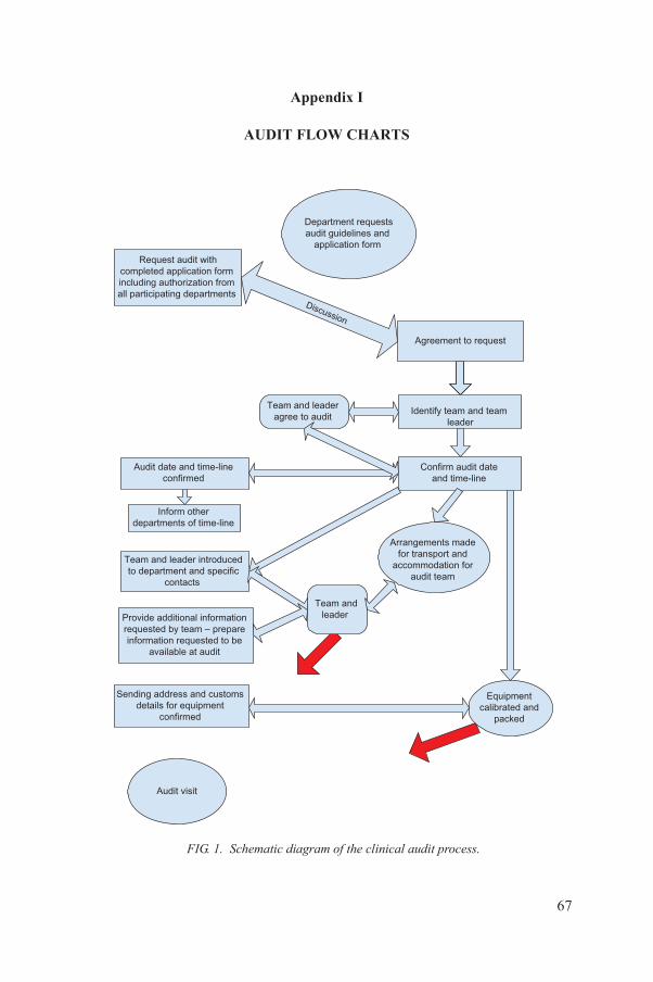
Appendix I
AUDIT FLOW CHARTS
Request audit with
completed application form
including authorization from
all participating departments
Agreement to request
Identify team and team
leader
Team and leader introduced
to department and specific
contacts
Team and leader
agree to audit
Confirm audit date
and time-line
Team and
leader
Audit date and time-line
confirmed
Department requests
audit guidelines and
application form
Provide additional information
requested by team – prepare
information requested to be
available at audit
Inform other
departments of time-line
Arrangements made
for transport and
accommodation for
audit team
Equipment
calibrated and
packed
Sending address and customs
details for equipment
confirmed
Discussion
67
Audit visit
FIG. 1. Schematic diagram of the clinical audit process.
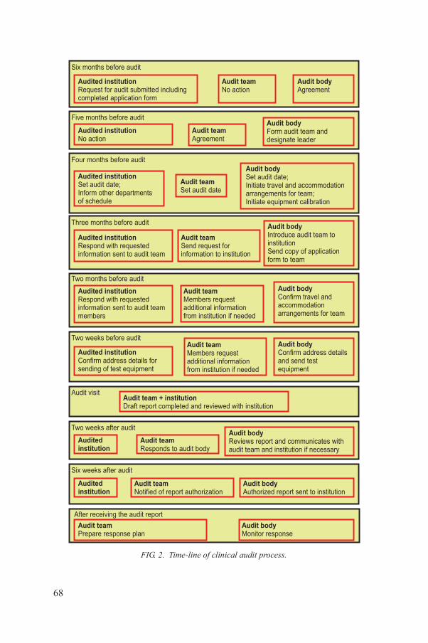
Six months before audit
Audit visit
Six weeks after audit
Two weeks after audit
Two weeks before audit
Two months before audit
Three months before audit
Four months before audit
Five months before audit
Audited institution
Request for audit submitted includingcompleted application form
Audited institution
Set audit date;Inform other departmentsof schedule
Audited institution
Respond with requestedinformation sent to audit team members
Audit team + institution
Draft report completed and reviewed with institution
Audited
institution
Audited
institution
Audit body
Form audit team anddesignate leader
Audit team
Members request additional information from institution if needed
Audit team
Members request additional information from institution if needed
Audit team
Send request forinformation to institution
Audit team
Set audit date
Audit team
Agreement
Audit body
Confirm address detailsand send testequipment
Audit body
Confirm travel and accommodationarrangements for team
Audit body
Introduce audit team to institutionSend copy of application form to team
Audit body
Set audit date;Initiate travel and accommodationarrangements for team; Initiate equipment calibration
Audit body
Authorized report sent to institution
Audit body
Reviews report and communicates with audit team and institution if necessary
Audit team
Notified of report authorization
Audit team
Responds to audit body
Audited institution
Confirm address details for sending of test equipment
Audited institution
Respond with requestedinformation sent to audit team
Audited institution
No action
Audit team
No actionAudit body
Agreement
68
After receiving the audit report
Audit body
Monitor responseAudit team
Prepare response plan
FIG. 2. Time-line of clinical audit process.
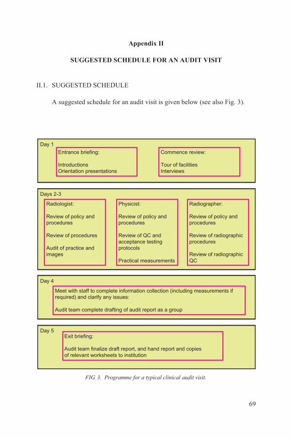
Appendix II
SUGGESTED SCHEDULE FOR AN AUDIT VISIT
II.1. SUGGESTED SCHEDULE
A suggested schedule for an audit visit is given below (see also Fig. 3).
Day 1
Entrance briefing:
Introductions
Orientation presentations
Commence review:
Tour of facilities
Interviews
Day 5
Day 4
Days 2-3
Exit briefing:
Meet with staff to complete information collection (including measurements if
required) and clarify any issues:
Audit team complete drafting of audit report as a group
Radiologist:
Review of policy and
procedures
Review of procedures
Audit of practice and
images
Physicist:
Review of policy and
procedures
Review of QC and
acceptance testing
protocols
Practical measurements
Radiographer:
Review of policy and
procedures
Review of radiographic
procedures
Review of radiographic
QC
69
Audit team finalize draft report, and hand report and copies
of relevant worksheets to institution
FIG. 3. Programme for a typical clinical audit visit.
Day 1:
Morning: All audit team members and relevant facility/institution personnel are involved in:
—A meeting with key facility/institution personnel; —An entrance briefing; —A tour of the facilities.
End of morning: Group audit team discussion time: approximately 30 minutes. Afternoon: Individual team members to meet with their departmental counterparts and other staff for discussions and a review.End of afternoon: Group audit team discussion time: approximately one hour.
Day 2:
Morning: Team members continue with discipline-specific reviews. End of morning: Group audit team discussion time: approximately one hour. Afternoon: Team members continue with discipline-specific reviews.End of afternoon: Group audit team discussion time: approximately one hour.
Day 3:
Morning: Team members continue with discipline-specific reviews and clarify any issues.End of morning: Group audit team discussion time: approximately one hour.Afternoon: Group audit team commences drafting of audit report.
Day 4:
Morning: Group audit team continues with drafting of audit report. Team members meet with staff as necessary to clarify any issues.Afternoon: Group audit team completes drafting of audit report.
Day 5:
Morning: All audit team members and relevant facility/institution personnel are involved in an exit briefing and a discussion of the recommendations.
70
Afternoon: Group audit team to:
—Amend draft report as necessary;—Give report and copies of audit documents to facility/institution authorities.
Audit team departs.

II.2. REVIEW PROCESS
All team members are to review the following Section 3 (in the main text) items with respect to their own discipline:
(a) Relevant policy and procedure documentation;(b) Compliance with policies and procedures;(c) Organizational chart, lines of authority, relevant quality manager(s), and
management commitment and support;(d) Relevant personnel issues, such as qualifications, employment, orientation,
training, professional supervision, continuing education, access to publications/information and communication;
(e) Relevant facility issues, such as equipment and available space, condition of equipment and work areas, meeting of patient requirements, meeting of general and radiation safety requirements, inventories and information backup;
(f) Handling of patient confidentiality, information, consent and feedback;(g) Adverse events recording and response.
The radiologist in the audit team is to address Section 4 items by reviewing: request forms; scheduling; imaging examinations; patient verification; images; viewing conditions; reports; contents and storage of patient files; report communication; radiologist QA activities such as audit, multidisciplinary meetings and attendance at conferences; nursing support; training and facilities for contrast use, sedation, anaesthesia, monitoring and resuscitation; and infection control and waste management.
The radiographer in the audit team is to address Section 4 and 5 items by reviewing: imaging examinations; patient verification; examination techniques and guidance charts; examination technique records; images and labelling; QC procedures and records; radiographer QA/quality improvement activities, such as image quality reviews; and CPD activities, such as attendance at meetings and conferences.
The physicist in the audit team is to address Section 5 items by reviewing: imaging examinations; technique guidance charts and technique records; image quality; QC procedures and records; QA/QC checks of equipment; staff, patient
71
and public radiation protection; room radiation protection and safety; personnel protective devices; patient dose audits; dosimetry data; and QA processes and process.
The physicist in the audit team also carries out QA testing of some of the diagnostic radiology systems, such as fluoroscopy, mammography, interventional radiology and CT systems.
Appendix III
AUDIT REPORT FORMAT: SUMMARY
III.1. TITLE PAGE
This title page should include the name and address of the facility and institution audited, the names of the audit team, identifying the leader, the date of the audit and any identification required by the auditing institution.
III.2. EXECUTIVE SUMMARY
The summary will not exceed three pages in length, should indicate the purpose of the audit and should comment on the extent to which the facility/institution has met the criteria for good practice as outlined in the guidelines, and summary of main recommendations.
III.3. RECOMMENDATIONS
This section of the report will be prepared for three distinct audiences:
(1) The audited facility/institution: These recommendations should address minor or major problems potentially resolvable by the institution.
(2) External organizations (e.g. government agencies): These recommendations should address major problems that may require intervention from outside the institution for resolution and other recommendations that should be brought to the attention of the government.
(3) The auditing body: These recommendations should address issues within the authority of the auditing body, for example, the need for additional training of auditors. (These recommendations will not be distributed outside of the auditing body.)
All recommendations should be prioritized in order of importance as
72
determined by the audit team. Any issues involving serious safety issues should be amongst the highest priority issues and be appropriately highlighted.
While it is the role of the audit team to identify areas for improvement of the services provided by the institution, it is not the responsibility of the auditing body to rectify any deficiencies identified. Rather, its role is to facilitate improvement and learning.
III.4. MAIN BODY
The main body of the report will include:
(a) The names of the individuals involved with the audit:—Those requesting the audit, —The heads of the audited departments, —The contact persons for the audit;
(b) The objectives of the audit as proposed by the facility/institution;(c) A description of audit activities and findings in the following areas:
—Quality management procedures and infrastructure,—Patient related procedures,—Equipment related procedures;
(d) Conclusions;(e) Annexes:
—A full list of individuals interviewed during the audit,—The application of the facility/institution,—Any other documents relevant to the audit.
73

Appendix IV
AUDIT REPORT FORMS
The completed audit report forms, which follow the issues outlined in Sections 3–6 of these guidelines, will form the body of the report.
The key for this appendix is as follows:
Y – Yes (i.e. available, performed or adequate);NI – Needs improvement;N – No (i.e. not available, not performed or not adequate);NA – Not applicable.
Note that these forms are for field use and relate to appropriate sections as indicated. For example, Form 3.3 relates to Section 3 (Quality management procedures and infrastructure), specifically Section 3.1 (Principles and criteria of good practice) should be read with Section 3.2 (Audit programme), which suggests approaches to verifying Section 3.1, the results of this are then recorded in Form 3.3. Note that the subheading notation is consistent throughout Sections 3.1 and 3.2 and also in Form 3.3 to allow ease of use. A similar approach is used for Forms 4.3, 5.3 and 6.3, which relate, respectively, to Sections 4 (Patient related procedures), 5 (Technical procedures) and 6 (Education, training and research programmes). Soft copies of these forms are available for optimal field application.
74
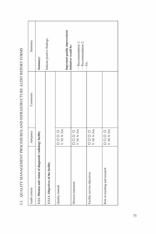
MA
NA
GE
ME
NT
PR
OC
ED
UR
ES
AN
D I
NF
RA
ST
RU
CT
UR
E A
UD
IT R
EP
OR
T F
OR
MS
A
dequ
acy
Com
men
tsSu
mm
ary
visi
on o
f d
iagn
osti
c ra
dio
logy
fac
ilit
ySu
mm
ary:
Indi
cate
pos
itive
fin
ding
s.
Impo
rtant
qua
lity
impr
ovem
ent
initi
ativ
es w
ould
be:
— R
ecom
men
dati
on 1
;—
Rec
omm
enda
tion
2;
— E
tc.
of
the
faci
lity
Y N
I N
NA
Y N
I N
NA
ctiv
es
Y N
I N
NA
d re
sear
ch
Y N
I N
NA
75
3.3.
QU
AL
ITY
Aud
it cr
iter
ia
3.3.
1. M
issi
on a
nd
3.3.
1.1.
Ob
ject
ives
Qua
lity
man
ual
Mis
sion
sta
tem
ent
Faci
lity
serv
ice
obje
Rol
e in
teac
hing
an
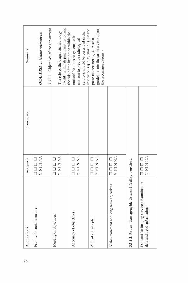
uctu
re
Y N
I N
NA
QU
AAD
RIL
guid
elin
e re
fere
nces
:
3.3.
1.1.
Obj
ectiv
es o
f th
e de
part
men
t
The
rol
e of
the
diag
nost
ic r
adio
logy
fa
cili
ty w
ithin
its
pare
nt in
stitu
tion
and
th
e ro
le o
f th
e in
stitu
tion
wit
hin
the
natio
nal h
ealth
car
e sy
stem
, or
its
mis
sion
to p
rovi
de r
adio
logi
cal
serv
ices
, sho
uld
be d
escr
ibed
in th
e in
stit
utio
n’s
qual
ity
man
ual.
(Cut
and
pa
ste
the
pert
inen
t QU
AA
DR
IL
guid
elin
e in
to th
e su
mm
ary
to s
uppo
rt
the
reco
mm
enda
tion
s.)
es
Y N
I N
NA
ives
Y
NI
N N
A
Y
NI
N N
A
d lo
ng te
rm o
bjec
tive
s
Y
NI
N N
A
ogra
ph
ic d
ata
and
fac
ilit
y w
ork
load
g se
rvic
es: E
xam
inat
ion
mat
ion
Y
NI
N N
A
A
dequ
acy
Com
men
tsSu
mm
ary
76
Faci
lity
fina
ncia
l str
Mee
ting
of o
bjec
tiv
Ade
quac
y of
obj
ect
Ann
ual a
ctiv
ity
plan
Vis
ion
stat
emen
t an
3.3.
1.2.
Pat
ien
t de
m
Dem
and
for
imag
inda
ta a
nd tr
end
info
r
Aud
it cr
iter
ia

Y N
I N
NA
ervi
ce d
ata
Y N
I N
NA
ans
Y N
I N
NA
s
Y
NI
N N
A
Y
NI
N N
A
agem
ent
syst
emSu
mm
ary:
Indi
cate
pos
itive
fin
ding
s.
Y
NI
N N
A
A
dequ
acy
Com
men
tsSu
mm
ary
77
Patie
nt a
ge d
ata
Em
erge
ncy/
urge
nt s
Staf
fing
dat
a an
d pl
Fund
ing
mec
hani
sm
Dev
elop
men
t pla
ns
3.3.
2. Q
ual
ity
man
Qua
lity
man
ager
Aud
it cr
iter
ia
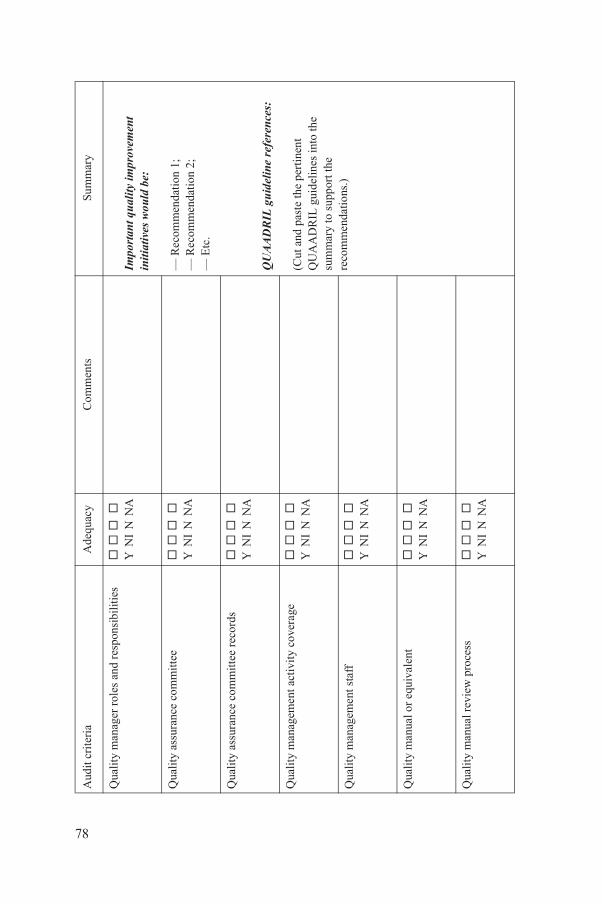
les
and
resp
onsi
bilit
ies
Y N
I N
NA
Impo
rtant
qua
lity
impr
ovem
ent
initi
ativ
es w
ould
be:
— R
ecom
men
dati
on 1
;—
Rec
omm
enda
tion
2;
— E
tc.
QU
AAD
RIL
guid
elin
e re
fere
nces
:
(Cut
and
pas
te th
e pe
rtin
ent
QU
AA
DR
IL g
uide
lines
into
the
sum
mar
y to
sup
port
the
reco
mm
enda
tions
.)
omm
itte
e
Y
NI
N N
A
omm
itte
e re
cord
s
Y
NI
N N
A
t act
ivit
y co
vera
ge
Y N
I N
NA
t sta
ff
Y N
I N
NA
quiv
alen
t
Y
NI
N N
A
ew p
roce
ss
Y
NI
N N
A
A
dequ
acy
Com
men
tsSu
mm
ary
78
Qua
lity
man
ager
ro
Qua
lity
ass
uran
ce c
Qua
lity
ass
uran
ce c
Qua
lity
man
agem
en
Qua
lity
man
agem
en
Qua
lity
man
ual o
r e
Qua
lity
man
ual r
evi
Aud
it cr
iter
ia
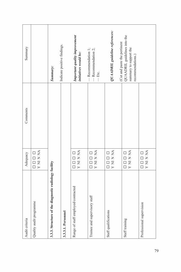
mm
e
Y
NI
N N
A
the
dia
gnos
tic
rad
iolo
gy f
acili
tySu
mm
ary:
Indi
cate
pos
itive
fin
ding
s.
Impo
rtant
qua
lity
impr
ovem
ent
initi
ativ
es w
ould
be:
— R
ecom
men
dati
on 1
;—
Rec
omm
enda
tion
2;
— E
tc.
QU
AAD
RIL
guid
elin
e re
fere
nces
:
(Cut
and
pas
te th
e pe
rtin
ent
QU
AA
DR
IL g
uide
lines
into
the
sum
mar
y to
sup
port
the
reco
mm
enda
tions
.)
oyed
/con
trac
ted
Y N
I N
NA
sory
sta
ff
Y
NI
N N
A
Y N
I N
NA
Y N
I N
NA
isio
n
Y
NI
N N
A
A
dequ
acy
Com
men
tsSu
mm
ary
79
Qua
lity
aud
it p
rogr
a
3.3.
3. S
tru
ctu
re o
f
3.3.
3.1.
Per
son
nel
Ran
ge o
f st
aff
empl
Tra
inee
and
sup
ervi
Staf
f qu
alif
icat
ions
Staf
f tr
aini
ng
Prof
essi
onal
sup
erv
Aud
it cr
iter
ia
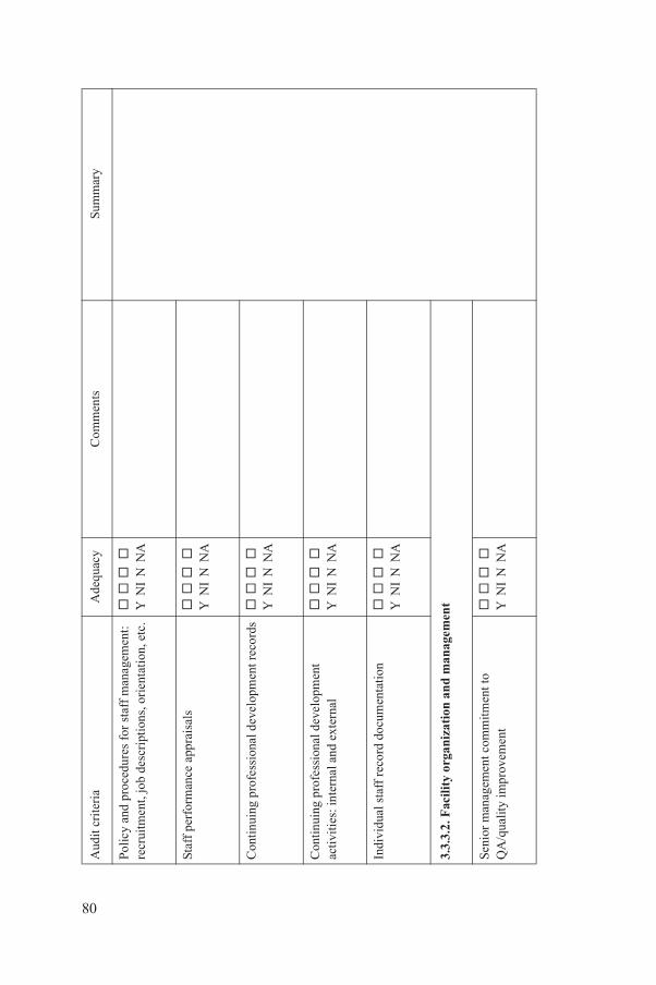
es f
or s
taff
man
agem
ent:
cr
ipti
ons,
ori
enta
tion
, etc
.
Y
NI
N N
A
ppra
isal
s
Y
NI
N N
A
onal
dev
elop
men
t rec
ords
Y N
I N
NA
onal
dev
elop
men
t nd
ext
erna
l
Y
NI
N N
A
rd d
ocum
enta
tion
Y N
I N
NA
aniz
atio
n a
nd
man
agem
ent
com
mitm
ent t
om
ent
Y N
I N
NA
A
dequ
acy
Com
men
tsSu
mm
ary
80
Polic
y an
d pr
oced
urre
crui
tmen
t, jo
b de
s
Staf
f pe
rfor
man
ce a
Con
tinu
ing
prof
essi
Con
tinu
ing
prof
essi
acti
viti
es: i
nter
nal a
Indi
vidu
al s
taff
rec
o
3.3.
3.2.
Fac
ility
org
Seni
or m
anag
emen
tQ
A/q
ualit
y im
prov
e
Aud
it cr
iter
ia

umen
tati
on
Y
NI
N N
A
t and
line
s of
aut
hori
ty
Y
NI
N N
A
atio
nal s
truc
ture
Y N
I N
NA
sta
ff n
umbe
rs a
nd r
oste
rs
Y
NI
N N
A
Y N
I N
NA
vacy
and
spe
cial
nee
ds
Y
NI
N N
A
A
dequ
acy
Com
men
tsSu
mm
ary
81
Qua
lity
man
ual d
oc
Org
aniz
atio
nal c
har
Inte
rfac
ilit
y or
gani
z
Hou
rs o
f op
erat
ion,
3.3.
3.3.
Pre
mis
es
Cle
anli
ness
Patie
nt c
omfo
rt, p
ri
Aud
it cr
iter
ia
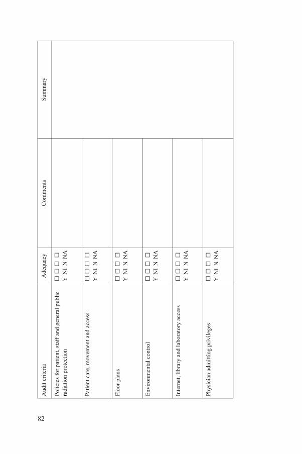
sta
ff a
nd g
ener
al p
ublic
Y
NI
N N
A
ent a
nd a
cces
s
Y
NI
N N
A
Y N
I N
NA
rol
Y N
I N
NA
labo
rato
ry a
cces
s
Y
NI
N N
A
pri
vile
ges
Y N
I N
NA
A
dequ
acy
Com
men
tsSu
mm
ary
82
Polic
ies
for
pati
ent,
radi
atio
n pr
otec
tion
Patie
nt c
are,
mov
em
Floo
r pl
ans
Env
iron
men
tal c
ont
Inte
rnet
, lib
rary
and
Phys
icia
n ad
mitt
ing
Aud
it cr
iter
ia
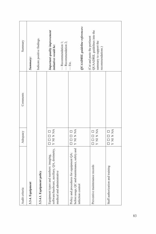
Sum
mar
y:
Indi
cate
pos
itive
fin
ding
s.
Impo
rtant
qua
lity
impr
ovem
ent
initi
ativ
es w
ould
be:
— R
ecom
men
dati
on 1
;—
Rec
omm
enda
tion
2;
— E
tc.
QU
AAD
RIL
guid
elin
e re
fere
nces
:
(Cut
and
pas
te th
e pe
rtin
ent
QU
AA
DR
IL g
uide
lines
into
the
sum
mar
y to
sup
port
the
reco
mm
enda
tions
.)
pol
icy
d nu
mbe
rs: i
mag
ing,
au
xilia
ry, Q
A, d
osim
etry
, st
rativ
e
Y N
I N
NA
es f
or e
quip
men
t QA
: d
mai
nten
ance
, saf
ety
and
Y
NI
N N
A
ance
rec
ords
Y N
I N
NA
nd tr
aini
ng
Y
NI
N N
A
A
dequ
acy
Com
men
tsSu
mm
ary
83
3.3.
4. E
quip
men
t
3.3.
4.1.
Eq
uip
men
t
Equ
ipm
ent t
ypes
an
soft
war
e/ha
rdw
are,
m
edic
al a
nd a
dmin
i
Polic
y an
d pr
oced
urbe
fore
usa
ge, Q
C a
nin
fect
ion
cont
rol
Prev
entiv
e m
aint
en
Staf
f au
thor
izat
ion
a
Aud
it cr
iter
ia
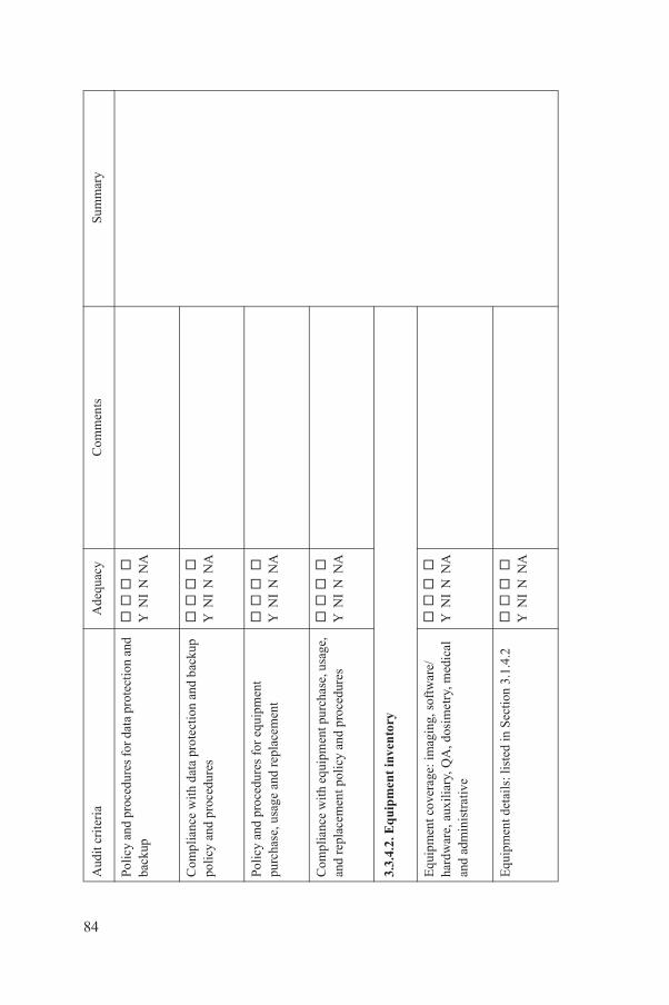
es f
or d
ata
prot
ecti
on a
nd
Y
NI
N N
A
ta p
rote
ctio
n an
d ba
ckup
es
Y N
I N
NA
es f
or e
quip
men
t r
epla
cem
ent
Y N
I N
NA
uipm
ent p
urch
ase,
usa
ge,
licy
and
pro
cedu
res
Y N
I N
NA
inve
nto
ry
e: im
agin
g, s
oftw
are/
QA
, dos
imet
ry, m
edic
al
Y
NI
N N
A
list
ed in
Sec
tion
3.1
.4.2
Y N
I N
NA
A
dequ
acy
Com
men
tsSu
mm
ary
84
Polic
y an
d pr
oced
urba
ckup
Com
plia
nce
wit
h da
polic
y an
d pr
oced
ur
Polic
y an
d pr
oced
urpu
rcha
se, u
sage
and
Com
plia
nce
wit
h eq
and
repl
acem
ent p
o
3.3.
4.2.
Eq
uip
men
t
Equ
ipm
ent c
over
agha
rdw
are,
aux
iliar
y,an
d ad
min
istr
ativ
e
Equ
ipm
ent d
etai
ls:
Aud
it cr
iter
ia
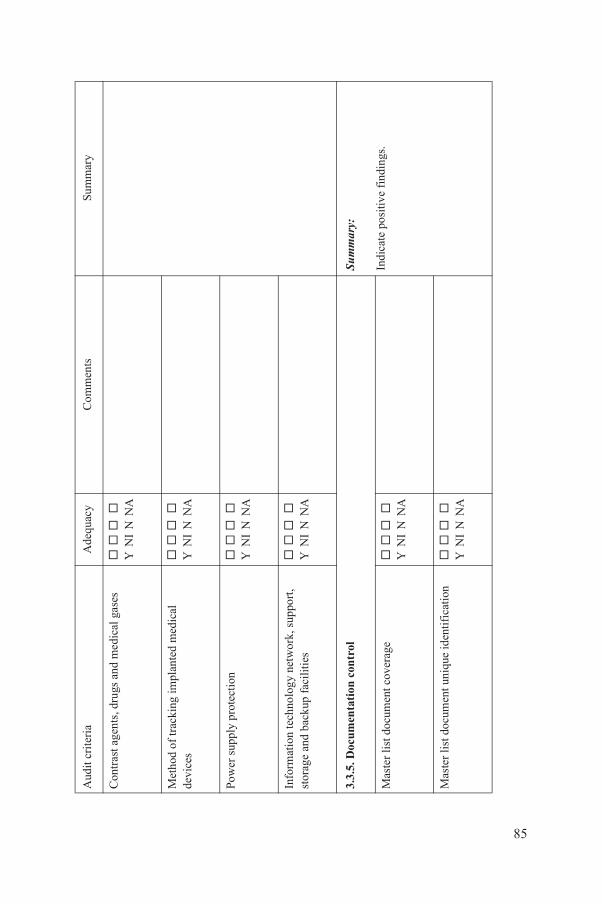
gs a
nd m
edic
al g
ases
Y N
I N
NA
impl
ante
d m
edic
al
Y N
I N
NA
ctio
n
Y
NI
N N
A
ogy
netw
ork,
sup
port
, fa
cilit
ies
Y N
I N
NA
on c
ontr
ol
Sum
mar
y:
Indi
cate
pos
itive
fin
ding
s.t c
over
age
Y N
I N
NA
t uni
que
iden
tifi
cati
on
Y
NI
N N
A
A
dequ
acy
Com
men
tsSu
mm
ary
85
Con
tras
t age
nts,
dru
Met
hod
of tr
acki
ng
devi
ces
Pow
er s
uppl
y pr
ote
Info
rmat
ion
tech
nol
stor
age
and
back
up
3.3.
5. D
ocum
enta
ti
Mas
ter
list
doc
umen
Mas
ter
list
doc
umen
Aud
it cr
iter
ia

ion
and
avai
labi
lity
Y N
I N
NA
Impo
rtant
qua
lity
impr
ovem
ent
initi
ativ
es w
ould
be:
— R
ecom
men
dati
on 1
;—
Rec
omm
enda
tion
2;
— E
tc.
QU
AAD
RIL
guid
elin
e re
fere
nces
:
(Cut
and
pas
te th
e pe
rtin
ent
QU
AA
DR
IL g
uide
lines
into
the
sum
mar
y to
sup
port
the
reco
mm
enda
tions
.)
te?
Y N
I N
NA
es f
or d
ocum
ent r
evie
w
Y
NI
N N
A
cum
ent r
evie
w/
and
pro
cedu
res
Y N
I N
NA
den
tial
ity,
fee
dba
ck a
nd
com
pla
ints
Sum
mar
y:
Indi
cate
pos
itive
fin
ding
s.es
for
pat
ient
Y
NI
N N
A
A
dequ
acy
Com
men
tsSu
mm
ary
86
Doc
umen
t dis
trib
ut
Doc
umen
ts u
p to
da
Polic
y an
d pr
oced
uran
d am
endm
ents
Com
plia
nce
wit
h do
amen
dmen
ts p
olic
y
3.3.
6. P
atie
nt
con
fi
Polic
y an
d pr
oced
urco
nfid
entia
lity
Aud
it cr
iter
ia
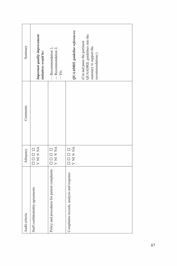
agr
eem
ents
Y N
I N
NA
Impo
rtant
qua
lity
impr
ovem
ent
initi
ativ
es w
ould
be:
— R
ecom
men
dati
on 1
;—
Rec
omm
enda
tion
2;
— E
tc.
QU
AAD
RIL
guid
elin
e re
fere
nces
:
(Cut
and
pas
te th
e pe
rtin
ent
QU
AA
DR
IL g
uide
lines
into
the
sum
mar
y to
sup
port
the
reco
mm
enda
tions
.)
es f
or p
atie
nt c
ompl
aint
s
Y
NI
N N
A
, ana
lysi
s an
d re
spon
se
Y
NI
N N
A
A
dequ
acy
Com
men
tsSu
mm
ary
87
Staf
f co
nfid
enti
alit
y
Polic
y an
d pr
oced
ur
Com
plai
nts
reco
rds
Aud
it cr
iter
ia
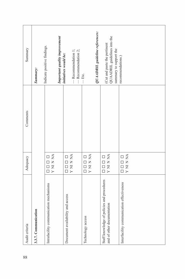
ion
Sum
mar
y:
Indi
cate
pos
itive
fin
ding
s.
Impo
rtant
qua
lity
impr
ovem
ent
initi
ativ
es w
ould
be:
— R
ecom
men
dati
on 1
;—
Rec
omm
enda
tion
2;
— E
tc.
QU
AAD
RIL
guid
elin
e re
fere
nces
:
(Cut
and
pas
te th
e pe
rtin
ent
QU
AA
DR
IL g
uide
lines
into
the
sum
mar
y to
sup
port
the
reco
mm
enda
tions
.)
nica
tion
mec
hani
sms
Y N
I N
NA
ity
and
acce
ss
Y
NI
N N
A
Y N
I N
NA
polic
ies
and
proc
edur
es
enta
tion
Y
NI
N N
A
nica
tion
eff
ecti
vene
ss
Y
NI
N N
A
A
dequ
acy
Com
men
tsSu
mm
ary
88
3.3.
7. C
omm
uni
cat
Intr
afac
ilit
y co
mm
u
Doc
umen
t ava
ilab
il
Tech
nolo
gy a
cces
s
Staf
f kn
owle
dge
of
and
of o
ther
doc
um
Inte
rfac
ilit
y co
mm
u
Aud
it cr
iter
ia
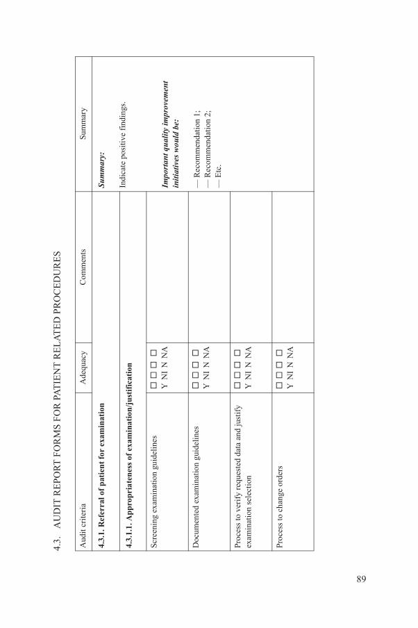
PO
RT
FO
RM
S F
OR
PA
TIE
NT
RE
LA
TE
D P
RO
CE
DU
RE
S
Ade
quac
yC
omm
ents
Sum
mar
y
atie
nt
for
exam
inat
ion
Sum
mar
y:
Indi
cate
pos
itive
fin
ding
s.
Impo
rtant
qua
lity
impr
ovem
ent
initi
ativ
es w
ould
be:
— R
ecom
men
dati
on 1
;—
Rec
omm
enda
tion
2;
— E
tc.
ten
ess
of e
xam
inat
ion/
just
ific
atio
n
ion
guid
elin
es
Y
NI
N N
A
natio
n gu
idel
ines
Y N
I N
NA
ques
ted
data
and
just
ify
on
Y N
I N
NA
rder
s
Y
NI
N N
A
89
4.3.
AU
DIT
RE
Aud
it cr
iter
ia
4.3.
1. R
efer
ral o
f p
4.3.
1.1.
Ap
prop
ria
Scre
enin
g ex
amin
at
Doc
umen
ted
exam
i
Proc
ess
to v
erif
y re
exam
inat
ion
sele
cti
Proc
ess
to c
hang
e o
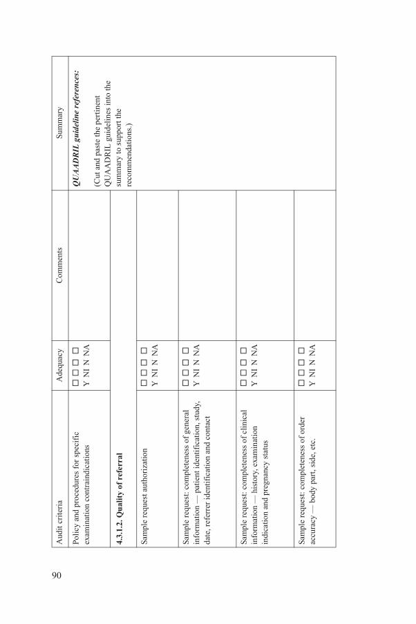
es f
or s
peci
fic
ndic
atio
ns
Y N
I N
NA
QU
AAD
RIL
guid
elin
e re
fere
nces
:
(Cut
and
pas
te th
e pe
rtin
ent
QU
AA
DR
IL g
uide
lines
into
the
sum
mar
y to
sup
port
the
reco
mm
enda
tions
.)
refe
rral
oriz
atio
n
Y
NI
N N
A
plet
enes
s of
gen
eral
nt
iden
tifi
cati
on, s
tudy
, ic
atio
n an
d co
ntac
t
Y N
I N
NA
plet
enes
s of
clin
ical
ry
, exa
min
atio
n an
cy s
tatu
s
Y N
I N
NA
plet
enes
s of
ord
er
art,
side
, etc
.
Y
NI
N N
A
Ade
quac
yC
omm
ents
Sum
mar
y
90
Polic
y an
d pr
oced
urex
amin
atio
n co
ntra
i
4.3.
1.2.
Qu
alit
y of
Sam
ple
requ
est a
uth
Sam
ple
requ
est:
com
info
rmat
ion
— p
atie
date
, ref
erre
r id
enti
f
Sam
ple
requ
est:
com
info
rmat
ion
— h
isto
indi
catio
n an
d pr
egn
Sam
ple
requ
est:
com
accu
racy
— b
ody
p
Aud
it cr
iter
ia
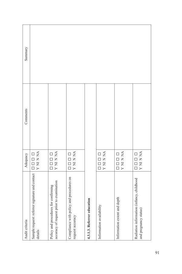
rrer
sig
natu
re a
nd c
onta
ct
Y
NI
N N
A
es f
or c
onfi
rmin
g p
rior
to e
xam
inat
ion
Y N
I N
NA
licy
and
pro
cedu
res
on
Y N
I N
NA
uca
tion
ilit
y
Y
NI
N N
A
nd d
epth
Y N
I N
NA
on (
infa
ncy,
chi
ldho
od
s)
Y
NI
N N
A
Ade
quac
yC
omm
ents
Sum
mar
y
91
Sam
ple
requ
est:
refe
deta
ils
Polic
y an
d pr
oced
urac
cura
cy o
f re
ques
t
Com
plia
nce
wit
h po
requ
est a
ccur
acy
4.3.
1.3.
Ref
erre
r ed
Info
rmat
ion
avai
lab
Info
rmat
ion
exte
nt a
Rad
iati
on in
form
ati
and
preg
nanc
y st
atu
Aud
it cr
iter
ia
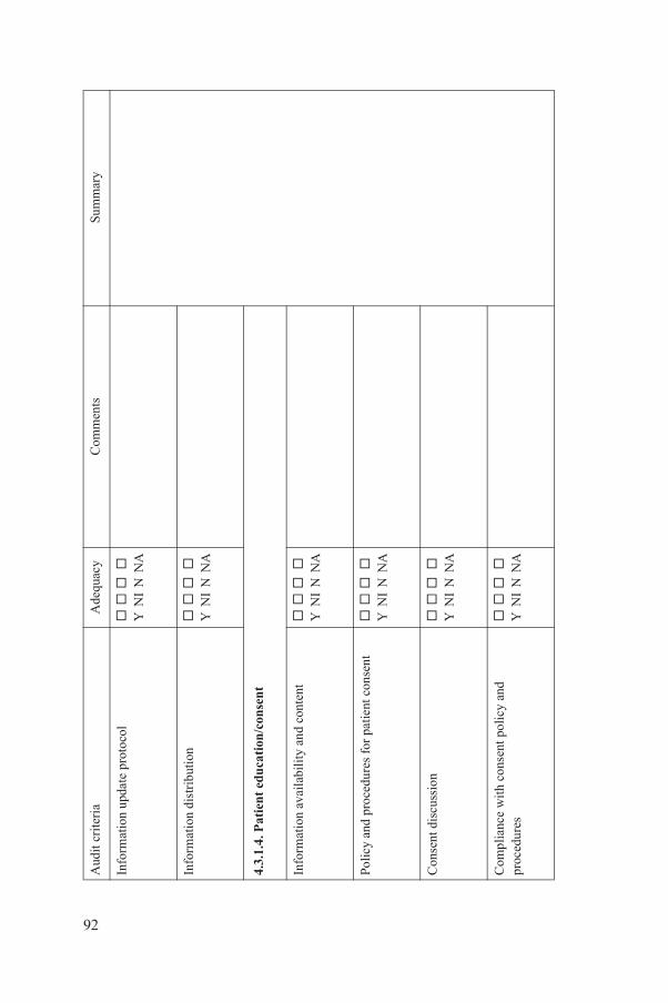
prot
ocol
Y N
I N
NA
tion
Y N
I N
NA
cati
on/c
onse
nt
ilit
y an
d co
nten
t
Y
NI
N N
A
es f
or p
atie
nt c
onse
nt
Y N
I N
NA
Y N
I N
NA
nsen
t pol
icy
and
Y N
I N
NA
Ade
quac
yC
omm
ents
Sum
mar
y
92
Info
rmat
ion
upda
te
Info
rmat
ion
dist
ribu
4.3.
1.4.
Pat
ien
t ed
u
Info
rmat
ion
avai
lab
Polic
y an
d pr
oced
ur
Con
sent
dis
cuss
ion
Com
plia
nce
wit
h co
proc
edur
es
Aud
it cr
iter
ia
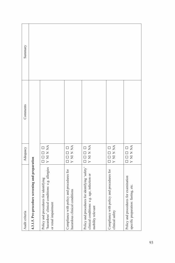
ure
scr
een
ing
and
pre
par
atio
n
es f
or id
enti
fyin
g c
ondi
tions
: e.g
. all
ergi
es
Y
NI
N N
A
licy
and
pro
cedu
res
for
ondi
tions
Y N
I N
NA
es fo
r ide
ntif
ying
‘saf
ety’
e.
g. a
ge, i
nfec
tion
or
Y N
I N
NA
licy
and
pro
cedu
res
for
Y N
I N
NA
es f
or e
xam
inat
ion
: fas
ting
, etc
.
Y
NI
N N
A
Ade
quac
yC
omm
ents
Sum
mar
y
93
4.3.
1.5.
Pre
-pro
ced
Polic
y an
d pr
oced
ur‘h
azar
dous
’ cl
inic
alor
ren
al im
pair
men
t
Com
plia
nce
wit
h po
haza
rdou
s cl
inic
al c
Polic
y an
d pr
oced
urcl
inic
al c
ondi
tion
s:
mob
ility
rel
evan
t
Com
plia
nce
wit
h po
clin
ical
saf
ety
Polic
y an
d pr
oced
ursp
ecif
ic p
repa
ratio
n
Aud
it cr
iter
ia
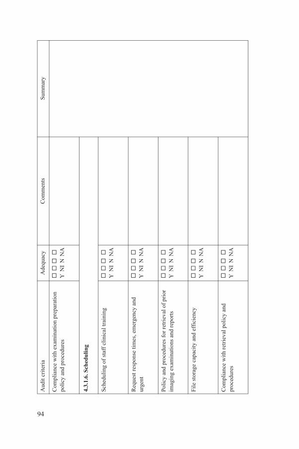
amin
atio
n pr
epar
atio
n es
Y N
I N
NA
clin
ical
trai
ning
Y N
I N
NA
mes
, em
erge
ncy
and
Y N
I N
NA
es f
or r
etri
eval
of
prio
r ns
and
rep
orts
Y
NI
N N
A
y an
d ef
fici
ency
Y
NI
N N
A
trie
val p
olic
y an
d
Y
NI
N N
A
Ade
quac
yC
omm
ents
Sum
mar
y
94
Com
plia
nce
wit
h ex
polic
y an
d pr
oced
ur
4.3.
1.6.
Sch
edu
ling
Sche
duli
ng o
f st
aff
Req
uest
res
pons
e ti
urge
nt
Polic
y an
d pr
oced
urim
agin
g ex
amin
atio
File
sto
rage
cap
acit
Com
plia
nce
wit
h re
proc
edur
es
Aud
it cr
iter
ia
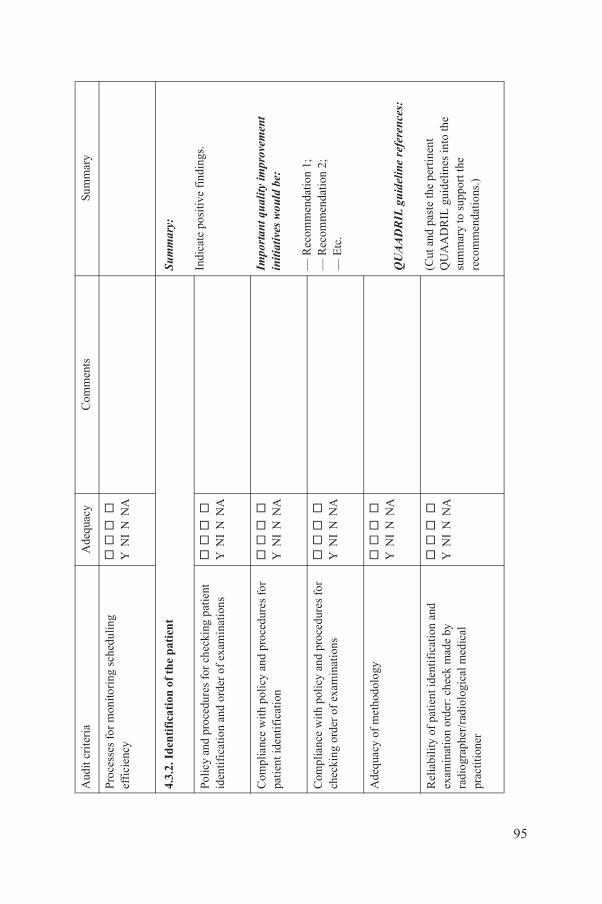
orin
g sc
hedu
ling
Y N
I N
NA
of
the
pat
ien
tSu
mm
ary:
Indi
cate
pos
itive
fin
ding
s.
Impo
rtant
qua
lity
impr
ovem
ent
initi
ativ
es w
ould
be:
— R
ecom
men
dati
on 1
;—
Rec
omm
enda
tion
2;
— E
tc.
QU
AAD
RIL
guid
elin
e re
fere
nces
:
(Cut
and
pas
te th
e pe
rtin
ent
QU
AA
DR
IL g
uide
lines
into
the
sum
mar
y to
sup
port
the
reco
mm
enda
tions
.)
es f
or c
heck
ing
patie
nt
rder
of
exam
inat
ions
Y N
I N
NA
licy
and
pro
cedu
res
for
n
Y
NI
N N
A
licy
and
pro
cedu
res
for
xam
inat
ions
Y
NI
N N
A
dolo
gy
Y N
I N
NA
t ide
ntif
icat
ion
and
chec
k m
ade
by
gica
l med
ical
Y N
I N
NA
Ade
quac
yC
omm
ents
Sum
mar
y
95
Proc
esse
s fo
r m
onit
effi
cien
cy
4.3.
2. I
den
tifi
cati
on
Polic
y an
d pr
oced
urid
entif
icat
ion
and
o
Com
plia
nce
wit
h po
pati
ent i
dent
ific
atio
Com
plia
nce
wit
h po
chec
king
ord
er o
f e
Ade
quac
y of
met
ho
Rel
iabi
lity
of
pati
enex
amin
atio
n or
der:
ra
diog
raph
er/r
adio
lopr
actit
ione
r
Aud
it cr
iter
ia
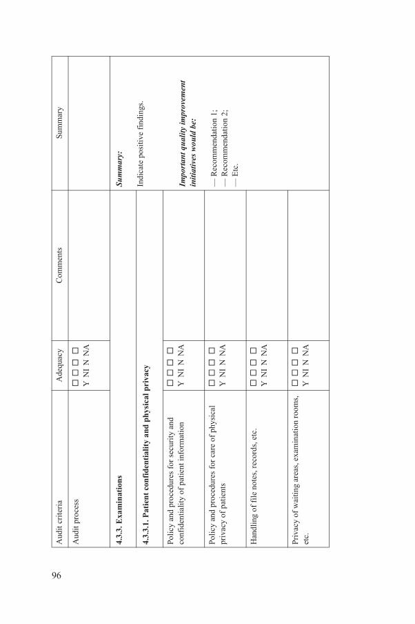
Y N
I N
NA
sSu
mm
ary:
Indi
cate
pos
itive
fin
ding
s.
Impo
rtant
qua
lity
impr
ovem
ent
initi
ativ
es w
ould
be:
— R
ecom
men
dati
on 1
;—
Rec
omm
enda
tion
2;
— E
tc.
fide
ntia
lity
and
ph
ysic
al p
riva
cy
es f
or s
ecur
ity
and
tien
t inf
orm
atio
n
Y
NI
N N
A
es f
or c
are
of p
hysi
cal
Y N
I N
NA
es, r
ecor
ds, e
tc.
Y N
I N
NA
reas
, exa
min
atio
n ro
oms,
Y
NI
N N
A
Ade
quac
yC
omm
ents
Sum
mar
y
96
Aud
it pr
oces
s
4.3.
3. E
xam
inat
ion
4.3.
3.1.
Pat
ien
t co
n
Polic
y an
d pr
oced
urco
nfid
entia
lity
of p
a
Polic
y an
d pr
oced
urpr
ivac
y of
pat
ient
s
Han
dlin
g of
fil
e no
t
Priv
acy
of w
aitin
g a
etc.
Aud
it cr
iter
ia

roto
cols
Y
NI
N N
AQ
UAA
DRI
L gu
idel
ine
refe
renc
es:
(Cut
and
pas
te th
e pe
rtin
ent
QU
AA
DR
IL g
uide
lines
into
the
sum
mar
y to
sup
port
the
reco
mm
enda
tions
.)
licy
and
pro
cedu
res
for
Y N
I N
NA
licy
and
pro
cedu
res
for
Y N
I N
NA
chn
iqu
es
s im
agin
g ex
amin
atio
ns:
amm
ogra
phy,
CT,
T
/CT,
imag
e gu
ided
et
c.
Y N
I N
NA
s fo
r ex
amin
atio
ns in
e
.g.,
paed
iatr
ic p
atie
nts
Y N
I N
NA
Ade
quac
yC
omm
ents
Sum
mar
y
97
Staf
f aw
aren
ess
of p
Com
plia
nce
wit
h po
conf
iden
tialit
y
Com
plia
nce
wit
h po
phys
ical
pri
vacy
4.3.
3.2.
Im
agin
g te
Prot
ocol
s fo
r va
riou
plai
n, f
luor
osco
py, m
ultr
asou
nd, M
RI,
PE
proc
edur
es, d
enta
l,
Polic
y an
d pr
otoc
olsp
ecia
l pop
ulat
ions
,
Aud
it cr
iter
ia
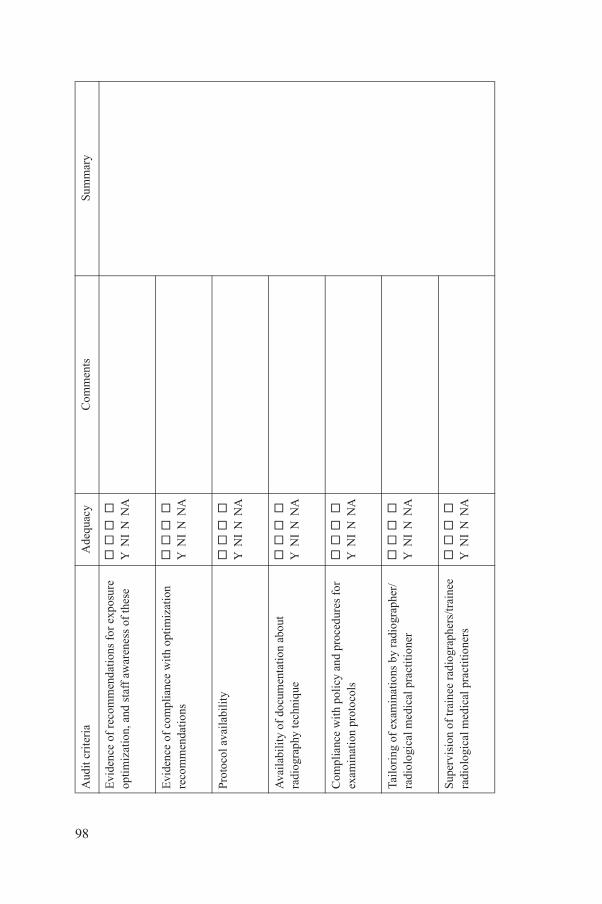
enda
tions
for
exp
osur
e af
f aw
aren
ess
of th
ese
Y N
I N
NA
ance
wit
h op
tim
izat
ion
Y N
I N
NA
Y
NI
N N
A
men
tati
on a
bout
ue
Y
NI
N N
A
licy
and
pro
cedu
res
for
ols
Y N
I N
NA
atio
ns b
y ra
diog
raph
er/
l pra
ctiti
oner
Y
NI
N N
A
ee r
adio
grap
hers
/trai
nee
l pra
ctiti
oner
s
Y
NI
N N
A
Ade
quac
yC
omm
ents
Sum
mar
y
98
Evi
denc
e of
rec
omm
optim
izat
ion,
and
st
Evi
denc
e of
com
pli
reco
mm
enda
tion
s
Prot
ocol
ava
ilab
ilit
y
Ava
ilab
ilit
y of
doc
ura
diog
raph
y te
chni
q
Com
plia
nce
wit
h po
exam
inat
ion
prot
oc
Tail
orin
g of
exa
min
radi
olog
ical
med
ica
Supe
rvis
ion
of tr
ain
radi
olog
ical
med
ica
Aud
it cr
iter
ia

ng a
nd r
ecor
d ls
of
mac
hine
, t,
exam
inat
ion
fact
ors,
, a
dver
se r
eact
ions
, n,
etc
.
Y N
I N
NA
es f
or c
ontr
ol o
f
Y
NI
N N
A
fect
ion
cont
rol p
olic
y an
d es
s, w
aste
, sha
rps,
ase
ptic
es
, ste
rili
zati
on, e
tc.
Y N
I N
NA
re, p
atie
nt
seda
tion
/ana
esth
esia
and
con
tras
t ag
ents
l car
e: m
onit
orin
g,
tc.
Y N
I N
NA
nvir
onm
ent
Y N
I N
NA
Ade
quac
yC
omm
ents
Sum
mar
y
99
Exa
min
atio
n la
bell
ico
mpl
eten
ess:
det
aira
diog
raph
er, p
atie
nco
ntra
st, l
imit
atio
nsse
datio
n, m
edic
atio
Polic
y an
d pr
oced
urin
fect
ions
Com
plia
nce
wit
h in
proc
edur
es: c
lean
lin
and
ster
ile te
chni
qu
4.3.
3.3.
Cli
nica
l ca
Prov
isio
n of
cli
nica
intr
aven
ous
lines
, e
Safe
ty o
f ph
ysic
al e
Aud
it cr
iter
ia
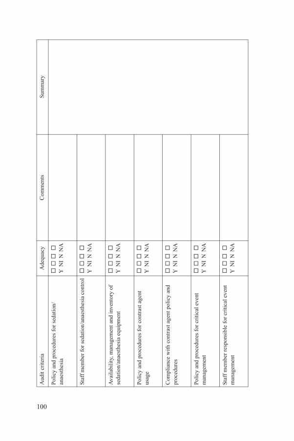
es f
or s
edat
ion/
Y N
I N
NA
dati
on/a
naes
thes
ia c
ontr
ol
Y
NI
N N
A
emen
t and
inve
ntor
y of
e
quip
men
t
Y
NI
N N
A
es f
or c
ontr
ast a
gent
Y
NI
N N
A
ntra
st a
gent
pol
icy
and
Y N
I N
NA
es f
or c
riti
cal e
vent
Y
NI
N N
A
nsib
le f
or c
riti
cal e
vent
Y
NI
N N
A
Ade
quac
yC
omm
ents
Sum
mar
y
100
Polic
y an
d pr
oced
uran
aest
hesi
a
Staf
f m
embe
r fo
r se
Ava
ilab
ilit
y, m
anag
seda
tion/
anae
sthe
sia
Polic
y an
d pr
oced
urus
age
Com
plia
nce
wit
h co
proc
edur
es
Polic
y an
d pr
oced
urm
anag
emen
t
Staf
f m
embe
r re
spo
man
agem
ent
Aud
it cr
iter
ia
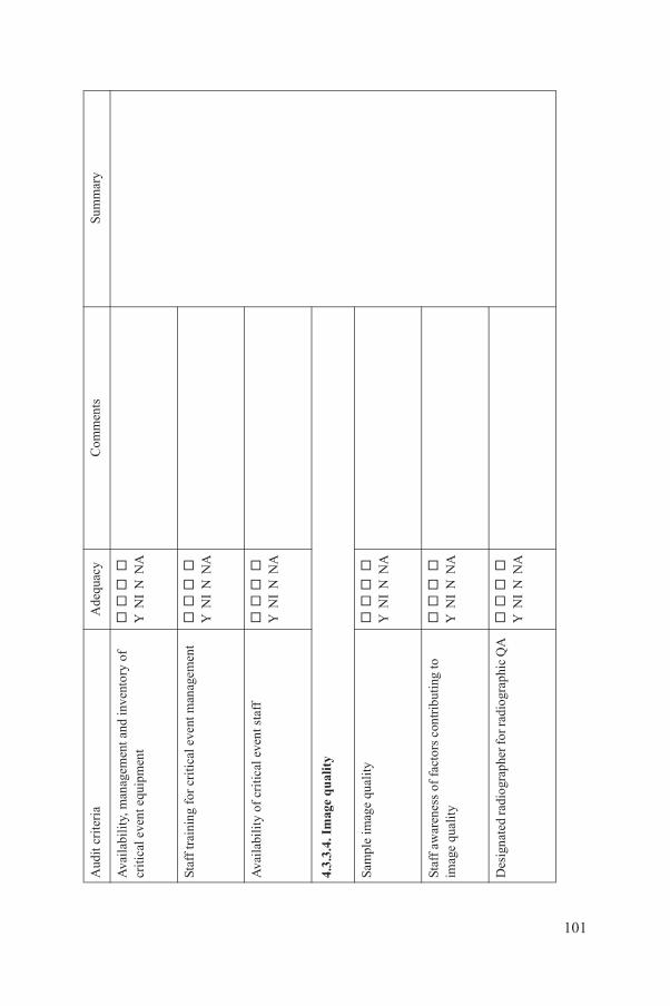
emen
t and
inve
ntor
y of
en
t
Y
NI
N N
A
tica
l eve
nt m
anag
emen
t
Y
NI
N N
A
al e
vent
sta
ff
Y N
I N
NA
lity ty
Y N
I N
NA
acto
rs c
ontr
ibut
ing
to
Y N
I N
NA
pher
for r
adio
grap
hic
QA
Y
NI
N N
A
Ade
quac
yC
omm
ents
Sum
mar
y
101
Ava
ilab
ilit
y, m
anag
criti
cal e
vent
equ
ipm
Staf
f tr
aini
ng f
or c
ri
Ava
ilab
ilit
y of
cri
tic
4.3.
3.4.
Im
age
qu
a
Sam
ple
imag
e qu
ali
Staf
f aw
aren
ess
of f
imag
e qu
alit
y
Des
igna
ted
radi
ogra
Aud
it cr
iter
ia
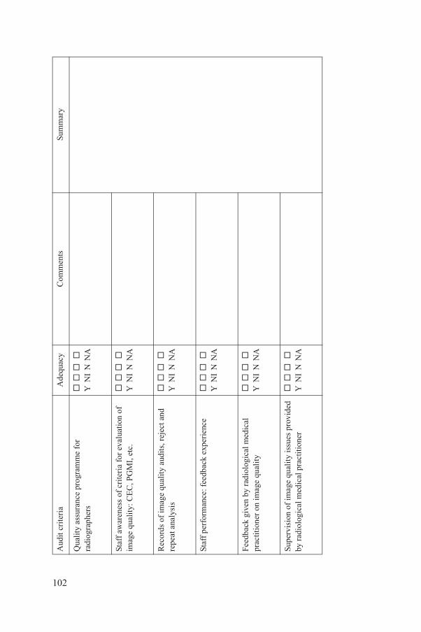
rogr
amm
e fo
r
Y
NI
N N
A
rite
ria
for
eval
uati
on o
f , P
GM
I, e
tc.
Y N
I N
NA
ualit
y au
dits
, rej
ect a
nd
Y N
I N
NA
eedb
ack
expe
rien
ce
Y
NI
N N
A
radi
olog
ical
med
ical
e
qual
ity
Y N
I N
NA
e qu
alit
y is
sues
pro
vide
d ic
al p
ract
itio
ner
Y N
I N
NA
Ade
quac
yC
omm
ents
Sum
mar
y
102
Qua
lity
ass
uran
ce p
radi
ogra
pher
s
Staf
f aw
aren
ess
of c
imag
e qu
alit
y: C
EC
Rec
ords
of
imag
e q
repe
at a
naly
sis
Staf
f pe
rfor
man
ce: f
Feed
back
giv
en b
y pr
actit
ione
r on
imag
Supe
rvis
ion
of im
agby
rad
iolo
gica
l med
Aud
it cr
iter
ia

rep
ort
Sum
mar
y:
Indi
cate
pos
itive
fin
ding
s.
Impo
rtant
qua
lity
impr
ovem
ent
initi
ativ
es w
ould
be:
— R
ecom
men
dati
on 1
;—
Rec
omm
enda
tion
2;
— E
tc.
QU
AAD
RIL
guid
elin
e re
fere
nces
:
(Cut
and
pas
te th
e pe
rtin
ent
QU
AA
DR
IL g
uide
lines
into
the
sum
mar
y to
sup
port
the
reco
mm
enda
tions
.)
exa
min
atio
ns a
nd re
port
s
Y
NI
N N
A
amin
atio
n re
port
: ref
erre
r, g
phys
icia
n id
entif
icat
ion,
re
and
dat
e
Y N
I N
NA
amin
atio
n re
port
:an
d ex
amin
atio
n
Y
NI
N N
A
amin
atio
n re
port
: s,
cor
rela
tion
and
Y N
I N
NA
amin
atio
n re
port
: adv
ice
igat
ions
Y
NI
N N
A
amin
atio
n re
port
: e
effe
cts,
inci
dent
s, e
tc.
Y N
I N
NA
Ade
quac
yC
omm
ents
Sum
mar
y
103
4.3.
4. T
he im
agin
g
Ava
ilab
ilit
y of
pri
orat
tim
e of
rep
orti
ng
Com
plet
enes
s of
ex
pati
ent a
nd re
port
inan
d re
port
er s
igna
tu
Com
plet
enes
s of
ex
clin
ical
indi
cati
ons
Com
plet
enes
s of
ex
exam
inat
ion
find
ing
rele
vanc
e
Com
plet
enes
s of
ex
abou
t fur
ther
inve
st
Com
plet
enes
s of
ex
reco
rdin
g of
adv
ers
Aud
it cr
iter
ia
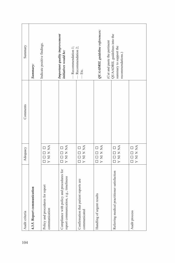
unic
atio
n Su
mm
ary:
Indi
cate
pos
itive
fin
ding
s.
Impo
rtant
qua
lity
impr
ovem
ent
initi
ativ
es w
ould
be:
— R
ecom
men
dati
on 1
;—
Rec
omm
enda
tion
2;
— E
tc.
QU
AAD
RIL
guid
elin
e re
fere
nces
:
(Cut
and
pas
te th
e pe
rtin
ent
QU
AA
DR
IL g
uide
lines
into
the
sum
mar
y to
sup
port
the
reco
mm
enda
tions
.)
es f
or r
epor
t
Y
NI
N N
A
licy
and
pro
cedu
res
for
on, e
.g.,
tim
elin
ess
Y N
I N
NA
atie
nt r
epor
ts a
re
Y N
I N
NA
resu
lts
Y N
I N
NA
ract
itio
ner
satis
fact
ion
Y N
I N
NA
Y N
I N
NA
Ade
quac
yC
omm
ents
Sum
mar
y
104
4.3.
5. R
epor
t co
mm
Polic
y an
d pr
oced
urco
mm
unic
atio
n
Com
plia
nce
wit
h po
repo
rt c
omm
unic
ati
Con
firm
atio
n th
at p
com
mun
icat
ed
Han
dlin
g of
urg
ent
Ref
erri
ng m
edic
al p
Aud
it pr
oces
s
Aud
it cr
iter
ia
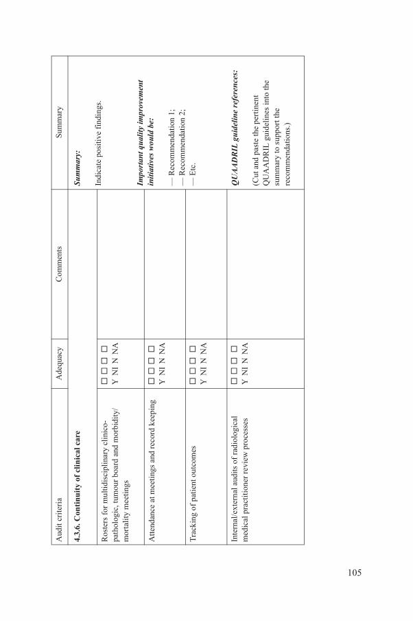
f cl
inic
al c
are
Sum
mar
y:
Indi
cate
pos
itive
fin
ding
s.
Impo
rtant
qua
lity
impr
ovem
ent
initi
ativ
es w
ould
be:
— R
ecom
men
dati
on 1
;—
Rec
omm
enda
tion
2;
— E
tc.
QU
AAD
RIL
guid
elin
e re
fere
nces
:
(Cut
and
pas
te th
e pe
rtin
ent
QU
AA
DR
IL g
uide
lines
into
the
sum
mar
y to
sup
port
the
reco
mm
enda
tions
.)
cipl
inar
y cl
inic
o-bo
ard
and
mor
bidi
ty/
Y N
I N
NA
ngs
and
reco
rd k
eepi
ng
Y
NI
N N
A
outc
omes
Y
NI
N N
A
dits
of
radi
olog
ical
r
evie
w p
roce
sses
Y N
I N
NA
Ade
quac
yC
omm
ents
Sum
mar
y
105
4.3.
6. C
onti
nuit
y o
Ros
ters
for
mul
tidis
path
olog
ic, t
umou
r m
orta
lity
mee
tings
Atte
ndan
ce a
t mee
ti
Tra
ckin
g of
pat
ient
Inte
rnal
/ext
erna
l au
med
ical
pra
ctit
ione
r
Aud
it cr
iter
ia
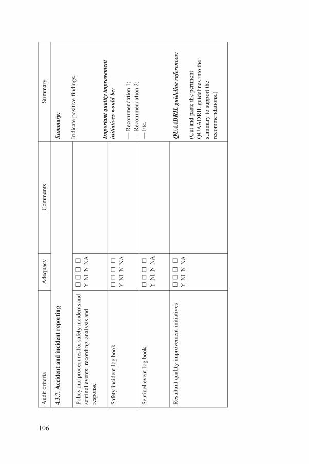
inci
den
t re
por
tin
gSu
mm
ary:
Indi
cate
pos
itive
fin
ding
s.
Impo
rtant
qua
lity
impr
ovem
ent
initi
ativ
es w
ould
be:
— R
ecom
men
dati
on 1
;—
Rec
omm
enda
tion
2;
— E
tc.
QU
AAD
RIL
guid
elin
e re
fere
nces
:
(Cut
and
pas
te th
e pe
rtin
ent
QU
AA
DR
IL g
uide
lines
into
the
sum
mar
y to
sup
port
the
reco
mm
enda
tions
.)
es fo
r saf
ety
inci
dent
s an
d rd
ing,
ana
lysi
s an
d
Y
NI
N N
A
book
Y
NI
N N
A
ook
Y N
I N
NA
prov
emen
t ini
tiat
ives
Y N
I N
NA
Ade
quac
yC
omm
ents
Sum
mar
y
106
4.3.
7. A
ccid
ent
and
Polic
y an
d pr
oced
urse
ntin
el e
vent
s: r
eco
resp
onse
Safe
ty in
cide
nt lo
g
Sent
inel
eve
nt lo
g b
Res
ulta
nt q
uali
ty im
Aud
it cr
iter
ia
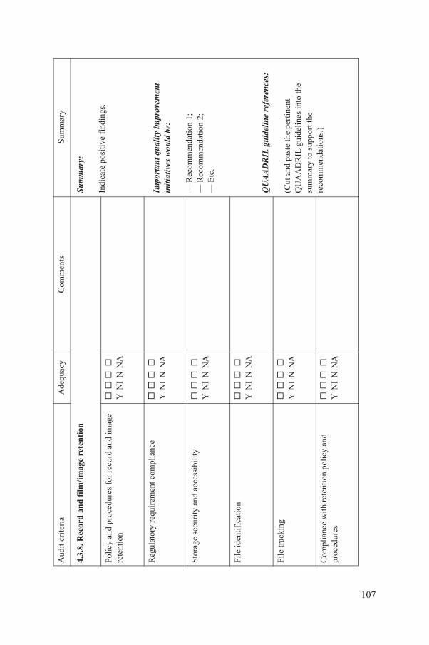
ilm
/im
age
rete
nti
onSu
mm
ary:
Indi
cate
pos
itive
fin
ding
s.
Impo
rtant
qua
lity
impr
ovem
ent
initi
ativ
es w
ould
be:
— R
ecom
men
dati
on 1
;—
Rec
omm
enda
tion
2;
— E
tc.
QU
AAD
RIL
guid
elin
e re
fere
nces
:
(Cut
and
pas
te th
e pe
rtin
ent
QU
AA
DR
IL g
uide
lines
into
the
sum
mar
y to
sup
port
the
reco
mm
enda
tions
.)
es f
or r
ecor
d an
d im
age
Y N
I N
NA
ent c
ompl
ianc
e
Y
NI
N N
A
acc
essi
bilit
y
Y
NI
N N
A
Y N
I N
NA
Y N
I N
NA
tent
ion
poli
cy a
nd
Y N
I N
NA
Ade
quac
yC
omm
ents
Sum
mar
y
107
4.3.
8. R
ecor
d a
nd
f
Polic
y an
d pr
oced
urre
tent
ion
Reg
ulat
ory
requ
irem
Stor
age
secu
rity
and
File
iden
tifi
catio
n
File
trac
king
Com
plia
nce
wit
h re
proc
edur
es
Aud
it cr
iter
ia
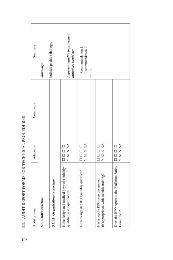
PO
RT
FO
RM
S F
OR
TE
CH
NIC
AL
PR
OC
ED
UR
ES
Ade
quac
yC
omm
ents
Sum
mar
y
re
Sum
mar
y:
Indi
cate
pos
itive
fin
ding
s.
Impo
rtant
qua
lity
impr
ovem
ent
initi
ativ
es w
ould
be:
— R
ecom
men
dati
on 1
;—
Rec
omm
enda
tion
2;
— E
tc.
onal
str
uctu
re
edic
al p
hysi
cist
sui
tabl
y en
ced?
Y N
I N
NA
O s
uita
bly
qual
ifie
d?
Y
NI
N N
A
een
desi
gnat
ed
h su
itab
le tr
aini
ng?
Y N
I N
NA
rt to
the
Rad
iati
on S
afet
y
Y
NI
N N
A
108
5.3.
AU
DIT
RE
Aud
it cr
iter
ia
5.3.
1. I
nfr
astr
uctu
5.3.
1.1.
Org
aniz
ati
Is th
e de
sign
ated
mqu
alif
ied
and
expe
ri
Is th
e de
sign
ated
RP
Has
a d
eput
y R
PO
b(i
f ap
prop
riat
e), w
it
Doe
s th
e R
PO
rep
oC
omm
ittee
?

Safe
ty C
omm
ittee
mee
t sp
onsi
ble
for t
he ra
diat
ion
inst
ituti
on/f
acili
ty, t
he
olic
y an
d it
s ev
alua
tion
?
Y N
I N
NA
QU
AAD
RIL
guid
elin
e re
fere
nces
:
(Cut
and
pas
te th
e pe
rtin
ent
QU
AA
DR
IL g
uide
lines
into
the
sum
mar
y to
sup
port
the
reco
mm
enda
tions
.)
man
ual a
vail
able
, doe
s it
r
elev
ant a
spec
ts o
f re
gula
rly
revi
ewed
and
is
ary?
Y N
I N
NA
ure
of r
adia
tion
saf
ety
Y N
I N
NA
dica
l phy
sici
st, i
n is
ing
QA
act
ivit
ies,
a
nd d
osim
etry
, cle
arly
tiv
e?
Y N
I N
NA
Ade
quac
yC
omm
ents
Sum
mar
y
109
Doe
s th
e R
adia
tion
re
gula
rly
and
is it
resa
fety
pol
icy
of th
e m
onito
ring
of
that
p
Is a
rad
iatio
n sa
fety
adeq
uate
ly d
escr
ibe
radi
atio
n sa
fety
, is
itit
upda
ted
as n
eces
s
Is th
e cu
rren
t str
uct
sati
sfac
tory
?
5.3.
1.2.
Per
son
nel
Is th
e ro
le o
f th
e m
edi
rect
ing
and
supe
rvra
diat
ion
prot
ectio
nde
scri
bed
and
oper
a
Aud
it cr
iter
ia
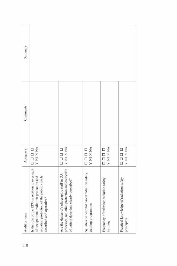
O in
rel
atio
n to
ove
rsig
ht
atio
n pr
otec
tion
and
o
f th
e pu
blic
cle
arly
tiv
e?
Y N
I N
NA
iogr
aphi
c st
aff
in Q
A
pro
tect
ion
and
colle
ctio
n c
lear
ly d
escr
ibed
?
Y N
I N
NA
bas
ed r
adia
tion
saf
ety
s
Y
NI
N N
A
her
radi
atio
n sa
fety
Y
NI
N N
A
of
radi
atio
n sa
fety
Y
NI
N N
A
Ade
quac
yC
omm
ents
Sum
mar
y
110
Is th
e ro
le o
f th
e R
Pof
occ
upat
iona
l rad
ira
diat
ion
prot
ectio
nde
scri
bed
and
oper
a
Are
the
dutie
s of
rad
proc
esse
s, r
adia
tion
of p
atie
nt d
ose
data
Sylla
bus
of h
ospi
tal
trai
ning
pro
gram
me
Freq
uenc
y of
ref
res
trai
ning
Prac
tical
kno
wle
dge
prin
cipl
es
Aud
it cr
iter
ia

nt s
upp
ort
ade
avai
labl
e fo
r Q
A a
nd
pro
tect
ion
acti
viti
es?
Y N
I N
NA
men
t ava
ilab
le?
Y N
I N
NA
ns ta
ken
with
in th
e
Y
NI
N N
A
ort e
vide
nt?
Y N
I N
NA
tion
atio
n cu
rren
t, av
aila
ble
adia
tion
safe
ty
olic
ies
and
proc
edur
es,
Y N
I N
NA
Ade
quac
yC
omm
ents
Sum
mar
y
111
5.3.
1.3.
Man
agem
e
Is s
uffi
cien
t tim
e m
safe
ty a
nd r
adia
tion
Is a
ppro
pria
te e
quip
Are
cor
rect
ive
acti
ora
diol
ogy
faci
lity?
Is m
anag
emen
t sup
p
5.3.
1.4.
Doc
umen
ta
Is f
acil
ity d
ocum
ent
and
acce
ssib
le f
or r
requ
irem
ents
, QA
pan
d do
sim
etry
?
Aud
it cr
iter
ia

otec
tion
an
d s
afet
ySu
mm
ary:
Indi
cate
pos
itive
fin
ding
s.
Impo
rtant
qua
lity
impr
ovem
ent
initi
ativ
es w
ould
be:
— R
ecom
men
dati
on 1
;—
Rec
omm
enda
tion
2;
— E
tc.
QU
AAD
RIL
guid
elin
e re
fere
nces
:
(Cut
and
pas
te th
e pe
rtin
ent
QU
AA
DR
IL g
uide
lines
into
the
sum
mar
y to
sup
port
the
reco
mm
enda
tions
.)
pos
ure
and
man
agem
ent p
olic
ies
l req
uire
men
ts o
r rel
evan
t
Y
NI
N N
A
ose
leve
ls f
rom
com
mon
at
ed a
nd c
ompa
red
with
nc
e le
vels
(D
RL
s)?
Y N
I N
NA
lici
es c
onsi
sten
t wit
h r
rele
vant
gui
deli
nes?
Y N
I N
NA
for
pre
gnan
cy s
uita
ble?
Y N
I N
NA
ancy
ade
quat
e?
Y
NI
N N
A
Ade
quac
yC
omm
ents
Sum
mar
y
112
5.3.
2. R
adia
tion
pr
5.3.
2.1.
Med
ical
ex
Are
risk
ass
essm
ent
cons
iste
nt w
ith
lega
guid
elin
es?
Are
typi
cal p
atie
nt d
exam
inat
ions
est
imre
leva
nt d
ose
refe
re
Are
opt
imiz
atio
n po
lega
l req
uire
men
ts o
Is th
e fa
cili
ty p
olic
y
Is s
igna
ge f
or p
regn
Aud
it cr
iter
ia

egna
ncy
stat
us s
uita
ble?
Y N
I N
NA
olic
y fo
r pr
egna
nt
Y N
I N
NA
fort
ers
and
care
rs in
si
sten
t wit
h le
gal
vant
gui
deli
nes?
Y N
I N
NA
ures
ava
ilabl
e fo
r p
aram
eter
s ap
prop
riat
e ts
?
Y N
I N
NA
erse
pat
ient
dos
e in
cide
nts
l req
uire
men
ts o
r rel
evan
t
Y
NI
N N
A
Ade
quac
yC
omm
ents
Sum
mar
y
113
Is v
erif
icat
ion
of p
r
Are
sta
ff a
war
e of
ppa
tien
ts?
Are
pol
icie
s fo
r co
mha
zard
ous
area
s co
nre
quir
emen
ts o
r re
le
Are
cha
rts
or p
roce
dad
just
men
t of
X r
ayfo
r pa
edia
tric
pat
ien
Are
pol
icie
s fo
r adv
cons
iste
nt w
ith
lega
guid
elin
es?
Aud
it cr
iter
ia
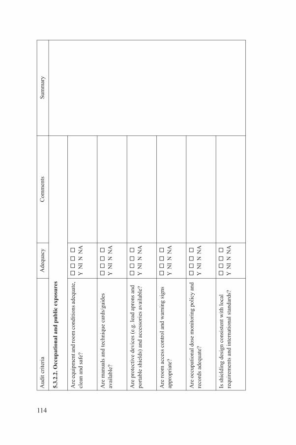
al a
nd
pub
lic e
xpos
ure
s
oom
con
diti
ons
adeq
uate
,
Y
NI
N N
A
chni
que
card
s/gu
ides
Y
NI
N N
A
es (
e.g.
lead
apr
ons
and
d ac
cess
orie
s av
aila
ble?
Y N
I N
NA
ntro
l and
war
ning
sig
ns
Y N
I N
NA
se m
onit
orin
g po
licy
and
Y
NI
N N
A
cons
iste
nt w
ith
loca
l te
rnat
iona
l sta
ndar
ds?
Y N
I N
NA
Ade
quac
yC
omm
ents
Sum
mar
y
114
5.3.
2.2.
Occ
upat
ion
Are
equ
ipm
ent a
nd r
clea
n an
d sa
fe?
Are
man
uals
and
teav
aila
ble?
Are
pro
tect
ive
devi
cpo
rtab
le s
hiel
ds)
an
Are
roo
m a
cces
s co
appr
opri
ate?
Are
occ
upat
iona
l do
reco
rds
adeq
uate
?
Is s
hiel
ding
des
ign
requ
irem
ents
and
in
Aud
it cr
iter
ia

nt s
taff
sui
tabl
e?
Y
NI
N N
A
nt s
taff
kno
wn
and
Y N
I N
NA
sifi
catio
n an
d ar
dous
are
as c
onsi
sten
t en
ts o
r re
leva
nt
Y N
I N
NA
tors
in h
azar
dous
are
as
l req
uire
men
ts o
r rel
evan
t
Y
NI
N N
A
ed to
ver
ify
shie
ldin
g
Y
NI
N N
A
lace
to a
dequ
atel
y ie
ldin
g?
Y
NI
N N
A
Ade
quac
yC
omm
ents
Sum
mar
y
115
Is p
olic
y fo
r pr
egna
Is p
olic
y fo
r pr
egna
unde
rsto
od b
y st
aff?
Are
pol
icie
s on
cla
sid
entif
icat
ion
of h
azw
ith le
gal r
equi
rem
guid
elin
es?
Are
pol
icie
s fo
r vi
sico
nsis
tent
wit
h le
gagu
idel
ines
?
Are
the
met
hods
us
adeq
uate
?
Is th
ere
a po
licy
in p
mon
itor
radi
atio
n sh
Aud
it cr
iter
ia
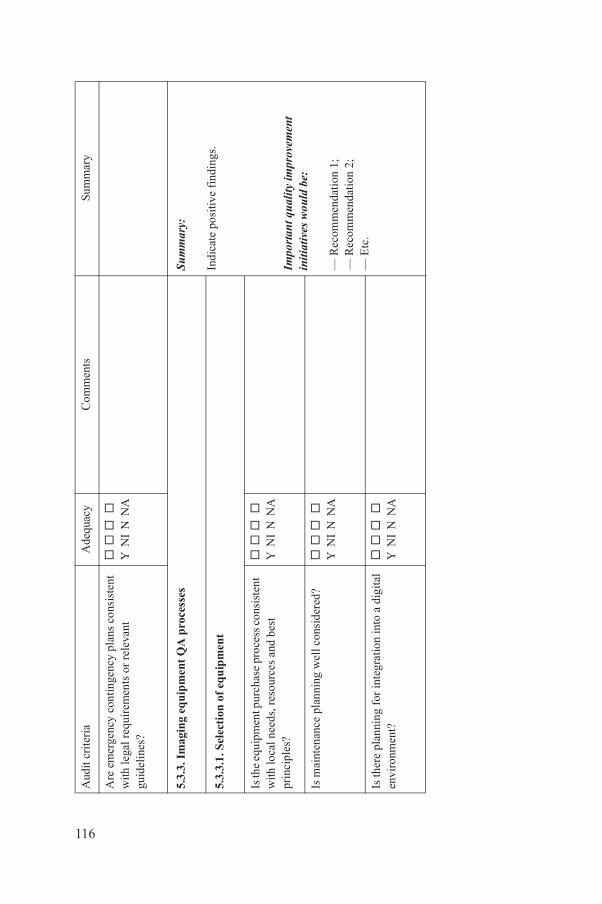
tinge
ncy
plan
s co
nsis
tent
en
ts o
r re
leva
nt
Y N
I N
NA
ipm
ent
QA
pro
cess
esSu
mm
ary:
Indi
cate
pos
itive
fin
ding
s.
Impo
rtant
qua
lity
impr
ovem
ent
initi
ativ
es w
ould
be:
— R
ecom
men
dati
on 1
;—
Rec
omm
enda
tion
2;
— E
tc.
f eq
uip
men
t
rcha
se p
roce
ss c
onsi
sten
t
sour
ces
and
best
Y
NI
N N
A
ning
wel
l con
side
red?
Y N
I N
NA
r in
tegr
atio
n in
to a
dig
ital
Y
NI
N N
A
Ade
quac
yC
omm
ents
Sum
mar
y
116
Are
em
erge
ncy
con
with
lega
l req
uire
mgu
idel
ines
?
5.3.
3. I
mag
ing
equ
5.3.
3.1.
Sel
ecti
on o
Is th
e eq
uipm
ent p
uw
ith lo
cal n
eeds
, re
prin
cipl
es?
Is m
aint
enan
ce p
lan
Is th
ere
plan
ning
fo
envi
ronm
ent?
Aud
it cr
iter
ia
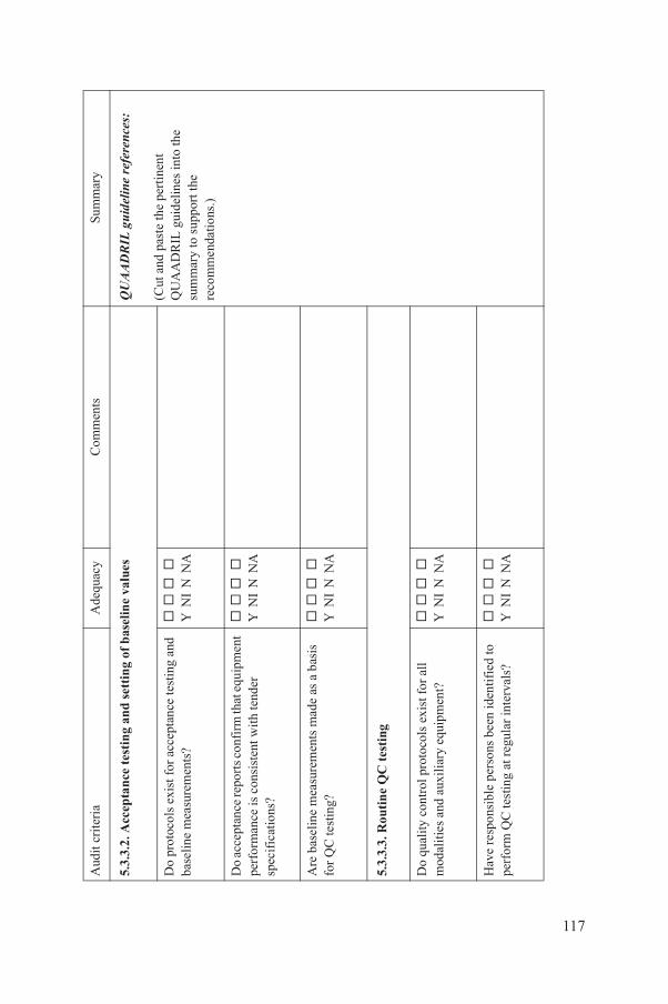
tes
tin
g an
d se
ttin
g of
bas
elin
e va
lues
QU
AAD
RIL
guid
elin
e re
fere
nces
:
(Cut
and
pas
te th
e pe
rtin
ent
QU
AA
DR
IL g
uide
lines
into
the
sum
mar
y to
sup
port
the
reco
mm
enda
tions
.)
or a
ccep
tanc
e te
stin
g an
d nt
s?
Y
NI
N N
A
ts c
onfi
rm th
at e
quip
men
t is
tent
wit
h te
nder
Y
NI
N N
A
rem
ents
mad
e as
a b
asis
Y
NI
N N
A
tes
ting
roto
cols
exi
st f
or a
ll li
ary
equi
pmen
t?
Y
NI
N N
A
rson
s be
en id
enti
fied
to
at r
egul
ar in
terv
als?
Y N
I N
NA
Ade
quac
yC
omm
ents
Sum
mar
y
117
5.3.
3.2.
Acc
epta
nce
Do
prot
ocol
s ex
ist f
base
line
mea
sure
me
Do
acce
ptan
ce re
por
perf
orm
ance
is c
ons
spec
ific
atio
ns?
Are
bas
elin
e m
easu
for
QC
test
ing?
5.3.
3.3.
Rou
tin
e Q
C
Do
qual
ity c
ontr
ol p
mod
alit
ies
and
auxi
Hav
e re
spon
sibl
e pe
perf
orm
QC
test
ing
Aud
it cr
iter
ia
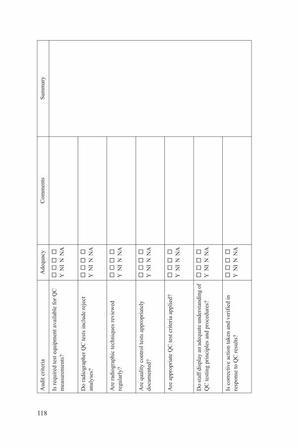
pmen
t ava
ilab
le f
or Q
C
Y N
I N
NA
test
s in
clud
e re
ject
Y
NI
N N
A
chni
ques
rev
iew
ed
Y N
I N
NA
test
s ap
prop
riat
ely
Y N
I N
NA
test
cri
teri
a ap
plie
d?
Y
NI
N N
A
dequ
ate
unde
rsta
ndin
g of
s
and
proc
edur
es?
Y N
I N
NA
take
n an
d ve
rifi
ed in
lt
s?
Y
NI
N N
A
Ade
quac
yC
omm
ents
Sum
mar
y
118
Is r
equi
red
test
equ
im
easu
rem
ents
?
Do
radi
ogra
pher
QC
anal
yses
?
Are
rad
iogr
aphi
c te
regu
larl
y?
Are
qua
lity
cont
rol
docu
men
ted?
Are
app
ropr
iate
QC
Do
staf
f dis
play
an
aQ
C te
stin
g pr
inci
ple
Is c
orre
ctiv
e ac
tion
re
spon
se to
QC
res
u
Aud
it cr
iter
ia
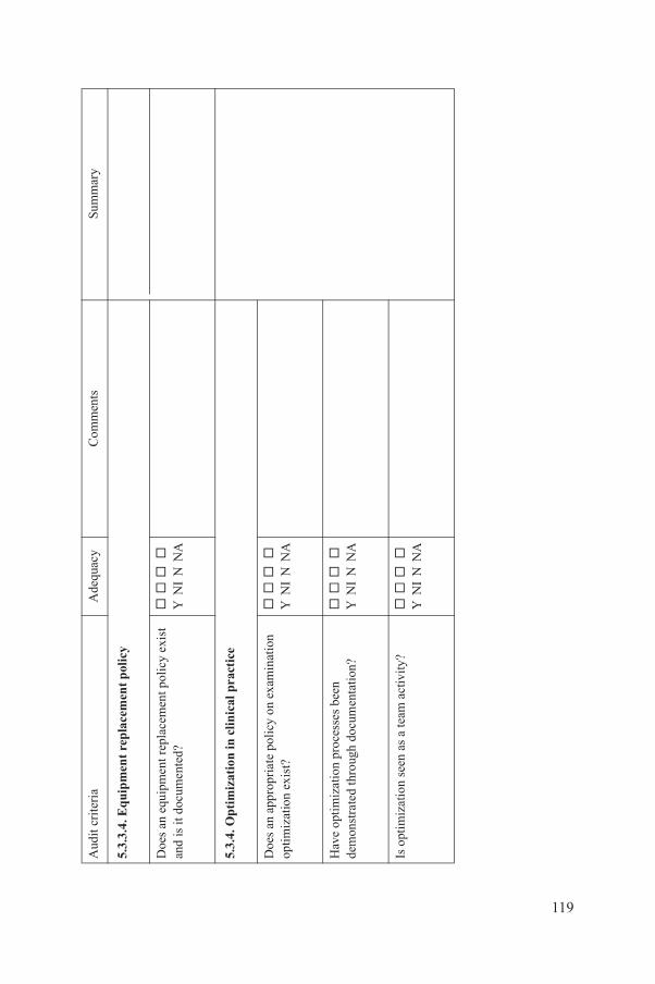
rep
lace
men
t p
olic
y
repl
acem
ent p
olic
y ex
ist
d?
Y
NI
N N
A
in c
lin
ical
pra
ctic
e
pol
icy
on e
xam
inat
ion
Y N
I N
NA
roce
sses
bee
n gh
doc
umen
tati
on?
Y N
I N
NA
as
a te
am a
ctiv
ity?
Y N
I N
NA
Ade
quac
yC
omm
ents
Sum
mar
y
119
5.3.
3.4.
Eq
uip
men
t
Doe
s an
equ
ipm
ent
and
is it
doc
umen
te
5.3.
4. O
pti
miz
atio
n
Doe
s an
app
ropr
iate
optim
izat
ion
exis
t?
Hav
e op
tim
izat
ion
pde
mon
stra
ted
thro
u
Is o
ptim
izat
ion
seen
Aud
it cr
iter
ia

Sum
mar
y:
Indi
cate
pos
itive
fin
ding
s.
Impo
rtant
qua
lity
impr
ovem
ent
initi
ativ
es w
ould
be:
— R
ecom
men
dati
on 1
;—
Rec
omm
enda
tion
2;
— E
tc.
QU
AAD
RIL
guid
elin
e re
fere
nces
:
(Cut
and
pas
te th
e pe
rtin
ent
QU
AA
DR
IL g
uide
lines
into
the
sum
mar
y to
sup
port
the
reco
mm
enda
tions
.)
prin
cip
les
iple
s ba
sed
on
rds
usin
g a
suit
able
Y
NI
N N
A
imet
ry
ce to
est
imat
e th
e do
ses
for
requ
ired
ur
es?
Y N
I N
NA
are
used
for
dos
e
Y
NI
N N
A
dits
reg
ular
ly p
erfo
rmed
pa
red
wit
h ap
prop
riat
e
Y
NI
N N
A
Ade
quac
yC
omm
ents
Sum
mar
y
120
5.3.
5. D
osim
etry
5.3.
5.1.
Dos
imet
ry
Are
dos
imet
ry p
rinc
inte
rnat
iona
l sta
nda
refe
renc
e?
5.3.
5.2.
Pat
ien
t do
s
Are
pro
toco
ls in
pla
appr
opri
ate
pati
ent
exam
inat
ion
proc
ed
Is a
ppro
pria
te s
oftw
esti
mat
es?
Are
pat
ient
dos
e au
and
the
resu
lts c
omD
RL
s?
Aud
it cr
iter
ia
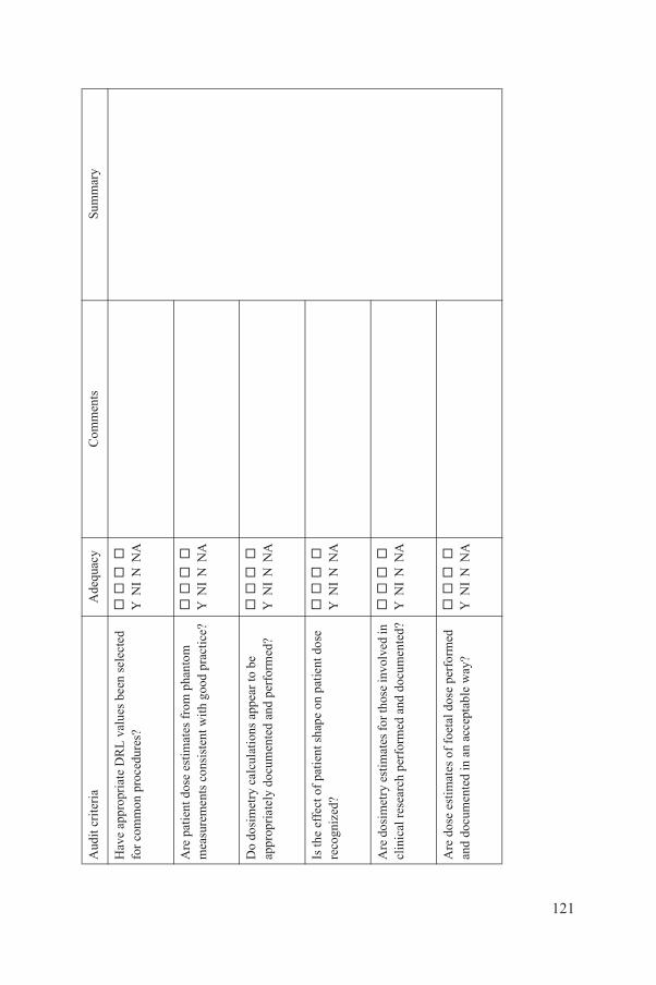
RL
val
ues
been
sel
ecte
d ur
es?
Y N
I N
NA
imat
es f
rom
pha
ntom
is
tent
wit
h go
od p
ract
ice?
Y N
I N
NA
lati
ons
appe
ar to
be
ente
d an
d pe
rfor
med
?
Y
NI
N N
A
nt s
hape
on
patie
nt d
ose
Y N
I N
NA
ates
for t
hose
invo
lved
in
form
ed a
nd d
ocum
ente
d?
Y
NI
N N
A
of f
oeta
l dos
e pe
rfor
med
an
acc
epta
ble
way
?
Y
NI
N N
A
Ade
quac
yC
omm
ents
Sum
mar
y
121
Hav
e ap
prop
riat
e D
for
com
mon
pro
ced
Are
pat
ient
dos
e es
tm
easu
rem
ents
con
s
Do
dosi
met
ry c
alcu
appr
opri
atel
y do
cum
Is th
e ef
fect
of
pati
ere
cogn
ized
?
Are
dos
imet
ry e
stim
clin
ical
res
earc
h pe
r
Are
dos
e es
tim
ates
an
d do
cum
ente
d in
Aud
it cr
iter
ia

ates
of
patie
nt d
oses
ur
emen
ts c
onsi
sten
t wit
h T
and
mam
mog
raph
ic
Y N
I N
NA
ates
of
patie
nt d
oses
ur
emen
ts u
sing
AE
C
ith
good
pra
ctic
e fo
r nd
abd
omin
al
Y N
I N
NA
al d
osim
etry
ysic
ist p
erfo
rm
cas
e, s
taff
dos
imet
ry?
Y N
I N
NA
w e
xam
ples
of
spec
ial
?
Y
NI
N N
A
Ade
quac
yC
omm
ents
Sum
mar
y
122
Are
aud
it te
am e
stim
from
pha
ntom
mea
sgo
od p
ract
ice
for
Cex
amin
atio
ns?
Are
aud
it te
am e
stim
from
pha
ntom
mea
sde
vice
s co
nsis
tent
wch
est r
adio
grap
hy a
fluo
rosc
opy?
5.3.
5.3.
Occ
upat
ion
Doe
s th
e m
edic
al p
hcu
stom
ized
, spe
cial
Are
ther
e pe
er r
evie
case
sta
ff d
osim
etry
Aud
it cr
iter
ia

tion
an
d c
alib
rati
onSu
mm
ary:
Indi
cate
pos
itive
fin
ding
s.
Impo
rtant
qua
lity
impr
ovem
ent
initi
ativ
es w
ould
be:
— R
ecom
men
dati
on 1
;—
Rec
omm
enda
tion
2;
— E
tc.
QU
AAD
RIL
guid
elin
e re
fere
nces
:
(Cut
and
pas
te th
e pe
rtin
ent
QU
AA
DR
IL g
uide
lines
into
the
sum
mar
y to
sup
port
the
reco
mm
enda
tions
.)
ram
me
avai
labl
e fo
r nt
atio
n?
Y
NI
N N
A
inst
rum
enta
tion
trac
eabl
e rd
s?
Y
NI
N N
A
rds
avai
labl
e, in
clud
ing
cks?
Y N
I N
NA
ss-c
heck
ed b
y th
e au
dit
ble
tole
ranc
es?
Y N
I N
NA
met
ers
calib
rate
d in
sit
u?
Y
NI
N N
A
Ade
quac
yC
omm
ents
Sum
mar
y
123
5.3.
6. I
nst
rum
enta
Is a
cal
ibra
tion
pro
gdo
sim
etri
c in
stru
me
Is m
edic
al p
hysi
cist
to c
alib
rati
on s
tand
a
Are
cal
ibra
tion
rec
ore
cord
s of
cro
ss-c
he
Are
inst
rum
ents
cro
team
wit
hin
acce
pta
Are
equ
ipm
ent K
AP
Aud
it cr
iter
ia

PO
RT
FO
RM
S F
OR
ED
UC
AT
ION
, TR
AIN
ING
AN
D R
ES
EA
RC
H P
RO
GR
AM
ME
S
Ade
quac
yC
omm
ents
Sum
mar
y
d t
rain
ing
prog
ram
mes
Sum
mar
y:
Indi
cate
pos
itive
fin
ding
s.
Impo
rtant
qua
lity
impr
ovem
ent
initi
ativ
es w
ould
be:
— R
ecom
men
dati
on 1
;—
Rec
omm
enda
tion
2;
— E
tc.
nsib
le f
or e
duca
tion
and
d?
Y N
I N
NA
rogr
amm
e
duca
tion
pro
gram
me
exis
t ic
al p
ract
ition
ers?
Y N
I N
NA
duca
tion
pro
gram
me
exis
t
Y
NI
N N
A
duca
tion
pro
gram
me
exis
t ts
?
Y
NI
N N
A
124
6.3.
AU
DIT
RE
Aud
it cr
iter
ia
6.3.
1. E
duca
tion
an
Can
an
offi
cer
resp
otr
aini
ng b
e id
entif
ie
6.3.
1.1.
Aca
dem
ic p
Doe
s an
aca
dem
ic e
for
radi
olog
ical
med
Doe
s an
aca
dem
ic e
for
radi
ogra
pher
s?
Doe
s an
aca
dem
ic e
for
med
ical
phy
sici
s

to a
llow
rel
ease
fro
m
ctur
es a
nd s
tudy
?
Y
NI
N N
AQ
UAA
DRI
L gu
idel
ine
refe
renc
es:
(Cut
and
pas
te th
e pe
rtin
ent
QU
AA
DR
IL g
uide
lines
into
the
sum
mar
y to
sup
port
the
reco
mm
enda
tion
s.)
equa
te li
brar
y, I
nter
net
ities
?
Y
NI
N N
A
ectu
re fa
cili
ties
, inc
ludi
ng
imag
es w
ith
a
Y
NI
N N
A
ers
avai
labl
e?
Y N
I N
NA
twor
k th
at a
llow
s id
es s
tudy
mat
eria
ls?
Y N
I N
NA
Ade
quac
yC
omm
ents
Sum
mar
y
125
Doe
s a
poli
cy e
xist
cl
inic
al d
utie
s fo
r le
Is th
ere
acce
ss to
ad
and
com
puti
ng f
acil
Are
ther
e ad
equa
te l
prov
isio
n to
dis
play
sati
sfac
tory
qua
lity?
Are
qua
lifie
d le
ctur
Is th
ere
a su
ppor
t ne
disc
ussi
on a
nd p
rov
Aud
it cr
iter
ia
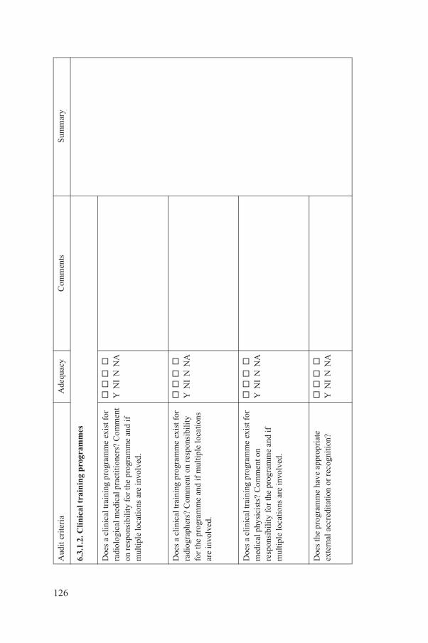
inin
g p
rogr
amm
es
ing
prog
ram
me
exis
t for
l p
ract
ition
ers?
Com
men
t r
the
prog
ram
me
and
if
re in
volv
ed.
Y N
I N
NA
ing
prog
ram
me
exis
t for
m
ent o
n re
spon
sibi
lity
an
d if
mul
tiple
loca
tions
Y N
I N
NA
ing
prog
ram
me
exis
t for
C
omm
ent o
n e
prog
ram
me
and
if
re in
volv
ed.
Y N
I N
NA
e ha
ve a
ppro
pria
te
n or
rec
ogni
tion
?
Y
NI
N N
A
Ade
quac
yC
omm
ents
Sum
mar
y
126
6.3.
1.2.
Cli
nica
l tra
Doe
s a
clin
ical
trai
nra
diol
ogic
al m
edic
aon
res
pons
ibil
ity
fom
ulti
ple
loca
tions
a
Doe
s a
clin
ical
trai
nra
diog
raph
ers?
Com
for
the
prog
ram
me
are
invo
lved
.
Doe
s a
clin
ical
trai
nm
edic
al p
hysi
cist
s?re
spon
sibi
lity
for
thm
ulti
ple
loca
tions
a
Doe
s th
e pr
ogra
mm
exte
rnal
acc
redi
tati
o
Aud
it cr
iter
ia

ondu
cted
in a
fac
ility
that
ov
ed fo
r cli
nica
l tra
inin
g?
Y
NI
N N
A
e ha
ve a
per
son
ondu
ct in
the
faci
lity
?
Y
NI
N N
A
ruct
ured
and
doe
s it
syl
labu
s?
Y
NI
N N
A
ifie
d or
trai
ned
clin
ical
nt
ifie
d fo
r the
pro
gram
me
to in
stru
ct th
ose
enro
lled
Y N
I N
NA
e m
eet t
he r
adia
tion
ring
req
uire
men
ts o
f th
e
Y
NI
N N
A
Ade
quac
yC
omm
ents
Sum
mar
y
127
Is th
e pr
ogra
mm
e c
is a
ccre
dite
d or
app
r
Doe
s th
e pr
ogra
mm
resp
onsi
ble
for
its c
Is th
e pr
ogra
mm
e st
foll
ow a
rec
ogni
zed
Hav
e sp
ecia
lly q
ual
supe
rvis
ors b
een
ide
who
are
com
pete
nt
in th
e pr
ogra
mm
e?
Doe
s th
e pr
ogra
mm
trai
ning
and
mon
ito
part
icip
ants
?
Aud
it cr
iter
ia

num
bers
of s
uper
viso
rs to
e
part
icip
ants
invo
lved
?
Y
NI
N N
A
in a
cli
nica
l env
iron
men
t rt
icip
ants
the
requ
ired
Y
NI
N N
A
f su
ffic
ient
dur
atio
n to
s
to b
e ac
quir
ed?
Y N
I N
NA
smen
t use
d ap
prop
riat
e?
Y
NI
N N
A
ous
pro
gram
mes
e.g.
imag
e ev
alua
tion,
qu
es, e
quip
men
t us
cita
tion
)
Y N
I N
NA
Ade
quac
yC
omm
ents
Sum
mar
y
128
Are
ther
e su
ffic
ient
in
stru
ct o
r m
ento
r th
Is a
pro
gram
me
held
that
can
off
er th
e pa
expe
rien
ce?
Is th
e pr
ogra
mm
e o
allo
w a
dequ
ate
skil
l
Is th
e fo
rm o
f as
ses
6.3.
1.3.
Mis
cell
ane
Clin
ical
edu
cati
on (
radi
ogra
phic
tech
niin
form
atio
n an
d re
s
Aud
it cr
iter
ia
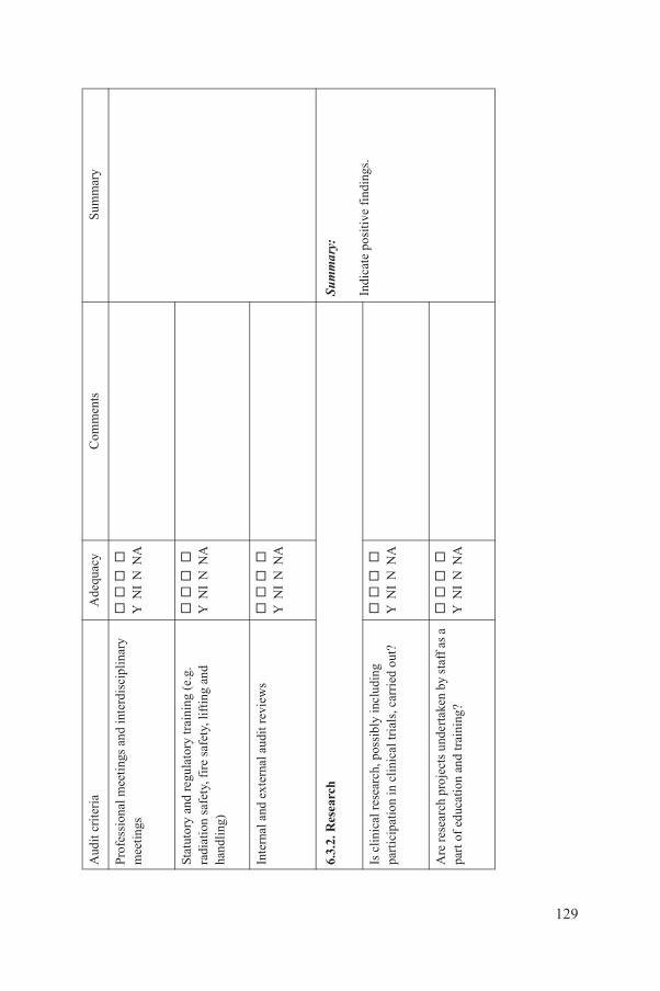
gs a
nd in
terd
isci
plin
ary
Y N
I N
NA
tory
trai
ning
(e.
g.
saf
ety,
lift
ing
and
Y N
I N
NA
l aud
it r
evie
ws
Y N
I N
NA
Sum
mar
y:
Indi
cate
pos
itive
fin
ding
s. p
ossi
bly
incl
udin
g ic
al tr
ials
, car
ried
out
?
Y
NI
N N
A
ts u
nder
take
n by
sta
ff a
s a
d tr
aini
ng?
Y N
I N
NA
Ade
quac
yC
omm
ents
Sum
mar
y
129
Prof
essi
onal
mee
tin
mee
ting
s
Stat
utor
y an
d re
gula
radi
atio
n sa
fety
, fir
eha
ndli
ng)
Inte
rnal
and
ext
erna
6.3.
2. R
esea
rch
Is c
linic
al r
esea
rch,
part
icip
atio
n in
cli
n
Are
res
earc
h pr
ojec
part
of
educ
atio
n an
Aud
it cr
iter
ia

gram
me
a cl
ear
poli
cy
des
mat
ters
of
ethi
cs,
rol
e an
d st
ruct
ure
of th
e o
f th
e in
stit
ute/
faci
lity?
Y N
I N
NA
Impo
rtant
qua
lity
impr
ovem
ent
initi
ativ
es w
ould
be:
— R
ecom
men
dati
on 1
;—
Rec
omm
enda
tion
2;
— E
tc.
QU
AAD
RIL
guid
elin
e re
fere
nces
:
(Cut
and
pas
te th
e pe
rtin
ent
QU
AA
DR
IL g
uide
lines
into
the
sum
mar
y to
sup
port
the
reco
mm
enda
tion
s.)
rogr
amm
e co
nfor
m to
ta
ndar
ds, i
nclu
ding
the
stim
ates
for
res
earc
h?
Y N
I N
NA
h in
fras
truc
ture
ava
ilab
le?
Y
NI
N N
A
ient
ific
per
sonn
el w
ith
a va
ilabl
e?
Y
NI
N N
A
ubli
shed
?
Y
NI
N N
A
t ade
quat
e re
sear
ch
Y N
I N
NA
Ade
quac
yC
omm
ents
Sum
mar
y
130
Has
the
rese
arch
pro
stat
emen
t tha
t inc
lugo
vern
ance
, and
the
rese
arch
com
mit
tee
Doe
s th
e re
sear
ch p
reco
gniz
ed e
thic
al s
use
of p
atie
nt d
ose
e
Is s
uffi
cien
t res
earc
Are
goo
d qu
alit
y sc
reco
rd in
res
earc
h a
Is r
esea
rch
outp
ut p
Doe
s re
sear
ch a
ttrac
fund
ing?
Aud
it cr
iter
ia
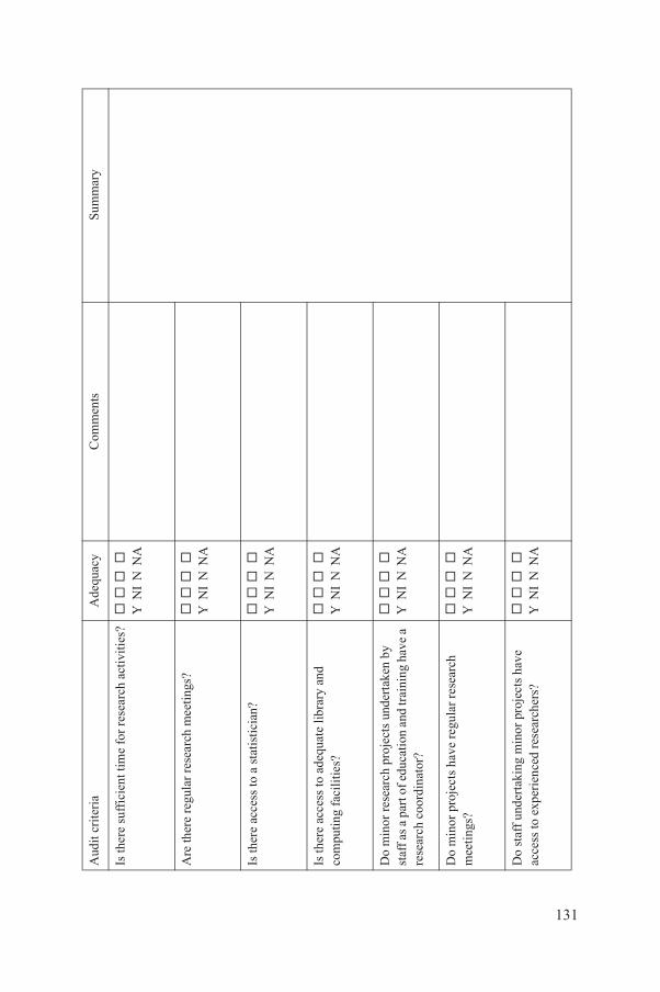
me
for
rese
arch
act
ivit
ies?
Y
NI
N N
A
sear
ch m
eeti
ngs?
Y N
I N
NA
stat
isti
cian
?
Y
NI
N N
A
equa
te li
brar
y an
d ?
Y N
I N
NA
roje
cts
unde
rtak
en b
y ca
tion
and
trai
ning
hav
e a
r?
Y N
I N
NA
ave
regu
lar
rese
arch
Y
NI
N N
A
g m
inor
pro
ject
s ha
ve
d re
sear
cher
s?
Y
NI
N N
A
Ade
quac
yC
omm
ents
Sum
mar
y
131
Is th
ere
suff
icie
nt ti
Are
ther
e re
gula
r re
Is th
ere
acce
ss to
a
Is th
ere
acce
ss to
ad
com
putin
g fa
cili
ties
Do
min
or r
esea
rch
pst
aff a
s a
part
of e
dure
sear
ch c
oord
inat
o
Do
min
or p
roje
cts
hm
eeti
ngs?
Do
staf
f un
dert
akin
acce
ss to
exp
erie
nce
Aud
it cr
iter
ia
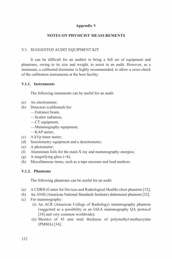
Appendix V
NOTES ON PHYSICIST MEASUREMENTS
V.1. SUGGESTED AUDIT EQUIPMENT KIT
It can be difficult for an auditor to bring a full set of equipment and phantoms, owing to its size and weight, to assist in an audit. However, as a minimum, a calibrated dosimeter is highly recommended, to allow a cross-check of the calibration instruments at the host facility.
V.1.1. Instruments
The following instuments can be useful for an audit:
(a) An electrometer;(b) Detectors (calibrated) for:
—Entrance beam,—Scatter radiation,—CT equipment,—Mammography equipment,—KAP meter;
(c) A kVp timer meter;(d) Sensitometry equipment and a densitometer;(e) A photometer;(f) Aluminium foils for the main X ray and mammography energies;(g) A magnifying glass (×4);(h) Miscellaneous items, such as a tape measure and lead markers.
V.1.2. Phantoms
The following phantoms can be useful for an audit:
(a) A CDRH (Center for Devices and Radiological Health) chest phantom [32];(b) An ANSI (American National Standards Institute) abdomenal phantom [32];
132
(c) For mammography: (i) An ACR (American College of Radiology) mammography phantom
(suggested as a possibility in an IAEA mammography QA protocol [34] and very common worldwide);
(ii) Sheet(s) of 45 mm total thickness of polymethyl-methacrylate (PMMA) [34];
(d) A CT phantom (made from PMMA — a 16 cm diameter head phantom and a 32 cm diameter body phantom).
V.2. SUGGESTED MEASUREMENTS
The suggested measurements by the physicist auditor are described in Section 5.2 and are summarized below. Suggested worksheets for the measurements are provided in Annex IV. Note that there is limited time available and that the number of measurements should be adjusted to the scope of the activities of the facility/institution.
V.2.1. Cross-checks for measuring instruments
Cross-checks should be made for the following:
(a) During the audit, cross-checks should be made of selected dosimetric instruments, as time allows: (i) Instruments for entrance surface air kerma measurements (these can be
done as part of a dosimetry check for a chest and abdomen phantom, as specified in Section 5.2.5.3);
(ii) Instruments for measurements of scattered radiation (these could be done in connection with a dosimetry check for CT dosimetry, as specified in Section 5.2.5.3);
(iii) Instruments for mammography and CT (these could be done as part of a dosimetry check for these modalities, as specified in Section 5.2.5.3);
(iv) At least one KAP meter in situ [32].(b) If time permits, a cross-check of available kVp meters could also be useful
during the audit period.
V.2.2. Phantom dosimetry
The resident medical physicist could also be asked to demonstrate dosimetry measurements with phantoms associated with the following examination types:
133
(a) Chest; (b) Abdominal fluoroscopy; (c) Mammography; (d) Computed tomography, including verifying a dose–length product (DLP)
indicator, if possible.

V.2.3. Other measurements
The other tasks for the physicist auditor are as follows:
(a) Examine a mammography film exposed with an image quality phantom (see Section 5.2.5.3);
(b) Examine the film reading conditions (this may involve making measurements);
(c) Review a demonstration by local staff of how to set up an AEC system;(d) Review a demonstration by local staff of some QA measurements, such as a
half-value layer (HVL) measurement or an entrance air kerma measurement;
(e) Verify the displayed DLP for the CT examination sequences.
134
REFERENCES
[1] THE SCOTTISH OFFICE, The Interface between Clinical Audit and Management — A Report of a Working Group Set Up by the Clinical Resource and Audit Group, Scottish Office, Edinburgh (1993) 9.
[2] COMMISSION OF THE EUROPEAN COMMUNITIES, Council Directive 97/43/Euratom of 30 June 1997 on health protection of individuals against the dangers of ionizing radiation in relation to medical exposure, and repealing Directive 84/466 Euratom, Off. J. Eur. Commun. Rep. L. 180 (1997) 22–27.
[3] INTERNATIONAL ATOMIC ENERGY AGENCY, Comprehensive Audits of Radiotherapy Practices: A Tool for Quality Improvement, IAEA, Vienna (2007).
[4] FOOD AND AGRICULTURE ORGANIZATION OF THE UNITED NATIONS, INTERNATIONAL ATOMIC ENERGY AGENCY, INTERNATIONAL LABOUR ORGANISATION, OECD NUCLEAR ENERGY AGENCY, PAN AMERICAN HEALTH ORGANIZATION, WORLD HEALTH ORGANIZATION, International Basic Safety Standards for Protection against Ionizing Radiation and for the Safety of Radiation Sources, IAEA Safety Series No. 115, IAEA, Vienna (1996).
[5] COMMISSION OF THE EUROPEAN COMMUNITIES, European Commission Guidelines on Clinical Audit for Medical Radiological Practices (Diagnostic Radiology, Nuclear Medicine, and Radiotherapy), CEC, Luxembourg (in preparation).
[6] INTERNATIONAL ATOMIC ENERGY AGENCY, The Management System for Facilities and Activities, IAEA Safety Standards Series No. GS-R-3, IAEA, Vienna (2006).
[7] DEMING, W.E., Out of Crisis, Cambridge University Press, Cambridge (1986).[8] DONABEDIAN, A., The Definition of Quality and Approaches to its Assessment,
Health Administration Press, Ann Arbor, MI (1980).[9] AMERICAN COLLEGE OF RADIOLOGY, ACR Appropriateness Criteria (2008),
http://www.acr.org/SecondaryMainMenuCategories/quality_safety/app_criteria.aspx[10] SCHNEIDER, K., et al., Micturition cystourethrography in paediatric patients in
selected children’s hospitals in Europe: Evaluation of fluoroscopy technique, image quality criteria and dose, Radiat. Prot. Dosim. 90 1–2 (2000) 197–201.
[11] WALL, B.F., Quality criteria development within the fourth framework research programme: Paediatric patients, Radiat. Prot. Dosim. 90 1–2 (2000) 73–78.
[12] COMMISSION OF THE EUROPEAN COMMUNITIES, European Guidelines on Quality Criteria for Diagnostic Radiographic Images in Paediatrics, Rep. EUR 16261 EN, CEC, Luxembourg.
[13] COMMISSION OF THE EUROPEAN COMMUNITIES, European Guidelines on Quality Criteria for Diagnostic Radiographic Images, Rep. EUR 16260 EN, CEC, Luxembourg (1996).
[14] COMMISSION OF THE EUROPEAN COMMUNITIES, European Guidelines on
135
Quality Criteria for Computed Tomography, Rep. EUR 16262 EN, CEC, Luxembourg (1998), http://www.drs.dk/guidelines/ct/quality/mainindex.htm
[15] COMMISSION OF THE EUROPEAN COMMUNITIES, Guidance on Diagnostic Reference Levels for Medical Exposure, Radiation Protection 109, CEC, Luxembourg (2001).
[16] COMMISSION OF THE EUROPEAN COMMISSION, Referral Guidelines for Imaging, Radiation Protection 118, CEC, Luxembourg (2001),http://ec.europa.eu/energy/nuclear/radioprotection/publication/doc/118_update_en.pdf
[17] ROYAL COLLEGE OF RADIOLOGISTS, Standards for Self-assessment of Performance, Rep. BFCR(07)9, RCR, London (2007).
[18] ROYAL COLLEGE OF RADIOLOGISTS, Guidelines for Nursing Care in Interventional Radiology: The Role of the Registered Nurse and Nursing Support, Rep. BFCR(06)7, RCR, London (2006).
[19] ROYAL COLLEGE OF RADIOLOGISTS, Recommendations for Cross-sectional Imaging in Cancer Management, Rep. RCR(06)1, RCR, London (2006).
[20] ROYAL COLLEGE OF RADIOLOGISTS, Standards for the Reporting and Interpretation of Imaging Investigations, Rep. BFCR(06)1, RCR, London (2006).
[21] ROYAL COLLEGE OF RADIOLOGISTS, Standards for Patient Consent Particular to Radiology, Rep. BFCR(05)8, RCR, London (2005).
[22] ROYAL COLLEGE OF RADIOLOGISTS, Making the Best Use of the Department of Clinical Radiology, Rep. BFCR(07)10, RCR, London,http://www.rcr.ac.uk/publications.aspx?PageID=310&PublicationID=261
[23] ROYAL COLLEGE OF RADIOLOGISTS, Referral Guidelines: Making the Best Use of Clinical Radiology Services, 6th edn, Rep. MBUR6 (2007),http://www.rcr.ac.uk/content.aspx?PageID=995
[24] ROYAL COLLEGE OF RADIOLOGISTS, Advice on Exposure to Ionising Radiation During Pregnancy in Children, RCL, London (2007), http://www.rcr.ac.uk/docs/radiology/pdf/Guidance_diagnosticmedicalexposure_April07.pdf
[25] NATIONAL HEALTH SERVICE, Breast Screening Programme: Quality Assurance Guidelines for Radiographers, NHS, Sheffield (1994) 30.
[26] INTERNATIONAL ATOMIC ENERGY AGENCY, Radiation Protection of Patients, Interventional Radiology, http://rpop.iaea.org/RPOP/Content/index.htm
[27] INTERNATIONAL COMMISSION ON RADIOLOGICAL PROTECTION, Pregnancy and Medical Radiation, Publication 84, Pergamon Press, Oxford and New York (2000), http://www.icrp.org/docs/ICRP_84_Pregnancy_s.pps
[28] AMERICAN COLLEGE OF RADIOLOGY, ACR Practice Guideline for Imaging Pregnant or Potentially Pregnant Adolescents and Women with Ionizing Radiation (2008), http://www.acr.org/SecondaryMainMenuCategories/quality_safety/guidelines/dx/Pregnancy.aspx
[29] SHARP, C., SHRIMPTON, J.A., BURY, R., Diagnostic Medical Exposures: Advice on Exposure to Ionising Radiation During Pregnancy,http://www.hpa.org.uk/web/HPAweb&HPAwebStandard/HPAweb_C/1195733757920
[30] NATIONAL COUNCIL ON RADIATION PROTECTION AND MEASUREMENT, Structural Shielding Design for Medical X-Ray Imaging Facilities, Rep. No. 147, NCRP, Bethesda, MD (2004).
136
[31] SUTTON, D.G., WILLIAMS, J.R. (Eds), Radiation Shielding for Diagnostic X-rays, British Institute of Radiology, London (2000).
[32] INTERNATIONAL ATOMIC ENERGY AGENCY, Dosimetry in Diagnostic Radiology: An International Code of Practice, Technical Reports Series No. 457, IAEA, Vienna (2007).

[33] INTERNATIONAL COMMISSION ON RADIATION UNITS AND MEASUREMENTS, Patient Dosimetry for X Rays Used in Medical Imaging, Rep. 74, ICRU, Bethesda, MD (2005).
[34] INTERNATIONAL ATOMIC ENERGY AGENCY, Quality Assurance Programme for Screen Film Mammography, IAEA Human Health Series No. 2, IAEA, Vienna (2009).
137

.
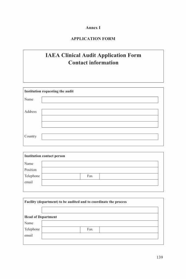
Annex I
APPLICATION FORM
IAEA Clinical Audit Application FormContact information
Institution requesting the audit
Name Address Country
Institution contact person
Name Position Telephone Fax email
Facility (department) to be audited and to coordinate the process
139
Head of Department Name Telephone Fax email
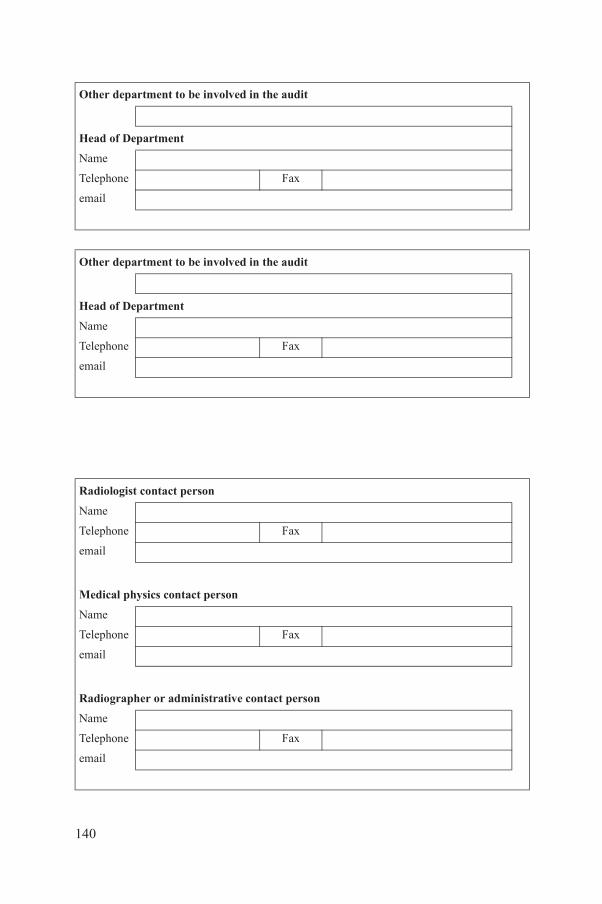
Other department to be involved in the audit
Head of Department Name Telephone Fax email
Other department to be involved in the audit
Head of Department Name Telephone Fax email
Radiologist contact person
Name Telephone Fax email
Medical physics contact person
Name Telephone Fax email
140
Radiographer or administrative contact person
Name Telephone Fax email
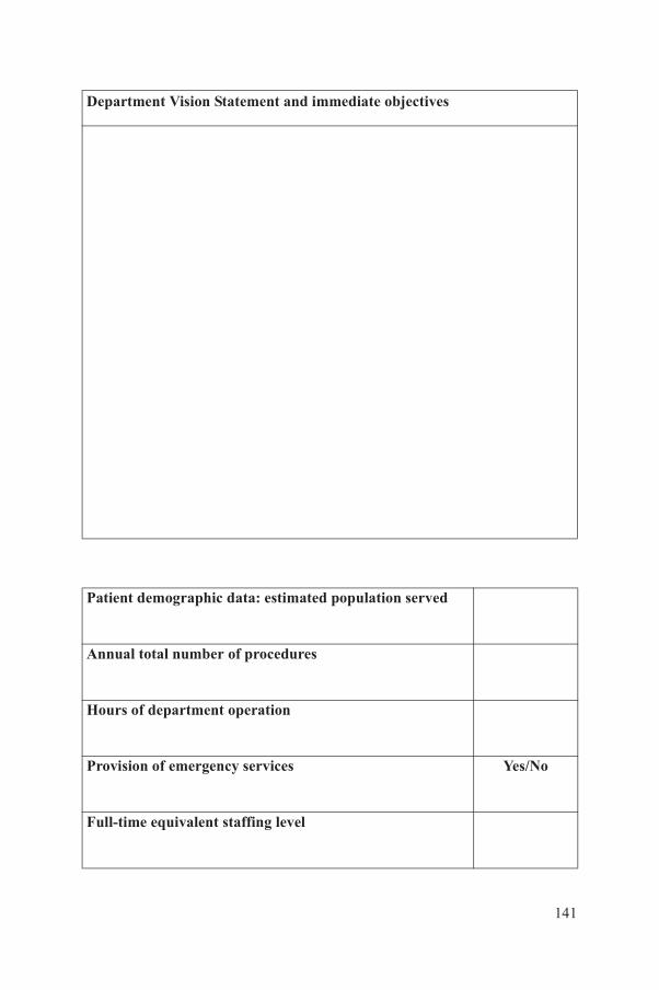
Department Vision Statement and immediate objectives
Patient demographic data: estimated population served
Annual total number of procedures
Hours of department operation
141
Provision of emergency services Yes/No
Full-time equivalent staffing level
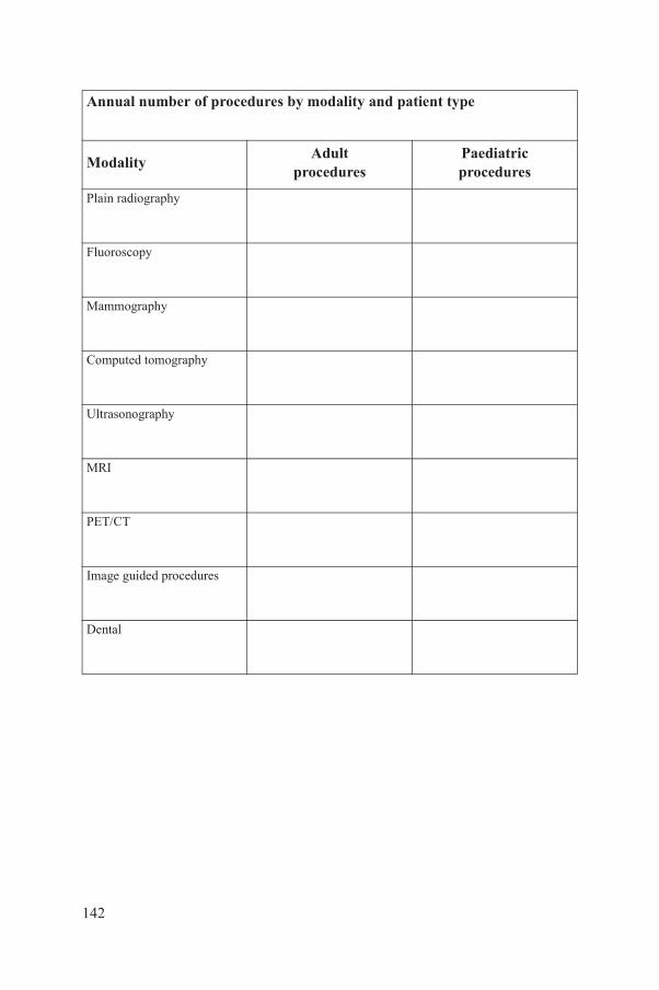
Annual number of procedures by modality and patient type
ModalityAdult
proceduresPaediatricprocedures
Plain radiography
Fluoroscopy
Mammography
Computed tomography
Ultrasonography
MRI
PET/CT
Image guided procedures
Dental
142
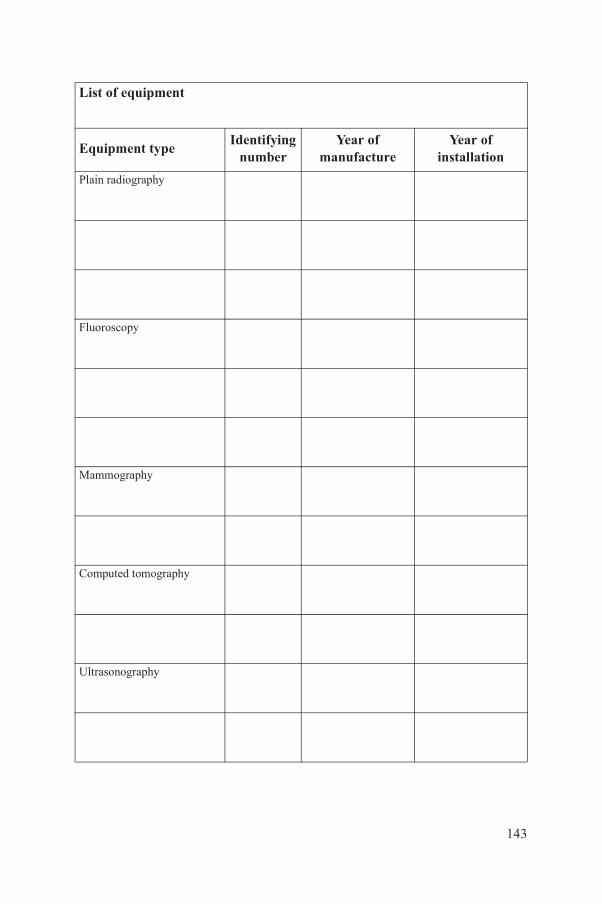
List of equipment
Equipment typeIdentifying
numberYear of
manufactureYear of
installation
Plain radiography
Fluoroscopy
Mammography
Computed tomography
Ultrasonography
143
MRI
PET/CT
Image guided procedures
Dental
Please extend table as necessary.
144
Annex II
LIST OF ITEMS REQUESTED TO BE AVAILABLE ON-SITE
Arrange for a private room to be available for the auditors to discuss or work on matters concerned with the audit during their site visit, as well as for them to have access to a printer and paper for the draft report.
Copies of the following documents, under their related headings, should be available for review by the audit team at the start of the audit period. Access to similar documents, if requested, will be appreciated.
II–1. TECHNICAL PROCEDURES
II–1.1. Quality assurance infrastructure
The following items are required:
—Documentation of the roles and responsibilities of radiographers and medical physicists;
—Syllabuses of the radiation safety training programmes undertaken by staff.
II–1.2. Radiation protection and general safety
The following items are required:
—The radiation safety manual used;—A sample of the shielding design for an X ray installation;—A sample of occupational dose monitoring records;—A statement of pregnancy policy, and associated records.
II–1.3. Equipment quality assurance processes
The following items are required:
145
—A tender document or equivalent for the purchase of a CT scanner or similar piece of equipment;
—An acceptance report for a major piece of equipment;—Protocols for a regular QC programme;—A statement of the equipment replacement programme policy.
II–1.4. Calibration of instrumentation
Instrument calibration records and protocols are required.
II–1.5. Dosimetry
Relevant dosimetry documentation, including phantom dose studies and patient survey results, are required.
II–1.6. Optimization
Documentation of examination optimization processes is required.
146
Annex III
EQUIPMENT-SPECIFIC CHECKLIST FORMS FOR SECTIONS 3–5
The audit team members should use the equipment-specific checklist forms as internal tools to assist them in conducting the audit review. The assessments made using these checklists will be summarized on the audit report forms given in Appendix IV.
The key for this annex is as follows:
Y — Yes (i.e. available, performed or adequate);NI — Needs improvement;N — No (i.e. not available, not performed or not adequate);NA — Not applicable.
147
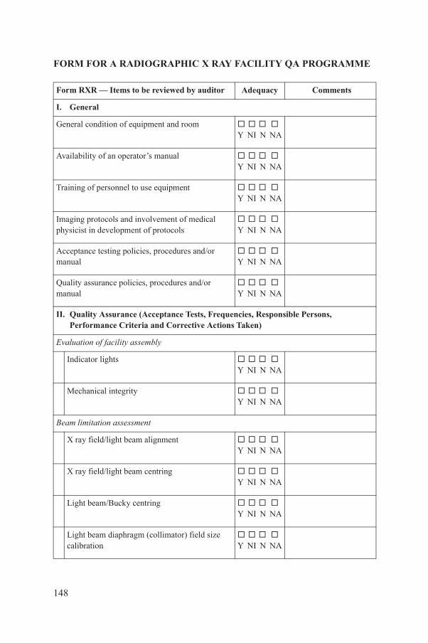
FORM FOR A RADIOGRAPHIC X RAY FACILITY QA PROGRAMME
Form RXR — Items to be reviewed by auditor Adequacy Comments
I. General
General condition of equipment and room Y NI N NA
Availability of an operator’s manual Y NI N NA
Training of personnel to use equipment Y NI N NA
Imaging protocols and involvement of medical physicist in development of protocols
Y NI N NA
Acceptance testing policies, procedures and/or manual
Y NI N NA
Quality assurance policies, procedures and/or manual
Y NI N NA
II. Quality Assurance (Acceptance Tests, Frequencies, Responsible Persons, Performance Criteria and Corrective Actions Taken)
Evaluation of facility assembly
Indicator lights Y NI N NA
Mechanical integrity Y NI N NA
Beam limitation assessment
X ray field/light beam alignment Y NI N NA
X ray field/light beam centring Y NI N NA
148
Light beam/Bucky centring Y NI N NA
Light beam diaphragm (collimator) field size calibration
Y NI N NA
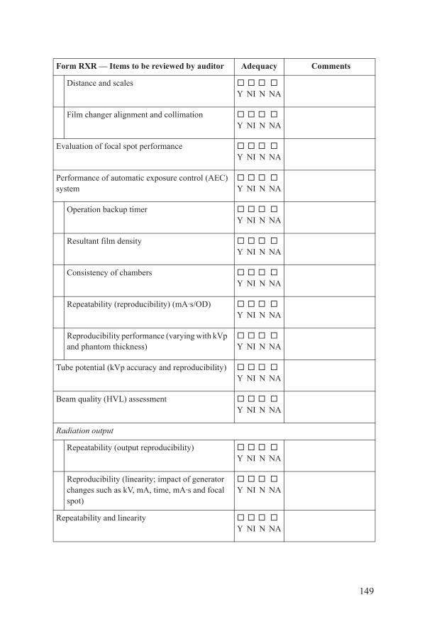
Distance and scales Y NI N NA
Film changer alignment and collimation Y NI N NA
Evaluation of focal spot performance Y NI N NA
Performance of automatic exposure control (AEC) system
Y NI N NA
Operation backup timer Y NI N NA
Resultant film density Y NI N NA
Consistency of chambers Y NI N NA
Repeatability (reproducibility) (mA·s/OD) Y NI N NA
Reproducibility performance (varying with kVp and phantom thickness)
Y NI N NA
Tube potential (kVp accuracy and reproducibility) Y NI N NA
Beam quality (HVL) assessment Y NI N NA
Radiation output
Repeatability (output reproducibility) Y NI N NA
Reproducibility (linearity; impact of generator changes such as kV, mA, time, mA·s and focal
Y NI N NA
Form RXR — Items to be reviewed by auditor Adequacy Comments
149
spot)
Repeatability and linearity Y NI N NA

Timer accuracy Y NI N NA
Image quality (phantom) Y NI N NA
Artefact evaluation Y NI N NA
Technique charts Y NI N NA
Repeat analysis Y NI N NA
Visual checklist Y NI N NA
Tomographic performance assessment
Cut height Y NI N NA
Angle of swing Y NI N NA
III. Radiation Protection and Safety
Radiation protection equipment available, accepted, calibrated and passed QC test
Y NI N NA
Room shielding checked Y NI N NA
Quality control of immobilization devices: storage and replacement
Y NI N NA
Control of access to X ray room Y NI N NA
Form RXR — Items to be reviewed by auditor Adequacy Comments
150
Checks of warning lights/signs Y NI N NA
Checks of door interlocks Y NI N NA

Radiation protection surveys (e.g. what is done, including frequency, methods and records, and actions taken as a result)
Y NI N NA
IV. Patient Dosimetry
Dosimetry equipment and methodology Y NI N NA
Patient dose estimates and reference values Y NI N NA
Equipment based dosimetry (e.g. kerma–area product (KAP) and input kerma (Ki) display)
Y NI N NA
Dosimetry in pregnancy Y NI N NA
Form RXR — Items to be reviewed by auditor Adequacy Comments
151
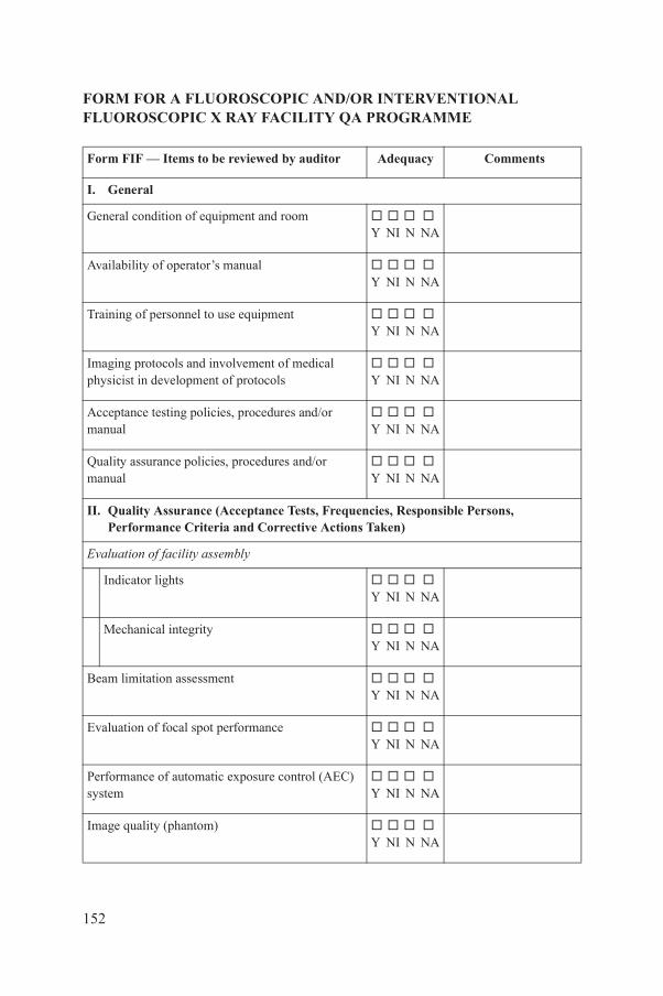
FORM FOR A FLUOROSCOPIC AND/OR INTERVENTIONAL FLUOROSCOPIC X RAY FACILITY QA PROGRAMME
Form FIF — Items to be reviewed by auditor Adequacy Comments
I. General
General condition of equipment and room Y NI N NA
Availability of operator’s manual Y NI N NA
Training of personnel to use equipment Y NI N NA
Imaging protocols and involvement of medical physicist in development of protocols
Y NI N NA
Acceptance testing policies, procedures and/or manual
Y NI N NA
Quality assurance policies, procedures and/or manual
Y NI N NA
II. Quality Assurance (Acceptance Tests, Frequencies, Responsible Persons, Performance Criteria and Corrective Actions Taken)
Evaluation of facility assembly
Indicator lights Y NI N NA
Mechanical integrity Y NI N NA
Beam limitation assessment Y NI N NA
Evaluation of focal spot performance Y NI N NA
152
Performance of automatic exposure control (AEC) system
Y NI N NA
Image quality (phantom) Y NI N NA
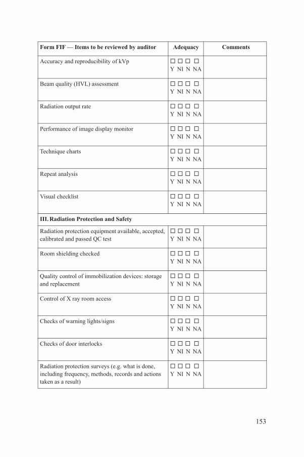
Accuracy and reproducibility of kVp Y NI N NA
Beam quality (HVL) assessment Y NI N NA
Radiation output rate Y NI N NA
Performance of image display monitor Y NI N NA
Technique charts Y NI N NA
Repeat analysis Y NI N NA
Visual checklist Y NI N NA
III. Radiation Protection and Safety
Radiation protection equipment available, accepted, calibrated and passed QC test
Y NI N NA
Room shielding checked Y NI N NA
Quality control of immobilization devices: storage and replacement
Y NI N NA
Control of X ray room access Y NI N NA
Checks of warning lights/signs Y NI N NA
Checks of door interlocks
Form FIF — Items to be reviewed by auditor Adequacy Comments
153
Y NI N NA
Radiation protection surveys (e.g. what is done, including frequency, methods, records and actions taken as a result)
Y NI N NA
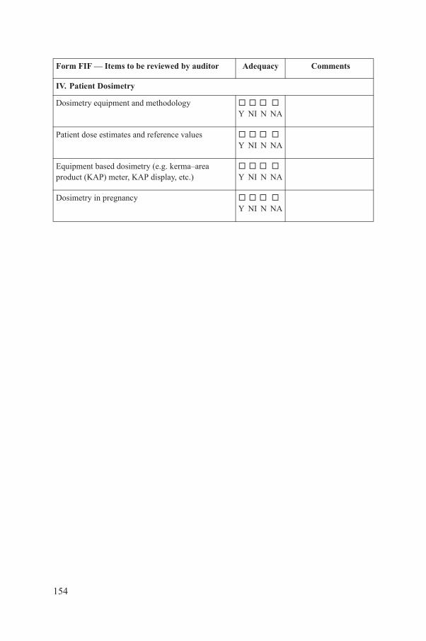
IV. Patient Dosimetry
Dosimetry equipment and methodology Y NI N NA
Patient dose estimates and reference values Y NI N NA
Equipment based dosimetry (e.g. kerma–area product (KAP) meter, KAP display, etc.)
Y NI N NA
Dosimetry in pregnancy Y NI N NA
Form FIF — Items to be reviewed by auditor Adequacy Comments
154
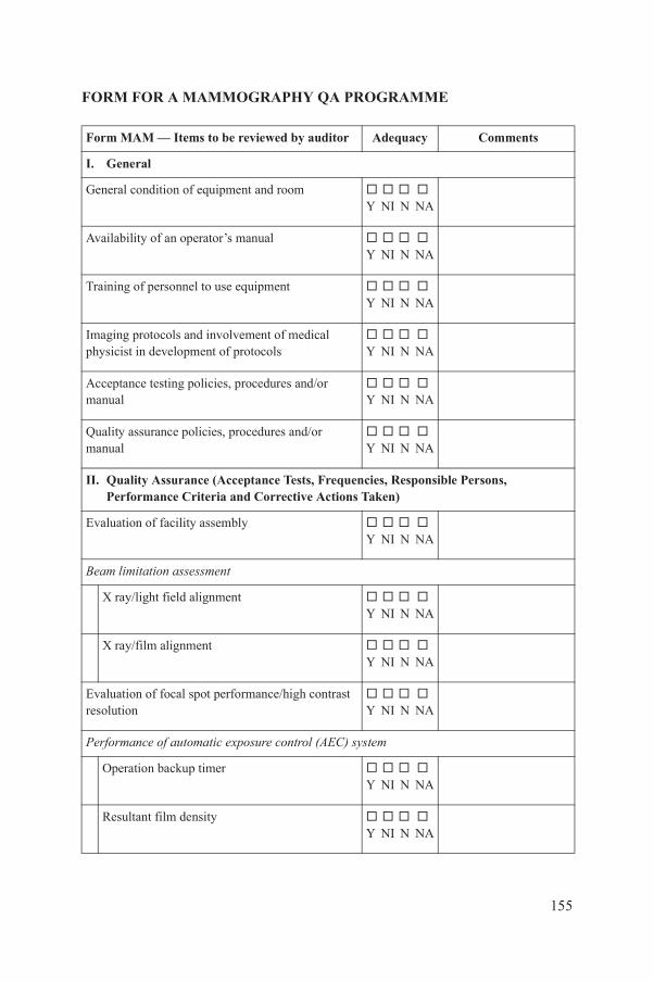
FORM FOR A MAMMOGRAPHY QA PROGRAMME
Form MAM — Items to be reviewed by auditor Adequacy Comments
I. General
General condition of equipment and room Y NI N NA
Availability of an operator’s manual Y NI N NA
Training of personnel to use equipment Y NI N NA
Imaging protocols and involvement of medical physicist in development of protocols
Y NI N NA
Acceptance testing policies, procedures and/or manual
Y NI N NA
Quality assurance policies, procedures and/or manual
Y NI N NA
II. Quality Assurance (Acceptance Tests, Frequencies, Responsible Persons, Performance Criteria and Corrective Actions Taken)
Evaluation of facility assembly Y NI N NA
Beam limitation assessment
X ray/light field alignment Y NI N NA
X ray/film alignment Y NI N NA
Evaluation of focal spot performance/high contrast resolution
Y NI N NA
Performance of automatic exposure control (AEC) system
155
Operation backup timer Y NI N NA
Resultant film density Y NI N NA

Consistency (reproducibility) (mA·s) Y NI N NA
Device breast thickness and kVp calibration Y NI N NA
Artefact evaluation Y NI N NA
Image quality (phantom) and threshold contrast Y NI N NA
Tube potential (accuracy and reproducibility of kVp) Y NI N NA
Beam quality (HVL) assessment Y NI N NA
Radiation output repeatability Y NI N NA
Radiation output rate Y NI N NA
Non-uniformity of X ray field Y NI N NA
Technique charts Y NI N NA
Repeat analysis Y NI N NA
Compression Y NI N NA
Visual checklist Y NI N NA
Form MAM — Items to be reviewed by auditor Adequacy Comments
156
General condition of equipment and room Y NI N NA
Accuracy of stereotactic device Y NI N NA
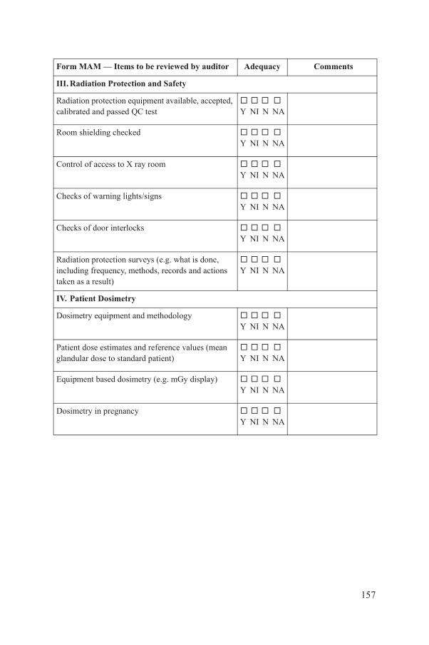
III. Radiation Protection and Safety
Radiation protection equipment available, accepted, calibrated and passed QC test
Y NI N NA
Room shielding checked Y NI N NA
Control of access to X ray room Y NI N NA
Checks of warning lights/signs Y NI N NA
Checks of door interlocks Y NI N NA
Radiation protection surveys (e.g. what is done, including frequency, methods, records and actions taken as a result)
Y NI N NA
IV. Patient Dosimetry
Dosimetry equipment and methodology Y NI N NA
Patient dose estimates and reference values (mean glandular dose to standard patient)
Y NI N NA
Equipment based dosimetry (e.g. mGy display) Y NI N NA
Dosimetry in pregnancy Y NI N NA
Form MAM — Items to be reviewed by auditor Adequacy Comments
157

FORM FOR A COMPUTED TOMOGRAPHY QA PROGRAMME
Form CTQ — Items to be reviewed by auditor Adequacy Comments
I. General
General condition of equipment and room Y NI N NA
Availability of operator’s manual Y NI N NA
Training of personnel to use equipment Y NI N NA
Imaging protocols and involvement of medical physicist in development of protocols
Y NI N NA
Acceptance testing policies, procedures and/or manual
Y NI N NA
Quality assurance policies, procedures and/or manual
Y NI N NA
II. Quality Assurance (Acceptance Tests, Frequencies, Responsible Persons, Performance Criteria and Corrective Actions Taken)
Facility assembly evaluation
Visual inspection Y NI N NA
Audible/visual signals Y NI N NA
Mechanical integrity Y NI N NA
Alignment light accuracy Y NI N NA
Alignment of table to gantry
158
Y NI N NA
Table/gantry tilt Y NI N NA

Slice localization from radiograph (scout image) Y NI N NA
Table incrementation accuracy Y NI N NA
Slice thickness Y NI N NA
Artefact evaluation Y NI N NA
Computed tomography number assessment Y NI N NA
High contrast resolution Y NI N NA
Low contrast resolution Y NI N NA
Image uniformity Y NI N NA
Noise Y NI N NA
Video display Y NI N NA
Hard copy display Y NI N NA
CTDIvol Y NI N NA
Warm-up procedure Y NI N NA
Form CTQ — Items to be reviewed by auditor Adequacy Comments
159
Data display, data transfer and data manipulation Y NI N NA
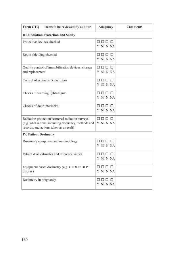
III. Radiation Protection and Safety
Protective devices checked Y NI N NA
Room shielding checked Y NI N NA
Quality control of immobilization devices: storage and replacement
Y NI N NA
Control of access to X ray room Y NI N NA
Checks of warning lights/signs Y NI N NA
Checks of door interlocks Y NI N NA
Radiation protection/scattered radiation surveys (e.g. what is done, including frequency, methods and records, and actions taken as a result)
Y NI N NA
IV. Patient Dosimetry
Dosimetry equipment and methodology Y NI N NA
Patient dose estimates and reference values Y NI N NA
Equipment based dosimetry (e.g. CTDI or DLP display)
Y NI N NA
Dosimetry in pregnancy Y NI N NA
Form CTQ — Items to be reviewed by auditor Adequacy Comments
160
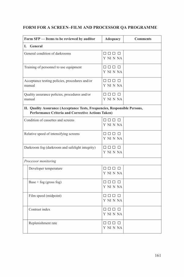
FORM FOR A SCREEN–FILM AND PROCESSOR QA PROGRAMME
Form SFP — Items to be reviewed by auditor Adequacy Comments
I. General
General condition of darkrooms Y NI N NA
Training of personnel to use equipment Y NI N NA
Acceptance testing policies, procedures and/or manual
Y NI N NA
Quality assurance policies, procedures and/or manual
Y NI N NA
II. Quality Assurance (Acceptance Tests, Frequencies, Responsible Persons, Performance Criteria and Corrective Actions Taken)
Condition of cassettes and screens Y NI N NA
Relative speed of intensifying screens Y NI N NA
Darkroom fog (darkroom and safelight integrity) Y NI N NA
Processor monitoring
Developer temperature Y NI N NA
Base + fog (gross fog) Y NI N NA
Film speed (midpoint) Y NI N NA
Contrast index
161
Y NI N NA
Replenishment rate Y NI N NA
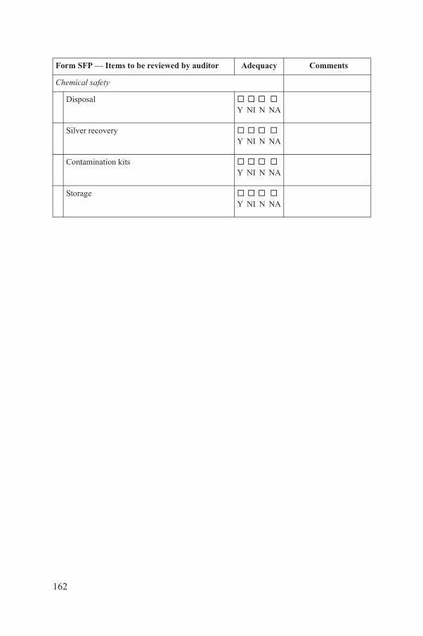
Chemical safety
Disposal Y NI N NA
Silver recovery Y NI N NA
Contamination kits Y NI N NA
Storage Y NI N NA
Form SFP — Items to be reviewed by auditor Adequacy Comments
162
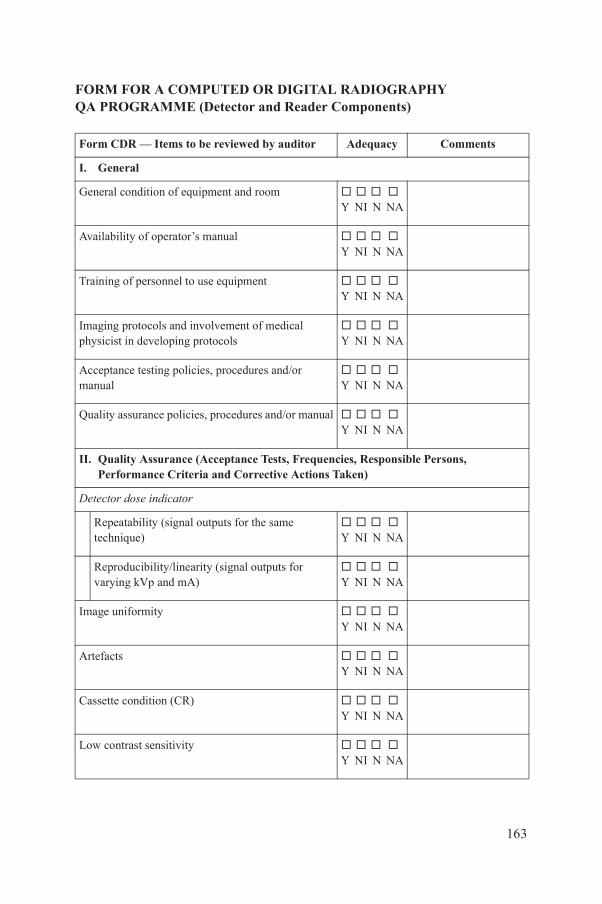
FORM FOR A COMPUTED OR DIGITAL RADIOGRAPHY QA PROGRAMME (Detector and Reader Components)
Form CDR — Items to be reviewed by auditor Adequacy Comments
I. General
General condition of equipment and room Y NI N NA
Availability of operator’s manual Y NI N NA
Training of personnel to use equipment Y NI N NA
Imaging protocols and involvement of medical physicist in developing protocols
Y NI N NA
Acceptance testing policies, procedures and/or manual
Y NI N NA
Quality assurance policies, procedures and/or manual Y NI N NA
II. Quality Assurance (Acceptance Tests, Frequencies, Responsible Persons, Performance Criteria and Corrective Actions Taken)
Detector dose indicator
Repeatability (signal outputs for the same technique)
Y NI N NA
Reproducibility/linearity (signal outputs for varying kVp and mA)
Y NI N NA
Image uniformity Y NI N NA
Artefacts Y NI N NA
163
Cassette condition (CR) Y NI N NA
Low contrast sensitivity Y NI N NA
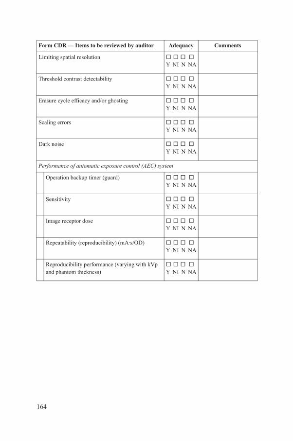
Limiting spatial resolution Y NI N NA
Threshold contrast detectability Y NI N NA
Erasure cycle efficacy and/or ghosting Y NI N NA
Scaling errors Y NI N NA
Dark noise Y NI N NA
Performance of automatic exposure control (AEC) system
Operation backup timer (guard) Y NI N NA
Sensitivity Y NI N NA
Image receptor dose Y NI N NA
Repeatability (reproducibility) (mA·s/OD) Y NI N NA
Reproducibility performance (varying with kVp and phantom thickness)
Y NI N NA
Form CDR — Items to be reviewed by auditor Adequacy Comments
164
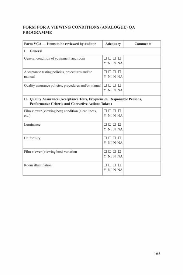
FORM FOR A VIEWING CONDITIONS (ANALOGUE) QA PROGRAMME
Form VCA — Items to be reviewed by auditor Adequacy Comments
I. General
General condition of equipment and room Y NI N NA
Acceptance testing policies, procedures and/or manual
Y NI N NA
Quality assurance policies, procedures and/or manual Y NI N NA
II. Quality Assurance (Acceptance Tests, Frequencies, Responsible Persons, Performance Criteria and Corrective Actions Taken)
Film viewer (viewing box) condition (cleanliness, etc.)
Y NI N NA
Luminance Y NI N NA
Uniformity Y NI N NA
Film viewer (viewing box) variation Y NI N NA
Room illumination Y NI N NA
165
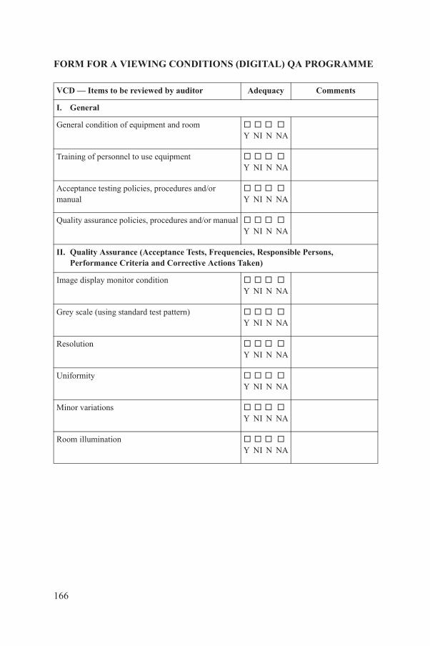
FORM FOR A VIEWING CONDITIONS (DIGITAL) QA PROGRAMME
VCD — Items to be reviewed by auditor Adequacy Comments
I. General
General condition of equipment and room Y NI N NA
Training of personnel to use equipment Y NI N NA
Acceptance testing policies, procedures and/or manual
Y NI N NA
Quality assurance policies, procedures and/or manual Y NI N NA
II. Quality Assurance (Acceptance Tests, Frequencies, Responsible Persons, Performance Criteria and Corrective Actions Taken)
Image display monitor condition Y NI N NA
Grey scale (using standard test pattern) Y NI N NA
Resolution Y NI N NA
Uniformity Y NI N NA
Minor variations Y NI N NA
Room illumination Y NI N NA
166
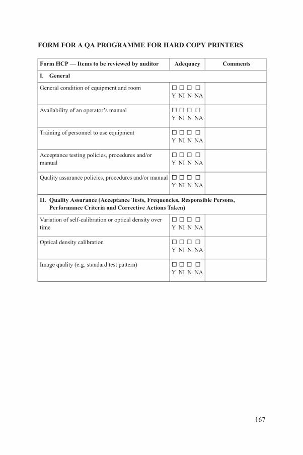
FORM FOR A QA PROGRAMME FOR HARD COPY PRINTERS
Form HCP — Items to be reviewed by auditor Adequacy Comments
I. General
General condition of equipment and room Y NI N NA
Availability of an operator’s manual Y NI N NA
Training of personnel to use equipment Y NI N NA
Acceptance testing policies, procedures and/or manual
Y NI N NA
Quality assurance policies, procedures and/or manual Y NI N NA
II. Quality Assurance (Acceptance Tests, Frequencies, Responsible Persons, Performance Criteria and Corrective Actions Taken)
Variation of self-calibration or optical density over time
Y NI N NA
Optical density calibration Y NI N NA
Image quality (e.g. standard test pattern) Y NI N NA
167
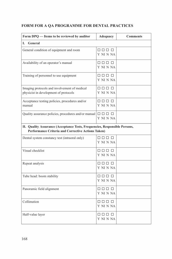
FORM FOR A QA PROGRAMME FOR DENTAL PRACTICES
Form DPQ — Items to be reviewed by auditor Adequacy Comments
I. General
General condition of equipment and room Y NI N NA
Availability of an operator’s manual Y NI N NA
Training of personnel to use equipment Y NI N NA
Imaging protocols and involvement of medical physicist in development of protocols
Y NI N NA
Acceptance testing policies, procedures and/or manual
Y NI N NA
Quality assurance policies, procedures and/or manual Y NI N NA
II. Quality Assurance (Acceptance Tests, Frequencies, Responsible Persons, Performance Criteria and Corrective Actions Taken)
Dental system constancy test (intraoral only) Y NI N NA
Visual checklist Y NI N NA
Repeat analysis Y NI N NA
Tube head: boom stability Y NI N NA
Panoramic field alignment Y NI N NA
168
Collimation Y NI N NA
Half-value layer Y NI N NA

Focal spot (at installation only) Y NI N NA
Timer accuracy and reproducibility Y NI N NA
Accuracy and reproducibility of kVp Y NI N NA
Linearity of mA Y NI N NA
Exposure reproducibility Y NI N NA
Technique chart evaluation Y NI N NA
III. Radiation Protection and Safety
Protective devices checked Y NI N NA
Room shielding checked Y NI N NA
Control of access to X ray room Y NI N NA
Warning lights/signs checked Y NI N NA
Door interlocks checked Y NI N NA
Radiation protection surveys (e.g. what is done, frequency, methods and records, and actions takenas a result)
Y NI N NA
Form DPQ — Items to be reviewed by auditor Adequacy Comments
169
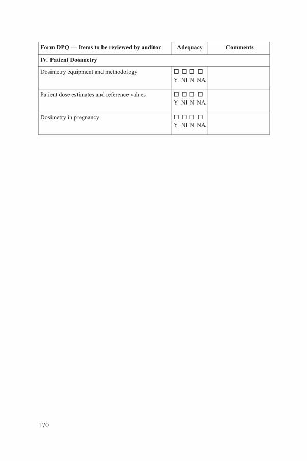
IV. Patient Dosimetry
Dosimetry equipment and methodology Y NI N NA
Patient dose estimates and reference values Y NI N NA
Dosimetry in pregnancy Y NI N NA
Form DPQ — Items to be reviewed by auditor Adequacy Comments
170
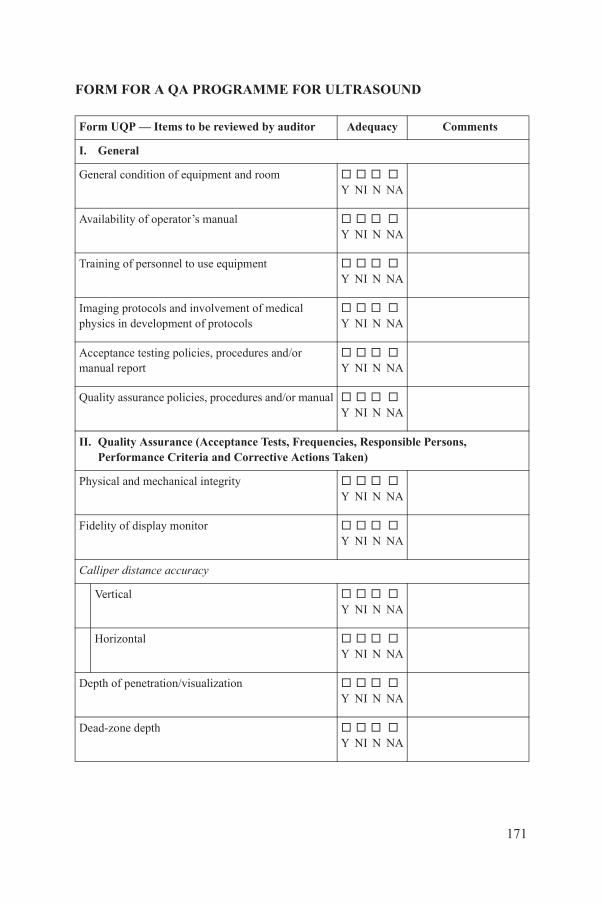
FORM FOR A QA PROGRAMME FOR ULTRASOUND
Form UQP — Items to be reviewed by auditor Adequacy Comments
I. General
General condition of equipment and room Y NI N NA
Availability of operator’s manual Y NI N NA
Training of personnel to use equipment Y NI N NA
Imaging protocols and involvement of medical physics in development of protocols
Y NI N NA
Acceptance testing policies, procedures and/or manual report
Y NI N NA
Quality assurance policies, procedures and/or manual Y NI N NA
II. Quality Assurance (Acceptance Tests, Frequencies, Responsible Persons, Performance Criteria and Corrective Actions Taken)
Physical and mechanical integrity Y NI N NA
Fidelity of display monitor Y NI N NA
Calliper distance accuracy
Vertical Y NI N NA
Horizontal Y NI N NA
Depth of penetration/visualization
171
Y NI N NA
Dead-zone depth Y NI N NA
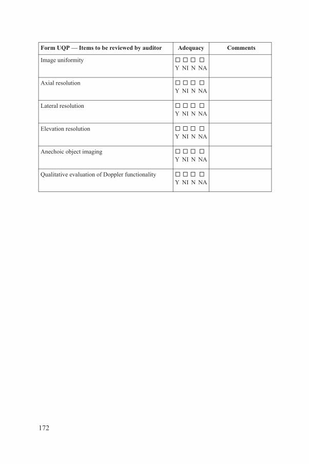
Image uniformity Y NI N NA
Axial resolution Y NI N NA
Lateral resolution Y NI N NA
Elevation resolution Y NI N NA
Anechoic object imaging Y NI N NA
Qualitative evaluation of Doppler functionality Y NI N NA
Form UQP — Items to be reviewed by auditor Adequacy Comments
172
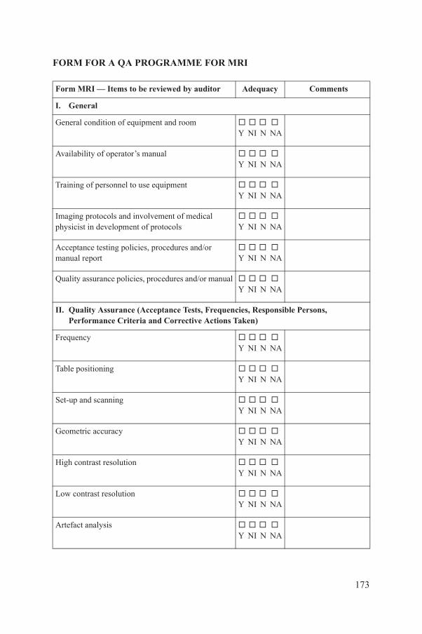
FORM FOR A QA PROGRAMME FOR MRI
Form MRI — Items to be reviewed by auditor Adequacy Comments
I. General
General condition of equipment and room Y NI N NA
Availability of operator’s manual Y NI N NA
Training of personnel to use equipment Y NI N NA
Imaging protocols and involvement of medical physicist in development of protocols
Y NI N NA
Acceptance testing policies, procedures and/or manual report
Y NI N NA
Quality assurance policies, procedures and/or manual Y NI N NA
II. Quality Assurance (Acceptance Tests, Frequencies, Responsible Persons, Performance Criteria and Corrective Actions Taken)
Frequency Y NI N NA
Table positioning Y NI N NA
Set-up and scanning Y NI N NA
Geometric accuracy Y NI N NA
High contrast resolution Y NI N NA
173
Low contrast resolution Y NI N NA
Artefact analysis Y NI N NA

Visual checklist Y NI N NA
Magnetic field homogeneity Y NI N NA
Slice position accuracy Y NI N NA
Slice thickness accuracy Y NI N NA
Radiofrequency coil checks
Volume radiofrequency coils Y NI N NA
Surface radiofrequency coils Y NI N NA
Interslice radiofrequency interference Y NI N NA
III. MRI Safety
MRI safety policies and procedures Y NI N NA
Site access restrictions
Zoning Y NI N NA
MRI personnel and non-MRI personnel Y NI N NA
Screening of patients and non-MRI personnel Y NI N NA
Screening of MRI personnel
Form MRI — Items to be reviewed by auditor Adequacy Comments
174
Y NI N NA
Screening of device and object Y NI N NA
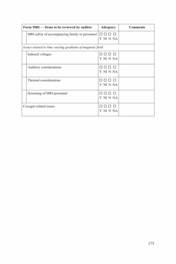
MRI safety of accompanying family or personnel Y NI N NA
Issues related to time varying gradients of magnetic field
Induced voltages Y NI N NA
Auditory considerations Y NI N NA
Thermal considerations Y NI N NA
Screening of MRI personnel Y NI N NA
Cryogen related issues Y NI N NA
Form MRI — Items to be reviewed by auditor Adequacy Comments
175

Annex IV
WORKSHEET FORMS FOR PHYSICS PROCEDURES
The audit team members should use the following worksheet forms for physics procedures as internal tools to assist them in conducting the audit review.
176
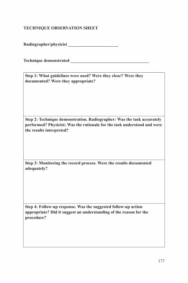
TECHNIQUE OBSERVATION SHEET
Radiographer/physicist _______________________
Technique demonstrated ____________________________________
Step 1: What guidelines were used? Were they clear? Were they documented? Were they appropriate?
Step 2: Technique demonstration. Radiographer: Was the task accurately performed? Physicist: Was the rationale for the task understood and were the results interpreted?
Step 3: Monitoring the record process. Were the results documented adequately?
Step 4: Follow-up response. Was the suggested follow-up action appropriate? Did it suggest an understanding of the reason for the
177
procedure?
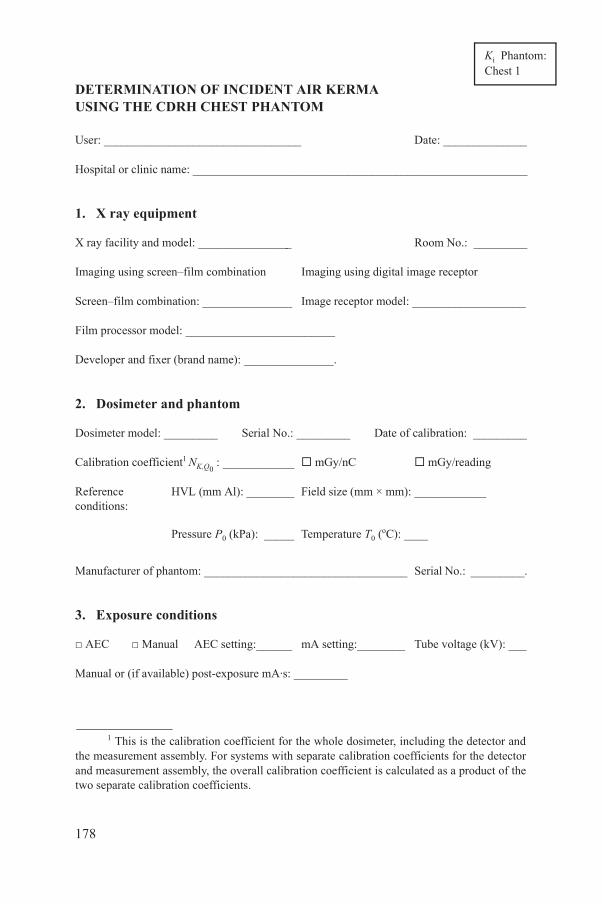
:
DETERMINATION OF INCIDENT AIR KERMAUSING THE CDRH CHEST PHANTOM
User: _________________________________ Date: ______________
Hospital or clinic name: ________________________________________________________
1. X ray equipment
X ray facility and model: ________________ Room No.: _________
Imaging using screen–film combination Imaging using digital image receptor
Screen–film combination: _______________ Image receptor model: ___________________
Film processor model: _________________________
Developer and fixer (brand name): _______________.
2. Dosimeter and phantom
Dosimeter model: _________ Serial No.: _________ Date of calibration: _________
Calibration coefficient1 NK,Q0 : ____________ mGy/nC mGy/reading
Reference conditions:
HVL (mm Al): ________ Field size (mm × mm): ____________
Pressure P0 (kPa): _____ Temperature T0 (oC): ____
Manufacturer of phantom: __________________________________ Serial No.: _________.
3. Exposure conditions
□ AEC □ Manual AEC setting:______ mA setting:________ Tube voltage (kV): ___
Manual or (if available) post-exposure mA·s: _________
Ki PhantomChest 1
178
1 This is the calibration coefficient for the whole dosimeter, including the detector and the measurement assembly. For systems with separate calibration coefficients for the detector and measurement assembly, the overall calibration coefficient is calculated as a product of the two separate calibration coefficients.
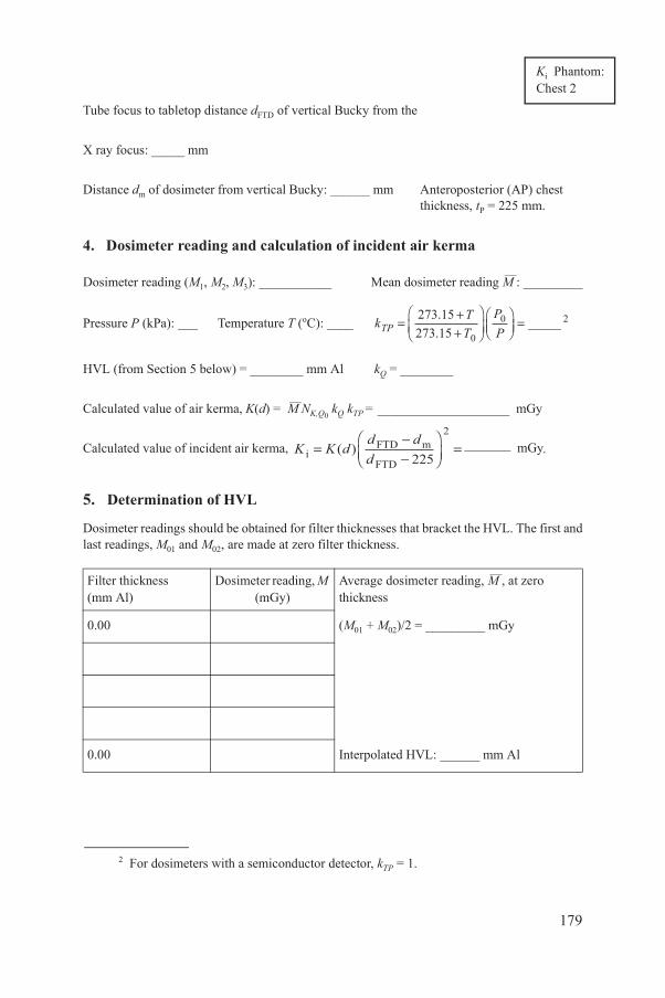
:
5. Determination of HVL
Tube focus to tabletop distance dFTD of vertical Bucky from the
X ray focus: _____ mm
Distance dm of dosimeter from vertical Bucky: ______ mm Anteroposterior (AP) chest thickness, tP = 225 mm.
4. Dosimeter reading and calculation of incident air kerma
Dosimeter reading (M1, M2, M3): ___________ Mean dosimeter reading M—
: _________
Pressure P (kPa): ___ Temperature T (oC): ____
HVL (from Section 5 below) = ________ mm Al kQ = ________
Calculated value of air kerma, K(d) = M—
NK,Q0 kQ kTP = ____________________ mGy
Calculated value of incident air kerma, _______ mGy.
Dosimeter readings should be obtained for filter thicknesses that bracket the HVL. The first and last readings, M01 and M02, are made at zero filter thickness.
Filter thickness(mm Al)
Dosimeter reading, M (mGy)
Average dosimeter reading, M—, at zero thickness
0.00 (M01 + M02)/2 = _________ mGy
0.00 Interpolated HVL: ______ mm Al
k TT
PPTP = +
+Ê
ËÁˆ
¯̃ÊËÁ
ˆ¯̃=273 15
273 15 0
0 2..
_____
K K dd ddi
FTD m
FTD=
--
ÊËÁ
ˆ¯̃
=( )225
2
Ki PhantomChest 2
179
2 For dosimeters with a semiconductor detector, kTP = 1.
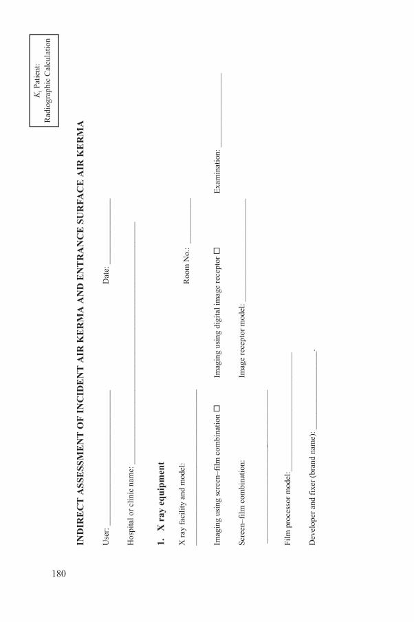
Ki P
atie
nt:
ESS
ME
NT
OF
IN
CID
EN
T A
IR K
ER
MA
AN
D E
NT
RA
NC
E S
UR
FAC
E A
IR K
ER
MA
____
____
____
____
____
_
Dat
e: _
____
____
____
___
me:
___
____
____
____
____
____
____
____
____
____
____
____
____
____
____
ent
del:
__
____
____
____
____
___
R
oom
No.
: __
____
____
_
–film
com
bina
tion
Im
agin
g us
ing
digi
tal i
mag
e re
cept
or
Exa
min
atio
n: _
____
____
____
____
_
atio
n:
____
____
____
____
____
__
I
mag
e re
cept
or m
odel
: ___
____
____
____
____
____
__
l:__
____
____
____
____
____
____
___
(bra
nd n
ame)
: ___
____
____
____
____
.
Rad
iogr
aphi
c C
alcu
latio
n
180
IND
IRE
CT
AS
S
Use
r: _
____
____
___
Hos
pita
l or
clin
ic n
a
1.
X r
ay e
qui
pm
X r
ay f
acil
ity
and
mo
____
____
____
____
_
Imag
ing
usin
g sc
reen
Scre
en–f
ilm c
ombi
n
___
____
____
____
_
Film
pro
cess
or m
ode
Dev
elop
er a
nd f
ixer
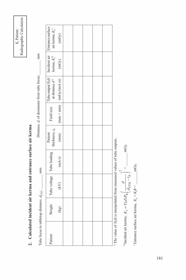
Ki P
atie
nt:
cid
ent
air
ker
ma
and
ent
ran
ce s
urf
ace
air
ker
ma
p di
stan
ce, d
FTD: _
____
___
mm
Dis
tanc
e, d
, of
dosi
met
er f
rom
tube
foc
us:_
____
___
mm
Wei
ght
Tub
e vo
ltage
Tub
e lo
adin
gPa
tien
t th
ickn
ess,
t PF
ield
siz
eT
ube
outp
ut Y
(d)
at d
ista
nce
da
Inci
dent
air
ke
rma,
Kib
Ent
ranc
e su
rfac
e ai
r ke
rma,
Kec
(kg)
(kV
)(m
A·s
)(m
m)
(mm
× m
m)
(mG
y/(m
A·s
))(m
Gy)
(mG
y)
s in
terp
olat
ed f
rom
mea
sure
d va
lues
of
tube
out
puts
.
=
___
____
mG
y.
r ke
rma,
Ke =
KiB
= _
____
__m
Gy.
KY
dP
dd
ti
ItFT
DP
=-
Ê ËÁˆ ¯̃
()
2
Rad
iogr
aphi
c C
alcu
latio
n
181
2.
Cal
cula
ted
in
Tub
e fo
cus
to ta
blet
o
Patie
nt
a The
val
ue o
f Y(
d) i
b In
cide
nt a
ir k
erm
a,
c E
ntra
nce
surf
ace
ai
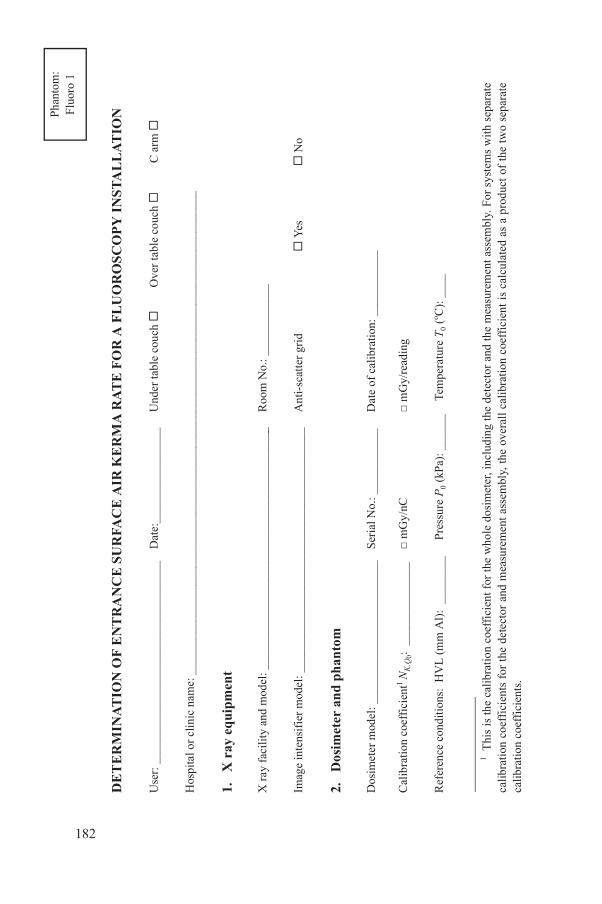
Pha
ntom
:
ON
OF
EN
TR
AN
CE
SU
RFA
CE
AIR
KE
RM
A R
AT
E F
OR
A F
LU
OR
OS
CO
PY
IN
ST
AL
LA
TIO
N
____
____
____
____
____
__D
ate:
____
____
____
____
Und
er ta
ble
couc
h
Ove
r ta
ble
couc
h
C a
rm
me:
___
____
____
____
____
____
____
____
____
____
____
____
____
____
____
____
____
____
____
____
__
ent
odel
: ___
____
____
____
____
____
____
____
____
____
__
Roo
m N
o.: _
____
____
___
del:
___
____
____
____
____
____
____
____
____
____
__A
nti-
scat
ter
grid
Yes
N
o
d p
han
tom
____
____
____
____
____
__S
eria
l No.
: ___
____
____
Dat
e of
cal
ibra
tion
: ___
____
____
nt1 N
K,Q
0: __
____
____
____
□ m
Gy/
nC□
mG
y/re
adin
g
s:H
VL
(m
m A
l):
____
____
P
ress
ure
P0
(kP
a): _
____
_Te
mpe
ratu
re T
0 (o C
): _
___
alib
rati
on c
oeff
icie
nt f
or th
e w
hole
dos
imet
er, i
nclu
ding
the
dete
ctor
and
the
mea
sure
men
t ass
embl
y. F
or s
yste
ms
wit
h se
para
te
ts f
or t
he d
etec
tor
and
mea
sure
men
t as
sem
bly,
the
ove
rall
cal
ibra
tion
coe
ffic
ient
is
calc
ulat
ed a
s a
prod
uct
of t
he t
wo
sepa
rate
ts
.
Flu
oro
1
182
DE
TE
RM
INA
TI
Use
r: _
____
____
___
Hos
pita
l or
clin
ic n
a
1.
X r
ay e
qui
pm
X r
ay f
acil
ity
and
m
Imag
e in
tens
ifie
r m
o
2.
Dos
imet
er a
n
Dos
imet
er m
odel
: __
Cal
ibra
tion
coe
ffic
ie
Ref
eren
ce c
ondi
tion
1 Thi
s is
the
cca
libra
tion
coe
ffic
ien
calib
rati
on c
oeff
icie
n
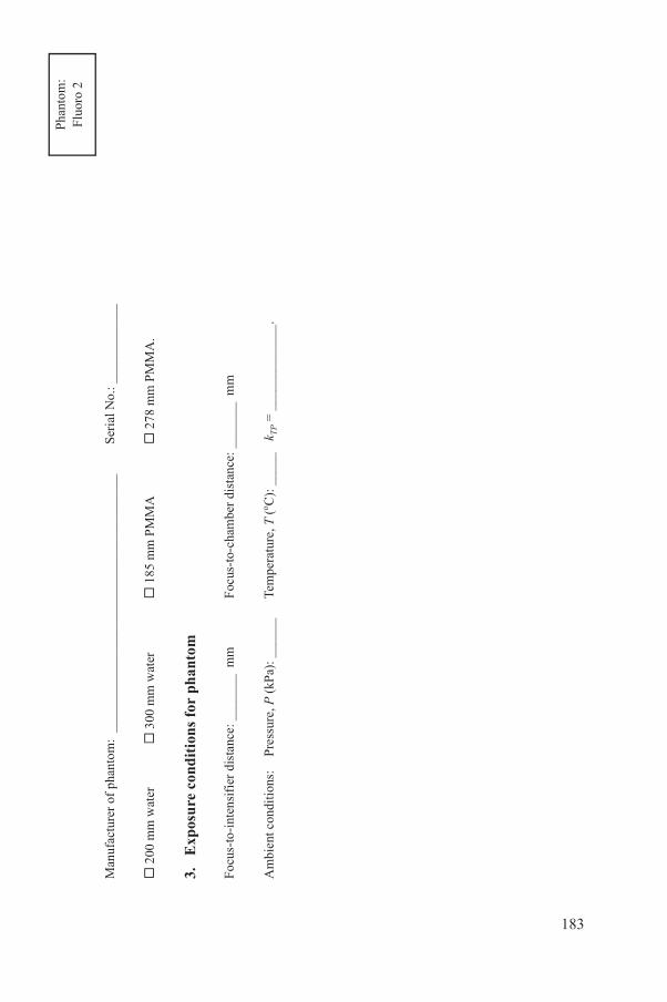
Pha
ntom
:
ntom
: __
____
____
____
____
____
____
____
____
____
_S
eria
l No.
: ___
____
____
_
3
00 m
m w
ater
185
mm
PM
MA
2
78 m
m P
MM
A.
dit
ion
s fo
r ph
anto
m
dist
ance
: ___
____
mm
F
ocus
-to-
cham
ber
dist
ance
: ___
____
mm
Pre
ssur
e, P
(kP
a): _
____
_
Tem
pera
ture
, T (
o C):
___
__ k
TP =
___
____
____
__.
Flu
oro
2
183
Man
ufac
ture
r of
pha
2
00 m
m w
ater
3.
Exp
osu
re c
on
Focu
s-to
-int
ensi
fier
Am
bien
t con
diti
ons:

Pha
ntom
:
adin
g an
d c
alcu
lati
on o
f en
tran
ce s
urf
ace
air
kerm
a ra
te
Tub
e vo
ltag
e(k
V)
Tub
e cu
rren
t(m
A)
Fil
trat
ion
(mm
Al)
Man
ual o
rau
to-m
ode
sett
inga
Fie
ld s
ize
(mm
× m
m)
Dos
imet
erre
adin
gs,
M· 1, M
· 2, M
· 3
(mG
y/m
in)
Mea
ndo
sim
eter
read
ing,
(m
Gy/
min
)
k Q
Ent
ranc
esu
rfac
e ai
rke
rma
rate
, K· eb
(mG
y/m
in)
or
auto
-mod
e de
tails
.
use
the
foll
owin
g fo
rmul
a:
, whe
re
tom
use
the
fol
low
ing
form
ula:
,
whe
re
k T
P =
1 f
or s
yste
ms
with
ture
and
pre
ssur
e co
rrec
tion
s an
d fo
r se
mic
ondu
ctor
dos
imet
ers.
M
KM
Nk
kK
TPe
,=
0k
T TP P
TP=
+ +Ê ËÁ
ˆ ¯̃Ê ËÁˆ ¯̃
273
227
32
0
0. .
.
KM
Nk
kB
BK
TPe
,w
PMM
A=
0k
T TP P
TP=
+ +Ê ËÁ
ˆ ¯̃Ê ËÁˆ ¯̃
273
227
32
0
0. .
;
Fluo
ro 3
184
4.
Dos
imet
er r
e
Sele
cted
opt
ion
aE
nter
M (
man
ual)
bF
or w
ater
pha
ntom
For
PM
MA
pha
n
auto
mat
ic te
mpe
ra
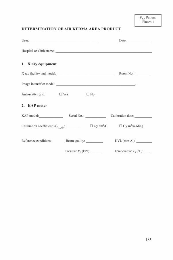
:
DETERMINATION OF AIR KERMA AREA PRODUCT
User: _______________________________________ Date: ______________
Hospital or clinic name: ________________________________________________________
1. X ray equipment
X ray facility and model: _________________________________ Room No.: _________
Image intensifier model: ______________________________________________.
Anti-scatter grid: Yes No
2. KAP meter
KAP model:______________ Serial No.: ____________ Calibration date: __________
Calibration coefficient, NPKA,Q0: ________ Gy·cm2/C Gy·m2/reading
Reference conditions: Beam quality: __________ HVL (mm Al): _________
Pressure P0 (kPa): _______ Temperature T0 (oC): ____.
PKA PatientFluoro 1
185

3. KAP reading and calculation of air kerma area product
Ambient conditions: Pressure, P (kPa): ______ Temperature, T (oC): ____ kTP = _________
Examinationdetails
Tube voltage
(kV)
AECsetting
KAP meterreading, M
kQ
Air kerma area product, PKA
a (Gy·cm2)
Fluoroscopy time (s)
a P MN k k TT
PPP Q TP TPKA ,KA
where = = ++
Ê
ËÁˆ
¯̃ÊËÁ
ˆ¯̃
, ..
.273 2273 2 0
0
PKA Patient:Fluoro 2
186
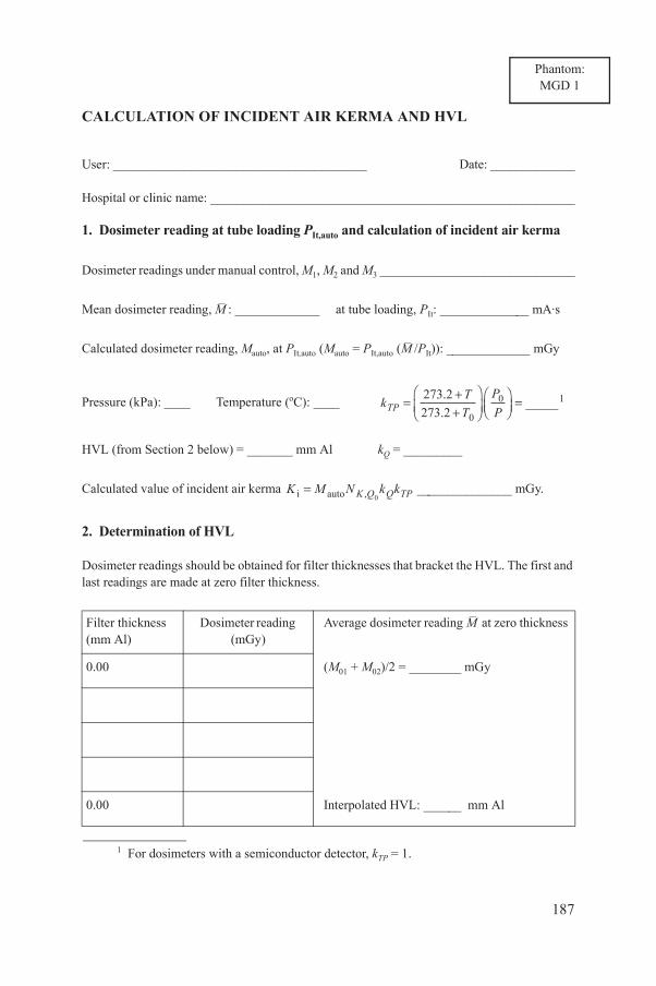
CALCULATION OF INCIDENT AIR KERMA AND HVL
User: _______________________________________ Date: _____________
Hospital or clinic name: ________________________________________________________
1. Dosimeter reading at tube loading PIt,auto and calculation of incident air kerma
Dosimeter readings under manual control, M1, M2 and M3 ______________________________
Mean dosimeter reading, M– : _____________ at tube loading, PIt: ______________ mA·s
Calculated dosimeter reading, Mauto, at PIt,auto (Mauto = PIt,auto (M– /PIt)): _____________ mGy
Pressure (kPa): ____ Temperature (oC): ____
HVL (from Section 2 below) = _______ mm Al kQ = _________
Calculated value of incident air kerma _______________ mGy.
2. Determination of HVL
Dosimeter readings should be obtained for filter thicknesses that bracket the HVL. The first and last readings are made at zero filter thickness.
Filter thickness(mm Al)
Dosimeter reading (mGy)
Average dosimeter reading M– at zero thickness
0.00 (M01 + M02)/2 = ________ mGy
k TT
PPTP = +
+Ê
ËÁˆ
¯̃ÊËÁ
ˆ¯̃=273 2
273 2 0
0 1..
_____
K M N k kK Q Q TPi auto ,=0
Phantom:MGD 1
187
0.00 Interpolated HVL: ______ mm Al
1 For dosimeters with a semiconductor detector, kTP = 1.
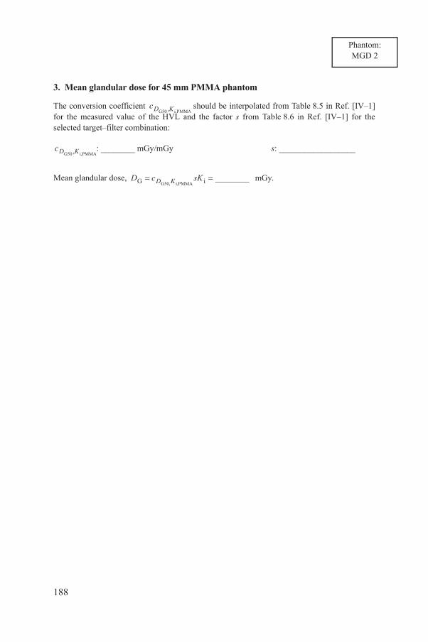
3. Mean glandular dose for 45 mm PMMA phantom
The conversion coefficient should be interpolated from Table 8.5 in Ref. [IV–1] for the measured value of the HVL and the factor s from Table 8.6 in Ref. [IV–1] for the selected target–filter combination:
: ________ mGy/mGy s: __________________
Mean glandular dose,
cD KG50 i,PMMA,
cD KG50 i,PMMA,
D c sKD KG iG50, i,PMMA mGy.= = ________
Phantom:MGD 2
188

MEASUREMENT OF Ca,100, CPMMA,100,c, CPMMA,100,p AND CALCULATION OF Cw
User: ____________________________________ Date: ________________
Hospital or clinic name: ________________________________________________________
1. X ray equipment
CT scanner model:_____________________________________ Room No.: _______.
2. Ionization chamber, electrometer and phantom
Dosimeter model: ___________________
Serial No.: _______ Calibration date:
____________
Calibration coefficient
NPKL,Q0:1__________________
mGy·cm/nC mGy·cm/reading
Reference conditions: Beam quality: ___________ HVL (mm Al): _________
Pressure, P0 (kPa): ________ Temperature, T0 (oC): ____
Manufacturer of phantom: ________________________ Serial No.: _______
Conditions during measurement:
Pressure (kPa): ____ Temperature (oC): ____ k TT
PPTP = +
+Ê
ËÁˆ
¯̃ÊËÁ
ˆ¯̃=273 2
273 2 0
0..
_______.
Phantom:CT1
189
1 This is the calibration coefficient for the whole dosimeter, including the detector and the measurement assembly. For systems with separate calibration coefficients for the detector and measurement assembly, the overall calibration coefficient is calculated as a product of the two separate calibration coefficients.

inde
x is
cal
cula
ted
usin
g1 :
inde
x C a
,100
filt
er, e
tc.)
Nom
inal
sli
ce
thic
knes
s, T
Num
ber
of s
lices
,N
Tub
e lo
adin
g,P
It
Dos
imet
er
read
ings
,M
1, M
2, M
3
Mea
n do
sim
eter
re
adin
g, M–
Cal
cula
ted
valu
e of
Ca,
100
Cal
cula
ted
valu
e of
nC
a,10
0
(mm
)(m
A·s
)(m
Gy)
(mG
y/(m
A·s
))
f 10
in
the
form
ulas
for
Ca,
100,
CP
MM
A,1
00,c a
nd C
PMM
A,1
00,p t
akes
acc
ount
of
the
use
of a
dos
imet
er c
alib
rati
on i
n m
Gy·
cm a
nd a
fi
ed in
mm
.
CnT
MN
kk
PQ
QTP
aK
L,
,;
100
100
=nC
CP
aa
It,
,.
100
100
=
Pha
ntom
:C
T2
190
The
CT
air
ker
ma
3.
CT
air
ker
ma
Sca
nner
set
ting
s (t
ube
volta
ge, b
eam
1 T
he f
acto
r o
slic
e th
ickn
ess
spec
i
REFERENCE TO ANNEX IV
[IV–1] INTERNATIONAL ATOMIC ENERGY AGENCY, Dosimetry in Diagnostic Radiology: An International Code of Practice, Technical Reports Series No. 457, IAEA, Vienna (2007).
191

Annex V
CONTENTS OF THE ATTACHED CD
Comprehensive Clinical Audits of Diagnostic Radiology Practices: A Tool for Quality Improvement
Audit report template
Audit report forms
Audit application form
List of items requested to be available on-site
Equipment-specific checklist forms
Worksheet forms for physics procedures
192

CONTRIBUTORS TO DRAFTING AND REVIEW
Abdullah, B. University of Malaya, Malaysia
Butler, P. American College of Radiology,United States of America
Christofides, S. European Federation of Organisationsfor Medical Physics
Ebdon-Jackson, S. Health Protection Agency, United Kingdom
Faulkner, K. Newcastle General Hospital, United Kingdom
Järvinen, H. Finnish Centre for Radiation and Nuclear Safety (STUK), Finland
Le Heron, J. International Atomic Energy Agency
McLean, I.D. International Atomic Energy Agency
Pentecost, M. American College of Radiology,United States of America
Rickard, M. Sydney Breast Clinic, Australia
Slack, K. Health Protection Agency, United Kingdom
Thomson, K. Asian–Oceanian Society of Radiology
Consultants Meetings
Vienna, Austria: 23–27 April 2007, 10–14 December 2007, 31 March–11 May 2008, 3–7 November 2008
193

In the following countries IAEA publications may be purchased from the sources listed below, or frommajor local booksellers. Payment may be made in local currency or with UNESCO coupons.
AustraliaDA Information Services, 648 Whitehorse Road, Mitcham Victoria 3132Telephone: +61 3 9210 7777 � Fax: +61 3 9210 7788Email: [email protected] � Web site: http://www.dadirect.com.au
BelgiumJean de Lannoy, avenue du Roi 202, B-1190 BrusselsTelephone: +32 2 538 43 08 � Fax: +32 2 538 08 41Email: [email protected] � Web site: http://www.jean-de-lannoy.be
CanadaBernan Associates, 4611-F Assembly Drive, Lanham, MD 20706-4391, USATelephone: 1-800-865-3457 � Fax: 1-800-865-3450Email: [email protected] � Web site: http://www.bernan.com
Renouf Publishing Company Ltd., 1-5369 Canotek Rd., Ottawa, Ontario, K1J 9J3Telephone: +613 745 2665 � Fax: +613 745 7660Email: [email protected] � Web site: http://www.renoufbooks.com
ChinaIAEA Publications in Chinese: China Nuclear Energy Industry Corporation, Translation Section, P.O. Box 2103, Beijing
Czech RepublicSuweco CZ, S.R.O. Klecakova 347, 180 21 Praha 9Telephone: +420 26603 5364 � Fax: +420 28482 1646Email: [email protected] � Web site: http://www.suweco.cz
FinlandAkateeminen Kirjakauppa, PL 128 (Keskuskatu 1), FIN-00101 HelsinkiTelephone: +358 9 121 41 � Fax: +358 9 121 4450Email: [email protected] � Web site: http://www.akateeminen.com
FranceForm-Edit, 5, rue Janssen, P.O. Box 25, F-75921 Paris Cedex 19Telephone: +33 1 42 01 49 49 � Fax: +33 1 42 01 90 90 � Email: [email protected]
Lavoisier SAS, 14 rue de Provigny, 94236 Cachan Cedex Telephone: + 33 1 47 40 67 00 � Fax +33 1 47 40 67 02 Email: [email protected] � Web site: http://www.lavoisier.fr
GermanyUNO-Verlag, Vertriebs- und Verlags GmbH, August-Bebel-Allee 6, D-53175 BonnTelephone: +49 02 28 949 02-0 � Fax: +49 02 28 949 02-22Email: [email protected] � Web site: http://www.uno-verlag.de
HungaryLibrotrade Ltd., Book Import, P.O. Box 126, H-1656 BudapestTelephone: +36 1 257 7777 � Fax: +36 1 257 7472 � Email: [email protected]
IndiaAllied Publishers Group, 1st Floor, Dubash House, 15, J. N. Heredia Marg, Ballard Estate, Mumbai 400 001,Telephone: +91 22 22617926/27 � Fax: +91 22 22617928Email: [email protected] � Web site: http://www.alliedpublishers.com
Bookwell, 24/4800, Ansari Road, Darya Ganj, New Delhi 110002Telephone: +91 11 23268786, +91 11 23257264 � Fax: +91 11 23281315Email: [email protected] � Web site: http://www.bookwellindia.com
ItalyLibreria Scientifica Dott. Lucio di Biasio �AEIOU�, Via Coronelli 6, I-20146 MilanTelephone: +39 02 48 95 45 52 or 48 95 45 62 � Fax: +39 02 48 95 45 48
JapanMaruzen Company, Ltd., 13-6 Nihonbashi, 3 chome, Chuo-ku, Tokyo 103-0027Telephone: +81 3 3275 8582 � Fax: +81 3 3275 9072Email: [email protected] � Web site: http://www.maruzen.co.jp
Where to order IAEA publications
No. 21, July 2006
Korea, Republic of�KINS Inc., Information Business Dept. Samho Bldg. 2nd Floor, 275-1 Yang Jae-dong SeoCho-G, Seoul 137-130�Telephone: +02 589 1740 � Fax: +02 589 1746�Email: [email protected] � Web site: http://www.kins.co.kr�
Netherlands�Martinus Nijhoff International, Koraalrood 50, P.O. Box 1853, 2700 CZ Zoetermeer�Telephone: +31 793 684 400 � Fax: +31 793 615 698 � Email: [email protected] � Web site: http://www.nijhoff.nl�
Swets and Zeitlinger b.v., P.O. Box 830, 2160 SZ Lisse�Telephone: +31 252 435 111 � Fax: +31 252 415 888 � Email: [email protected] � Web site: http://www.swets.nl�
New Zealand�DA Information Services, 648 Whitehorse Road, MITCHAM 3132, Australia�Telephone: +61 3 9210 7777 � Fax: +61 3 9210 7788�Email: [email protected] � Web site: http://www.dadirect.com.au�
Slovenia�Cankarjeva Zalozba d.d., Kopitarjeva 2, SI-1512 Ljubljana�Telephone: +386 1 432 31 44 � Fax: +386 1 230 14 35�Email: [email protected] � Web site: http://www.cankarjeva-z.si/uvoz�
Spain�Díaz de Santos, S.A., c/ Juan Bravo, 3A, E-28006 Madrid�Telephone: +34 91 781 94 80 � Fax: +34 91 575 55 63 � Email: [email protected]�[email protected] � [email protected] � [email protected] Web site: http://www.diazdesantos.es�
United Kingdom�The Stationery Office Ltd, International Sales Agency, PO Box 29, Norwich, NR3 1 GN�Telephone (orders): +44 870 600 5552 � (enquiries): +44 207 873 8372 � Fax: +44 207 873 8203�Email (orders): [email protected] � (enquiries): [email protected] � Web site: http://www.tso.co.uk�
On-line orders:�DELTA Int. Book Wholesalers Ltd., 39 Alexandra Road, Addlestone, Surrey, KT15 2PQ�Email: [email protected] � Web site: http://www.profbooks.com�
Books on the Environment:�Earthprint Ltd., P.O. Box 119, Stevenage SG1 4TP�Telephone: +44 1438748111 � Fax: +44 1438748844�Email: [email protected] � Web site: http://www.earthprint.com�
United Nations (UN)�Dept. I004, Room DC2-0853, First Avenue at 46th Street, New York, N.Y. 10017, USA�Telephone: +800 253-9646 or +212 963-8302 � Fax: +212 963-3489�Email: [email protected] � Web site: http://www.un.org�
United States of America�Bernan Associates, 4611-F Assembly Drive, Lanham, MD 20706-4391�Telephone: 1-800-865-3457 � Fax: 1-800-865-3450�Email: [email protected] � Web site: http://www.bernan.com�
Renouf Publishing Company Ltd., 812 Proctor Ave., Ogdensburg, NY, 13669�Telephone: +888 551 7470 (toll-free) � Fax: +888 568 8546 (toll-free)�Email: [email protected] � Web site: http://www.renoufbooks.com�
Orders and requests for information may also be addressed directly to: �
����������� ���� Unit, International Atomic Energy Agency�Vienna International Centre, PO Box 100, 1400 Vienna, Austria�Telephone: +43 1 2600 22529 (or 22530) � Fax: +43 1 2600 29302 �Email: [email protected] � Web site: http://www.iaea.org/books�
INTERNATIONAL ATOMIC ENERGY AGENCYVIENNA
ISBN 978–92–0–112009–0 ISSN 2075–3772
Currently there is great interest in quality assurance processes and quality improvement in diagnostic radiology. This publication is a guide for the organization and conduct of a comprehensive clinical audit. It includes a structured set of standards appropriate for diagnostic radiology and an audit guide to their clinical review. The data collection sheets included, which have already been successfully tested in pilot studies, have been designed for the rapid production of reports.
IAEA HumAn HEAltH SErIES
Com
prehensive Clinical A
udits of Diagnostic R
adiology Practices:
A Tool for Q
uality Improvem
entIAEA Hum
An HEAltH SErIES no. 4
IAEA HumAn HEAltH SErIESno. 4
Comprehensive Clinical Audits of Diagnostic Radiology
Practices: A Tool for Quality Improvement
Quality Assurance Audit for Diagnostic Radiology Improvement and Learning (QUAADRIL)
09-43621_P1425_cover.indd 1 2010-01-22 10:59:08