Embed Size (px)
Citation preview


Comprehensive
Cardiovascular Risk
Stratification
Role of Biomarkers
Szilard Voros, MD, FACC,
FSCCT, FAHA

The Main Message
Advanced biomarker testing in
aggregate provides refined risk-
stratification for cardiovascular
disease, and individual biomarkers
provide mechanistic explanation and
individualized treatment targets, such
that targeted therapies will improve
overall cardiovascular outcomes

Main Points
1. ApoB-containing lipoprotein particles are potentially atherogenic; the best clinical measure of overall atherogenic particle burden is a measure of all circulating apoB- containing particles
2. A mal-adaptive inflammation is a response to the primary injury of atherogenic lipoprotein deposition; elevated serum inflammatory markers indicate that a systemic response has been mounted against atherogenic lipoprotein deposition
3. Traditional risk-assessment only provides a population-based probability and no insight into individual abnormalities in individual patients
4. A biology-based risk assessment provides mechanistic explanation for the patients’ atherosclerotic risk, and provides tangible treatment targets

Storyline
1. CV Disease Burden is High
2. We know the steps leading to CVD
3. We can assess cardiovascular risk
4. We can treat cardiovascular risk factors
5. Treating cardiovascular risk factors improves outcomes
6. CV Disease Burden is Reduced

Storyline
1. High burden of cardiovascular disease
2. Development of cardiovascular disease
3. Assessment of cardiovascular disease risk
4. Management of CVD risk factors
5. Improving patient outcomes by CVD risk
factor management

High Burden of Cardiovascular
Disease
Roger et al. Circulation 2011,
123:e18-e209.

Storyline
1. High burden of cardiovascular disease
2. Development of cardiovascular disease
3. Assessment of cardiovascular disease risk
4. Management of CVD risk factors
5. Improving patient outcomes by CVD risk
factor management

Development of Cardiovascular
Disease
Genetic Factors Intermediate
Phenotypes
Ultimate
Phenotype
Environmental Factors
DNA RNA Lipoproteins,
Inflammation, BP Atherosclerosis
Biomarkers Imaging

Atherosclerosis: Plaque
Fixed Stenosis
Demand Ischemia
Necrosis/Fibrosis
Exertional Angina
Resting Ischemia Rest Angina
Dyspnea
Myocardial Dysfunction Dyspnea
End-Organ Damage Renal, hepatic dysfunction
Genetic Predisposition
DNA Environmental Factors
Gene Expression (RNA)
Atherogenic milieu (Lipoproteins, IR, BP, shear)
Plaque Rupture: ACS
Resting chest pain Thrombosis: ACS

Atherosclerosis: Plaque
Fixed Stenosis
Demand Ischemia
Necrosis/Fibrosis
Resting Ischemia
Myocardial Dysfunction
End-Organ Damage
Genetic Predisposition
DNA Environmental Factors
Gene Expression (RNA)
Atherogenic milieu
(Lipoproteins, IR, BP, shear)
Plaque Rupture: ACS
Resting chest pain Thrombosis: ACS
Genotyping:
ApoE, Factor II, V
CYP2C9
Lipoprotein testing
(FLP, apoB, LDL-P, sdLDL, Lp(a),
apoA, HDL-P, HDL classes, sterols,
FFA)
Insulin resistance, MS, DM
(insulin, glu, HbA1c, leptin, AN)
Inflammatory markers
(hs-CRP, LpPLA2, MPO, fibrinogen)
hs-cTn
hs-cTn
hs-cTn, galectin 3
NT-proBNP
creat, cystatin-C
Hepatic panel
VitD, PTH, Ca, Phos

Development of Atherosclerosis
Development of Cardiovascular Disease

1. Atherogenic lipoprotein retention
2. Maladaptive inflammation
3. Apoptosis/Necrosis
4. Calcification
5. Fibrosis
Development of Cardiovascular Disease Atherosclerosis

Lipoprotein
Deposition
Maladaptive
Inflammation
Apoptosis
VSMC
LRNC
Calcification Fibrosis
Deposition of
apoB particles
• LDL
• sdLDL
• Lp(a)
• Chylo-R
Counteracted
by apoA/HDL
• ApoA
• HDL
• Large HDL
Inflammarory
Response
• CRP
• LpPLA2
• MPO
• Fibronigen
Ca-Metabolism
• Ca, Phos
• VitD
• PTH
Development of Cardiovascular Disease Atherosclerosis: Major Steps

1. Atherogenic lipoprotein retention
2. Maladaptive inflammation
3. Apoptosis/Necrosis
4. Calcification
5. Fibrosis
Development of Cardiovascular Disease Atherosclerosis: Major Steps

Lipoproteins
Lipids Proteins
(Apoproteins)
Development of Cardiovascular Disease Atherosclerosis: Lipoprotein Retention

Lipoproteins
Apo B Apo A
Intestines
Apo B48
Liver
Apo B100
Chylomicrons
ApoB48 VLDL
Apo B100
IDL Apo B100
LDL Apo B100
Lp(a) Apo B100
HDL Apo AI
Development of Cardiovascular Disease Atherosclerosis: Lipoprotein Retention
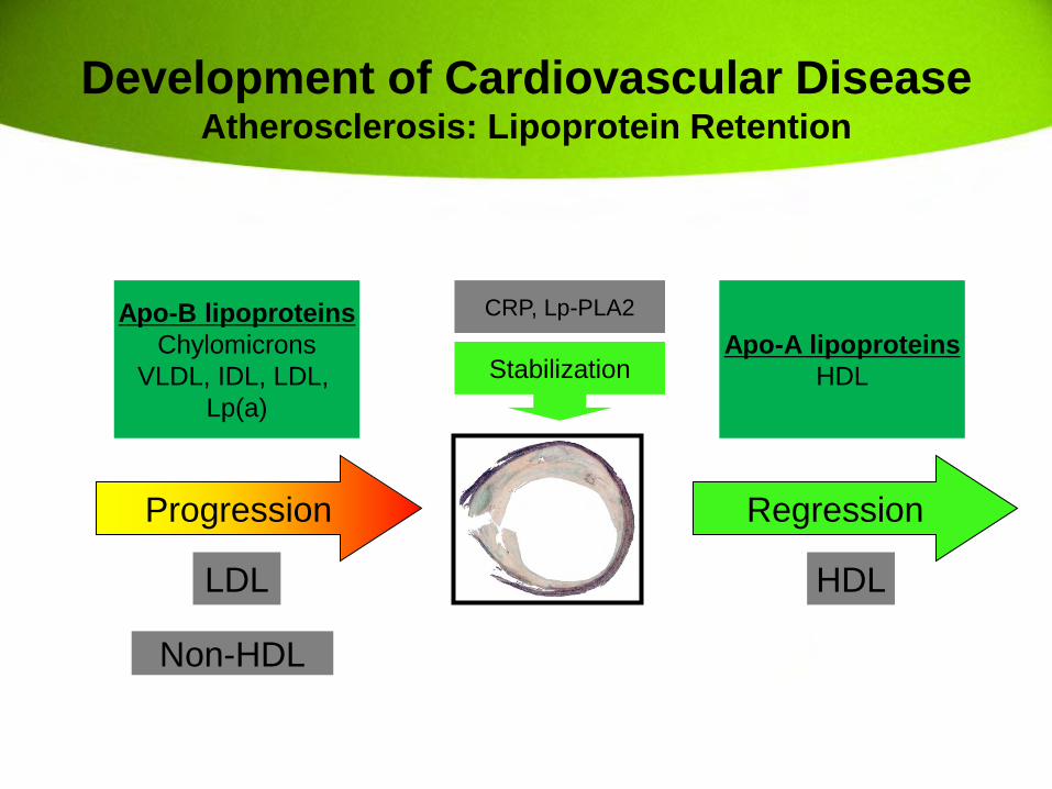
Progression Regression
Apo-B lipoproteins
Chylomicrons
VLDL, IDL, LDL,
Lp(a)
LDL
Non-HDL
Apo-A lipoproteins
HDL
HDL
Stabilization
CRP, Lp-PLA2
Development of Cardiovascular Disease Atherosclerosis: Lipoprotein Retention

1. ApoB-containing lipoprotein particles are atherogenic
• LDL-particles
• Small-dense LDL particles
• Lp(a) particles
• Chylomicron remnants
Development of Cardiovascular Disease Atherosclerosis: Lipoprotein Retention

Vazquez, Anderson, Voros. 2009.
Development of Cardiovascular Disease Atherosclerosis: Lipoprotein Retention

Joshi, Krivitsky, Qian, Vazquez,
Voros, Miller. Current Treatment
Options in Cardiovascular
Medicine (2010) 12:396–407.
Development of Cardiovascular Disease Atherosclerosis: Lipoprotein Retention

Take Home Message #1:
…all apoB-containing lipoprotein particles are potentially atherogenic…
…the best clinical measure of overall atherogenic particle burden is a measure of all circulating apoB containing
particles…
• ApoB, non-HDL-C, (LDL-P) – Specific apoB-containing atherogenic particle subtypes:
• Lp(a)
• sdLDL
Development of Cardiovascular Disease Atherosclerosis: Lipoprotein Retention

Lipoprotein
Deposition
Maladaptive
Inflammation
Apoptosis
VSMC
LRNC
Calcification Fibrosis
Inflammarory
Response
• CRP
• LpPLA2
• Fibronigen
Ca-Metabolism
• Ca, Phos
• VitD
• PTH
Development of Cardiovascular Disease Atherosclerosis: Major Steps
Atherogenic LP:
TC, ApoB, LDL-
C, LDL-P, sdLDL,
Lp(a), TG, FFA
Protective LP:
ApoA, HDL-C,
HDL subclasses

2. ApoA-containing lipoprotein particles participate in reverse cholesterol transport, and their maturation by removing excess
cholesterol is protective against atherosclerosis
• ApoA
• HDL particles
Development of Cardiovascular Disease Atherosclerosis: Reverse Cholesterol Transport

Lipoprotein
Deposition
Maladaptive
Inflammation
Apoptosis
VSMC
LRNC
Calcification Fibrosis
Deposition of
apoB particles
• LDL
• sdLDL
• Lp(a)
• Chylo-R
Counteracted
by apoA/HDL
• ApoA
• HDL
• Large HDL
Inflammarory
Response
• CRP
• LpPLA2
• Fibronigen
Ca-Metabolism
• Ca, Phos
• VitD
• PTH
Development of Cardiovascular Disease Atherosclerosis: Major Steps

1. Atherogenic lipoprotein retention
2. Maladaptive inflammation
3. Apoptosis/Necrosis
4. Calcification
5. Fibrosis
Development of Cardiovascular Disease Atherosclerosis: Major Steps

Development of Cardiovascular Disease Atherosclerosis: Mal-Adaptive Inflammation
Macrophage Infiltration

Lipoprotein
Deposition
Macrophage
recruitment
Cytokine release IL-6 Liver CRP Plaque LpPLA-2
Development of Cardiovascular Disease Atherosclerosis: Mal-Adaptive Inflammation

… There is no CRP found in
atherosclerotic plaques…
(only in miniscule amounts)
Development of Cardiovascular Disease Atherosclerosis: Mal-Adaptive Inflammation

Mannheim et al. Stroke.
2008;39:1448-1455.
Development of Cardiovascular Disease Atherosclerosis: Mal-Adaptive Inflammation

Development of Cardiovascular Disease Atherosclerosis: Mal-Adaptive Inflammation
Take Home Message #2:
…mal-adaptive inflammation is a response to the primary injury of atherogenic lipoprotein deposition…
…elevated serum inflammatory markers indicate that a systemic response has been mounted against atherogenic lipoprotein
deposition…
• CRP
• LpPLA2
• MPO
• (Fibrinogen, IL-6, SAA)

Lipoprotein
Deposition
Maladaptive
Inflammation
Apoptosis
VSMC
LRNC
Calcification Fibrosis
Ca-Metabolism
• Ca, Phos
• VitD
• PTH
Development of Cardiovascular Disease Atherosclerosis: Major Steps
Atherogenic LP:
TC, ApoB, LDL-
C, LDL-P, sdLDL,
Lp(a), TG, FFA
Protective LP:
ApoA, HDL-C,
HDL subclasses
Inflammatory
Markers:
CRP
LpPLA2
MPO
Fibrinogen

1. Atherogenic lipoprotein retention
2. Maladaptive inflammation
3. Apoptosis/Necrosis
4. Calcification
5. Fibrosis
Development of Cardiovascular Disease Atherosclerosis: Major Steps

ACP Amorphous
Calcium Phosphate
Nucleation CHA Calcium
Hydroxyapatite
Bone Formation: Osteoblasts (ALP)
Bone Degradation (Osteoclasts: Acidic)
VSMC
Osteoblastic Transformation
Ox-LDL, AngII, TNF-alpha SMA rOAT1
Adijiang et al. Nephrol Dial Transplant
2008;23:1892-1901.
Development of Cardiovascular Disease Atherosclerosis: Calcification

Lipoprotein
Deposition
Maladaptive
Inflammation
Apoptosis
VSMC
LRNC
Calcification Fibrosis
Development of Cardiovascular Disease Atherosclerosis: Major Steps
Atherogenic LP:
TC, ApoB, LDL-
C, LDL-P, sdLDL,
Lp(a), TG, FFA
Protective LP:
ApoA, HDL-C,
HDL subclasses
Inflammatory
Markers:
CRP
LpPLA2
Fibrinogen
Ca-
Metabolism
Ca, Phos
VitD
PTH

Atherosclerosis: Plaque
Fixed Stenosis
Demand Ischemia
Necrosis/Fibrosis
Resting Ischemia
Myocardial Dysfunction
End-Organ Damage
Genetic Predisposition
DNA Environmental Factors
Gene Expression (RNA)
Atherogenic milieu
(Lipoproteins, IR, BP, shear)
Plaque Rupture: ACS
Resting chest pain Thrombosis: ACS
Genotyping:
ApoE, Factor II, V
CYP2C9
Lipoprotein testing
(FLP, apoB, LDL-P, sdLDL,
Lp(a),
apoA, HDL-P, HDL classes,
sterols,
FFA)
Insulin resistance, MS, DM
(insulin, glu, HbA1c, leptin,
AN)
Inflammatory markers
(hs-CRP, LpPLA2, MPO,
fibrinogen)
hs-cTn
hs-cTn
hs-cTn, galectin 3
NT-proBNP
creat, cystatin-C
Hepatic panel
VitD, PTH, Ca, Phos

Storyline
1. High burden of cardiovascular disease
2. Development of cardiovascular disease
3. Assessment of cardiovascular disease risk
4. Management of CVD risk factors
5. Improving patient outcomes by CVD risk
factor management

The Main Message
Advanced biomarker testing in
aggregate provides refined risk-
stratification for cardiovascular
disease, and individual biomarkers
provide mechanistic explanation and
individualized treatment targets, such
that targeted therapies will improve
overall cardiovascular outcomes

Assessment of Cardiovascular Risk
1. Traditional paradigm
– Population-based
– Probabilistic
2. Biology-based paradigm
– Individualized
– Mechanistic and pragmatic

Assessment of Cardiovascular Risk
1. Traditional paradigm
– Population-based
– Probabilistic
2. Biology-based paradigm
– Individualized
– Mechanistic and pragmatic

http://hp2010.nhlbihin.net/atpiii/cal
culator.asp
1. Age
2. Gender
3. Tobacco
4. Blood pressure
5. Total cholesterol
6. HDL-cholesterol
7. (Diabetes)
http://hp2010.nhlbihin.net/atpiii/calculator.asp
Framingham, MA
Assessment of Cardiovascular Risk Traditional Risk Assessment

NCEP Expert Panel. NIH
Publication 02-5215.
…The FRS tells you that for example,
the probability of a cardiovascular
event in a given individual is 15% in the
next 10 years...
“Probability Paradigm”
Assessment of Cardiovascular Risk Traditional Risk Assessment

1. Low Risk: < 10% 10-Year Risk
2. Intermediate Risk: 10-20% 10-Year Risk
3. High Risk: > 10 % 10-Year Risk
Assessment of Cardiovascular Risk Traditional Risk Assessment

…This all sounds very nice, but…
…Almost not a single physician actually calculates the Framingham score…
…They “eyeball” the risk-factors; probably count them and come up with a “ballpark” estimate…
“I think this patient is probably low risk”
Assessment of Cardiovascular Risk Traditional Risk Assessment

PR Consent on File at PHI
Assessment of Cardiovascular Risk Traditional Risk Assessment

PR Consent on File at PHI
Lisa: Age 38; no heart disease
• Exercises
• Height: 5’3”
• Weight: 165
• No diabetes
• No tobacco
• Pre-menopausal
Assessment of Cardiovascular Risk Traditional Risk Assessment

What is her 10-year risk?
1. < 1%
2. 1-10%
3. 10-20%
4. > 20%
Answer <1%
“Low Risk”
Traditional Risk
Assessment
1. Age: 38
2. Gender: Female
3. Total cholesterol: 165
4. HDL cholesterol: 48
5. Tobacco: Never
6. High BP: Yes
Exercise
Diet
Flu shot
Pap-Smear
Seatbelt
Rx
Assessment of Cardiovascular Risk Traditional Risk Assessment
Low Risk

Main Point #3
…the traditional evaluation only provides
a population-based probability and no
insight into individual abnormalities in
individual patients…
Assessment of Cardiovascular Risk Traditional Risk Assessment

Assessment of Cardiovascular Risk
1. Traditional paradigm
– Population-based
– Probabilistic
2. Biology-based paradigm
– Individualized
– Mechanistic and pragmatic

Atherosclerosis: Plaque
Fixed Stenosis
Demand Ischemia
Necrosis/Fibrosis
Resting Ischemia
Myocardial Dysfunction
End-Organ Damage
Genetic Predisposition
DNA Environmental Factors
Gene Expression (RNA)
Atherogenic milieu
(Lipoproteins, IR, BP, shear)
Plaque Rupture: ACS
Resting chest pain Thrombosis: ACS
Genotyping:
ApoE, Factor II, V
CYP2C9
Lipoprotein testing
(FLP, apoB, LDL-P, sdLDL, Lp(a),
apoA, HDL-P, HDL classes, sterols,
FFA)
Insulin resistance, MS, DM
(insulin, glu, HbA1c, leptin, AN)
Inflammatory markers
(hs-CRP, LpPLA2, MPO, fibrinogen)
hs-cTn
hs-cTn
hs-cTn, galectin 3
NT-proBNP
creat, cystatin-C
Hepatic panel
VitD, PTH, Ca, Phos

1. Evaluating genetic susceptibility
2. Evaluating the current atherogenic milieu
3. Evaluating myocardial ischemia, necrosis
4. Evaluating myocardial fibrosis
5. Evaluating myocardial dysfunction
6. Evaluating end-organ damage
Assessment of Cardiovascular Risk Biology-Based Evaluation

1. Evaluating genetic susceptibility
1. Family history
2. Genotypes (DNA; SNP’s)
2. Evaluating the current atherogenic milieu
3. Evaluating myocardial ischemia, necrosis
4. Evaluating myocardial fibrosis
5. Evaluating myocardial dysfunction
6. Evaluating end-organ damage
Assessment of Cardiovascular Risk Biology-Based Evaluation

1. Genotyping for risk-stratification
– Genes identified in GWAS
2. Genotyping for intermediate phenotypes
– ApoE: LDL vs. TG-related problems
• Aids with dietary counseling
• ?Aids with statin versus fibrate selection
– CYP2C9 and VKORC1: Aids with warfarin dosing
– CYP2C19: aids with clopidogrel resistance
Assessment of Cardiovascular Risk Evaluation of Genetic Susceptibility: Genotyping

1. Evaluating genetic susceptibility
2. Evaluating the current atherogenic milieu 1. (Gene expression: RNA)
2. Lipoproteins 1. Atherogenic particle burden: apoB-particles
2. Protective particles: apoA/HDL particles
3. Inflammation: CRP, LpPLA2, MPO, fibrinogen
4. Insulin-resistant states: Glu, HbA1c, insulin, leptin, adiponectin, etc.
3. Evaluating myocardial ischemia, necrosis
4. Evaluating myocardial fibrosis
5. Evaluating myocardial dysfunction
6. Evaluating end-organ damage
Assessment of Cardiovascular Risk Biology-Based Evaluation

Progression Regression
Apo-B lipoproteins
Chylomicrons
VLDL, IDL, LDL,
Lp(a)
LDL
Non-HDL
Apo-A lipoproteins
HDL
HDL
Stabilization
CRP, Lp-PLA2
Development of Cardiovascular Disease Atherosclerosis: Lipoprotein Retention

Apo B Apo A
Intestines
Apo B48
Liver
Apo B100
Chylomicrons
ApoB48 VLDL
Apo B100
IDL Apo B100
LDL Apo B100
Lp(a) Apo B100
HDL Apo AI
Assessment of Cardiovascular Risk Biology-Based Evaluation: Lipoproteins

Apo B Apo A
Intestines
Apo B48
Liver
Apo B100
Chylomicrons
ApoB48 VLDL
Apo B100
IDL Apo B100
LDL Apo B100
Lp(a) Apo B100
HDL Apo AI
Assessment of Cardiovascular Risk Biology-Based Evaluation: Lipoproteins
Total Cholesterol

Apo B Apo A
Intestines
Apo B48
Liver
Apo B100
Chylomicrons
ApoB48 VLDL
Apo B100
IDL Apo B100
LDL Apo B100
Lp(a) Apo B100
HDL Apo AI
Assessment of Cardiovascular Risk Biology-Based Evaluation: Lipoproteins
ApoB or Non-HDL-C

Apo B Apo A
Intestines
Apo B48
Liver
Apo B100
Chylomicrons
ApoB48 VLDL
Apo B100
IDL Apo B100
LDL Apo B100
Lp(a) Apo B100
HDL Apo AI
Assessment of Cardiovascular Risk Biology-Based Evaluation: Lipoproteins
LDL-C or LDL-P

Apo B Apo A
Intestines
Apo B48
Liver
Apo B100
Chylomicrons
ApoB48 VLDL
Apo B100
IDL Apo B100
LDL Apo B100
Lp(a) Apo B100
HDL Apo AI
Assessment of Cardiovascular Risk Biology-Based Evaluation: Lipoproteins
sdLDL

Apo B Apo A
Intestines
Apo B48
Liver
Apo B100
Chylomicrons
ApoB48 VLDL
Apo B100
IDL Apo B100
LDL Apo B100
Lp(a) Apo B100
HDL Apo AI
Assessment of Cardiovascular Risk Biology-Based Evaluation: Lipoproteins
Lp(a)-C or Lp(a)-mass

• Overall atherogenic particle burden
– ApoB and non-HDL-C
– LDL-P (captures about 90%)
• Specific atherogenic lipoprotein sub-
types
– sdLDL
– Lp(a)-C and Lp(a) mass
Assessment of Cardiovascular Risk Biology-Based Evaluation: Lipoproteins

• ApoB versus LDL-C
– In most studies, apoB better than LDL-C
• ApoB versus LDL-P
– Mixed data
• sdLDL provides incremental information
over LDL-C
• Lp(a) provides independent,
incremental information
Assessment of Cardiovascular Risk Biology-Based Evaluation: Lipoproteins

Pischon et al. Circulation
2005;112:3375-83.
1.81
2.76 3.01
0
LDL-C Non-HDL-C ApoB
Relative Risk
… in 18 primary and secondary prevention
studies…
14 studies: apoB superior to LDL-C
2 studies: apoB and LDL-C are equivalent
2 studies: LDL-C was superior to apoB
Assessment of Cardiovascular Risk Biology-Based Evaluation: Lipoproteins

Net Proportion of Subjects Correctly Reclassified Using Model including sdLDL-C in Females: 6%
Females:
AUC = 0.81
AUC = 0.72
p = 0.05
All Patients:
AUC = 0.807
AUC = 0.802
p = 0.76
p = 0.43
AUC = 0.81
AUC = 0.79
Males:
Assessment of Cardiovascular Risk Biology-Based Evaluation: Lipoproteins
Joshi, Voros et al. Manuscript in
review.

Joshi, Krivitsky, Qian, Vazquez,
Voros, Miller. Current Treatment
Options in Cardiovascular
Medicine (2010) 12:396–407.
Assessment of Cardiovascular Risk Biology-Based Evaluation: Lipoproteins

Kamstrup et al. JAMA.
2009;301(22):2331-2339.
Assessment of Cardiovascular Risk Biology-Based Evaluation: Lipoproteins

Assessment of Cardiovascular Risk Biology-Based Evaluation: Inflammation-CRP
Ridker et al. NEJM-JUPITER
Trial.

Assessment of Cardiovascular Risk Biology-Based Evaluation: LpPLA2
CP1211911B-2
OR ORStudy 95% CI 95% CI
MONICA 1.37 (1.16, 1.62)
WOSCOPS 1.19 (1.06, 1.33)
WHS 1.73 (0.87, 3.44)
ARIC 1.78 (1.33, 2.38)
Brilakis 1.28 (1.06, 1.54)
HELICOR 1.61 (1.06, 2.44)
ROTTERDAM 2.36 (1.58, 3.52)
Random effects pooled 1.47 (1.25, 1.72)estimates (95% CI)
0.2 0.5 1 2 5
Favors no CVD Favors CVD
Garza, McConnell, et al. Mayo
Clinic Proceedings.

Assessment of Cardiovascular Risk Biology-Based Evaluation: LpPLA2
Ryu et al. Circulation.
2012;125:757-766.

1. Evaluating genetic susceptibility
2. Evaluating the current atherogenic milieu
3. Evaluating myocardial ischemia, necrosis
1. High-sensitivity cardiac troponin
4. Evaluating myocardial fibrosis
5. Evaluating myocardial dysfunction
6. Evaluating end-organ damage
Assessment of Cardiovascular Risk Biology-Based Evaluation

Assessment of Cardiovascular Risk Biology-Based Evaluation: hs-cTn
Body et al. J. Am. Coll. Cardiol.
2011;58;1332-1339.

1. Evaluating genetic susceptibility
2. Evaluating the current atherogenic milieu
3. Evaluating myocardial ischemia, necrosis
4. Evaluating myocardial fibrosis
1. Galectin-3
5. Evaluating myocardial dysfunction
6. Evaluating end-organ damage
Assessment of Cardiovascular Risk Biology-Based Evaluation

Gullestad et al. ESC 2011.
Assessment of Cardiovascular Risk Biology-Based Evaluation

1. Evaluating genetic susceptibility
2. Evaluating the current atherogenic milieu
3. Evaluating myocardial ischemia, necrosis
4. Evaluating myocardial fibrosis
5. Evaluating myocardial dysfunction
1. NT-proBNP
6. Evaluating end-organ damage
Assessment of Cardiovascular Risk Biology-Based Evaluation

Assessment of Cardiovascular Risk Biology-Based Evaluation: pro-NT-BNP
Januzzi et al. J. Am. Coll. Cardiol.
2011;58;1881-1889.

1. Evaluating genetic susceptibility
2. Evaluating the current atherogenic milieu
3. Evaluating myocardial ischemia, necrosis
4. Evaluating myocardial fibrosis
5. Evaluating myocardial dysfunction
6. Evaluating end-organ damage 1. Renal dysfunction
1. Abnormal calcium/phosphorus metabolism 1. Ca, Phos, PTH, VitD
2. Hepatic dysfunction
3. Cerebral ischemia 1. NR2 antibody
Assessment of Cardiovascular Risk Biology-Based Evaluation

Main Point #4
…a biology-based risk assessment
provides mechanistic explanation for the
patients’ atherosclerotic risk, and
provides tangible treatment targets…
Assessment of Cardiovascular Risk Biology-Based Risk Assessment

Assessment of Cardiovascular Risk Biology-Based Risk Assessment
PR Consent on File at PHI
Traditional Risk
Assessment
1. Age: 38
2. Gender: Female
3. Total cholesterol: 165
4. HDL cholesterol: 48
5. Tobacco: Never
6. High BP: Yes
Exercise
Diet
Flu shot
Pap-Smear
Seatbelt
Rx
Low Risk

PR Consent on File at PHI
Comprehensive Assessment
Genetic Predisposition
1. Family History:
• Father had CABG
2. Genotyping:
• Apo E3 (E3/E3)
• E-selectin: -/-
• AGT: +/+
Assessment of Cardiovascular Risk Biology-Based Risk Assessment

Comprehensive Assessment
Phenotyping
1. Lipoproteins • ApoB: 74 mg/dL
• LDL-C: 111 mg/dL • LDL size: 21.54 nm
• Lp(a): 9 mg/dL
• ApoA: 121 mg/dL
• HDL: 58 mg/dL
• TG: 79 mg/dL
2. Inflammation • CRP: 0.3 mg/dL
• Lp-PLA2: 160 ng/mL
3. Blood Pressure: • 143/89
Assessment of Cardiovascular Risk Biology-Based Risk Assessment

Assessment of Cardiovascular Risk Biology-Based Risk Assessment
Comprehensive Assessment
Phenotyping
Imaging: Coronary Calcium
Has calcified plaque Calcium score: 13
Percentile: 98%
! 98% of women her age should have
less calcium!
high Risk
Exercise
Diet
Flu shot
Pap-Smear
Seatbelt
Rx
Exercise
Diet
Vytorin
Norvasc
Folic acid
Rx

Main Points
1. ApoB-containing lipoprotein particles are potentially atherogenic; the best clinical measure of overall atherogenic particle burden is a measure of all circulating apoB- containing particles
2. A mal-adaptive inflammation is a response to the primary injury of atherogenic lipoprotein deposition; elevated serum inflammatory markers indicate that a systemic response has been mounted against atherogenic lipoprotein deposition
3. Traditional risk-assessment only provides a population-based probability and no insight into individual abnormalities in individual patients
4. A biology-based risk assessment provides mechanistic explanation for the patients’ atherosclerotic risk, and provides tangible treatment targets

Atherosclerosis: Plaque
Fixed Stenosis
Demand Ischemia
Necrosis/Fibrosis
Resting Ischemia
Myocardial Dysfunction
End-Organ Damage
Genetic Predisposition
DNA Environmental Factors
Gene Expression (RNA)
Atherogenic milieu
(Lipoproteins, IR, BP, shear)
Plaque Rupture: ACS
Resting chest pain Thrombosis: ACS
Genotyping:
ApoE, Factor II, V
CYP2C9
Lipoprotein testing
(FLP, apoB, LDL-P, sdLDL,
Lp(a),
apoA, HDL-P, HDL classes,
sterols,
FFA)
Insulin resistance, MS, DM
(insulin, glu, HbA1c, leptin,
AN)
Inflammatory markers
(hs-CRP, LpPLA2, MPO,
fibrinogen)
hs-cTn
hs-cTn
hs-cTn, galectin 3
NT-proBNP
creat, cystatin-C
Hepatic panel
VitD, PTH, Ca, Phos

The Main Message
Advanced biomarker testing in
aggregate provides refined risk-
stratification for cardiovascular
disease, and individual biomarkers
provide mechanistic explanation and
individualized treatment targets, such
that targeted therapies will improve
overall cardiovascular outcomes

Comprehensive
Cardiovascular Risk
Stratification
Role of Biomarkers
Szilard Voros, MD, FACC,
FSCCT, FAHA