Embed Size (px)
Citation preview

COMPREHENSIVE ANALYSIS OF ORGANOPHOSPHOROUS POISONING IN
POISON CENTRE, GOVERNMENT GENERAL HOSPITAL, CHENNAI
Dissertation Submitted For
M.D. DEGREE IN GENERAL MEDICINE BRANCH - I
TAMILNADU DR.M.G.R. MEDICAL UNIVERSITY
CHENNAI MARCH 2007
brought to you by COREView metadata, citation and similar papers at core.ac.uk
provided by ePrints@TNMGRM (Tamil Nadu Dr. M.G.R. Medical University)

CERTIFICATE
Certified that this dissertation entitled "COMPREHENSIVE ANALYSIS OF ORGANOPHOSPHOROUS POISONING IN POISON CENTRE, GOVERNMENT GENERAL HOSPITAL, CHENNAI" is a bonafide work done by DR.N.EZHILAN, post graduate student of Internal Medicine, Institute of Internal Medicine, Madras Medical College, Chennai - 600 003, during the academic year 2004-2007.
Prof.Dr.P.Thirumalaikolundusubramanian,M.D., Director and Professor, Institute of Internal Medicine, Madras Medical College & Govt. General Hospital, Chennai - 600 003.
Prof. C.Rajendiran, M.D., Addl. Professor, Institute of Internal Medicine, Madras Medical College & Govt. General Hospital, Chennai - 600 003.
THE DEAN,
Madras Medical College & Govt. General Hospital,
Chennai - 600 003.

DECLARATION
I solemnly declare that this dissertation entitled
"COMPREHENSIVE ANALYSIS OF ORGANOPHOSPHOROUS
POISONING IN POISON CENTRE" was done by me at Madras Medical
College and Govt. General Hospital, during 2004-2007 under the guidance
and supervision of Prof.C.Rajendiran, M.D. This dissertation is submitted
to the Tamil Nadu Dr.M.G.R. Medical University towards the partial
fulfillment of requirements for the award of M.D. Degree in General
Medicine (Branch - I).
Place :
Date :
DR.N.EZHILAN
ACKNOWLEDGEMENT
At the outset I thank Prof. Kalavathy Ponniraivan, B.Sc., M.D.,
Dean, Madras Medical College, for having permitted me to use the hospital
resources for the study.
I am immensely grateful to Prof.
P.Thirumalaikolundusubramanian, M.D., Director and Professor,
Institute of Internal Medicine, for his suggestions and encouragement.
I express my deep gratitude to Prof.C.Rajendiran, M.D., Addl.
Professor, Institute of Internal Medicine, for his inspiration, advise,
comments, corrections and guidance in making this work complete.
I am ever grateful to Prof.A..Manamalli, M.D., Director of the
Institute of Bio-Chemistry who has extended excellent guidance.
I express my sincere thanks to Dr.R.Muthuselvan, M.D.,
Dr.S.Basker, M.D., Asst. Professors of Medicine for their help.
Lastly my gratitude and thanks to the patients who were kind and
cooperative during the course of study.

CONTENTS
S.NO. TITLE PAGE NO.
1. INTRODUCTION 1
2. REVIEW OF LITERATURE 2
3. OBJECTIVES 23
4. MATERIALS AND METHODS 24
5. RESULTS AND OBSERVATIONS 28
6. DISCUSSION 60
7. CONCLUSIONS 73
8. SUMMARY 77
9. BIBLIOGRAPHY 79
10. PROFORMA 87
11. LIST OF TABLES 90
12. LIST OF FIGURES 93
13. MASTER CHART 94
ABBREVIATIONS ACH – ACETYL CHOLINE
AcHE-ACETYL CHOLINESTERASE
ABG- ARTERIAL BLOOD GAS ANALYSIS
CK - CREATINE KINASE
CK-MB-CREATINE KINASE MB FRACTION
ECG- ELECTROCARDIOGRAM
GIT- GASTROINTESTINAL TRACT
GBS- GUILLAIN BARRE SYNDROME
GGH- GOVERNMENT GENERAL HOSPITAL
IMS INTERMEDIATE SYNDROME
LDH- LACTATE DEHYDROGENASE
PAM- PRALIDOXIME

INTRODUCTION
NECESSITY IS THE MOTHER OF INVENTION
Humans in this modern world have made numerous inventions for their
survival and existence. Organophosphorous compounds are one of those creations.
Being developed as pesticides, now they form important ingredient of weapons
of mass destruction. In spite of increased dividends in food production and vector
control, these compounds have resulted in serious ill effects to man and his
ecosystem.
More than 225 groups of organophosphorous pesticides are being marketed
world wide. These pesticides are misused as an important commodity for
deliberate self harm in developing world. Self poisoning with agricultural
pesticides is an important cause of mortality in many rural areas(1) Case fatality
rate of organophosphates may exceed 60% in third world countries.(1). They form
the major group of suicidal poisoning in developing countries because of their
easy availability, cheap cost and their toxic nature. The developed nations have
also renewed their interest in these compounds because of its potency as weapons
of mass destruction. In this current scenario medical management of acute opc
poisoning is having lacunae in evidence based treatment protocols and research
tools that would reduce mortality. This calls for an urgent comprehensive analysis
of opc poisoning to promote a preventive, educational and management
programme in countries like India where economy is predominantly based upon
agricultural sector . (1)
REVIEW OF LITERATURE
HISTORY
In the era of industrial revolution many organophosphorous compounds
were synthesized. As early 1854 CLERMONT prepared tetra ethyl pyrophosphate
(TEPP) OPC made its mark in modern chemistry when LANGE and KRUEGER
recorded the synthesis of di-methyl di-ethyl phosphor fluoridates in 1932(2). They
observed that inhalation of these compounds produced blurring of vision and
choking sensation. But opc as pesticides were produced by a group of German
scientists led by Gerhard Schrader, FarbenFebriken and Bayer AG 1937.The
possibilities of potential chemical warfare with these agents were explored by
Nazis in World War II. Indeed Sarin (isopropyl methyl phosphono fluoridate) was
used by Iraq against Kurdish rebels in villages of north IRAQ. (3)The residues of
Sarin were still found in analysis of the soil. In 1944 SCHRADER synthesized
parathion which was widely used as a pesticide.In1870 FRASER developed
atropine as an antidote to physostigmine. COLLOMP in 1949 experimented
atropine against muscarinic effects of nerve gas .In 1955 Davies introduced
oximes and I.B WILSON confirmed its usefulness in opc acute poisoning
.NAMBA for the first time used pralidoxime in his victims of opc poisoning in
1956. Lallement studied GK-11 an anti Glutamimmetic drug as a neuro protective
agent in OPC in 1997.In 1998 adenosine receptor antagonist role was studied in
opc necessitating further studies.

TABLE 1 CLASSIFICATION AND THEIR STRUCTURE
Sl. No. Common Name Trade Name
1. Acephate Asataf, Orthene, Starthene
2. Chlorpyrifos Dursban, Durmet, Lorsban
3. Dichlorvas Noovan
4. Dimethoate Rogar, Tara 909, Fosfamid
5. Fenitrothian Surmunion, Nitrophos
6. Fenthion Baycid, Baytex
7. Malathion Cythion, Chemathion
8. Methyl Parathion Metacid, Folitav, Paramox, Metaphos
9. Monocrotophos Monocron, Nivacron, Luphos
10. Phorate Thimet, Pempart
11. Parathion Folidol, Ekatox
12. Phosphomidon Dimecron, Famfos
13. Quinalphos Ekalux
GENERAL CHEMICAL STRUCTURE
Organophosphorous compounds are basically esters of phosphoric acid or
of phosphorothioic acids (4).The R denotes either ethyl or methyl group. The

organothiophosphates which contains double bonded sulphur group are converted
into organophosphates in the liver.Phosphonate contains an alkyl(R-) in place of
one alkoxy group (RO-). The X is called the leaving group and is the principal
metabolite for species identification. (5).
RO S(or O) P RO X (Leaving Group) KINETICS
The kinetics of each group are highly dependent upon many factors such
as route of administration such as ingestion, injection ,inhalation ,transdermal and
transmucosal exposure, distance from target organs, local versus systemic
metabolism and activation, route of elimination, endogenous hydrolysis and
consumption of the compound by non specific esterases. Each group has its
chemical structure ,R- groups attached to the sulphur, carbon, or phosphorus
entity, tightness of the bond to the central atom and the inherent affinity to
cholinesterase.(6) After absorption the chemicals are equally distributed in all
tissues but predominantly in liver and the renal. Lipophilic compounds reach
maximum concentration in neural and other lipid rich tissues. Plasma half life after
single dose administration depends upon the type of opc and route of exposure and
it may range from few minutes to few hours. Metabolism occurs mainly by three
ways namely oxidation, hydrolysis by esterases and transfer of portion of molecule
to glutathione. Urinary and faecal excretion occurs in 48hours where in 80 -90%
of the compound is eliminated.(7).Most of the agents show some symptoms and

signs within six to ten hours(8) with the exception of fat soluble compounds where
it may take several days to weeks to manifest because the substance must be
leached out of the fat. Some opc have to be activated to active toxic state (hepatic
activation of parathion to paraxon).Studies reveal that these residues may remain
for days to week even after treatment.(8)
HIGHLY TOXIC
FOLIDOL, DIMECRON, TRITHION, CELATHION, SYSTOX
MODERATELY TOXIC
BAYTEX, TIC20, ABATE, FINIT (9)
LOW TOXIC
MALATHION, DICHLORVOS (10)
LIPHOPHILIC
TRITHION, BAYTEX, FENTHION
MECHANISM OF ACTION
Acetylcholine (ACh) is the neurotransmitter released at all postganglionic
parasympathetic nerve endings and at the synapses of both sympathetic and
parasympathetic ganglia. It is also released at the skeletal muscle myoneural
junction, and serves as a neurotransmitter in the central nervous system. ACh is
hydrolyzed by acetyl cholinesterase into two fragments: acetic acid and choline.

Acetyl cholinesterase is present in two forms: True acetyl cholinesterase
which is found primarily in the tissues and erythrocytes, and pseudo cholinesterase
which is found in the serum and liver.
Organophosphorous compounds are acid-transferring inhibitors of
cholinesterase. They cause cholinesterase to become firmly (and sometimes
irreversibly) phosphorylated. This means that the action of cholinesterase will be
inhibited. Cleavage of the carbon-enzyme bond from ACh is complete in a few
microseconds. However, the breaking of the phosphorus-enzyme bond requires a
period varying from 60 minutes to several weeks, depending on the
organophosphorous compound involved.
Reactivation of the inhibited enzyme may occur spontaneously. The rate of
reactivation will depend on the species, the tissue, and the chemical group attached
to the enzyme. Reactivation may be enhanced by hydrolysis of the acid-radical-
enzyme through the use of oximes (i.e. reactivating agents). Response to
reactivating agent's declines with time; this process being caused by "ageing" of
the inhibited enzyme. Ageing is probably the result of the loss of one alkyl or
alkoxy group, leaving a much more stable acetyl cholinesterase. The aged
phosphorylated enzyme cannot be reactivated by oximes.
Accumulation of acetylcholine causes over stimulation of both muscarinic
and nicotinic receptors, and subsequently disrupts the transmission of nerve
impulses in both the peripheral and central nervous system. (11)

Most of these opc do not possess a positive charge, hence they react with
esteratic site but not with anionic site. Splitting of the acid group the occurs.
The bond between phosphorous and esteratic site (free site) is more stable
than the bond between carbon atom of acetyl choline and the same site. Thus
blocking of the active site and consequent inactivation of enzymes results in
accumulation of acetylcholine at cholinergic sites .Pseudo cholinesterase is a less
specialized enzyme as it lacks an anionic site in a position that specially adapts it
to react with acetylcholine. It does however react with acetyl choline, albeit, more
slowly and also with a wide range of other esters
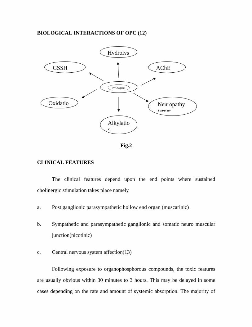
BIOLOGICAL INTERACTIONS OF OPC (12)
Fig.2
CLINICAL FEATURES
The clinical features depend upon the end points where sustained
cholinergic stimulation takes place namely
a. Post ganglionic parasympathetic hollow end organ (muscarinic)
b. Sympathetic and parasympathetic ganglionic and somatic neuro muscular
junction(nicotinic)
c. Central nervous system affection(13)
Following exposure to organophosphorous compounds, the toxic features
are usually obvious within 30 minutes to 3 hours. This may be delayed in some
cases depending on the rate and amount of systemic absorption. The majority of
P=O agent
Alkylation
GSSH
Oxidatio
AChE
Neuropathy target
Hydrolys

patients give a history of intentional or accidental ingestion of organophosphorous
compounds. Toxicity is produced by the rapid absorption of the compound
through the gastrointestinal, respiratory tracts and skin.
The clinical symptoms and signs are non-specific and will depend on the
specific agent, the quantity and the route of entry. Some patients present with
vomiting, diarrhea and abdominal pain, whilst others may be unconscious on
arrival at the hospital. A high index of suspicion is therefore needed to make an
early diagnosis. Early cases present predominantly with parasympathetic over-
activity, and a characteristic garlic smell. The end result may be a multi-system
manifestation involving the gastrointestinal, respiratory, and cardiovascular and
nervous systems, as well as involvement of skeletal muscle, other organs and
metabolic effects such as hypo or hyperglycemia. Most fatalities occur within 24
hours and those who recover usually do so within 10 days.
CARDIAC MANIFESTATIONS
The commonest cardiac manifestations following poisoning are hypotension
(with warm, dilated peripheries), and bradycardia. Patients seldom present with
tachycardia and hypertension due to predominant nicotinic receptor blockade.
Cardiac manifestations are often the cause of serious complications and fatality.
(14).

Table 2. Symptoms and signs of organophosphorous poisoning
Muscarinic receptors Nicotinic receptors Central receptors
Cardiovascular
• Bradycardia • Hypotension
Respiratory
• Rhinorrhoea • Bronchorrhoea • Bronchospasm • Cough
Gastrointestinal
• Nausea/vomiting • Increased
salivation • Abdominal
cramps • Diarrhea • Faecal
incontinence
Genitourinary
• Urinary continence
Eyes
• Blurred vision • Increased
lacrimation • Miosis
Glands
Cardiovascular
• Tachycardia • Hypertension
Musculoskeletal
• Weakness • Fasciculation • Cramps • Paralysis
General effects
• Anxiety • Restlessness • Ataxia • Convulsions • Insomnia • Dysarthria • Tremors • Coma • Absent reflexes • Respiratory
depression • Circulatory
collapse
• Excessive salivation
The mechanism of cardiac toxicity is unclear and the following has been
postulated:
• A direct toxic effect on the myocardium
• Over activity of cholinergic or nicotinic receptors causing hemodynamic
alteration
• Hypoxia
• Acidosis
• Electrolyte abnormalities
• High dose atropine therapy (used as treatment for organophosphate
poisoning).
RESPIRATORY MANIFESTATIONS
Respiratory manifestations of acute organophosphorous poisoning include
bronchorrhoea, rhinorrhoea, bronchospasm and laryngeal spasm. This is due to the
action of the organophosphate on muscarinic receptors. The integrity of the airway
may be compromised by excessive secretions. The nicotinic effects lead to
weakness and subsequent paralysis of respiratory and oropharyngeal muscles. This

increases the likelihood of both airway obstruction and aspiration of gastric
contents. Finally, central neurological depression may lead to respiratory arrest.

GASTROINTESTINAL MANIFESTATIONS
Symptoms resembling gastroenteritis such as vomiting, diarrhea and
abdominal cramps are the first to occur after oral ingestion of an
organophosphorous compound.
NEUROLOGICAL MANIFESTATIONS
A large number of patients, following acute exposure to
organophosphorous compounds, require prolonged ventilatory support in the
intensive care unit due to neuromuscular weakness. The neurological
manifestations have therefore been a primary focus of interest. There has been an
emphasis on reducing the incidence of neuro-muscular respiratory failure. Three
different types of paralysis are recognized based largely on the time of occurrence
and their differing path physiology:
• Type I paralysis or acute paralysis
• Type II paralysis or Intermediate syndrome
• Type III paralysis or Organophosphate- induced delayed polyneuropathy
Type I paralysis or acute paralysis is seen during the initial cholinergic phase.
This is when large numbers of both muscarinic and nicotinic receptors are
occupied by acetylcholine, leading to persistent depolarization at the

neuromuscular junction. Clinical features include muscle fasciculation, cramps,
twitching and weakness. At this stage the patient may require ventilatory support
due to the weakness of the respiratory muscles leading to respiratory depression
and arrest.
Type II paralysis or Intermediate syndrome. This was first described in 1974
by Wadia et al (14)as type II paralysis and subsequently termed "The Intermediate
Syndrome" by Senanayake. This syndrome develops 24-96 hours after the
poisoning. Following recovery from the acute cholinergic crisis, and before the
expected onset of delayed neuropathy, some patients develop a state of muscle
paralysis. The cardinal feature of the syndrome is muscle weakness affecting the
proximal limb muscles and neck flexors. There is a relative sparing of the distal
muscle group. One of the earliest manifestations in these patients is the inability to
lift their head from the pillow (due to a marked weakness in neck flexion). This is
a useful test to establish whether or not a patient is likely to develop respiratory
muscle weakness. Of the cranial nerves, those supplying the extra-ocular muscles
are mostly involved, with a lesser effect on VII and X. This syndrome persists for
about 4-18 days and most patients will survive unless infection or cardiac
arrhythmias complicate the course.
Type III paralysis or organophosphate- induced delayed polyneuropathy
(OPIDP) is a sensory-motor distal axonopathy that usually occurs after ingestion
of large doses of an organophosphorous compound (15-17)The neuropathy
presents as weakness and ataxia following a latent period of 2-4 weeks. Initial
stimulation causes excitatory fasciculation, which then progresses to an inhibitory
paralysis. The cardinal symptoms are distal weakness of the hands and feet. This is
often preceded by calf pain, and in some cases, parasthesia of the distal part of the
limbs. Delayed CNS signs include tremor, anxiety and coma.
OTHER EFFECTS OF OPC MAY INCLUDE
• Neuropsychiatric effects: Impaired memory, confusion, irritability,
lethargy, psychosis, and chronic organophosphate-induced
Neuropsychiatric disorders have been reported. The mechanism is not
proven.(18)
• Extra pyramidal effects: These are characterized by dystonia, cogwheel
rigidity, and parkinsonian features (basal ganglia impairment after recovery
from acute toxicity).
• Other neurological and/or psychological effects: Guillain-Barré–like
syndrome and isolated bilateral recurrent laryngeal nerve palsy are
possible.(19)
• Ophthalmic effects: Optic neuropathy, retinal degeneration, defective
vertical smooth pursuit, myopia, and miosis (due to direct ocular exposure
to organophosphates) are possible.
• Ears: Ototoxicity is also possible(20).
MODIFIED DREISBACH’ CLINICAL CRITERIA – KARNIT(9)
GRADE I - Mild symptoms related to portal of entry.
Nausea, vomiting in case of ingestion
Cough, burning sensation in the chest in case of
inhalation Mild systemic symptoms like headache,
dizziness, weakness
GRADE II - Moderate systemic intoxication
Abdominal pain, diarrhea in case of ingestion.
Tightness in chest, difficulty in breathing in case of
inhalation
Salivation,lacrimation, sweating, papillary changes.
Bradycardia, confusion, tremor, restlessness.
GRADE III - Severe systemic intoxication
Respiratory depression, generalized weakness
Cyanosis, peripheral circulatory failure,
convulsions coma.
DIAGNOSTIC CRITERIA (13)
• INVESTIGATORY MODALITES: ESTIMATION OF
CHOLINEESTERASE LEVELS: Organophosphate (OP) toxicity is a
clinical diagnosis. Confirmation of organophosphate poisoning is based on
the measurement of cholinesterase activity; typically, these results are not
readily available. Although RBC and plasma (pseudo) cholinesterase levels

can both be used, RBC cholinesterase correlates better with CNS acetyl
cholinesterase (AChE) and is, therefore, a more useful marker of
organophosphate poisoning.
o Measurement of RBC and plasma cholinesterase levels prior to
treatment with pralidoxime (2-PAM). Monitoring serial levels can be
used to determine a response to therapy.
o RBC AChE represents the AChE found on RBC membranes, similar
to that found in neuronal tissue. Therefore, measurement more
accurately reflects nervous system OP AChE inhibition.
o Plasma cholinesterase is a liver acute-phase protein that circulates in
the blood plasma. It is found in CNS white matter, the pancreas, and
the heart. It can be affected by many factors, including pregnancy,
infection, and medical illness. Additionally, a patient's levels can
vary up to 50% with repeated testing.
o RBC cholinesterase is the more accurate of the 2 measurements, but
plasma cholinesterase is easier to assay and is more readily
available.
• Cholinesterase levels do not always correlate with severity of clinical
illness.
• The level of cholinesterase activity is relative and is based on population
estimates. Neonates and infants have baseline levels that are lower than

adults. Because most patients do not know their baseline level, the
diagnosis can be confirmed by observing a progressive increase in the
cholinesterase value until the values plateau over time.
• Falsely depressed levels of erythrocyte cholinesterase can be found in
pernicious anemia, hemoglobinopathies, use of antimalarial drugs, and
oxalate blood tubes.
• Falsely depressed levels of plasma cholinesterase are observed in liver
dysfunction, low-protein conditions, neoplasia, hypersensitivity reactions,
use of certain drugs (succinylcholine, codeine, and morphine), pregnancy,
and genetic deficiencies.
• Other laboratory findings include leukocytosis, hemoconcentration,
metabolic acidosis, hyperglycemia, hypokalemia, and hypomagnesemia.
IMAGING STUDIES
A chest radiograph may reveal pulmonary edema but typically adds little to
the clinical management of a poisoned patient. Electrocardiographic
manifestations include prolonged Q-Tc intervals, elevation of the ST segment,
inverted T waves and a prolonged PR interval. There may also be rhythm
abnormalities such as sinus bradycardia, ventricular extra- systoles, ventricular
tachycardia and fibrillation. Ludomirsky et al described three phases of cardiac
toxicity following organophosphate poisoning:
• Phase I: A brief period of increased sympathetic tone

• Phase II: A prolonged period of parasympathetic activity including AV
node blockade
• Phase III: Q-T prolongation followed by torsades de pointes, ventricular
tachycardia and ventricular fibrillation (35)
THERAPEUTIC CONSIDERATIONS
Medical Care: Airway control and adequate oxygenation are paramount in
organophosphate (OP) poisonings. Intubations may be necessary in cases of
respiratory distress due to laryngospasm, bronchospasm, bronchorrhoea, or
seizures. Immediate aggressive use of atropine may eliminate the need for
intubation.(21) Succinylcholine should be avoided because it is degraded by acetyl
cholinesterase (AChE) and may result in prolonged paralysis.
• Continuous cardiac monitoring and pulse oximetry should be established;
an ECG should be performed. Torsades de Pointes should be treated in the
standard manner. The use of intravenous magnesium sulfate has been
reported as beneficial for organophosphate toxicity.(22) The mechanism of
action may involve acetylcholine antagonism or ventricular membrane
stabilization.
• Remove all clothing and gently cleanse patients suspected of
organophosphate exposure with soap and water because organophosphates
are hydrolyzed readily in aqueous solutions with a high pH. Consider
clothing hazardous waste and discard accordingly.

• Health care providers must avoid contaminating themselves while handling
patients. Use personal protective equipment, such as neoprene or nitrile
gloves and gowns, when decontaminating patients because hydrocarbons
can penetrate non polar substances such as latex and vinyl. Use charcoal
cartridge masks for respiratory protection when decontaminating patients
who are significantly contaminated.
• Irrigate the eyes of patients who have ocular exposure using isotonic
sodium
If ingestion occurred within 30 to 60minutes placement of nasogastric tube
and administration of activated charcoal in a dose, 1g/kg orally. Cathartics are
contraindicated because of electrolyte imbalance. (23)
Atropine administration: 3-5mg rapid intravenous should be given to reach
effective atropinisation. The adequate atropinisation is gauged by five parameters.
1. Pulse rate >85/min
2. Systolic blood pressure >80mmHg
3. Absence of lung crepitations
4. Dry axilla
5. No constricted pupils(9)

Then after three to five minutes if the parameters are not attained double the
initial dosing. Atropine should be given in doubling dose pattern till adequate
atropinisation.
Maintenance of atropinisation: 10 -20% of the bolus dose should be given
as infusion in100ml normal saline and the parameters are assessed every
15minutes.If there is inadequate atropinisation superimposed bolus in the dosage
of 3-5mg should be given. Atropine should be tailored down hourly for six hours
and then every two to three hours for the next 24hrs.Meticulous watch for atropine
toxicity such as agitation, confusion, retention of bladder, hyperthermia, ileus and
tachycardia should be done.
OXIMES: PRALIDOXIME (2-PAM OR PROTO PAM) (24)
Cholinesterase reactivation
Oximes are nucleophilic agents that re-activate the phosphorylated acetyl
cholinesterase by binding to the organophosphorous molecule. The use of oximes
in acute organophosphorous poisoning has been a controversial subject for the last
two decades as there have been very few randomized controlled trials that have
addressed the role of pralidoxime (PAM).
Pralidoxime has three main actions
• A direct reaction converting the organophosphate to a harmless compound.
• A transient reaction protecting the enzyme from further inhibition.
• Reactivation of the inhibited alkyl phosphorylated enzyme to free the active
unit (if given early enough)
The reactivating action of pralidoxime is most marked at the nicotinic
skeletal neuromuscular junction. It does not reverse the muscarinic manifestations
of organophosphorous poisoning. Pralidoxime should be started as early as
possible to prevent permanent binding of the organophosphate to acetyl
cholinesterase. Once this has occurred, receptor regeneration is required to allow
recovery. The recommended dose of pralidoxime in organophosphorous poisoning
is 1 gram, by intravenous injection, every 6-12 hour in adults (maximum dose
12g/24 hours) and 25-50mg/kg in children. Pralidoxime should be continued until
adequate spontaneous ventilation is achieved by the patient. The effective plasma
concentration is 4mg/litre and the patient should show signs of improvement 10-
40 minutes after its administration. Plasma and pseudo cholinesterase levels
should ideally be monitored during treatment. Side effects of pralidoxime include
drowsiness, visual disturbances, nausea, tachycardia and muscle weakness, so
treatment should be reserved for potentially fatal cases.
PREVENTION AND EDUCATION
Improved regulation of the availability of pesticides, strict regulation of
vendors, and modifications in packaging of pesticides may all help reduce the use
of organophosphates as poisons. Adequate provision of information to the public,
regular training of health care providers, better availability of drugs / antidotes and
the establishment of poison information centers will facilitate in reducing the
morbidity and mortality related to organophosphorous poisoning. Insecticides
should be kept out of reach of children, to prevent accidental poisoning. During
agricultural spraying, proper precautions should be taken to prevent inhalation and
accidental ingestion of the substance. The greatest incidence of
organophosphorous poisoning was reported from Japan where there were 19,436
cases over a period of 17 years (1953-1969).WHO used data from 19 countries
and reported approximately5, 00,000cases of pesticide poisoning annually. Of
these 99% belong to third world countries(24).In1981 the estimate was 75,000
cases annually and rose to 3million in 1983 and majority were under the age of
30(36). Pesticide poisoning contributes more than forty percent of cases in
Poison Centre GGH Chennai. The mortality rate due to this poison is 42.29%
(case register2001-2004). The victims are farmers of rural South India. Since only
limited studies are available in analyzing the predictors of mortality in this most
common form of agricultural poison, the outcome analysis of the predictors form
the basis of the thesis

OBJECTIVES
1. To describe the epidemiology of organophosphorous poisoning in poison
centre Government General Hospital Chennai.
2. To study the clinical profile of organophosphorous poisoning in poison
centre Government General Hospital Chennai
3. To analyze the epidemiological and clinical factors in correlation to
severity and outcome of organophosphorous poisoning in poison centre
Government General Hospital Chennai.
4. To identify prognostic indicators from biochemical parameters (renal
function test, liver enzymes, arterial blood gas analysis, enzymes such as
creatine kinase, creatine kinase mb fraction, serum amylase and lactate
dehydrogenase,) to predict the severity, outcome and the need of ventilatory
support.
5. To asses the clinical severity and outcome of organophosphorous poisoning
and correlate the same with pseudo cholinesterase levels
6. To do outcome analysis of clinical and biochemical parameters in
mechanically ventilated patients .
7. To compare the effectiveness of low dose versus high dose administration
of pralidoxime in organophosphorous poisoning in relation to the need of
ventilatory support and mortality.

MATERIALS AND METHOD
SETTING: This study was conducted in the Poison centre GGH Chennai in
collaboration of Institute of Internal Medicine and Institute of Biochemistry. It was
a cross sectional prospective study done during the period from September2005-
September2006. 87patients with history and clinical features suggestive of
organophosphorous poisoning were selected irrespective of age and sex. 46 age
and sex matched controls from normal population (patient attendants) were studied
for biochemical markers.
EXCLUSION CRITERIA
1. Patients with other pesticide poisoning were excluded (eg., organo
carbamate organochlorous compounds) by clinical features and confirmed
by thin layer chromatographic analysis of gastric aspirate.
2. Patients with other poisoning such as oleander, oduvanthazai , and drug
over dosage(nicotine replacements, inocybe clitocybe, pilocarpine,
opioids,phenothiazines,bethanechol.etc )known to mimic the clinical
picture of organophosphorous poisoning were excluded.
3. Patients with known medical illness such as neuromuscular disorders like
myasthenia gravis or muscular dystrophy, hypokalemic periodic paralysis
and conditions known to alter biochemical parameters were excluded.

• Patients with known history of exposure to organophosphorous compounds
were taken into consideration. The reliability on exposure was reinforced
by definite history from the patients and their attendants. The container
from which the poison where consumed were studied for the type and the
quantity estimated. Each patient registered for the study went through
detailed clinical evaluation as per the proforma.The cases were divided into
three groups as mild moderate and severe by modified DREISBACH
criteria. All patients underwent baseline laboratory investigations. The
gastric aspirate was analyzed by thin layer chromatography method for the
detection of poison.
All patients underwent renal function test, arterial blood gas analysis, ecg
chest roentogram and sonogram of the abdomen.
Serum AChE were measured on day 1 3 and 5.
The other biochemical markers such as liver enzymes, CK,CK-MB,LDH
and amylase were taken on day1.
BI0CHEMICAL MARKERS AND THE METHODS EMPLOYED- (KITS)
1.CHOLINESTERASE - KINETIC COLOIMETRIC METHOD(26)
2.SGOT (AST) - UV KINETIC METHOD (27)
3.SGPT - (MODIFIED IFCC METHOD)- (28)

4.ALKALINE PHOSPHATASE - PNPP METHOD(29) 5.ALBUMIN - BCG METHOD(30)
6. CK - UV KINETIC METHOD(31)
7.AMYLASE - CNP – G3 METHOD(32)
8. LDH - UV KINETIC METHOD(28)
On treatment aspect, average total dose of atropine administered per patient
was calculated in mg. All patients in the study were given pralidoxime . They
were sub divided into two groups –low dose and high dose based upon the total
grams per day(<4gm/dayand >4gm /day)(33)
• Ventilatory support was considered in patients with following
parameteres
a. RR >35/min
b. Apnea or obvious hypoventilation
c. Persistent cyanosis,excessive secretions,depressed level pf
unconsciousness,inability to protect the air way(34)
d. PaO2 < 60 mmHg, FiO2 >0.6
PaCO2 > 50 mmHg, pH <7.2
Mechanical ventilation was performed with assist control mode and simv
either as volume or pressure control. Positive end expiratory pressure was titrated
to keep SaO2 above 94% with 40% FIO2. Weaning was performed using either T-

tube trials or pressure support weaning. All patients were monitored meticulously
and vital signs assessed till the outcome.
STATISTICAL METHOD
The chi-square test was used for statistical analysis. Logistic Regression
analysis was run to identify the parameters that would predict the clinical severity,
need for mechanical ventilation and the outcome. Data are presented as mean ±
standard deviation.
Detailed evaluation of each patient was made. Each variable in the pro-
forma was correlated with severity and mortality.

RESULTS
Epidemiological Pattern of OPC in Poison Centre, Govt. General Hospital
TABLE 3 - Age Distribution
Sl.No. Age Group in years Frequency(n) Percentage
1. <= 20 17 19.54
2. 21-30 40 45.98
3. 31-40 15 17.24
4. > 40 15 17.24
Total 87 100.00
Majority of the patients were in the 21 to 30 age group. The number of
people between the age group 20 to 40 accounts for 55 out of 87 cases.
TABLE 4 – Age Distribution and Outcome
Sl.No. Age Group in years Outcome Discharge Death
1. <= 20 14 3
2. 21-30 33 7
3. 31-40 10 5
4. > 40 8 7
Total 65 22

The correlation between the age group and the outcome was not
statistically significant (P value – 0.11)
Table.5 Sex Distribution
Sl.No. Sex Frequency(n) Percentage
1. Male 76 87.36
2. Female 11 12.64
Incidence of OPC was more in males when compared to females in our
series.
Table.6 - Occupation
Sl.No. Occupation Frequency(n) Percentage Expired
1. Agriculture 40 45.98 9
2. Unskilled Labour 37 42.53 9
3. Others 10 11.49 4
More than 80 percent of the cases of OP poisoning were among
agriculturalists and unskilled labourers. The death rate was also more among these
groups.

Locality – Distance from the Poison Centre, Govt. General Hospital in Kilo
Meters.
Table 7 – Locality
Sl.No. Locality (kms) Frequency(n) Percentage
1. <= 100 40 45.98
2. > 100 47 54.02
Table 8 - Type of Poison consumed
Sl.No. Type of Poison consumed (kms) Frequency(n) Percentage
1. CHLORPYIFOS 6 6.90 2. DIMECRON 26 29.89 3. QUINALPHOS (EKALUX) 3 3.45 4. FENTHION (BAYTEX) 2 3.45 5. HINOXAN 3 2.30 6. MALATHION
(CHEMATHION) 2 2.30
7. METHYL PARATHION (METAPHOS)
1 1.15
8. MONOCROTOPHOS 2 2.30 9. PARATHION (FOLIDOL,
EKATOX) 30 34.49
10. PHORATE 3 3.45 11. SYSTOX 1 1.15 12. TRIPHOS 6 6.90
Total 87 100.00
Most of the patients in this series consumed parathion (n=30) and
Dimecron (n=26). But the type of poison does not contribute both to severity and
mortality.

Table.9 - Route Of Exposure
Sl.No. Route of Exposure Frequency(n) Percentage
1. Ingestional 75 86.2
2. Inhalational 10 11.5
3. Topical 2 2.3
Ingestional exposure was more common among in the series(86.2%) and
most of the patients in this entity fell under the severe grade of Dreisbach’s
criteria (P value = 0.03).
Intention Of Poison: Suicidal (75) - Accidental (12)
Table.10 Duration of stay in hospitals
Sl.No. Duration of Stay in Hospitals (in days)
Frequency(n) Percentage
1. <= 5 38 43.68
2. 6-10 32 36.78
3. > 10 17 19.54
87 100.00
The mean duration of stay in the hospital is 7.25 days (SD 4.64)

Table 11 - Duration Of Hospital Stay Correlation With Clinical Severity-
Clinical Severity at admission
Mild Moderate Severe
0-5
n=9
23.7% n=15
39.5% n=14
36.8%
6-10
n=6 18.8%
n=14 43.8%
n=12 37.5%
> 10
0
0
n=17 100% D
urat
ion
of S
tay
in H
ospi
tals
(in
days
)
In this series of 87 cases, more severe grade of intoxication had longer
duration of stay in the hospital (P value =0.0002)
Table 12 - Quantum of exposure
Sl.No. Quantity consumed (ml.) Frequency(n) Percentage
1. NA 25 28.74
2. <=50 29 33.33
3. 51-100 19 21.84
4. >100 14 16.09
Total 87 100.00

The non availability of data in 25 patients was due to two reasons. 1. 12
cases had inhalataional and topical forms of exposure so the quantity cannot be
ascertained 2. In 13 cases reliable information regarding the quantity of
consumption was not known. The average quantity of consumption in this series
amounts to 101.58ml.
Table 13 – Quantity of Consumption and Clinical Severity
Clinical Severity at Admission
Mild Moderate Severe
< =5
0
n=5 17.2%
n=13 44.8%
n=11 37.9%
50-1
00 n=4
21.1% | n=2 10.5% |
n=13 68.4%
Qua
ntity
of C
onsu
mpt
ion
(ml.)
>100
n=2 14.3%
n=12 85.7%
Patients who have consumed more than 100 ml. of OPC have higher grade
of severity (P value = 0.004). The quantity consumed had no relation to the
mortality rate. 21 cases had OPC mixed with alcohol. Two cases consumed OPC
mixed with phenol and kerosene.

Table.14 - Onset Of Symptoms After Exposure
Sl.No. Time of onset of
Poisoning (Mins.)
Frequency(n) Percentage
1. NA 1 1.15
2. <=30 40 45.98
3. 31-60 36 41.38
4. >60 10 11.49
Total 87 100.00
Most of the patients had their initial symptom of intoxication between 30
minutes to one hour.
PERIOD INTERVAL FOR INITIATION OF TREATMENT
a) Initiation of First Aid (Gastric Lavage/activated charcoal)
Table 15 –Initiation Of First Aid
Sl.No. Initiation of First aid (Hours.)
Frequency(n) Percentage
1. <=1 33 37.93
2. 1-2 32 36.78
3. >2 22 25.29
Total 87 100.00

Out of 87 patients 65 received first aid either at household or nearby
medical facility within two hours.
b) Initiation of Atropine
Table.16 - Time of initiation of Atropine
Sl.No. Time of initiation of
Atropine (Hours.)
Frequency(n) Percentage
1. <=1 35 35.63
2. 1-3 38 43.68
3. >3 18 20.69
Total 87 100.00
c) Initiation of 2- PAM
Table 17 - Time Of Initiation Of 2-PAM
Sl.No. Time of initiation of
2-P AM (Hours.)
Frequency(n) Percentage
1. NA 4 4.60
2. <=1 27 31.03
3. 1-3 36 41.38
4. >3 20 22.99
Total 87 100.00
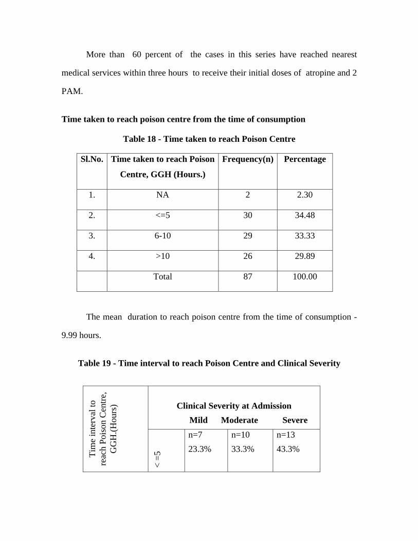
More than 60 percent of the cases in this series have reached nearest
medical services within three hours to receive their initial doses of atropine and 2
PAM.
Time taken to reach poison centre from the time of consumption
Table 18 - Time taken to reach Poison Centre
Sl.No. Time taken to reach Poison
Centre, GGH (Hours.)
Frequency(n) Percentage
1. NA 2 2.30
2. <=5 30 34.48
3. 6-10 29 33.33
4. >10 26 29.89
Total 87 100.00
The mean duration to reach poison centre from the time of consumption -
9.99 hours.
Table 19 - Time interval to reach Poison Centre and Clinical Severity
Clinical Severity at Admission
Mild Moderate Severe
Tim
e in
terv
al to
re
ach
Pois
on C
entre
, G
GH
.(Hou
rs)
< =5
n=7 23.3%
n=10 33.3%
n=13 43.3%
6-10
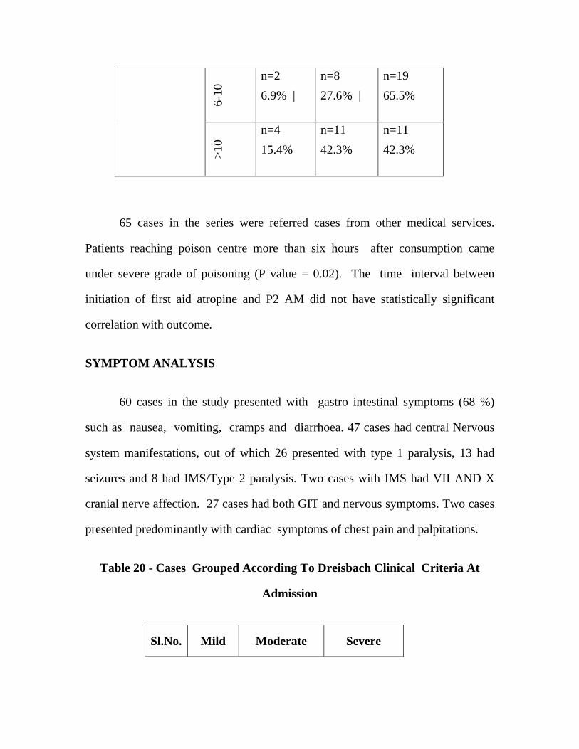
n=2 6.9% |
n=8 27.6% |
n=19 65.5%
>10
n=4 15.4%
n=11 42.3%
n=11 42.3%
65 cases in the series were referred cases from other medical services.
Patients reaching poison centre more than six hours after consumption came
under severe grade of poisoning (P value = 0.02). The time interval between
initiation of first aid atropine and P2 AM did not have statistically significant
correlation with outcome.
SYMPTOM ANALYSIS
60 cases in the study presented with gastro intestinal symptoms (68 %)
such as nausea, vomiting, cramps and diarrhoea. 47 cases had central Nervous
system manifestations, out of which 26 presented with type 1 paralysis, 13 had
seizures and 8 had IMS/Type 2 paralysis. Two cases with IMS had VII AND X
cranial nerve affection. 27 cases had both GIT and nervous symptoms. Two cases
presented predominantly with cardiac symptoms of chest pain and palpitations.
Table 20 - Cases Grouped According To Dreisbach Clinical Criteria At
Admission
Sl.No. Mild Moderate Severe
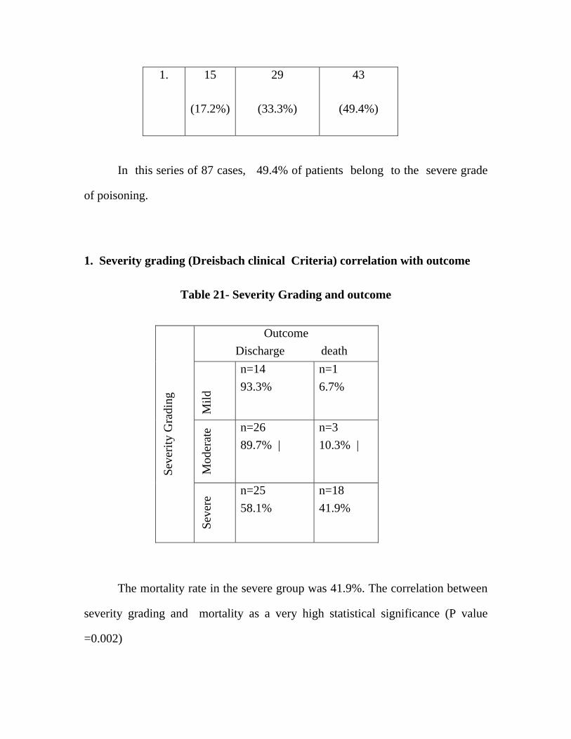
1. 15
(17.2%)
29
(33.3%)
43
(49.4%)
In this series of 87 cases, 49.4% of patients belong to the severe grade
of poisoning.
1. Severity grading (Dreisbach clinical Criteria) correlation with outcome
Table 21- Severity Grading and outcome
Outcome Discharge death
Mild
n=14 93.3%
n=1 6.7%
Mod
erat
e n=26
89.7% | n=3 10.3% |
Seve
rity
Gra
ding
Seve
re n=25 58.1%
n=18 41.9%
The mortality rate in the severe group was 41.9%. The correlation between
severity grading and mortality as a very high statistical significance (P value
=0.002)

2. Severity grading (Dreisbach clinical Criteria) correlation to the need of
ventilatory support
Table 22 - Severity Grading and Ventilatory Management
Ventilatory Management
No Support Ventilatory Support
Mild
n=15 100.0%
n=0
Mod
erat
e n=26
89.7% | n=3
10.3% |
Seve
rity
Gra
ding
Seve
re n=4
9.3% n=39
90.7%
(P value = 0.0000)
Out of 42 cases on mechanical ventilation, 39 cases were initiated on assist
control mode and three were put on synchronized intermittent mandatory
ventilation. (SIMV)
PROXIMAL MUSCLE GROUP INVOLVEMENT IN OPC
47 out of 87 cases had involvement of proximal muscles such as neck
flexors, bulbar muscles and muscles of the shoulder and pelvic griddle. Out of
this group, 8 cases developed IMS. The correlation of proximal muscle group
affection was studied in relation to clinical severity, outcome and the need of

mechanical ventilation. These three variables studied had statistically significant
relationship.
Table 23 - Proximal Muscle Group Involvement and Clinical Severity
Clinical Severity
Mild Moderate Severe Y
es
n=1
2.1%
n=13
27.7%
n=33
70.2%
Prox
imal
Mus
cle
Gro
up
Invo
lvem
ent
No
n=14
35.0% |
n=16
40.0% |
n=10
25.0%
(P value = 0.00001)
33 cases of proximal muscle group involvement had severe grade of
poisoning.
Table 24 - Proximal Muscle Group Involvement and Ventilatory
Management
Ventilatory Management
No Support Assist control SIMV
Prox
imal
Mus
cle
Gro
up
Invo
lvem
ent
Yes
N=14
29.8%
n=30
63.8.7%
n=3
6.4%

No n=31
77.5%
n=9
22.5%
n=0
(P value = 0.0004)
Table 25 - Proximal Muscle Group Involvement and Outcome
Outcome
Discharge Death
Yes
N=31
66.0%
n=16
34%
Prox
imal
Mus
cle
Gro
up
Invo
lvem
ent
No
n=34
85.0%
n=6
15.0%
(P value = 0.04170)
The mortality rate among the group which had proximal muscle
involvement was 34 percent.
LABORATORY PARAMETERS
1. Average hemoglobin observed in these series – 13.2 g/dl/(SD =2.40)
2. Total WBC count had a range between 6500 and 27000 cells per cubic
mm. and it had a mean value -14,038(S.D-5233.3)
3. Routine Bio-chemistry

Table 26 – Bio Chemistry Values
Sl.No. Values Mean S.D. (+/-)
1. Blood Sugar mg/dl. 103.7 32.26
2. Urea mg/dl. 27.92 13.22
3. Creatinine |0.91 0.27
4. Sodium 134 6.3
5. Potassium 3.83 0.55
6. Chloride 111.95 6.14
4. Liver Function Test (These parameters did not have any correlation with
severity and outcome. The values of 87 patients are given below:
Table 27 - LFT
Sl.No. Values Mean S.D. (+/-)
1. SGOT IU/L 52.89 22.55
2. SGPT IU/L 47.23 27.23
3. Total Protein g/dl 6.54 0.48
4. Albumin g/dl 3.60 0.70
5. SAP IU/L 152.4 69.4

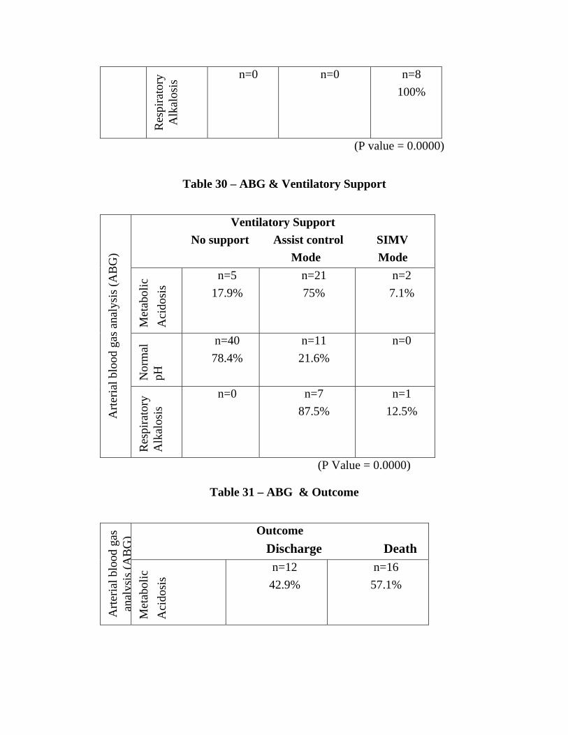
5. Arterial blood gas analysis
Table 28 - ABG
Sl.No. Values Mean S.D. (+/-)
1. ph 7.33 0.14
2. PaCo2 mm/hg 43.31 13.17
3. PaO2 87.05 16.28
4. O2Sat 90.99 14.30
5. Bicarbonate 23.44 5.58
Interpretation of arterial blood gas analysis revealed that 51 patients had
normal pH. 28 patients had metabolic acidosis and 8 had respiratory alkalosis.
The arterial blood gas analysis was studied in relation to severity of poisoning,
need of ventilatory support of outcome. The results are as follows:
Table 29 – ABG & Clinical Severity
Clinical Severity Mild Moderate Severe
Met
abol
ic
Aci
dosi
s
n=0 n=6 21.4%
n=22 78.6%
Arte
rial b
lood
gas
ana
lysi
s (A
BG
)
Nor
mal
pH
n=15 29.4%
n=23 45.1%
n=13 25.5%
Res
pira
tory
A
lkal
osis
n=0
n=0
n=8 100%
(P value = 0.0000)
Table 30 – ABG & Ventilatory Support
Ventilatory Support No support Assist control SIMV Mode Mode
Met
abol
ic
Aci
dosi
s
n=5 17.9%
n=21 75%
n=2 7.1%
Nor
mal
pH
n=40 78.4%
n=11 21.6%
n=0
Arte
rial b
lood
gas
ana
lysi
s (A
BG
)
Res
pira
tory
A
lkal
osis
n=0
n=7 87.5%
n=1 12.5%
(P Value = 0.0000)
Table 31 – ABG & Outcome
Outcome Discharge Death
Arte
rial b
lood
gas
an
alys
is(A
BG
)M
etab
olic
A
cido
sis
n=12 42.9%
n=16 57.1%

Nor
mal
pH
n=45 88.2%
n=5 11.8%
Res
pira
tory
A
lkal
osis
n=8 100%
n=0
(P Value = 0.0001)
The three variables namely, clinical severity, need of ventilatory support
and the outcome had statistically significant correlation with pH.
5. ECG changes are classified into three groups (35)
ECG with normal limits n=22
Phase 1GROUP - n=43
Phase 2 GROUP n=18
Phase 3 GROUP n= 4
Out of 18 in phase 2 group of ECG changes seven needed ventilatory
support and the morality rate was 33.34%(n=6).All the four cases in phase3 group
needed ventilatory support and every one recovered.
6. Gastric aspirate analysis by Thin layer chromatography in detection of
OPC.
In this series out of 75 cases of ingestional form of exposure the TLC
method detection rate was 84 percent.

7. Pseudocholinesterase Levels in OP Poisoned Patients
Table 32 - Pseudocholinesterase Levels in OPC poisoned Patients
Serum
CHe Level
IU/L
N Mean SD Minimum
Value
Maximum
Value
Range
Day 1 87 841.32 1426.7 31.00 7000.0 6969.0
Day 3 87 1301.7 1697.4 48.00 7259.0 7211.0
Day 5 87 1664.3 1965.7 50.00 7259.0 7840.0
The mean level of Serum CHe among the age and sex matched control
population (n=46) is 7090 IU per litre. The total number of 87 patients were
classified on the basis of serum CHe level in the following manner :
1. Mild (20 to 50% of 7090)
2. Moderate (10 to 20% of 7090) and
3. Severe (Less than 10%)
In these series, Day 1 CHe estimation correlated well with Dreishbach
clinical criteria of severity but it has no statistically significant relationship to
predict the need of mechanical ventilation and outcome.

Table 33 – Day 1 Serum AcHE Level & Clinical Severity
Clinical Severity Mild Moderate Severe
Nor
mal
(>
50%
)
n=2 50%
n=2 50%
n=0
Mild
(2
0 ton=2 20%
n=6 60%
n=2 20%
Mod
era
te
(10
to
n=5 50%
n=0 n=5 50%
Day
1 S
erum
CH
e Le
vel a
s % o
f N
orm
alSe
ver
e
(<10
n=6 9.5%
n=21 33.3%
n=36 57.1%
(P value = 0.00167)
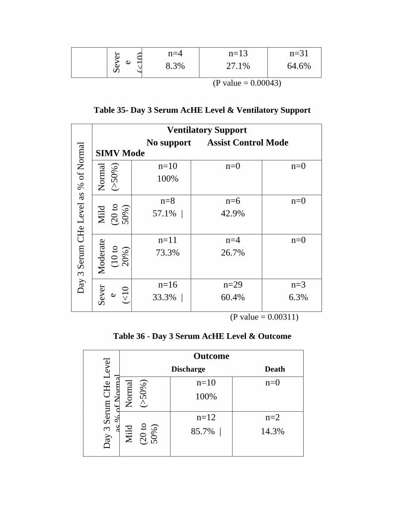
Day 3 and day 5 serum CHes estimation had significant and very
significant statistical relationship respectively with three variables of clinical
severity, to predict the need of mechanical ventilation and outcome.
Table 34 - Day 3 Serum AcHE Level & Clinical Severity
Clinical Severity Mild Moderate Severe
Nor
mal
(>
50%
)
n=6 60%
n=4 40%
n=0
Mild
(2
0 to
n=2 14.3%
n=4 28.6%
n=8 57.1%
D
ay 3
Ser
um C
He
Leve
l as %
of
Nor
mal
Mod
erat
e
(10
to
n=3 20%
n=8 53.3%
n=4 26.7%
Seve
re
(<
10) n=4
8.3% n=13
27.1% n=31
64.6%
(P value = 0.00043)
Table 35- Day 3 Serum AcHE Level & Ventilatory Support
Ventilatory Support No support Assist Control Mode SIMV Mode
Nor
mal
(>
50%
) n=10 100%
n=0
n=0
Mild
(2
0 to
50
%) n=8
57.1% | n=6
42.9% n=0
Mod
erat
e
(10
to
20%
)
n=11 73.3%
n=4 26.7%
n=0
Day
3 S
erum
CH
e Le
vel a
s % o
f Nor
mal
Seve
re
(<
10
n=16 33.3% |
n=29 60.4%
n=3 6.3%
(P value = 0.00311)
Table 36 - Day 3 Serum AcHE Level & Outcome
Outcome Discharge Death
Nor
mal
(>
50%
) n=10 100%
n=0
Day
3 S
erum
CH
e Le
vel
as%
ofN
orm
alM
ild
(20
to
50%
)
n=12 85.7% |
n=2 14.3%
Mod
erat
e
(10
to
20%
)
n=13 86.7%
n=2 13.3%
Seve
re
(<10
%) n=30
62.5% | n=18
37.5%
(P value = 0.02668)
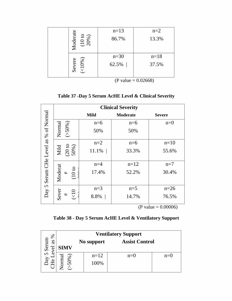
Table 37 -Day 5 Serum AcHE Level & Clinical Severity
Clinical Severity Mild Moderate Severe
Nor
mal
(>
50%
) n=6 50%
n=6 50%
n=0
Mild
(2
0 to
50
%) n=2
11.1% | n=6
33.3% n=10
55.6%
Mod
erat
e
(10
to
20%
) n=4 17.4%
n=12 52.2%
n=7 30.4%
Day
5 S
erum
CH
e Le
vel a
s % o
f Nor
mal
Seve
re
(<
10 %) n=3
8.8% | n=5
14.7% n=26
76.5%
(P value = 0.00006)
Table 38 - Day 5 Serum AcHE Level & Ventilatory Support
Ventilatory Support No support Assist Control SIMV
Day
5 S
erum
C
He
Leve
l as %
Nor
mal
(>
50%
) n=12 100%
n=0
n=0
Mild
(2
0 to
50
%) n=10
55.6% |n=8
44.4% n=0
Mod
erat
e
(10
to
20%
) n=14 60.9%
n=7 30.4%
n=2 8.7%
Seve
re
(<10
%) n=9
26.5% |n=24
70.6% n=1
2.9%
(P value = 0.00044)
Table 39 - Day 5 Serum AcHE Level & Outcome
Outcome Discharge Death
Nor
mal
(>
50%
) n=12 100%
n=0
Mild
(2
0 to
50
%) n=17
94.4% | n=1
5.6%
Mod
erat
e
(10
to
20%
)
n=19 82.6%
n=4 17.4%
Day
5 S
erum
CH
e Le
vel a
s % o
f Nor
mal
Seve
re
(<10
%) n=17
50% | n=17 50%
(P value = 0.00021)
Table 40 - Creatine kinase (CK) and its MB Fraction (CKMB)

Sl.No. Values Normal Mean S.D. (+/-)
1. CPK IU/L 15-130 723.20 827.75
2. CPK MB IU/L 0-24 100.33 112.73
(P Value = 0.0000)
Both the enzymes level elevation in the cases of OP Compound poisoning
were statistically significant.
Both serum CK and CK MB fraction elevation correlated very well with
clinical severity grading and the need of mechanical ventilation. There is no
statistical significant relationship between these enzymes elevation and the
outcome.
Table 41 - Serum CK Level & Clinical Severity
Clinical Severity
Mild Moderate Severe
Nor
mal
n=5
38.5%
n=8
61.5%
n=0
Seru
m C
K L
evel
Abn
orm
al
n=10
13.5%
n=21
28.4%
n=43
58.1
(P value = 0.00050)

Table 42 - Serum CK Level & Ventilatory Support
Ventilatory Support No support Assist Control SIMV
Nor
mal
n=13 100%
n=0
n=0
Seru
m C
K L
evel
Abn
orm
al n=32
43.2% | n=39
52.7% n=3
4.1%
(P value = 0.00080)
Table 43 - Serum CKMB Level & Clinical Severity
Clinical Severity Mild Moderate Severe
Nor
mal
n=6 37.5%
n=9 56.3.5%
n=1 6.3%
Seru
m C
KM
B
Leve
l
Abn
orm
al
n=9 12.7%
n=20 28.2%
n=42 59.2%
(P value = 0.00051)
Table 44 - Serum CKMB Level & Ventilatory Support
Seru m
Ventilatory Support No support Assist Control SIMV

Nor
mal
n=14 87.5%
n=2 12.5%
n=0
Abn
orm
al n=31
43.7% | n=37
52.1% n=3
4.2%
(P value = 0.00642)
SERUM AMYLASE
In all 87 cases, serum amylase and ultra sonogram of the abdomen were
done. Sonography of the abdomen were normal in all cases. The mean serum
amylase level in the series 110 U/L. The rise of serum amylase levels was directly
related to the severity and the need of mechanical ventilation.
Table 45 - Serum amylase Level & Clinical Severity
Clinical Severity Mild Moderate Severe
Nor
mal
n=11 22.9%
n=19 39.6%
n=18 37.5%
Seru
m a
myl
ase
Leve
l
Abn
orm
al
n=4 10.3%
n=10 25.6%
n=25 64.1%
(P value = 0.04209)
Table 46 - Serum Amylase Level & Ventilatory Support

Ventilatory Support No support Assist Control SIMV
Nor
mal
n=33 68.8%
n=15 31.3%
n=0
Seru
m A
myl
ase
Le
vel
Abn
orm
al n=12
30.8% | n=24
61.5% n=3
61.5%
(P value = 0.00087)
SERUM LDH
The mean LDH level in the series – 540 U/L (SD- (+/-) 247.02)
The Serum LDH elevation in cases of OPC poisoning was highly
significant (P value = 0.0000). The LDH elevation did not have any correlation
with clinical severity, need of mechanical ventilation and the outcome.
TREATMENT CONTRIBUTES
In our series of 87 patients mean atropine requirement: 268.51mg
(S.D179.96) 2PAM was given to all patients. They were grouped in a non random
fashion into two groups as low dose and high dose on basis of total dose given per
day.(<4gm/day and >4gm/day). Average mean requirement of 2PAM - 31.64gm
(SD 18.62|)36 patients came under the low dose group and 51 patients in the high
dose group. These groups were compared in relation to severity, need of
ventilatory support and outcome and their correlation was statistically significant.

Table 47 - 2PAM & Clinical Features
Clinical Features Mild Moderate Severe
Low
D
ose
n=10 27.8%
n=16 44.4%
n=10 27.8% 2P
AM
Hig
h D
ose
n=5
9.8% n=13
25.5% n=33
64.7% (P value = 0.00242)
Table 48 - 2PAM & Ventilatory Support
Ventilatory Support No Support Assist Control SIMV
Low
Do
n=30 83.3%
n=5 13.9%
n=1 2.8%
2PA
M
Hig
h D
ose
n=15 29.4%
n=34 66.7 %
n=2 3.9%
(P value = 0.0000)
Table 49 - 2PAM & Outcome
Outcome Discharge Death
Low
D
ose
n=32 88.9%
n=4 11.2%
2PA
M
Hig
h D
ose
n=33 64.7%
n=18 35.3%
(P value = 0.01059)

VENTILATORY SUPPORT
Out of 87 patients 42 needed ventilatory support.(48.27%).39 patients were
initiated on assist control mode and three were started on simv mode.
The average duration of mechanical ventilation in the series was
7.11days (S.D= 4.53) .The more the severe grade of poisoning more was the need
of mechanical ventilation.(pvalue=0.0000).19 out of 42 cases on mechanical
ventilaton expired .The morality rate among patients on mechanical ventilation
was 45.23%. Their correlation was very significant. The mortality rate differed
with patients with ventilatory support and with that of no support (6.7%). The
difference was statistically significant.
Table 50 - Ventilatory Support & Outcome
Ventilatory Support No Support No.of Patients 42 45 Recovered 23 42 Expired 19 3
(P Value =0.001)
All the 8 cases of IMS (type2 )syndrome were on mechanical ventilation.The
mean duration of these cases on mechanical ventilation-17.5 days. One case
expired and seven got discharged.Eight cases needed tracheostomy during
mechanical ventilation and three cases out of them had type2paralysis.
Outcome In this series 22 expired. The mortality rate was 25.29%. In 18
cases primary cause of death was respiratory failure with secondary cardiac arrest.

It has resulted from central respiratory depression, respiratory muscle weakness,
increased bronchial secretions, broncho spasm and acute pulmonary oedema.4
cases developed torsades de pointes and they expired due to ventricular
fibrillation.19 out of 22 cases needed ventilatory support.

LOGISTIC REGRESSION ANALYSIS
in order to identify important variables that would predict the need of mechanical
ventilation and outcome this stastical method was adopted.
a. Need for ventilatory support
Dependant variable – ventilatory support.
Independent variables- 1.Duration of hospital stay
2. Clinical severity grading
3. Proximal muscle affection
4.Creatine kinase (CK)
5.Total dose of atropine(mg)
6.Day 1serum AcHE level
7.Total dose of 2PAM
The correlation was found out to be very significant (p=0.0000) by chi
square test. The prediction percentage was high as 96.55%.
INFERENCES: Variables with positivity were clinical severity,total dose
of atropine and total dose of 2PAM, ie more the clinical grade of severity, greater
the dose of atropine and 2PAM the need for mechanical ventilation was high.
Variables with negativity were duration of hospital stay, proximal muscle
affection, day1 serum ChE and creatine kinase (i.e.) shorter the hospital stay, non
involvement of proximal muscle group, lesser inhibition of day 1 serum che and
low creatine kinase the need for ventilatory support was low.
B PREDICTORS OF.OUTCOME
Dependant variable-Outcome
Independent variables 1.Duration of hospital stay
2.Clinical severity grading
3. Proximal muscle affection
4.Creatine kinase (CK)
5.Day 1serum AcHE level
6.Day 5serum AcHE level
7. Total dose of atropine
8.Total dose of 2PAM
9.Ventilatory support.
The correlation was found out to be very significant(p=0.0000) by chi
square test. The prediction percentage was 87.36%.
INFERENCE: Patients with severe poisoning (driesch bach criteria),
elevated levels of creatine kinase, more the total dose of atropine , greater
reduction of day 1 che levels were associated with significant mortality.
In the series, patients with shorter duration of stay in the
hospital,noninvolvement of proximal muscles, lesser inhibition of day 5 che levels
and lower the dose of total p2am administered were associated with good
outcome,.

DISCUSSION
Comprehensive analysis of 87 cases of acute organophosphorous poisoning
EPIDEMIOLOGY OF ORGANOPHOSPHOROUS POISONING:
Age Pattern.
In this series the opc poisoning was prevalent in the 21-30 age
group.57cases were below the age of 30.WHO has reported about 3million cases
of opc exposureand40,000deaths annually and majority were under the age group
of thirty. (36) Murat Sungur and Muhammed Güven et al of TURKEY observed
the mean age group of opc exposure was 30±15 years.(12) Karalliedde L,
Senanayake N.et al of SRILANKA documented 91% of their cases were under
the age of 30.(37). In Kashmir valley Malik et al. revealed that 33.5% of the
cases of opc were under the age of 25.(38) In Mangalore, Karnataka, India the
most common age group to be affected was between 20-30 (36.6%) (36). The
series reported in this thesis had the similar pattern of age group affection. The
reason could be that this age group by all probability is vulnerable to various
emotional conflicts that occur during this phase of life. This young age group
affected by exposure form the viable entity of any population both in terms of
procurement and productivity. This case study and the case reports mentioned
above throw light on the target age group for educative and preventive
programmes to reduce the incidence of opc poisoning.

SEX DISTRIBUTION
In poison centre GGH Chennai males were exposed more when compared
to female population.(87.36% versus 12.64%). On the contrary to the series in
Murat Sungur study of 47 cases of opc in Turkey(12) [(female n=25,
male=22)]and. Malik et al. observation of 122 cases in Kashmir valley[(female
n=114,male=50)] female intoxication was more. In Srilanka and Mangalore had
similar pattern to the case series of the poison centre.(male 86%-female
14%).S.Shivakumar and K.Raghavan et al of Tamil Nadu reported 165 cases of
organo phosphorous poisoning and sex distribution was similar to the case series
(male n=122, female n=45)(39).This variation was due to handling of poison by
the respective sex in their respective locality. In Kashmir the female population
are predominantly employed in apple orchards and they are involved in pesticide
control. In southern part of India males are actively involved in spraying fertilizers
and pesticides.
OCCUPATION AND SOCIOECONOMIC STATUS
Wesselling C, McConnell R, Partanen T, Hogstedt C et al reported that
large worker populations in the Third World were exposed to increasing amounts
of pesticides, including pesticides severely restricted and banned in industrialized
countries. Studies on knowledge, attitudes, and practices indicate that unsafe use
of pesticides was the rule in Third World countries.(40).In our case series 77out
of 87 cases were agriculturists and unskilled labourers.54.02% of the cases were
from rural areas.(n=47).The mortality rate were also high among the these group.

In Kashmir valley two third of the population who had exposure were engaged in
apple orchard.In Karnataka agriculturists formed the majority.Students formed the
predominant group in Nepal (41) whereas in Almeria exposure was more in green
house workers.(42).
INTENTION OF POISON.
Estimates from the WHO indicate that each year, 1 million accidental
poisonings and 2 million suicide attempts involving pesticides occur
worldwide. (10). In the series majority of cases were suicidal in nature
which was consistent with other workers.75cases(86.21%) were suicidal and
12 cases(13.79%) had accidental exposure.
Murat Sungur and Muhammed Güven et al observed 68%of opc
poisoning reported were of suicidal exposure and Karalliedde L,
Senanayake N et al of SRILANKA had similar pattern. Malik et al. reported
74.4% of cases were of suicidal in nature and rest(25.6%) had accidental
exposure. Palimar Vikram MD, Arun M MD, DNB et al in their case study
of 153 cases in Mangalore reported 98.7%of sucidal exposure. Most of the
agriculturists consume for the fact of failure of crops, increasing debts
coupled with the easy availability of poison.
2700 people are referred to hospital for self poisoning each week in the
United Kingdom alone. It is likely to be even more difficult for the developing
world, with its limited resources, to address this problem effectively. However, we

think that the time has come to acknowledge the seriousness of the situation as a
first step towards preventing this massive unnecessary loss of life.(43)
ROUTE OF EXPOSURE:
Ingestion of the opc poisoing (n=75) formed th majority in this series apart
of inhalational(n=10) and topical (n=2) form of exposure.
In the series most cases of grade III severity were ingested OPC but the
correlation with the outcome was not significant. The mortality rate in the
ingestional group-26.7% Arup Kumar Kundu, JD Mukhopadhyay, AK Saha, S
Das et al(44)studied 108patients in sub urban West bengal poison.90 consumed
the poison and the death rate was12% and it had positive correlation with
outcome. In Kashmir Valley out of 164 cases-ingestional route(n=140),
inhalational (n=7) and topical exposure(n=17). In the Turkey study the
gastrointestinal route was the main route in 44 (93.6%) patients.
TYPE OF POISON
In this study parathion(34.49%) and dimecron(29.89) were the most
common type of poison consumed when compared to other workers. The type of
poison did not contribute much to the severity and mortality.

Murat Sungurb et al in TURKEY observed the three common type of opc were:
Agent Number of patients
Dichlorvos 24 (51.1%)
Ethyl-Parathion 5 (10.6%)
Fenthion 4 (8.5%
In sub urban West Bengal Arup kumar kundu et al showed that mortality was high
in poisoning with monocrotophos and dimethoate (31%) and nil with malathion. In
the Mangalore study methyl parathion was the most common poison consumed.
Karalliedde L, Senanayake N. reported dimethoate, methamidophos, malathion,
monocrotophos and fenthion as the common type opc consumed in Srilanka. In the
Kashmir valley phosphamidon(55%), malathion(12%) and dichlorovas(8.5%)
were the commonly used opc compounds for ingestion.In study from Tamil nadu
S.Shiva Kumar and K.Raghavan et al of Tamil Nadu reported methyl parathion as
the most common form of exposure in their study group.
In this case series the severity of poison was directly proportional to
quantity consumed. (pvalue=0.004).There was no relation to mortality. But the
study from West Bengal showed there was a correlation between the quantity
consumed and the mortality.

CLINICAL CONTRIBUTES
In this case study, the average duration between the onset of symptoms
and consumption was 44.73 (SD 26.76). Dr.Sumathi Joshi et.al. reported that the
symptoms of OPC poisoning developed between 30 minutes and 1 hour (45). In
this study group, patient reaching the Poison centre by more than 6 hours had
grade 3 severity (P value 0.02). More than 60 per cent of the cases have the
accessibility to have their first aid, first doses of atropine and 2PAM before 3
hours. But these variables did not have significant correlation with the
outcome. But the West Bengal workers reported increased mortality among the
patients who had increased time interval before initial atrophinazation (44). In
this series, presenting muscuranic symptom predominantly involved the GIT
(68%) followed by the central nervous system manifestations. Murat Sungurb et
al in Turkey reported that the CNS symptoms such as depressed mental status,
confusion and muscle weakness were the common presentation. Kenneth D.Ketz
et.al. from Pittsburg reported neuro psychiatric manifestations, ototoxity,
Guillain-Barré (46) –like syndrome and isolated bilateral recurrent laryngeal nerve
palsy in OPC poisoning. In this series, there were two cases with lower cranial
involvement, one with bilateral LMN type of facial palsy and the other with tenth
cranial nerve affection.

CLINICAL SEVERITY (MODIFIED DREISHBACH CRITERIA)
In this case series, most of the patients admitted fell into the grade 3
group of clinical severity (n=43) 49.4%. The reason could be that this centre
being a referral unit, complicated cases, warranting specialist treatment, intensive
care monitoring, ventilatory support (n=65) were transferred from primary health
centres, district hospitals and private nursing homes. Arup Kumar Kundu (et.al)
reported, mild 15 (14%), moderate 55 (50.9%), and severe 32 (29.6%) in his
study based on OPC poisoning in sub-urban West Bengal. In that study, the
severe grade of poisoning was associated with increased ventilatory support and
poor outcome. Similarly, in this case series, clinical severity association with
need of ventilatory support and mortality was statistically significant (P value =
0.002).
In this case study, proximal muscle group affection formed an important
parameter to assess the need of ventilatory support, progression to grade 3
severity and predictor of outcome.
LAB CONTRIBUTES
In this study group, the average total WBC count 14,038 cells cubic mm.
(SD. 5233). Leucocytosis were also reported in the studies from Turkey and
Pittsburg. The liver function tests observed in this study had similar pattern of
mild elevation when compared to the study from Murat Sungurb et al in
TURKEY. The arterial blood gas analysis performed in this study had significant
correlation with severity, outcome and the need of ventilatory support similar to

the observations made by A.Goe, S.Joseph, et.al.(47). Kenneth D. Ketz, et.al. who
documented metabolic acidosis in his study group.
ECG
Most of the cases in this series had sinus tachycardia (n=43) 18 cases had
AV nodal inhibition and 4 cases had QTC prolongation followed by torsade de
pointes, ventricular tachycardia and ventricular fibrillation. Patients with phase 2
and phase 3 ecg changes as described by Ludomirsky et al. (25) were
associated with grade 3 severity and outcome similar to observations made by
Dalvi, CP, et.al.(48).
CHOLINESTERASE LEVELS AND ITS SIGNIFICANCE
In this case study, day 1 Che correlated very well with clinical criteria of
dreishbach (P value = 0.001). This finding was also observed by Bobba
R, Venkataraman, BV, Pais P, Joseph T. et.al. in Bangalore (49). In contrary to
this observation, S.N. Chugh and Navneeth Agarwal, et.al. found no relationship
between serum CHE estimation and severity grading (50). In this case series,
logistic regression analysis was run to predict the outcome in which day 1 and
day 5 CHE level were important parameters. Sequential post exposure estimations
of the ChEs upto 5 days not reveal any rise in the values though there was
substantial clinical improvement. This observation was in par with the study in
Bangalore (49). Day 3 and day 5 ChE levels in this study had statistically
significant association with need of ventilatory support.
SERUM AMYLASE
In all 87 patients serum amylase and ultra sonogram of the abdomen was
performed in order to identify transient pancreatits which is a rare complication
(51). Ultra sonography of the abdomen was done in all the cases. Serum
Amylase elevation was statistically significant. 4 cases out of 87, had serum
amylase level > 300 U/L out of which two cases expired. Sahin I, Onbasi K,
Sahin H, Karakaya C, Ustun Y, Noyan T. et.al. observed 4 out of 47 cases in
their series, had amylase elevation more than 300 U/L.(52). Lee WC, Yang CC,
Deng JF, Wu ML, Ger J, Lin HC, Chang FY, Lee SD, et.at. stated that :
hyperamylasemia was frequent in severe organophosphate poisoning. However,
hyperamylasemia was not synonymous with acute pancreatitis and pancreatic
amylase was not a reliable parameter in the diagnosis of organophosphate-induced
pancreatitis due to its low sensitivity and specificity (53). In this series of 87
patients in Poison centre serum amylase level was directly related to clinical
severity and the need of ventilatory support (P value < 0.001). This observation
was also documented in the Taiwan study (53).
SERUM LDH
Serum LDH elevation denotes oxidative damage done to skeletal muscles
during OPC. (55). In this series, mean LDH levels were 540.53 U/L (SD 247.02)
and the elevation was significant. Murat Sungur and Muhammed Güven of
Turkey observed mean LDH levels of 548.8 ± 45.5 U/L. Sahin I, Onbasi K,

Sahin H, Karakaya C, Ustun Y, Noyan T. et.al. also found out LDH levels to be
elevated. But in this series, LDH did not have any correlation with clinical
severity, outcome nor to the need of ventilatory support.
CK AND CK MB
In this series, both enzyme level elevation were significant. CK rise may
indicate skeletal muscle injury due to sustained stimulation of acetylcholine at
the neuro muscular junction. The MB fraction of CK suggest myocardial
dysfunction due to primary effect of OPC compounds on the heart as well as
overzealous administration of atropine. Both CK and CK MB elevation were able
to predict the clinical severity and the need of ventilatory support but not in
outcome assessment. John M, Oommen A, Zachariah A. et.al. studied the
correlation between muscle injury and Type 1/Type 2 paralysis with enzymes
markers in OPC poisoning and made similar observations (54).
TREATMENT CONTRIBUTES
The average total dose of atropine administered in the case series, 268.51
mg (SD 179.96) Murat Sungur and Muhammed Güven of Turkey study of 47
patients their average total dose of atropine was 79.1 ± 62.9 mg. S.N. Chugh and
Navneeth Agarwal, et.al. observed the average total dose for atropine 248 ±
196mg.
In this series, 2PAM was given to all patients.They were grouped in a non
random fashion into two groups as low dose and high dose on basis of total dose
given per day.(<4gm/day and >4gm/day). Average mean requirement of 2PAM -
31.64gm (SD 18.62)came under the low dose group and 51 patients in the high
dose group. The grade of clinical severity, the need of ventilatory support and
the mortality were more among the high dose group when compared to the low
dose administration of 2PAM. Arun Kumar Kundu, et.al. in a study in sub-
urban West Bengal related high dose 2PAM administered in first 24 hours was
associated with adverse outcome (44). In 1991, De Silva studied the treatment of
organophosphate poisoning with atropine and 2-PAM and, later the same year,
with atropine alone. He found that atropine seemed to be as effective as atropine
plus 2-PAM in the treatment of acute organophosphate poisoning. The controversy
continued when other authors observed more respiratory complications and higher
mortality rates with use of high-dose 2-PAM. Low-dose (1-2 g slow IV) 2-PAM is
the current recommendation. Studies are underway to assess the role of low-dose
2-PAM (39) A recent meta-analysis performed by Peter et al (2006) revealed that
oxime therapy was associated with a ”null effect or possible harm” in terms of
organophosphate poisoning (56). Johnson S, Peter JV, Thomas K, Jeyaseelan L,
Cherian AM. et.al. in their double blinded randomized control study revealed high
dose 2PAM group were associated with risk of intermediate syndrome, longer
duration of ventilation and increased mortality rate (39) S.Shivakumar and K.
Raghavan, et.al. compared the effectiveness of high dose 2PAM with low dose
by selection of patients in non-random manner. They found out the high dose
2PAM group had better survival, lesser incidence of type 2 paralysis and
shortened duration of ventilation.

VENTILATORY SUPPORT
Out of 87 patients 42 needed ventilatory support.(48.27%).39 patients were
initiated on assist control mode and three were started on simv mode.19 out of 42
cases on mechanical ventilaton expired. The morality rate among patients on
mechanical ventilation was 45.23%. The difference in mortality between the
ventilatory and the non-ventilatory was significant (P value 0.00013)
The average duration of mechanical ventilation in the series was
7.11days (SD = 4.53). Grade 3 clinical severity, proximal muscle affection,
elevation of creatinine Kinase, low values of day 1 serum ChE and higher dose of
2PAM were the predictors for the need of mechanical ventilation. Murat Sungur
and Muhammed Güven of Turkey in the study of 47 patients of OPC reported
Mechanical ventilatory support was needed for 10 (21.2%) patients The duration
of mechanical ventilation was 4.1 ± 3.2 days. The mortality rate for the patients
who were mechanically ventilated was 50% (5 patients), although the mortality
rate was 27.6% (13 patients) for all patients. The mortality rate for the
mechanically ventilated patients was not statistically different compared with
those patients not mechanically ventilated. Two patients who are mechanically
ventilated died with sudden cardiorespiratory arrest following ventricular
tachycardia, and three died from pneumonia and complicating adult respiratory
distress syndrome S.Shivakumar and K. Raghavan, et.al. in their case series of
165 patients 22 to patients were mechanical ventilated, out of which 14 died.

OUTCOME ANALYSIS
In this series 22 expired. The mortality rate was 25.29%. In 18 cases
primary cause of death was respiratory failure with secondary cardiac arrest. It
has resulted from central respiratory depression, respiratory muscle weakness,
increased bronchial secretions, broncho spasm and acute pulmonary oedema.4
cases developed torsades de pointes and they expired due to ventricular
fibrillation.19 out of 22 cases needed ventilatory support. By statistical method of
logistic regression, the following parameters predict the outcome. The prolonged
duration of hospital stay grade 3 clinical severity, proximal muscle involvement,
suppressed day 1 and day 5 ChE levels, elevated creatine kinase, high dose of
2PAM and mechanical ventilation were associated with poor outcome.
Karalliedde L, Senanayake N. et.al. reported mortality rate of 18% in their study
of 92 cases of OPC in Sri Lanka. Arup Kumar Kundu, JD Mukhopadhyay, AK
Saha, S Das Burdwan Medical College and Hospital, Burdwan, West Bengal
analysed that increased time interval before initial atropinisation, higher clinical
grading, respiratory paralysis at the time of admission, higher quantity of poison
consumed, type of poison (e.g., monocrotophos and dimethoate), poison ingested
rather than inhaled, development of encephalopathy and/or is ECG changes (pulse
rate < 45/min, complete heart block QT-prolongation), and higher dosage of PAM
used in first 24 hours were associated with increased mortality. In Kashmir
valley a study of 164 patients of OPC poisoning, the mortality rate was 5.5%. In
the Mangalore study, the mortality was 26.2%. Murat Sungur and Muhammed
Güven of Turkey reported the mortality rate of 32%.
CONCLUSIONS
Organophosphorous poisoning is most prevalent in the 21-30 age
group.Incidence is more common in males .
Organophosphorous poisoning is more common among agricultural
labourers and unskilled workers.
The most important cause for consumption of organophosphorous poison is
self harm.
The common route of exposure is ingestion of poison and it is associated
with clinical severity.(p<.0.05).
The quantity consumed has direct proportional relationship with severity of
poisoning(p<0.01).
The duration of stay in the hospital has significant correlation with clinical
severity(p<0.01)
The higher the clinical grade of poisoning at initial presentation more the
need of ventilatory support(p<0.01) and adverse the outcome(p<0.01)
The proximal muscle involvement in organophosphorous poisoning forms
an important indicator in assessment of clinical severity(p<0.01),need of
ventilatory support(p<0.01) and the outcome.(p<0.05).

The day one serum cholinesterase level can be used as a diagnostic marker
and a tool to gauge the clinical severity.(P<0.01).
The day three and day five serum cholinesterase level estimation has a
strong prognostic value in assessment for the need of ventilatory
support(p<0.01)and outcome(p<0.01).
The blood gas analysis forms an important investigation in
organophosphorous poisoning in correlation with clinical
severity(p<0.01),prediction of ventilatory support(p<0.01) and the
outcome.(p<0.01)
Serum creatine kinase and lactate dehydrogenase elevation are significant
in organophosphorous poisoning(p<0.01).They reflect skeletal muscle
injury and oxidative stress caused by the poison.
Serum creatine kinase and the MB fraction prove to be a prognostic indices
in predicting the grade of clinical severity(p<0.01) and need of ventilatoy
support(p<0.01).
Hyperamylasemia is frequent in severe organophosphate
poisoning.(p<0.01) However, hyperamylasemia is not synonymous with
acute pancreatitis of organophosphorous poisoniong.The enzyme elevation
is directly related to severity(p<0.05) and predicts the need of ventilatory
support.(p<0.01).

The need of ventilatory support and the duration of mechanical ventilation
are more for the high dose pralidoxime group(p<0.01) when compared to
the low dose group. Patients in the high dose pralidoxime group had
adverse outcome.(p<0.01).
The following parameters predict the need of ventilatory support, grade 3
clinical severity, proximal muscle affection, elevation of creatine kinase,
low values of day 1 serum cholinesterase and the high dose of pralidoxime.
The parameters were evolved by statistical method of logistic regression
with predictive percent of 96.55%.
The following parameters predict the outcome. The prolonged duration of
hospital stay, grade 3 clinical severity, proximal muscle involvement,
suppressed day 1 and day 5 ChE levels, elevated creatine kinase, high
dose of pralidoxime and mechanical ventilation are associated with
increased mortality. The parameters were evolved by statistical method of
logistic regression with predictive percent of 87.36%.
RECOMMENDATIONS AND AREAS OF FUTURE RESEARCH
1. In the management of organophosphorous poisoning the role of oximes is
challenged by many studies(57).Hence a large high-quality RCT comparing
the current WHO-recommended regimen with placebo is required to
definitively assess the value of pralidoxime in acute OP poisoning.
2. Fresh frozen plasma therapy increases BuChE levels in patients with
organophosphate poisonings. The administration of plasma may also
prevent the development of intermediate syndrome and related mortality
(58). Plasma (fresh frozen or freshly prepared) therapy may be used as an
alternative or adjunctive treatment method in patients with
organophosphate pesticide poisoning, especially in cases not given
pralidoxime. Further randomized controlled are required to infer a
definitive result.
3. Magnesium sulfate administration to cases of organophosphorous
poisoning reduced mortality rate and decreased the duration of stay in the
hospital (22).The role of magnesium sulfate need to be studied in our
Indian context.
SUMMARY
Organophosphorous poisoning is a menace to the human race both as a
weapon of mass destruction and a misused pesticide of self harm. The case fatality

rate exceed 60% in developing countries where there are many pit falls in
treatment protocol and research activities. So a comprehensive analysis of 87
patients of organophosphorous poisoning was done in Poison Centre Government
General hospital Chennai.
All the cases included in the studied underwent detailed clinical evaluation
and extensive laboratory work up.Each patient was monitored periodically till the
outcome. 47 intensive care unit. There were 76 males and 22 female patients. The
predominant age group was 21- 30years.75 patients were suicide attempts and 12
had accidental exposure. 75 (86.2%) of the patients were poisoned through the
gastrointestinal route. 10 (11.5%) patients had inhalational poisoning and two
(2.3%) patients had topical exposure There were 12 different types of OP
insecticide agents involved. The estimated average time for the admission to
POISON CENTRE after the exposure was 9.9hours. According to Dreisbach
clinical criteria at admission, 49.4% of patients belong to the severe grade of
poisoning. 47 out of 87 cases had involvement of proximal muscles such as neck
flexors, bulbar muscles and muscles of the shoulder and pelvic griddle. The
blood gas analysis and serum cholinesterase levels had significant correlation with
clinical severity,mechanical ventilation and outcome. Serum amylase, LDH,
creatine kinase and ck-mb elevation were significant.
The average total dose of atropine was 268.51mg.All patients received
pralidoxime. The grade of clinical severity, the need of ventilatory support and
the mortality were more among the high dose group when compared to the low

dose group of patients who received pralidoxime. Mechanical ventilatory support
was needed for 42(48.27%) patients
The duration of mechanical ventilation was 7.11 ± 4.5days. The mortality
rate for the patients who were mechanically ventilated was 45.23%(19 patients),
although the mortality rate was 25.29% (22 patients) for all patients. The mortality
rate for the mechanically ventilated patients was statistically significant when
compared with those patients not mechanically ventilated. The grade 3 clinical
severity, proximal muscle affection, elevation of creatine kinase, low values of
day 1 serum cholinesterase and the high dose of pralidoxime predicted the need of
ventilatory support.
The predictors of adverse outcome were prolonged duration of hospital
stay, grade 3 clinical severity, proximal muscle involvement, suppressed day 1
and day 5 levels serum cholinesterase ,elevated creatine kinase, high dose of
pralidoxime and mechanical ventilation.

BIBLIOGRAPHY
1. Michael Eddleston, M H Rezvi Sheriff, Keith Hawton, et.al. (1998),
Deliberate self harm in Sri Lanka: an overlooked tragedy in the developing
world, British Medical Journal 98;317:133-135 ( 11 July )
2. Behera,et.al. (2001), Clinical electrophysiological and histopathological
study of acute organophosphorous poisoning, APICON Medicine Update
3. Taylor and Francis (1994), Environmental health perspectives, Journal of
Toxicology and Environmental Health, Vol.102:1 (Jan.)
4. Gleen Sipes, et.al. (1997), Comprehensive Toxicology; Nervous System
and Behavioral Toxicology, vol.II-Ist edition.
5. Gallo MA and Lawryk, (1991), Classes of pesticides, Sandiago, CA,
Academic Press Inc.
6. Ford,D.J.Ling, et,al. (2001), Clinical Toxicology Ist Edition, 814-828.
7. Karalliedde L, Senanayake N.(1988), Acute organophosphorus insecticide
poisoning in Sri Lanka. Forensic Sci Int. Jan; 36 (1-2): 97-100.
8. Willems JL. Verstaete, et.al. (1992), Plasma concentrations of pralidoxime
methylsulphate in OP poisoned patients. Archtoxicol, 66:260-266.
9. Journal of Critical Care (2004), Management of Organophosphorous
poisoning, 22, September, 391-397.

10. Sumati Joshi, Binaya Biswas and Gyanendra Malla, et.al. (2005),
Management of Organophosphorous Poisoning, Update in Anaesthesia,
19:13:1.
11. Haddad LM. (1990), Organophosphates and other insecticides. In: Haddad
LM, Winchester J, Eds. Clinical management of poisoning and drug
overdose. W.B. Saunders Company 1076-87
12. Ames RG, et.al. (1995), Neurological sequeale of organophosphorous
poisoning, Arch Environ Health, 50:440-444.
13. Coye MJ., Lowe, JA, et.al. (1986), Biological monitoring of Agricultural
workers exposed to pesticides, Journal of Occupational Medicine, 28;
619-627.
14. Wadia RS, Sadagopan C, Amin RB Sardesai HV, (1974), Neurological
manifestations of organophosphorus insecticide poisoning. J Neurol
Neurosurg Psychiatry,37:841-7
15. Moretto A, Lotti M.et.al. (1998), Poisoning by organophosphorus
insecticides and sensory neuropathy. J Neurol Neurosurg
Psychiatry;64:463-8
16. Lotti M, Becker CE, et.al. (1984), Amioff MJ. Organophosphates
polyneuropathy: pathogenesis and prevention. Neurology, 34:658-62

17. Senanayake N. (1985) Polyneuropathy following insecticide poisoning; a
clinical and electrophysiological study. Abstract J Neurol, suppl:203-23
18. Joshi V.R., et.al. (1987), Neurology of OP compounds intoxication,
JAPI, 35:615-6
19. Fihser, J.R. et.al. (1977), GBS following exposure to Organophosphorous
poisoning, JAMA,238.
20. Kenneth D.Ketz, et.al. (2006), Organophosphate toxicity, Emedicine,
Sec.1.
21. Eddleston M, Buckley NA, Checketts H, Senarathna L, Mohamed F,
Sheriff MH, Dawson J Toxicol Clin Toxicol et.al..(2004), Speed of
initial atropinisation in significant organophosphorus pesticide poisoning--a
systematic comparison of recommended regimens, 42(6):865-75.
22. Pajoumand A, Shadnia S, Rezaie A, Abdi M, Abdollahi M.(2004),
Benefits of magnesium sulfate in the management of acute human
poisoning by organophosphorus insecticides, Hum Exp. Toxocol,
Dec;23(12):565
23. Michael Eddleston, Surjit Singh, and Nick Buckley, et.al. (2006)
Organophosphorus poisoning (acute), BMJ.
24. Bull D, et.al. (1983), A growing problem of pesticides in third world
countries, OXFAM, 24 (34), 560.

25. Ludomirsky A, Klein H, Sarelli P, Becker B et al. (1982) Q-T prolongation
and polymorphous (Torsade de pointes- ventricular arrhythmias associated
with organophosphorus insecticide poisoning. Am J Cardiol; 49: 1654-58.
26. Knedel M and Al.Kin.Wschr (1967), 45, 325
27. Expert Panel of the IFCC on enzymes, (1976), Clin.Chem. Acta, 70, F19
28. IFCC methods for the measurement of catalytic concentrations of
enzymes (1986), J.Clin. Chem. Clin Biochem, 24:481
29. Z.Klin, Chem.U.Klin (1972), Biochem, 8,658.
30. Webster, D.Clin (1977), . Chem.23, 663
31. Winn-Deen, E.S. David, H.Sigler E and Chavez,R (2005), Clin. Chem.
34,10.
32. Ann, Niol, (1982), Clin 40, 99.
33. S.Shivakumar, K.Raghavan, et.al. (2006), Organophosphorous poisoning
: A Study on effectiveness of therapy with oximes, JAPI, 54. 250-51.
34. Namba T, Nolte CT, Jackrel J, Grob D, et.al. (1971), Poisoning due to
organophosphate insecticides. Acute and chronic manifestations.
Am J Med, 50:475-492.
35. Dalvi CP, Abraham PP, Iyer S, et.al. (1986), Correlation of
electrocardiagraphic changes with prognosis in organophosphorous
poisoning, 32:3:115-9.
36. Palimar Vikram, Arun M, aralaya K M, Singh Bhoopendra, et.al.(2005),
Spectrum of Organophoshorous Poisoning in Manipal, Vol5, No.2.
37. De Silva HJ, Wijewickrema R, Senanayake N, (1992), Does pralidoxime
affect outcome in acute organophosphorus poisoning? 339:1136:1138.
38. Malik, G.M. (1998), Organophosphorus Poisoning in the Kashmir Valley,
1994 to 1997, NEJM, Vol. 338:1078-1079, No.15
39. Johnson S, Peter JV, Thomas K, Jeyaseelan L, Cherian AM.(1996),
Evaluation of two treatment regimens of pralidoxime (1 gm single bolus
dose vs 12 gm infusion) in the management of organophosphorus
poisoning. J Assoc Physicians India. Aug;44(8):529-31
40. World Health Organisation (1996) Constitution of the World Health
Organisation. Basic documents (forty-first edition) including amendments
adopted up to 31 October Geneva:
41. Karki, et.al (2001), A Clinico epidemiology of organophosphorus
poisoning, TROP doct. 31(1)-32-4.
42. Martin, et.al. (1996), A Study on pesticide poisoning, Clin.Esp. 196(3),
145-9.

43. Hawton K, Fagg J, Simkin S, Bale E, Bond A. (1997), Trends in
deliberate self harm in Oxford, 1985-1995: implications for clinical
services and the prevention of suicide. Br J Psychiatry 1997; 171: 556-560.
44. Arup Kumar Kundu, JD Mukhopadhyay, AK Saha, S Das. (2001),
Predictors of Mortality in Organophosphorus (OP) Compound Poisoning -
Hospital-Based Study from Suburban West Bengal. APICON Medicine
Update
45. Bardin PG, Van Eeden SF (1994), Organophosphates and carbamate
poisoning. Arch Intern Med. 154:1433-41
46. Shahar E, Bentur Y, Bar-Joseph G (2005), Extrapyramidal parkinsonism
complicating acute organophosphate insecticide poisoning. Pediatr Neurol
Nov; 33(5): 378-82.
47. Tafuri J,Roberts J., (1987) Oraganophosphate poisoning. Ann Emerg Med.
16:193-202.
48. Chuang FR, Jang SW, Lin JL, (1996), QTc prolongation indicates a poor
prognosis in patients with organophosphate poisoning. Am J Emerg Med,
Sep; 14(5): 451-3.
49. Bobba R, Venkataraman BV, Pais P, Joseph (1996), Correlation
between the severity of symptoms in organophosphorus poisoning and
cholinesterase activity (RBC and plasma) in humans. Indian Journal of
Physiol Pharmacol. Jul.40(3):249-52.

50. Environmental Health Committee on organophosphorus poisoning,
(1980), A General introduction, WHO, Geneva.
51. Yamashita M, Yamashita M, Tanaka J, (1997), Human mortality in
organophosphate poisonings. Vet Hum Toxicol, Apr; 39(2): 84-5
52. Sahin I, Onbasi K, Sahin H, Karakaya C, Ustun Y, Noyan T. (2002).
The prevalence of pancreatitis in organophosphate poisonings. Hum Exp
Toxicol. Apr;21(4):175-7.
53. Lee WC, Yang CC, (1998), The clinical significance of hyperamylasemia
in organophosphate poisoning, Journal of Clinical Toxicology, 36(7):673-
81.
54. Sasaki, T (1988), Organophosphate pesticides and cardiac enzymes, IV
Asian Pacific Congress of Clinical Biochemistry, 258-261.
55. Bardin PG, Van Eden SF (1990), Organophosphate poisoning: grading the
severity and comparing treatment between atropine and glycopyrolate,
18:956-960.
56. Peter JV, Moran JL, Graham P, (2006), Oxime therapy and outcomes in
human organophosphate poisoning: an evaluation using meta-analytic
techniques. Crit Care Med., Feb; 34(2): 502-10.

57. Worek F, Backer M, Thiermann H, Szinicz L, Mast U, Klimmek R,
Eyer, (1997), Reappraisal of indications and limitations of oxime therapy
in organophosphate poisoning, Hum, Exp. Toxicology, Aug;16(8), 466-72.
58. Guven M, Sungur M, Eser B, Sari I, Altuntas F.(2004), The effects of
fresh frozen plasma on cholinesterase levels and outcomes in patients with
organophosphate poisoning, J Toxicol Clin Toxicol.;42(5):617-23

PROFOMA - COMPREHENSIVE ANALYSIS OF
ORGANOPHOSPHOROUS POISONING
NAME : AGE: SEX: OCCUPATION:
LOCALITY: D.O.A D.O.D
DURATION OF STAY : MORTALITY:
HISTORY TYPE OF POISON
ROUTE OF EXPOSURE INGESTIONAL TOPICAL INHALATIONAL QUANTITY TIME OF ONSET OF POISONING TIME OF INITISATION OF FIRST AID (LAVAGE ACTIVATED
CHARCOAL) TIME OF INITIATION of ATROPINE TIME OF INITIATION of P2 AM TIME TAKEN TO REACH HOSPITAL
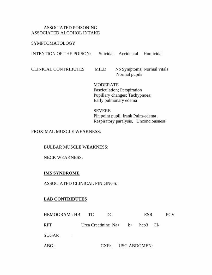
ASSOCIATED POISONING ASSOCIATED ALCOHOL INTAKE SYMPTOMATOLOGY
INTENTION OF THE POISON: Suicidal Accidental Homicidal
CLINICAL CONTRIBUTES MILD No Symptoms; Normal vitals Normal pupils MODERATE
Fasciculation; Perspiration Pupillary changes; Tachypnoea; Early pulmonary edema
SEVERE Pin point pupil, frank Pulm-edema , Respiratory paralysis, Unconciousness
PROXIMAL MUSCLE WEAKNESS: BULBAR MUSCLE WEAKNESS: NECK WEAKNESS: IMS SYNDROME ASSOCIATED CLINICAL FINDINGS: LAB CONTRIBUTES
HEMOGRAM : HB TC DC ESR PCV
RFT Urea Creatinine Na+ k+ hco3 Cl-
SUGAR : ABG : CXR: USG ABDOMEN:

ECG: ST-T CHANGES QTC PROLONGATION LOW VOLTAGE PROGRESSIVE LOSS OF VOLTAGE GASTRIC ASPIRATE SERUM PSEUDO CHOLINEESTRASE LEVELS DAY1 DAY2 DAY3 CPK CPK MB LFT : SGPT SGOT SAP TB LDH AMYLASE TREATMENT CONTRIBUTES TOTAL DOSE OF ATROPINE TOTAL DOSE OF P2 AM ( Less than 4 gms per day | More than 4 gms per day)
ANTIBIOTICS:TYPE DURATION VENTILATORY SUPPORT: INTIATIONMODE: DURATION :
OUTCOME MORTALITY RATE CAUSE OF DEATH

LIST OF TABLES
TABLE NO.
TITLE PAGE NO.
1. CLASSIFICATION AND THEIR STRUCTURE
3
2. Symptoms and signs of organophosphorous poisoning
10
3. Age Distribution 28
4. Age Distribution and Outcome 28
5. Sex Distribution 29
6. Occupation 29
7. Locality 30
8. Type of Poison consumed 30
9. Route Of Exposure 31
10. Duration of stay in hospitals 31
11. Duration Of Hospital Stay Correlation
With Clinical Severity-
32
12. Quantum of exposure 32
13. Quantity of Consumption and Clinical
Severity
33
14. Onset Of Symptoms After Exposure 34
15. Initiation Of First Aid 34

TABLE NO.
TITLE PAGE NO.
16. Time of initiation of Atropine 35
17. Time Of Initiation Of 2-PAM 35
18. Time taken to reach Poison Centre 36
19. Time interval to reach Poison Centre and
Clinical Severity
36
20. Cases Grouped According To Dreisbach
Clinical Criteria
37
21. Severity Grading and outcome 38
22. Severity Grading and Ventilatory
Management
39
23. Proximal Muscle Group Involvement and
Clinical Severity
40
24. Proximal Muscle Group Involvement and
Ventilatory Management
40
25. Proximal Muscle Group and outcome 41
26. Bio Chemistry Values 42
27. LFT 42
28. ABG 43
29. – ABG & Clinical Severity 43

TABLE NO.
TITLE PAGE NO.
30. ABG & Ventilatory Support 44
31. ABG & Outcome 44
32. Pseudocholinesterase Levels in OPC
poisoned Patients
46
33. Day 1 Serum AcHE Level & Clinical Severity
47
34. Day 3 Serum AcHE Level & Clinical Severity
47
35. Day 3 Serum AcHE Level & Ventilatory
Support
48
36. Day 3 Serum AcHE Level & Outcome 48
37. Day 5 Serum AcHE Level & Clinical
Severity
49
38. Day 5 Serum AcHE Level & Ventilatory
support Severity
49
39. Day 5 Serum AcHE Level & Outcome 50
40. Creatine kinase (CK) and its MB Fraction
(CKMB)
50
41. Serum CK Level & Clinical Severity 51
TABLE NO.
TITLE PAGE NO.
42. Serum CK Level & Ventilatory Support 51
43. Serum CKMB Level & Clinical Severity 52
44. Serum CKMB Level & Ventilatory
Support
52
45. Serum amylase Level & Clinical Severity 53
46. Serum Amylase Level & Ventilatory
Support
53
47. 2PAM & Clinical Features 54
48. 2PAM & Ventilatory Support 54
49. 2PAM & Outcome 55
50. Ventilatory Support & Outcome 55

LIST OF FIGURES
FIG 1. MECHANISM OF OPC
FIG 2.BIOLOGICAL INTERACTIONS OF OPC
FIG 3.AGE DISTRIBUTION
FIG 4. SEX DISTRIBUTION
FIG 5.OCCUPATION
FIG 6. TYPE OF POISON
FIG 7. ROUTE OF EXPOSURE
FIG 8. QUANTITY CONSUMED
FIG 9. DREISBACH CLINCAL CRITERIA
FIG 10.OUTCOME
FIG 11.CORRELATION OF SERUM CHOLINESTERASE WITH
CLINCAL GRADING

Fig.3 Age Distribution
17
40
15 15
0
5
10
15
20
25
30
35
40
45
<= 20 21-30 31-40 > 40Age group (in years)
Num
ber o
f Pat
ient
s

Fig.4 Sex Distribution Fig.2 Sex
76
11
0 10 20 30 40 50 60 70 80
Male
Female
Frequency(n)

Fig.5 Occupation
40, 46%
37, 43%
10, 11%
Agriculture Unskilled Labour Others

Fig. 6 Type of Poison
6
26
3
2
3
2
1
2
30
3
1
6
CHLORPYIFOS
DIMECRON
QUINALPHOS (EKALUX)
FENTHION (BAYTEX)
HINOXAN
MALATHION (CHEMATHION)
METHYL PARATHION (METAPHOS)
MONOCROTOPHOS
PARATHION (FOLIDOL, EKATOX)
PHORATE
SYSTOX
TRIPHOS
No. of Patients
Type of Poison

Fig.7 Route of
87%
11%
2%IngestionalInhalationalTopical

25
29
19
14
0
5
10
15
20
25
30
No. of Patients
NA <=50 51-100 >100in ml
Fig.8 Quantity consumed ( )

Fig.9 Dreischbach clinical criteria of
17%
33%
50% MildModerateSevere
65
22
0
10
20
30
40
50
60
70
No. of Patients
Recovered Expired
Fig. 10 Outcome

Fig.11 Correlation of Serum Cholinesterase levels with Clinical grading
Day 1
Day 3Day 5
Clinical Contributes
SevereModerateMild
95%
CI o
f Enz
ymes
5000
4000
3000
2000
1000
0

KEY Sex : Male = 1 Female = 2
Occupation : Agriculturist = 1 Unskilled workers = 2 Others = 3
Locality : <100 km = 1 >100 km =2
Intention : Suicide = 1 Accidental = 2
Association poison : Yes = 1 No = 2
Alcohol Intake : Yes = 1 No=2
Severity : Mild = 1 Moderate = 2 Severe = 3
Proximal muscle Involvement : Yes = 1 No.2
ECG : 1= PI 2=PII 3 = PIII 4 = Normal 5 = Sinus Bradycardia
Ventilatory support : 1=Nil 2=Assist Control 3 =Simv
Outcome : 1=Discharge 2 = Death