Embed Size (px)
Citation preview

Accepted Manuscript
Compositional and morphological analyses of wax in northern wild berry spe-cies
Priyanka Trivedi, Katja Karppinen, Linards Klavins, Jorens Kviesis, PetriSundqvist, Nga Nguyen, Esa Heinonen, Maris Klavins, Laura Jaakola, JuhaVä änänen, Janne Remes, Hely Häggman
PII: S0308-8146(19)30938-0DOI: https://doi.org/10.1016/j.foodchem.2019.05.134Reference: FOCH 24860
To appear in: Food Chemistry
Received Date: 8 January 2019Revised Date: 13 May 2019Accepted Date: 20 May 2019
Please cite this article as: Trivedi, P., Karppinen, K., Klavins, L., Kviesis, J., Sundqvist, P., Nguyen, N., Heinonen,E., Klavins, M., Jaakola, L., Vä änänen, J., Remes, J., Häggman, H., Compositional and morphological analyses ofwax in northern wild berry species, Food Chemistry (2019), doi: https://doi.org/10.1016/j.foodchem.2019.05.134
This is a PDF file of an unedited manuscript that has been accepted for publication. As a service to our customerswe are providing this early version of the manuscript. The manuscript will undergo copyediting, typesetting, andreview of the resulting proof before it is published in its final form. Please note that during the production processerrors may be discovered which could affect the content, and all legal disclaimers that apply to the journal pertain.
1
Compositional and morphological analyses of wax in northern wild berry species
Priyanka Trivedia,*, Katja Karppinena, Linards Klavinsb, Jorens Kviesisb, Petri Sundqvistc, Nga
Nguyena, Esa Heinonenc, Maris Klavinsb, Laura Jaakolad,e, Juha Väänänenc, Janne Remesc, Hely
Häggmana
a Department of Ecology and Genetics, University of Oulu, FI-90014 Oulu, Finland
b Department of Environmental Science, University of Latvia, LV-1004, Riga, Latvia
c Centre of Microscopy and Nanotechnology, University of Oulu, FI-90014 Oulu, Finland
d NIBIO, Norwegian Institute of Bioeconomy Research, NO-1431 Ås, Norway
e Climate laboratory Holt, Department of Arctic and Marine Biology, UiT The Arctic University of
Norway, NO-9037 Tromsø, Norway
Priyanka Trivedi: [email protected]
Katja Karppinen: [email protected]
Linards Klavins: [email protected]
Jorens Kviesis: [email protected]
Petri Sundqvist: [email protected]
Nga Nguyen: [email protected]
Esa Heinonen: [email protected]
Maris Klavins: [email protected]
Laura Jaakola: [email protected]
Juha Väänänen: [email protected]
Janne Remes: [email protected]
Hely Häggman: [email protected]
*Corresponding author
2
ABSTRACT
Aerial surfaces of plants are covered by a waxy cuticle protecting plants from excessive water loss
and UV light. In the present study, composition and morphology of cuticular waxes of northern
wild berry species bilberry (Vaccinium myrtillus L.), lingonberry (V. vitis-idaea L.), bog bilberry
(V. uliginosum L.) and crowberry (Empetrum nigrum L.) were investigated. Scanning electron
microscopy (SEM) revealed differences in epicuticular wax morphology, and gas chromatography–
mass spectrometry (GC–MS) analysis confirmed variation in chemical composition of cuticular
waxes between the berry species. The dominant compounds in bilberry and lingonberry cuticular
waxes were triterpenoids, while fatty acids and alkanes were the dominant type in bog bilberry and
crowberry, respectively. Wax extracted by supercritical fluid extraction (SFE) from industrial press
cakes of bilberry and lingonberry contained linoleic acid and γ-linolenic acid as the dominant
compounds. Furthermore, in vitro sun protection factor (SPF) of berry waxes depicted good UV-B
absorbing capacities.
Keywords: Vaccinium, Empetrum, fruits, cuticular wax, chemical composition, morphology,
triterpenoids
Chemical compounds studied in this article
β-amyrin (PubChem CID: 73145); α-amyrin (PubChem CID: 73170); Lupeol (PubChem CID:
259846); Nonacosane (PubChem CID:12409); Hentriacontane (PubChem CID: 12410); Linoleic
acid (PubChem CID: 5280450); γ-linolenic acid (PubChem CID: 5280933); γ-tocopherol
(PubChem CID: 92729); Adriaticol (PubChem CID:00000)
3
1. Introduction
Cuticle acts as an interface between plant and environment covering the aerial parts of land
plants, including leaves, stems and fruits. Plant cuticle evolved 450 million years ago as a protection
against non-transpirational water loss but it also protects plants from UV light and pathogen attacks
(Yeats & Rose, 2013). Solar radiation reaching the earth includes 10% UV light, of which UV-B
(280-320 nm) has the highest energy, creating a need for protection, not only for plants, but also for
humans, due to risk of skin cancer.
The plant cuticle is composed of a polyester polymer called cutin and cuticular wax. Cuticular
wax is a complex mixture of very-long-chain fatty acids and their derivatives, such as alkanes,
ketones, primary and secondary alcohols, aldehydes and esters, but also includes secondary
metabolites, such as triterpenoids, sterols, tocopherols and phenolic compounds (Yeats & Rose,
2013). Cuticular wax composition can vary greatly depending on species, organ and developmental
stage (Yeats & Rose, 2013; Trivedi et al., 2019). The cuticular wax is present as intracuticular wax,
an amorphous mixture of lipids embedded in the cutin, and outermost epicuticular wax (Barthlott,
Mail, Bhushan, & Koch, 2017). The epicuticular wax has different forms, such as films or different
types of three-dimensional crystalloid structures on plant surfaces (Jeffree, 2006). The epicuticular
wax can be visible to the naked eye, either as whitish, dull or glossy coating.
The studies on plant cuticular waxes have largely focussed on vegetative parts, such as leaves,
while surfaces of fruits and berries have been less studied (Trivedi et al., 2019). Berries are an
important component of a healthy diet and it is well established that the dietary intake of berries has
a positive and profound impact on human health. The health effects are mainly due to bioactive
compounds, such as polyphenols, flavonoids, carotenoids and vitamins (Jimenez-Garcia, Guevara-
Gonzalez, Miranda-Lopez, Feregrino-Perez, Torres-Pacheco, & Vazquez-Cruz, 2013). However,
berries also include other types of bioactive components, such as compounds present in wax. For
example, triterpenoids have various health beneficial properties, such as anticancer, anti-
inflammatory, antimicrobial and cardioprotective effects (Szakiel, Pa̧czkowski, Pensec, & Bertsch,
4
2012b). The juice industry is one of the major users of berries and the industrial leftovers, berry
press cakes, form a potential source for bioactive compounds and berry wax fractions to be utilized
in commercial products.
Bilberry (Vaccinium myrtillus L.) and lingonberry (V. vitis-idaea L.) are economically the
most important wild berries of Northern Europe widely utilized by food industry including juice
industry. Crowberry (Empetrum nigrum L.) and bog bilberry (V. uliginosum L.) are less utilized
nevertheless widely distributed wild berries in northern areas. These berry species have been
studied extensively for secondary metabolites (Jurikova et al., 2016; Karppinen, Zoratti,
Nguyenquynh, Häggman, & Jaakola, 2016). However, they have not been investigated for their
cuticular wax composition, although the triterpenoid profile of bilberry cuticular wax has been
reported earlier (Szakiel, Pączkowski, & Huttunen, 2012a).
The objective of the present study was to investigate the amount, chemical composition as
well as morphology of cuticular wax in important northern wild berries, including bilberry,
lingonberry, bog bilberry and crowberry. Also, the berry press cakes (residues of juice industry) of
bilberry and lingonberry were extracted by supercritical fluid extraction (SFE), and the composition
was analyzed. In addition, in vitro sun protection factor (SPF) of the waxes is reported and the
potential commercial use of berry waxes discussed.
2. Materials and methods
2.1. Plant material
Berries of four different wild species were utilized in the present study, namely bilberry,
lingonberry, bog bilberry and crowberry. Ripe fruits of the berry species were collected carefully
using forceps in August 2017 from natural forest stands in Oulu region, Finland. Industrial press
cakes of bilberry and lingonberry were obtained from Polarica Ltd., Tornio, Finland.
2.2. Scanning electron microscopy (SEM)

5
For SEM analysis, the fresh berries were immediately dried after collection by using a
vacuum freeze-drier (Edwards High Vacuum International, West Sussex, England) before fixing on
aluminium stubs. The berry surfaces were then sputter-coated with a 20 nm layer of platinum by
using a sputter coater (Agar High Resolution Sputter Coater, Agar Scientific Ltd, Essex, UK) and
then investigated for the three-dimensional surface micromorphology by using SEM (Helios
Nanolab 600, Oregon, USA). SEM was operated at 5 kV with a current value of 86 pA at secondary
electron mode. Images were taken at 2500X and 10000X magnification.
2.3. Cuticular wax extraction and determination of wax amount
The cuticular wax from the ripe berries of different berry species was separately extracted
with chloroform (Sigma-Aldrich, St. Louis, USA) at ambient temperature immediately after
collection and transportation to the laboratory. One hundred berries per species were individually
dipped twice in 5 ml chloroform for 30 seconds. The two extracts were combined, evaporated to
dryness under nitrogen flow at room temperature and the dry weight was measured. The cuticular
wax extraction was performed in triplicates for each berry species. The amount of wax was
expressed as weight per unit surface area (µg/cm2). For calculating the surface areas, images of the
dipped berries on a white surface were taken immediately after extraction. Image J software v1.50i
(NIH, Maryland, USA) was used to calculate the total surface area of the berries as S = 4 πr2, where
r is the radius of berry (assuming that the berries are spherical).
2.4. Wax extraction from industrial berry press cakes
The berry press cakes of bilberry and lingonberry were dried in an oven at 60 °C and milled to
fine powder by using a handheld grinder before wax extraction. Supercritical fluid extraction (SFE)
was performed by using an Xtractor (Chematur Ecoplanning Pvt Ltd, Tampere, Finland). The
operating parameters used for extraction were 350 bar at 60 °C with a CO2 flow rate of 0.4-0.5
6
L/min for 10-L extraction. The yield of the wax was expressed as mg/g dry weight of starting
material.
2.5. GC-MS analysis
Derivatization of fatty acids was performed as previously described (Dobson, Shrestha, Hilz,
Karjalainen, McDougall, & Stewart, 2012). Extracted berry wax was dissolved in 0.5 ml toluene
(Sigma-Aldrich). Then, 3 ml of 14% boron trifluoride-methanol solution (Sigma-Aldrich) were
added and the mixture and heated at 60 C for 180 min. Resulting fatty acid methyl esters were
dissolved in hexane and used for GC-MS analysis.
GC-MS analysis was performed using a PerkinElmer Clarus 580 system equipped with a
Clarus SQ 8 C mass-selective detector (Waltham, MA, USA) and an Omegawax 250 column (30 m
× 0.25 mm, 0.25 µm, Darmstadt, Germany). Analysis of FAME’s and polyfunctional compounds as
trimethylsilyl derivatives was performed on an Elite-5MS column (30 m × 0.25 mm, 0.25 μm,
PerkinElmer, Waltham, MA, USA) after derivatization of hexane fraction with 60 µL N,O-Bis
(trimethylsilyl)trifluoroacetamide (Sigma-Aldrich). Analysis in both columns was initiated at 75 °C
for 2 min and then increased from 75 °C to 150 °C at a rate of 20 °C/min. For Omegawax 250
column, temperature was further increased from 150 °C to 270 °C and for Elite-5MS the increase
was from 150 °C to 310 C at 4 °C/min. In the final isothermal step, temperature was held for 5 min
at 270 °C for Omegawax 250 and 310 C for Elite-5MS. The total runtime was 39.50 min and 54.75
min for Omegawax 250 and Elite-5MS, respectively. Injection volume was 0.5 µl with injection and
interface temperatures kept at 290 C. Helium (AGA, Riga, Latvia) was used as a carrier gas at the
flow rate of 1.0 ml/min and split flow of 10.0 ml/min. Electron impact was set to 70 eV and scan
range from 42 to 750 m/z. Identification of the compounds was done using the NIST MS 2.2 library
(Gaithersburg, MD, USA). The analysis was performed in triplicate. Quantification of compounds
was done using standard solutions of methyl heptadecanoate (≥99.0%), ergosterol (≥99.0%),
hexadecanol (≥99.0%), 1-dodecanal (≥98.0%), (±)-α-tocopherol (99.0%), 1-octadecanol (99.0%)
7
and n-tetracosane (≥99.5%) obtained from Sigma-Aldrich in the concentration range of 1.5–500
µg/ml.
2.6. Determination of in vitro sun protection factor (SPF)
Extracted wax was dissolved in methanol (Fisher Scientific, Waltham, USA) for bilberry and
lingonberry waxes, and hexane (Fisher Scientific) for bog bilberry and crowberry waxes. The
choice of solvent was based on the maximal solubility of wax in the respective solvents. The
absorption spectra of the wax solutions in quartz cuvette were obtained in the range of 290 to 320
nm every 1 nm by using a UV-Vis spectrophotometer (Genesys 10S, Thermo Scientific, Waltham,
USA). Measurements were done in triplicates. The SPF was calculated by the following equation
(Mansur, Breder, Mansur, & Azulay, 1986):
𝑆𝑃𝐹 = 𝐶𝐹 ×320
∑290
𝐸𝐸 (𝜆) × 𝐼 (𝜆) × 𝐴𝑏𝑠 (𝜆)
In the equation, EE (λ) is erythemal efficiency spectrum, I (λ) is solar simulator intensity spectrum,
Abs (λ) is the absorbance of the measured sample, and CF is a correction factor (= 10). The constant
values of normalized product function (EE × I; Sayre, Agin, Levee, & Marlowe, 1979) used in the
calculations can be found in Supplementary Table 1.
2.7. Statistical analysis
One-way analysis of variance (ANOVA) with Duncan’s multiple range test at p < 0.05 was
performed using SPSS statistical program version 25.0 (IBM, Chicago, USA). Principal component
analysis was performed by using SAS JMP®, Version 13 (SAS Institute Inc., Cary, NC, USA).
3. Results and discussion
3.1. Morphology of epicuticular wax
8
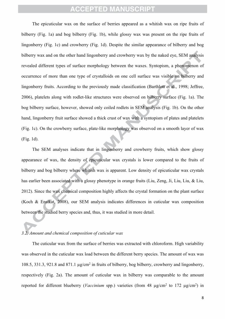
The epicuticular wax on the surface of berries appeared as a whitish wax on ripe fruits of
bilberry (Fig. 1a) and bog bilberry (Fig. 1b), while glossy wax was present on the ripe fruits of
lingonberry (Fig. 1c) and crowberry (Fig. 1d). Despite the similar appearance of bilberry and bog
bilberry wax and on the other hand lingonberry and crowberry wax by the naked eye, SEM analysis
revealed different types of surface morphology between the waxes. Syntopism, a phenomenon of
occurrence of more than one type of crystalloids on one cell surface was visible on bilberry and
lingonberry fruits. According to the previously made classification (Barthlott et al., 1998; Jeffree,
2006), platelets along with rodlet-like structures were observed on bilberry surface (Fig. 1a). The
bog bilberry surface, however, showed only coiled rodlets in SEM analysis (Fig. 1b). On the other
hand, lingonberry fruit surface showed a thick crust of wax with a syntopism of plates and platelets
(Fig. 1c). On the crowberry surface, plate-like morphology was observed on a smooth layer of wax
(Fig. 1d).
The SEM analyses indicate that in lingonberry and crowberry fruits, which show glossy
appearance of wax, the density of epicuticular wax crystals is lower compared to the fruits of
bilberry and bog bilberry where whitish wax is apparent. Low density of epicuticular wax crystals
has earlier been associated with a glossy phenotype in orange fruits (Liu, Zeng, Ji, Liu, Liu, & Liu,
2012). Since the wax chemical composition highly affects the crystal formation on the plant surface
(Koch & Ensikat, 2008), our SEM analysis indicates differences in cuticular wax composition
between the studied berry species and, thus, it was studied in more detail.
3.2. Amount and chemical composition of cuticular wax
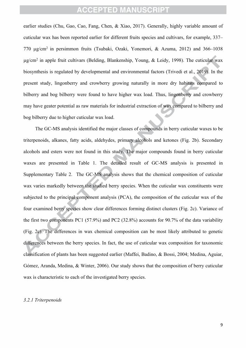
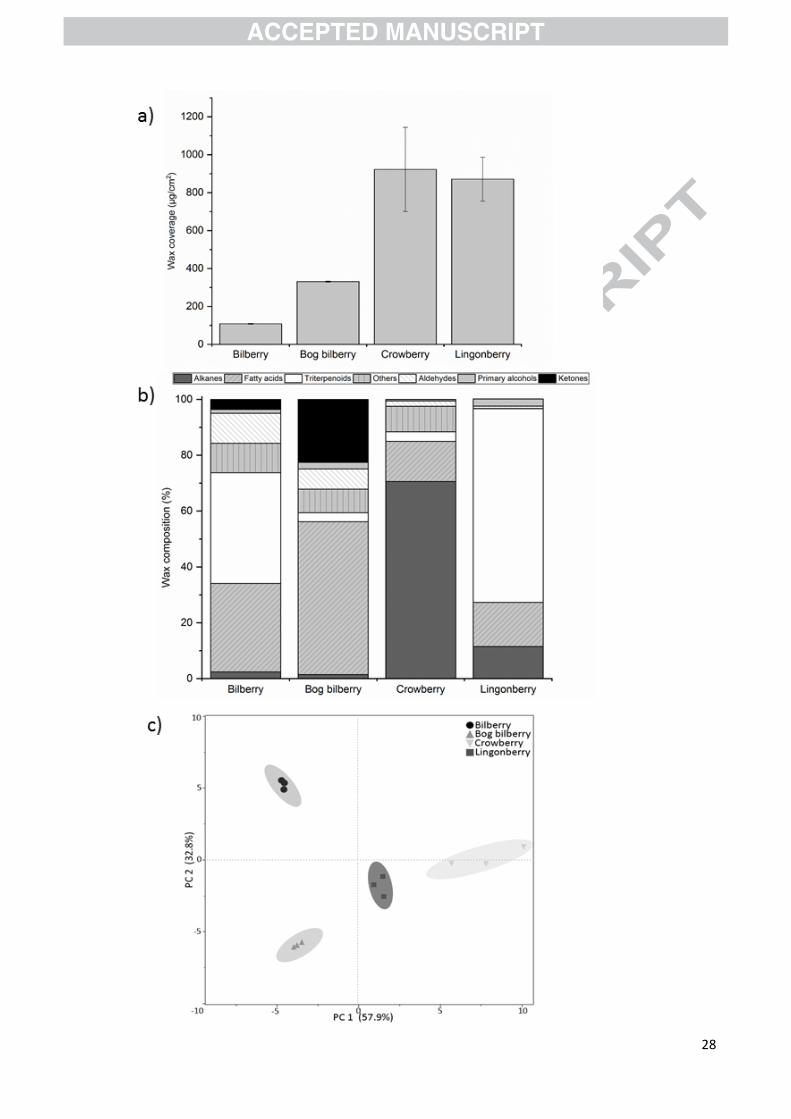
The cuticular wax from the surface of berries was extracted with chloroform. High variability
was observed in the cuticular wax load between the different berry species. The amount of wax was
108.5, 331.3, 921.8 and 871.1 µg/cm2 in fruits of bilberry, bog bilberry, crowberry and lingonberry,
respectively (Fig. 2a). The amount of cuticular wax in bilberry was comparable to the amount
reported for different blueberry (Vaccinium spp.) varieties (from 48 µg/cm2 to 172 µg/cm2) in
9
earlier studies (Chu, Gao, Cao, Fang, Chen, & Xiao, 2017). Generally, highly variable amount of
cuticular wax has been reported earlier for different fruits species and cultivars, for example, 337–
770 µg/cm2 in persimmon fruits (Tsubaki, Ozaki, Yonemori, & Azuma, 2012) and 366–1038
µg/cm2 in apple fruit cultivars (Belding, Blankenship, Young, & Leidy, 1998). The cuticular wax
biosynthesis is regulated by developmental and environmental factors (Trivedi et al., 2019). In the
present study, lingonberry and crowberry growing naturally in more dry habitats compared to
bilberry and bog bilberry were found to have higher wax load. Thus, lingonberry and crowberry
may have geater potential as raw materials for industrial extraction of wax compared to bilberry and
bog bilberry due to higher cuticular wax load.
The GC-MS analysis identified the major classes of compounds in berry cuticular waxes to be
triterpenoids, alkanes, fatty acids, aldehydes, primary alcohols and ketones (Fig. 2b). Secondary
alcohols and esters were not found in this study. The major compounds found in berry cuticular
waxes are presented in Table 1. The detailed result of GC-MS analysis is presented in
Supplementary Table 2. The GC-MS analysis shows that the chemical composition of cuticular
wax varies markedly between the studied berry species. When the cuticular wax constituents were
subjected to the principal component analysis (PCA), the composition of the cuticular wax of the
four examined berry species show clear differences forming distinct clusters (Fig. 2c). Variance of
the first two components PC1 (57.9%) and PC2 (32.8%) accounts for 90.7% of the data variability
(Fig. 2c). The differences in wax chemical composition can be most likely attributed to genetic
differences between the berry species. In fact, the use of cuticular wax composition for taxonomic
classification of plants has been suggested earlier (Maffei, Badino, & Bossi, 2004; Medina, Aguiar,
Gómez, Aranda, Medina, & Winter, 2006). Our study shows that the composition of berry cuticular
wax is characteristic to each of the investigated berry species.
3.2.1 Triterpenoids
10
Triterpenoids are commonly found in cuticular waxes of fruits (Szakiel et al., 2012b; Trivedi
et al., 2019). Triterpenoids represented the most abundant class of compounds in bilberry and
lingonberry wax accounting for 39.6% and 69.6% of total cuticular wax content, respectively, while
in bog bilberry and crowberry, triterpenoids accounted for only 3.2% and 3.4% of the total cuticular
wax, respectively (Fig. 2b). Both in bilberry and lingonberry wax, triterpene alcohols (β-amyrin, α-
amyrin, lupeol) and triterpene acids (oleanolic acid, ursolic acid) were identified, while only
lingonberry wax contained adriaticol and uvaol (Table 1). In bog bilberry, oleanolic acid (3.1%) and
ursolic acid (1.8%) were identified (Table 1).
Among Vaccinium species, reports of triterpenoid profiles in bilberry and blueberry cuticular
waxes have indicated triterpene acids, namely oleanolic acid and ursolic acid, as the dominant
compounds (Szakiel et al., 2012a; Chu et al., 2017). This result is different from our study that
indicated β-amyrin as the most abundant triterpenoid (20.2% of total wax) followed by oleanolic
acid (8.9%) and α-amyrin (7.1%) in bilberry cuticular wax. The variability in bilberry triterpenoid
profiles between the two studies can be due to the difference in geographical origin of the sample,
environmental factors or timing of sample collection.
In lingonberry cuticular wax, adriaticol (14.2%) followed by α-amyrin (13%) and β-amyrin
(12.8%) were the dominant triterpenoids. Adriaticol has a structure similar to isoarborinol, a C3-
oxygenated pentacyclic triterpenol. Isoarborinol derivatives, which can be used as plant biomarkers,
are rarely reported in cuticular wax of plants. Isoarborinol derivatives have been reported earlier in
leaf epicuticular wax of Euphorbia lathyris and plants of angiosperm families, such as Gramineae
(Van Bree et al., 2018). The finding of adriaticol in lingonberry wax gives possibility to use the
compound as a biomarker for the identification of lingonberry, for example in lingonberry products.
However, for that purpose, further studies of the wax profiles of different berry species are still
required.
Wax morphology and chemical composition are closely related, and the dominant wax
compounds play important role in crystal assembly (Jeffree et al., 2006). The dominance of
11
triterpenoids in the cuticular wax of bilberry and lingonberry may lead to the formation platelets
that were detected in the SEM analysis of our study. Triterpenoid rich platelets have been reported
in Sedum rupestre leaf wax (Stevens, 1995). Also, triterpenoid rich wax from olive fruits (Olea
europaea) and leaves of southern mahogany (Eucalyptus botryoides) recrystallized as platelets
(Baker, 1982). However, caution must be taken when drawing connections between epicuticular
wax morphology and cuticular wax chemical composition since wax extracts are complex mixtures
of compounds and extracts may also contain compounds from intracuticular wax.
3.2.2. Fatty acids
Fatty acids were the major components of cuticular wax in bog bilberry accounting for 54.8%
of the total wax (Fig. 2b), with arachidic acid as dominant compound (Table 1). To the best of our
knowledge, fatty acids have not earlier been reported as the dominant component in fruit cuticular
waxes although some fruits contain high amounts of fatty acids in their cuticles, such as Asian pear
(Yin et al., 2011) and cucumber (Wang et al., 2015). In bilberry, lingonberry and crowberry, fatty
acids accounted for 31.7%, 15.9% and 14.4% of the total wax content, respectively (Fig. 2b). In
bilberry cuticular wax, a vast variety of fatty acids was detected of which montanic acid (C28:0) and
cerotic acid (C26:0) were the predominant types (Table 1). Crowberry fruit contained oleic acid (C18:
1n-9) as the most abundant cuticular fatty acid while lingonberry wax contained mainly lignoceric
acid (C24:0).
3.2.3. Alkanes
Alkanes were the predominant compounds in cuticular wax of crowberry fruits, constituting
70.6% of the total wax (Fig. 2b). Especially the amount of nonacosane was high followed by
hentriacontane in crowberry (Table 1). In lingonberry cuticular wax, alkanes represented 11.5% of
total wax (Fig. 2b) with nonacosane as the predominant alkane (Table 1). Both nonacosane and
hentriacontane are common compounds found in fruit epicuticular waxes (Trivedi et al., 2019).
12
Nonacosane has been reported as the dominant compound in cuticular waxes of fruits, such as apple
(Belding, Sutton, & Blankenship, 2000), Asian pear (Yin et al., 2011) and cucumber (Wang et al.,
2015). Alkanes were a minor fraction in cuticular wax of bilberry and bog bilberry fruits
constituting only 2.4% and 1.4% of total wax, respectively (Fig. 2b). Previous studies have
suggested that waxes containing high amounts of nonacosane and hentriacontane often form plate-
like morphologies (Jeffree et al., 2006). Thus, plates found in crowberry and lingonberry surface in
this study may be due to the presence of high levels of alkanes in cuticular wax.
3.2.4. Other very-long-chain aliphatic compounds
Aldehydes were found in cuticular wax of fruits of all studied berry species: 10.3%, 7.2%,
1.8% and 0.9% in bilberry, bog bilberry, crowberry and lingonberry, respectively (Fig. 2b).
Octacosanal was the predominant aldehyde in bilberry and bog bilberry fruits while in crowberry
and lingonberry, tetracosanal was the predominant type (Table 1). Aldehydes are rarely found
abundantly in fruit cuticles with the exception in cucumber, cranberry (Vaccinium macrocarpon)
and Citrus fruits (Trivedi et al., 2019). The detected aldehyde amount in bilberry cuticular wax in
this study is close to that reported earlier for cranberry (Croteau & Fagerson, 1971).
Ketones accounted for the second largest fraction in bog bilberry wax (22.5%), with 2-
heneicosanone as the most prominent ketone (Table 1). Ketones were also present in small
quantities in bilberry (3.6%) and crowberry (0.03%) fruit cuticular wax (Fig. 2b). Ketones can be
responsible for the formation of rodlets on the surface of bog bilberry that were detected in our
SEM analysis since ketone-containing waxes have been reported earlier to form different types of
rodlets (Ensikat, Boese, Mader, Barthlott, & Koch, 2006). However, further data is needed to
confirm this.
Primary alcohols were present in berry waxes, only in small quantities, accounting for 1.3%,
2.3%, 0.6% and 2.6% in cuticular wax of bilberry, bog bilberry, crowberry and lingonberry fruits,
13
respectively (Fig. 2b). Cinnamic acid in small quantities was found in bog bilberry cuticular wax. A
minor quantity of p-coumaric acid was found in lingonberry cuticular wax (Table 1).
3.3 SFE extraction and chemical composition of wax
By SFE extraction, green semisolid wax was obtained from bilberry press cake while the wax
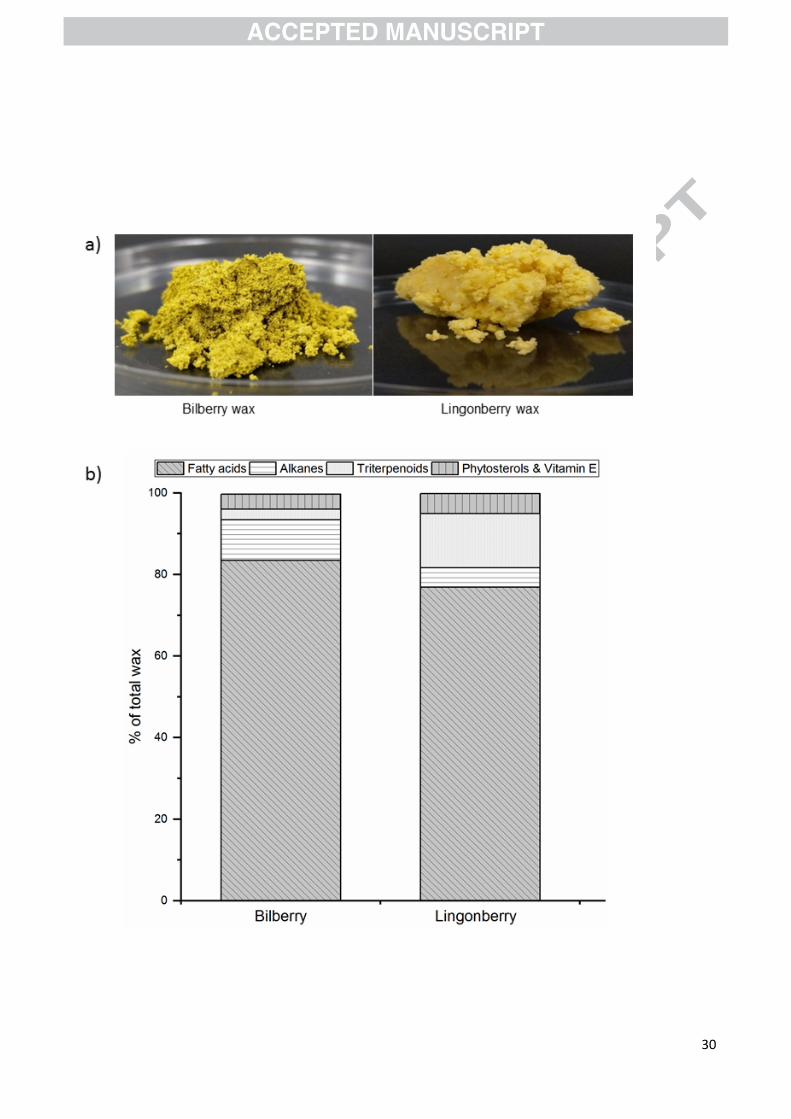
from lingonberry press cake was more yellow and greasy (Fig. 3a). The yield of lingonberry fruit
wax was 1.02 % (10.2 mg/g DW) while the yield for bilberry was 0.45% (4.5 mg/g DW). The
higher wax yield obtained from lingonberry by SFE is in line with our results from cuticular wax
extraction, confirming that lingonberry as a raw material can be more effective than bilberry for
industrial scale berry wax extraction.
Compositional analysis by GC-MS showed that fatty acids were the most abundant
constituents in the SFE extracts, accounting for 83.4% and 76.9% in bilberry and lingonberry wax,
respectively (Fig. 3b). The wax components also included alkanes, triterpenoids, phytosterols,
vitamin E and minute amounts of aldehydes and cinnamic acid (Fig. 3b, Supplementary Table 3). In
our study, linoleic acid and γ-linolenic acid were the predominant compounds, constituting a total of
53.9% and 54.8% of total wax for bilberry and lingonberry, respectively (Supplementary Table 3),
that is similar to the amount reported from SFE extraction of bilberry earlier (Jumaah, Sandahl, &
Turner, 2015). Triterpenoids accounted for 3.0% and 14.3% for bilberry and lingonberry wax,
respectively, with β-amyrin and lupeol as predominant compounds. Alkanes formed a minor
fraction (9.9% for bilberry and 4.8% for lingonberry wax), with nonacosane as the dominant alkane
in both bilberry and lingonberry press cakes. β-sitosterol was found in bilberry and lingonberry
wax, constituting 3% and 4.7% of total wax composition, respectively.
The wax derived from bilberry and lingonberry press cakes, in the present study, had different
chemical composition compared to berry cuticular waxes. Phytosterols and vitamin E were detected
in press cakes, but not in cuticular wax of berries. The difference in composition is most likely due
to the presence of seeds in the berry press cakes. Bilberry and lingonberry seed oil have been
14
reported to contain vitamin E and other bioactive compounds (Yang, Ahotupa, Määttä & Kallio,
2011; Gustinelli et al., 2018). Since SFE is a clean, sustainable method to extract valuable waxes
from various agricultural residues, it is well suited for the extraction of waxes rich in bioactive
compounds, for commercial applications in the food and cosmetic industries. Dietary as well as
topical application of γ-linolenic acid has been reported to have a protective effect on the structure
and physiology of skin (Andreassi, Forleo, Lorio, Masci, Abate, & Amerio, 1997; Kawamura et al.,
2011). Minor quantities of benzoic acid were also detected in bilberry and lingonberry wax in our
study (Supplementary Table 3), and can contribute to the shelf-life of wax (Brul & Coote, 1999).
Therefore, berry waxes can also have potential applications in food packaging and preservation.
Taken together, our study signifies that residues of berry juice industry can be used to extract wax
using sustainable extraction process (SFE), and this wax has potential as an effective additive for
applications in food formulations as well as in the cosmetic industry.
3.4. SPF of berry wax
SPF is a universal term used to assess the UV absorption/blocking potential of compounds.
There are studies on in vitro SPFs of various plant extracts (Kumar, Datta, & Dutta Gupta, 2009;
Maske, Lokapure, Nimbalkar, Malavi, & D’souza, 2015). However, to our knowledge the UV
blocking potential of berry waxes has not been studied yet. All extracted berry waxes were tested
for SPF in our study. The results show high UV-B absorption properties as revealed by their SPFs
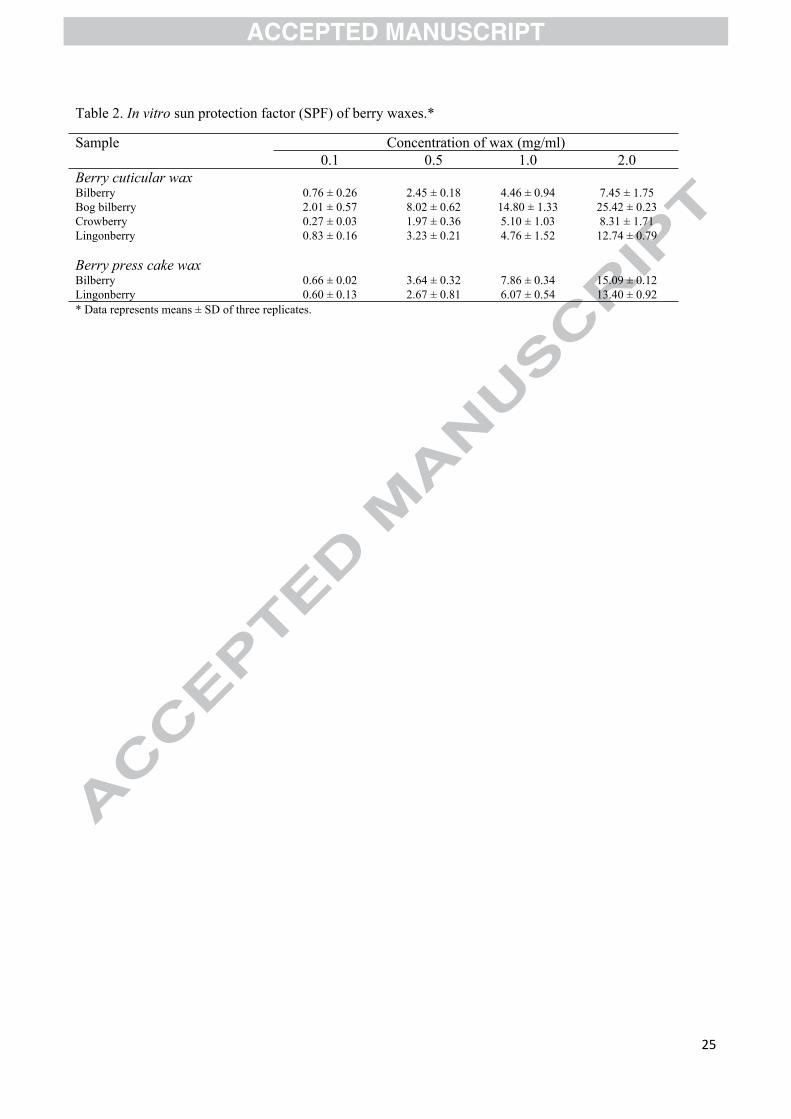
as well as demonstrate dose dependent increase of SPF with the wax concentration (Table 2).
Cuticular wax of bog bilberry fruit showed the highest SPF. From SFE extracted waxes of berry
press cakes, bilberry wax showed slightly higher SPF than lingonberry wax (Table 2). The high SPF
could be attributed to the presence of cinnamic acid and vitamin E, which were both at higher levels
in bilberry wax compared to lingonberry wax (SFE extractions), and high cinnamic acid levels were
also found in bog bilberry cuticular wax. Cinnamic acid has been shown to absorb broad range of
UV-light, and it is used as a UV-absorber in cosmetic products (Li, Tang, Ma, Wang, & Zhou,
15
2017) while vitamin E has photoprotective effects against UV radiation (Podhaisky & Wohlrab,
2002).
One of the most important physiological functions of cuticular wax in plants is the protection
from UV-B and, thus, it is not surprising that the extracted cuticular waxes showed high UV-B
absorption in this study. The wild berry waxes showed SPFs that are comparable to SPFs of
common commercial sunscreen products (Dutra, Oliveira, Kedor-Hackmann, & Santoro, 2004).
Secondary metabolites, such as triterpenoids (Hashim, Sidek, Helan, Sabery, Palanisamy, & Ilham,
2011) and phenolic acids (Kumar et al., 2009) have been attributed for the potential UV-B
absorption activities of plant extracts. Thus, the presence of triterpenoids and phenolic acids in the
studied cuticular berry wax could be responsible for UV-B absorbing properties.
4. Conclusion
There is an increasing demand for natural plant based waxes due to irregular supply of
petroleum based waxes, as well as consumer inclination for natural products. It is therefore,
imperative to explore more plant based waxes for a sustainable greener economy. In this study we
have reported the chemical composition and SPF of fruit wax of four important northern wild berry
species. The variation in amount, morphology and chemical composition, as well as high SPF of
cuticular wax in different berry species was detected. Furthermore, we utilized SFE of industrial
residual berry press cakes from the berry juice industry to present a potential source of natural berry
waxes. Our results may contribute the exploration of various applications of berry waxes in food
engineering, food packaging/preservation and cosmetic industries. Berry wax might be suitable for
applications in food grade films/coatings to improve the water barrier, and optical and mechanical
properties of films.
5. Acknowledgements
16
We acknowledge the research grant from InterregNord (Natural Wax of Arctic Berries as Our
Treasure – WAX project (number 20201089 to University of Oulu and grant IR16-020 and grant
RMF16-026 to Troms Fylkeskommune and NIBIO). The work of PT was financially supported by
I4future doctoral program funded by EU Horizon 2020 research and innovation program under
Marie Skoldowska Curie grant agreement number 713606. The work of JK was financially
supported by the ERDF project No. 1.1.1.1/16/A/047 “Genus Vaccinium berry processing using
"green technologies and innovative, pharmacologically characterized biopharmaceutical products”
and the work of LK was financially supported by the patron “Mikrotīkls” Ltd. administered by the
Foundation of University of Latvia.
Conflicts of interest
The authors declare no conflict of interest.
References
Andreassi, M., Forleo, P., Di, Lorio A., Masci, S., Abate, G., & Amerio, P. (1997). Efficacy of
gamma-linolenic acid in the treatment of patients with atopic dermatitis. Journal of International
Medical Research, 25(5), 266–274. https://doi.org/10.1177/030006059702500504
Baker, E.A. (1982). Chemistry and morphology of plant epicuticular waxes. In D.F. Cutler, K.L.
Alvin, & C.E. Price (Eds.). The plant cuticle (pp. 139-166). London Academic Press Publishing
Ltd.
Barthlott, W., Mail, M., Bhushan, B., & Koch, K. (2017). Plant surfaces: Structures and functions
for biomimetic innovations. Nano-Micro Letters, 9, 23. https://doi.org/10.1071/FP06139
Barthlott, W., Neinhuis, C., Cutler, D., Ditsch, F., Meusel, I., Theisen, I., & Wilhelmi, H. (1998).
Classification and terminology of plant epicuticular waxes. Journal of the Linnean Society, 126,
237–260. https://doi.org/10.1111/j.1095-8339.1998.tb02529.x
17
Belding, R. D., Blankenship, S. M., Young, E., & Leidy, R. B. (1998). Composition and variability
of epicuticular waxes in apple cultivars. Journal of American Society of Horticultural Science,
123, 348–356.
Belding, R. D., Sutton, T. B., & Blankenship, S. M. (2000). Relationship between apple fruit
epicuticular wax and growth of Pelaster fructicola and Leptodontidium elatius, two fungi that
cause sooty blotch disease. Plant Disease, 84(7), 767–772.
https://doi.org/10.1094/PDIS.2000.84.7.767
Brul, S., & Coote, P (1999). Preservative agents in foods. Mode of action and microbial resistance
mechanisms. International Journal of Food Microbiology, 50(1-2), 1-17.
https://doi.org/10.1016/S0168-1605 (99) 00072-0
Chu, W., Gao, H., Cao, S., Fang, X., Chen, H., & Xiao, S. (2017). Composition and morphology of
cuticular wax in blueberry (Vaccinium spp.) fruits. Food Chemistry, 219, 436–442.
https://doi.org/10.1016/j.foodchem.2016.09.186
Croteau, R., & Fagerson, I. S. (1971). The chemical composition of the cuticular wax of
cranberry. Phytochemistry, 10, 32393245. https://doi.org/10.1016/S0031-9422(00)97379-5
Dobson, G., Shrestha, M., Hilz, H., Karjalainen, R., McDougall, G., & Stewart, D. (2012).
Lipophilic components in black currant seed and pomace extracts. European Journal of Lipid
Science and Technology, 114, 575-582. https://doi.org/10.1002/ejlt.201100313
Dutra, E. A., Oliveira D. A., Kedor-Hackmann, E. R., & Santoro, M. I. (2004). Determination of
sun protection factor (SPF) of sunscreens by ultraviolet spectrophotometry. Brazilian Journal of
Pharmaceutical Sciences, 40(3), 381-385. http://dx.doi.org/10.1590/S1516-
93322004000300014
Ensikat, H. J., Boese, M., Mader, W., Barthlott, W., & Koch, K. (2006). Crystallinity of plant
epicuticular waxes: electron and X-ray diffraction studies. Chemistry and Physics of Lipids, 144
(1), 45–59. https://doi.org/10.1016/j.chemphyslip.2006.06.016
18
Gustinelli, G., Eliasson, L., Svelander, C., Andlid, T., Lundin, L., Ahrné, L., & Alminger, M.
(2018). Supercritical fluid extraction of berry seeds: Chemical composition and antioxidant
activity. Journal of Food Quality, vol. 2018, Article ID 6046074, 10 pages.
https://doi.org/10.1155/2018/6046074
Hashim, P., Sidek, H., Helan, M. H., Sabery, A., Palanisamy, U. D., & Ilham, M. (2011). Triterpene
composition and bioactivities of Centella asiatica. Molecules, 16(2), 1310–1322.
https://doi.org/10.3390/molecules16021310
Jeffree, C. E. (2006). The fine structure of the plant cuticle, In M. Riederer, C. Müller (Eds) Biology
of the plant cuticle. (pp. 11-125). Oxford Blackwell Publishing.
Jimenez-Garcia, S. N., Guevara-Gonzalez, R. G., Miranda-Lopez, R., Feregrino-Perez, A. A.,
Torres-Pacheco, I., & Vazquez-Cruz, M. A. (2013). Functional properties and quality
characteristics of bioactive compounds in berries: Biochemistry, biotechnology, and genomics.
Food Research International, 54, 1195-1207. https://doi.org/10.1016/j.foodres.2012.11.004
Jumaah, F., Sandahl, M., & Turner, C. (2015). Supercritical fluid extraction and chromatography of
lipids in bilberry. Journal of American Oil Chemical Society, 92, 1103-1111.
https://doi.org/10.1007/s11746-015-2680-x
Jurikova, T., Mlcek, J., Skrovankova, S., Balla, S., Sochor, J., Baron, M., & Sumczynski, D. (2016).
Black crowberry (Empetrum nigrum L.) flavonoids and their health promoting activity.
Molecules, 21(12), 685. https://doi.org/10.3390/molecules21121685.
Karppinen, K., Zoratti, L., Nguyenquynh, N., Häggman, H., & Jaakola, L. (2016). On the
developmental and environmental regulation of secondary metabolism in Vaccinium spp. berries.
Frontiers in Plant Science, 7, 655. https://doi.org/10.3389/fpls.2016.00655.
Kawamura, A., Ooyama, K., Kojima, K., Kachi, H., Abe, T., Amano, K., & Aoyama, T. (2011).
Dietary supplementation of gamma-linolenic acid improves skin parameters in subjects with dry
skin and mild atopic dermatitis. Journal of Oleo Science, 60(12), 597-607.
https://doi.org/10.5650/jos.60.597
19
Koch, K., & Ensikat, H. J. (2008). The hydrophobic coatings of plant surfaces: epicuticular wax
crystals and their morphologies, crystallinity and molecular self-assembly. Micron, 39(7), 759–
772. https://doi.org/10.1016/j.micron.2007.11.010
Kumar, M. S., Datta, P. K., & Dutta Gupta, S. (2009). In vitro evaluation of UV opacity potential of
Aloe vera L. gel from different germplasms. Journal of Natural Medicine, 63(2), 195-199.
https://doi.org/10.1007/s11418-008-0299-z
Li, Y., Tang, L., Ma, X., Wang, X., & Zhou, W. (2017). Synthesis and characterization of Zn-Ti
layered double hydroxide intercalated with cinnamic acid for cosmetic application. Journal of
Physics and Chemistry of Solids, 107, 62-67. https://doi.org/10.1016/j.jpcs.2017.02.018
Liu, D. C., Zeng, Q., Ji, Q. X., Liu, C. F., Liu, S. B., & Liu, Y. (2012). A comparison of the
ultrastructure and composition of fruits cuticular wax from the wild-type ‘Newhall’ navel orange
(Citrus sinensis [L.] Osbeck cv. Newhall) and its glossy mutant. Plant Cell Reports, 31(12),
2239–2246. https://doi.org/10.1007/s00299-012-1333-x
Maffei, M., Badino, S., & Bossi, S. (2004). Chemotaxonomic significance of leaf wax n-alkanes in
the Pinales (Coniferales). Journal of Biological Research, 1, 3-19.
Mansur, J. S., Breder, M. N. R., Mansur, M. C. A., & Azulay, R. D. (1986). Determinação do fator
de proteção solar por espectrofotometria (in Portuguese). Anais Brasileiros de Dermatologia,
61(3), 121-124.
Maske, P. P., Lokapure, S. G., Nimbalkar, D., Malavi, S., & D’souza, J. I. (2015). In vitro
determination of sun protection factor and chemical stability of Rosa kordesii extract gel.
Journal of Pharmacy Research, 7(6), 520-524. https://doi.org/10.1016/j.jopr.2013.05.021
Medina, E., Aguiar, G., Gómez, M., Aranda, J., Medina, J. D., & Winter, K. (2006). Taxonomic
significance of the epicuticular wax composition in species of the genus Clusia from Panama.
Biochemical Systematics and Ecology, 34(4), 319–326. https://doi.org/10.1016/j.bse.2005.10.009

20
Podhaisky, H. P., & Wohlrab, W. (2002) Is the photoprotective effect of vitamin E based on its
antioxidative capacity? Journal of Dermatological Science, 28(1), 84–86.
https://doi.org/10.1016/S0923-1811(01)00140-2
Sayre, R. M., Agin, P. P., Levee G. J, & Marlowe, E. (1979). Comparison of in vivo and in vitro
testing of sunscreening formulas. Photochemistry and Photobiology, 29, 559-566.
https://doi.org/10.1111/j.1751-1097.1979.tb07090.x
Stevens, J. F. (1995). The systematic and evolutionary significance of phytochemical veriation in
the Eurasian sedoideae and sempervivoideae (crassulaceae). Ponsen & Looijen BV,
Wageningen, Netherlands.
Szakiel, A., Pączkowski, C., & Huttunen, S. (2012a). Triterpenoid content of berries and leaves of
bilberry Vaccinium myrtillus from Finland and Poland. Journal of Agricultural and Food
Chemistry, 60, 11839−11849. https://pubs.acs.org/doi/10.1021/jf3046895.
Szakiel, A., Pa̧czkowski, C., Pensec, F., & Bertsch, C. (2012b). Fruit cuticular waxes as a source of
biologically active triterpenoids. Phytochemistry Reviews, 11, 263–284.
https://doi.org/10.1007/s11101-012-9241-9
Trivedi, P., Nguyen, N., Hykkerud, A. L., Häggman, H., Martinussen, I., Jaakola, L., Karppinen, K.
(2019). Developmental and environmental regulation of cuticular wax biosynthesis in fleshy
fruits. Frontiers in Plant Science, 10, 431. https://doi.org/10.3389/fpls.2019.00431
Tsubaki, S., Ozaki, Y., Yonemori, K., & Azuma, J. I. (2012). Mechanical properties of fruit
cuticular membranes isolated from 27 cultivars of Diospyros kaki Thunb. Food Chemistry,
132(4), 2135–2139. https://doi.org/10.1016/j.foodchem.2011.12.039
Van Bree, L. G. J., Islam, M. M., Rijpstra, W. I. C., Verschuren, D., van Duin, A. C. T., Sinninghe
Damsté, J. S., & de Leeuw, J. W. (2018). Origin, formation and environmental significance of
des-A-arborenes in the sediments of an East African crater lake. Organic Geochemistry, 125, 95-
108. https://doi.org/10.1016/j.orggeochem.2018.09.001
21
Wang, W., Zhang, Y., Xu, C., Ren, J., Liu, X., Black, K., Gai, X., Wang, Q., & Ren, H. (2015).
Cucumber ECERIFERUM1 (CsCER1), which influences the cuticle properties and drought
tolerance of cucumber, plays a key role in VLC alkanes biosynthesis. Plant
Molecular Biology, 87(3), 219–233. https://doi.org/10.1007/s11103-014-0271-0
Yang, B., Ahotupa, M., Määttä, P., & Kallio, H. (2011). Composition and antioxidative activities of
supercritical CO2-extracted oils from seeds and soft parts of northern berries. Food Research
International, 44(7), 2009-2017. https://doi.org/10.1016/j.foodres.2011.02.025.
Yeats, T. H., & Rose, J. K. C. (2013). The formation and function of plant cuticles. Plant
Physiology, 163, 5–20. https://doi.org/10.1104/pp.113.222737
Yin, Y., Bi, Y., Chen, S., Li, Y., Wang, Y., Ge, Y., Ding, B., Li, Y., & Zhang, Z. (2011). Chemical
composition and antifungal activity of cuticular wax isolated from Asian pear fruit (cv.
Pingguoli). Scientia Horticulturae, 129(4), 577–582.
https://doi.org/10.1016/j.scienta.2011.04.028
22
Figure legends
Figure 1. Morphology of epicuticular wax on the surface of the wild berries: bilberry (a), bog
bilberry (b), lingonberry (c) and crowberry (d). Red arrows in figures indicate platelets on bilberry
surface and plates on lingonberry surface.
Figure 2. Amount of cuticular wax (µg/cm2) in wild berry species (a). Cuticular wax profile in wild
berry species (b). Principal component analysis of cuticular berry wax composition (c). Bars
represent means ± SD of three replicates.
Figure 3. Wax obtained from bilberry and lingonberry press cakes by SFE (a). Wax profile in berry
press cakes (b).
23
Table 1. Quantities (μg/cm2) of cuticular wax compounds in the different wild berry species.*
Quantity (μg/cm2)Wax compounds Bilberry Bog bilberry Crowberry Lingonberry
Triterpenoidsβ-Amyrin 6.12 ± 0.51 nd nd 49.48 ± 1.59 α-Amyrin 2.16 ± 0.08 nd nd 50.37 ± 0.72 Lupeol 0.30 ± 0.29 nd nd 37.83 ± 0.98 Oleanolic acid 2.73 ± 0.27 2.47 ± 1.24 nd 20.67 ± 00.92 Ursolic acid 0.61 ± 0.03 1.41 ± 0.71 nd 36.93 ± 0.31 Adriaticol nd nd nd 55.05 ± 0.68Uvaol nd nd nd 17.17 ± 0.20Others 0.10 ±0.10 nd 6.24 ± 0.36 nd
Fatty acidsLinoleic acid 0.01 ± 0.01 nd 2.01 ± 0.16 0.90 ± 0.01Oleic acid 0.01 ± 0.01 nd 4.16 ± 0.22 0.88 ± 0.02 Elaidic acid 0.01 ± 0.01 nd nd nd Stearic acid 0.43 ± 0.03 a 2.46 ± 0.03 b 1.76 ±0.08 c 2.70 ± 0.05 c9,10-Dihydroxystearicacid 0.29 ± 0.02 nd nd 5.53 ± 0.26Nonadecanoic acid 0.03 ± 0.02 0.45 ± 0.01 nd nd 11-Eicosenoic acid 0.03 ± 0.02 0.62 ± 0.05 nd nd Arachidic acid 1.21 ± 0.17 a 27.21 ± 0.17 b 1.01 ± 0.08 a 2.93 ± 0.07 aHeneicosanoic acid 0.11 ± 0.01 0.45 ± 0.01 1.20 ± 0.05 ndLignoceric acid 0.44 ± 0.01 a 3.54 ± 0.07 b 1.55 ± 0.17 a 30.69 ± 0.50 cHyenic acid 0.07 ± 0.01 0.77 ± 0.01 nd nd Cerotic acid 2.10 ± 0.14 a 2.75 ± 0.10 b 0.59 ± 0.06 c 4.54 ± 0.17 dCarboceric acid 0.18 ± 0.02 0.26 ± 0.01 nd nd Montanic acid 2.93 ± 0.18 a 2.77 ± 0.14 a 1.81 ± 0.21 b 5.03 ± 0.15 cNonacosanoic acid 0.18 ± 0.02 nd nd nd Melissic acid 0.65 ± 0.05 a 0.05 ± 0.05 b 2.50 ± 0.34 c 0.71 ± 0.02 aLacceric acid 0.08 ± 0.03 nd nd nd
AlkanesTricosane 0.01 ± 0.01 0.34 ± 0.05 0.42 ± 0.02 nd Pentacosane 0.10 ± 0.00 0.26 ± 0.02 0.17 ± 0.05 ndHexacosane 0.04 ± 0.00 0.16 ± 0.02 nd nd Heptacosane 0.23 ± 0.01 0.53 ± 0.02 4.67 ± 0.39 3.34 ± 0.07 Octacosane nd nd 1.41 ± 0.18 0.57 ± 0.04 aNonacosane 0.09 ± 0.00 a 0.05 ± 0.02 a 97.71 ±11.40 b 37.11 ± 0.63 Triacontane 0.21 ± 0.21 nd 1.67 ± 0.25 0.73 ± 0.00 aHentriacontane 0.04 ± 0.01 a 0.15 ± 0.05 a 24.66 ± 3.22 b 2.14 ± 0.03Dotriacontane nd nd nd ndTritriacontane nd nd 0.76 ± 0.06 nd
AldehydesTetracosanal 0.06 ± 0.00 a 1.56 ±0.04 b 1.39 ± 0.16 b 1.02 ± 0.08 cHexacosanal 0.96 ± 0.04 a 3.02 ± 0.37 b 0.48 ± 0.04 a 0.83 ± 0.10 aHeptacosanal 0.02 ± 0.02 nd nd ndOctacosanal 1.68 ± 0.10 a 3.64 ± 0.13 b 0.79 ± 0.07 c 0.96 ± 0.10 cTriacontanal 0.02 ± 0.00 nd nd 0.54 ± 0.10 abHentriacontanal 0.52 ± 0.02 ab 0.05 ± 0.03 b 0.69 ± 0.31 c nd
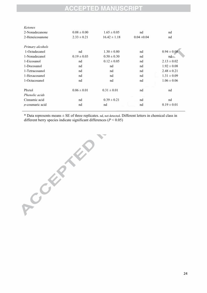
24
Ketones2-Nonadecanone 0.08 ± 0.00 1.65 ± 0.05 nd nd 2-Heneicosanone 2.33 ± 0.21 16.42 ± 1.18 0.04 ±0.04 nd
Primary alcohols 1-Octadecanol nd 1.30 ± 0.80 nd 0.94 ± 0.081-Nonadecanol 0.19 ± 0.03 0.50 ± 0.30 nd nd 1-Eicosanol nd 0.12 ± 0.05 nd 2.13 ± 0.021-Docosanol nd nd nd 1.92 ± 0.081-Tetracosanol nd nd nd 2.48 ± 0.211-Hexacosanol nd nd nd 1.31 ± 0.091-Octacosanol nd nd nd 1.06 ± 0.06
Phytol 0.06 ± 0.01 0.31 ± 0.01 nd nd Phenolic acidsCinnamic acid nd 0.39 ± 0.21 nd nd p-coumaric acid nd nd nd 0.19 ± 0.01
* Data represents means ± SE of three replicates. nd, not detected. Different letters in chemical class in different berry species indicate significant differences (P < 0.05)
25
Table 2. In vitro sun protection factor (SPF) of berry waxes.*
Sample Concentration of wax (mg/ml)0.1 0.5 1.0 2.0
Berry cuticular waxBilberry 0.76 ± 0.26 2.45 ± 0.18 4.46 ± 0.94 7.45 ± 1.75Bog bilberry 2.01 ± 0.57 8.02 ± 0.62 14.80 ± 1.33 25.42 ± 0.23Crowberry 0.27 ± 0.03 1.97 ± 0.36 5.10 ± 1.03 8.31 ± 1.71Lingonberry 0.83 ± 0.16 3.23 ± 0.21 4.76 ± 1.52 12.74 ± 0.79
Berry press cake waxBilberry 0.66 ± 0.02 3.64 ± 0.32 7.86 ± 0.34 15.09 ± 0.12 Lingonberry 0.60 ± 0.13 2.67 ± 0.81 6.07 ± 0.54 13.40 ± 0.92* Data represents means ± SD of three replicates.
26
Highlights
Composition and morphology of cuticular wax varies between different berry species.
Triterpenoids were the dominant compounds in bilberry and lingonberry cuticular wax.
Fatty acids and alkanes were prominent in bog bilberry and crowberry cuticular wax, respectively.
All studied berry waxes showed good UV-B absorption capacities.
Bilberry and lingonberry press cakes are sources of wax rich in linoleic acid and γ-linolenic acid.

27
28
29
30