Embed Size (px)
Citation preview

City University of New York (CUNY) City University of New York (CUNY)
CUNY Academic Works CUNY Academic Works
Publications and Research City College of New York
2019
Composite biomaterial repair strategy to restore biomechanical Composite biomaterial repair strategy to restore biomechanical
function and reduce herniation risk in an ex vivo large animal function and reduce herniation risk in an ex vivo large animal
model of intervertebral disc herniation with varying injury severity model of intervertebral disc herniation with varying injury severity
Warren W. Hom Icahn School of Medicine at Mount Sinai
Melanie Tschopp Icahn School of Medicine at Mount Sinai
Huizi A. Lin CUNY City College
Philip Nasser Icahn School of Medicine at Mount Sinai
Damien M. Laudier Icahn School of Medicine at Mount Sinai
See next page for additional authors
How does access to this work benefit you? Let us know!
More information about this work at: https://academicworks.cuny.edu/cc_pubs/729
Discover additional works at: https://academicworks.cuny.edu
This work is made publicly available by the City University of New York (CUNY). Contact: [email protected]

Authors Authors Warren W. Hom, Melanie Tschopp, Huizi A. Lin, Philip Nasser, Damien M. Laudier, Andrew C. Hecht, Steven B. Nicoll, and James C. Iatridis
This article is available at CUNY Academic Works: https://academicworks.cuny.edu/cc_pubs/729

RESEARCH ARTICLE
Composite biomaterial repair strategy to
restore biomechanical function and reduce
herniation risk in an ex vivo large animal
model of intervertebral disc herniation with
varying injury severity
Warren W. HomID1, Melanie Tschopp1, Huizi A. Lin2, Philip Nasser1, Damien M. Laudier1,
Andrew C. Hecht1, Steven B. Nicoll2, James C. IatridisID1*
1 Leni & Peter W. May Department of Orthopaedics, Icahn School of Medicine at Mount Sinai, New York,
New York, United States of America, 2 Department of Biomedical Engineering, The City College of New York,
New York, New York, United States of America
Abstract
Back pain commonly arises from intervertebral disc (IVD) damage including annulus fibro-
sus (AF) defects and nucleus pulposus (NP) loss. Poor IVD healing motivates developing
tissue engineering repair strategies. This study evaluated a composite injectable IVD bioma-
terial repair strategy using carboxymethylcellulose-methylcellulose (CMC-MC) and genipin-
crosslinked fibrin (FibGen) that mimic NP and AF properties, respectively. Bovine ex vivo
caudal IVDs were evaluated in cyclic compression-tension, torsion, and compression-to-fail-
ure tests to determine IVD biomechanical properties, height loss, and herniation risk follow-
ing experimentally-induced severe herniation injury and discectomy (4 mm biopsy defect
with 20% NP removed). FibGen with and without CMC-MC had failure strength similar to
discectomy injury suggesting no increased risk compared to surgical procedures, yet no bio-
materials improved axial or torsional biomechanical properties suggesting they were incapa-
ble of adequately restoring AF tension. FibGen had the largest failure strength and was
further evaluated in additional discectomy injury models with varying AF defect types (2 mm
biopsy, 4 mm cruciate, 4 mm biopsy) and NP removal volume (0%, 20%). All simulated dis-
cectomy defects significantly compromised failure strength and biomechanical properties.
The 0% NP removal group had mean values of axial biomechanical properties closer to
intact levels than defects with 20% NP removed but they were not statistically different and
0% NP removal also decreased failure strength. FibGen with and without CMC-MC failed at
super-physiological stress levels above simulated discectomy suggesting repair with these
tissue engineered biomaterials may perform better than discectomy alone, although
restored biomechanical function may require additional healing with the potential application
of these biomaterials as sealants and cell/drug delivery carriers.
PLOS ONE | https://doi.org/10.1371/journal.pone.0217357 May 28, 2019 1 / 20
a1111111111
a1111111111
a1111111111
a1111111111
a1111111111
OPEN ACCESS
Citation: Hom WW, Tschopp M, Lin HA, Nasser P,
Laudier DM, Hecht AC, et al. (2019) Composite
biomaterial repair strategy to restore
biomechanical function and reduce herniation risk
in an ex vivo large animal model of intervertebral
disc herniation with varying injury severity. PLoS
ONE 14(5): e0217357. https://doi.org/10.1371/
journal.pone.0217357
Editor: Lachlan J. Smith, University of
Pennsylvania, UNITED STATES
Received: January 14, 2019
Accepted: May 9, 2019
Published: May 28, 2019
Copyright: © 2019 Hom et al. This is an open
access article distributed under the terms of the
Creative Commons Attribution License, which
permits unrestricted use, distribution, and
reproduction in any medium, provided the original
author and source are credited.
Data Availability Statement: Data files are
available from the Figshare database (DOI: 10.
6084/m9.figshare.7579877).
Funding: This study was supported by the National
Institute of Arthritis and Musculoskeletal and Skin
Diseases of the National Institutes of Health under
award number R01AR057397. The funders had no
role in study design, data collection and analysis,

Introduction
Intervertebral disc (IVD) injuries can result in herniation when the central nucleus pulposus
(NP) tissue protrudes or extrudes through defects in the surrounding annulus fibrosus (AF).
Herniated IVD tissue can impinge upon surrounding nerves to cause radiculopathy with pain
and disability in the back, neck, arms, and/or legs depending on the IVD level of the injury [1].
Conservative methods to address these symptoms include physical therapy and pain medica-
tion, but if these treatments fail then the next step is to remove the herniated tissue through
discectomy surgery. There are approximately 300,000 annual discectomy procedures per-
formed in the United States and this procedure is very successful for improving acute pain and
disability due to neuropathy [2,3]. Current discectomy procedures do not replace the removed
NP tissue or repair AF defects. Furthermore, the IVD is avascular and has a low cell-density,
which contributes to its poor healing potential [4,5]. As such, there is a need to develop
improved IVD repair strategies to prevent disc height loss, altered biomechanics, and acceler-
ated degeneration from IVD injury and complications from discectomy procedures, including
reherniation and recurrent pain at the same level [6–10].
Several structural biomaterials and hydrogels have been developed to repair or replace IVD
tissues that are damaged or degenerated during injury, herniation, and degeneration but few
have undergone rigorous biomechanical testing to assess functional restoration and rehernia-
tion risk [11,12]. The NP and AF each have distinct material properties and biomechanical
roles and both must be addressed to fully repair the IVD. The NP has large swelling potential
and exerts large intradiscal pressures [13–15] that put the fibrocartilaginous AF into circum-
ferential tension which acts to constrain the NP. To achieve these distinct mechanical func-
tions, the AF and NP have large differences in material properties and structure [8,16–18]. A
successful IVD repair strategy similarly must achieve multiple mechanical functions to mimic
the IVD and would therefore ideally incorporate two biomaterials: one to replace the removed
NP and another to seal the AF and prevent reherniation. Such combination repair strategies
are still in the early stages of development and currently there is one study by Borem et al. that
utilized decellularized tissues to create an AF patch and an NP replacement scaffold [19] which
demonstrated improvement in axial biomechanical properties [20] and another study by Sloan
et al. which demonstrated that a combined hydrogel repair had the best restoration of NP
hydration [21].
Injectable hydrogels are ideal candidates for IVD repair because they can fill in irregularly
shaped defects and are amenable to minimally invasive procedures. Two previously investi-
gated hydrogels which demonstrated promising IVD biomechanical restoration and biocom-
patibility were redox-polymerized carboxymethylcellulose-methylcellulose (CMC-MC) for NP
replacement and a genipin-crosslinked fibrin (FibGen) hydrogel for AF repair. CMC-MC has
high swelling potential and has demonstrated its ability to restore disc height and biomechani-
cal restoration following a discectomy injury in bovine IVDs ex vivo [22,23]. FibGen has been
tuned to match AF shear mechanical properties and has been shown to seal AF defects and
resist reherniation under cyclic loading at physiological levels [24–29]. Both CMC-MC and
FibGen are capable of rapid in situ gelation which is important for clinical translation when
considering ease of application without extending surgical procedure times.
Herniation of NP tissue can occur through various AF defect types ranging in size from
small tears up to large fissures spanning 50% or more of the IVD height. Ex vivo studies have
been conducted to determine the biomechanical effects of varying AF defect sizes and found
that biomechanical changes generally increase with AF defect size and require a defect span-
ning 40% or more of the IVD height in order to induce significant biomechanical changes
Composite biomaterial repair strategy to restore biomechanical function and reduce herniation risk
PLOS ONE | https://doi.org/10.1371/journal.pone.0217357 May 28, 2019 2 / 20
decision to publish, or preparation of the
manuscript.
Competing interests: The authors have declared
that no competing interests exist.

[30–32]. However, those studies mainly focused on modeling IVD injury and degeneration
and examined AF defects alone without trying to model discectomy procedures by removing
NP tissue, and without efforts at repair. Therefore, there is limited data on whether the size
and severity of AF defects have an effect on IVD biomechanical behaviors following discect-
omy surgery and how this influences repair performance.
This 2 part ex vivo biomechanical study evaluated how different biomaterial strategies influ-
enced the biomechanical performance and failure strength of IVD injuries of varying severity.
Part 1 focused on effects of biomaterial repair strategy and investigated the combined use of
CMC-MC and FibGen to restore IVD biomechanical properties and failure strength back to
intact levels following discectomy with an experimentally-induced severe defect and discect-
omy injury involving a 4-mm biopsy AF defect with 20% NP removal. Histological assess-
ments determined if hydrogels filled void space and adhered to IVD tissues. Part 2 focused on
AF defect type and NP volume removed and investigated whether varying AF defect size, type,
and NP removal volume in the experimentally-induced injury and discectomy scenarios
affected biomechanical performance and failure strength with FibGen, which was found to
have lowest herniation risk from Part 1.
Materials and methods
Motion segment preparation and potting
Bovine caudal IVD motion segments from coccygeal levels cc2/3 to cc4/5 were isolated and
prepared as previously described [26]. Briefly, oxtails were obtained from a local abattoir
(Green Village Packing, Green Village, NJ) and the vertebral processes and surrounding mus-
cles and ligaments were removed. Caudal motion segments were isolated and potted in poly
(methyl methacrylate) and submerged in phosphate-buffered saline (PBS) (Fisher Scientific,
Hampton, NH) at 4˚C overnight under free-swelling conditions without a preload to allow full
hydration before mechanical testing.
Hydrogel fabrication and injection
FibGen was prepared as previously described [24]. Briefly, 200 mg of bovine fibrinogen
(Sigma-Aldrich, St. Louis, MO) was dissolved in 1140 μL of PBS and mixed with 40 U of
thrombin (Sigma-Aldrich, St. Louis, MO) and 8 mg of genipin (FUJIFILM Wako Chemicals
U.S.A. Corporation, Richmond, VA) using a 4:1 dual barrel syringe and a mixing tip (Pac-
Dent, Brea, CA). The tip of the syringe was then placed in the defect and slowly retracted as
FibGen was injected to fill the void created by injury.
CMC and MC polymers (Sigma-Aldrich, St. Louis, MO) were methacrylated as previously
described [23,33] and mixed along with 20 mM each of the redox initiators ammonium persul-
fate (Sigma-Aldrich) and N,N,N’,N’-tetramethylethylenediamine (Fisher Scientific, Hampton,
NH) using a 1:1 dual barrel syringe (Pac-Dent, Brea, CA). The filled syringe was then warmed
in a 37˚C water bath for 30 minutes before injection into an injured motion segment. For the
combined repair (Combo) samples, CMC-MC hydrogel was injected first and then the
remaining void of the AF defect was filled in with FibGen. The volume of hydrogel injected
per repair sample was carefully controlled to completely fill the defect to the AF periphery, as
might be expected in a surgical repair setting. However, the precise volume injected was not
measured and the amount may have varied depending on the size of the AF defect and the NP
void space.
Composite biomaterial repair strategy to restore biomechanical function and reduce herniation risk
PLOS ONE | https://doi.org/10.1371/journal.pone.0217357 May 28, 2019 3 / 20
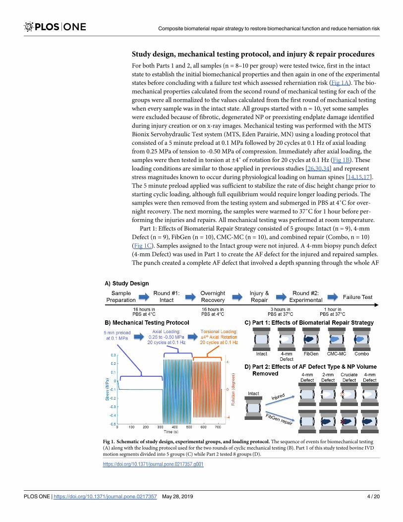
Study design, mechanical testing protocol, and injury & repair procedures
For both Parts 1 and 2, all samples (n = 8–10 per group) were tested twice, first in the intact
state to establish the initial biomechanical properties and then again in one of the experimental
states before concluding with a failure test which assessed reherniation risk (Fig 1A). The bio-
mechanical properties calculated from the second round of mechanical testing for each of the
groups were all normalized to the values calculated from the first round of mechanical testing
when every sample was in the intact state. All groups started with n = 10, yet some samples
were excluded because of fibrotic, degenerated NP or preexisting endplate damage identified
during injury creation or on x-ray images. Mechanical testing was performed with the MTS
Bionix Servohydraulic Test system (MTS, Eden Parairie, MN) using a loading protocol that
consisted of a 5 minute preload at 0.1 MPa followed by 20 cycles at 0.1 Hz of axial loading
from 0.25 MPa of tension to -0.50 MPa of compression. Immediately after axial loading, the
samples were then tested in torsion at ±4˚ of rotation for 20 cycles at 0.1 Hz (Fig 1B). These
loading conditions are similar to those applied in previous studies [26,30,34] and represent
stress magnitudes known to occur during physiological loading on human spines [14,15,17].
The 5 minute preload applied was sufficient to stabilize the rate of disc height change prior to
starting cyclic loading, although full equilibrium would require longer loading periods. The
samples were then removed from the testing system and submerged in PBS at 4˚C for over-
night recovery. The next morning, the samples were warmed to 37˚C for 1 hour before per-
forming the injuries and repairs. All mechanical testing was performed at room temperature.
Part 1: Effects of Biomaterial Repair Strategy consisted of 5 groups: Intact (n = 9), 4-mm
Defect (n = 9), FibGen (n = 10), CMC-MC (n = 10), and combined repair (Combo, n = 10)
(Fig 1C). Samples assigned to the Intact group were not injured. A 4-mm biopsy punch defect
(4-mm Defect) was used in Part 1 to create the AF defect for the injured and repaired samples.
The punch created a complete AF defect that involved a depth spanning through the whole AF
Fig 1. Schematic of study design, experimental groups, and loading protocol. The sequence of events for biomechanical testing
(A) along with the loading protocol used for the two rounds of cyclic mechanical testing (B). Part 1 of this study tested bovine IVD
motion segments divided into 5 groups (C) while Part 2 tested 8 groups (D).
https://doi.org/10.1371/journal.pone.0217357.g001
Composite biomaterial repair strategy to restore biomechanical function and reduce herniation risk
PLOS ONE | https://doi.org/10.1371/journal.pone.0217357 May 28, 2019 4 / 20

thickness. After creating the AF defect, the healthy NP tissue was disrupted with a curette and
then ~20% of the total NP was removed with a rongeur, based on weight. The amount of NP
volume removed varied with IVD size. A preliminary test removed the NP from 15 intact
IVDs and correlated the weight of the NP vs. the IVD diameter. Since there was no way of
measuring the exact amount of NP in an intact disc prior to performing the injury, we used
this correlation and removed between 0.15–0.20 g of NP depending on the IVD diameter, with
an average removal of 0.188 ± 0.025 g.
Part 2: Effects of AF Defect Type and NP Removal Volume consisted of 7 groups: Intact
(n = 9), 3 different AF defects (2-mm biopsy punch (n = 9), 4-mm biopsy punch (n = 9), and
4-mm cruciate (n = 10)), and the repair of each of the AF defects with FibGen (n = 10 per
group) (Fig 1D). The cruciate defect (Cruciate Defect) was made with a #15 scalpel blade with
each cut approximately 4 mm long. Biopsy defects were created with punches of 2 mm (2-mm
Defect) and 4 mm (4-mm Defect) diameter. All AF defects in both parts of the study were cre-
ated on the left posterolateral side of the IVD. As with Part 1, after creating the complete AF
defects, the healthy NP was disrupted and ~20% of NP tissue removed.
To determine the influence of NP removal on severely injured IVDs and the ability of Fib-
Gen to restore AF radial tension, motion segments with the NP disrupted using a curette but
not removed (0% NP Removed) were divided into two groups, 4-mm Defect (n = 8) and Fib-
Gen repair (n = 8), and tested using the previous mechanical loading protocol. Results from
the 0% NP Removed groups were compared against the 4-mm Defect and FibGen samples
which had 20% NP removed.
Following the injury, the samples were repaired using injection of the appropriate biomate-
rial or combination biomaterial, as described above. The combination repair included 2 sepa-
rate syringes first filling the NP space with CMC-MC and next filling the AF defect space with
FibGen. Following repairs, IVDs remained at room temperature for 10 minutes to allow initial
gelation before they were submerged in PBS and kept at 37˚C for 3 hours to allow full solidifi-
cation of both hydrogels. All samples were then tested a second time following the same load-
ing protocol used for the first test.
After the second round of testing, samples were subjected to failure testing which consisted
of an axial compression to failure at a rate of 2 mm/min at 5˚ of bending with the left postero-
lateral side facing the open side of the wedge (to push NP towards he defect), using procedures
as previously described [35].
Biomechanical parameters
A custom MATLAB (Mathworks, Natrick, MA, USA) code was used to analyze axial and tor-
sional data as follows. The last axial loading cycle was identified and the corresponding force-
displacement curve was drawn (Fig 2A). Compressive and tensile stiffness values were defined
as the slope of the linear regions of the force-displacement curve and calculated from the top
20% of the data points for the loading portion of the compressive and tensile curves. Range of
motion (ROM) was defined as the total distance traveled from maximum compression to max-
imum tension in the last axial loading cycle.
Axial neutral zone (NZ) region was defined as the region of the force-displacement curve
with the smallest slope. This region was found using the MATLAB code with a 15-point mov-
ing slope calculation along the compressive and tensile curves to find the position where a lin-
ear regression could be drawn with minimal slope. Once found, the NZ region was expanded
by adding data points to each side of the initial 15 points until any additional data points
would increase the standard error by 10%. This set of parameters for the algorithm were
selected since they provided the NZ that best matched the NZ manually determined by three
Composite biomaterial repair strategy to restore biomechanical function and reduce herniation risk
PLOS ONE | https://doi.org/10.1371/journal.pone.0217357 May 28, 2019 5 / 20
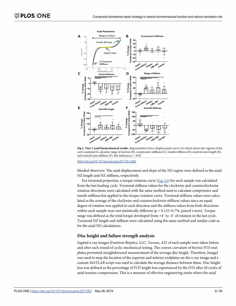
blinded observers. The axial displacement and slope of the NZ region were defined as the axial
NZ length and NZ stiffness, respectively.
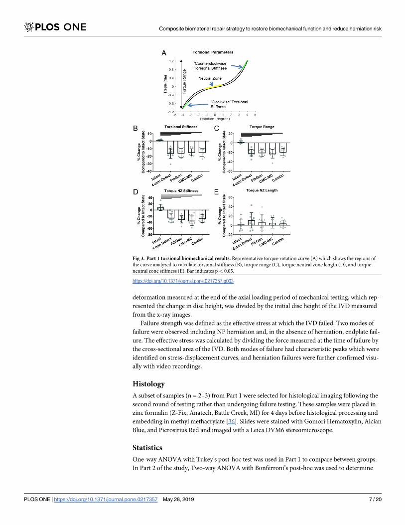
For torsional properties, a torque-rotation curve (Fig 3A) for each sample was calculated
from the last loading cycle. Torsional stiffness values for the clockwise and counterclockwise
rotation directions were calculated with the same method used to calculate compressive and
tensile stiffness but applied to the torque-rotation curve. Torsional stiffness values were calcu-
lated as the average of the clockwise and counterclockwise stiffness values since an equal
degree of rotation was applied in each direction and the stiffness values from both directions
within each sample were not statistically different (p = 0.125–0.776, paired t-tests). Torque
range was defined as the total torque developed from +4˚ to -4˚ of rotation in the last cycle.
Torsional NZ length and stiffness were calculated using the same method and similar code as
for the axial NZ calculations.
Disc height and failure strength analysis
Sagittal x-ray images (Faxitron Bioptics, LLC, Tucson, AZ) of each sample were taken before
and after each round of cyclic mechanical testing. The convex curvature of bovine IVD end-
plates prevented straightforward measurement of the average disc height. Therefore, ImageJ
was used to map the location of the superior and inferior endplates on the x-ray image and a
custom MATLAB script was used to calculate the average distance between them. Disc height
loss was defined as the percentage of IVD height loss experienced by the IVD after 20 cycles of
axial tension-compression. This is a measure of effective engineering strain where the axial
Fig 2. Part 1 axial biomechanical results. Representative force-displacement curve (A) which shows the regions of the
curve analyzed to calculate range of motion (B), compressive stiffness (C), tensile stiffness (D), neutral zone length (E),
and neutral zone stiffness (F). Bar indicates p< 0.05.
https://doi.org/10.1371/journal.pone.0217357.g002
Composite biomaterial repair strategy to restore biomechanical function and reduce herniation risk
PLOS ONE | https://doi.org/10.1371/journal.pone.0217357 May 28, 2019 6 / 20
deformation measured at the end of the axial loading period of mechanical testing, which rep-
resented the change in disc height, was divided by the initial disc height of the IVD measured
from the x-ray images.
Failure strength was defined as the effective stress at which the IVD failed. Two modes of
failure were observed including NP herniation and, in the absence of herniation, endplate fail-
ure. The effective stress was calculated by dividing the force measured at the time of failure by
the cross-sectional area of the IVD. Both modes of failure had characteristic peaks which were
identified on stress-displacement curves, and herniation failures were further confirmed visu-
ally with video recordings.
Histology
A subset of samples (n = 2–3) from Part 1 were selected for histological imaging following the
second round of testing rather than undergoing failure testing. These samples were placed in
zinc formalin (Z-Fix, Anatech, Battle Creek, MI) for 4 days before histological processing and
embedding in methyl methacrylate [36]. Slides were stained with Gomori Hematoxylin, Alcian
Blue, and Picrosirius Red and imaged with a Leica DVM6 stereomicroscope.
Statistics
One-way ANOVA with Tukey’s post-hoc test was used in Part 1 to compare between groups.
In Part 2 of the study, Two-way ANOVA with Bonferroni’s post-hoc was used to determine
Fig 3. Part 1 torsional biomechanical results. Representative torque-rotation curve (A) which shows the regions of
the curve analyzed to calculate torsional stiffness (B), torque range (C), torque neutral zone length (D), and torque
neutral zone stiffness (E). Bar indicates p< 0.05.
https://doi.org/10.1371/journal.pone.0217357.g003
Composite biomaterial repair strategy to restore biomechanical function and reduce herniation risk
PLOS ONE | https://doi.org/10.1371/journal.pone.0217357 May 28, 2019 7 / 20
effects of the different AF defect types and FibGen repair status. Data presented as mean ±standard deviation along with individual data points. Statistical outliers were detected and
removed using the integrated Robust regression and Outlier removal (ROUT) method [37] in
GraphPad Prism 7 (GraphPad Software, San Diego, California) with Q = 1%. Inclusion or
exclusion of the outliers did not affect the statistical results. Significance was set at p< 0.05
and all statistical analysis was performed using GraphPad Prism 7.
Results
Part 1 mechanical testing results
Compressive stiffness (Fig 2B) was not significantly altered by injury or repair (p>0.076)
whereas tensile stiffness (Fig 2C) was significantly decreased due to injury (p<0.005) and not
restored by any repair group (p>0.995). ROM (Fig 2D) for 4-mm Defect and all three repair
groups were significantly increased compared to Intact values (p<0.032). Axial NZ length (Fig
2E) for 4-mm Defect and CMC-MC samples were increased (p<0.006) but FibGen and
Combo repair samples were not significantly different from either Intact (p>0.110) or 4-mm
Defect samples (p>0.378). Axial NZ stiffness (Fig 2F) was significantly decreased in 4-mm
Defect, FibGen and CMC-MC samples (p<0.026).
Torsional stiffness, torque range, and torque NZ stiffness (Fig 3B–3D) were significantly
decreased in 4-mm Defect and all repair groups (p<0.0001). Torque NZ length (Fig 3E) was
not significantly affected by any group (p>0.581).
Part 1 disc height and failure strength
Disc height measurements obtained from x-ray images and MATLAB processing (Fig 4A)
were used to calculate disc height loss (Fig 4B), which was significantly larger (i.e., disc height
was smaller) for 4-mm Defect and CMC-MC samples (p<0.038). Disc height loss was not sig-
nificantly different for FibGen and Combo samples as compared to either Intact (p>0.062) or
the 4-mm Defect (p>0.872) samples. Prior to failure testing, visual assessment of the hydrogels
did not reveal signs of cracking in FibGen or CMC-MC, although CMC-MC did appear to be
closer to the edge of the AF defect (but still within the boundaries of the disc) than before
mechanical testing.
In failure testing, samples failed either through herniation or endplate failure (Fig 4C). All
4-mm Defect and hydrogel repaired samples failed by herniation and had significantly reduced
failure strength compared to Intact samples (p<0.0001) (Fig 4D), which all experienced end-
plate failure. None of the repaired samples had significant increased failure strength compared
to 4-mm Defect samples (p>0.380), which represents the simulated discectomy condition.
Failure strength was also larger than the physiological stress levels for IVDs.
Morphology and histology
In samples cut transversely prior to histological fixation (Fig 5), FibGen and CMC-MC can
be seen within the IVD as dark and light blue hydrogels, respectively. Hydrogels and native
IVD tissues swelled so there were no obvious void spaces. The stained images of sagittal sec-
tions show some void spaces following the fixation and staining processes which highlighted
NP damage from the injury procedures. FibGen filled the AF defect space and remained
adherent to the outer AF. CMC-MC was adjacent to native IVD tissues without obvious
adhesion.
Composite biomaterial repair strategy to restore biomechanical function and reduce herniation risk
PLOS ONE | https://doi.org/10.1371/journal.pone.0217357 May 28, 2019 8 / 20

Fig 4. Disc height loss and failure strength for Part 1. X-ray images (A) were used to calculate disc height loss (B).
Force-displacement curves (C) were used to calculate failure strength (D) of each sample. Horizontal grey shading
indicates range of human physiological loads based on in vivo measurements of intradiscal pressure up to 2.3 MPa
[14]. Bar indicates p< 0.05.
https://doi.org/10.1371/journal.pone.0217357.g004
Fig 5. Qualitative assessment of hydrogel repair. Samples were cut transversely to determine extent of injury and repair. Sagittal histological sections
were stained with picrosirius red and alcian blue to determine the amount of adhesion between native IVD tissue and the hydrogel repairs. Scale bar = 1
mm. Black arrows indicate hydrogel in situ.
https://doi.org/10.1371/journal.pone.0217357.g005
Composite biomaterial repair strategy to restore biomechanical function and reduce herniation risk
PLOS ONE | https://doi.org/10.1371/journal.pone.0217357 May 28, 2019 9 / 20

Part 2 mechanical testing results
All axial biomechanical parameters measured were significantly altered with injury for all
defect types assessed (p<0.040) (Fig 6A–6E), except for compressive stiffness which had no
change (p>0.206). No significant differences between injury types were observed (p>0.083).
FibGen repair of each injury type (Fig 6F–6J) had no significant improvements for most axial
parameters (p>0.205) except for the tensile stiffness of the Cruciate FibGen repair and the
axial NZ length of the 4-mm FibGen repair which both demonstrated partial restoration back
to Intact levels since all injuries significantly changed biomechanical properties from Intact
levels but FibGen did not (p>0.184).
Torsional stiffness and torque range decreased due to injury (p<0.0002), with no differ-
ences between the AF defects (p>0.669) (Fig 7A and 7B). Torsional NZ stiffness decreased for
the 4-mm Defect (p = 0.0005) and Cruciate Defect (p = 0.049) but the 2-mm Defect was not
significantly different from Intact values (p = 0.190) (Fig 7C). Torsional NZ length had no sig-
nificant differences between Intact or injured samples (p>0.167) (Fig 7D). FibGen repairs of
the injuries (Fig 7E–7H) showed no significant differences compared to injured samples
(p>0.269). Volume of NP removal did not significantly affect axial or torsional biomechanics
of the 4-mm Defect (p>0.109).
Fig 6. Part 2 axial biomechanical results. The compressive stiffness (A) was not significantly altered from intact levels
by any of the three AF defects. Tensile stiffness (B), range of motion (C), axial neutral zone length (D), and axial
neutral zone length (E) were significantly altered by each of the AF defects but there were no significant differences
between the AF defects. There was no significant improvement with the addition of FibGen (F-J). � = significantly
different from Intact Samples.
https://doi.org/10.1371/journal.pone.0217357.g006
Composite biomaterial repair strategy to restore biomechanical function and reduce herniation risk
PLOS ONE | https://doi.org/10.1371/journal.pone.0217357 May 28, 2019 10 / 20

Part 2 disc height and failure strength
No significant differences in disc height loss were observed between the Intact and injured
groups (p>0.110) (Fig 8A), likely because of the free swelling recovery and high NP swelling
propensity of these healthy bovine caudal IVDs. Since the injury groups had no significant dif-
ferences, the disc height loss of the FibGen repairs of each defect type also had no significant
differences between Intact and injured samples (p>0.180) (Fig 8B). Failure strength of the
injured samples was significantly decreased from Intact levels (p<0.003) (Fig 8C). Among the
three defect types that had 20% NP removed, the 2-mm Defect samples had significantly
greater failure strength than the 4-mm Defect group (p = 0.032), highlighting that the 4-mm
Defect was most severe. Furthermore, two out of the nine 2-mm Defect samples had endplate
failure rather than herniation. All 4-mm and Cruciate Defect samples herniated. The 4-mm
Defect with 0% NP removed all herniated and had significantly reduced failure strength com-
pared to 2-mm Defect (p = 0.002) and Cruciate Defect samples (p = 0.037). FibGen repair did
Fig 7. Part 2 torsional biomechanical results. The torque range (A), torsional stiffness (B), and torsional neutral zone (C) were significantly
decreased from intact levels due to injury but there were no differences between groups. Torsional neutral zone length (D) was not significantly
altered from intact levels by any group. FibGen repair of each defect type (E-H) did not provide any significant restoration. � = significantly
different from Intact Samples.
https://doi.org/10.1371/journal.pone.0217357.g007
Composite biomaterial repair strategy to restore biomechanical function and reduce herniation risk
PLOS ONE | https://doi.org/10.1371/journal.pone.0217357 May 28, 2019 11 / 20

not significantly improve the failure strength for any of the injury types (p>0.360) but values
of the FibGen 4-mm Defect and FibGen Cruciate groups were above the injury levels and were
all above physiological levels of loading typically experienced in vivo (Fig 8D). FibGen repairs
of 4-mm and Cruciate Defects all herniated while two out of the ten 2-mm FibGen repaired
samples failed via endplate instead of herniation.
Discussion
Discectomy procedures alleviate many symptoms associated with IVD herniation but do not
address the damage and loss of NP and AF tissues which can result in reherniation and recur-
rent pain [6–10]. This 2 part biomechanical study developed and characterized injectable IVD
biomaterial repair strategies capable of being applied during discectomy procedures to repair
herniated IVDs. CMC-MC as an NP hydrogel replacement and FibGen as an AF sealant were
applied. Part 1 evaluated effects of biomaterial repair strategy using hydrogel alone and in
combination with large AF defects and NP tissue removal that simulate herniation and an
‘aggressive’ discectomy procedure. The combined hydrogel repair did not significantly
improve IVD biomechanical properties suggesting no repair strategy was capable of restoring
NP swelling and AF tension. However, failure strength of all repair strategies were greater than
physiological loading levels (discussed below) and mean failure strength was largest for the
FibGen group, although it was not significantly different from other groups and had the great-
est variability. Part 2 evaluated effects of AF defect type and NP removal volume to determine
their influence on biomechanical behaviors and FibGen repair capacity. Axial and torsional
Fig 8. Part 2 disc height loss and failure strength. Disc height for injured (A) and FibGen repaired samples (B) were
not significantly different from intact levels or different between groups. Failure strength was significantly decreased
from intact by all four AF defects (C) but the 2 mm Defect had a greater failure strength than the 4 mm Defect at both
0% and 20% NP removed. The Cruciate Defect group also had greater failure strength than 4 mm Defect with 0% NP
removed. Failure strength for FibGen repaired samples (D) were not significantly greater than the injured samples.
Horizontal grey shading (C,D) indicates range of human physiological loads. Dashed line indicates mean of Intact
values. Solid bars indicates p< 0.05.
https://doi.org/10.1371/journal.pone.0217357.g008
Composite biomaterial repair strategy to restore biomechanical function and reduce herniation risk
PLOS ONE | https://doi.org/10.1371/journal.pone.0217357 May 28, 2019 12 / 20

biomechanical properties were not significantly different between the three AF defects tested
suggesting that amount of NP volume removed was more important than the AF defect type
in determining the extent of biomechanical changes. Failure strength was greater for the
2-mm Defect and Cruciate Defect, highlighting that AF defect size and type was an important
determinant of herniation potential. FibGen repair resulted in greater failure strength for
larger defects indicating the importance of an AF sealant for a large defect.
AF and NP have distinct biomechanical properties and both of these tissues are disrupted
and removed during herniation injury and discectomy procedures. The NP has high swelling
capacity and exerts hydrostatic pressure, which places the surrounding AF fibers in circumfer-
ential tension [13,14]. When AF structural integrity is compromised, the NP pressurization is
lost and IVD biomechanical properties are significantly altered [30,32,38]. Few studies investi-
gate a combination repair and we hypothesized that a combination biomaterial repair strategy
following simulated herniation and discectomy injury was necessary to restore NP hydrostatic
pressure and AF tension and return biomechanical properties to the Intact condition. CMC-
MC was selected as the NP replacement hydrogel due to its swelling potential [22] and FibGen
was selected as the AF sealant for its ability to resist reherniation under physiological loads
[26–29]. Both hydrogels were also previously determined to be biocompatible, injectable and
capable of in situ gelation suggesting they show promise for application to augment current
discectomy procedures [25,26,33,39]. The gross morphological images and sagittal histology
performed in this study confirmed the ability of CMC-MC and FibGen to gel in situ. Imaging
demonstrated that FibGen adhered to the native tissue while CMC-MC remained in place
without adhering with the native tissue. A similar combination repair strategy was previously
applied to rat coccygeal motion segments demonstrating that the NP replacement material
had largest effects on biomechanical properties, highlighting the importance of NP swelling on
axial biomechanical properties [21]. Biomaterial strategies involving composite repairs that
mimic the whole IVD structure are also under development, although most of these strategies
are being developed for a fusion alternative rather than a herniation repair [12,40,41].
Part 1 of this study applied a 4-mm biopsy punch injury spanning ~50% of the disc height
and 20% NP removal, as might be observed with a severe herniation and aggressive discect-
omy. This severe defect altered nearly all biomechanical parameters with reduced tensile stiff-
ness, torsional stiffness and torque range and increased axial range of motion. Multiple NZ
properties were also affected for both axial and torsional tests. Interestingly, compressive stiff-
ness was not significantly altered in the 4-mm Defect group or by the amount of NP removal.
We believe the relatively small % difference (4-mm Defect group with 6.96±5.93% change) in
compressive stiffness can be attributed to the healthy NP tissue undergoing free swelling to fill
the void space and partially restore intradiscal pressure even with the large AF defect and
removal of ~20% of the NP. The three hydrogel repair groups did not restore any axial or tor-
sional parameter back to Intact levels and these groups were also not different from injury.
The lack of restoration of torsional properties suggests that no biomaterial strategies were able
to restore AF tension. The lack of restoration of axial biomechanical properties to Intact levels
further suggests no biomaterial strategies were able to restore NP pressurization.
Prior studies, which used the same axial loading magnitudes and similar durations as this
study, showed that repair of IVD defects with FibGen and CMC-MC significantly improved
axial biomechanical properties, and that FibGen had a trend of improved torsional properties
[23,26,27]. In this study, neither FibGen and CMC-MC nor the combination repair restored
axial or torsional biomechanical properties. We believe these differences between studies are
most likely attributed to differences in injury severity since this study involving larger AF
defects and 20% NP removal (as compared to less NP removal in the study by Likhitpanichkul
et al. [26] and less severe AF defects used by Varma et al. [23]). Results therefore highlight the
Composite biomaterial repair strategy to restore biomechanical function and reduce herniation risk
PLOS ONE | https://doi.org/10.1371/journal.pone.0217357 May 28, 2019 13 / 20

importance of controlling for IVD injury severity in study design as well as a continuing need
to develop repair strategies for more severe injuries.
Part 2 determined that different AF defect types had similar effects on biomechanical prop-
erties and that FibGen had little biomechanical improvement on any AF defects. AF defects
from herniation observed during discectomy procedures vary in size and, in some cases, the
AF defect is difficult to find and the surgeon has to create an incision to remove the protruding
NP tissue [42]. Several biomechanical studies have also determined how different sizes and
shapes of AF injury influence biomechanical changes in models of IVD degeneration [30–
32,38,43], yet few evaluate how different injury models influence failure strength, which relates
to IVD herniation risk [44,45]. The biopsy punch injuries selected for this study represent
severe AF defects that can be seen clinically whereas the cruciate injury represents smaller AF
tears or the incision surgeons make when the AF is still intact. The 4-mm and 2-mm biopsy
punch defects were selected to induce injuries of 50% or 25% of the disc height, respectively.
These percentages were selected based on a prior study by Elliot et al. which demonstrated that
needle puncture injuries of 52% disc height caused greater biomechanical changes than inju-
ries that were 26% of the disc height [30]. The Cruciate Defect involved 4-mm scalpel cuts that
also spanned 50% of the disc height, cutting similar numbers of fibers as the 4-mm Defect but
retaining the cut AF tissue. FibGen was selected to repair the AF defects in Part 2 because it
had the most promising biomechanical restoration and was easier to apply than the Combo
repair, which had similar performance as FibGen. All 3 defect models of Part 2 significantly
diminished axial and torsional biomechanical parameters but were not significantly different
from each other. This indicates that IVD biomechanical properties were equally altered
regardless of the size or type of AF defect present after discectomy. Therefore, it is likely that
the amount of NP removal dominates the influence of AF defect type on biomechanical prop-
erties, particularly since all AF defects were somewhat severe. This is contrary to previous stud-
ies which found that an AF defect size of 40% or more of the disc height is necessary to induce
significant biomechanical changes [30]. However, those studies were modeling IVD degenera-
tion and used needle puncture injuries with no NP removal whereas the injuries induced in
this study did remove NP tissue to model the condition of the IVD after discectomy. We con-
clude that biomechanical changes in this study were driven largely by the amount of NP tissue
removed once the AF was substantially disrupted, and that improved biomechanical perfor-
mance following severe injuries requires restoration of both NP swelling and AF tension that
our biomaterial strategies had little capacity to do in a severe injury model.
To determine whether FibGen could restore AF circumferential tension with NP tissue in
place, IVDs with 0% NP removed were tested with the 4-mm Defect type and 4-mm FibGen
repair conditions. Although the average change in axial and torsional properties for 0% NP
removed samples was lower than injured samples with 20% NP removed, the four injury
groups were not significantly different. FibGen repair of the 4-mm Defect with 0% NP
removed did not provide significant biomechanical restoration. These results suggest that Fib-
Gen was unable to restore AF radial tension even when healthy NP tissue remained in place
and was able to swell. Restoring AF tension might be possible if IVDs are unloaded prior to AF
repair in order to allow restoration of AF pre-stress and/or NP pressurization with resumption
of loading. However, such a strategy was not employed because it would be very difficult to
apply in a clinical scenario.
Biomaterial repairs performed better for herniation risk and disc height loss than for bio-
mechanical properties. Herniation risk and disc height loss are likely to be more important
than biomechanical properties in improving clinical outcomes since IVD height loss and her-
niation risk are key factors that could directly impact nerve compression and must be
addressed for clinical translation of any IVD repair strategy. Herniation risk was assessed by
Composite biomaterial repair strategy to restore biomechanical function and reduce herniation risk
PLOS ONE | https://doi.org/10.1371/journal.pone.0217357 May 28, 2019 14 / 20

failure testing in which the IVD was compressed at 5˚ of posterolateral flexion until failure
which was defined as NP herniation, hydrogel extrusion, or endplate fracture [35]. Although
FibGen, CMC-MC and the Combo repair were unable to significantly improve failure strength
compared to the injured condition, values for FibGen and Combo repairs were all above 2.3
MPa which is the upper range of physiological loading typically experienced by lumbar IVDs
during moderate levels of physical activity [14]. On the other hand, half of the 4-mm Defect
samples herniated within this range of physiological loading. This suggests that FibGen and
the Combo repair may provide protection against reherniation even though the results were
not statistically significant. CMC-MC is not an adhesive hydrogel and the 4-mm biopsy punch
defect is very large and spanned 50% of the disc height so it was unsurprising that there was no
improvement in failure strength highlighting that injectable CMC-MC is likely to be preferable
for early IVD degeneration conditions with small IVD defects and little potential for hernia-
tion. Part 2 compared three AF defect types and found the 2-mm Defect and Cruciate Defect
groups had greater failure strength than the 4-mm Defect, although the Cruciate Defect group
was not significantly different from the 4-mm Defect. The 4-mm Defect was also used to assess
the impact of the volume of NP removed on failure strength. Although the mean failure
strength of the 0% NP removed group was lower than the 20% NP removed group, and most
of the samples herniated at physiological loading levels, the two groups were not significantly
different. This indicates that size and relative severity of the AF defect are important factors
when considering the reherniation of any repair strategy and that the amount of NP removed
in severe AF defects may also influence biomechanics and failure strength. Intact IVDs failed
via endplate fracture rather than AF rupture and herniation, and this suggests it may never be
possible to fully restore failure strength back to Intact levels using injectable hydrogels alone.
In that case, the goal should be to prevent repairs from failing at physiological levels of loading
while introducing bioactive factors or cells to promote healing.
Disc height loss is a common symptom of herniation and discectomy due to removal of NP
tissue and this can lead to nerve root impingement and radicular pain. In clinical scenarios
three months after discectomy, an 18% loss of disc height was observed which increased to
26% disc height loss two years after surgery [7]. In this study, the magnitude of disc height loss
only reached 9% in injured samples and was not significantly different from Intact samples. A
previous study has demonstrated that loading magnitude and duration both affect disc height
loss [46]. As such, we believe that these disc height loss measurements are not so sensitive
since the short duration of low-force loading and free-swelling recovery used in this study was
not sufficient to simulate the disc height loss that would be observed under physiological con-
ditions that more severely test these biomaterials. For more accurate IVD height measure-
ments, we recommend application of a longer axial load such as during long-term organ
culture experiments or creep experiments, since our prior studies showed both FibGen and
CMC-MC were able to reduce or prevent disc height loss using those test protocols [23,26].
One of the limitations of this study is the relatively high variance observed. There are several
sources of potential variance in this complex study of in situ repair using multiple biomaterials.
We evaluated biomaterial formulations from different batches and found them to have rela-
tively low variance. Previous studies by Amin et al. have attributed approximately 20% of the
biomechanical variation to biological variability, experimental technique, and injury mecha-
nisms [47,48]. Biological, anatomical, and technical factors in the multi-step potting and test-
ing processes all contribute to variability and were controlled for in this study by normalizing
each sample to their intact state and by having the same person, who was well-practiced, per-
form the same procedures to reduce this precision error. Relative injury severity also contrib-
uted to variance since NP removal volume was difficult to precisely control and since we used
a fixed-size defect that did not account for variations in disc heights and diameters between
Composite biomaterial repair strategy to restore biomechanical function and reduce herniation risk
PLOS ONE | https://doi.org/10.1371/journal.pone.0217357 May 28, 2019 15 / 20

samples. Differences in hydrogel repair procedures, such as the varied amounts of hydrogel
injected, were an additional source of variation indicated by the repair groups having the larg-
est variation for many variables.
Additional limitations may be related to the testing protocol. Axial and torsional loading
were selected since they are physiologically relevant loading conditions and because torsional
biomechanical properties are sensitive to AF integrity while axial biomechanical properties are
sensitive to NP pressurization and AF integrity [8]. However, physiological loading of the IVD
involves loading in six degrees of freedom and at different loading rates, which can affect bio-
mechanical properties [49]. Although no significant differences between the different AF
defect types were observed at a loading frequency of 0.1 Hz, testing with different loading fre-
quencies or measuring viscoelastic parameters are interesting future investigations to further
characterize injury effects and repair potential. The amount of IVD hydration can influence
biomechanical properties [50] and the 5 minute preload applied prior to cyclic loading in this
study may not have been sufficient to establish equilibrium in all samples. The addition of a
recovery period between the axial and torsional tests in future studies would restore any poten-
tial hydration loss induced by the cyclic axial loading. While the defect models used in this
study simulate clinically relevant AF defects, studies using human IVDs with facet joints intact
which increase torsional stiffness and bear part of the compressive loads [51,52] would be
more representative of in vivo biomechanical changes caused by injury and repair. Intact and
healthy facet joints in the human spine may limit the magnitude of biomechanical changes
caused by AF defects with the facets sharing part of the applied loads.
Conclusion
This study investigated the combination of CMC-MC, an NP replacement, and FibGen, an AF
sealant to restore IVD biomechanics after discectomy by restoring NP pressurization and AF
tension. Although the biomechanical properties were not restored back to Intact levels, FibGen
and the combined hydrogel remained adherent in the large 4-mm AF defect during axial and
torsional cyclic loading and had failure strength above moderate levels of physiological loading.
CMC-MC and FibGen have both previously demonstrated the ability to support the encapsula-
tion of cells [28,33] and future work will continue to develop both hydrogels for sustained deliv-
ery of cells and soluble therapeutic factors with in vivo assessments. Results also demonstrated
that NP removal volume had a greater impact on biomechanical dysfunction and that AF defect
type had a larger impact on failure risk with implications for discectomy procedures with cruci-
ate cuts exhibiting higher failure strength than similarly sized biopsy defects. IVD repair with
FibGen showed the greatest increase in failure strength for nearly all conditions suggesting it is
particularly important for large AF defects, although small defects had high failure strength and
did not show an increase with repair. Variability observed in the results also highlights the need
to optimize delivery methods as well as the biomaterial being delivered. No biomaterials were
capable of recovering AF tension and NP swelling to restore biomechanical function, suggesting
healing may be required to achieve this objective. The current study and literature lead us to
conclude that FibGen and CMC-MC remain biomaterial candidates with potential benefit as
sealants with low reherniation risk and reduced disc height loss. Therefore, their use as a carrier
to deliver cells and bioactive factors that promote healing may be the most promising method
to promote healing and allow for restored IVD biomechanical function.
Author Contributions
Conceptualization: Warren W. Hom, Melanie Tschopp, Huizi A. Lin, Andrew C. Hecht, Ste-
ven B. Nicoll, James C. Iatridis.
Composite biomaterial repair strategy to restore biomechanical function and reduce herniation risk
PLOS ONE | https://doi.org/10.1371/journal.pone.0217357 May 28, 2019 16 / 20
Data curation: Warren W. Hom, Melanie Tschopp, Philip Nasser.
Formal analysis: Warren W. Hom, Melanie Tschopp, Philip Nasser.
Funding acquisition: James C. Iatridis.
Investigation: Warren W. Hom, Melanie Tschopp, Huizi A. Lin.
Methodology: Warren W. Hom, Huizi A. Lin, Philip Nasser, Steven B. Nicoll, James C.
Iatridis.
Project administration: Warren W. Hom.
Resources: Warren W. Hom, Huizi A. Lin, Damien M. Laudier, Steven B. Nicoll.
Software: Warren W. Hom, Philip Nasser.
Supervision: Warren W. Hom, James C. Iatridis.
Validation: Warren W. Hom, Melanie Tschopp.
Visualization: Warren W. Hom, Melanie Tschopp, Damien M. Laudier.
Writing – original draft: Warren W. Hom.
Writing – review & editing: Warren W. Hom, Melanie Tschopp, Huizi A. Lin, Philip Nasser,
Damien M. Laudier, Andrew C. Hecht, Steven B. Nicoll, James C. Iatridis.
References1. Deyo RA, Mirza SK. CLINICAL PRACTICE. herniated lumbar intervertebral disk. N Engl J Med. 2016;
374: 1763–1772. https://doi.org/10.1056/NEJMcp1512658 PMID: 27144851
2. Sherman J, Cauthen J, Schoenberg D, Burns M, Reaven NL, Griffith SL. Economic impact of improving
outcomes of lumbar discectomy. Spine J. 2010; 10: 108–116. https://doi.org/10.1016/j.spinee.2009.08.
453 PMID: 19819761
3. Weinstein JN, Lurie JD, Tosteson TD, Skinner JS, Hanscom B, Tosteson ANA, et al. Surgical vs nonop-
erative treatment for lumbar disk herniation: the Spine Patient Outcomes Research Trial (SPORT)
observational cohort. JAMA. 2006; 296: 2451–2459. https://doi.org/10.1001/jama.296.20.2451 PMID:
17119141
4. Hampton D, Laros G, McCarron R, Franks D. Healing potential of the anulus fibrosus. Spine. 1989; 14:
398–401. https://doi.org/10.1097/00007632-198904000-00009 PMID: 2718042
5. Melrose J, Ghosh P, Taylor TK, Hall A, Osti OL, Vernon-Roberts B, et al. A longitudinal study of the
matrix changes induced in the intervertebral disc by surgical damage to the annulus fibrosus. J Orthop
Res. 1992; 10: 665–676. https://doi.org/10.1002/jor.1100100509 PMID: 1500980
6. Carragee EJ, Spinnickie AO, Alamin TF, Paragioudakis S. A prospective controlled study of limited ver-
sus subtotal posterior discectomy: short-term outcomes in patients with herniated lumbar intervertebral
discs and large posterior anular defect. Spine. 2006; 31: 653–657. https://doi.org/10.1097/01.brs.
0000203714.76250.68 PMID: 16540869
7. McGirt MJ, Eustacchio S, Varga P, Vilendecic M, Trummer M, Gorensek M, et al. A prospective cohort
study of close interval computed tomography and magnetic resonance imaging after primary lumbar
discectomy: factors associated with recurrent disc herniation and disc height loss. Spine. 2009; 34:
2044–2051. https://doi.org/10.1097/BRS.0b013e3181b34a9a PMID: 19730212
8. Iatridis JC, Nicoll SB, Michalek AJ, Walter BA, Gupta MS. Role of biomechanics in intervertebral disc
degeneration and regenerative therapies: what needs repairing in the disc and what are promising bio-
materials for its repair? Spine J. 2013; 13: 243–262. https://doi.org/10.1016/j.spinee.2012.12.002
PMID: 23369494
9. Yorimitsu E, Chiba K, Toyama Y, Hirabayashi K. Long-term outcomes of standard discectomy for lum-
bar disc herniation: a follow-up study of more than 10 years. Spine. 2001; 26: 652–657. PMID:
11246379
10. Grunert P, Moriguchi Y, Grossbard BP, Ricart Arbona RJ, Bonassar LJ, Hartl R. Degenerative changes
of the canine cervical spine after discectomy procedures, an in vivo study. BMC Vet Res. 2017; 13: 193.
https://doi.org/10.1186/s12917-017-1105-5 PMID: 28645289
Composite biomaterial repair strategy to restore biomechanical function and reduce herniation risk
PLOS ONE | https://doi.org/10.1371/journal.pone.0217357 May 28, 2019 17 / 20

11. Sharifi S, Bulstra SK, Grijpma DW, Kuijer R. Treatment of the degenerated intervertebral disc; closure,
repair and regeneration of the annulus fibrosus. J Tissue Eng Regen Med. 2015; 9: 1120–1132. https://
doi.org/10.1002/term.1866 PMID: 24616324
12. Bowles RD, Setton LA. Biomaterials for intervertebral disc regeneration and repair. Biomaterials. 2017;
129: 54–67. https://doi.org/10.1016/j.biomaterials.2017.03.013 PMID: 28324865
13. Nachemson AL. Disc pressure measurements. Spine. 1981; 6: 93–97. PMID: 7209680
14. Wilke HJ, Neef P, Caimi M, Hoogland T, Claes LE. New in vivo measurements of pressures in the inter-
vertebral disc in daily life. Spine. 1999; 24: 755–762. https://doi.org/10.1097/00007632-199904150-
00005 PMID: 10222525
15. Sato K, Kikuchi S, Yonezawa T. In vivo intradiscal pressure measurement in healthy individuals and in
patients with ongoing back problems. Spine. 1999; 24: 2468–2474. https://doi.org/10.1097/00007632-
199912010-00008 PMID: 10626309
16. Guterl CC, See EY, Blanquer SBG, Pandit A, Ferguson SJ, Benneker LM, et al. Challenges and strate-
gies in the repair of ruptured annulus fibrosus. Eur Cell Mater. 2013; 25: 1–21. https://doi.org/10.22203/
eCM.v025a01 PMID: 23283636
17. Long RG, Torre OM, Hom WW, Assael DJ, Iatridis JC. Design requirements for annulus fibrosus repair:
review of forces, displacements, and material properties of the intervertebral disk and a summary of
candidate hydrogels for repair. J Biomech Eng. 2016; 138: 021007. https://doi.org/10.1115/1.4032353
PMID: 26720265
18. Humzah MD, Soames RW. Human intervertebral disc: structure and function. Anat Rec. 1988; 220:
337–356. https://doi.org/10.1002/ar.1092200402 PMID: 3289416
19. Fernandez C, Marionneaux A, Gill S, Mercuri J. Biomimetic nucleus pulposus scaffold created from
bovine caudal intervertebral disc tissue utilizing an optimal decellularization procedure. J Biomed Mater
Res A. 2016; 104: 3093–3106. https://doi.org/10.1002/jbm.a.35858 PMID: 27507100
20. Borem R, Madeline A, Walters J, Mayo H, Gill S, Mercuri J. Angle-ply biomaterial scaffold for annulus
fibrosus repair replicates native tissue mechanical properties, restores spinal kinematics, and supports
cell viability. Acta Biomater. 2017; https://doi.org/10.1016/j.actbio.2017.06.006 PMID: 28587986
21. Sloan SR, Galesso D, Secchieri C, Berlin C, Hartl R, Bonassar LJ. Initial investigation of individual and
combined annulus fibrosus and nucleus pulposus repair ex vivo. Acta Biomater. 2017; 59: 192–199.
https://doi.org/10.1016/j.actbio.2017.06.045 PMID: 28669721
22. Lin HA, Gupta MS, Varma DM, Gilchrist ML, Nicoll SB. Lower crosslinking density enhances functional
nucleus pulposus-like matrix elaboration by human mesenchymal stem cells in carboxymethylcellulose
hydrogels. J Biomed Mater Res A. 2016; 104: 165–177. https://doi.org/10.1002/jbm.a.35552 PMID:
26256108
23. Varma DM, Lin HA, Long RG, Gold GT, Hecht AC, Iatridis JC, et al. Thermoresponsive, redox-polymer-
ized cellulosic hydrogels undergo in situ gelation and restore intervertebral disc biomechanics post dis-
cectomy. Eur Cell Mater. 2018; 35: 300–317. https://doi.org/10.22203/eCM.v035a21 PMID: 29845998
24. Schek RM, Michalek AJ, Iatridis JC. Genipin-crosslinked fibrin hydrogels as a potential adhesive to aug-
ment intervertebral disc annulus repair. Eur Cell Mater. 2011; 21: 373–383. PMID: 21503869
25. Guterl CC, Torre OM, Purmessur D, Dave K, Likhitpanichkul M, Hecht AC, et al. Characterization of
mechanics and cytocompatibility of fibrin-genipin annulus fibrosus sealant with the addition of cell adhe-
sion molecules. Tissue Eng Part A. 2014; 20: 2536–2545. https://doi.org/10.1089/ten.TEA.2012.0714
PMID: 24684314
26. Likhitpanichkul M, Dreischarf M, Illien-Junger S, Walter BA, Nukaga T, Long RG, et al. Fibrin-genipin
adhesive hydrogel for annulus fibrosus repair: performance evaluation with large animal organ culture,
in situ biomechanics, and in vivo degradation tests. Eur Cell Mater. 2014; 28: 25–37; discussion 37.
https://doi.org/10.22203/eCM.v028a03 PMID: 25036053
27. Long RG, Burki A, Zysset P, Eglin D, Grijpma DW, Blanquer SBG, et al. Mechanical restoration and fail-
ure analyses of a hydrogel and scaffold composite strategy for annulus fibrosus repair. Acta Biomater.
2016; 30: 116–125. https://doi.org/10.1016/j.actbio.2015.11.015 PMID: 26577987
28. Cruz MA, Hom WW, DiStefano TJ, Merrill R, Torre OM, Lin HA, et al. Cell-Seeded Adhesive Biomaterial
for Repair of Annulus Fibrosus Defects in Intervertebral Discs. Tissue Eng Part A. 2018; 24: 187–198.
https://doi.org/10.1089/ten.TEA.2017.0334 PMID: 29214889
29. Scheibler A-G, Gotschi T, Widmer J, Holenstein C, Steffen T, Camenzind RS, et al. Feasibility of the
annulus fibrosus repair with in situ gelating hydrogels—A biomechanical study. PLoS ONE. 2018; 13:
e0208460. https://doi.org/10.1371/journal.pone.0208460 PMID: 30521633
30. Elliott DM, Yerramalli CS, Beckstein JC, Boxberger JI, Johannessen W, Vresilovic EJ. The effect of rel-
ative needle diameter in puncture and sham injection animal models of degeneration. Spine. 2008; 33:
588–596. https://doi.org/10.1097/BRS.0b013e318166e0a2 PMID: 18344851
Composite biomaterial repair strategy to restore biomechanical function and reduce herniation risk
PLOS ONE | https://doi.org/10.1371/journal.pone.0217357 May 28, 2019 18 / 20
31. Hsieh AH, Hwang D, Ryan DA, Freeman AK, Kim H. Degenerative anular changes induced by puncture
are associated with insufficiency of disc biomechanical function. Spine. 2009; 34: 998–1005. https://doi.
org/10.1097/BRS.0b013e31819c09c4 PMID: 19404174
32. Michalek AJ, Iatridis JC. Height and torsional stiffness are most sensitive to annular injury in large ani-
mal intervertebral discs. Spine J. 2012; 12: 425–432. https://doi.org/10.1016/j.spinee.2012.04.001
PMID: 22627276
33. Reza AT, Nicoll SB. Characterization of novel photocrosslinked carboxymethylcellulose hydrogels for
encapsulation of nucleus pulposus cells. Acta Biomater. 2010; 6: 179–186. https://doi.org/10.1016/j.
actbio.2009.06.004 PMID: 19505596
34. Beckstein JC, Sen S, Schaer TP, Vresilovic EJ, Elliott DM. Comparison of Animal Discs Used in Disc
Research to Human Lumbar Disc. Spine. 2008; 33: E166–E173. https://doi.org/10.1097/BRS.
0b013e318166e001 PMID: 18344845
35. Vergroesen P-PA, Bochyn Ska AI, Emanuel KS, Sharifi S, Kingma I, Grijpma DW, et al. A biodegrad-
able glue for annulus closure: evaluation of strength and endurance. Spine. 2015; 40: 622–628. https://
doi.org/10.1097/BRS.0000000000000792 PMID: 25608249
36. Laudier D, Schaffler MB, Flatow EL, Wang VM. Novel procedure for high-fidelity tendon histology. J
Orthop Res. 2007; 25: 390–395. https://doi.org/10.1002/jor.20304 PMID: 17149746
37. Motulsky HJ, Brown RE. Detecting outliers when fitting data with nonlinear regression—a new method
based on robust nonlinear regression and the false discovery rate. BMC Bioinformatics. 2006; 7: 123.
https://doi.org/10.1186/1471-2105-7-123 PMID: 16526949
38. Michalek AJ, Funabashi KL, Iatridis JC. Needle puncture injury of the rat intervertebral disc affects tor-
sional and compressive biomechanics differently. Eur Spine J. 2010; 19: 2110–2116. https://doi.org/10.
1007/s00586-010-1473-z PMID: 20544231
39. Varma DM, Gold GT, Taub PJ, Nicoll SB. Injectable carboxymethylcellulose hydrogels for soft tissue
filler applications. Acta Biomater. 2014; 10: 4996–5004. https://doi.org/10.1016/j.actbio.2014.08.013
PMID: 25152355
40. Martin JT, Gullbrand SE, Kim DH, Ikuta K, Pfeifer CG, Ashinsky BG, et al. In Vitro Maturation and In
Vivo Integration and Function of an Engineered Cell-Seeded Disc-like Angle Ply Structure (DAPS) for
Total Disc Arthroplasty. Sci Rep. 2017; 7: 15765. https://doi.org/10.1038/s41598-017-15887-4 PMID:
29150639
41. Gullbrand SE, Ashinsky BG, Bonnevie ED, Kim DH, Engiles JB, Smith LJ, et al. Long-term mechanical
function and integration of an implanted tissue-engineered intervertebral disc. Sci Transl Med. 2018;
10. https://doi.org/10.1126/scitranslmed.aau0670 PMID: 30463917
42. Azab WA, Nasim K, Najibullah M. Lumbar microendoscopic discectomy: surgical technique and
nuances. Acta Neurochir (Wien). 2016; 158: 749–753. https://doi.org/10.1007/s00701-016-2743-2
PMID: 26903050
43. Hu M-H, Yang K-C, Chen Y-J, Sun Y-H, Lin F-H, Yang S-H. Optimization of puncture injury to rat caudal
disc for mimicking early degeneration of intervertebral disc. J Orthop Res. 2018; 36: 202–211. https://
doi.org/10.1002/jor.23628 PMID: 28594131
44. Borde B, Grunert P, Hartl R, Bonassar LJ. Injectable, high-density collagen gels for annulus fibrosus
repair: An in vitro rat tail model. J Biomed Mater Res A. 2015; 103: 2571–2581. https://doi.org/10.1002/
jbm.a.35388 PMID: 25504661
45. Yang C-H, Chiang Y-F, Chen C-H, Wu L-C, Liao C-J, Chiang C-J. The effect of annular repair on the fail-
ure strength of the porcine lumbar disc after needle puncture and punch injury. Eur Spine J. 2016; 25:
906–912. https://doi.org/10.1007/s00586-015-4316-0 PMID: 26553105
46. Masuoka K, Michalek AJ, MacLean JJ, Stokes IAF, Iatridis JC. Different effects of static versus cyclic
compressive loading on rat intervertebral disc height and water loss in vitro. Spine. 2007; 32: 1974–
1979. https://doi.org/10.1097/BRS.0b013e318133d591 PMID: 17700443
47. Amin DB, Lawless IM, Sommerfeld D, Stanley RM, Ding B, Costi JJ. Effect of potting technique on the
measurement of six degree-of-freedom viscoelastic properties of human lumbar spine segments. J Bio-
mech Eng. 2015; 137: 054501. https://doi.org/10.1115/1.4029698 PMID: 25646970
48. Amin DB, Lawless IM, Sommerfeld D, Stanley RM, Ding B, Costi JJ. The effect of six degree of freedom
loading sequence on the in-vitro compressive properties of human lumbar spine segments. J Biomech.
2016; 49: 3407–3414. https://doi.org/10.1016/j.jbiomech.2016.09.009 PMID: 27663622
49. Costi JJ, Stokes IA, Gardner-Morse MG, Iatridis JC. Frequency-dependent behavior of the intervertebral
disc in response to each of six degree of freedom dynamic loading: solid phase and fluid phase contribu-
tions. Spine. 2008; 33: 1731–1738. https://doi.org/10.1097/BRS.0b013e31817bb116 PMID: 18628705
50. Bezci SE, Nandy A, O’Connell GD. Effect of hydration on healthy intervertebral disk mechanical stiff-
ness. J Biomech Eng. 2015; 137: 101007. https://doi.org/10.1115/1.4031416 PMID: 26300418
Composite biomaterial repair strategy to restore biomechanical function and reduce herniation risk
PLOS ONE | https://doi.org/10.1371/journal.pone.0217357 May 28, 2019 19 / 20

51. Jaumard NV, Welch WC, Winkelstein BA. Spinal facet joint biomechanics and mechanotransduction in
normal, injury and degenerative conditions. J Biomech Eng. 2011; 133: 071010. https://doi.org/10.
1115/1.4004493 PMID: 21823749
52. Bezci SE, Eleswarapu A, Klineberg EO, O’Connell GD. Contribution of facet joints, axial compression,
and composition to human lumbar disc torsion mechanics. J Orthop Res. 2018; https://doi.org/10.1002/
jor.23870 PMID: 29431237
Composite biomaterial repair strategy to restore biomechanical function and reduce herniation risk
PLOS ONE | https://doi.org/10.1371/journal.pone.0217357 May 28, 2019 20 / 20


![BIOMATERIAL [SEM and TEM analysis]nuristianah.lecture.ub.ac.id/files/2016/09/Biomaterial-12.pdf · BIOMATERIAL [SEM and TEM analysis] NurIstianah, ST.,MT.,M.Eng. Scale of Structure](https://img.pdfslide.us/doc/110x75/5e618afba57d6d7f196476ae/biomaterial-sem-and-tem-analysis-biomaterial-sem-and-tem-analysis-nuristianah.jpg)
![BIOMATERIAL [XRD and FTIR analysis]nuristianah.lecture.ub.ac.id/files/2016/09/Biomaterial-13.pdf · BIOMATERIAL [XRD and FTIR analysis] ... • Historical retrospective CHAPTER 3:](https://img.pdfslide.us/doc/110x75/5b0d75de7f8b9a952f8d8c05/biomaterial-xrd-and-ftir-analysis-xrd-and-ftir-analysis-historical-retrospective.jpg)