Embed Size (px)
Citation preview

Complications of Organ Transplantation
edited by
LUIS H. TOLEDO-PEREYRA Chief, Transplantation
Director, Research Department of Surgery
Mount Carmel Mercy Hospital Detroit, Michigan
MARCEL DEKKER, INC. New York and Basel
Copyright © 1987 by Marcel Dekker, Inc.

18 Complications of Liver Transplantation
ROBERT D. GORDON, LEONARD MAKOWKA, OSCAR L. BRONSTHER,* JAN P. LERUT,t CARLOS O. ESQUIVEL, SHUNZABURO IWATSUKI, and THOMAS E. ST ARZL University of Pittsburgh School of Medicine, Pittsburgh, Pennsylvania
I. INTRODUCTION
Liver transplantation was recognized in 1983 by a National Institutes of Health Consensus Conference as an acceptable therapy for the treatment of end-stage liver disease [1]. Currently, pediatric and adult I-year survival after liver transplantation at the University of Pittsburgh are 77 and 64%. Nevertheless, the complications, often requiring reoperation, remain significant. Patients undergoing liver transplantation are usually chronically ill and severely debilitated. The stress of difficult surgery, which lasts 6-24 hr, must be withstood, followed by the additional demands of immunosuppressive therapy.
Many of the complications, such as gastrointestinal bleeding, are common to all stressed surgical patients, and many of the infectious complications, such as Pneumocystis carinii and Legionella pneumonia, are seen in other immunosuppressed patients. In this chapter discussion is limited to complications with a special relevance to transplantation of the liver. Each complication is presented with an illustrative case report.
*Present affiliation: University of California San Diego Medical Center, San Diego, California. tPresentaffiliation: Catholic University of Leuven Medical School, Leuven, Belgium.
329

330 Gordon et a!.
II. PRIMARY GRAFT FAILURE
As the demand for liver transplantation has increased exponentially in the last several years, we have been forced at the University of Pittsburgh to reassess criteria for donor selection. As criteria are liberalized, donor utilization is expanded. We have become increasingly dependent upon both the clinical judgment and surgical expertise of the donor surgical team. Primary failure of the donor liver to function promptly in the recipient creates a crisis with very high mortality. Although many injured donor livers can and do recover, approximately 5% of the livers retrieved fail to function well enough to sustain the recipient. Only urgent retransplantation can save the patient faced with this predicament. Between March 1, 1980, and December 31, 1984, 83 liver retransplants were performed. Primary graft failure was the indication for retransplantation in 15 (18.3%) of these cases and carried a mortality of 87%. This is stark contrast to rejection, the most frequent indication for retransplantation (49 cases, 59.8%), for which mortality was 49%.
Case History
A 7 -month-old white male with biliary atresia had undergone a laparotomy and liver biopsy at 1 month of age. No biliary reconstruction was done. He recovered from operation but developed progressive jaundice and gastrointestinal hemorrhage. Esophageal varices were seen by endoscopy. At the time of transplantation, his bilirubin was 31.2 mg/dL, prothrombin time 18.1 sec (control, 11.7), albumin 3.0 g per 100 mL, and his weight 7.8 kg.
The transplant was difficult, but the operation went according to plan. The portal vein and hepatic artery were reconstructed in routine primary fashion; the biliary tract reconstruction was our preferred technique of Roux-en-Y choledochojejunostomy.
Postoperatively, the patient woke up but remained agitated. His initial prothrombin time was 14.5 sec. However, this rapidly deteriorated to 27.5 sec within 36 hr of surgery despite replacement of clotting factors. On postoperative day 2 the patient became hypoglycemic and required a constant infusion of 25% dextrose to maintain acceptable serum glucose levels. A sonogram confirmed patency of the hepatic artery and portal vein. By postoperative day 3 the patient had lapsed into coma. Computed tomographic (CT) scan of the head was unremarkable. Urine output was still good, and serum creatinine remained between 0.2 and 0.6 mg per 100 mL.
On postoperative day 4 the patient was completely unresponsive with absent spontaneous respirations and absent reflexes. Blood cultures grew Pseudomonas aernginosa. The patient arrested and died on postoperative day 6.
The liver function profile was consistent with severe ischemic injury to the graft. SGOT peaked at 7763 IU and SGPT at 3085 IU. Bilirubin rose from 11.6

Liver Transplantation Complications 331
mg/dL on the first day to 34.6 mg/dL on the day of death. The patient was kept on urgent status for retransplantation during the entire postoperative course, but a suitable donor could not be found in time.
The postoperative course of this patient illustrates most of the features of primary graft failure. The most sensitive indicator of early liver function is prothrombin time (PT). PT frequently remains elevated during the first 1-3 days after transplantation but usually is below 18 sec within 24-36 hr of surgery. When the PT remains above 20 sec and fails to improve with clotting factor replacement (fresh-frozen plasma, calcium, and cryoprecipitate), primary graft failure should be suspected. The most likely alternative diagnosis in this setting, especially in small children, is hepatic artery thrombosis. If Doppler ultrasound cannot detect hepatic artery pulsations, an arteriogram is indicated.
Hypoglycemia is uncommon after transplantation and is an ominous sign of diffuse hepatocellular dysfunction. Adult patients with primary graft failure may wake up after surgery and be promptly extubated, but they too will slip into hepatic coma. Brain death tends to occur later than in children. Children with severely compromised hepatic grafts often do not wake up after surgery.
Hepatic transaminases usually peak on postoperative day 2 or 3. Grafts with peak SGOT and SGPT in the range 2000-4000 IU are not uncommon and usually recover. Even grafts with higher peak transaminase levels have recovered, but enzyme elevations in the 5000-10,000 range are worrisome. In every case, the liver function profile can only be interpreted in the context of the overall clinical condition of the patient and does not by itself reliably predict outcome except in extreme cases.
Hepatorenal failure is common with primary graft injury and is a poor prognostic sign. However, it is almost always reversible with retransplantation.
The ischemic liver is an excellent culture medium, and sepsis is a frequent complication of primary graft failure, usually with gram-negative organisms typical of biliary tract flora. In patients who survive long enough to be retransplanted, sepsis is the most common cause of death.
Assessment of donors remains an imprefect science. The history of the donor, including cause of brain death, the number and length of ischemic episodes, the amount, type, and length of pressor support, and the clinical liver profile, are all important to varying degrees. Transient elevations of bilirubin or transaminases are acceptable if significant improvement has been demonstrated.
An important aspect of donor evaluation is the assessment of the graft by the donor surgeon at the time of donor hepatectomy. A discolored, congested, hard liver is perhaps the most ominous finding. If the appearance of the liver does not improve with volume unloading, the graft should probably not be taken. Precooling by infusion of fluid in the portal vein should be minimized in patients with congested livers to reduce the risk of perfusion injury.
The treatment of primary graft failure is retransplantation. The usual medical

332 Gordon et aJ.
measures for the management of acute liver failure are applied while the search for a graft is made.
III. VENOVENOUS BYPASS WITHOUT SYSTEMIC HEPARINIZATION
Despite the extensive experience with the liver transplant operation in the 17 years before the introduction of cyc1osporine therapy, only a few important technical improvements have contributed to improved patient survival. Even during the first 3 years in which cyc1osporine was used, mortality related to a difficult intraoperative course remained a disturbing problem. In 1980, 30-day surgical mortality was 19.2%, and in 1982 it was 27.4%. In 1983 it declined to 15.8% and for 1984 fell to the lowest level, 8.9%. The routine use of a pumpdriven yenovenous bypass system since 1983 has been the major development contributing to this improvement in survival [2] .
For the most part, the technical difficulties have centered around the anhepatic phase, with its associated splanchnic congestion, cardiodynamic instability, renal vein hypertension, hemorrhage, and limited exposure and time. The development of a pump-driven venovenous bypass was stimulated by the need to overcome these problems.
The acute interruption of portal and systemic venous return required in liver transplantation creates several problems:
1. A sudden fall in central venous return and pulmonary artery wedge pressure resulting in a decrease in ventricular preload and cardiac output and an increase in systemic vascular resistance (afterload)
2. Severe venous hypertension in the obstructed portal and systemic venous beds with edema and congestion of the kidneys, intestines, and pancreas and exacerbation of hemorrhage from thin-walled high-pressure venous collaterals
3. Sudden return of stagnant blood rich in acid and potassium from the obstructed venous beds into the systemic circulation upon revascularization of the hepatic graft
Passive shunts were employed during the first human trials of liver transplantation but were abandoned because of pulmonary emboli and patient mortality. In 1982, in collaboration with the cardiac surgery division, trials began of a venous bypass using conventional systemic heparinization but the bleeding complications were unacceptable. Development of the pump-driven nonheparin bypass system was then undertaken, and the success of this effort has recently been reported by this institution [2,3]. The bypass is used routinely in adult patients and in older children. Application in small children has been hindered by lack of a suitable site for venous return to the patient and the low flow rates attainable.

Liver Transplantation Complications 333
The technique of bypass has been discussed elsewhere and is beyond the scope of this discussion [2-4]. The advantages of the system include
1. Maintenance of mean arterial pressure, central venous pressure, and pulmonary artery wedge pressure at prehepatectomy levels with a decreased incidence of cardiac arrhythmias, hypotension, and organ hypoperfusion
2. Elimination of the need for excessive volume preloading of the patient and therefore less risk of pulmonary edema at the time of liver revascularization
3. Protection of the kidneys from renal venous hypertension and hypoperfusion with a significant reduction in postoperative renal dysfunction
4. A significant decrease in the mean operative and early postoperative blood replacement requirements
5. A significant improvement in 3~-day patient survival
The routine use of venovenous bypass has converted the anhepatic phase of liver transplantation into a controlled, calm procedure, but it is not without inherent problems. A major risk of using a nonheparinized bypass system is the development of thrombus in the bypass tubing and in the cannulated veins, which could result in pulmonary embolization. Although this has been an infrequent occurrence, both pulmonary emboli and deep venous thrombosis have been documented and have been a rare cause of patient death. The case report below is one example. One effort to alleviate this problem has led to the development of a commercially produced, heparin-bonded single-circuit polyvinyl chloride tubing system that incorporates the bypass pump head. The system is primed before use with Ringer's lactate containing dilute (1: 1000) heparin. In patients with small veins, smaller caliber chest tubes can be adapted to the ends of the tubing to facilitate cannulation.
During bypass, the rotation of the centrifugal blood pump is kept to the minimum revolutions per minute required to maintain an acceptable flow of at least 1000 mL/min. Usually flows of 2-3 L/min are obtained. In children, using heparin-bonded and dilute heparin-primed tubing, flows down to 500 mL/min may be acceptable. Maintenance of adequate flow reduces the risk of thrombus formation. The use of one-piece tubing systems, careful priming of the system to eliminate air bubbles, and moderate pump speed minimizes the risk of introducing air into the bypass.
Certain patients may present special risks for use of the venovenous bypass. One patient with polycystic disease of the liver who was transplanted to relieve massive hepatomegaly and inferior vena cava obstruction suffered a fatal pulmonary embolus soon after initiating bypass. Two patients with Budd-Chiari syndrome had attempts at thrombectomy of the clotted portal vein and vena cava followed by establishment of bypass. Flows in the circuit were critically low with great risk of thrombus formation in the bypass circuit. Bypass was abandoned in

334 Gordon et al.
both patients without further incidence. Such conditions, with preexisting thrombus in the venous beds, are contraindications to the use of bypass.
Wound hematomas, seromas, or lymphoceles in the access wounds in the groin or axilla have occurred in approximately 25% of patients. Usually these are mild, self-limited problems, but surgical exploration and drainage are occasionally required. There has been one significant brachial plexus injury in a child. Meticulous dissection and gentle handling of neural tissues are essential to minimize these complications.
The following case report illustrates the risk of thrombotic complications asso· ciated with the use of venovenous bypass.
Case Report
A 30-year-old female was admitted in fulminant hepatic failure from B virus hepatitis. She failed to improve after 12 hr of vigorous medical therapy. An appropriate liver donor was found within 24 hr. At surgery, the hilum and ligaments of the liver were dissected without incident. The patient was placed on venovenous bypass. Heparin-bonded Gott shunts were used to cannulate the vena cava via the saphenofemoral system, the axillary vein, and the portal vein. Bypass flow was 3 L/min. During anastomosis of the suprahepatic vena cava, there was a sudden rise in pulmonary artery pressure followed by a cardiac arrest. All efforts at resuscitation failed, and the patient expired on the operating table. The right atrium was opened and approximately 100 mL of soft clot was found. Thrombus was also found in the bypass tubing.
IV. GRAFT-VERSUS-HOST DISEASE
Whenever possible, liver transplantation is carried out between ABO blood group matched donors and recipients. In kidney transplantation, ABO matching is essential, since the presence of ABO isoagglutinating antibodies against a renal graft almost always result in hyperacute rejection of the graft. The liver, however, is privileged, and hyperacute rejection has not been observed when liver transplantation is performed between ABO-mismatched donor and recipients. The use of ABO-mismatched grafts, such as transplantation of a group 0 liver to an nongroup 0 recipient or a group A or B liver to a AB recipient, is done frequently for patients in urgent need of transplantation and for small children, for whom the supply of available donors is especially limited.
The liver graft, a reticuloendothelial organ, can produce isoagglutinating antibodies against the ABO blood determinants of the recipient. A large number of lymphocytes accompany the liver from donor to recipient in both the lymphatics of the liver and the lymph nodes in the hepatic hilum. Therefore, the graft contains a significant number of B lymphocytes with the potential to produce antiABO group determinant antibodies. Ramsey et al. [5] were able to detect such

Liver Transplantation Complications 335
ABO antibodies in 27.6% of recipients of ABO-mismatched livers. No such antibodies were found in 180 recipients of ABO-matched transplants. In five of the eight patients with detectable antibodies, there was clinical evidence of significant hemolysis occurring well after the period of perioperative transfusion requirement. Antirecipient ABO antibodies were first detected 8-16 days after transplantation and were last seen 11-41 days after surgery. Hemolysis, when present, was seen between 5 and 8 days after surgery and lasted 7-19 days.
The hemolytic graft-versus-host reaction that occurs with transplantation of an ABO-mismatched graft is usually self-limited and treatment with immunosuppression is already in place. The following case report illustrates the clinical course.
Case Report
An ll-year-old male, blood group A, with type II homozygous hypercholesterolemia was admitted for hepatic transplantation to reverse his life-threatening enzyme deficiency. His disease was manifested by cutaneous xanthomas, atypical angina pectoris with demonstrable coronary artery stenosis, and two right parietal cerebrovascular accidents. The best available donor, blood group 0, had a larger liver requiring a left lateral segmentectomy at transplantation.
The patient made a prompt recovery from surgery but developed enzyme and electrocardiographic findings of acute myocardial infarction. This resulted in mild, transient pulmonary edema. The patient improved, but a drop in hematocrit from 32 to 18% occurred on postoperative day 10 with no evidence of external bleeding. Direct Coombs test was positive, and Circulating anti-A blood group antibody was detectable. The patient was treated with a 2-unit packed red blood cell transfusion and an intravenous bolus of 500 mg methylprednisolone. He developed a severe thrombocytopenia (platelet count 4000 mm 3 )
and epistaxis 14 days after transplantation. He received another bolus of 500 mg methylprednisolone followed by a 5-day burst of steroids starting at 200 mg methylprednisolone on the first day and tapering by 40 mg/day until reaching a maintenance dose of 20 mg/ day. Azathioprine, 50 mg/day, was added. The platelet count remained below 10,000 mm 3 for the next 6 days, and a splenectomy was performed. Over the next 10 days the platelet count slowly recovered.
The self-limited hemolytic anemia experienced by this patient was typical of the findings in the graft-versus-host response seen with ABO-mismatched donor and recipient. The thrombocytopenia is unusual but could also have been related to antibody coating and damage to platelets, which were then removed from the circulation by the spleen and possibly also by the liver. This case is exceptional in that previously we have not had to perform a splenectomy in patients with ABO-mismatched grafts.
336 Gordon et al.
V. VASCULAR THROMBOSIS
Technical complications remain an important cause of morbidity and mortality after liver transplantation. Thrombosis of the hepatic artery is one of the most common and most serious complications. In a recent review of 244 patients who received 309 liver transplants at the University of Colorado and the University of Pittsburgh from March 1980 to June 1984,22 cases (7.1 %) of hepatic artery thrombosis were identified [6]. Of these, 14 cases occurred in children (11.8%) and 6 in adults (3.4%). Mortality was 37.5% in children and 50% in adults. A total of 66 liver retransplants were performed during this period: 13 (19.7%) were for hepatic artery thrombosis; 9 patients (62.9%) survived. Only 27.3% of the patients not retransplanted survived. A specific anatomic cause of thrombosis was identified in only 3 cases: an intimal dissection of the hepatic artery in 2 and medial necrosis and intramural hemorrhage of the celiac axis in 1.
The clinical presentation of hepatic artery thrombosis usually follows one of three patterns:
1. Fulminant hepatic necrosis (8 patients) with frank hepatic gangrene characterized by a rapid rise in serum transaminase levels and rapid clinical deterioration between 9 and 33 (mean 16.4) days after transplantation (Fig. 1)
2. Delayed biliary leak (7 grafts in 6 patients) at 7-65 (mean 29.0) days after transplantation from ischemic necrosis of the bile duct manifested by subhepatic fluid collections, drainage of bile through abdominal drains, frank bile peritonitis, bacteremia, and changes in the clinical liver profile (bilirubin and transaminases) often similar to those seen with graft rejection
3. Relapsing bacteremia in 7 cases characterized by an indolent clinical course presenting 1545 (mean 26.7) days after transplantation, usually with only minor abnormalities in the liver function profile
Case Report
A seriously ill 4-year-old male with £\(1 -antitrypsin deficiency received a liver transplant with a double hepatic arterial supply. The left hepatic artery arose from the celiac axis and the right hepatic artery from the superior mesenteric artery. Reconstruction required the use of donor throack aorta left attached to the celiac and superior mesenteric arteries and implanted into the right common iliac artery. The patient had Klebsiella septicemia with moderate elevations of liver function studies 4 weeks after surgery. No obvious source of sepsis was found, but the patient responded well to antibiotic therapy. The patient had a second episode of Klebsiella septicemia 1 week later. A CT scan of the abdomen suggested the presence of thrombus in the thoracic aortic conduit, and an arteriogram was obtained that confirmed arterial thrombosis. Initially the patient was managed conserva-

Liver Transplantation Complications 337
Figure 1 Complications of hepatic artery thrombosis. Selective celiac angiography demonstrating hepatic artery thrombosis (arrow) and resulting in air in the liver.

338 Gordon et al .
tively since he remained clinically stable. However , when antibiotics were discontinued he again became septic and developed gastrointestinal bleeding. He was retransplanted at 53 days. The bile ducts in the first graft were impacted with inspissated bile and old blood clots. Disimpaction of this material resulted in vigorous hemorrhage from a portobiliary fistula. The patient died 15 days after retransplantation of hemorrhage caused by rupture of a mycotic aneurysm at the suture line between the new aortic conduit and the native aorta.
Hepatic artery thrombosis must be considered in all patients presenting with fever and a positive gram-negative blood culture after transplantation. If hepatic arterial pulsations cannot be detected with the Doppler ultrasound, arteriography is indicated (Figs. ] and 2). Laparotomy is a potentially dangerous diagnostic maneuver. Manipulation of the devascularized liver may result in severe hemorrhage and septicemia. Evaluation of the hepatic artery is sometimes difficult since an artery with a palpable pulse may be thrombosed at a more distal level or a transmitted pulse in an occluded vessel may be misinterpreted as a sign of vessel patency.
Although patients with arterial thrombosis may have a fulminant clinical course, many patients have a subtle and indolent course. The principal injury of hepatic arterial thrombosis is ischemic necrosis of intrahepatic or extrahepatic bile ducts. If the extrahepatic ducts are involved, extrahepatic bile abscess or frank bile peritonitis occurs, which requires laparotomy for drainage and eventualliver replacement. If the intrahepatic ducts are involved, hepatic abscess is common. In milder cases, late biliary strictures may develop. CT scans are useful in detecting bile collections inside or outside the liver (Fig. 3a). Percutaneous or T-tube cholangiography may be necessary to define the biliary tract anatomy (Fig. 3b). Percutaneous transhepatic catheter manipulation can drain intrahepatic collections and permit dilatation of strictures. In the occasional case this may be sufficient to permit healing and avoid the need for retransplantation.
Hepatic artery thrombosis must be differentiated from primary graft failure and primary biliary tract technical complications. Herpetic hepatitis, a rare complication, can attack the biliary system and result in disruption of the bile duct. Bile duct stricture, intra-abdominal abscess, or other undetected sources of persistent infection must also be excluded.
Most patients with hepatic arterial thrombosis require retransplantation. Children and some young adults appear to tolerate hepatic arterial thrombosis more readily than older patients and are more likely to present with an indolent course. Younger patients are better able to develop hepa topedal arterial colla terals (Fig. 2). Although a limited number of patients have been initially managed successfully with antibiotics and , as indicated, percutaneous transhepatic manipulation, the long-term durability of such management remains to be established.

Liver Transplantation Complications 339
Figure 2 Selective superior mesenteric arteriography showing the extensive collateral circulation that has developed in the right upper quadrant following thrombosis of the hepatic graft artery.
Portal vein thrombosis (Fig. 4) is much less common after liver transplantation and has occurred in only 6 of 313 recipients. The majority of cases occurred in patients with previous surgery for portal hypertension, such as portacaval shunting or splenectomy. Other potential causes include technical mistakes, such as stricture, misalignment , or excessive length (Fig . 5), hypercoagulable states, and thrombus formation resulting from the presence of the venovenous bypass cannula.

340 Gordon et al.
(A)
Figure 3 Biliary tract complications after hepatic arterial thrombosis (same patient as Fig. 2). (A) CT scan demonstrating a single hypodense zone (arrow) in the transplan ted liver. (B) Percll taneOllS transhepatic cholangiogram demonstrating a significant biloma (arrow) that corresponds to the findings on the CT scan.

Liver Transplantation Complications 341
(8)
Figure 3 (continued)

342 Gordon et al.
Figure 4 Venous phase of a selective superior mesenteric arteriogram demonstrating thrombosis of the portal vein (arrow) and formation of secondary venous collaterals.
Clinical presentation of portal vein thrombosis is variable. It may present acutely with hepatic failure, intestinal swelling, and massive ascites or, more often, with chronic ascites and variceal hemorrhage. The diagnosis is confirmed by arteriography and has been made at laparotomy. Treatment is retransplantation or, when feasible, conservative management of portal hypertension .
Other vascular complications that may have a similar clinical presentation and require retransplantation are mycotic aneurysms and pseudoaneurysms. In small children and in patients with significant disease of the native hepatic artery, it is often necessary to use donor aortic or iliac arterial segmen ts as grafts for rearterialization of the liver. These grafts are usually sewn into the recipient infrarenal aorta and tunneled under the pancreas to reach the hepatic hilum. A short tunnel can be made above the left renal vein and superior mesenteric artery and under

Figure 5 Portal vein thrombosis resulting from technical error. (a) Stenosis of the portal vein anastomosis (arrow) diagnosed at transhepatic portography. (b) Operative venogram through a mesenteric vein demonstrates complete portal obstruction (arrow). (c) Management of this problem consisted of resection of the stenosis, thrombectomy, and reanastomosis (arrow).
r < '" ..., --t ~ ::l ..,. "2-I>J ::l
~ .... o· ::l
n o 3 "2-". I>J :!. o :::l ..,.
w ~ w

344 Gordon et al.
(A)
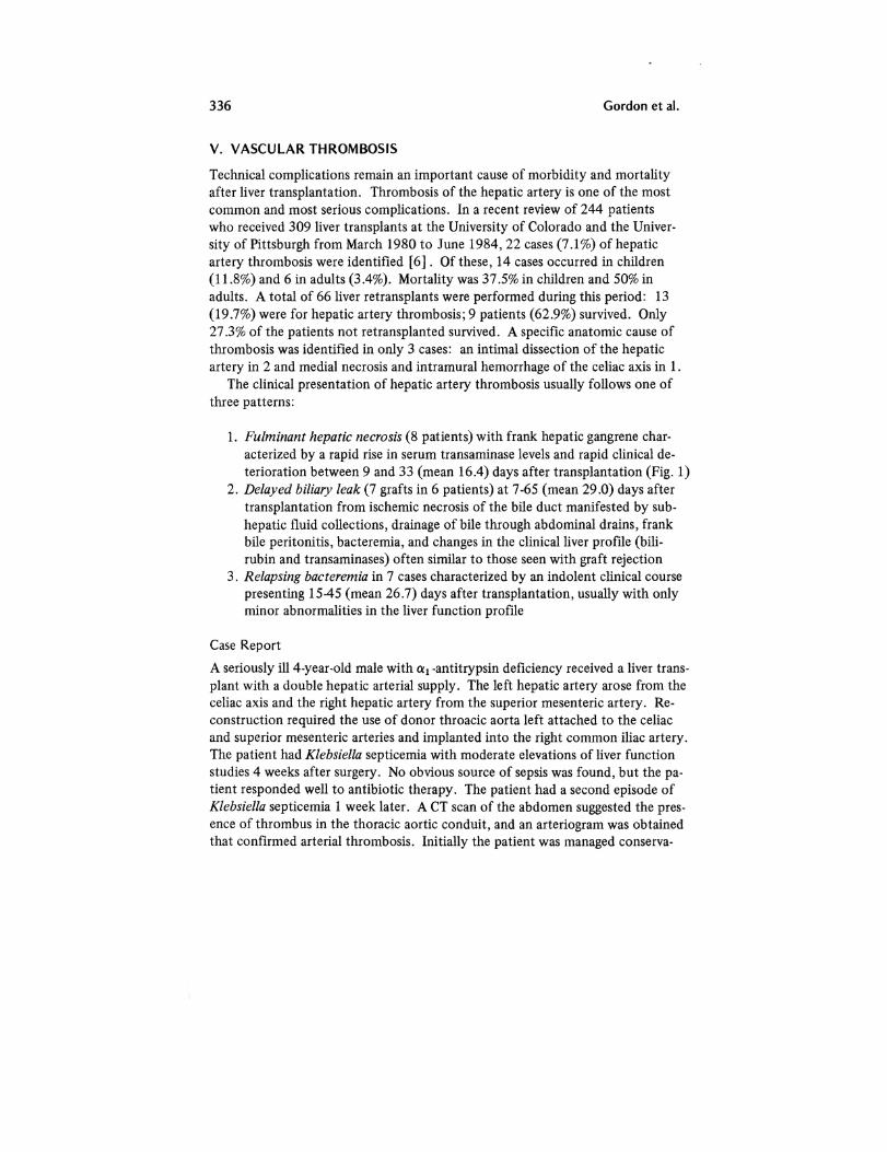
Figure 6 Pseudoaneurysm (arrows) of an aortic graft in a pediatric liver transplant recipient demonstrated by both CT scan (A) and aortography (B) .
the neck of the pancreas, or a longer tunnel can be made crossing over the inferior vena cava and under the superior mesenteric artery, head of the pancreas, and duodenum. The anastomosis between the donor arterial conduit and the recipient aorta has been the most frequent site of pseudoaneurysm or mycotic aneurysm formation (Fig. 6) . Ultrasound and CT scanning have been helpful in making the diagnosis.
VI. BILIARY TRACT COMPLICATIONS
The two principal technical advances in liver transplantation in recent years have been the development of a reliable, effective venovenous bypass technique and standardization of the techniques for biliary reconstruction. Direct duct-to-duct (choledochocholedochostomy) over aT-tube stent in adults is the preferred method of reconstruction when possible. In children with biliary atresia and other patients with disease rendering the recipient bile duct unsuitable for direct
Liver Transplantation Complications 345
(B)
Figure 6 (continued)

346 Gordon et aL
reconstruction, anastomosis of the donor bile duct to Roux-en-Y limb of jejunum (choledochojejunostomy) is the reconstruction of choice . These reconstructions are now successful in over 90% of cases .
In 313 patients between March I, 1980, and December 31, 1984, biliary tract complications occurred in 52 patients [7] . Of these patients, 5 (9 .6%) died as a direct result of the complication . Prior to this period, the incidence of biliary tract complications was even higher. It had been thought that a deficient blood supply of the graft duct system was responsible for the high incidence of complications, but, in fact, the majority of problems were probably the result of inappropriate surgical techniques, such as choledochoduodenostomy, and the failure to diagnose complications promptly. With the advent of cyclosporine-steroid immunosuppression in 1980, frequent use of postoperative cholangiography, standardization of choledochocholedochostomy and Roux-en-Y choledochojejunostomy, and prompt operative intervention for complications, the incidence of complications and morbidity and mortality have decreased markedly.
It has already been emphasized that primary biliary tract complications must be differentiated from those secondary to arterial insufficiency. Other less common and unusual biliary tract complications include bile cast syndrome, transplantation of a traumatized liver, and unexplained changes in the peripheral intrahepatic bile ducts not related to the biliary anastomosis.
Case Report
A 34-year-old female received a liver transplant for chronic aggressive hepatitis. The operation went without incident, and a direct duct-to-duct biliary reconstruction over a T tube was performed. Completion and postoperative cholangiography demonstrated a normal biliary tree and patent leak-free anastomosis. The early postoperative course was complicated by pneumonia and several episodes of acute rejection treated with corticosteroids.
A cholangiogram performed on postoperative day 53 showed a stricture at the confluence of the hepatic ducts of the graft and dislocation of one T -tube limb out of the biliary tract. The tube was replaced percutaneously by a straight tube stent. The stricture was treated by seven percutaneous balloon dilatations. The stricture was dilated to 12 mm, resulting in a patent duct with no detectable pressure gradient across the site of the stricture.
The patient was again hospitalized 18 months later with an abnormal liver function profile. A liver biopsy showed moderate allograft rejection. Percutaneous transhepatic cholangiography and ERCP demonstrated recurrence of the stricture. Percutaneous balloon dilatation was unsuccessful, and the patient was reconstructed with a Rou x-en-Y hepaticojejunostomy. Histology showed only mild fibrosis of the duct. The patient made an uneventful recovery . U1trasound of the liver prior to hospital discharge showed no dilatation of the biliary tree.

Liver Transplantation Compl ications 347
This case demonstrates several important aspects of biliary tract reconstruction in liver transplantation (Fig. 7). First, the differential diagnosis between rejection and biliary tract complications can be difficult. Second, T tubes and stents may dislocate or migrate out of proper position predisposing to stricture. Third, biliary tract obstruction need not involve the anastomosis even in the absence of ischemic injury to the graft ductal system. Fourth, percutaneous transhepatic dilatation is only an interim method of management. Definitive management usually requires resection of the obstruction and mucosa-to-mucosa repair of the bile duct to a Roux-en-Y jejunal loop.
The biliary tract reconstruction is no longer the "Achilles heel" of liver transplantation. The use of standard reconstructive techniques, appropriate postoperative investigation, and aggressive intervention when a complication is identified can minimize morbidity and mortality and save most grafts.
VII. VIRAL HEPATITIS
Infection still accounts for much of the morbidity and mortality after liver transplantation. The differential diagnosis of liver dysfunction after transplantation is complicated by the difficulty of distinguishing rejection from postoperative viral hepatitis. Cytomegalovirus (CMV) infection of the liver is a particularly troublesome and still frequent occurrence. Between March 1980 and September 1985 we have identified 17 patients with pathologically proven CMV hepatitis after liver transplantation.
CMV hepatitis tends to occur relatively late after transpIantation (15-132 days ; mean, 44 days). It is characterized by elevation of bilirubin and transaminases with a prolonged clinical course of spiking temperatures (ranging 5-68 days in 15 of the 17 patients). Since the management of viral infection and rejection is radical1y different, the establishment of the correct diagnosis is essential. Percutaneous liver biopsy is the method of choice and is performed frequently on our service. Prior to our liberal use of biopsy, three patients had .liver retransplantation for an incorrect diagnosis of rejection based only on liver function abnormalities . AIl three patients had been overimmunosuppressed because of the mistaken diagnosis. AIl three patients ultimately died. At autopsy CMV hepatitis, not rejection, was found .
More striking evidence of the need for liver biopsy is found in further evaluation of the 17 confirmed cases of CMV hepatitis in our series. Of the first 8 patients,S were diagnosed at the time of retransplantation. The next 9 patients were all diagnosed by percutaneous biopsy . Only 4 of 12 patients diagnosed by biopsy have died compared with 4 of the 5 patients diagnosed at reoperation.
CMV infection is diagnosed by serological changes and/or isolation of the virus. Liver biopsy may show the typical inclusion bodies in the hepatocytes,

348 Gordon et al.
(A)
Figure 7 Bile duct stricture after liver transplantation. (A) Operative cholangiogram (left) demonstrating normal biliary tree after the initial reconstruction . Cholangiography (right) demonstrating stricture of the donor common hepatic duct. (B) Cholangiogram showing a dislodged T tube lying outside the biliary tree, which was subsequently replaced with a straight tube stent. (C) The dislodged T tube has been replaced with a straight tube stent passed up toward the hepatic duct bifurcation. (D) Cholangiography showing resolution of the stricture after repetitive percutaneous transhepatic balloon dilatations. No pressure gradient was detectable across the stricture site. (E) Cholangiography showing recurrence of the stricture 2 years later subsequently treated by conversion of the choledochocholedochostomy to a Roux-en-Y choledochojejunostomy.

Liver Transplantation Complications 349
( 8)
Figure 7 (continued)

(e )
(0)
Figure 7 (continued)

Liver Transplantation Complications 351
(E)
Figure 7 (continued)

352 Gordon et al.
but biopsy material should always be cultured since isolation of virus is the best way to establish the diagnosis . CMV infection can occur primarily, as established by seroconversion, or by reactivation of prior infection, as documented by a fourfold or greater rise in antibody titer. Reactivation is promoted by immunosuppression. Primary infection in the transplant patient can originate from many sources, including the graft itself and blood products. Greater than 35% of the U.S. population is serologically positive for CMV and thus the virus is frequently transmitted from donor to recipient. Of the 17 cases we reviewed, 8 patients had primary infection and 1 had definite and 5 had probable reactivation of infection. In 3 cases, insufficient serum samples were available to make a determination.
The finding of primary infection in 8 of 14 (57%) patients with CMV hepatitis is higher than one would expect and suggests that patients with primary infection are more at risk of developing significant hepatitis [8]. It is well known that primary CMV infections tend to be more symptomatic and result in higher morbidity and mortality in immunocompromised patients, such as kidney transplant patients [7]. It is important to note, however, that in the group of 17 patients with CMV hepatitis we recently reviewed, we found no difference in clinical prognosis between primary infection or reactivated infection.
Most CMV infections are self-limited if immunosuppression is managed with restraint. It is also worth noting that CMV infection is by itself immunosuppressive . Full maintenance of immunosuppressive agents in the face of CMV infection may easily result in dangerous overimmunosuppression and increase susceptibility to other opportunistic infections.
Once the diagnosis of CMV hepatitis is established, our management strategy includes reduction of cyclosporine to achieve moderate blood levels (400-500 ngJmL by whole-blood RIA) and reduction of maintenance steroids from 20 to 10 mg/day or less. As the disease resolves, cyclosporine is increased to maintain blood levels in the usual therapeutic range (800-1000 ng/mL).
The management of the occasional patient with both rejection and CMV hepatitis is difficult. We often reduce the daily maintenance dose of steroids but supplement therapy with boluses of 0.5-1.0 g of methylprednisolone or hydrocortisone every 4-5 days. CMV hepatitis may worsen after therapy for rejection, requiring judicious lowering of immunosuppression. Finally, in some cases , only retransplantation can save the patient.
Hepatitis B virus, adenovirus, and herpesvirus are other less common offenders . Hepatitis B is also often self-limited and managed by conservative immunosuppressive therapy. Herpes and adenovirus hepatitis are rare but often ominous and severe.

Liver Transplantation Complications 353
Case Report
A 23-year-old male underwent an uneventful orthotopic liver transplant in December 1983 for end-stage sclerosing cholangitis. He did well postoperatively until about day 16, when he developed significant elevations in bilirubin and hepatic transaminases. His temperature reached 39°C, and fever lasted for 6 days. A cholangiogram and an arteriogram were normal. Liver biopsy was not performed. He was treated for rejection with steroids and antilymphocyte globulin without significant improvement in liver function.
The patient suddenly deteriorated with disorientation, hypoxia, and bilateral pulmonary infiltrates requiring intubation and high-pressure ventilatory support. Retransplantation was performed, but the patient died 7 days later. Pathological examination of the first liver graft showed severe cytomegalovirus hepatitis without evidence of rejection. CMV was also identified in the spleen, which had also been removed at surgery. CMV was isolated in urine and from buffy coat blood. In addition, Clostridium difficile was isolated from the spleen and Staphylococcus aureus and Enterobacter were isolated from blood.
This case demonstrates the severe penalty for treating viral hepatitis as rejection. A liver biopsy can be especially helpful in this situation and prevent inappropriate immunosuppressive treatment.
VIII. CONCLUSION
Just a few years ago, the patient with end-stage liver disease had little to hope for. Liver transplantation was still an experimental procedure with a high morbidity and mortality. Today, the surgical technique, though still demanding, has become standardized and operative mortality is below 10%. Cyclosporine and monoclonal antibody therapy have made rejection more manageable. Nonetheless, the majority of patients undergoing liver transplantation will experience at least one major complication and many will require at least one additional operation. Fortunately, our experience in recognizing and managing the complications of liver transplantation has also grown in recent years, and this also has contributed significantly to the improved survival now being enjoyed by our patients.
REFERENCES
1. NIH Consensus Development Conference Statement: Liver transplantation. June 20-23,1983. Hepatology 4:1075-1095,1984.
2. Shaw, B. W., Martin, D. J., Marquez, J. M., Kang, Y. G., Bugbee, A. C., lwat-
354 Gordon et al.
suki, S., Griffith, B. P., Hardesty, R. L., Bahnson, H. T., and Starz1, T. E.: Venous bypass in elinicalliver transplantation. Ann. Surg. 200:524-534, 1984.
3. Griffith, B. P., Shaw, B. W., Hardesty, R. L., Iwatsuki, S., Bahnson, H. T., and Starz1, T. E.: Veno-venous bypass without systemic anticoagulation for transplantation of the human liver. Surg. Gynecol. Obstet. 160:270-272, 1985.
4. Starzl, T. E., Iwatsuki, S., Shaw, B. W., Gordon, R. D., Esquivel, C. 0., Todo, S., Kam, I., and Lynch, S.: Factors in the development of liver transplantation. Transplant. Proe. 17(Suppl. 2): 107-119, 1985.
5. Ramsey, G., Nusbacher, J., Starzl, T. E., and Lindsay, G. D.: Isohemagglutinins of graft origin after ABO-unmatched liver transplantation. N. Engl. J. Med. 311:1167-1170,1984.
6. Tzakis, A., Gordon, R. D., Shaw, B. W., Iwatsuki, S., and Starzl, T. E.: Clinical presentation of hepatic artery thrombosis after liver transplantation in the cyclosporine era. Transplantation 40:667-671,1985.
7. Ho, M.: Cytomegalovirus: Biology and Infection. New York: Plenum, 1982.
8. Ho, M., Wajszczuk, C. P., Hardy, A., Dunmer, J. S., Starzl, T. E., Hakala, T. T., and Bahnson, H. T.: Infections in kidney, heart and liver transplant recipients on eyc1osporine. Transplant. Proc. 15: 2768-2772, 1983.
9. Lerut, J., Gordon, R. D., Iwatsuki, I., Shaw, B. W., Jr., Esquivel, C. 0., Tzakis, A., and Starzl, T. E.: Biliary tract complications in 393 human liver transplantations. Transplantation (in press).


![Kidney Transplantation (Renal Transplantation) Auto Saved]](https://img.pdfslide.us/doc/110x75/577d22b31a28ab4e1e9807d7/kidney-transplantation-renal-transplantation-auto-saved.jpg)