Embed Size (px)
Citation preview
Complications Incidence of Non-union 1 cm displacement of fracture caused 55% Non-union It takes 5-20 yrs to develop SNAC. SNAC appears to be more common with waist fracture than a proximal pole. However this is controversial Union rate: Union: Stable 94% All fracture 90% Union after surgery for nonunion with surgery 80% OA in healed scaphoid: 9% Malunion: Interscaphoid angle > 35° [Humpback scaphoid] AVN of proximal 90% [MRI: T1 shows decreased signals] 1. Scaphoid Nonunion Nonunion of the scaphoid occurs in around 10% of nonoperatively managed scaphoid fractures. The risk is probably greater in unstable fractures and correspondingly less in stable fractures. The natural history of a scaphoid nonunion depends on the stability of the nonunion with progressive degenerative changes. Stable Nonunions Is characterized by a firm fibrous nonunion that prevents deformity from occurring. The length and shape of the scaphoid remain well preserved, and the risk of osteoarthritis is small. Radiographs show an indistinct fracture line with variable cystic changes affecting the adjacent bone fragments. The patients are usually relatively symptom-free unless the wrist is subjected to further trauma, which often leads to an unstable nonunion with all of the associated problems of carpal collapse, osteoarthritis, pain, and weakness. Although there are patients who seem to have an asymptomatic, stable nonunion of the scaphoid for many years, most patients will become symptomatic if the stable nonunion progresses to an unstable one and osteoarthritis occurs. Treatment: Open palmar approach The sheath of the flexor carpi radialis tendon is incised and the tendon retracted ulnarly. The capsule should be incised longitudinally. The superficial palmar branch of the radial artery is distal at the end of incision and needs to be ligated in cases of wider exposure of the distal scaphoid. It is important to prepare the nonunion surfaces by removing any fibrous tissue and sclerotic bone. Leave the dorsal cartilage in place. Cancellous bone graft from the distal radius usually provides sufficient volume, although iliac crest bone graft can be used if necessary. Screw fixation of the scaphoid is then used. 2. Unstable Nonunions (Herbert Type D2) Bone grafts A. Matti: Inlay grafts thro dorsal approach Russe : Palmar B. Tricortical shaped iliac graft anterior to correct deformity. Better for deformity correction. C. Vascularised bone graft with PQ or I or II dorsal M pedicle Overall union is 80%.
The unstable scaphoid nonunion is characterized 1) sclerotic bone surfaces 2) synovial erosion, fibrous cysts 3) A marked discrepancy between the sizes of the two bone fragments. Unstable scaphoid nonunion leads: SNAC Humpback deformity Success rates of achieving union with internal fixation and bone grafting range from 60% to 95%. The lateral intrascaphoid angle
and the height-to-length ratio of the bone help identify angulation and collapse of the
scaphoid. The lateral intrascaphoid angle is formed by the intersection of the perpendicular lines to the diameters of the proximal and distal poles. An angle of more than 35 degrees has been shown [Trumble] to be associated with an increased incidence of arthrosis even in fractures that went on to unite.
Green reported that the number of punctuate bleeding points is a good indicator of vascularity of the bone. When the
proximal pole was completely avascular, the likelihood of successful healing with a graft was virtually nil and an alternative procedure such as intercarpal fusion, excision of the proximal scaphoid, interposition arthroplasty, proximal row carpectomy, or scaphoid allograft should be considered. Herbert stated that carpal collapse and secondary arthritis are rarely associated with proximal pole nonunion. He therefore argued against the use of interposition bone grafts and suggested screw fixation by a dorsal approach, with or without cancellous bone grafting, depending on the findings at the time of surgery. Although the fracture may not unite, the patient's own bone should be better than any implant. Another alternative is a vascularized bone graft.
Standard Russe bone graft
Technique relied on packing a corticocancellous bone graft into a trough curretted through the volar cortex of both fragments. Because the volar cortex is often foreshortened by erosion of the fragments, loss of length is difficult to correct without introducing a cortical graft (center). Modified Russe winged graft that is impacted into a volar trough to lengthen the scaphoid (bottom).
The standard palmar approach should be used for most reconstructions of unstable scaphoid nonunions to avoid damaging the dorsal blood supply. Scaphoid nonunions might not be visible macroscopically and often need sharp division with the knife. Anterior wedge grafting procedures are now in common use as humpback deformities can be corrected with the Russe’s technique. My approach A volar approach is necessary to correct a humpback deformity. The nonunion gap is exposed and débrided, and the fracture fragments are mobilized. It is best to leave a cartilage hinge posteriorly to provide a fulcrum around which the fragments may be hinged open| If the hinge is released in an effort to regain all of the scaphoid length, the fracture fragments will become extremely unstable
The wrist is extended and the two fragments gently distracted with small spreaders. This maneuver usually achieves adequate correction of the carpal deformity and a satisfactory improvement in wrist extension. The fracture surfaces are excised with a small osteotome, burr, or curette. A corticocancellous wedge graft from the iliac crest. Vascularized bone grafts from the distal radius (radial artery) or distal ulna (ulnar artery) have also been described.
We prefer the pronator quadratus graft. A partial radial styloidectomy
can be performed in patients with radiologic
signs of stage I radioscaphoid arthritis, this being arthritis that is limited to the scaphoid and radial styloid. This is undertaken to relieve pain arising from arthritic joints or osteophyte impingement. If there are no radiologic signs of arthritis, a styloidectomy should not be undertaken at the same time as a scaphoid reconstruction
often relieves symptoms.
Salvage Procedures for Scaphoid Nonunion
1. Excision of Part or all of the Scaphoid. A very small fragment can be excised with impunity. However, most surgeons are
aware that if it is more than 8 mm long, the results are poor and the wrist feels weak.
2.Wrist Denervation. Wrist denervation is often helpful as it is combined with significant pain relief. However, pain relief
can be temporary.
3.Proximal Row Carpectomy. The results of excising the scaphoid, lunate, and triquetrum have been disappointing in some
series, which is why we prefer wrist arthrodesis in cases of panscaphoid arthritis and severe pain.
4. Bentzon Operation: May be worth trying after a failed bone graft
5. Radial styloidectomy: Useful in limited OA with pain on radial deviation when pain is from OA and not from nonunion.
6. 4 corner fusion
7. Scaphoid Prosthesis. In selected patients with panscaphoid osteoarthritis, total replacement of the scaphoid is worth
considering. Silicone implants induced progressive silicone arthritis in many cases,
and the technique was abandoned 20
years ago. Other methods of replacement titanium implants are currently in clinical trials.
8. Wrist arthrodesis: Young and active patients are likely to complain of continued pain after this procedure, and wrist
arthrodesis is therefore preferable in these patients. Arthrodesis is an accepted
surgical treatment option for patients with markedly restricted and painful wrist
motion. Instability and deformity of the wrist affect hand function significantly
but pain diminishes both strength and dexterity. Wrist arthrodesis achieves good
pain relief, especially in younger patients with high functional demands. The pain
relief associated with a successful fusion results in significant improvement of
hand function and grip strength.
2. Scaphoid Malunion
Although the malunited scaphoid fracture is a recognized entity that causes altered carpal kinematics and abnormal load
distribution which may cause premature wrist arthrosis, the reported number of patients treated with early osteotomy is
surprisingly small. Indications for osteotomy are pain, weakness, limited range of motion, and deformity of the scaphoid
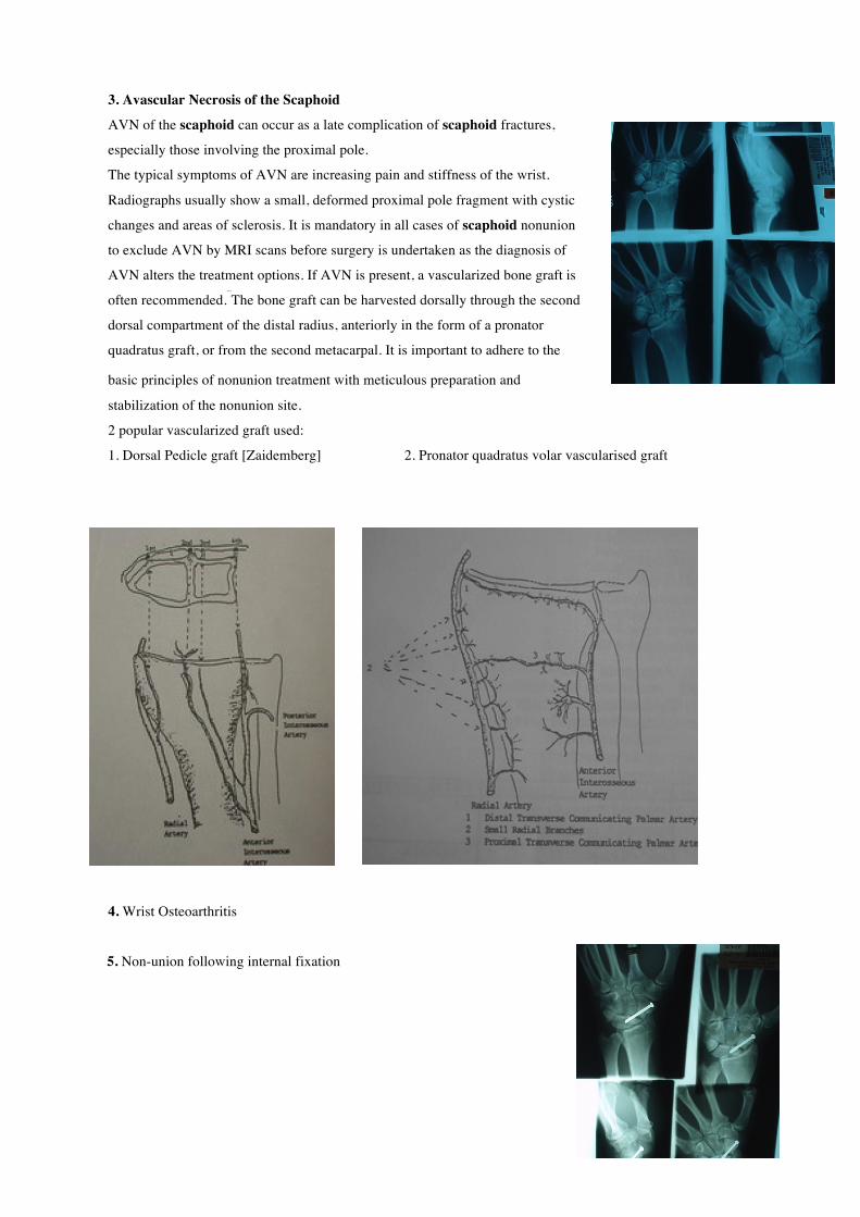
detected on radiographs and evaluated by CT scans.
Sagittal CT image: >35° can cause pain and arthritis.
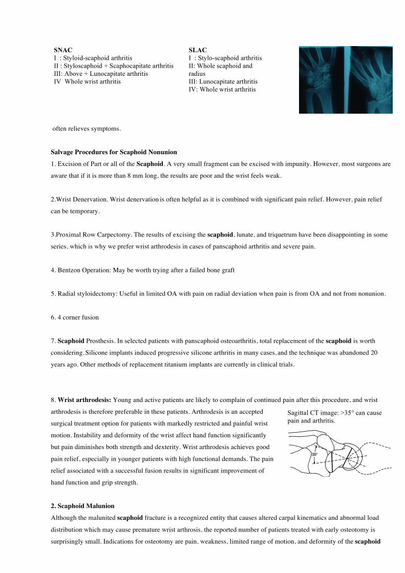
SNAC I : Styloid-scaphoid arthritis II : Styloscaphoid + Scaphocapitate arthritis III: Above + Lunocapitate arthritis IV Whole wrist arthritis
SLAC I : Stylo-scaphoid arthritis II: Whole scaphoid and radius III: Lunocapitate arthritis IV: Whole wrist arthritis
3. Avascular Necrosis of the Scaphoid
AVN of the scaphoid can occur as a late complication of scaphoid fractures,
especially those involving the proximal pole.
The typical symptoms of AVN are increasing pain and stiffness of the wrist.
Radiographs usually show a small, deformed proximal pole fragment with cystic
changes and areas of sclerosis. It is mandatory in all cases of scaphoid nonunion
to exclude AVN by MRI scans before surgery is undertaken as the diagnosis of
AVN alters the treatment options. If AVN is present, a vascularized bone graft is
often recommended. The bone graft can be harvested dorsally through the second
dorsal compartment of the distal radius, anteriorly in the form of a pronator
quadratus graft, or from the second metacarpal. It is important to adhere to the
basic principles of nonunion treatment with meticulous preparation and
stabilization of the nonunion site.
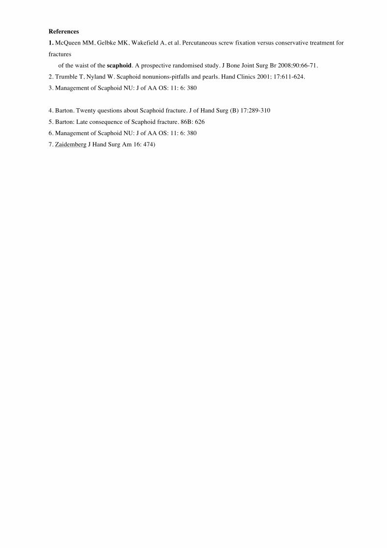
2 popular vascularized graft used:
1. Dorsal Pedicle graft [Zaidemberg] 2. Pronator quadratus volar vascularised graft
4. Wrist Osteoarthritis
5. Non-union following internal fixation
References
1. McQueen MM, Gelbke MK, Wakefield A, et al. Percutaneous screw fixation versus conservative treatment for
fractures
of the waist of the scaphoid. A prospective randomised study. J Bone Joint Surg Br 2008;90:66-71.
2. Trumble T, Nyland W. Scaphoid nonunions-pitfalls and pearls. Hand Clinics 2001; 17:611-624.
3. Management of Scaphoid NU: J of AA OS: 11: 6: 380
4. Barton. Twenty questions about Scaphoid fracture. J of Hand Surg (B) 17:289-310
5. Barton: Late consequence of Scaphoid fracture. 86B: 626
6. Management of Scaphoid NU: J of AA OS: 11: 6: 380
7. Zaidemberg J Hand Surg Am 16: 474)


![Research Article Early Enteral Nutrition Using Oligomeric ... · decreases the incidence of infectious complications in critically ill patients [1]. In addition, although the mechanism](https://img.pdfslide.us/doc/110x75/602e6e06f7bd50188852216c/research-article-early-enteral-nutrition-using-oligomeric-decreases-the-incidence.jpg)