Embed Size (px)
Citation preview
53
COMPLEX REGIONAL PAIN SYNDROME: PART I
2Complex regional painsyndrome: Part 1
LOUIS GIFFORD AND MICK THACKER
IntroductionThis section of Topical Issues in Pain 3 is devoted to conditions that have beenlargely attributed to pathobiology of the sympathetic nervous system. Theprevious chapter overviewed the anatomy and basic physiology of theautonomic nervous system. This chapter discusses the cluster of presentationsthat are classified under the umbrella term complex regional pain syndrome.The chapter describes and discusses their features and some aspects of theirmechanisms and manifestations that may be of interest to physiotherapistsseeking a broader understanding of the complex multifactorial underpinnings.The following two chapters discuss the pathobiology relating to the knownpain mechanisms involved in CRPS.
Why is it that some people develop incredibly complex symptompresentations, often after relatively little in the way of injury? Remarkablecases where minor traumas quickly develop dramatic presentations—asimple bang on the elbow is followed by swelling of the hand, marked lossof joint range of motion and intense pain over the whole forearm that soonbecomes resistant to all attempts to relieve the pain and which can thendevelop into an ongoing and devastating problem—are part of the experienceof most physiotherapists. Cases of patients who present with quite long termpain and disability that includes observations and/or complaints by thepatient of feelings of swelling, temperature changes, blotchy skin, alteredsweating, poor skin health etc., are not uncommon and are often attributedto ‘sympathetic’ mechanisms. Is this true? What do we know about the roleof the sympathetic nervous system in producing these states?
Further, can these sometimes awful presentations be prevented fromhappening in the first place, and once they are established how should webe thinking about their management?
54
TOPICAL ISSUES IN PAIN 3
This chapter, and the next two, explores and reviews postulated andknown mechanisms relating to pain states where the sympathetic nervoussystem has been assumed to have a significant role.
It seems logical to assume that the autonomic nervous system, and hencethe SNS, has to be involved in some way in all injuries, pathologies and painstates. The SNS, under normal conditions, has been shown to respond tonociceptive activity and to the presence of pain (see previous chapter andalso reviews by Vallbo et al 1979, Wallin & Elam 1997). The reasoning is thatsince tissue damaging events represent a threat to the system’s homeostasisand survival, the SNS, whose prime role is to engage defensive mechanisms,is likely to play a role in activating and co-ordinating the body’s responsesto restore or maintain homeostasis. Unfortunately, this isn’t how the bulk ofthe literature on the role of the sympathetic nervous system in pain statesfollowing pathology or injury appears to view the situation.
An important point is that in normal circumstances, while the SNS respondsto pain, its activity does not significantly excite sensory neurones and causepain. Thus, the ‘adaptive’ role of sympathetic activity in the presence of painis focused more on ‘recovery physiology’ rather than on the production ofpain (Michaelis 2000).
Complex regional pain syndrome—newterminology to replace oldThe role of the sympathetic nervous system in pain states has been a longheld and reasonable assumption. It is based on the belief that sympatheticactivity or hyperactivity is in some way involved in symptom generation.This belief is backed up by the clinical finding that sympathetic blocks, ordestruction of sympathetic nerves when used in conditions diagnosed ashaving sympathetically maintained pain (SMP), like ‘reflex sympatheticdystrophy’ (RSD), are occasionally good at relieving pain as well as symptomslike oedema and sweating that are so often attributed to abnormal SNSbehaviour. Further support comes from the finding that in some patientspain associated with neuropathy and RSD can be provoked by injection ofadrenaline or noradrenaline or other ‘adrenoreceptor agonists’ (see Torebjorket al 1995, Wall 1995), whereas in normal people these hormones have nopain producing effect.
However, there is considerable argument surrounding the hypothesisedrole of the sympathetic system and also great difficulty in classifying patientpresentations accurately. For example, many patients who fit into the RSDcategory do not respond at all to sympathetic blocking and a great many whodo respond find that the relief is only temporary (see Chapter 4). Some patientsmay respond to a sympathetic block early on in the course of their problem,but not later, or vice versa (Torebjork et al 1995, Wall 1995). It seems that somepatients can shift from having pain that may be dependent on sympatheticactivity to having pain that is not. Some pain mechanisms appear to move andchange over time, even though the presenting symptoms may appear stable.
55
COMPLEX REGIONAL PAIN SYNDROME: PART I
For the past 10 years or so the golden nugget status that the sympatheticsystem has achieved and held on to has been subjected to quite a strongchallenge that has resulted in a revised downward rating (see Chapters 3& 4.)
In an attempt to clarify the nature of RSD and the role of the sympatheticnervous system in pain states a working party of world experts was set upfollowing an International Association for the Study of Pain ‘SpecialConsensus Workshop’ held in Orlando, Florida USA in 1993 (Stanton-Hickset al 1995). As an example of some of the challenges facing the group, theproblems with the term RSD are representative: ‘The term RSD is usedimprecisely as it refers to changes in soft tissue which may or may not dependupon the sympathetic nervous system, may not be the consequence of a reflex,and may occur later in the disorder’ (see Stanton-Hicks et al 1995, p 128).The goals set out in the workshop were to examine the terms RSD, causalgia,sympathetically maintained pain (SMP) and sympathetically independentpain (SIP) and to revise the classifications and definitions for better clinicalutility.
One overriding theme was that the continued use of the term ‘sympathetic’was unhelpful as it implied a pathological mechanism attributable to thesympathetic system that lacked conclusive evidence and which may haveprovided a barrier to the consideration of alternative mechanisms or amultiplicity of mechanisms acting in concert.
The group chose to use ‘Complex Regional Pain Syndrome’ (CRPS) as anoverall term: complex to denote the varied clinical phenomena in addition topain (see below); regional, since the distribution of symptoms and findingsare so general, widespread and beyond the area of the original lesion—oftenbeing in the distal part of the extremities and rarely on the trunk and face,but having the potential to spread to other body areas; and pain since it wasthe cardinal symptom and being so disproportionate to the inciting event(Stanton-Hicks et al 1995, Boas 1996).
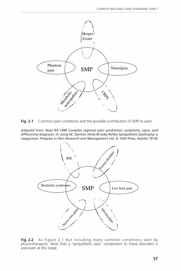
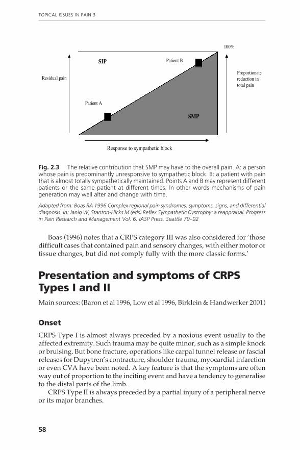
Within the CRPS designation are two subsets: Types I and II; both dependon the apparent cause and on the presentation—see Box 2.1. Type Icorresponds to the ‘old’ RSD and Type II to ‘causalgia.’ Essentially thedifference between the two is that Type I follows a tissue injury and Type IIfollows a frank nerve injury. A major departure as a result of this classificationis the separation of the previously implied focus on sympathetic elements ormechanisms from the definitions. The result of this is the recognition thatvirtually any disorder, including CRPS, may have a component ofsympathetically maintained pain alongside pains whose mechanisms arequite independent of the system, hence the terms sympathetically maintainedpain (SMP) and sympathetically independent pain (SIP) (see Figs 2.1 and2.2).
A major effect of this new classification is the focus on the presentation’shistory and the constellation of signs and symptoms, rather than on apresumed mechanism that seems to have unfairly overburdened thesympathetic system with responsibility and ignored other mechanisms thatmay be more relevant.
56
TOPICAL ISSUES IN PAIN 3
An important point for physiotherapy is that mechanistic approaches topain syndromes like this are not necessarily helpful since they represent aninterventionist biomedical paradigm whose object is to recognise apathobiological mechanism, usually related to a pain mechanism, and thento directly alter the mechanism in some way. The recognition that pain is onething but function and having a life are others too, does not seem to enterthe therapeutic equation too easily. Even so, most physiotherapists like tofeel comfortable with the problem they are dealing with and the belief hereis that knowing about the current state of the art with regard to mechanismsis an important issue.
Box 2.1 Classification: complex regional pain syndrome (CRPS)
CRPS describes a variety of painful conditions that usually follow injury, occurregionally, have a distal predominance of abnormal findings, exceed in bothmagnitude and duration the expected clinical course of the inciting event, oftenresult in significant impairment of motor function, and show variable progressionover time.
CRPS Type I (RSD)The syndrome follows an initiating noxious event.
1. Spontaneous pain or allodynia/hyperalgesia occurs beyond the territory ofa single peripheral nerve(s) and is disproportionate to the inciting event.
2. There is or has been evidence of oedema, skin blood flow abnormality, orabnormal sudomotor activity, in the region of the pain since the incitingevent.
3. This diagnosis is excluded by the existence of conditions that would otherwiseaccount for the degree of pain and dysfunction
CRPS Type II (Causalgia)This syndrome follows nerve injury. It is similar in all other respects to Type I.
1. Is a more regionally confined presentation about a joint (e.g. ankle, knee,wrist) or area (e.g. face, eye, penis), associated with a noxious event.
2. Spontaneous pain or allodynia/hyperalgesia is usually limited to the areainvolved but may spread variably distal or proximal to the area, not in theterritory of a dermatomal or peripheral nerve distribution.
3. Intermittent and variable oedema, skin blood flow change, temperature change,abnormal sudomotor activity, and motor dysfunction, disproportionate to theinciting event, are present about the area involved.
Sympathetically maintained painPain that is maintained by sympathetic efferent activity or neurochemical orcirculating catecholamine action, as determined by pharmacological orsympathetic nerve blockade. SMP may be a feature of several types of paindisorder, and is not an essential component of any one condition. Conditionswithout any response to sympathetic block are, by contrast, designated as havingsympathetic independent pain states (SIP).
57
COMPLEX REGIONAL PAIN SYNDROME: PART I
SMPPhantom pain
HerpesZoster
Neuralgias
CRPS
Met
abol
icne
urop
athi
es
SMP
RSI
Low back painHeadache syndromes
Nerve
root
pain
s fibromyalgia
Froz
en sh
ould
er
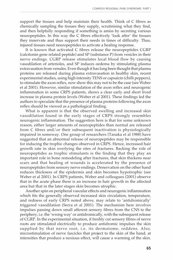
Fig. 2.1 Common pain conditions and the possible contribution of SMP to each.
Adapted from: Boas RA 1996 Complex regional pain syndromes: symptoms, signs, anddifferential diagnosis. In: Janig W, Stanton-Hicks M (eds) Reflex Sympathetic Dystrophy: areappraisal. Progress in Pain Research and Management Vol. 6. IASP Press, Seattle 79–92
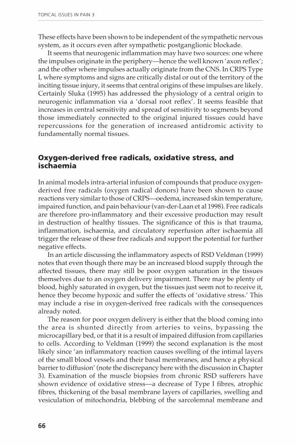
Fig. 2.2 As Figure 2.1 but including many common conditions seen byphysiotherapists. Note that a ‘sympathetic pain’ component to these disorders isunproven at this stage.
58
TOPICAL ISSUES IN PAIN 3
Boas (1996) notes that a CRPS category III was also considered for ‘thosedifficult cases that contained pain and sensory changes, with either motor ortissue changes, but did not comply fully with the more classic forms.’
Presentation and symptoms of CRPSTypes I and IIMain sources: (Baron et al 1996, Low et al 1996, Birklein & Handwerker 2001)
Onset
CRPS Type I is almost always preceded by a noxious event usually to theaffected extremity. Such trauma may be quite minor, such as a simple knockor bruising. But bone fracture, operations like carpal tunnel release or fascialreleases for Dupytren’s contracture, shoulder trauma, myocardial infarctionor even CVA have been noted. A key feature is that the symptoms are oftenway out of proportion to the inciting event and have a tendency to generaliseto the distal parts of the limb.
CRPS Type II is always preceded by a partial injury of a peripheral nerveor its major branches.
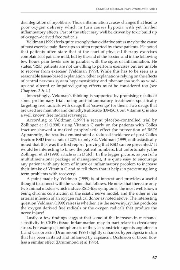
SIP
SMP
Response to sympathetic block
Proportionatereduction intotal pain
Residual pain
100%
Patient A
Patient B
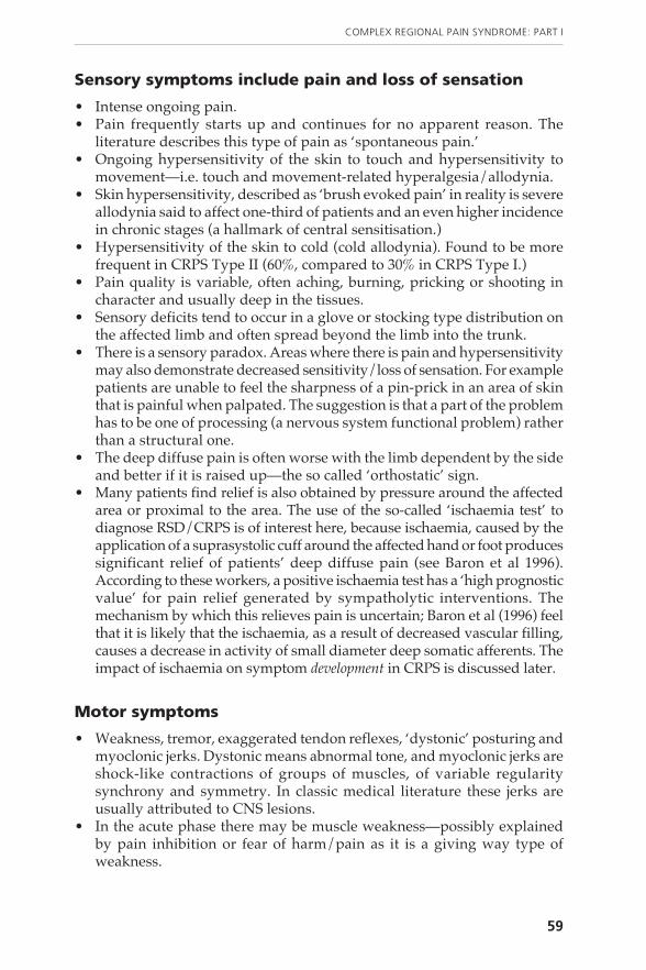
Fig. 2.3 The relative contribution that SMP may have to the overall pain. A: a personwhose pain is predominantly unresponsive to sympathetic block. B: a patient with painthat is almost totally sympathetically maintained. Points A and B may represent differentpatients or the same patient at different times. In other words mechanisms of paingeneration may well alter and change with time.
Adapted from: Boas RA 1996 Complex regional pain syndromes: symptoms, signs, and differentialdiagnosis. In: Janig W, Stanton-Hicks M (eds) Reflex Sympathetic Dystrophy: a reappraisal. Progressin Pain Research and Management Vol. 6. IASP Press, Seattle 79–92
59
COMPLEX REGIONAL PAIN SYNDROME: PART I
Sensory symptoms include pain and loss of sensation
• Intense ongoing pain.• Pain frequently starts up and continues for no apparent reason. The
literature describes this type of pain as ‘spontaneous pain.’• Ongoing hypersensitivity of the skin to touch and hypersensitivity to
movement—i.e. touch and movement-related hyperalgesia/allodynia.• Skin hypersensitivity, described as ‘brush evoked pain’ in reality is severe
allodynia said to affect one-third of patients and an even higher incidencein chronic stages (a hallmark of central sensitisation.)
• Hypersensitivity of the skin to cold (cold allodynia). Found to be morefrequent in CRPS Type II (60%, compared to 30% in CRPS Type I.)
• Pain quality is variable, often aching, burning, pricking or shooting incharacter and usually deep in the tissues.
• Sensory deficits tend to occur in a glove or stocking type distribution onthe affected limb and often spread beyond the limb into the trunk.
• There is a sensory paradox. Areas where there is pain and hypersensitivitymay also demonstrate decreased sensitivity/loss of sensation. For examplepatients are unable to feel the sharpness of a pin-prick in an area of skinthat is painful when palpated. The suggestion is that a part of the problemhas to be one of processing (a nervous system functional problem) ratherthan a structural one.
• The deep diffuse pain is often worse with the limb dependent by the sideand better if it is raised up—the so called ‘orthostatic’ sign.
• Many patients find relief is also obtained by pressure around the affectedarea or proximal to the area. The use of the so-called ‘ischaemia test’ todiagnose RSD/CRPS is of interest here, because ischaemia, caused by theapplication of a suprasystolic cuff around the affected hand or foot producessignificant relief of patients’ deep diffuse pain (see Baron et al 1996).According to these workers, a positive ischaemia test has a ‘high prognosticvalue’ for pain relief generated by sympatholytic interventions. Themechanism by which this relieves pain is uncertain; Baron et al (1996) feelthat it is likely that the ischaemia, as a result of decreased vascular filling,causes a decrease in activity of small diameter deep somatic afferents. Theimpact of ischaemia on symptom development in CRPS is discussed later.
Motor symptoms
• Weakness, tremor, exaggerated tendon reflexes, ‘dystonic’ posturing andmyoclonic jerks. Dystonic means abnormal tone, and myoclonic jerks areshock-like contractions of groups of muscles, of variable regularitysynchrony and symmetry. In classic medical literature these jerks areusually attributed to CNS lesions.
• In the acute phase there may be muscle weakness—possibly explainedby pain inhibition or fear of harm/pain as it is a giving way type ofweakness.
60
TOPICAL ISSUES IN PAIN 3
• In chronic stages it is hypothesised that weakness may be due to impairedmuscle nutrition or/as well as abnormalities of central processing. Fearof pain and loss of confidence should also be a consideration.
• Tremor, myoclonic jerks and dystonia are reported to be more commonin CRPS Type II.
• Reduced range of motion—by joint effusion and pain in acute stages andby contraction and fibrosis in chronic stages. Again, fear of pain and/ordamage may be an important consideration.
• About 45% have exaggerated tendon reflexes on the affected side. Yet nopyramidal tract signs—but good correlation to level of pain. Thereforethere may be a pain facilitation of tendon reflexes.
Autonomic symptoms
• Distal limb oedema.• Skin colour changes—red in the early stages, turning bluish later.• Skin temperature differences from affected side to good side—usually
about 1˚C. The affected side is warmer in the acute stages and cooler inthe more chronic.
• In 50% of patients with CRPS increased skin sweating changes can beobserved in the affected limb.
• Some researchers report that sweating may be increased or decreasedand that it occurs most commonly on the palmar surface of the hand orplantar surface of the foot (Blumberg et al 1994). It is also noted thatpatients report a strange reaction of the skin temperature to changes ofenvironmental temperature. For example, the affected hand in comparisonwith the healthy hand cools too slowly or too quickly when exposed tocold. Abnormal responses of skin blood flow to warming or coolingcompared to the non affected side have been demonstrated in patientswith RSD (Blumberg et al 1994).
Trophic changes
• Trophic changes occur in more than 50% of cases. Within a few weeks ofthe initiating traumatic incident there may be increased hair and nailgrowth. Thus there is an upregulation of growth in the early stages—socalled ‘plus signs.’
• Later, a decrease in tissue growth is observed—‘minus signs’—here thereis decreased hair and nail growth and atrophy of the skin. Skin maybecome thin and glossy and in severe cases there may even be skin ulcers.
• Other trophic changes noted are: changes in the texture of the skin, nailsand hair; the skin may become fibrotic; there may be alterations insubcutaneous tissues as well as in bone density (osteoporosis). Plainradiographs show a diffuse and spotty distal distribution ofdemineralisation of small bones with a periarticular dominance at thelonger bones (Baron et al 1996). These changes may not occur for manymonths. However, changes in bone metabolism picked up by ‘three phasebone scans’ apparently appear quite early on in CRPS Type I. Intraossary
61
COMPLEX REGIONAL PAIN SYNDROME: PART I
plasma extravasation has also been demonstrated, which lends supportto those who view the condition as an inflammatory disorder.
• It is not clear whether trophic changes are responsible for joint stiffnessand tendon shortening. Disuse and functional motor changes may alsohave a role to play.
Note that it can be difficult to diagnose CRPS from some of the abovereactions in the acute stage as many are the symptoms of normal trauma.For example, oedema, skin temperature differences and pain are commonlyobserved. Birklein & Handwerker (2001) state that the best symptoms to usefor early recognition are motor signs, trophic changes and increased sweating.
Diagnostic tools
• In research laboratories and specialist clinics some workers use skintemperature differences—by measuring the affected and non-affectedlimbs repeatedly over time and in a variety of room temperature settings.
• Increased sweating in both the acute and chronic stages of the disorderare measured using a sudorometer.
• X-rays can reveal ‘spotty osteoporotic changes’ within 4–8 weeks, butonly in 40% of cases. There is some evidence for increased bonemetabolism.
• MRI scans show that muscle and periarticular tissues may becomeoedematous and there is an increased permeability of blood vessels, butit is a good deal less dramatic than that seen in arthritis.
• Diagnostic blocks alone are not helpful to diagnose CRPS but they arethe only method to date that can indicate the possibility of a sympatheticcomponent to the pain (see Chapter 4).
• Ischaemia test noted above.
Stages of the disorder
For some time CRPS (RSD/causalgia) has been considered to pass throughthree distinct stages (Bonica 1990) as an accepted fact in the literature. Thestages or phases that were described by Bonica are:
1. The acute stage, characterised by pain and vasomotor changes such asgeneralised oedema, warm skin and sweating changes in areas not affectedby the preceding lesion. These symptoms may occur within hours or daysof the preceding incident.
2. The ‘dystrophic’ stage at 3–6 months after onset, characterised by moremarked pain and sensory dysfunction, continued evidence of vasomotorabnormalities and markedly increased trophic and motor changes. Theskin tended to shift from warm to cold.
3. The ‘atrophic’ last stage, with a decrease in pain/sensory disturbance,but continued vasomotor disturbance and markedly increased motor andtrophic changes. For example, atrophy of muscle and bone andcontractures of joints. This stage is seen as permanent.
62
TOPICAL ISSUES IN PAIN 3
This concept has now been challenged seriously. Recently Bruehl et al(2002), using a cluster analysis technique to look for stages in the disorder aswell as different subgroups, were unable to support the existence ofsequential stages. Their conclusions pointed to the existence of three differentsub-types of the disorder:
1. A relatively limited syndrome in which vasomotor signs predominate.2. A relatively limited syndrome in which neuropathic pain/sensory
abnormalities predominate.3. A florid CRPS syndrome similar to descriptions of classic RSD.
Interestingly, in their discussion Bruehl et al (2002) noted that in this thirdsubgroup, work on animals as well as humans was suggesting a significantcontribution of disuse to the development of the CRPS changes. They listedallodynia, hyperalgesia, motor dysfunction and temperature/colour changes.The negative effects of immobility are discussed further in the next section.
The results of this analysis reveal how highly respected and prominentindividuals in a specialist area may be publishing unsubstantiated or eveninaccurate material. We may need to be more wary of trusted experts andalways in mind to check the sources on which their information is provided.Sadly, a good deal of the clinical ‘facts’ physiotherapists rely on may comeinto the same category and are really due for serious scrutiny.
Some mechanistic perspectives on CRPSImmobilisation and disuse
Physiotherapists who have worked in a fracture clinic will be familiar withthe appearance of a patient’s limb when it is freshly taken out of plaster. Onremoval of the cast the limb is invariably atrophied, often with abnormalhair growth; it may be significantly warm with the skin being blotchy inappearance; it is markedly stiff; and occasionally it is very painful to touchand move. It actually looks very much like classic ‘RSD.’ Fortunately, onlyrelatively few cases are known to go on to have a documented case of RSD orCRPS. However, the clinical reality, which urgently needs quantifying, isthat many of the stubborn conditions physiotherapists treat have often hadsignificant periods of quite unjustified immobility or disuse imposed on them.
That the features of CRPS may relate to disuse has been noted and theeffects of immobilisation on humans and rats has been investigated (see Butleret al 2000). For example, Ushida and Willis (1996) immobilised rat wrists infull flexion for 3–4 weeks, some with radius fractures and some without.The results showed an increase in mechanosensitivity (touch and movementallodynia) in both groups and central plastic changes in spinal cord dorsalhorn cells that process sensory information from the immobilised areas. Evenimmobilisation of rat hindpaws for one week will increase levels of sensitivityto heat, cold and mechanical stimulation (Maves & Smith 1996).
Four week immobilisation of the wrist in 21 human volunteers (Butler etal 2000) showed:
63
COMPLEX REGIONAL PAIN SYNDROME: PART I
• All subjects had a temperature difference compared to the non-immobilised side ranging from 0.5–2.7°C (10 were warmer and 11 cooler).In three of the subjects the difference persisted longer than two weeks.
• 16 subjects had decreased range of movement of the thumb, 12 had alteredsensation to sensory testing, of whom 4 had summation to pinprick (anamplifying sensation, often termed ‘hyperpathia,’ with every pinprick),and another four had hyperalgesia to pin prick.
• Pain was present in seven of the subjects—burning in two and aching infive.
• 18 reported stiffness and 14 had symptoms and signs of a ‘neglect-likestate.’
• Six subjects had abnormal sweating, seven had skin, hair or nail changes,and one had abnormal swelling.
• There was great variability in sensitivity to different and changingtemperatures—some subjects becoming more sensitive while othersbecame less. For example, in some there was a decreased tolerance tocold, often producing cold pain on the immobilised side. In contrast, some(two-thirds) had pain at a higher temperature on the immobilised side,indicating a decreased sensitivity; seven of the subjects were found to beable to detect the sensation of warmth earlier on their immobilised side;while in five others their warmth detection threshold was raisedsignificantly.
• Some of the above changes lasted many weeks, but most were back tonormal by 4–5 weeks.
There are some important clinical messages:
• Immobilisation is not healthy, it is detrimental. Immobilisation of normalwrists, let alone an injured one, causes quite dramatic changes in sensoryprocessing, physical function, and physiological processing that can takequite a long time to recover in some individuals. Recovery appears toneed activity and movement for return to normal. It seems that normalsensitivity and tissue health is maintained by normal use, a vitalconsideration for all those who would promote immobility or rest as asignificant element of their intervention. What seems clear is thatimmobilisation is not a good thing and quickly leads to marked and quiteremarkable changes that are not compatible with normal function, ornormal sensory or physiological processing.
• We need to understand that there is great variability betweenindividuals—there does not seem to be any typical response pattern. Inan evolutionary setting ‘in the wild’ (see Gifford’s Chapter in Volume 4of this series, Gifford 2002a), immobility is only a very short term optionno matter what is wrong. It is unlikely that an adaptive, biologically usefulresponse has evolved to combat longer term immobility. Immobility issimply not compatible with survival. As a result, immobility can perhapsbe classified from the very beginning as maladaptive.
• The constellation of findings associated with immobilisation are verysimilar to many of those found in CRPS Type I. It is not difficult to envisage
64
TOPICAL ISSUES IN PAIN 3
that a combination of injury and immobilisation in certain vulnerableindividuals could lead to a long term disruption of sensory, motor andlocal physiological homeostatic mechanisms typical of those found in theCRPS conditions.
• The findings of the studies discussed provides some useful and reassuringinformation that can be passed on to patients who have been immobilisedand are concerned about what they feel and see as well as the slow rate ofprogress. For example, even normal non-injured individuals immobilisedfor four weeks can have changes in temperature and touch sensitivity(up or down), swelling, pain, changes in skin and nail health, and sweatingchanges that can take many weeks to return to normal after cast removal.For the patient it is very reassuring to know that this is normal.
Neurogenic inflammation
Since the time of Sudeck at the turn of the last century the symptomsassociated with the conditions now classified as CRPS (formerly Sudeck’satrophy, RSD, causalgia etc.) have been similar to inflammation, i.e. swelling,redness, pain and impaired function.
An important perspective is to look upon early inflammation followingtissue injury as an adaptive process. Inflammation, albeit a much malignedprocess, should really be seen as the response that not only provides a hostileenvironment for potential invaders and attackers but also sets the scene forsubsequent healing processes to develop. Part of the healing process of course,is for inflammation to subside and to be followed by processes ofregeneration, repair, and remodelling. If inflammation continues on too long,in effect outstaying its welcome, it can then be seen as maladaptive. Theongoing, unsupressed nature of CRPS signs and symptoms, including thosethat appear inflammatory in nature, are surely maladaptive and hence havea negative impact.
Although classic humoral inflammation has never been proved, there isnow good evidence to suggest that various measurable inflammatory andimmune reactions are occurring in CRPS (see Veldman 1999). There are alsosome workers in the field who have pointed out how much of what isobserved in CRPS resembles neurogenic inflammation (Birklein &Handwerker 2001, Weber et al 2001).
Recall that neurogenic inflammation is the effect caused by the stimulationof unmyelinated primary afferents—the C fibre nociceptors. Classically the‘axon reflex’ is described as leading to the triple response: redness, flare andwheal in the skin following scratching. This circulatory response is knownto be due to the release of neuropeptides from C fibres in the tissues of theskin.
The important message is that C fibres have a secretory or ‘efferent’function (see Raja et al 1999) as well as a sensory or ‘afferent’ function. Theend terminals of C fibres contain vesicles of neuropeptides that can bereleased into the tissues. Neuropeptides are thought to have an importanttrophic function in the skin (Tanaka et al 1988, Raja et al 1999) in that they
65
COMPLEX REGIONAL PAIN SYNDROME: PART I
support the tissues and help maintain their health. Think of C fibres aschemically sampling the tissues they supply, scrutinising what they find,and then helpfully responding if something is amiss by secreting variousneuropeptides. In this way the C fibres effectively ‘look after’ the tissuesthey innervate and thus support their needs in times of difficulty. Thus,injured tissues need neuropeptides to activate a healing response.
It is known that activated C fibres release the neuropeptides CGRP(calcitonin gene related peptide) and SP (substance P) from vesicles in theirnerve endings. CGRP release stimulates local blood flow by causingvasodilation of arterioles, and SP induces oedema by stimulating plasmaextravasation from venules. Even though it has long been thought that plasmaproteins are released during plasma extravasation in healthy skin, recentexperimental studies, using high intensity TENS or capsaicin (chilli peppers),to stimulate the axon reflex, now show this may not to be the case (see Weberet al 2001). However, similar stimulation of the axon reflex and neurogenicinflammation in some CRPS patients, shows a clear early and short livedincrease in plasma protein levels (Weber et al 2001). These findings led theauthors to speculate that the presence of plasma proteins following the axonreflex should be viewed as a pathological finding.
What is apparent is that the observed swelling and increased skinvasodilation found in the early stages of CRPS strongly resemblesneurogenic inflammation. The suggestion here is that for some unknownreason, either larger amounts of neuropeptides than normal are releasedfrom C fibres and/or their subsequent inactivation is physiologicallyimpaired in someway. One group of researchers (Tanaka et al 1988) havesuggested that an abnormal release of neuropeptides may be responsiblefor inducing the trophic changes observed in CRPS. Hence, increased hairgrowth rate in skin overlying the sites of fractures. Backing the role ofneuropeptides as trophic stimulants is the finding that they play animportant role in bone remodeling after fractures, that skin thickens nearscars and that healing of wounds is accelerated by the presence ofneuropeptides from sensory nerve endings. Denervation on the other handreduces thickness of the epidermis and skin becomes hypotrophic (seeWeber et al 2001). In CRPS patients, Weber and colleagues (2001) observethat in the acute phase there is an increase in hair growth in the affectedarea but that in the later stages skin becomes atrophic.
Another spin on peripheral vascular effects and neurogenic inflammationwhich fits the generally observed increased skin circulation, temperature,and redness of early CRPS noted above, may relate to ‘antidromically’triggered vasodilation (Serra et al 2001). The mechanism here involvesimpulses passing down small afferent sensory fibres from the CNS to theperiphery, i.e. the ‘wrong way’ or antidromically, with the subsequent releaseof CGRP. In the experimental situation, if freshly cut sensory fibres of nerveroots are stimulated electrically to produce antidromic impulses the skinsupplied by that nerve root, i.e. its dermatome, reddens. Also,microstimulation of nerve fascicles that project to the skin of the hand, atintensities that produce a noxious effect, will cause a warming of the skin.
66
TOPICAL ISSUES IN PAIN 3
These effects have been shown to be independent of the sympathetic nervoussystem, as it occurs even after sympathetic postganglionic blockade.
It seems that neurogenic inflammation may have two sources: one wherethe impulses originate in the periphery—hence the well known ‘axon reflex’;and the other where impulses actually originate from the CNS. In CRPS TypeI, where symptoms and signs are critically distal or out of the territory of theinciting tissue injury, it seems that central origins of these impulses are likely.Certainly Sluka (1995) has addressed the physiology of a central origin toneurogenic inflammation via a ‘dorsal root reflex’. It seems feasible thatincreases in central sensitivity and spread of sensitivity to segments beyondthose immediately connected to the original injured tissues could haverepercussions for the generation of increased antidromic activity tofundamentally normal tissues.
Oxygen-derived free radicals, oxidative stress, andischaemia
In animal models intra-arterial infusion of compounds that produce oxygen-derived free radicals (oxygen radical donors) have been shown to causereactions very similar to those of CRPS—oedema, increased skin temperature,impaired function, and pain behaviour (van-der-Laan et al 1998). Free radicalsare therefore pro-inflammatory and their excessive production may resultin destruction of healthy tissues. The significance of this is that trauma,inflammation, ischaemia, and circulatory reperfusion after ischaemia alltrigger the release of these free radicals and support the potential for furthernegative effects.
In an article discussing the inflammatory aspects of RSD Veldman (1999)notes that even though there may be an increased blood supply through theaffected tissues, there may still be poor oxygen saturation in the tissuesthemselves due to an oxygen delivery impairment. There may be plenty ofblood, highly saturated in oxygen, but the tissues just seem not to receive it,hence they become hypoxic and suffer the effects of ‘oxidative stress.’ Thismay include a rise in oxygen-derived free radicals with the consequencesalready noted.
The reason for poor oxygen delivery is either that the blood coming intothe area is shunted directly from arteries to veins, bypassing themicrocapillary bed, or that it is a result of impaired diffusion from capillariesto cells. According to Veldman (1999) the second explanation is the mostlikely since ‘an inflammatory reaction causes swelling of the intimal layersof the small blood vessels and their basal membranes, and hence a physicalbarrier to diffusion’ (note the discrepancy here with the discussion in Chapter3). Examination of the muscle biopsies from chronic RSD sufferers haveshown evidence of oxidative stress—a decrease of Type I fibres, atrophicfibres, thickening of the basal membrane layers of capillaries, swelling andvesiculation of mitochondria, blebbing of the sarcolemnal membrane and
67
COMPLEX REGIONAL PAIN SYNDROME: PART I
disintegration of myofibrils. Thus, inflammation causes changes that lead topoor oxygen delivery which in turn causes hypoxia with yet furtherinflammatory effects. Part of the effect may well be driven by toxic build upof oxygen-derived free radicals.
Veldman (1999) feels quite strongly that oxidative stress may be the causeof post exercise pain flare-ups so often reported by these patients. He notedthat patients often state that at the start of physical therapy exercisescomplaints of pain are mild, but by the end of the session and in the followingfew hours pain levels rise in parallel with the signs of inflammation. Hestates, ‘RSD patients are not unwilling to perform exercises but are unableto recover from exercise’ (Veldman 1999). While this has to be seen as areasonable tissue-based explanation, other explanations relying on the effectsof central nervous system hypersensitivity and phenomena such as wind-up and altered or impaired gating effects must be considered too (seeChapters 3 & 4.)
Interestingly, Veldman’s thinking is supported by promising results ofsome preliminary trials using anti-inflammatory treatments specificallytargeting free radicals with drugs that ‘scavenge’ for them. Two drugs thatare used are mannitol and dimethylsulfoxide (DMSO), but Vitamin C is alsoa well known free radical scavenger.
According to Veldman (1999) a recent placebo-controlled trial byZollinger et al (1998) using Vitamin C early on for patients with Collesfracture showed a marked prophylactic effect for prevention of RSD!Apparently, the results demonstrated a reduced incidence of post-Collesfracture RSD from a rate of 22% to only 8%. Veldman (1999) enthusiasticallynoted that this was the first report ‘proving that RSD can be prevented.’ Itwould be interesting to know the patient numbers, but unfortunately, theZollinger et al (1998) article is in Dutch! In the bigger picture, as part of amultidimensional package of management, it is quite easy to encourageany patient with any form of injury or inflammatory problem to increasetheir intake of Vitamin C and to tell them that it helps in preventing longterm problems with recovery.
A point made by Veldman (1999) is of interest and provides a usefulthought to connect with the section that follows. He notes that there are onlytwo animal models which induce RSD-like symptoms, the most well knownbeing chronic constriction of the sciatic nerve model, and the other is viaarterial infusion of an oxygen radical donor as noted above. The interestingquestion Veldman (1999) raises is whether it is the nerve injury that producesthe oxygen derived free radicals or the oxygen radicals that produce thenerve injury!
Lastly, a few findings suggest that some of the increases in mechano-sensitivity in CRPS/tissue inflammation may in part relate to circulatorystress. For example, iontophoresis of the vasoconstrictor agents angiotensinII and vasopressin (Drummond 1998) slightly enhances hyperalgesia in skinthat has been irritated and inflamed by capsaicin. Occlusion of blood flowhas a similar effect (Drummond et al 1996).
68
TOPICAL ISSUES IN PAIN 3
Temperature changes, impaired sympathetic function,and supersensitivity development
The reader may recall from the previous chapter that noxious stimulation ofviscera and the consequent sensory afferent barrage caused a change insympathetic activity resulting in increased skin sweating and increased skincirculation with a consequent temperature increase. This activity may berelevant to the mechanisms proposed for abnormal skin temperatureobserved in CRPS. It may also provide one possible pathway for thedevelopment of the condition following deeper tissue injury. It would beinteresting to know whether noxious stimulation of deep muscles or jointsproduces a similar change in sympathetic activity.
According to Veldman et al (1993) within the first weeks of CRPS, skintemperature and sweating on the affected limb are increased in nearly allpatients, with the exception of a very few ‘primary cold’ cases. For thosepatients with increased temperature, this suggests, that sympathetic activityis in fact reduced. Increased sympathetic activity to skin causes vasoconstictionand hence less circulation to the skin (but see last paragraph below). Increasedsweating on the other hand, is due to an increase in sudomotor sympatheticactivity to sweat glands. Since most patients present with increasedtemperature and increased sweating it is suggested that the impairment ofsympathetic output function (down for one, up for the other) is most likelyto be of central origins (Wasner et al 2001)—but may have been instigatedby afferent sensory barrages from injury or disease to other, possibly deeper,tissues.
If the clinical picture was one of increased temperature with a decrease insweating the most obvious explanation, biased to the sympathetic system,would be loss of SNS activity. This might be due to pre- or postganglionicfibre injury/neuropathy/dysfunction, or to more complex central inhibitoryeffects.
Later on in the condition, when the disorder becomes chronic, skintemperature of the affected limb is almost always reduced compared to thegood side. Patients complain that their limbs feel constantly cold. Indeed,clinical experience suggests that this is commonly the case in many chronicpain patients who may not necessarily be diagnosed as having CRPS. Is itdue to altered or impaired sympathetic activity, or should we view it asmerely the most likely consequence of disuse related to pain andhypersensitivity? Biomedical research tends not to think like this and mostlyargues the case for altered physiological mechanisms. What mechanismscould explain this change in presentation?
Oedema
Most early CRPS presentations combine a heat increase with oedema of thedistal limb. Could it be that the oedema is partly responsible for the secondphase cooling? Oedema may produce an increase in tissue pressure sufficientto overcome normal venous filling pressure, causing collapse of the vessel
69
COMPLEX REGIONAL PAIN SYNDROME: PART I
and hence a point whereby circulatory perfusion is prevented. Oncesuperficial blood flow velocity decreases or stops surface skin temperaturewill adapt to the ambient temperature which is normally colder than thecore temperature. Loss of superficial circulation like this could also explainthe tendency for skin health to suffer and lesions to develop. Clinically, andnormally when we have cold hands or feet, it takes only a few minutes ofactive limb work to increase the circulation to the point where our extremitiesare very warm.
Oedema is discussed further below.
Supersensitivity development
In the nervous system it is known that following a peripheral nerve injurywhere nerve cells are actually killed and degenerate, i.e. where there is afrank neuropathy, the central sensory cells that normally connect to theseprimary sensory fibres that are ‘lost’, can develop a ‘supersensitivity’ state.It seems that central nerve cells involved in sensory pathways need theirnormal peripheral inputs and if they lose them they increase their sensitivityin an attempt to try and restore them. It is as if they are desperately lookingfor the slightest sign that their former fellows may still be found and therelationship rekindled.
This process of ‘supersensitivity’ development may occur outside thenervous system too. For example, with relevance here, it is postulated that a‘supersensitising’ process may occur in the blood vessels of the skin whenthey are subjected to a decrease, or ‘loss’ of normal sympathetic activity(Birklein & Handwerker 2001). This may be due to ongoing reduced activityin sympathetic vascular output (discussed above), or, due to actual die-offof sympathetic postganglionic fibres if there has been a frank nerve injury(Wakisaka et al 1991).
Decreased sympathetic activity to the skin vascular bed effectively meansreduced levels of the catecholamine transmitter noradrenaline in them. It ispostulated that if this goes on for long enough the smooth muscle in thevasculature starts to increase its sensitivity to noradrenaline, for example byup-regulating the number of adrenoreceptors (Drummond et al 1996), withthe result that they go on to develop ‘catecholamine supersensitivity’. Thevessels now become hyper-reactive to adrenaline whether derived from thesympathetic nerve terminals or from the adrenal glands via the circulationitself, and hence shift their activity state from being predominantly one ofvasodilation to vasoconstriction. Support for this reasonable model is stillthin (Birklein & Handwerker 2001) and alternative models have beenproposed. One is the development of supersensitivity of central neuronesassociated with the production of vasoconstriction—hence a shift towardshyperactivity of skin vasoconstrictor neurones. This obviously requires anintact peripheral sympathetic supply. Another relates to the finding that re-innervated vessels demonstrate increased responses to sympathetic dischargeand also to circulating levels of noradrenaline and adrenaline (Janig 1993).A possibility here is that, in the early stages, increased temperature relates
70
TOPICAL ISSUES IN PAIN 3
to loss of sympathetic neurones while, later, decreased temperature relatesto the supersensitivity that accompanies reinnervation.
Before leaving this section, with its interesting and competing hypothesesthat attempt to explain temperature, sweating and circulatory changes, it isworth noting that there is some research available demonstrating avasodilatory effect of the SNS to the skin on the dorsum of the hand and foot(Bell 1983, Lundberg et al 1989, Janig 1991). This is in direct contrast to theconventional belief, outlined at the beginning of this section and in the lastchapter, that the activity of the SNS on the skin vasculature always causesvasoconstriction. For CRPS, the circulatory innervation and control to thedorsum of the hand and foot are of obvious interest. One wonders whetherthe innervation of CRPS sufferers is anatomically and functionally differentfrom normals.
What some of the mechanisms highlighted in this section illustrate,whatever the underlying causative pathobiology, is that a decrease or lossof sympathetic activity needs to be entertained in the understanding anddevelopment of CRPS. What is also illustrated is the shifting nature of themechanisms, the possible plethora of mechanisms and the potential for awide symptomatic presentation, all of which serve to make any targetedbiomedical approach to the problem very difficult. Unfortunately, the stabilitymyth of nearly all pain states is often held down by diagnostic labels like‘reflex sympathetic dystrophy’ and ‘sympathetically maintained pain’ (seeChapter 4).
Circulatory effects: sympathetically generated oedema
Blumberg et al (1994) describe four fascinating case histories of patients whodeveloped ‘RSD’ with significant oedema that was dramatically relieved bysympathetic blocking procedures. The article is well worth reviewing andcontains some very convincing pictures of the patients’ condition before andafter management.
To whet your appetite, what is so fascinating is the history of eachand the dramatic onset of symptoms. For example,in the first case, a 37year old healthy male merely banged his left elbow doing housework,carried on, had a nap for two hours and then woke to find severe swellingof the left hand and loss of sensation. The swelling was reduceddramatically by guanethidine blocks. The second patient, a 50 year oldmale who had kidney dysfunction for which he was on regular dialysis,underwent surgery to remove a subperitoneal haematoma. After cominground from the operation he complained of pain in his left leg that overthe next few days focused on his foot and was accompanied by a massiveswelling of the whole leg, in particular his thigh. The lower leg and footwere not only swollen but developed blisters. Here again, guanethidineblocks significantly and progressively alleviated the swelling and helpedthe problem to resolve.
71
COMPLEX REGIONAL PAIN SYNDROME: PART I
These authors, in attempting to explain the oedema in these patients, afterruling out clear cut circulatory impairment, myocardial dysfunction or renalimpairment, go on to dismiss an inflammatory or a neurogenic mechanismand propose a model that embraces an abnormal sympathetic dischargeaffecting in particular the vasoconstriction of veins.
The proposed hypothesis can be summarised:
• A lesion generates a nociceptive barrage into the CNS that sensitises‘spinal circuits’ (see Chapter 3).
• The result of this spinal circuit excitation is an abnormal discharge patternof sympathetic vasoconstrictor fibres.
• The oedema is generated because of the biased effect of the vasoconstrictoractivity to the venous/postcapillary side rather than the arterial/precapillary side. Blood can flow in but cannot get out, hence an increased‘filtration pressure’ which forces fluid out of the circulation and into thetissues.
• Increased pressure within the oedematous tissues then excites andactivates their nociceptive population producing an afferent barrage thatfurther maintains the sympathetic vasoconstrictor tone.
Whether vasoconstriction biased to the post-capillary side of the vascularbed is possible is discussed. Two things are proposed to support theirhypothesis. First, that each section of the vascular bed is innervated separatelyby postganglionic neurones and therefore provides the potential forindependent regulation of the calibre of pre- and postcapillary vessels.Second, pre- and postcapillary vessels react differently to the same activityin postganglionic vasoconstrictor neurones, the predominant action beingbiased towards the venous side.
A key element of the management paradigm is that it requires theexistence of a vicious circle which can be interrupted in some way. Blockingthe sympathetic side of the circle produced dramatic results. Vicious circles,if they are well founded, also leave open other therapeutic options; forexample, could similar results be obtained by blocking the afferent traffic innerves from the area affected? Since a peripheral nerve block prevents bothafferent and efferent (hence sympathetic) activity it is feasible that this mightbe another approach.
Further, anything that could reduce the impact of afferent traffic or thepotential for ongoing central sensitisation may be appropriate. Thus anyform of therapy that reduces nociception or reduces the psychologicalimpact of pain may be of some help. In a sense this is a simple plea for thepossibility of multiple management strategies used ‘in parallel’ (Gifford2002). This includes at one end of a spectrum cognitive-behaviouralstrategies and normal movement approaches and at the other the carefuluse of any active or passive input to help reduce the impact of nociceptiveactivity. Clearly, physiotherapists have a wide variety of skills andtechniques to offer.
72
TOPICAL ISSUES IN PAIN 3
Unhealthy target tissues: a source of nociceptive input
This chapter needs now to hand on to the next two that take a closer look atthe role of the sympathetic nervous system in pain. A final point before doingso is that associations between output systems and the input systems linkedto pain may be quite indirect. Sympathetic or autonomic activity has thecapacity to change the tissues that it innervates—for example a significantpart of the discussion above relates to competing hypotheses focused onchanges in circulatory perfusion. Thus, it seems quite feasible that alteredsympathetic activity, especially if it is functionally disturbed in some wayand is persistent (i.e. maladaptive), can lead to detrimental effects andimpairments in the tissues it regulates and fosters. Long-term alteredcirculation, hence altered interstitial pressures and altered nutrition,oxygenation and clearance, is an example that cannot be compatible withpositive tissue health. Nociceptive detection or ‘sampling’ systems may passthis information into the central nervous system with the potential to set upcentral sensitivity changes and hence pain states that may contain complexand multifaceted vicious circles.
REFERENCES
Baron R, Blumberg H, Janig W 1996 Clinical characteristics of patients with complexregional pain syndrome in Germany with special emphasis on vasomotorfunction. In: Janig W, Stanton-Hicks M (eds) Reflex Sympathetic Dystrophy: Areappraisal. IASP Press, Seattle 25–48
Birklein F, Handwerker HO 2001 Complex regional pain syndrome: how to resolvethe complexity? Pain 94: 1–6
Blumberg H, Hoffmann U, Mohadjer M et al 1994 Clinical phenomenology andmechanisms of reflex sympathetic dystrophy: Emphasis on edema. In: GebhartGF, Hammond DL, Jensen TS (eds) Proceedings of the 7th World Congress onPain, Progress in Pain Research and Management. IASP press, Seattle 455–481
Boas R A 1996 Complex regional pain syndromes: symptoms, signs, and differentialdiagnosis. In: Janig W, Stanton-Hicks M (eds) Reflex Sympathetic Dystrophy: AReappraisal. Progress in Pain Research and Management, Vol 6. IASP Press,Seattle 79–92
Bonica JJ 1990 Causalgia and other reflex sympathetic dystrophies. In: Bonica JJ (ed)Management of pain 2nd edition. Lea and Feibiger, Philadelphia, PA 220–243
Bruehl S, Harden RN, Galer BS et al 2002 Complex regional pain syndrome: arethere distinct subtypes and sequential stages of the syndrome? Pain 95: 119–124
Butler SH, Nyman M, Gordh T 2000 Immobility in volunteers transiently producessigns and symptoms of complex regional pain syndrome. In: Devor M,Rowbotham MC, Wiesenfeld-Hallin Z (eds) Proceedings of the 9th WorldCongress on Pain, Progress in Pain Research and Management, Vol 16. IASPPress, Seattle 657–660
Drummond PD 1998 The effect of noradrenaline, angiotensin II and vasopressin onblood flow and sensitivity to heat in capsaicin-treated skin. Clinical AutonomicResponse 8: 87–93
Drummond PD, Skipworth S, Finch PM 1996 Alpha (1) adrenoreceptors in normaland hyperalgesic human skin. Clinical Science 91: 73–77
Gifford LS 2002 Perspectives on the biopsychosocial model part 2: The shoppingbasket approach. In Touch, The Journal of the Organisation of CharteredPhysiotherapists in Private Practice, Spring issue No 99
73
COMPLEX REGIONAL PAIN SYNDROME: PART I
Gifford LS 2002a An Introduction to evolutionary reasoning: Diet, discs and theplacebo. In: Gifford LS (ed) Topical Issues in Pain 4. Placebo and nocebo. Musclesand pain. Pain management. CNS Press, Falmouth
Janig W 1993 Pain and the autonomic nervous system : pathophysiologicalmechanisms. In: Bannister R, Mathias CJ (eds) Autonomic Failure, 3rd edn.Oxford University Press, Oxford
Low PA, Wilson PR, Sandroni P et al 1996 Clinical characteristics of patients withreflex sympathetic dystrophy (sympathetically maintained pain) in the USA.In: Janig W, Stanton-Hicks M (eds) Reflex Sympathetic Dystrophy: A reappraisal.IASP Press, Seattle 49–66
Maves TJ, Smith B 1996 Pain behaviours and sensory alterations followingimmobilization of the rat hindpaw. Abstracts: 8th World Congress in Pain. IASPPress, Seattle 118
Michaelis M 2000 Coupling of sympathetic and somatosensory neurons followingnerve injury: Mechanisms and potential significance for the generation of pain.In: Devor M, Rowbotham MC, Wiesenfeld-Hallin Z (eds) Proceedings of the 9thWorld Congress on Pain, Progress in Pain Research and Management, Vol 16.IASP Press, Seattle 645–656
Raja S N, Meyer RA, Ringkamp M et al 1999 Peripheral mechanisms of nociception.In: Wall PD, Melzack R (eds) Textbook of Pain 4th edn. Churchill Livingstone,Edinburgh 11–57
Serra J, Ochoa J, Campero M 2001 Human studies of primary nociceptors inneuropathic pain. In: Hansson PT, Fields H, Hill RG et al (eds) NeuropathicPain: Pathophysiology and Treatment, Progress in Pain Research andManagement Vol 21. IASP Press, Seattle 63–83
Sluka KA, Willis WD, Westlund KN 1995 The role of dorsal root reflexes in neurogenicinflammation. Pain Forum 4 (3): 141–149
Stanton-Hicks M, Janig W, Hassenbusch S et al 1995 Reflex sympathetic dystrophy:changing concepts and taxonomy. Pain 63: 127–133
Tanaka T, Danno K, Ikai K et al 1988 Effects of substance P and substance K on thegrowth of cultured keratinocytes. Journal of Investigations in Dermatology 90:399–401
Torebjork E, Wahren L, Wallin G et al 1995 Noradrenaline-evoked pain in neuralgia.Pain 63: 11–20
Ushida T, Will is WD 1996 Effect of contracture-induced pain in rat :Electrophysiological and behavioural study. Abstracts: 8th World Congress inPain. IASP Press, Seattle 6
Vallbo A, Hagbarth K-E, Torebjork HE et al 1979 Somatosensory, pro–rioceptive andsympathetic activity in human peripheral nerves. Physiological Review 59: 919-957
van-der-Laan L, Kapitein P, Verhofstad A et al 1998 Clinical signs and symptoms ofacute reflex sympathetic dystrophy in one hindlimb of the rat, induced byinfusion of a free-radical donor. Acta Orthopaedica Belgium 64: 210–217
Veldman PHJM 1999 Inflammatory aspects of RSD. In: Max M (ed) Pain 1999—anupdated review. Refresher course syllabus. IASP Press, Seattle 343–345
Veldman PHJM, Reynen HM, Arntz IE et al 1993 Signs and symptoms of reflexsympathetic dystrophy: prospective study of 829 patients. Lancet 342: 1012–1016
Wakisaka S, Kajander KC, Bennett GJ 1991 Abnormal skin temperature and abnormalsympathetic vasomotor innervation in an experimental painful peripheralneuropathy. Pain 46: 299–313
Wall PD 1995 Noradrenaline-evoked pain in neuralgia. Pain 63: 1–2Wallin BG, Elam M 1997 Cutaneous sympathetic nerve activity in humans. In: Morris
JL, Gibbins IL (eds) Autonomic innervation of the skin. Harwood Academic,Amsterdam 111–132
Wasner G, Schattschneider J, Heckmann K et al 2001 Vascular abnormalities in reflexsympathetic dystropohy (CRPS I): mechanisms and diagnostic value. Brain 124(Pt 3): 587–599
74
TOPICAL ISSUES IN PAIN 3
Weber M, Birklein F, Neundorfer B et al 2001 Facilitated neurogenic inflammationin complex regional pain syndrome. Pain 91: 251–257
Zollinger PE, Tuinebreijer WE, Kreis RW, Breederveld RS 1998 Lagere incidentiesympathische reflex dystrofie bij polsfracturen na profylactische toediening vanvitamine C. In: Van Mourik J B (ed) Posttraumatisch dystrofie. Gorssel:Symposium-commissie Chirurgie Nederland: 52–58


![Cronicon · neck pain, and pain of the shoulder region (cervicobrachial syndrome, rotator cuff syndrome), lumbar region (lumbar syndrome), chest pain and so on [2]. It is important](https://img.pdfslide.us/doc/110x75/5fa22c69706ace092c52fd11/cronicon-neck-pain-and-pain-of-the-shoulder-region-cervicobrachial-syndrome-rotator.jpg)