Embed Size (px)
Citation preview

ABSTRACT
SANDFOSS, MARK ROBERT. The Serosurvey of Feral Pigs (Sus scrofa) in Eastern North Carolina. (Under the direction of Christopher S. Deperno.)
Feral pigs (Sus scrofa) survive in many climates, reproduce year-round, and are dietary
generalists. In the United States, the size and range of the feral pig population has expanded,
resulting in greater interaction with humans and domestic swine and increased potential for
disease transmission. I conducted a serosurvey in feral pigs from eastern North Carolina to
determine exposure to the zoonotic parasites, Toxoplasma gondii and Trichinella spp. Between
September 2007 and March 2009, blood serum was collected from 83 feral pigs harvested at
Howell Woods Environmental Learning Center, Four Oaks, North Carolina. We used a
modified agglutination test (MAT) to test for T. gondii antibodies and an enzyme-linked
immunosorbent assay (ELISA) to test for Trichinella spp. antibodies. The seroprevalence of
antibodies to T. gondii and Trichinella spp. was 27.7% and 13.3%, respectively, and three pigs,
3.6% had antibodies to both diseases. We detected an increased risk of T. gondii antibodies
with age (χ22 = 6.89, P = 0.032), whereas the risk of exposure to T. gondii across years (χ2
1 =
1.79, P = 0.181) and sex (χ21
= 0.001, P = 0.939) were similar. In eastern North Carolina, feral
pigs have been exposed to T. gondii and Trichinella spp. and may pose a health risk to
domestic swine and humans.
To further investigate the health risk feral pigs pose we conducted a serosurvey for
antibodies to porcine circovirus type 2 (PCV-2), Brucella suis, pseudorabies virus (PRV), and
classical swine fever (CSF) in 13 North Carolina counties and Howell Woods from September
2007 to May 2009. Feral pigs were collected by trapping and hunter harvest. At Howell

Woods, we detected PCV-2 antibodies in 58.9% (53/90) of feral pigs that differed between
collection years (χ21 = 6.08, P = 0.01) but was similar across age classes (χ2
2 = 2.62, P = 0.27)
and sexes (χ21 = 0.39, P = 0.53); no feral pigs collected in the 13 North Carolina counties were
screened for PCV-2 for this study. We detected B. suis antibodies in 7.5% (6/80) of feral pigs
at Howell Woods which differed between collection years (P = 0.005, Fisher’s exact test), and
0/265 in the ten North Carolina counties. We did not detect antibodies for PRV (n = 61, 264)
or CSF (n = 40, 130) at Howell Woods or the 13 North Carolina counties, respectively. The
detection of feral pigs with antibodies to B. suis for the first time in North Carolina warrants
increased surveillance of the feral pig population surrounding areas to evaluate how quickly the
disease spreads and to establish the potential risk to commercial pig producers.

© Copyright 2010 by Mark Robert Sandfoss
All Rights Reserved

A Serosurvey of Feral Pigs (Sus scrofa) in Eastern North Carolina
by Mark Robert Sandfoss
A thesis submitted to the Graduate Faculty of North Carolina State University
in partial fulfillment of the requirements for the Degree of
Master of Science
Fisheries and Wildlife Sciences
Raleigh, North Carolina
2010
APPROVED BY: ___________________________ ____________________________ Richard A. Lancia Kevin Gross ___________________________ _____________________________ Suzanne Kennedy-Stoskopf Christopher S. DePerno
Chair of Advisory Committee

ii
DEDICATION
In dedication to what many might read as the usual people, my supportive parents and family,
friends, professors, dog, and of course my wife, but I insist these people are by no means
“usual” and that has made all the difference.

iii
BIOGRAPHY
Mark Sandfoss was born in Fort Thomas, Kentucky to parents Steve and MaryAnn. He
received his Bachelor’s degree in 2006 in Wildlife Biology from Murray State University in
Murray, Kentucky.

iv
ACKNOWLEDGMENTS
I want to thank my advisor Dr. Chris DePerno for helping me with this research and
giving me the hope that I could finish. Also, I want to thank my advisory committee: Dr.
Richard Lancia, Dr. Suzanne Kennedy-Stoskopf, and Dr. Kevin Gross. Funding was provided
by Howell Woods Environmental Learning Center, NCSU Fisheries, Wildlife, and
Conservation program, and the Department of Biology. I thank Ms. Patty Aune for all her help
and encouragement during my long tenure as a teaching assistant. Much statistical assistance
was provided by Dr. Clay Barker, which was a tremendous help. Many thanks to the staff at
Howell Woods for teaching a herpetologist about hunting and even letting him participate with
those whose expertise made this project possible: Mike Rose, Jason Parker, and James Sasser.
Mr.Carl Betsill was a great resource for all things feral pig, and his cooperation was greatly
appreciated. Dr. James Flowers assisted with laboratory work and answered many questions.
Thanks to the help and support of many friends and colleagues: my first office mates
Aimee Rockhill, Neil Chartier, and Kate Golden, roommate Chris Ayers, office mates Stan
Hutchens, Gabe Karns, Charlotte Matthews, Amelia Savage, Liz Jones, Liz Rutledge, James
Tomberlin, Idaho cultural guide Corey Shake, and the NCSU Leopold Wildlife Club. My
classmates and professors at Murray State University and the dynamic duo at Clarks River
National Wildlife Refuge, Michael Johnson and Alan Whited who were an inspiration. Thank
you to my uncles that took me fishing, my family that vacationed at every state park in
Kentucky, and Colonel Sanders who has made all of us Kentuckians famous. My interest in
wildlife began with the inspiration of two great wildlife advocates that I spent many weekend
mornings with, the great Jack Hannah and the departed Steve Irwin. There were many others
along the way that inspired or taught whether I or they realized it at the time. I want to

v
acknowledge the scientific achievements of Aldo Leopold who made watching and catching
animals all day a science and a valid career and Charles Darwin, whose brilliance and ideas
continue to inspire us all. Last but not least, I want to thank my wife, Carolina, who supports
me and shares my passion for biology and life.

vi
TABLE OF CONTENTS LIST OF TABLES ...................................................................................................................... vii LIST OF FIGURES ................................................................................................................... viii
STUDY INTRODUCTION .......................................................................................................... 1
Literature Cited ................................................................................................................. 4 SEROPREVALENCE OF TOXOPLASMA GONDII AND TRICHINELLA SPP. IN FERAL PIGS (SUS SCROFA) OF EASTERN NORTH CAROLINA
Abstract ................................................................................................................................. 8 Introduction ........................................................................................................................... 8
Methods ............................................................................................................................... 10
Results ................................................................................................................................. 13
Discussion ........................................................................................................................... 13
Acknowledgements ............................................................................................................. 15
Literature Cited ................................................................................................................... 16
A SEROSURVEY OF BRUCELLA SUIS, CLASSICAL SWINE FEVER, PORCINE CIRCOVIRUS TYPE 2, AND PSEUDORABIES VIRUS IN FERAL PIGS (SUS SCROFA) OF EASTERN NORTH CAROLINA
Abstract ............................................................................................................................... 24 Introduction ......................................................................................................................... 25
Methods ............................................................................................................................... 27
Results ................................................................................................................................. 30
Discussion ........................................................................................................................... 31
Literature Cited ................................................................................................................... 36
Tables .................................................................................................................................. 42
Figures ................................................................................................................................. 45

vii
LIST OF TABLES
Table 2.1 Age and gender of all feral pigs collected in North Carolina, 2007-2009.…………………………………………………...…………….42 Table 2.2 Summary of feral pigs tested for antibodies organized by year, county, and disease from North Carolina, 2007-2009………..……………43 Table 2.3 Serosurveillance results of feral pigs harvested from Howell Woods, Four Oaks, North Carolina, 2007-2009………………………..…………..44

viii
LIST OF FIGURES
Figure 2.1 Feral pig collection sites within North Carolina 13 counties from September 2007 to May 2009…...…………………...………………45

1
STUDY INTRODUCTION
The European wild boar (Sus scrofa) was first introduced as free-range livestock, in
Florida, during the mid 16th century by early European explorers (Towne and Wentworth,
1950). Since their original introduction to the United States, pigs have increased in numbers
and distribution, with large populations in California, Texas, and the Southeast (Clay, 2007).
Throughout the United States the feral pig population is estimated to be ~ 5 million animals
(Clay, 2007). The history and origins of the wild pigs of North Carolina are not clear; there are
pure wild boar, escaped domestic “feral” pigs, and mixes of the two. In North Carolina, pure
European wild boar of German, Polish, or Russian origin (Bratton, 1977) were introduced to
Graham County by the Whiting Manufacturing Company in 1912, for the purpose of a game
preserve (Jones, 1959). These pigs have expanded their range at approximately 2.5 km/year
(Singer, 1981) and are now concentrated in 6 western counties where they have been classified
by the state as a “game animal,” and have a regulated harvest and protection outside of season
(Wood and Barrett, 1979). All other wild pigs in North Carolina are classified as “feral” pigs
and have no game status, regulated harvest, or protection. These pigs are most likely products
of escaped domestic pigs and/or are transplanted feral pigs from other parts of the Southeast.
The pigs at our study site, Howell Woods Environmental Learning Center (hereafter Howell
Woods), located in eastern North Carolina, are “feral pigs” and have no game status or
protection.
The spread of feral pigs throughout the United States is primarily due to the movement
of feral pigs by humans for recreational harvest as opposed to natural dispersal (Wood and
Barrett, 1977; Gipson et al., 1997; Waithman et al., 1999). New populations are established by
releases of wild pigs for hunting, escape of wild pigs from shooting preserves, dispersal from

2
established populations, and domestic pigs avoiding capture in free-range commercial
operations (Gipson et al., 1997). Feral pig hunting popularity has increased because of, or in
concert with, the spread of populations throughout the U.S. In California where feral pigs have
game status, feral pig hunting has eclipsed deer (Odocoileus virginianus) hunting in popularity
(Barrett, 1993). It is difficult to track feral pig hunting statistics in states where feral pigs have
no regulated harvest. Feral pig hunting popularity has increased because feral pig hunting can
be undertaken year-round with no bag limits or fees, and the desire of landowners to manage
feral pig populations to control pig damage.
The reason feral pigs have been successfully transplanted throughout the U.S. is their
ability to adapt in many climates, reproduce year-round, and survive on a varied diet (Wilcox
and Van Vuren, 2009). The species is the most abundant wild, exotic ungulate in the U.S.
(McKnight, 1964; Decker, 1978) and possesses the highest reproductive potential of any North
American large mammal (Wood and Barrett, 1979). Reproductive production varies due to
nutrition, but under good conditions feral pigs can produce large litters (4-8 piglets) twice a
year (Taylor et al., 1998).
The spread of feral pigs is a serious ecological threat as pigs cause extensive
environmental damage. Feral pigs damage seedlings, agricultural crops, natural vegetation
(Singer et al., 1984; Tate, 1984; Cushman et al., 2004; Seward et al., 2004; Engeman et al.,
2007a), and ecologically sensitive areas (Engeman et al., 2004). Also, feral pigs cause soil
leaching (Tate, 1984), predate and compete with native wildlife for resources (Adkins and
Harveson, 2007; Kaller et al., 2007; Mersinger and Silvy, 2007; Jolley et al., 2010), and
transmit pathogens to native wildlife, livestock, and humans (Corn et al., 2004; Wood et al.,

3
1976). Pimentel et al. (2000) estimated feral pig damage in the U.S. to be US$ 800 million
annually and that amount has certainly increased over the past 10 years.
As feral pigs have spread throughout the world, with populations in all seven continents
with the exception of Antarctica, their ecological impacts have received more attention and
there has been increased research on methods of control and eradication. Due to the increased
size of the feral pig population in North Carolina and the threats they pose, the North Carolina
House of Assembly passed House Bill 1118 in 2009, which advocated the study of feral pig
importance in the state.
This study focused on the importance of feral pigs in North Carolina as disease
reservoirs. Little research has focused on evaluating feral pigs as potential reservoirs of
Toxoplasma gondii (Diderrich et al., 1996; Gresham et al., 2002; Blumenshine et al., 2009).
Recently, the role of feral pigs as reservoirs of Trichinella spiralis has been investigated as
many countries attempt to demonstrate free status for international pig production (Gamble et
al., 2005; Antolova et al., 2006; Nockler et al., 2006). Nevertheless, the numbers and range of
feral pigs has expanded, resulting in greater interaction with humans and domestic swine, and
increased potential for parasite transmission. Hence, an objective of my study was to
investigate antibody prevalence in feral pigs in eastern North Carolina to Trichinella spp. and
T. gondii.
Another objective of my study was to evaluate hunter harvest demographics over a 2-
year period on a privately owned property; we collected serum samples from hunter-killed feral
pigs screened for antibodies to CSF, pseudorabies, B. suis, and PCV-2. My objective was to
evaluate antibody prevalence in more feral swine from a smaller geographic area (2800 acres)
and compare to routine seroprevalence data from feral pigs sampled throughout North Carolina.

4
My final objective was to examine PCV-2 seroprevalence in an established feral pig population
over time.
LITERATURE CITED
ADKINS, R. N., AND L. A. HARVESON. 2007. Demographic and spatial characteristics of
feral hogs in the Chihuahuan Desert, Texas. Human-Wildlife Conflicts 1: 152-160.
BARRETT, R. H. 1993. Feral swine: the California experience. Pages 107-116 in C. W.
HanselkaandJ.F.Cadenhead.eds. Feral swine: a compendium for resource managers.
Texas Agric. Ext. Service, College Station, Tex.
BRATTON, S. P. 1977. Wild hogs in the United States origin and nomenclature. Pages 1-4 in
G. W. Wood, ed. Research and management of wild hog populations. The Belle W.
Baruch Forest Science Institute of Clemson University, Georgetown, SC.
CLAY, W. H. 2007. Hogs gone wild. Human-Wildlife Conflicts 1: 137-138.
CORN, J. L., D. E. STALLKNECHT, N. M. MECHLIN, M. P. LUTTRELL, AND J. R.
FISCHER. 2004. Persistence of Pseudorabies virus in feral swine populations. Journal
of Wildlife Diseases 40: 307-310.
CUSHMAN, J. H., T. A. TIERNEY, AND J. M. HINDS. 2004. Variable effects of feral hog
disturbances on native and exotic plants in a California grassland. Ecological
Applications 14: 1746-1756.
DECKER, E. 1978. Exotics. Pages 249-256 in J. L. Schmidt and D. L. Gilbert, editors. Big
game of North America: ecology and management. Stackpole Books, Harrisburg, PA,
USA.

5
ENGEMAN, R. M., H. T. SMITH, R. SEVERSON, M. A. SEVERSON, J. WOOLARD, S. A.
SHWIFF, B. CONSTANTIN, AND D. GRIFFIN. 2004. Damage reduction estimates
and benefit-cost ratios for feral swine control from the last remnant of a basin marsh
system in Florida. Environmental Conservation 31: 207-211.
ENGEMAN, R.M., A. STEVENS, J. ALLEN, J. DUNLAP, M. DANIAL, D. TEAGUE, AND
B. CONSTANTIN. 2007a. Feral swine management for conservation of an imperiled
wetland habitat: Florida’s vanishing seepage slopes. Biological Conservation 134: 440–
446.
GIPSON, P. S., B. HLAVACHICK, T. BERGER, C. D. LEE. 1997. Explanations for recent
range expansions by wild hogs into midwestern states. Great Plains Wildlife Damage
Control Workshop 13: 148–150.
JONES, P. 1959. The European wild boar in North Carolina. N.C. Wildlife Resources
Commission, Game Division, Raleigh, NC. 29pp.
JOLLEY, D. B., S. S. DITCHKOFF, B. D. SPARKLIN, L. B. HANSON, M. S. MITCHELL,
AND J. B. GRAND. 2010. Estimate of herpetofauna depredation by a population of
wild pigs. Journal of Mammalogy 91: 519–524.
KALLER, M. D., J. D. HUDSON III, E. C. ARCHBERGER, AND W. E. KELSO. 2007. Feral
hog research in western Louisiana: expanding populations and unforeseen
consequences. Human-Wildlife Conflicts 1:168-177.
MCKNIGHT, T. 1964. Feral livestock in Anglo-America. University of California Publisher of
Geology Number 16. Berkley, CA, USA.

6
MERSINGER, R. C., AND N. J. SILVY. 2007. Range size, habitat use and dial activity of feral
hogs on reclaimed surface-mined lands in east Texas. Human-Wildlife Conflicts 1: 161-
167.
PIMENTEL, D., L. LACH, R. ZUNIGA, AND D. MORRISON. 2000. Environmental and
economic costs of nonindigenous species in the United States. Bioscience 50: 53–65.
SEWARD, N. W., K. C. VERCAUTEREN, G. W. WITMER, AND R. M. ENGEMAN. 2004.
Feral swine impacts on agriculture and the environment. Sheep and Goat Research
Journal 19: 34-40.
SINGER, F. J. 1981. Wild pig populations in the national parks. Environmental Management
5: 263-270.
SINGER, F. J., W. T. SWANK, AND E. E. C. CLEBSCH. 1984. Effects of wild pig rooting in
a deciduous forest. Journal of Wildlife Management 48: 464-473.
TATE, J. 1984. Techniques for controlling wild hogs in Great Smoky Mountains National
Park. Proceedings of a workshop, November 29-30. U.S. Department of Interior,
National Park Service, Research/Resources Management Report SER-72.
TAYLOR, R. B., E. C. HELLGREN, T. M. GABOR, AND L. M. ILSE. 1998. Reproduction of
feral pigs in southern Texas. Journal of Mammalogy 79: 1325-1331.
TOWNE, C. W., AND E. N. WENTWORTH. 1950. Pigs from Cave to Cornbelt. University of
Oklahoma Press, Norman, OK. 305 pp.
WAITHMAN, J. D., R. A. SWEITZER, D. VAN VUREN, J. D. DREW, A. J. BRINKHAUS,
I. A. GARDNER, AND W. M. BOYCE. 1999. Range expansion, population sizes, and
management of wild pigs in California. The Journal of Wildlife Management 63: 298-
308.

7
WILCOX, J. T., AND D. H. VAN VUREN. 2009. Wild pigs as predators in oak woodlands of
California. Journal of Mammalogy 90: 114-118.
WOOD, G. W., AND R. H. BARRETT. 1979. Status of wild pigs in the United States. Wildlife
Society Bulletin 7: 237-246.
WOOD, G. W., J. B. HENDRICKS, AND D. E. GOODMAN. 1976. Brucellosis in feral swine.
Journal of Wildlife Diseases 12: 579-582.
8
Seroprevalence of Toxoplasma gondii and Trichinella spp. in Feral Pigs (Sus scrofa) of
Eastern North Carolina.
ABSTRACT
Feral pigs (Sus scrofa) survive in many climates, reproduce year-round, and are
dietary generalists. In the United States, the size and range of the feral pig population has
expanded, resulting in greater interaction with humans and domestic swine and increased
potential for disease transmission. A serosurvey was conducted in feral pigs from eastern
North Carolina to determine exposure to the zoonotic parasites, Toxoplasma gondii and
Trichinella spp. Between September 2007 and March 2009, blood serum was collected from
83 feral pigs harvested at Howell Woods Environmental Learning Center, Four Oaks, North
Carolina. We used a modified agglutination test (MAT) to test for T. gondii antibodies and an
enzyme-linked immunosorbent assay (ELISA) to test for Trichinella spp. antibodies. The
seroprevalence of antibodies to T. gondii and Trichinella spp. was 27.7% and 13.3%,
respectively, and three pigs, 3.6% had antibodies to both diseases. We detected an increased
risk of T. gondii antibodies with age (χ22
= 6.89, P = 0.032), whereas the risk of exposure to T.
gondii across years (χ21 = 1.79, P = 0.181) and sex (χ2
1 = 0.001, P = 0.939) were similar. In
eastern North Carolina, feral pigs have been exposed to T. gondii and Trichinella spp. and may
pose a health risk to domestic swine and humans.
INTRODUCTION
Since their original introduction to the United States from Europe, in the mid 16th
century, pigs (Sus scrofa) were raised as domestic livestock and pursued as a game animal

9
(Towne and Wentworth, 1950). However, because of their ability to adapt in many climates,
reproduce year-round, and survive on a varied diet (Wilcox and Van Vuren, 2009), feral pigs
have expanded their range and increased in numbers. Today, the United States’ feral pig
population is estimated to be ~4 million animals across 39 states, with large populations in
California, Texas, and the Southeast (Clay, 2007). The increasing feral pig population has
resulted in greater feral pig interactions with domestic swine and humans and increased risk of
zoonotic disease transmission, including the parasites Toxoplasma gondii (Dubey and Beattie,
1988) and Trichinella spiralis (Campbell, 1983).
Toxoplasma gondii is a protozoan parasite that infects domestic animals, wildlife, and
humans, through the uptake of an infective stage of the T. gondii life cycle (Dubey and Beattie,
1988). Toxoplasma gondii oocysts, the infective stage, are shed into the environment by the
definitive host, members of the family Felidae. Oocysts can persist in the environment from 46
to 410 days (Yilmaz and Hopkins, 1972) and survive in water up to 54 months (Benenson et al.,
1982; Bowie et al., 1997; Dubey, 2004). If oocysts are ingested by a non-felid host, including
humans, the parasite will invade and encyst in muscle tissue and organs. Further, transmission
of T. gondii may occur by consumption of parasite stages encysted in muscle tissue, including
improperly cooked meat (Dubey and Beattie, 1988). In feral pigs, it is unclear if infections
primarily occur by ingestion of oocysts from the environment or from ingesting muscle cysts in
prey or carrion.
There have been seven described species of nematode within the genus Trichinella
(Trichinella britovi, Trichinella murrelli, Trichinella nativa, Trichinella nelson, Trichinella
papuae, Trichinella pseudospiralis, and Trichinella spiralis) (Pozio et al., 1992, 1999; Nagano
et al., 1999; La Rosa and Pozio, 2000; Pozio and La Rosa, 2000), two of which have been

10
predicted to occur in eastern North Carolina, T. spiralis and T. murrelli (Pozio, 2000; Masuoka
et al., 2009). Trichinella spiralis is a widely distributed nematode parasite which has a direct
life cycle and can be transmitted interspecifically to mammals and humans (Campbell, 1983).
Infection of T. spiralis in humans is commonly associated with ingestions of raw or
undercooked game meat (Gamble et al., 1999) and may become clinical, potentially leading to
human fatalities. Similarly, domestic pigs may become infected by ingesting T. spiralis laden
tissue of other omnivorous or carnivorous species (Zimmermann et al., 1962), feces containing
gravid intestinal worms (Hill, 1968), or cannibalism (Leighty, 1983). Trichinella murrelli, has
been widely detected in wildlife within the United States, but the complete distribution is yet to
be defined (Pozio and La Rosa, 2000).
Little research has focused on evaluating feral pigs as potential reservoirs of T. gondii
(Diderrich et al., 1996; Gresham et al., 2002; Blumenshine et al., 2009). However, the role of
feral pigs as reservoirs of T. spiralis has been investigated as many countries attempt to
demonstrate free status for international pig production (Gamble et al., 2005; Antolova et al.,
2006; Nockler et al., 2006). The objective of this study was to investigate antibody prevalence
to Trichinella spp. and T. gondii in feral pigs in eastern North Carolina, where domestic swine
farms are concentrated.
METHODS
Study site and data collection
Our research was conducted between September 2007 and March 2009 at Howell
Woods Environmental Learning Center in eastern North Carolina (35.22’16.30N,
78.18’23.43W). Howell Woods encompassed 11 km2 with elevations ranging from 32 to 50 m.

11
The climate was temperate with an average rainfall of 120.4 cm and the average maximum
temperatures in July and January were 32.0° C and 11.0° C, respectively. Howell Woods was
primarily comprised of bottomland hardwood forest including red maple (Acer rubrum), willow
oak (Quercus phellos), loblolly pine (Pinus taeda), and sweetgum (Liquidambar styraciflua).
The understory consisted of giant river cane (Arundinaria gigantean) and possumhaw (Ilex
deciduas). Howell Woods was located within 5 km2 of ~13,320 domestic pigs.
Feral pigs were hunted at Howell Woods from September 2007 to March 2009. A
total of 30 hunts were conducted consisting of ≤ 20 hunters for ≤ 4 consecutive days. All
hunting was conducted from tree stands overlooking automated feeders programmed to
dispense corn at 1630 hours. Feral pig hunting did not occur from April to August in any year
of the study; therefore, pigs were grouped into two time periods: year one (September 2007 to
March 2008) or year two (September 2008 to March 2009).
Feral pigs killed by hunters were transported to a central processing site for field
dressing. Once at the processing site, pigs were weighed, sexed, aged, and blood was collected
by heart puncture, cranial sinus puncture, or directly from the wound site. Pigs were aged
based upon dental characteristics (Matschke, 1967), and divided into three age-classes: juvenile
(≤ 5 months), sub-adult (5-8 months), and adult (7-11+ months). Blood was centrifuged
(Vulcon Technologies Mobilespin Model #128 centrifuge, 718 Main St., Grandview, MO,
USA) at 3082 rpm for 10-15 minutes and stored at -80°C until tested. Serum samples were
tested for antibodies to T. gondii and Trichinella spp. by the Clinical Parasitology Diagnostic
Service at the University of Tennessee, College of Veterinary Medicine.

12
Serology
Serum was screened for Toxoplasma gondii IgG using the modified agglutination test ,
MAT, (Toxo-Screen DA, Biomerieux SA, Capital 12 029 370 EUR, 69280 Marcy-l’Etoile/
France, RCS Lyon B) as previously described (Desmonts and Remington, 1980; Patton et al.,
1991; Smith et al., 1992; Assadi-Rad et al., 1995; Dubey et al., 1995). Dubey et al., (1995)
concluded the sensitivity and specificity of the MAT for T. gondii antibodies in pigs was 82.9%
and 90.2% respectively. Also, the MAT detected antibody at titers of at least 1:80 in all pigs
recently infected as confirmed by bioassay (the gold standard) with low numbers of T. gondii
(Dubey et al., 1995. Serum was screened for T. gondii IgG antibodies at 1:16, 1:32, and 1:512
dilutions, and any serum with an IgG titer ≥ 1:32 was considered positive for T. gondii.
Serum antibodies (IgG) to Trichinella species were determined using a validated
commercial kit (Safepath Laboratory, Carlsbad, CA, USA now marketed by Bio-Rad) which is
a USDA-licensed serology enzyme-linked immunosorbent assay (ELISA) (Gamble, 1993;
Davies et al., 1998) and the recommended test for swine (OIE, 2000, Gamble et al., 2004).
Test sera was added to wells that came coated with excretory-secretory (ES) antigen derived
from Trichinella in the muscle of infected pigs. Sera were tested at a 1:200 dilution as
recommended by the manufacturer and positive and negative control sera were incubated on
each plate. According to the manufacturer, the ELISA values were considered positive if the
optical density (OD) exceeded 0.300 after subtraction of the negative control well. The test is
98.4% sensitive and 100% specific.
Statistical analyses
Seropositivity of T. gondii was analyzed by age, sex, and year using a likelihood ratio
test. A Fisher’s exact test (2-tailed) was used to compare years for Trichinella spp. All

13
statistical analyses were conducted using SAS’s JMP 7.0.2 (SAS Institute, Inc., 100 SAS
Campus Drive, Cary, North Carolina, USA) and alpha was set at 0.05.
RESULTS
Forty-three and 40 feral pigs were tested during year one (2007-2008) and year two
(2008-2009), respectively, for Trichinella spp. and T. gondii antibodies. In year one, 13 out of
43 (30.2%) feral pigs were positive for antibodies to T. gondii and eight out of 43 (18.6%) feral
pigs were positive for antibodies to Trichinella spp. In year two, 10 out of 40 (25%) feral pigs
had antibodies to T. gondii and three out of 40 (7.5%) feral pigs had antibodies to Trichinella
spp. When combined across years, the seroprevalence was 27.7% for T. gondii and 13.3% for
Trichinella spp.. In year one, three feral pigs (7%) had antibodies for both parasites.
We detected an increased risk of T. gondii antibodies with age (χ22
= 6.89, P = 0.032);
older feral pigs were more likely to be infected than younger. No effect of year (χ21=1.79, P =
0.181) or sex (χ21 = 0.001, P = 0.939) was detected on the presence of T. gondii or Trichinella
spp. antibodies. Further, the probability of feral pigs being infected in year one compared to
year two was similar (P = 0.198).
DISCUSSION
We detected antibodies to T. gondii and Trichinella spp. in feral pigs at Howell Woods
in eastern North Carolina. In our study, the seroprevalence (27.7%, n = 83) of antibodies to T.
gondii in feral pigs was similar to other studies (0.5-38%; Dubey et al. 1991, 1997; Diderrich et
al., 1996; Davies et al., 1998; Gauss et al., 2005). Currently in the United States, the
prevalence of T. gondii within domestic swine is reportedly zero, which is reduced from

14
previous levels, due to the implementation of modern biosecurity on commercial production
farms (Lubroth et al., 1983; Dubey and Weigel, 1996). However, as there has been a decrease
in the prevalence of T. gondii in domestic pigs, there has not been a corresponding drop in
human exposure to T. gondii based upon seropositivity, which remains around 25% of the adult
population (Jones et al., 2003). Hence, it is believed that human exposure is being maintained
from an underestimated or increasing oocyst presence in the environment and not from
domestic pork consumption (Conrad et al., 2005).
Feral cats have been trapped and removed from Howell Woods but none have been
tested for T. gondii antibodies. A seroprevalence study of feral cats in a central North Carolina
county detected antibodies in 63% of the cats tested (Nutter et al., 2004). In the United States,
surveys of domestic cats have detected T. gondii seroprevalences ranging from 8 to 74%
(Conrad et al., 2005). Further, up to 2% of feral cats may be shedding oocysts at any time
(Dubey, 1973; Christie et al., 1976; Guterbock and Levine, 1977) and an infected cat can shed
more than 100 million oocysts in its feces (Dubey et al., 1970; Dubey and Frenkel, 1972;
Tenter et al., 2000). Although only felids shed oocysts, a number of native wildlife species are
known to have antibodies to T. gondii and may serve as potential intermediate hosts including:
raccoons (Procyon lotor) (Smith et al., 1992), white-tailed deer (Odocoileus virginianus)
(Humphreys et al., 1995), shrews and mice (Kijlstra et al., 2008), striped skunk (Mephitis
mephitis) (Smith et al., 1992), opossum (Didelphis virginianus) (Smith et al., 1992), and birds
(Dubey, 2002). Notably, feral pigs may consume all of these species as prey or carrion, thus
ingesting infective T. gondii cysts.
During this study, we detected a 13.3% seroprevalence of antibodies to Trichinella spp.
in feral pigs in eastern North Carolina. The pigs positive for antibodies could have been

15
infected by one of the three species of the Trichinella genus predicted to occur within eastern
North Carolina (T. murrelli, T. pseudospiralis, and T. spiralis) as the ELISA test is not species
specific. Although, previous research within North Carolina has detected T. spiralis infection
within domestic swine (Davies et al., 1998), feral pigs have a slightly higher incidence of
antibodies to T. spiralis (1.3%) than domestic pigs (0.4%) (Gamble et al., 1999).
Modern market farm production practices have nearly eliminated T. gondii and T.
spiralis prevalence (Davies et al., 1998); however, the recent trend towards “organic” and free-
ranging pig production has increased domestic pigs’ exposure to infection and the possibility of
human infection through pork consumption (Kijlstra et al., 2004; Schulzig and Fehlhaber,
2006; van der Giessen et al., 2007; Gebreyes et al., 2008). Further, the importance of feral pigs
as sources of infection to humans and domestic swine has increased (Nelson et al., 1961;
Schultz, 1970; Bessonov, 1979; Dubey and Jones, 2008). As feral pig range and population
size expands, either naturally or with human assistance, the opportunity for feral pig hunting
increases. We recommend education programs be conducted for hunters to understand the risk
of exposure to zoonotic diseases during the cleaning process and meat consumption.
ACKNOWLEDGEMENTS
We thank the staff of Howell Woods for their assistance. Also, we thank the
University of Tennessee Clinical Parasitology Diagnostic Services at the Veterinary School for
providing serology results. This study was funded by Howell Woods Environmental Learning
Center and the Fisheries, Wildlife, and Conservation Biology Program at North Carolina State
University.
16
LITERATURE CITED
ANTOLOVA, D., K. REITEROVA, AND P. DUBINSKY. 2006. The role of wild boars (Sus
scrofa) in circulation of trichinellosis, toxocarosis and ascariosis in the Slovak
Republic. Helminthologia 43: 92-97.
ASSADI-RAD, A. M., J. C. NEW, AND S. PATTON. 1995. Risk factors associated with the
transmission of Toxoplasma gondii to sows kept in different management systems in
Tennessee. Veterinary Parasitology 57: 289-297.
BENENSON, M. W., E. T. TAKAFUJI, S. M. LEMON, R. L. GREENUP, AND A.J.
SULZER. 1982. Oocyst-transmitted toxoplasmosis associated with ingestion of
contaminated water. New England Journal of Medicine 307: 666-669.
BESSONOV, A. 1979. A short review on trichinellosis in the USSR (Proceedings of the
International Commission on Trichinellosis). Wiad Parazytol. 25: 578-579.
BLUMENSHINE, K. M., H. KINDE, AND S. PATTON. 2009. Biometric and disease
surveillance of an insular population of feral pigs on Santa Cruz Island, California.
Proceedings of the Seventh California Islands Symposium, February 5-8, 2008, C.C.
DAMIANI AND D.K. GARCELON, editors, published by the Institute of Wildlife
Studies, p 289-304.
BOWIE, W. R., A. S. KING, D. H. WERKER, J. L. ISAAC-RENTON, A. BELL, S. B. ENG,
AND S. A. MARION. 1997. Outbreak of toxoplasmosis associated with municipal
drinking water. Lancet 350:173-177.
CAMPBELL, W. C. 1983. Trichinella and trichinosis. Plenum Press, New York, New York,
USA, 581 pp.

17
CHRISTIE, E., J. P. DUBEY, AND P. W. PAPPAS. 1976. Prevalence of Sarcocystis infection
and other intestinal parasitisms in cats from a humane shelter in Ohio. Journal of the
American Veterinary Medical Association 168: 421-422.
CLAY, W. H. 2007. Hogs gone wild. Human-Wildlife Conflicts 1: 137-138.
CONRAD, P. A., M. A. MILLER, C. KREUDER, E. R. JAMES, J. MAZET, H. DABRITZ, D.
A. JESSUP, F. GULLAND, AND M. E. GRIGG. 2005. Transmission of Toxoplasma:
Clues from the study of sea otters as sentinels of Toxoplasma gondii flow into the
marine environment. International Journal for Parasitology 35: 1155-1168.
DAVIES, P. R., W. E. M. MORROW, J. DEEN, H. R. GAMBLE, AND S. PATTON. 1998.
Seroprevalence of Toxoplasma gondii and Trichinella spiralis in finishing swine raised
in different production systems in North Carolina, USA. Preventative Veterinary
Medicine 36: 67-76.
DESMONTS, G., AND J. S. REMINGTON. 1980. Direct agglutination test for diagnosis of
Toxoplasma gondii infection: Method for increasing sensitivity and specificity. Journal
of Clinical Microbiology 11: 562-568.
DIDERRICH, V., J. C. NEW, G. P. NOBLET, AND S. PATTON. 1996. Serologic survey of
Toxoplasma gondii antibodies in free-ranging wild hogs (Sus scrofa) from the Great
Smoky Mountains National Park and from sites in South Carolina. Journal of
Eukaryotic Microbiology 43: 122S.
DUBEY, J. P. 1973. Feline toxoplasmosis and coccidiosis: a survey of domiciled and stray
cats. Journal of the American Veterinary Medical Association 162: 873-877.
______. 2002. A review of toxoplasmosis in birds. Veterinary Parasitology 106: 121:153.
______. 2004. Toxoplasmosis-a waterborne zoonosis. Veterinary Parasitology 126: 57-72.

18
______, AND J. K. FRENKEL. 1972. Cyst-induced toxoplasmosis in cats. Journal of
Protozoology 19: 155-177.
______, AND C. P. BEATTIE. 1988. Toxoplasmosis of animals and man. CRC Press,
Incorporated, Boca Raton, Florida, 220 pp.
______ AND R. M. WEIGEL. 1996. Epidemiology of Toxoplasma gondii in farm ecosystems.
Journal of Eurkaryotic Microbiology 43: 124.
______, AND J. L. JONES. 2008. Toxoplasma gondii infection in humans and animals in the
United States. International Journal for Parasitology 38: 1257-1278.
______, N. L. MILLER, AND D. K. FRENKEL. 1970. Toxoplasma gondii life cycle in cats.
Journal of the American Veterinary Medical Association 157: 1767-1770.
______, J. C. LEIGHTY, V. C. BEAL, W. R. ANDERSON, C. D. ANDREWS, AND P.
THULLIEZ. 1991. National Seroprevalence of Toxoplasma gondii in pigs. Journal of
Parasitology 77: 517-521.
______, P. THULLIEZ, R. M. WEIGEL, C. D. ANDREWS, P. LIND, AND E. C. POWELL.
1995. Sensitivity and specificity of various serologic tests for the detection of
Toxoplasma gondii infection in naturally infected sows. American Journal of Veterinary
Research 56: 1030–1036.
______, E. A. ROLLOR, S. K. SMITH, O. C. H. KWOK, AND P. THULLIEZ. 1997. Low
seroprevalence of Toxoplasma gondii in feral pigs from a remote island lacking cats.
Journal of Parasitology 83: 839-841.
GAMBLE, H.R., 1993. Larval (L1) antigens for the serodiagnosis of trichinellosis in swine and
other species. In Trichinellosis, W. Campbell, W., E. Pozio, and F., Bruschi, (eds.),
Trichinellosis. Instituto Superiore di Sanita Press, Rome, pp. 323-330.

19
______, R. C. BRADY, L. L. BULAGA, C. L. BERTHOUD, W. G. SMITH, L. A.
DETWEILER, L. E. MILLER, AND E. A. LAUTNER. 1999. Prevalence and risk
association for Trichinella infection in domestic pigs in northeastern United States.
Veterinary Parasitology 82: 59-69.
______, E. POZIO, F. BRUSCHI, K. NOCKLER, C. M. O. KAPEL, AND A. A. GAJADHAR.
2004. International commission on Trichinellosis: recommendations on the use of
serological tests for the detection of Trichinella infection in animals and man. Parasite
11: 3-13.
______, E. POZIO, J. R. LICHTENFELS, D. S. ZARLENGA, AND D. E. HILL. 2005.
Trichinella pseudospiralis from a wild pig in Texas. Veterinary Parasitology 132: 147-
150.
GAUSS, C. B. L., J. P. DUBEY, D. VIDAL, F. RUIZ, J. VICENTE, I. MARCO, S. LAVIN, C.
GORTAZAR, AND S. ALMERIA. 2005. Seroprevalence of Toxoplasma gondii in wild
pigs (Sus scrofa) from Spain. Veterinary Parasitology 131: 151-156.
GEBREYES, W. A., P. BAHNSON, J. A. FUNK, J. MCKEAN, AND P. PATCHANEE. 2008.
Seroprevalence of Trichinella, Toxoplasma, and Salmonella in antimicrobial-free and
conventional swine production systems. Foodbourne Pathogens and Diseases 5: 199-
203.
GRESHAM, C.S., C. A. GRESHAM, M. J. DUFFY, C. T. FAULKNER, AND S. PATTON.
2002. Increased prevalence of Brucella suis and pseudorabies virus antibodies in adults
of an isolated feral swine population in Coastal South Carolina. Journal of Wildlife
Disease 38: 653-656.

20
GUTERBOCK, W. M., AND N. D. LEVINE. 1977. Coccidia and intestinal nematodes of east
central Illinois cats. Journal of the American Veterinary Medical Association 170:
1411-1413.
HILL, C. H. 1968. Fecal transmission of Trichinella spiralis in penned hogs. American Journal
of Veterinary Research 29: 1229-1234.
HUMPHREYS, J. G., R. L. STEWART, AND J. P. DUBEY. 1995. Prevalence of Toxoplasma
gondii antibodies in sera of hunter-killed white-tailed deer in Pennsylvania. American
Journal of Veterinary Research 56: 172-173.
JONES, J. L., D. KRUSZON-MORAN, AND M. WILSON. 2003. Toxoplasma gondii
infection in the United States, 1999-2000. Emerging Infectious Diseases 9: 1371-1374.
KIJLSTRA, A., O. A. EISSEN, J. CORNELISSEN, K. MUNNIKSMA, I. EIJCK, AND T.
KORTBEEK. 2004. Toxoplasma gondii infection in animal-friendly pig production
systems. Investigative Ophthalmology & Visual Science 45: 3165-3169.
______, B. MEERBURG, J. CORNELISSEN, S. DE CRAEYE, P. VEREIJKEN, AND E.
JONGERT. 2008. The role of rodents and shrews in the transmission of Toxoplasma
gondii to pigs. Veterinary Parasitology 156: 183-190.
LA ROSA, G., AND E. POZIO. 2000. Molecular investigations of African isolates of
Trichinella reveal genetic polymorphism in Trichinella nelsoni. International Journal of
Parasitology 30: 663–667.
LEIGHTY, J. C. 1983. Public health aspects (with special reference to the United States). In
Trichinella and Trichinosis, W. C. Campbell (ed.). Plenum Press, New York, New
York, pp. 501-514.

21
LUBROTH, J. S., D. W. DREESEN, AND R. A. RIDENHOUR. 1983. The role of rodents and
other wildlife in the epidemiology of swine toxoplasmosis. Preventative Veterinary
Medicine 1: 169-178.
MASUOKA, P. M., R. BURKE, M. COLACCICO, H. RAZURI, D. HILL, AND K. D.
MURRELL. 2009. Predicted geographic ranges for North American sylvatic Trichinella
Species. Journal of Parasitology 95: 829-837.
MATSCHKE, G. H. 1967. Aging European wild hogs by dentition. Journal of Wildlife
Management 31: 109-113.
NAGANO, I., Z. WU, A. MATSUO, E. POZIO, AND Y. TAKAHASHI. 1999. Identification
of Trichinella genotypes by polymerase chain reaction-restriction fragment length
polymorphism of mitochondrial cytochrome c oxidase subunit I gene. International
Journal of Parasitology 29: 1113–1120.
NELSON, G. S., R. RICKMAN, AND F. R. N. PESTER. 1961. Feral trichinosis in Africa.
Transactions of the Royal Society of Tropical Medicine and Hygiene 55: 514-517.
NOCKLER, K., S. RECKINGER, AND E. POZIO. 2006. Trichinella spiralis and Trichinella
pseudospiralis mixed infection in a wild boar (Sus scrofa) of Germany. Veterinary
Parasitology 137: 364-368.
NUTTER, F. B., J. P. DUBEY, J. F. LEVINE, E. B. BREITSCHWERDT, R. B. FORD, AND
M. K. STOSKOPF. 2004. Seroprevalences of antibodies against Bartonella henselae
and Toxoplasma gondii and fecal shedding of Cryptosporidium spp, Giardia spp, and
Toxocara cati in feral and pet domestic cats. Journal of American Veterinary Medical
Association 225: 1394-1398.

22
OFFICE INTERNATIONAL DES EPIZOOTIES. 2000. Manual of standards for diagnostic
tests and vaccines. Paris, France, 2000, p 322-327.
PATTON, S., A. M. LEGENDRE, M. D. MCGAVIN, AND D. PELLETIER. 1991. Concurrent
infection with Toxoplasma gondii and feline leukemia virus: Antibody response and
oocyst production. Journal of Veterinary Internal Medicine 5: 199-201.
POZIO, E.. 2000. Factors affecting the flow among domestic, synanthropic and sylvatic cycles
of Trichinella. Veterinary Parasitology 93: 241-262.
______, AND G. LA ROSA. 2000. Trichinella murrelli n. sp: etiological agent of sylvatic
trichinellosis in temperate areas of North America. Journal of Parasitology 86: 134–139.
______, G. LA ROSA, K. D. MURRELL, AND J. R. LICHTENFELS. 1992. Taxonomic
revision of the genus Trichinella. Journal of Parasitology 78: 654–659.
______, I. L. OWEN, G. LA ROSA, L. SACCHI, P. ROSSI, AND S. CORONA. 1999.
Trichinella papuae n. sp. (Nematoda), a new non-encapsulated species from domestic
and sylvatic swine of Papua New Guinea. International Journal of Parasitology 29:
1825–1839.
SCHULTZ, M. G. 1970. Reservoirs of Trichinella spiralis in nature and routes of transmission
to man. Journal of Parasitology 56: 309-310.
SCHULZIG, H. S., AND K. FEHLHABER. 2006. Seroprevalence of Toxoplasma gondii in
conventionally and organically produced pork and pork-products. Fleischwirtschaft 86:
106-108.
SMITH, K. E., J. J. ZIMMERMAN, S. PATTON, G. W. BERAN, AND H. T. HILL. 1992.
The epidemiology of toxoplasmosis in Iowa swine farms with an emphasis on the roles
of free-living mammals. Veterinary Parasitology 42: 199-211.

23
TENTER, A. M., A. R. HECKEROTH, AND L. M. WEISS. 2000. Toxoplasma gondii: from
animals to humans. International Journal for Parasitology 30: 1217-1258.
TOWNE, C. W., AND E. N. WENTWORTH. 1950. Pigs from Cave to Cornbelt. University of
Oklahoma Press, Norman, OK. 305 pp.
VAN DER GIESSEN, J., M. FONVILLE, M. BOUWKNEGT, AND A. VOLLEMA. 2007.
Seroprevalence of Trichinella spiralis and Toxoplasma gondii in pigs from different
housing systems in The Netherlands. Veterinary Parasitology 148: 371-374.
WILCOX, J. T., AND D. H. VAN VUREN. 2009. Wild pigs as predators in oak woodlands of
California. Journal of Mammalogy 90: 114-118.
YILMAZ, S. M., AND S. H. HOPKINS. 1972. Effects of different conditions on duration of
infectivity of Toxoplasma gondii oocysts. Journal of Parasitology 58: 938-939.
ZIMMERMANN, W, E. D. HUBBARD, L. H. SCHWARTE, AND H. E. BIESTER. 1962.
Trichinosis in Iowa swine with further studies on modes of transmission. Cornell
Veterinarian 52: 156-163.

24
A serosurvey of Brucella suis, classical swine fever, porcine circovirus type 2, and
pseudorabies in feral pigs (Sus scrofa) of eastern North Carolina.
ABSTRACT
As feral pig (Sus scrofa) populations expand their range and the popularity of feral pig
hunting increases, there is increased potential for disease transmission that may impact humans,
domestic swine, and wildlife. In the United States, North Carolina is the second largest
producer of pork, valued at US$ 2 billion dollars annually. From September 2007 to May
2009, in 13 North Carolina counties and at Howell Woods Environmental Learning Center
(hereafter Howell Woods), we conducted a serosurvey of feral pigs for antibodies to porcine
circovirus type 2 (PCV-2), Brucella suis, pseudorabies virus (PRV), and classical swine fever
(CSF). Feral pigs were collected by trapping and hunter harvest. At Howell Woods, we
detected PCV-2 antibodies in 58.9% (53/90) of feral pigs that differed between collection years
(χ21 = 6.08, P = 0.01) but was similar across age classes (χ2
2 = 2.62, P = 0.27) and sexes (χ21 =
0.39, P = 0.53); no feral pigs collected in the 13 North Carolina counties were screened for
PCV-2 for this study. We detected B. suis antibodies in 7.5% (6/80) of feral pigs at Howell
Woods which differed between collection years (P = 0.005, Fisher’s exact test), and 0/265 in
the ten North Carolina counties. We did not detect antibodies for PRV (n = 61, 264) or CSF (n
= 40, 130) at Howell Woods or the 13 North Carolina counties, respectively. The detection of
feral pigs with antibodies to B. suis for the first time in North Carolina warrants increased
surveillance of the feral pig population surrounding areas to evaluate how quickly the disease
spreads and to establish the potential risk to commercial pig producers.

25
INTRODUCTION
In the mid 16th century, pigs (Sus scrofa) were brought from Europe to mainland
United States, maintained in captive facilities, and released into the wild as free-range livestock
(Towne and Wentworth, 1950). Feral pigs have increased in numbers and expanded their range
making them the most abundant free-ranging, exotic ungulate in the United States (McKnight,
1964; Decker, 1978). In the United States, the feral pig population has quadrupled over the last
10 years and is estimated to be ~4 million animals distributed across 39 states (Nettles and
Erickson, 1984; Clay, 2007). This increase in feral pig numbers has resulted from intentional
or accidental introductions including 1) translocation to establish populations for hunting, 2)
escapes from shooting preserves or confinement operations, 3) avoidance of capture by
domestic pigs in free-ranging livestock operations, 4) abandonment by their owners, and 5)
dispersal from established feral populations (Gipson et al., 1997; Witmer et al., 2003; Seward et
al., 2004). As the feral pig population expands and the popularity of feral swine hunting
increases, there is increased feral pig interaction with domestic swine and humans, respectively.
The potential for disease transmission that can affect humans, commercial pigs, and wildlife
thus elevates (Evans, 1947; Capua et al., 1997; Starnes et al., 2004; Ruiz-Fons et al., 2008a).
The National Wildlife Disease Program within USDA-APHIS routinely screens feral
pigs for antibodies to classical swine fever (CSF), pseudorabies (PRV), and Brucella suis.
These diseases currently do not occur in U.S. commercial pig operations. Classical swine
fever, formerly called hog cholera, was eradicated from the U.S. in 1976 (USDA, 2005).
Active surveillance for CSF focuses primarily on domestic pigs and pork products because
reintroduction of this highly contagious Pestivirus into the country would most likely occur

26
through commercial sources. Surveillance of feral pigs, however, is important to reinforce the
country’s CSF free status. Brucella suis and pseudorabies do occur in feral pig populations in
the U.S. (Wood et al., 1976; Zygmont et al., 1982; Corn et al., 1986; Pirtle et al., 1989; van der
Leek et al., 1993; Gresham et al., 2002; Stoffregen et al., 2007; Cavendish et al., 2008);
although historically, North Carolina feral pigs have been seronegative for B. suis (Corn et al.,
2009; Erickson pers comm.). Pseudorabies has been detected in feral pigs in the western
portion of the state since 2005 (Cavendish et al., 2008).
Porcine circovirus type 2 is commonly found in domestic pigs in North America and
is associated with a variety of clinical presentations (Desrosiers, 2007). One of the more
problematic is post-weaning multisystemic wasting syndrome (PMWS; Ellis et al., 1998).
Until recently, there has been little information on the seroprevalence of PCV-2 in feral pigs
within the U.S. Corn et al. (2009) reported that PCV-2 seroprevalence is common in feral pigs
in North and South Carolina (66.7% and 59.2%, respectively).
Surveying feral pigs annually for select infectious diseases provides information on
whether populations have become established reservoirs and could potentially pose a risk to
domestic pigs. Sampling feral pigs can be labor intensive and costs of tests prohibit large
numbers of pigs being screened annually. In North Carolina, most of the sampling effort is in
eastern North Carolina where commercial pig operations are concentrated. Approximately
100-200 feral pigs are screened each year with sampling effort distributed over multiple
counties resulting in smaller numbers of pigs per area. Consequently, prevalence of disease
exposure would have to be relatively high before positive animals are detected.
As part of a study to evaluate hunter harvest demographics over a 2-year period in a
privately owned property, we collected serum samples from hunter-killed feral pigs and

27
screened for antibodies to CSF, pseudorabies, B. suis, and PCV-2. Our first objective was to
evaluate antibody prevalence in more feral swine from a smaller geographic area (2800 acres)
and compare to routine seroprevalence data from feral pigs sampled throughout North Carolina.
The second objective was to examine PCV-2 seroprevalence in an established feral pig
population over time.
METHODS
Study Area
From 2007 to 2009, we conducted a serosurvey of feral pigs. Our research was
conducted at sites within ten North Carolina counties including Bertie (36°01’28.3”N, -
76°57’51.5”W), Bladen (34°35’17.7”N, -78°33’57.9”W), Caswell (36°23’09.7”N, -
79°17’24.8”W), Columbus (34°15’17.5”N, -78°44’51.4”W), Craven (35°05’16.0”N, -
77°03’23.2”W) , Duplin (34°53’02.4”N, -78°01’10.3”W), Johnston (35°26’25.3”N, -
78°23’03.2”W), Pender (34°31’01.5”N, -77°50’12.2”W), Pitt (35°33’32.1”N, -77°25’27.5”W),
Richmond (35°00’11.0”N, -79°47’02.4”W), Robeson (34°38’18.2”N, -79°06’34.9”W),
Sampson (34°55’11.1”N, -78°23’03.2”W), and Wayne (35°21’23.6”N, -77°58’25.9”W)
(Figure 1). All counties were in eastern North Carolina where commercial pig production
occurs except Caswell County located at the border with Virginia. In 2007, 200 pigs were
culled in this county as a depopulation effort. Our research was also conducted at Howell
Woods (35°22’14.7”N, -78°18’23.4”W), an 11 km2 private property, located within Johnston
County, North Carolina.
In the 13 North Carolina counties, feral pigs were collected from January 2007 to May
2009 on private agricultural properties using walk-in drop door traps (i.e., 1.3x2x1-m box-style

28
traps and 6x6x2-m corral type traps) baited with corn, or shot with the aid of spotlights at night.
Feral pigs collected via traps and night-hunting were necropsied in the field. Between 1-3 cc of
whole blood was collected via heart puncture and serum was obtained by centrifugation,
frozen, and stored at -22°C.
At Howell Woods, feral pigs were hunted from September 2007 to March 2009, during
30 hunting sessions, each lasting four days. During each hunting session, there were 20 hunters
and all hunting was conducted from tree stands overlooking an automated feeder programmed
to dispense corn once daily at 1630 hours. At Howell Woods, feral pigs harvested by hunters
were transported to a central processing site for cleaning. Once at the processing site, feral pigs
were weighed, sexed, aged, and blood was collected by heart puncture, cranial sinus puncture,
or from the wound site. Blood was centrifuged (Vulcon Technologies Mobilespin Model #128
centrifuge) at 3082 rpm for 10-15 minutes and stored at -80°C until tested.
Feral pig hunting at Howell Woods did not occur during the months April to August in
any year of the study; therefore, feral pigs were grouped into two time periods as either season
one (September 2007 to March 2008) or season two (September 2008 to March 2009).
Additionally, all feral pigs collected were aged based upon dental characteristics (Matschke,
1967), and divided into three age-classes: juvenile (≤ 5 months), sub-adult (5-8 months), and
adult (7-11+ months).
Serum Analyses
Only feral pigs collected from Howell Woods were screened for antibodies to PCV-2 in
this study. Serum samples were sent to Rollins Animal Disease Diagnostic Laboratory (Rollins
Animal Disease Diagnostic Laboratory, Raleigh, North Carolina, USA) and analyzed using a
SERELISATM PCV2 Ab mono blocking kit (Synbiotics Europe, Lyon, France) (Corn et al.,

29
2009). The test kit uses a single well blocking immunoenzymatic technique for the detection of
anti-PCV-2 in serum. Samples with a negative corrected ratio of ≤ 0.50 are considered positive
for the presence of PCV-2 antibodies in serum and samples with a ratio of > 0.50 are
considered negative.
In 2007 and 2008, feral pig sera tested for PRV and B. suis were sent to the Rollins
Animal Disease Diagnotic Laboratory, whereas in 2009, samples were sent to the USDA-
APHIS-VS Eastern Region Federal Brucellosis Laboratory. Brucellosis suis test procedures
include three sequential analyses: the buffered acidified plate antigen (BAPA) test, card test
(Rose Bengal) and fluorescence polarization assay (FPA). Due to the high sensitivity of these
tests any negative result interrupted the test sequence whereas a positive result was analyzed by
all three tests.
The BAPA test is a latex agglutination assay that detects Brucella spp antibodies and is
used as the initial screening test for B. suis. Positive reactions are then followed by the card
test, another latex agglutination assay with roughly the same sensitivity and specificity as the
BAPA. The FPA is a qualitative test that detects antibodies to B. abortus O-polysaccharide
which is covalently linked with a fluorescein isothiocyanate tracer molecule. If antibody is
present in a serum sample, the resulting antibody-antigen complex reduces the rotation of the
fluorescein tracer and increases polarization of emitted light. Serum samples with polarization
values 20 above the negative control are considered positive for B. suis. The fluorescence
polarization assay is highly specific and used as a confirmatory test for B. suis.
The PRV serology tests were the AutolexTM Anti-PRV Screen (Viral Antigens, Inc.)
and the HerdChekTM Anti-PRV gpI (IDEXX). The AutolexTM is a highly specific, semi-
automated latex agglutination immunoassay for the detection of antibodies to pseudorabies

30
virus in swine serum. A negative reaction suspended further examinations with high
confidence. Positive reaction samples were subsequently screened for PRV gpI antibody to
distinguish between field strains and vaccine strains lacking gpI.
Feral pig sera were sent to the United States Department of Agriculture Veterinary
Services, Foreign Animal Disease Diagnostic Laboratory and screened for CSF antibodies.
Tests included an ELISA followed by an immunoperoxidase test and finally virus
neutralization. Any negative reaction stopped further testing.
Statistics
We analyzed seropositivity of PCV-2 by age, sex, and year using a likelihood ratio
test. We used a Fisher’s exact test (2-sided, α = 0.05) to compare B. suis across years. Due to
no detection of CSF and PRV antibodies, statistical analyses were not conducted. All statistical
analyses were conducted using SAS’s JMP 7.0.2 (SAS Institute, Inc., 100 SAS Campus Drive,
Cary, North Carolina, 27513, USA) and alpha was set at 0.05.
RESULTS
Between 2007 and 2009, there were 433 feral pigs harvested from the 13 North
Carolina counties and 104 harvested at Howell Woods (Table 1). Due to the variation in the
amount of serum collected, not all feral pigs harvested were tested for every disease. There
were 422 feral pigs tested for B. suis, 421 feral pigs tested for PRV, 216 feral pigs tested for
CSF (Table 2) and 90 feral pigs from Howell Woods tested for PCV-2 (Table 3). No feral pigs
had antibodies to CSF (n = 196) or PRV (n = 360). In the 13 North Carolina counties,
excluding Howell Woods, no feral pigs had antibodies to B. suis (n = 360).

31
Due to serum sample hemolysis, four samples from Howell Woods were excluded
from all laboratory analyses during year one and 10 during year two. In season one, 0/35 feral
pigs had antibodies to B. suis, whereas in season two 6/27 (22.2%) feral pigs had antibodies,
which differed between seasons (P = 0.005). During season one, 33/54 (66%) feral pigs tested
positive for antibodies to PCV-2 and 20/50 (40%) feral pigs tested positive for antibodies to
PCV-2 during season two. Presence of PCV-2 antibodies was significantly different between
seasons (χ21 = 6.08, P = 0.01) but similar across age classes (χ2
2 = 2.62, P = 0.27) and sexes (χ21
= 0.39, P = 0.53).
DISCUSSION
North Carolina’s domestic swine industry is ranked second in the nation earning US$
2 billion a year (NCACS, 2005). While the majority of pigs are raised in confinement facilities
with biosecurity measures to prevent contact with or contamination from feral pigs, there is a
growing shift to free-range operations that place domestic pigs at greater risk to diseases carried
by feral pigs as suggested for PRV by Ruiz-Fons et al. (2008b) and has been demonstrated with
Trichinella spiralis and Toxoplasma gondii (Kijlstra et al., 2004; van der Giessen et al., 2007).
No feral pigs had antibodies to CSF and PRV at any of the collection sites in North Carolina,
including Howell Woods. Classical swine fever is a foreign animal disease so negative
findings are expected. Absence of antibodies to PRV has been consistently seen in feral pigs in
eastern North Carolina since routine surveillance began in 2004, but has been detected yearly in
western North Carolina since 2005 (Cavendish et al., 2008) and is present in feral pigs of other
states (Nettles and Erickson, 1984; Corn et al., 1986; Pirtle et al., 1989; van der Leek et al.,
1993).

32
Swine are the natural host and reservoir of suid herpesvirus 1, etiology of
pseudorabies. Although a wide range of domestic and wildlife species are susceptible to PRV,
they are considered dead-end hosts because they do not live long enough to establish an
effective reservoir (Trainer and Karstad, 1963; Kirkpatrick et al., 1980, Wright and Thawley,
1980, Stallknecht and Howerth, 2001). In 1989, a joint private and public program was
initiated to eradicate PRV from domestic swine herds in the United States, which has proven
successful for many commercial operations (Romero et al., 1997), including those in North
Carolina. No such effort, however, was made to eradicate PRV from feral pigs, which
presumably could have maintained the infection in the state. Corn et al. (2009) suggested that
feral pig populations in North Carolina, unlike their counterparts in South Carolina, became
established following active eradication of PRV in commercial pigs during the ‘90’s.
However, there has been continuous presence of feral pigs in Howell Woods for over 50 years
(Betsill pers. comm.), which makes the alternative explanation that the population was
established originally with uninfected pigs before any disease eradication programs were
initiated more plausible.
Over the two collection seasons at Howell Woods, the seroprevalence of antibodies to
PCV-2 in feral pigs was 58.9% (n = 90). This is similar to a previous survey of feral pigs in
Johnston County where 60% (n = 45) were seropositive for PCV-2 (Corn et al., 2009).
However, there was a 26% decrease in PCV-2 seropositive animals in the second collection
season of our study. The significance of this is difficult to interpret. The impact of PCV-2 on
feral pigs is unknown. Even in domestic pigs, disease pathogenesis is complex and ranges
from inapparent to severe PMWS epidemics increasing post-weaning mortality rates 3-4 times
normal levels (Harding, 2004). Co-infections, age, management practices, and genetics have

33
all been implicated in contributing to PCV-2 associated clinical disease expression in domestic
pigs (Dorr et al., 2007; Desrosiers, 2007). Porcine circovirus type 2 antibodies have previously
been found in wild boar in Europe (Sanchez et al., 2001) and Ellis et al., (2003) concluded that
PCV-2 was associated with PMWS in wild boar that experienced poor body condition,
diarrhea, and rapid death. However, these animals were farm-reared and may have experienced
other co-factors relative to captivity that contributed to the mortalities. At this point, it is not
possible to determine whether PCV-2 is maintained in feral pig populations in the absence of
close proximity to domestic pig operations. Seroprevalence studies would need to be
conducted in feral pigs isolated from farm and commercial operations.
No Brucella antibodies were detected in any feral pig samples from North Carolina
with the exception of the second collection season at Howell Woods. Six samples out of 27
(22%) were seropositive for B. suis. The feral pig population at Howell Woods has been tested
for B. suis since 2004 with no positive individuals until this study. It is unknown how B. suis
was introduced to the population. Possibly the founder population had infected pigs that were
missed during previous disease screenings; but more likely, there were recent immigrations or
introductions of carrier pigs, transitional or feral, to the population. It is believed that feral pigs
were being moved by humans into and around the state for recreational hunting with the source
of these pigs most likely South Carolina, which has a large feral pig population positive for B.
suis (Stoffregen et al., 2007; Corn et al., 2009).
Howell Woods is ~ 134 km from the South Carolina state line making feral pig
natural movement unlikely. Further feral pig populations in South Carolina are greater in
counties that border North Carolina counties in the far west of the state and not the east (Corn
et al., 2009). Among the eastern North Carolina counties where feral pig samples were

34
collected for this study, Robeson and Columbus counties border South Carolina and Bladen and
Sampson counties are juxtaposed between those counties and Johnston County. A total of 39
serum samples were collected from these 4 counties situated between South Carolina and
Howell Woods and none were positive for Brucella (Table 2). Although sampling effort in
each county was relatively low, we can be 95% confident that the prevalence of B. suis in the 4
county population was not greater than 7.7% (Hanley and Lippman-Hand, 1983) which is much
lower than the 22% in Howell Woods suggesting that pigs were recently transplanted here.
The recent introduction of B. suis into a feral pig population that is routinely hunted
raises concern about zoonotic transmission. Howell Woods has facilities for processing
carcasses on-site, and although gloves are worn, extra care and attention are warranted. Recent
cases of B. suis in feral pig hunters were linked to butchering of pigs and not consumption of
the meat (CDC, 2009). In one instance, a hunter cut his hand during field dressing and was not
wearing gloves. Hunters need to be aware that clinical signs can develop weeks to months after
exposure and are relatively non-specific. It is important that hunters who develop febrile
illnesses inform their physicians of their activities so appropriate differentials, like B. suis, can
be included and screened.
The impact of B. suis on feral pig populations is not well known, but is probably
negligible. Despite the ability of B. suis to cause sterility in boars and abortions in sows, feral
pig populations with B. suis are able to maintain a high level of reproductive productivity
(Stoffregen et al., 2007). The problem is spillover into the domestic population where sterility
and abortions adversely impact profit margins, transmission can occur more readily because
there are more animals in closer proximity, and infected animals pose a greater zoonotic risk to
pork processing plant workers (CDC, 1994) and potentially consumers.
35
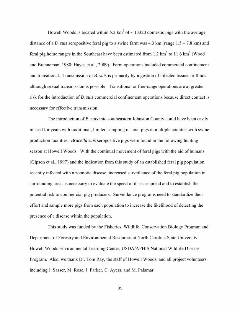
Howell Woods is located within 5.2 km2 of ~ 13320 domestic pigs with the average
distance of a B. suis seropositive feral pig to a swine farm was 4.3 km (range 1.5 – 7.8 km) and
feral pig home ranges in the Southeast have been estimated from 1.2 km2 to 11.6 km2 (Wood
and Brenneman, 1980; Hayes et al., 2009). Farm operations included commercial confinement
and transitional. Transmission of B. suis is primarily by ingestion of infected tissues or fluids,
although sexual transmission is possible. Transitional or free-range operations are at greater
risk for the introduction of B. suis commercial confinement operations because direct contact is
necessary for effective transmission.
The introduction of B. suis into southeastern Johnston County could have been easily
missed for years with traditional, limited sampling of feral pigs in multiple counties with swine
production facilities. Brucella suis seropositive pigs were found in the following hunting
season at Howell Woods. With the continual movement of feral pigs with the aid of humans
(Gipson et al., 1997) and the indication from this study of an established feral pig population
recently infected with a zoonotic disease, increased surveillance of the feral pig population in
surrounding areas is necessary to evaluate the speed of disease spread and to establish the
potential risk to commercial pig producers. Surveillance programs need to standardize their
effort and sample more pigs from each population to increase the likelihood of detecting the
presence of a disease within the population.
This study was funded by the Fisheries, Wildlife, Conservation Biology Program and
Department of Forestry and Environmental Resources at North Carolina State University,
Howell Woods Environmental Learning Center, USDA/APHIS National Wildlife Disease
Program. Also, we thank Dr. Tom Ray, the staff of Howell Woods, and all project volunteers
including J. Sasser, M. Rose, J. Parker, C. Ayers, and M. Palamar.

36
LITERATURE CITED
CAPUA, I., C. CASACCIA, G. CALZETTA, AND V. CAPORALE. 1997. Isolation and
characterization of an Aujeszky’s disease virus naturally infecting a wild boar (Sus
scrofa). Veterinary Microbiology 55: 141-146.
CAVENDISH, T., W. STIVER, AND E. K. DELOZIER. 2008. Disease surveillance of wild
hogs in Great Smoky Mountains National Park-a focus on pseudorabies. In
Proceedings: Wildlife Damage Management, 2008 National Conference on Feral Hogs,
Internet Center for National Conference on Feral Hogs, S. M. Vantassel (ed.). Missouri
Department of Conservation, St. Louis, MO, USA, pp. 1-10.
CENTERS FOR DISEASE CONTROL AND PREVENTION. 1994. Brucellosis outbreak at a
pork processing plant -- North Carolina, 1992. Morbidity and Mortality Weekly Report
43: 113-116.
CENTERS FOR DISEASE CONTROL AND PREVENTION. 2009. Brucella suis infection
associated with feral swine hunting - three states, 2007-2008. Morbidity and Mortality
Weekly Report 58: 618-621.
CLAY, W. H. 2007. Hogs gone wild. Human-Wildlife Conflicts 1: 137-138.
CORN, J.L., P. K. SWIDEREK, B. O. BLACKBURN, G. A. ERICKSON, A. B.
THIERMANN, AND V. F. NETTLES. 1986. Survey of selected diseases in wild swine
in Texas. Journal of the American Veterinary Medical Association 189: 1029-1032.
CORN, J. L., J. C. CUMBEE, R. BARFOOT, AND G. A. ERICKSON. 2009. Pathogen
exposure in feral swine populations geographically associated with high densities of
transitional swine premises and commercial swine production. Journal of Wildlife
Diseases 45: 713-721.

37
DECKER, E. 1978. Exotics. Pages 249-256 in J. L. Schmidt and D. L. Gilbert, editors. Big
game of North America: Ecology and management. Stackpole Books. Harrisburg, PA,
USA.
DESROSIERS, R. 2007. Overview of PCVD – the disease in Eastern Canada and US vs.
Europe. Proceedings of the 2007 Banff Pork Seminar, Alberta, Canada, pp. 35–48.
DORR, P. M., R. B. BAKER, G. W. ALMOND, S. R. WAYNE, AND W. A. GEBREYES.
2007. Epidemiologic assessment of porcine circovirus type 2 coinfection with other
pathogens in swine. Journal of the American Veterinary Medical Association 230: 244–
250.
ELLIS, J., L. HASSARD, E. CLARK, J. HARDING, G. ALLAN, P. WILLSON, J.
STROKAPPE, K. MARTIN, F. MCNEILLY, B. MEEHAN, D. TODD, AND D.
HAINES. 1998. Isolation of circovirus from lesions of pigs with postweaning
multisystemic wasting syndrome. Canadian Veterinary Journal 39: 44-51.
ELLIS, J., M. SPINATO, C. YONG, K. WEST, F. MCNEILLY, B. MEEHAN, S. KENNEDY,
E. CLARK, S. KRAKOWKA, AND G. ALLAN. 2003. Porcine circovirus 2-associated
disease in Eurasian wild boar. Journal of Veterinary Diagnostic Investigation 15: 364-
368
EVANS, A. C. 1947. Brucellosis in the United States. American Journal of Public Health 37:
139-151.
GIPSON, P. S., B. HLAVACHICK, T. BERGER, AND C. D. LEE 1997. Explanations for
recent range expansions by wild hogs into midwestern states. Great Plains Wildlife
Damage Control Workshop 13: 148-150.

38
GRESHAM, C. S., C. A. GRESHAM, M. J. DUFFY, C. T. FAULKNER, AND S. PATTON.
2002. Increased prevalence of Brucella suis and pseudorabies virus antibodies in adults
of an isolated feral swine population in Coastal South Carolina. Journal of Wildlife
Diseases 38: 653-656.
HANLEY, J. A., AND A. LIPPMAN-HAND. 1983. If nothing goes wrong, is everything all
right? Interpreting zero numerators. Journal of the American Medical Association 249:
1743–1745.
HARDING, J. C. S. 2004. The clinical expression and emergence of porcine circovirus 2.
Veterinary Microbiology 98: 131-135.
HAYES, R., S. RIFFELL, R. MINNIS, AND B. HOLDER. 2009. Survival and habitat use of
feral hogs in Mississippi. Southeastern Naturalist 8: 411-426.
KIJLSTRA, A., O. A. EISSEN, J. CORNELISSEN, K. MUNNIKSMA, I. EIJCK, AND T.
KORTBEEK. 2004. Toxoplasma gondii infection in animal-friendly pig production
systems. Investigative Ophthalmology and Visual Science 45: 3165-3169.
KIRKPATRICK, C. M., C. L. KANITZ, AND S. M. McCROCKLIN. 1980. Possible role of
wild mammals in transmission of pseudorabies to swine. Journal of Wildlife Diseases
16: 601-614.
MATSCHKE, G. H. 1967. Aging European wild hogs by dentition. Journal of Wildlife
Management 31: 109-113.
MCKNIGHT, T. 1964. Feral livestock in Anglo-America. University of California Publisher of
Geology Number 16. Berkley, CA, USA.
NETTLES, V. F., AND G. A. ERICKSON. 1984. Pseudorabies in wild swine. Proceedings of
the United States Animal Health Association 88: 505–506.

39
[NCACS] North Carolina Department of Agriculture and Consumer Services. 2005.
Agriculture Statistics Statewide Summary.
PIRTLE, E.C., J. M. SACKS, V. F. NETTLES, AND E. A. ROLLOR, III. 1989. Prevalence
and transmission of pseudorabies virus in an isolated population of feral swine. Journal
of Wildlife Diseases 25: 605-607.
ROMERO, C. H., P. MEADE, J. SANTAGATA, K. GILLIS, G. LOLLIS, E. C. HAHN, AND
E. P. J. GIBBS. 1997. Genital infection and transmission of pseudorabies virus in feral
swine in Florida, USA. Veterinary Microbiology 55: 131-139.
RUIZ-FONS, F, J. SEGALES, AND C. GORTAZAR. 2008a. A review of viral diseases of the
European wild boar: Effects of population dynamics and reservoir role. The Veterinary
Journal 176: 158-169.
RUIZ-FONS, F., D. VIDAL, J. VINCENTE, P. ACEVEDO, I. G. FERNANDEZ DE MERA,
V. MONTORO, AND C. GORTAZAR. 2008b. Epidemiological risk factors of
Aujeszky’s disease in wild boars (Sus scrofa) and domestic pigs in Spain. European
Journal of Wildlife Research 54: 549–555.
SANCHEZ, R., H. NAUWYNCK, AND M. PENSAERT. 2001. Serological survey of porcine
circovirus 2 antibodies in domestic and feral pig populations in Belgium. In
Proceedings of the ssDNA Viruses of Plants, Birds, Pigs and Primates (ESVV)
Meeting, Saint-Malo, France. Ploufragan, France: AFSSA.
SEWARD, N. W., K. C. VERCAUTEREN, G. W. WITMER, AND R. M. ENGEMAN. 2004.
Feral swine impacts on agriculture and the environment. Sheep and Goat Research
Journal 19: 34-40.

40
STALLKNECHT, D. E. AND E. W. HOWERTH. 2001. Pseudorabies (Aujeszky's disease).
Pages 164–170. in Williams, E. S. and I. K. Barker, editors. Infectious diseases of wild
mammals. Iowa State University Press. Ames, Iowa, USA.
STARNES, C. T., R. TALWANI, J. A. HORVATH, W. A. DUFFUS, AND C. S. BRYAN.
2004. Brucellosis in two hunt club members in South Carolina. Journal of the South
Carolina Medical Association 100: 113-115.
STOFFREGEN, W. C., S. C. OLSEN, C. J. WHEELER, B. J. BRICKER, M. V. PALMER, A.
E. JENSEN, S. M. HALLING, AND D. P. ALT. 2007. Diagnostic characterization of a
feral swine herd enzootically infected with Brucella. Journal of Veterinary Diagnostic
Investigation 19: 227-237.
TOWNE, C. W., AND E. N. WENTWORTH. 1950. Pigs from cave to cornbelt. University of
Oklahoma Press, Norman, OK. 305 pp.
TRAINER, D. O., AND L. KARSTAD. 1963. Experimental pseudorabies in some wild North
American mammals. Zoonoses Research 2: 135-151.
[USDA] United States Department of Agriculture. 2005. Feral/wild pigs: Potential problems
for farmers and hunters. United States Department of Agriculture Animal and Plant
Health Inspection Service Agriculture Information Bulletin No. 799.
VAN DER GIESSEN, J., M. FONVILLE, M. BOUWKNEGT, AND A. VOLLEMA. 2007.
Seroprevalence of Trichinella spiralis and Toxoplasma gondii in pigs from different
housing systems in the Netherlands. Veterinary Parasitology 148: 371-374.

41
VAN DER LEEK, M. L., H. N. BECKER, E. C. PIRTLE, P. HUMPHREY, C. L. ADAMS, B.
P., ALL, G. A. ERICKSON, R. C. BELDEN, W. B. FRANKENBERGER, AND E. P.
J. GIBBS. 1993. Prevalence of pseudorabies (Aujeszky's disease) virus antibodies in
feral swine in Florida. Journal of Wildlife Diseases 29: 403-406.
WITMER, G. W., R. B. SANDERS, AND A. C. TAFT. 2003. Feral swine-are they a disease
threat to livestock in the United States? Proceedings of the Wildlife Damage
Management Conference 10: 316-325.
WOOD, G. W., AND R. E. BRENNEMAN. 1980. Feral Hog movements and habitat use in
coastal South Carolina. Journal of Wildlife Management 44: 420–427.
WOOD, G. W., J. B. HENDRICKS, AND D. E. GOODMAN. 1976. Brucellosis in feral swine.
Journal of Wildlife Diseases 12: 579-582.
WRIGHT, J. C., AND D. G. THAWLEY. 1980. Role of the raccoon in the transmission of
pseudorabies: A field and laboratory investigation. American Journal of Veterinary
Research 41: 581-583.
ZYGMONT, S. M., V. F. NETTLES, E. B. SHOTTS, Jr., W. A. CARMEN, AND B. O.
BLACKBURN. 1982. Brucellosis in wild swine: a serologic and bacteriologic survey in
the southeastern United States and Hawaii. Journal of the American Veterinary Medical
Association 181: 1285-1287.

42
Table 2.1. Age and gender of all feral pigs collected in North Carolina*, 2007-2009.
Year Adult Sub‐adult Juvenile Unknown Male Female
2007 77 3 170 0 126 124
2008 57 93 28 7 95 90
2009 36 42 24 0 49 53
Total 170 138 222 7 270 267
*Includes feral pigs from Howell Woods.

43
Table 2.2. Summary of feral pigs tested for antibodies organized by year, county, and disease,
North Carolina 2007-2009.
*Howell Woods’ is located within Johnston County but the results from Howell Woods are
reported separately.
xSB refers to Brucella suis.
Year County SBx test SBx pos PRV test PRV pos CSF test CSF pos
2007 Bertie 1 0 1 0 1 0
Bladen 11 0 11 0 11 0
Caswell 195 0 195 0 0 0
Duplin 19 0 19 0 17 0
Johnston* 6 0 6 0 6 0
Pitt 5 0 5 0 5 0
Wayne 9 0 9 0 9 0
2008 Bertie 13 0 13 0 14 0
Bladen 47 0 47 0 52 0
Caswell 17 0 17 0 0 0
Columbus 10 0 10 0 14 0
Johnston* 23 0 23 0 22 0
Richmond 2 0 2 0 0 0
2009 Bladen 19 0 18 0 19 0
Columbus 7 0 7 0 7 0
Craven 5 0 5 0 6 0
Johnston* 8 0 8 0 8 0
Pender 12 0 12 0 12 0
Robeson 2 0 2 0 2 0
Sampson 11 0 11 0 11 0
Totals 422 0 421 0 216 0

44
Table 2.3. Serosurveillance results of feral pigs harvested from Howell Woods, Four Oaks,
North Carolina 2007-2009.
Season Harvest Site SB test
SB pos
PRV test
PRV pos
CSF test
CSF pos
PCV-2 test
PCV-2 pos
1 Howell Woods
35 0 34 0 - - 50 33
2 Howell Woods
27 6 27 0 20 0 40 20
Total 62 6 61 0 20 0 90 53
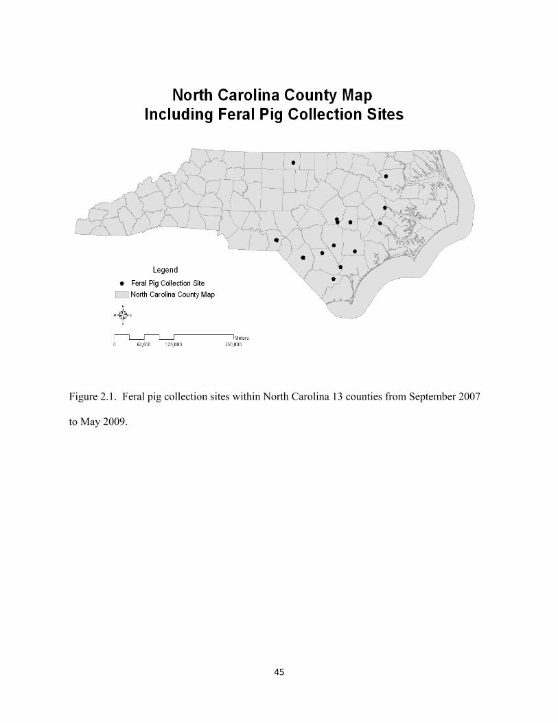
45
Figure 2.1. Feral pig collection sites within North Carolina 13 counties from September 2007
to May 2009.