Embed Size (px)
Citation preview
Complete replication of hepatitis B virus and hepatitisC virus in a newly developed hepatoma cell lineDarong Yanga, Chaohui Zuob, Xiaohong Wanga, Xianghe Menga, Binbin Xuea, Nianli Liua, Rong Yua, Yuwen Qina,Yimin Gaoa, Qiuping Wanga, Jun Hub, Ling Wangc, Zebin Zhoud, Bing Liud, Deming Tanc, Yang Guand,and Haizhen Zhua,b,1
aDepartment of Molecular Medicine, College of Biology, State Key Laboratory of Chemo/Biosensing and Chemometrics, Hunan University, Changsha 410082,China; bResearch Center of Cancer Prevention and Treatment, Translational Medicine Research Center of Liver Cancer, Hunan Provincial Tumor Hospital,affiliated with the Tumor Hospital of Xiangya Medical School of Central South University, Changsha 410013, China; cDepartment of Infectious Disease ofXiangya Hospital, Central South University, Changsha 410008, China; and dDepartment of Pathology of Tongji Medical College, Huazhong University ofScience and Technology, Wuhan 430030, China
Edited by Peter Palese, Icahn School of Medicine at Mount Sinai, New York, NY, and approved January 29, 2014 (received for review October 29, 2013)
The absence of a robust cell culture system for hepatitis B virus(HBV) and hepatitis C virus (HCV) infection has limited the analysisof the virus lifecycle and drug discovery. We have established ahepatoma cell line, HLCZ01, the first cell line, to the authors’ knowl-edge, supporting the entire lifecycle of both HBV and HCV. HBVsurface antigen (HBsAg)-positive particles can be observed in thesupernatant and the lumen of the endoplasmic reticulum of thecells via electron microscopy. Interestingly, HBV and HCV clinicalisolates propagate in HLCZ01 cells. Both viruses replicate in thecells without evidence of overt interference. HBV and HCV entryare blocked by antibodies against HBsAg and human CD81, re-spectively, and the replication of HBV and HCV is inhibited byantivirals. HLCZ01 cells mount an innate immune response to virusinfection. The cell line provides a powerful tool for exploring themechanisms of virus entry and replication and the interaction be-tween host and virus, facilitating the development of novel antiviralagents and vaccines.
cell culture model | primary human hepatocytes | cccDNA | interferon |ISGs
More than 500 million people worldwide are persistentlyinfected with hepatitis B virus (HBV) and/or hepatitis B
virus (HCV) and are at risk of developing chronic liver diseases(1). There is no vaccine against HCV, and many patients who arepersistently infected by HBV or HCV do not respond to cur-rently available therapies (2, 3). Improved understanding of thebiology and pathogenesis of these infections is required for thedevelopment of vaccine and antiviral drugs (4). The inability togrow HBV and HCV efficiently in cell culture has presenteda major obstacle to understanding the virus lifecycle and path-ogenesis and to developing improved therapeutics.HBV is a member of the hepadnavirus families, and its genome
is a relaxed circular, partially double-stranded DNA molecule.The negative strand has an invariable length of ∼3.2 kb, and thepositive strand is 50–100% of this length. Several key issues aboutthe biology of HBV remain to be explored, including the identi-fication of the cellular receptors, the role of the X gene, and themechanisms by which the viral minichromosome is formed. Co-valently closed circular DNA (cccDNA) is responsible for theestablishment of viral infection and persistence. Understandingthe mechanisms underlying cccDNA formation and regulation iscritical for understanding the HBV pathogenesis and findinga cure for hepatitis B. HepG2.2.15 cells derived from the hepa-toma cell line HepG2 transfected with the full genome of HBVhave been used to study HBV replication (5). Primary humanhepatocytes (PHH) are susceptible to HBV infection (6, 7), butthe use of this model is hampered by the limited availability andunpredictable variability of human liver. Several human hepa-toma cell lines support HBV replication after HBV DNAtransfection, and overexpression of sodium-taurocholate cotrans-porting polypeptide (NTCP) in HepG2 and Huh7 cells can render
these cells able to support HBV produced in cell culture at lowefficiency (5, 8, 9). HepaRG is a hepatoma cell line that is sus-ceptible to HBV infection (10), but the susceptibility of HepaRGcells to HBV is strictly dependent on the differentiation stateinduced by DMSO, causing variable toxic side effects and irre-producible results for HBV replication levels. Moreover, poorviral replication, low viral yields, the absence of reinfection, andlack of cccDNA amplification in HepaRG cells make the study ofHBV lifecycle and pathogenesis difficult (10). For reasons thatstill are unknown, HBV clinical isolates do not propagate incell culture.HCV is a positive-strand RNA virus of the Flaviviridae family.
Its genome encodes a large polyprotein that is processed to pro-duce viral structural proteins including core, E1, and E2 proteinsas well as nonstructural proteins consisting of p7, NS2, NS3,NS4A, NS4B, NS5A, and NS5B. Genotype 2a, 2b, and 1a isolateswere used to develop infectious cell culture systems for HCVstudy (11–15). However, other HCV genotypes cannot propagatein vitro. The permissive human hepatoma cell Huh7.5 may notmount an innate immune response to HCV infection, so it isdifficult to explore the interaction between the virus and host cells.Moreover, clinical isolates of HCV do not propagate in cell cul-ture. Although primary culture systems have been established forHCV (16, 17), these do not seem to extend the range of isolatesthat can be studied. We now report the establishment of a novelhepatoma cell line, HLCZ01, supporting the entire life cycles ofHBV and HCV. Interestingly, the cells can be infected by the serafrom patients with different HBV and HCV strains.
Significance
More than 500 million people are persistently infected withhepatitis B virus (HBV) and/or hepatitis C virus (HCV) and are ata risk of developing chronic hepatitis, cirrhosis, and liver can-cer. The absence of robust cell culture systems for both viralinfections limits the understanding of the virus lifecycle andpathogenesis required for the development of vaccine andantivirals. We have established a novel human hepatoma cellline termed “HLCZ01” that supports the entire lifecycle of bothHBV and HCV produced both in cell culture and clinically. Thiscell line provides a powerful tool for addressing the viruslifecycle and the development of antivirals and vaccines.
Author contributions: H.Z. designed research; D.Y., C.Z., X.W., X.M., B.X., N.L., R.Y., Y.Q.,Y. Gao, Q.W., J.H., L.W., Z.Z., B.L., D.T., and Y. Guan performed research; D.T. contributednew reagents/analytic tools; D.Y., X.M., B.X., and H.Z. analyzed data; and H.Z. wrotethe paper.
The authors declare no conflict of interest.
This article is a PNAS Direct Submission.1To whom correspondence should be addressed. E-mail: [email protected].
This article contains supporting information online at www.pnas.org/lookup/suppl/doi:10.1073/pnas.1320071111/-/DCSupplemental.
E1264–E1273 | PNAS | Published online March 10, 2014 www.pnas.org/cgi/doi/10.1073/pnas.1320071111
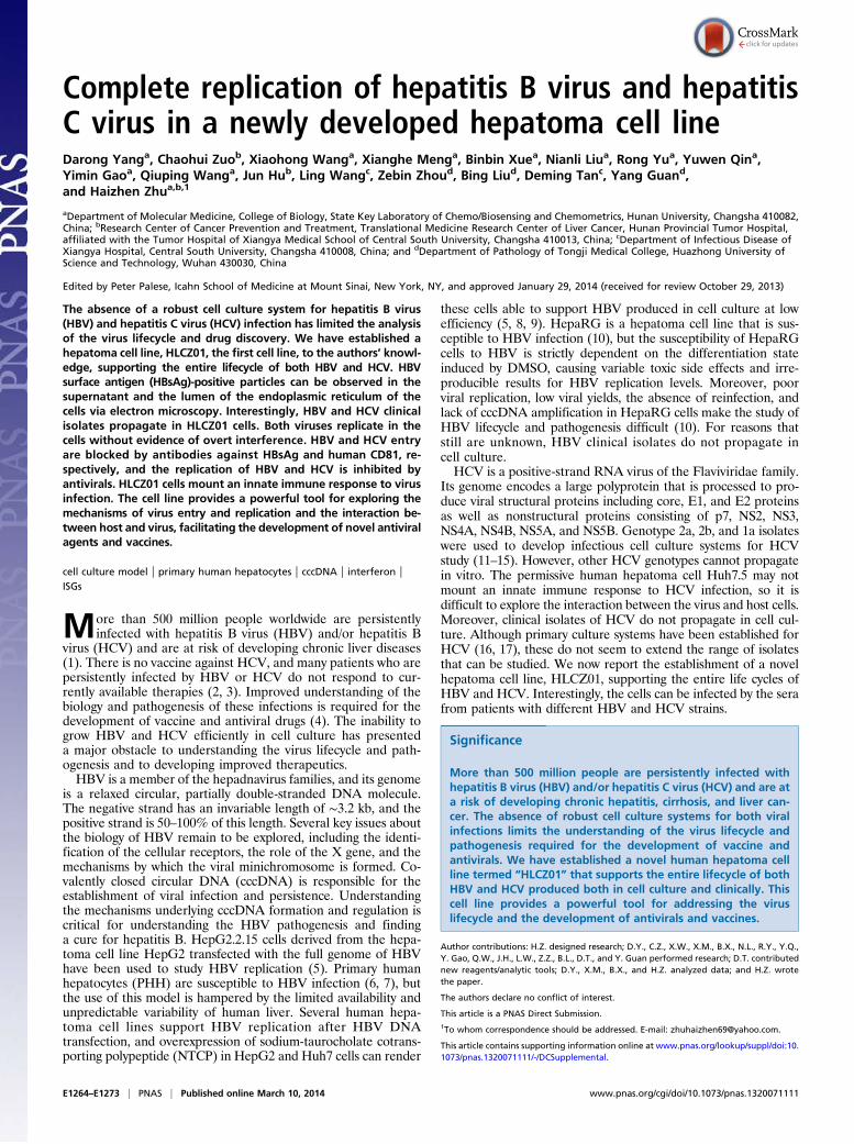
ResultsEstablishment of a Novel Hepatoma Cell Line, HLCZ01. To understandbetter the virus lifecycle and the interaction between host andvirus, we attempted to replicate HBV and HCV in hepatomacells, which have histological features characteristic of hepato-cellular carcinoma (Fig. 1A). Tumor tissue was harvested andcultured in DMEM/F12 medium, and several clones survived forlong-time culture. We One of these clones, HLCZ01 (Fig. 1B),was derived from the grade 2 differentiated hepatocellular car-cinoma of an HCV-infected male patient. To obtain hepatomacell lines, we injected clones into immunodeficient mice andcultured tumor tissues from the mice in DMEM/F12 medium.HLCZ01 cells express liver-specific genes, such as human albu-min (ALB), α1-antitrypsin (AAT), hepatocyte nuclear factor 4(HNF4), cytochrome P450 3A4 (CYP3A4), and microRNA122(miR-122) (Fig. 1C), and the liver-specific proteins human ALBand AAT (Fig. 1D). The presence of HCV RNA is not detectableby RT-PCR after the establishment of HLCZ01 cells in culture.
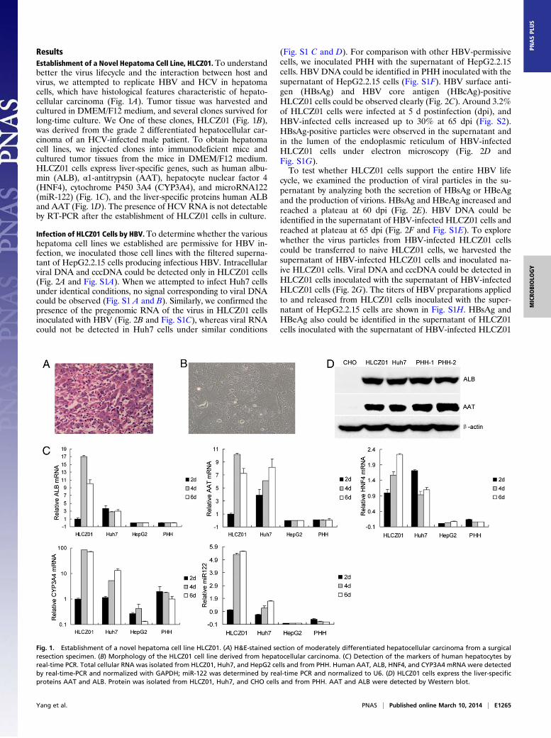
Infection of HLCZ01 Cells by HBV. To determine whether the varioushepatoma cell lines we established are permissive for HBV in-fection, we inoculated those cell lines with the filtered superna-tant of HepG2.2.15 cells producing infectious HBV. Intracellularviral DNA and cccDNA could be detected only in HLCZ01 cells(Fig. 2A and Fig. S1A). When we attempted to infect Huh7 cellsunder identical conditions, no signal corresponding to viral DNAcould be observed (Fig. S1 A and B). Similarly, we confirmed thepresence of the pregenomic RNA of the virus in HLCZ01 cellsinoculated with HBV (Fig. 2B and Fig. S1C), whereas viral RNAcould not be detected in Huh7 cells under similar conditions
(Fig. S1 C and D). For comparison with other HBV-permissivecells, we inoculated PHH with the supernatant of HepG2.2.15cells. HBV DNA could be identified in PHH inoculated with thesupernatant of HepG2.2.15 cells (Fig. S1F). HBV surface anti-gen (HBsAg) and HBV core antigen (HBcAg)-positiveHLCZ01 cells could be observed clearly (Fig. 2C). Around 3.2%of HLCZ01 cells were infected at 5 d postinfection (dpi), andHBV-infected cells increased up to 30% at 65 dpi (Fig. S2).HBsAg-positive particles were observed in the supernatant andin the lumen of the endoplasmic reticulum of HBV-infectedHLCZ01 cells under electron microscopy (Fig. 2D andFig. S1G).To test whether HLCZ01 cells support the entire HBV life
cycle, we examined the production of viral particles in the su-pernatant by analyzing both the secretion of HBsAg or HBeAgand the production of virions. HBsAg and HBeAg increased andreached a plateau at 60 dpi (Fig. 2E). HBV DNA could beidentified in the supernatant of HBV-infected HLCZ01 cells andreached at plateau at 65 dpi (Fig. 2F and Fig. S1E). To explorewhether the virus particles from HBV-infected HLCZ01 cellscould be transferred to naive HLCZ01 cells, we harvested thesupernatant of HBV-infected HLCZ01 cells and inoculated na-ive HLCZ01 cells. Viral DNA and cccDNA could be detected inHLCZ01 cells inoculated with the supernatant of HBV-infectedHLCZ01 cells (Fig. 2G). The titers of HBV preparations appliedto and released from HLCZ01 cells inoculated with the super-natant of HepG2.2.15 cells are shown in Fig. S1H. HBsAg andHBeAg also could be identified in the supernatant of HLCZ01cells inoculated with the supernatant of HBV-infected HLCZ01
Fig. 1. Establishment of a novel hepatoma cell line HLCZ01. (A) H&E-stained section of moderately differentiated hepatocellular carcinoma from a surgicalresection specimen. (B) Morphology of the HLCZ01 cell line derived from hepatocellular carcinoma. (C) Detection of the markers of human hepatocytes byreal-time PCR. Total cellular RNA was isolated from HLCZ01, Huh7, and HepG2 cells and from PHH. Human AAT, ALB, HNF4, and CYP3A4 mRNA were detectedby real-time-PCR and normalized with GAPDH; miR-122 was determined by real-time PCR and normalized to U6. (D) HLCZ01 cells express the liver-specificproteins AAT and ALB. Protein was isolated from HLCZ01, Huh7, and CHO cells and from PHH. AAT and ALB were detected by Western blot.
Yang et al. PNAS | Published online March 10, 2014 | E1265
MICRO
BIOLO
GY
PNASPL
US
cells (Fig. 2H). Taken together, these data indicate that HLCZ01cells support the entire HBV lifecycle.
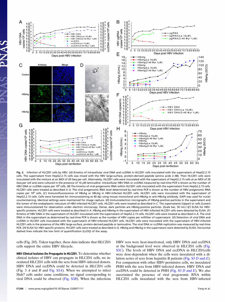
HBV Clinical Isolates Do Propagate in HLCZ01. To determine whetherclinical isolates of HBV can propagate in HLCZ01 cells, we in-oculated HLCZ01 cells with the sera from HBV-infected donors.HBV DNA and cccDNA could be detected in HLCZ01 cells(Fig. 3 A and B and Fig. S3A). When we attempted to infectHuh7 cells under same conditions, no signal corresponding toviral DNA could be observed (Fig. S3B). When the infectious
HBV sera were heat-inactivated, only HBV DNA and cccDNAat the background level were observed in HLCZ01 cells (Fig.S3C). The levels of HBV DNA and cccDNA in HLCZ01cellswere dose-dependent when the cells were inoculated with a di-lution series of sera from hepatitis B patients (Fig. S3 D and E).For comparison with other HBV-permissive cells, we inoculatedPHH with the sera from HBV-infected donors. HBV DNA andcccDNA could be detected in PHH (Fig. S3 D and E). We alsoascertained the presence of viral pregenomic RNA withinHLCZ01 cells inoculated with the sera from HBV-infected
Fig. 2. Infection of HLCZ01 cells by HBV. (A) Kinetics of intracellular viral DNA and cccDNA in HLCZ01 cells inoculated with the supernatant of HepG2.2.15cells. The supernatant from HepG2.2.15 cells was mixed with the HBV large-surface, protein-derived peptide (amino acids 2–48). Then HLCZ01 cells wereinoculated with the mixture at an MOI of 20 Geq per cell. Alternately, HLCZ01 cells were inoculated with the supernatant of HepG2.2.15 cells at an MOI of 20Geq per cell and were cultured in the presence of 10 μM lamivudine. Intracellular HBV DNA or cccDNA measured by real-time PCR is shown as the number ofHBV DNA or cccDNA copies per 106 cells. (B) The kinetics of viral pregenomic RNA within HLCZ01 cells inoculated with the supernatant from HepG2.2.15 cells.HLCZ01 cells were treated as described in A. The viral pregenomic RNA level determined by real-time PCR is shown as the number of HBV pregenomic RNAcopies per 106 cells. (C) Immunofluorescence of HBsAg or HBcAg in HBV-infected HLCZ01 cells. HLCZ01 cells were inoculated with the supernatant ofHepG2.2.15 cells. Cells were harvested for immunostaining at 40 dpi using mouse monoclonal anti-HBsAg or anti-HBcAg antibody. DAPI was used for nucleicounterstaining. Identical settings were maintained for image capture. (D) Immunoelectron micrographs of HBsAg-positive particles in the supernatant andthe lumen of the endoplasmic reticulum of HBV-infected HLCZ01 cells. HLCZ01 cells were treated as described in C. The supernatants (Upper) or cells (Lower)were immunostained for observation under electron microscopy. Dense, dark particles are HBsAg-positive particles. (Scale bar, 50 nm.) (E) ELISA for HBV-specific proteins. HLCZ01 cells were treated as described in A. HBsAg and HBeAg in the supernatant of HBV-infected HLCZ01 cells were detected by ELISA. (F)Kinetics of HBV DNA in the supernatant of HLCZ01 inoculated with the supernatant of HepG2.2.15 cells. HLCZ01 cells were treated as described in A. The viralDNA in the supernatant as determined by real-time PCR is shown as the number of HBV copies per milliliter of supernatant. (G) Detection of viral DNA andcccDNA in HLCZ01 cells inoculated with the supernatant of HBV-infected HLCZ01 cells. HLCZ01 cells were inoculated with the supernatant of HBV-infectedHLCZ01 cells in the presence of the HBV large-surface, protein-derived peptide or lamivudine. The viral DNA or cccDNA replication was measured by real-timePCR. (H) ELISA for HBV-specific proteins. HLCZ01 cells were treated as described in G. HBsAg and HBeAg in the supernatant were detected by ELISA. Horizontaldashed lines indicate the low limit of quantification (LLOQ) of the assay.
E1266 | www.pnas.org/cgi/doi/10.1073/pnas.1320071111 Yang et al.
donors (Fig. 3C and Fig. S3F). Similar to viral DNA, viral RNAwas undetectable in Huh7 cells inoculated with HBV sera (Fig.S3F). Flow cytometry results showed that HBsAg-positiveHLCZ01 cells increased from ∼3.5% at 7 dpi to 7% at 10 dpi(Fig. S3G), and when the cells were inoculated with the serum ofpatient #2465, which had high viral titer, HBsAg-positiveHLCZ01 cells increased from 8.7% at 7 dpi to 10.9% at 10 dpi(Fig. S3G). HBsAg could be found in the supernatant ofHLCZ01 cells inoculated with the sera from hepatitis B patients(Fig. 3D). Moreover, viral particles in the supernatant ofHLCZ01 cells infected by HBV sera could be transferred tonaive HLCZ01 cells (Fig. 3E). However, when we attempted toinfect Huh7 cells under similar conditions, no signal corre-sponding to viral DNA could be observed (Fig. S3H). Thesefindings clearly show that HBV clinical isolates can propagate inHLCZ01 cells.
Inhibition of HBV Infection in HLCZ01 Cells by Antiviral Drugs in Vitroand in Vivo. To verify further that infection of HLCZ01 cells byHBV follows the authentic entry pathway, we incubated HBVwith various concentrations of the anti-HBsAg antibody beforeincubation with HLCZ01 cells. Anti-HBsAg antibody blockedviral infection (Fig. 4A). In addition, we examined the inhibitioneffect of myristoylated peptide representing the N-terminal 48amino acids of the HBV large-surface protein required for HBVinfectivity. As expected, HBV infection could be inhibited inHLCZ01 cells when the peptide was added during the infectionprocess (Figs. 2 and 4B). One recent study showed that NTCP isa putative HBV receptor (9). The expression of NTCP protein inHLCZ01 and PHH is comparable (Fig. S4A). Silencing of NTCPinhibited HBV infection in HLCZ01 cells (Fig. S4 B and C). Aprevious study indicated that IFN-α inhibits HBV transcriptionand replication in vitro and in vivo (18), so we examined the
Fig. 3. HBV clinical isolates do propagate in HLCZ01. (A and B) HBV clinical isolates propagate in HLCZ01. HLCZ01 cells were inoculated with the sera fromhepatitis B patients in the presence of 10 μM lamivudine. Total cellular DNA was isolated. Real-time PCR was performed for HBV DNA (A) and cccDNA (B). Theviral DNA or cccDNA replication is shown as the number of HBV or cccDNA copies per 106 cells. (C) Viral pregenomic RNA is present in HLCZ01 cells inoculatedwith the sera from HBV-infected donors. HLCZ01 cells were treated as described in A. Intracellular HBV pregenomic RNA measured by real-time PCR is shownas the number of HBV pregenomic RNA copies per 106 cells. (D) HBsAg is detected in the supernatant of sera-infected HLCZ01 cells. HLCZ01 cells were treatedas described in A. HBsAg in the supernatant of HLCZ01 cells at 17 d pi was detected by ELISA. (E) Viral particles in the supernatant of HBV sera-infected HLCZ01cells can be transferred to naive HLCZ01 cells. HLCZ01 cells were treated as described in A. HLCZ01 cells were inoculated with the supernatant of HBV sera-infected HLCZ01 cells in the presence of lamivudine. Cellular DNA was isolated, and real-time PCR for HBV DNA was performed. The viral DNA replication isshown as the number of HBV copies per 106 cells. Horizontal dashed lines indicate the LLOQ of the assay.
Yang et al. PNAS | Published online March 10, 2014 | E1267
MICRO
BIOLO
GY
PNASPL
US
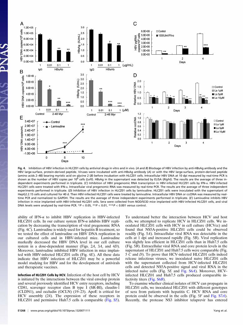
ability of IFN-α to inhibit HBV replication in HBV-infectedHLCZ01 cells. In our culture system IFN-α inhibits HBV repli-cation by decreasing the transcription of viral pregenomic RNA(Fig. 4C). Lamivudine is widely used for hepatitis B treatment, sowe tested the effect of lamivudine on HBV DNA replication inour cultured cells and in HBV-infected mice. Lamivudinemarkedly decreased the HBV DNA level in our cell culturesystem in a dose-dependent manner (Figs. 2A, 3A, and 4D).Moreover, lamivudine inhibited HBV infection in mice implan-ted with HBV-infected HLCZ01 cells (Fig. 4E). All these dataindicate that HBV infection of HLCZ01 may be a powerfulmodel studying for HBV and for the development of antiviralsand therapeutic vaccines.
Infection of HLCZ01 Cells by HCV. Infection of the host cell by HCVis initiated by the interactions between the viral envelop proteinand several previously identified HCV entry receptors, includingCD81, scavenger receptor class B type I (SR-BI), claudin-1(CLDN1), and occludin (OCLN) (19–23). ApoE is critical forHCV assembly (24). The expression of these receptors inHLCZ01 and permissive Huh7.5 cells is comparable (Fig. S5).
To understand better the interaction between HCV and hostcells, we attempted to replicate HCV in HLCZ01 cells. We in-oculated HLCZ01 cells with HCV in cell culture (HCVcc) andfound that NS5A-positive HLCZ01 cells could be observedreadily (Fig. 5A). Intracellular viral RNA was detectable in thecells at 1 dpi and increased rapidly (Fig. 5B). Viral replicationwas slightly less efficient in HLCZ01 cells than in Huh7.5 cells(Fig. 5B). Extracellular viral RNA and core protein levels in thesupernatant of HLCZ01 and Huh7.5 cells were comparable (Fig.5 C and D). To prove that HCV-infected HLCZ01 cells indeedrelease infectious viruses, we inoculated naive HLCZ01 cellswith the supernatant collected from HCV-infected HLCZ01cells and detected NS5A-positive signal and viral RNA in theinfected naive cells (Fig. 5E and Fig. S6A). Moreover, HCV-infected HLCZ01 and Huh7.5 cells produced comparable in-fectivity titers (Fig. S6B).To examine whether clinical isolates of HCV can propagate in
HLCZ01 cells, we inoculated HLCZ01 with different genotypesof sera from patients with hepatitis C. HCV RNA and coreprotein could be observed in the cells (Fig. 5F and Fig. S7A).Recently, the protease NS3 inhibitor telaprevir has entered
Fig. 4. Inhibition of HBV infection in HLCZ01 cells by antiviral drugs in vitro and in vivo. (A and B) Blockage of HBV infection by anti-HBsAg antibody and theHBV large-surface, protein-derived peptide. Viruses were incubated with anti-HBsAg antibody (A) or with the HBV large-surface, protein-derived peptide(amino acids 2–48) bearing myristic acid on glycine 2 (B) before incubation with HLCZ01 cells. Intracellular HBV DNA at 10 dpi measured by real-time PCR isshown as the number of HBV copies per 106 cells (Left). HBsAg in the supernatant was detected by ELISA (Right). The results are the average of three in-dependent experiments performed in triplicate. (C) Inhibition of HBV pregenomic RNA transcription in HBV-infected HLCZ01 cells by IFN-α. HBV-infectedHLCZ01 cells were treated with IFN-α. Intracellular viral pregenomic RNA was measured by real-time PCR. The results are the average of three independentexperiments performed in triplicate. (D) Inhibition of HBV infection in HLCZ01 cells by lamivudine. HLCZ01 cells were inoculated with the supernatant ofHepG2.2.15 cells and cultured for 40 d. Then HBV-infected HLCZ01 cells were treated by lamivudine. Intracellular HBV DNA or cccDNA was measured by real-time PCR and normalized to GAPDH. The results are the average of three independent experiments performed in triplicate. (E) Lamivudine inhibits HBVinfection in mice implanted with HBV-infected HLCZ01 cells. Sera were collected from NOD/SCID mice implanted with HBV-infected HLCZ01 cells, and viralDNA levels were analyzed by real-time PCR. *P < 0.05, **P < 0.01, ***P < 0.001 versus control.
E1268 | www.pnas.org/cgi/doi/10.1073/pnas.1320071111 Yang et al.
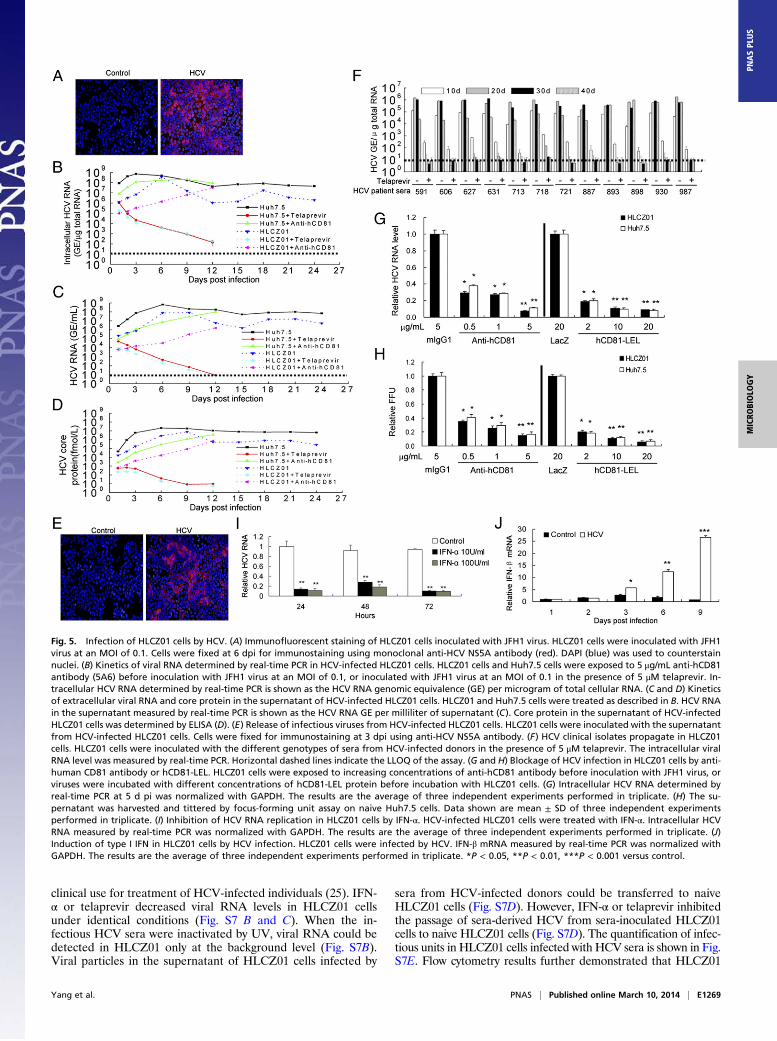
clinical use for treatment of HCV-infected individuals (25). IFN-α or telaprevir decreased viral RNA levels in HLCZ01 cellsunder identical conditions (Fig. S7 B and C). When the in-fectious HCV sera were inactivated by UV, viral RNA could bedetected in HLCZ01 only at the background level (Fig. S7B).Viral particles in the supernatant of HLCZ01 cells infected by
sera from HCV-infected donors could be transferred to naiveHLCZ01 cells (Fig. S7D). However, IFN-α or telaprevir inhibitedthe passage of sera-derived HCV from sera-inoculated HLCZ01cells to naive HLCZ01 cells (Fig. S7D). The quantification of infec-tious units in HLCZ01 cells infected with HCV sera is shown in Fig.S7E. Flow cytometry results further demonstrated that HLCZ01
Fig. 5. Infection of HLCZ01 cells by HCV. (A) Immunofluorescent staining of HLCZ01 cells inoculated with JFH1 virus. HLCZ01 cells were inoculated with JFH1virus at an MOI of 0.1. Cells were fixed at 6 dpi for immunostaining using monoclonal anti-HCV NS5A antibody (red). DAPI (blue) was used to counterstainnuclei. (B) Kinetics of viral RNA determined by real-time PCR in HCV-infected HLCZ01 cells. HLCZ01 cells and Huh7.5 cells were exposed to 5 μg/mL anti-hCD81antibody (5A6) before inoculation with JFH1 virus at an MOI of 0.1, or inoculated with JFH1 virus at an MOI of 0.1 in the presence of 5 μM telaprevir. In-tracellular HCV RNA determined by real-time PCR is shown as the HCV RNA genomic equivalence (GE) per microgram of total cellular RNA. (C and D) Kineticsof extracellular viral RNA and core protein in the supernatant of HCV-infected HLCZ01 cells. HLCZ01 and Huh7.5 cells were treated as described in B. HCV RNAin the supernatant measured by real-time PCR is shown as the HCV RNA GE per milliliter of supernatant (C). Core protein in the supernatant of HCV-infectedHLCZ01 cells was determined by ELISA (D). (E) Release of infectious viruses from HCV-infected HLCZ01 cells. HLCZ01 cells were inoculated with the supernatantfrom HCV-infected HLCZ01 cells. Cells were fixed for immunostaining at 3 dpi using anti-HCV NS5A antibody. (F) HCV clinical isolates propagate in HLCZ01cells. HLCZ01 cells were inoculated with the different genotypes of sera from HCV-infected donors in the presence of 5 μM telaprevir. The intracellular viralRNA level was measured by real-time PCR. Horizontal dashed lines indicate the LLOQ of the assay. (G and H) Blockage of HCV infection in HLCZ01 cells by anti-human CD81 antibody or hCD81-LEL. HLCZ01 cells were exposed to increasing concentrations of anti-hCD81 antibody before inoculation with JFH1 virus, orviruses were incubated with different concentrations of hCD81-LEL protein before incubation with HLCZ01 cells. (G) Intracellular HCV RNA determined byreal-time PCR at 5 d pi was normalized with GAPDH. The results are the average of three independent experiments performed in triplicate. (H) The su-pernatant was harvested and tittered by focus-forming unit assay on naive Huh7.5 cells. Data shown are mean ± SD of three independent experimentsperformed in triplicate. (I) Inhibition of HCV RNA replication in HLCZ01 cells by IFN-α. HCV-infected HLCZ01 cells were treated with IFN-α. Intracellular HCVRNA measured by real-time PCR was normalized with GAPDH. The results are the average of three independent experiments performed in triplicate. (J)Induction of type I IFN in HLCZ01 cells by HCV infection. HLCZ01 cells were infected by HCV. IFN-β mRNA measured by real-time PCR was normalized withGAPDH. The results are the average of three independent experiments performed in triplicate. *P < 0.05, **P < 0.01, ***P < 0.001 versus control.
Yang et al. PNAS | Published online March 10, 2014 | E1269
MICRO
BIOLO
GY
PNASPL
US
cells can be infected by sera from hepatitis C patients (Fig. S7F).All these data suggest that HLCZ01 cells indeed can be infectedby HCVcc and by the sera from hepatitis C patients.It has been reported that human CD81 is an essential coreceptor
for HCV entry (19). To provide evidence that the uptake ofHCV into HLCZ01 cells follows the authentic entry pathway, weperformed infection competition assays using mouse monoclonalanti-CD81 antibody or soluble CD81 large extracellular loop(hCD81-LEL). HCVcc infectivity could be blocked with eitheranti-CD81 antibody or hCD81-LEL (Fig. 5 G and H), indicatingthat the virus enters via the authentic HCV entry pathway.The current standard of care for chronic hepatitis C involves
IFN-α–based therapy. We found that IFN-α significantly inhibi-ted HCV RNA replication in HLCZ01 cells (Fig. 5I and Fig.S7B), demonstrating that HLCZ01 cells infected by HCV may beuseful for testing novel drugs.To understand better the interaction between HCV and host
cells, we infected HLCZ01 cells with HCVcc and tested the in-nate immune response in the cells. IFN-β and IFN-stimulatedgenes (ISGs) were induced in viral-infected cells (Fig. 5J and Fig.S8), indicating that HLCZ01 cells mount an innate immune re-sponse to HCV infection.
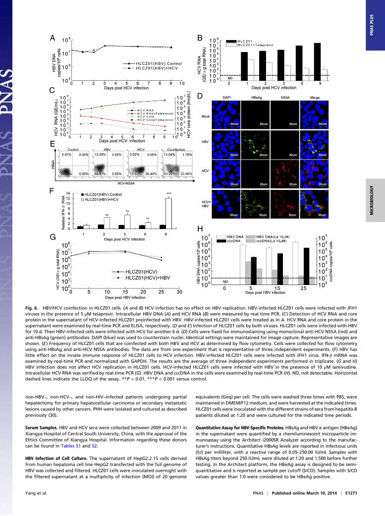
HBV/HCV Coinfection in HLCZ01 Cells. HBV/HCV coinfection is com-mon, with an estimated 7–20 million individuals affected world-wide. Patients with HBV/HCV coinfection have an increased riskfor cirrhosis, hepatocellular carcinoma, and death (26). The vi-rological and molecular aspects of HBV/HCV coinfection arepoorly understood. The lack of appropriate model systems hasmade the study of the interactions between HBV and HCV diffi-cult. Our novel cell culture system allows us to investigate the in-teractions between HBV and HCV. HCV infection did not affectHBV replication in HLCZ01 cells (Fig. 6 A and B and Fig. S9A).HCV RNA and core protein in the supernatant of the cells couldbe verified (Fig. 6C). Interestingly, HBV and HCV infected thesame cells (Fig. 6 D and E and Fig. S9A), providing new insightsinto the pathogenesis of HBV/HCV coinfection. The frequency ofcoinfected cells increased when HLCZ01 cells were infected withHBV and HCV at higher virus titers (Fig. S9A). Moreover, HBVhad little effect on the innate host response of HLCZ01 cells toHCV infection (Fig. 6F). HBV infection did not affect HCV rep-lication in HLCZ01 cells (Fig. 6G). HBV infection was confirmedin the cells (Fig. 6H). Finally, simultaneous infection of HLCZ01cells by HBV and HCV did not affect either HBV (Fig. S9B) orHCV (Fig. S9C) replication. These data indicate that HBV andHCV replicate in the same cells without evidence of direct inter-ference in vitro (27). Superinfection exclusion generally is restrictedto homologous viruses (28), whereas nonrelated viruses are ableto replicate normally. In agreement with the general concept, ourresults showed that there is no superinfection exclusion of HBVby HCV or of HCV by HBV.
DiscussionWe have established that HLCZ01 is a robust cell culture modelof HBV infection by showing the kinetics of several markers ofviral infection, including viral DNA replication, the formationand amplification of cccDNA, newly synthesized pregenomicviral RNA, the secretion of HBsAg and HBeAg, and the pro-duction and release of infectious viral particles from HBV-infected HLCZ01 cells. In addition, evidence that HBV infectionis blocked by specific anti-HBsAg antibody or by pre-S1–block-ing peptide strongly indicates that HBV infection of HLCZ01cells follows the authentic entry pathway and that the process ofviral adsorption and entry of HBV can be studied in this system.That the expression of NTCP protein is comparable in HLCZ01,HepG2, and Huh7 cells and in PHH but only HLCZ01 and PHHare susceptible to HBV infection suggests that other HBVreceptors exist. Our data show that HBV infection in HLCZ01
cells results in the formation of foci of infected cells and that thepercentage of HBV-infected cells increases, indicating that HBVmay spread via cell-to-cell transmission and/or by attaching pref-erentially to the adjacent cells after secretion. Interestingly, HBVclinical isolates can propagate HLCZ01 cells, providing a veryuseful tool for the analysis of clinical isolates of HBV and forthe development of antiviral drugs and vaccines. The HLCZ01 cellline provides a powerful tool for improving our understanding ofthe HBV life cycles, including the identification of the still un-known receptors and the mechanisms by which cccDNA is formedand amplified.The HLCZ01 cell line also is susceptible to HCV infection, as
shown by the kinetics of intracellular viral RNA replication, theexpression of viral protein, and the production and release ofinfectious virus particles. HCVcc infectivity could be blockedwith either anti-CD81 antibody or hCD81-LEL, indicating thatvirus enters via the authentic HCV entry pathway. The remark-able feature of HLCZ01 cells is their susceptibility to differentgenotypes of sera from hepatitis C patients, providing a usefultool for the analysis of clinical isolates of HCV and for the de-velopment of vaccines.Our novel culture system allows us to investigate the inter-
actions between HBV and HCV. Interestingly, the two viruses caninfect the same cells without evidence for direct interference, pro-viding new insights into the pathogenesis of HBV/HCV coinfection.In summary, we have established a robust cell culture model of
HBV and HCV infection in which infectious HBV and HCV canbe produced and passaged to naive cells. Supporting the entirelife cycles of both viruses, the HLCZ01 cell line provides a pow-erful tool for addressing aspects of the virus life cycles, includingthe identification of the yet unknown receptors of HBV, virusentry, formation and amplification of cccDNA, virus assembly,the analysis of clinical isolates of HBV and HCV, and HBV/HCVcoinfection. It will enable us to explain functionally the role ofviral surface proteins in the entry process and consequently willfacilitate the development of antiviral drugs interfering with theearly steps of viral infection. Evidence that type I IFN and ISGsare induced in HCV-infected HLCZ01 cells and that differentgenotypes of HBV and HCV clinical isolates can propagate inHLCZ01 cells suggests that this cell culture system may be usefulfor the analysis of host–virus interactions that should facilitatethe discovery of antiviral drugs and vaccines.
Materials and MethodsIsolation and Establishment of a Novel Hepatoma Cell Line. Cells were isolatedfrom the tissue of a liver tumor from a male patient with chronic HCV in-fection. Experimental procedures were performed in accordance with theprovisions of the Ethics Committee of Hunan Provincial Tumor Hospital(Changsha, China). The cells were isolated and cultured as described pre-viously (29). Briefly, immediately after surgical resection, the tumor tissuewas stored in PBS, and cells were dissociated within 1 h by two-step perfu-sions. Visible vessels were perfused first for 15 min with Liver PerfusionMedium (Invitrogen) to eliminate the blood cells. A second perfusion wasperformed with collagenase- and dispase-containing Liver Digest Medium(Invitrogen) until the tissue was digested. Then the liver tissue was cut intosmall pieces and shaken gently in Hepatocytes Wash Medium (Invitrogen).The cells and small pieces of liver tissue were cultured with DMEM/F12medium supplemented with 10% (vol/vol) FBS (Invitrogen). The cell cloneswere selected and cultured in a six-well plate until cell growth filled theculture well. To obtain the hepatoma cell line, 5 × 10⁶ cells were injectedinto NOD/SCID immunodeficient mice. Two months later, the tumor tissueswere removed from the mice, cut into small pieces, and cultured in DMEM/F12 medium supplemented with 40 ng/mL of dexamethasone (Sigma) and 10ng/mL of EGF (BD). Several clones were obtained, one of which we desig-nated HLCZ0. All cells were cultured in collagen-coated tissue culture plates.HLCZ01 cells were passaged every 5 d (1/3 dilution) by trypsinization.
Isolation of PHH. All procedures were performed in accordance with theprovision of the Ethical Commission of Hunan Provincial Tumor Hospital.PHH were obtained from healthy peritumoral liver resection specimens from
E1270 | www.pnas.org/cgi/doi/10.1073/pnas.1320071111 Yang et al.
non-HBV–, non-HCV–, and non-HIV–infected patients undergoing partialhepatectomy for primary hepatocellular carcinoma or secondary metastaticlesions caused by other cancers. PHH were isolated and cultured as describedpreviously (30).
Serum Samples. HBV and HCV sera were collected between 2009 and 2011 inXiangya Hospital of Central South University, China, with the approval of theEthics Committee of Xiangya Hospital. Information regarding these donorscan be found in Tables S1 and S2.
HBV Infection of Cell Culture. The supernatant of HepG2.2.15 cells derivedfrom human hepatoma cell line HepG2 transfected with the full genome ofHBV was collected and filtered. HLCZ01 cells were inoculated overnight withthe filtered supernatant at a multiplicity of infection (MOI) of 20 genome
equivalents (Geq) per cell. The cells were washed three times with PBS, weremaintained in DMEM/F12medium, andwere harvested at the indicated times.HLCZ01 cells were inoculated with the different strains of sera from hepatitis Bpatients diluted at 1:20 and were cultured for the indicated time periods.
Quantitative Assay for HBV-Specific Proteins. HBsAg and HBV e antigen (HBeAg)in the supernatant were quantified by a chemiluminescent microparticle im-munoassay using the Architect i2000SR Analyzer according to the manufac-turer’s instructions. Quantitative HBsAg levels are reported in infectious units(IU) per milliliter, with a reactive range of 0.05–250.00 IU/mL Samples withHBsAg titers beyond 250 IU/mL were diluted at 1:20 and 1:500 before furthertesting. In the Architect platform, the HBeAg assay is designed to be semi-quantitative and is reported as sample per cutoff (S/CO). Samples with S/COvalues greater than 1.0 were considered to be HBeAg positive.
Fig. 6. HBV/HCV coinfection in HLCZ01 cells. (A and B) HCV infection has no effect on HBV replication. HBV-infected HLCZ01 cells were infected with JFH1viruses in the presence of 5 μM telaprevir. Intracellular HBV DNA (A) and HCV RNA (B) were measured by real-time PCR. (C) Detection of HCV RNA and coreprotein in the supernatant of HCV-infected HLCZ01 preinfected with HBV. HBV-infected HLCZ01 cells were treated as in A. HCV RNA and core protein in thesupernatant were examined by real-time PCR and ELISA, respectively. (D and E) Infection of HLCZ01 cells by both viruses. HLCZ01 cells were infected with HBVfor 10 d. Then HBV-infected cells were infected with HCV for another 6 d. (D) Cells were fixed for immunostaining using monoclonal anti-HCV NS5A (red) andanti-HBsAg (green) antibodies. DAPI (blue) was used to counterstain nuclei. Identical settings were maintained for image capture. Representative images areshown. (E) Frequency of HLCZ01 cells that are coinfected with both HBV and HCV as determined by flow cytometry. Cells were collected for flow cytometryusing anti-HBsAg and anti-HCV NS5A antibodies. The data are from one experiment that is representative of three independent experiments. (F) HBV haslittle effect on the innate immune response of HLCZ01 cells to HCV infection. HBV-infected HLCZ01 cells were infected with JFH1 virus. IFN-β mRNA wasexamined by real-time PCR and normalized with GAPDH. The results are the average of three independent experiments performed in triplicate. (G and H)HBV infection does not affect HCV replication in HLCZ01 cells. HCV-infected HLCZ01 cells were infected with HBV in the presence of 10 μM lamivudine.Intracellular HCV RNA was verified by real-time PCR (G). HBV DNA and cccDNA in the cells were examined by real-time PCR (H). ND, not detectable. Horizontaldashed lines indicate the LLOQ of the assay. **P < 0.01, ***P < 0.001 versus control.
Yang et al. PNAS | Published online March 10, 2014 | E1271
MICRO
BIOLO
GY
PNASPL
US
Isolation and Analysis of Viral DNA. DNA was isolated from the whole-celllysates and supernatant. Real-time PCR for total HBV DNA and cccDNA wasperformed as described previously (31). Intracellular hepatic HBV DNA andcccDNA were measured using real-time PCR analysis. The primers used forPCR to detect HBV DNA were 5′-CACCTCTGCCTAATCATC-3′ (sense) and5′-GGAAAGAAGTCAGAAGGCAA-3′ (antisense). The primers used for PCR todetect cccDNA were 5′-GTGCCTTCTCATCTGCCGG-3′ (sense) and 5′-GGAAA-GAAGTCAGAAGGCAA-3′ (antisense). Plasmid-safe ATP-dependent DNase(Epicentre) was used to digest the single-strand region of HBV genome, allowingenrichment of cccDNA for subsequent real-time PCR detection.
Reverse Transcription and Analysis of HBV-Specific Transcripts. Total RNA wasextracted from the cells using TRIzol reagent (Invitrogen) as recommended bythemanufacturer. RNA samples were treated with RNase-free DNase (Promega)for 1 h at 37 °C to remove genomic DNA. Quantitative measurement of HBVpregenomic RNA was performed using real-time PCR as described previously(31). The primers for the detection of pregenomic were 5′-CTCAATCTCGG-GAATCTCAATGT-3′ (sense) and 5′-TGGATAAAACCTAGCAGGCATAAT-3′(antisense).
HCV Infection of HLCZ01 Cells. Huh7.5 cells were kindly provided by CharlesRice (Rockefeller University, New York). pJFH1 was a gift from Takaji Wakita(National Institute of Infectious Diseases, Tokyo) (13). HCVcc was generatedas described previously (12). HLCZ01 cells were inoculated overnight withHCVcc at an MOI of 0.1, and the inoculum was removed. The cells weremaintained and harvested at the indicated times. HLCZ01 cells were in-oculated with the sera from hepatitis C patients diluted at 1:20 and werecultured for various time periods.
Detection of HCV RNA and IFN-β by Real-Time PCR. Total cellular RNA wasisolated using TRIzol. The primers targeting HCV, IFN-β, G1P3, 1-8U, andGAPDH have been reported, and real-time PCR was performed as describedpreviously (30). The primers for amplification of ALB, AAT, HNF4, andCYP3A4 are given shown in SI Materials and Methods. The cDNA of miR-122was synthesized from total RNA using the stem-loop reverse-transcriptionprimer 5′-GTCGTATCCAGTGCGTGTCGTGGAGTCGGCAATTGCACTGGATACG-ACCAAACA-3′, and miR-122 was quantitated by real-time PCR using primers5′-GGGTGGAGTGTGACAATGG-3′ and 5′-TGCGTGTCGTG GAGTC -3′. The in-ternal control was U6. The cDNA of U6 was synthesized from total RNA usingthe stem-loop RT primer 5′-CGCTTCACGAATTTGCGTGTCAT-3′, and U6 wasquantitated by real-time PCR using primers 5′-GCTTCGGCAGCACATATACAAAAT and 5′-CGCTTCACGAATTTGCGTGTCAT-3′. Fold variations werecalculated after normalization to U6.
Southern Blot Analysis. HBV DNA was isolated from whole-cell lysates. Cells ina 60-mm dish recovered after trypsinization and one washing were lysedovernight at 37 °C in 1 mL lysis buffer [50 mM Tris·HCl (pH 8.0), 10 mM EDTA,1% SDS, 150 mM NaCl] supplemented with proteinase K (200 g/mL). ThecccDNA was selectively extracted from cells in a 60-mm dish recovered by
trypsinization. Cells were lysed for 1 h at 37 °C in 1 mL lysis buffer notsupplemented with proteinase K, followed by the addition of 0.25 mL of 2.5M KCl and incubation at 4 °C overnight. The lysate then was clarified bycentrifugation at 12,000 × g for 30 min at 4 °C. In both cases, the lysate wasextracted with phenol and phenol:chloroform, followed by ethanol pre-cipitation. For cccDNA detection, the prepared DNA sample was treated withplasmid-safe, ATP-dependent DNase (Epicentre Technologies) following themanufacturer’s instructions. HBV viral particles in cell supernatants wereconcentrated by ultracentrifugation at 28,000 rpm in a SW28 rotor (Beck-man Coulter) for 16 h at 4 °C. Fifteen milliliters of supernatant per samplewere used for the concentration and extraction of HBV viral DNA. Nucleicacids were separated on 1% agarose gel and analyzed by Southern blotprocedures with modifications (32). HBV-specific nucleic acids were detectedwith a digoxygenin (DIG)-labeled probe obtained by random priming (DIG-High primer DNA labeling and detection kit; Roche Diagnostics) on a 3.2-kbEcoRI fragment containing a complete linear HBV genome from HepG2.2.15cells, according to the manufacturer’s instructions. Biodyne B Nylon transfermembranes (0.45 μm) were from PALL.
Northern Blot Analysis. Total RNAwas isolated by using the TRIzol reagent andtreated with RNase-free DNase I. Thirty micrograms of total cellular RNA persample denatured for 5 min at 100 °C was separated on 1.2% agarose gel andanalyzed by Northern blot according to the procedures published previously(33) and using the DIG-labeled HBV probe described above.
Immunofluorescence of Viral Protein and Human Hepatocyte-Specific Markers.Cells were seeded on glass coverslips and fixedwith ice-cold acetone for 10min.Cells were blocked with 1:50 goat serum for 30 min and then were incubatedfor 1 h with mouse monoclonal anti-NS5A(HL1126), a gift from Chen Liu(University of Florida, Gainesville, FL), mouse monoclonal anti-HBsAg (S26) oranti-HBcAg (10E11) antibody (Pierce), mouse monoclonal anti-CD81 antibody(5A6) (Santa Cruz Biotechnology), mouse monoclonal anti–claudin-1 (2H10D10)or anti-occludin (OC-3F10) (Invitrogen) antibody, or rabbit anti–SR-BI antibody(ab137829) (Abcam). Cells were washed three times with PBS and stained withfluorescence-labeled secondary antibodies (Invitrogen) for 45 min. Finally, thecoverslips were washed with PBS, and the nuclei were counterstained withDAPI (Vector Laboratories, Inc.). Fluorescent images were obtained witha fluorescent microscope (Olympus). Titration of infectious HCV was reportedpreviously (12).
Statistical Analyses. The data were analyzed using a two-tailed Student t testand are presented as means ± SD.
ACKNOWLEDGMENTS. We thank Charles M. Rice for the Huh7.5 cell line;Takaji Wakita for pJFH1; and Chen Liu for sharing research materials andhelpful discussions. This work was supported by National Science and Tech-nology Major Project of the Ministry of Science and Technology of ChinaGrant 2009ZX10004-312 and National Natural Science Foundation of ChinaGrant 81271885 (to H.Z.).
1. El-Serag HB; EI-Serag HB (2012) Epidemiology of viral hepatitis and hepatocellular
carcinoma. Gastroenterology 142(6):1264–1273, e1.2. Tujios SR, Lee WM (2012) New advances in chronic hepatitis B. Curr Opin Gastro-
enterol 28(3):193–197.3. Rice CM (2011) New insights into HCV replication: Potential antiviral targets. Top
Antivir Med 19(3):117–120.4. Guidotti LG, Chisari FV (2006) Immunobiology and pathogenesis of viral hepatitis.
Annu Rev Pathol 1:23–61.5. Sells MA, Chen ML, Acs G (1987) Production of hepatitis B virus particles in Hep G2
cells transfected with cloned hepatitis B virus DNA. Proc Natl Acad Sci USA 84(4):
1005–1009.6. Ochiya T, et al. (1989) An in vitro system for infection with hepatitis B virus that uses
primary human fetal hepatocytes. Proc Natl Acad Sci USA 86(6):1875–1879.7. Galle PR, et al. (1994) In vitro experimental infection of primary human hepatocytes
with hepatitis B virus. Gastroenterology 106(3):664–673.8. Sureau C, Romet-Lemonne JL, Mullins JI, Essex M (1986) Production of hepatitis B virus
by a differentiated human hepatoma cell line after transfection with cloned circular
HBV DNA. Cell 47(1):37–47.9. Yan H, et al. (2012) Sodium taurocholate cotransporting polypeptide is a functional
receptor for human hepatitis B and D virus. Elife 1:e00049.10. Hantz O, et al. (2009) Persistence of the hepatitis B virus covalently closed circular
DNA in HepaRG human hepatocyte-like cells. J Gen Virol 90(Pt 1):127–135.11. Lindenbach BD, et al. (2005) Complete replication of hepatitis C virus in cell culture.
Science 309(5734):623–626.12. Zhong J, et al. (2005) Robust hepatitis C virus infection in vitro. Proc Natl Acad Sci USA
102(26):9294–9299.
13. Wakita T, et al. (2005) Production of infectious hepatitis C virus in tissue culture from
a cloned viral genome. Nat Med 11(7):791–796.14. Li YP, et al. (2012) Robust full-length hepatitis C virus genotype 2a and 2b infectious
cultures using mutations identified by a systematic approach applicable to patient
strains. Proc Natl Acad Sci USA 109(18):E1101–E1110.15. Li YP, et al. (2012) Highly efficient full-length hepatitis C virus genotype 1 (strain TN)
infectious culture system. Proc Natl Acad Sci USA 109(48):19757–19762.16. Steinmann E, Pietschmann T (2013) Cell culture systems for hepatitis C virus. Curr Top
Microbiol Immunol 369:17–48.17. Sheahan T, Jones CT, Ploss A (2010) Advances and challenges in studying hepatitis C
virus in its native environment. Expert Rev Gastroenterol Hepatol 4(5):541–550.18. Belloni L, et al. (2012) IFN-α inhibits HBV transcription and replication in cell culture
and in humanized mice by targeting the epigenetic regulation of the nuclear cccDNA
minichromosome. J Clin Invest 122(2):529–537.19. Pileri P, et al. (1998) Binding of hepatitis C virus to CD81. Science 282(5390):938–941.20. Scarselli E, et al. (2002) The human scavenger receptor class B type I is a novel can-
didate receptor for the hepatitis C virus. EMBO J 21(19):5017–5025.21. Evans MJ, et al. (2007) Claudin-1 is a hepatitis C virus co-receptor required for a late
step in entry. Nature 446(7137):801–805.22. Ploss A, et al. (2009) Human occludin is a hepatitis C virus entry factor required for
infection of mouse cells. Nature 457(7231):882–886.23. Dorner M, et al. (2013) Completion of the entire hepatitis C virus life cycle in ge-
netically humanized mice. Nature 501(7466):237–241.24. Chang KS, Jiang J, Cai Z, Luo G (2007) Human apolipoprotein e is required for in-
fectivity and production of hepatitis C virus in cell culture. J Virol 81(24):13783–13793.25. Schlütter J (2011) Therapeutics: New drugs hit the target. Nature 474(7350):S5–S7.
E1272 | www.pnas.org/cgi/doi/10.1073/pnas.1320071111 Yang et al.

26. Potthoff A, Manns MP, Wedemeyer H (2010) Treatment of HBV/HCV coinfection.Expert Opin Pharmacother 11(6):919–928.
27. Bellecave P, et al. (2009) Hepatitis B and C virus coinfection: A novel model systemreveals the absence of direct viral interference. Hepatology 50(1):46–55.
28. Lee YM, Tscherne DM, Yun SI, Frolov I, Rice CM (2005) Dual mechanisms of pestiviralsuperinfection exclusion at entry and RNA replication. J Virol 79(6):3231–3242.
29. Zhu H, et al. (2009) Primary human hepatocyte culture for HCV study. Methods MolBiol 510:373–382.
30. Yang D, et al. (2011) Innate host response in primary human hepatocytes with hep-atitis C virus infection. PLoS ONE 6(11):e27552.
31. Wong DK, et al. (2011) Occult hepatitis B infection and HBV replicative activity in pa-tients with cryptogenic cause of hepatocellular carcinoma. Hepatology 54(3):829–836.
32. Sambrook J, Fritsh EF, Maniatis T (1989) Molecular Cloning: A Laboratory Manual(Cold Spring Harbor Lab Press, NY).
33. Zhu H, et al. (2003) Gene expression associated with interferon alfa antiviral activity inan HCV replicon cell line. Hepatology 37(5):1180–1188.
Yang et al. PNAS | Published online March 10, 2014 | E1273
MICRO
BIOLO
GY
PNASPL
US


![Hepatitis B virus and hepatitis C virus play different ... · alcoholic cirrhosis, hepatitis viruses, tobacco and metabolic diseases[4]. Hepatitis viruses, including hepatitis B virus](https://img.pdfslide.us/doc/110x75/60e46cab5bd9101a6f539e91/hepatitis-b-virus-and-hepatitis-c-virus-play-different-alcoholic-cirrhosis.jpg)