Embed Size (px)
Citation preview

doi.org/10.26434/chemrxiv.12084468.v1
Compilation of Potential Protein Targets for SARS-CoV-2: Preparation ofHomology Model and Active Site Determination for Future RationalAntiviral DesignSourav Pal, Dr. Arindam Talukdar
Submitted date: 05/04/2020 • Posted date: 06/04/2020Licence: CC BY-NC-ND 4.0Citation information: Pal, Sourav; Talukdar, Dr. Arindam (2020): Compilation of Potential Protein Targets forSARS-CoV-2: Preparation of Homology Model and Active Site Determination for Future Rational AntiviralDesign. ChemRxiv. Preprint. https://doi.org/10.26434/chemrxiv.12084468.v1
The recent pandemic due to the novel coronavirus SARS-CoV-2 (COVID-19) is causing significant mortalityworldwide. However, there is a lack of specific drugs which can either prevent or treat the patient sufferingfrom COVID-19. To understand the SARS-CoV-2 receptor recognition causing infectivity and pathogenesis,we have compiled a list of 20 probable drug targets on host and virus based on viral life cycle along with theirPDB IDs for the rational development of future antivirals. We have prepared nine homology model for vitalproteins for which no crystal structure is reported, which includes protein from host, viral membrane proteinsand essential non-structural proteins (NSPs) of virus. The generated models were validated followed byRamachandran plot along with their sequence and structural alignment. The active site residues of all theprotein models are calculated by utilizing COACH meta-server and also cross verified with the CASTpwebservers. All the active sites of the homology build proteins were evaluated after superimposition of theclosely related X-ray crystallized structure bound with the co-crystal ligands. These information present in themanuscript can be used for the discovery effort towards new antivirals as well as repurposing FDA approveddrugs against SARS-CoV-2.
File list (2)
download fileview on ChemRxivManuscript_Compilation of Potential Protein Targets for S... (1.15 MiB)
download fileview on ChemRxivSupplementary_Compilation of Potential Protein Targets fo... (2.42 MiB)
Compilation of Potential Protein Targets for SARS-CoV-2: Preparation of Homology Model
and Active Site Determination for Future Rational Antiviral Design
Sourav Pal1,2
and Arindam Talukdar
1,2*,
1Department of Organic and Medicinal Chemistry, CSIR-Indian Institute of Chemical Biology, 4
Raja S. C. Mullick Road, Kolkata 700032, WB (India).
2 Academy of Innovative and Scientific Research, Ghaziabad-201002, India
ABSTRACT
The recent pandemic due to the novel coronavirus SARS-CoV-2 (COVID-19) is causing significant
mortality worldwide. However, there is a lack of specific drugs which can either prevent or treat the
patient suffering from COVID-19. To understand the SARS-CoV-2 receptor recognition causing
infectivity and pathogenesis, we have compiled a list of 20 probable drug targets on host and virus
based on viral life cycle along with their PDB IDs for the rational development of future antivirals.
We have prepared nine homology model for vital proteins for which no crystal structure is reported,
which includes protein from host, viral membrane proteins and essential non-structural proteins
(NSPs) of virus. The generated models were validated followed by Ramachandran plot along with
their sequence and structural alignment. The active site residues of all the protein models are
calculated by utilizing COACH meta-server and also cross verified with the CASTp webservers.
All the active sites of the homology build proteins were evaluated after superimposition of the
closely related X-ray crystallized structure bound with the co-crystal ligands. These information
present in the manuscript can be used for the discovery effort towards new antivirals as well as
repurposing FDA approved drugs against SARS-CoV-2.
Result: The compiled list of 20 probable drug targets on host and virus along with their role in
pathogenesis will provide a snap shot for the drug discovery scientists. These targets present an
attractive target for antiviral design. We have prepared homology model of TMPRSS2, Envelope
Protein (E), Membrane Protein (M), NSP1, NSP2, NSP4, NSP6, NSP7, NSP14, that have critical
role in the virus recognition to human cell receptors, replication and transcription. The generated
models were validated followed by Ramachandran plot along with their sequence and structural
alignment. The manuscript also reports the mapping of the active sites and the amino acids residues
involved for all these protein targets. The computational scientists and medicinal chemistry
researchers can use these homology models and the active site for rational designing and docking
studies for novel antivirals as well as can perform docking studying of the FDA approved drugs for
repurposing against COVID-19.
Keywords: COVID-19, homology model, active site, antivirals, drug design
INTRODUCTION
The recent pandemic due to the novel coronavirus SARS-CoV-2 (COVID-19) is causing significant
mortality worldwide (1). World Health Organization (WHO) named the recent viral pneumonia as
2019 novel coronavirus or "2019-nCoV" on January 12, 2020 (2,3). Because of profoundly
homologous nature of 2019-nCoV with SARS-CoV, it is considered as a nearby relative of SARS-
CoV. Thus, 2019-nCoV is grouped as a Severe Acute Respiratory Syndrome Coronavirus 2 (SARS-
CoV-2) on February 11, 2020 as per the ordered from The International Virus Classification
Commission (ICTV). Simultaneously, WHO named the infection brought about by 2019-nCoV as
COVID-19 (4). Coronaviruses (CoVs) belonging to the family Coronaviridae having a one of the
longest single stranded positive sense RNA genome (∼29.7 kb) protected by a enveloped structure
(5). The shape of the overall nCoV-2 is either pleomorphic or round which has club-molded
projections of glycoproteins on its surface having a diameter across 80–120 nm. Seven strains of
human CoVs are available; significantly Middle East respiratory disorder (MERS)- CoV, extreme
intense respiratory disorder (SARS)- CoV, and 2019-novel coronavirus (nCoV) are most vulnerable
for causing the severe infection on both upper and lower respiratory tract (6).
Unfortunately until this point, no particular remedial medication or vaccine has been
affirmed for the treatment of human coronavirus. The recent outbreak of novel coronavirus disease-
19 (COVID-19) calls for repurposing of the market drug as well as finding new antivirals based on
rational design. Treatment of COVID-19 can follow approaches by blocking the virus binding to
human cell receptors or inhibiting the virus’s self-assembly process through acting on some
structural proteins to prevent the synthesis of viral RNA through acting on the genetic material of
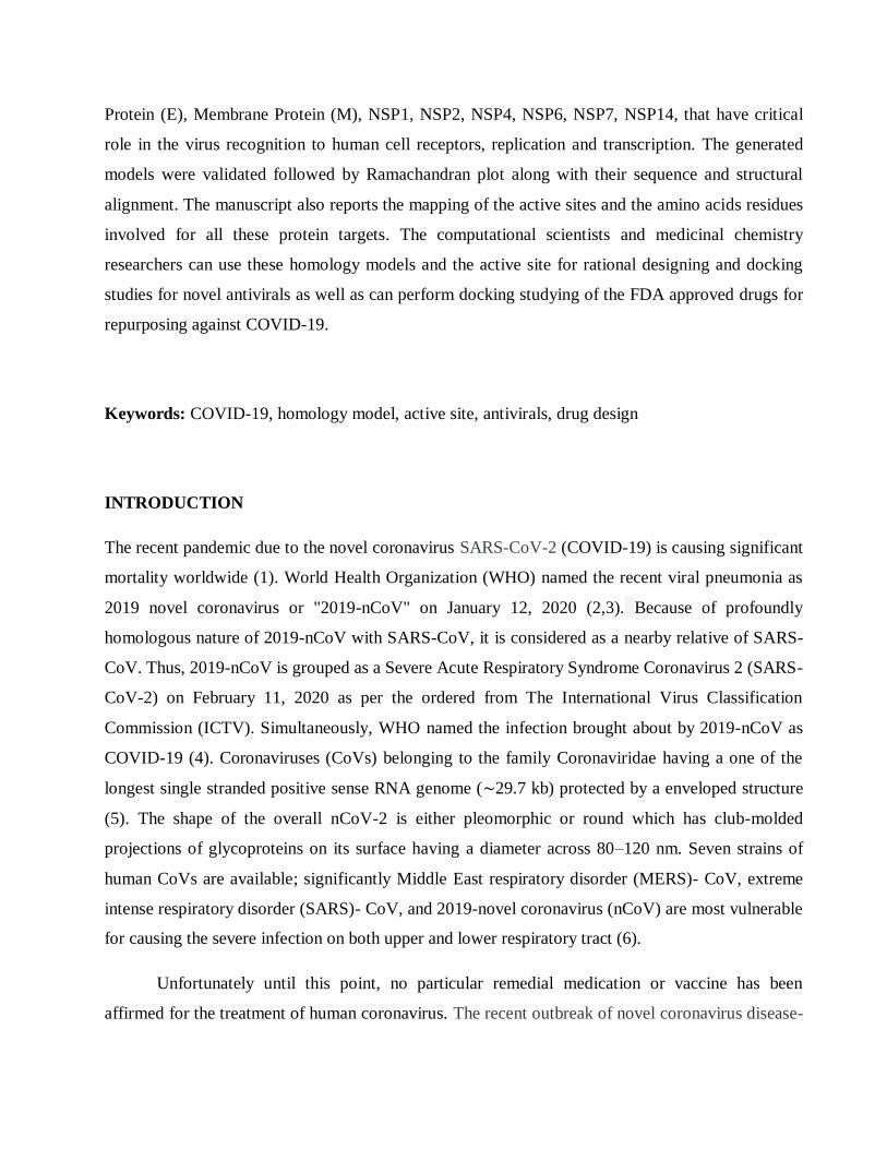
the virus, inhibiting virus replication through acting on critical enzymes of virus (figure 1). The
related viral target proteins include ACE2, TMPRSSS2, Spike protein, Envelop protein, Membrane
protein, non-structural proteins such as NSP1, NSP2, NSP4, RNA-dependent RNA polymerase
(RdRp), helicase, Papain-Like Proteases (PLpro), main protease (3CLpro, also named 3-
chymotrypsin-like protease) etc (7,8).
Figure 1. Life cycle of novel coronavirus SARS-CoV-2 and possible drug targets.
There is a lack of specific antiviral drugs for the treatment of COVID-19. Most treatment strategies
are still not widely accepted and focus on symptomatic management and supportive therapy only.
Antimalarial drug having lysosomotropic groups such as chloroquine (CQ) and hydroxychloroquine
(HCQ) were found to be effective. Some therapeutic agents that require further experimental
validation are ribavirin, remdesivir, ritonavir and lopinavir (8,9). In the present work, we have
compiled a list of 20 probable drug targets on host and virus based on viral life cycle which can
targeted for the rational development of future antivirals as well as computationally screening
marketed drugs for repurposing. We prepared homology model for ten vital proteins for which no
crystal structure is reported.
MATERIALS AND METHODS
We have searched thoroughly the RCSB protein data bank of the possible drug target for the
treatment against novel corona virus 2019. There are plenty of proteins associated with their life
cycle. Among them few are already available in PDB. For the preparation of rest of the protein
associated with SARS-n-Cov2, we followed up template based homology modeling. The most
readily accessible full genome sequence for the recently emerged COVID-19 is recovered from the
National Center for Biotechnology Information (NCBI) nucleotide database. The accession code for
that sequence is NC_045512.2. The required genome sequence of the respective structural target
protein isolated as a query sequence. The Protein-Protein Blast (BLASTp) tools was performed
against all known crystals structures deposited into protein data bank (PDB) in order to identify
closely related homologous template structure for modeling (10). Based on the structure and
sequence similarities with the template protein and E-value on the BLAST results, proper highes t
identical homologous template structure has been selected. Swiss model web-server was utilized to
build the models (Table 1). Homology models for TMPRSS2, Envelope Protein (E), Membrane
Protein (M), NSP1, NSP2, NSP4, NSP6, NSP7,, NSP14 protein targets were prepared for which no
crystal structure is available. The generated models were validated followed by Ramachandran plot
along with their sequence and structural alignment. The binding pocket residues of all the protein
models are calculated by utilizing COACH meta-server (11) and also cross verified with the
CASTp webservers. All the active sites of the homology build proteins were evaluated after
superimposition of the closely related X-ray crystallized structure bound with the co-crystal ligands.
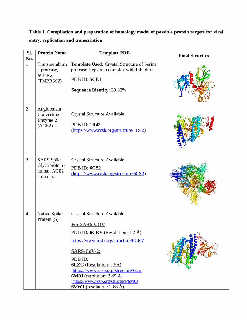
Table 1. Compilation and preparation of homology model of possible protein targets for viral
entry, replication and transcription
Sl.
No.
Protein Name Template PDB Final Structure
1. Transmembran
e protease,
serine 2
(TMPRSS2)
Template Used: Crystal Structure of Serine
protease Hepsin in complex with Inhibitor
PDB ID: 5CE1
Sequence Identity: 33.82%
2. Angiotensin
Converting
Enzyme 2
(ACE2)
Crystal Structure Available.
PDB ID: 1R42
(https://www.rcsb.org/structure/1R42)
3. SARS Spike
Glycoprotein -
human ACE2
complex
Crystal Structure Available.
PDB ID: 6CS2
(https://www.rcsb.org/structure/6CS2)
4. Native Spike
Protein (S)
Crystal Structure Available.
For SARS-COV
PDB ID: 6CRV (Resolution: 3.2 Å)
https://www.rcsb.org/structure/6CRV
SARS-CoV-2:
PDB ID:
6LZG (Resolution: 2.5Å)
https://www.rcsb.org/structure/6lzg
6M0J (resolution: 2.45 Å)
:https://www.rcsb.org/structure/6M0J
6VW1 (resolution: 2.68 Å) :
https://www.rcsb.org/structure/6VW1
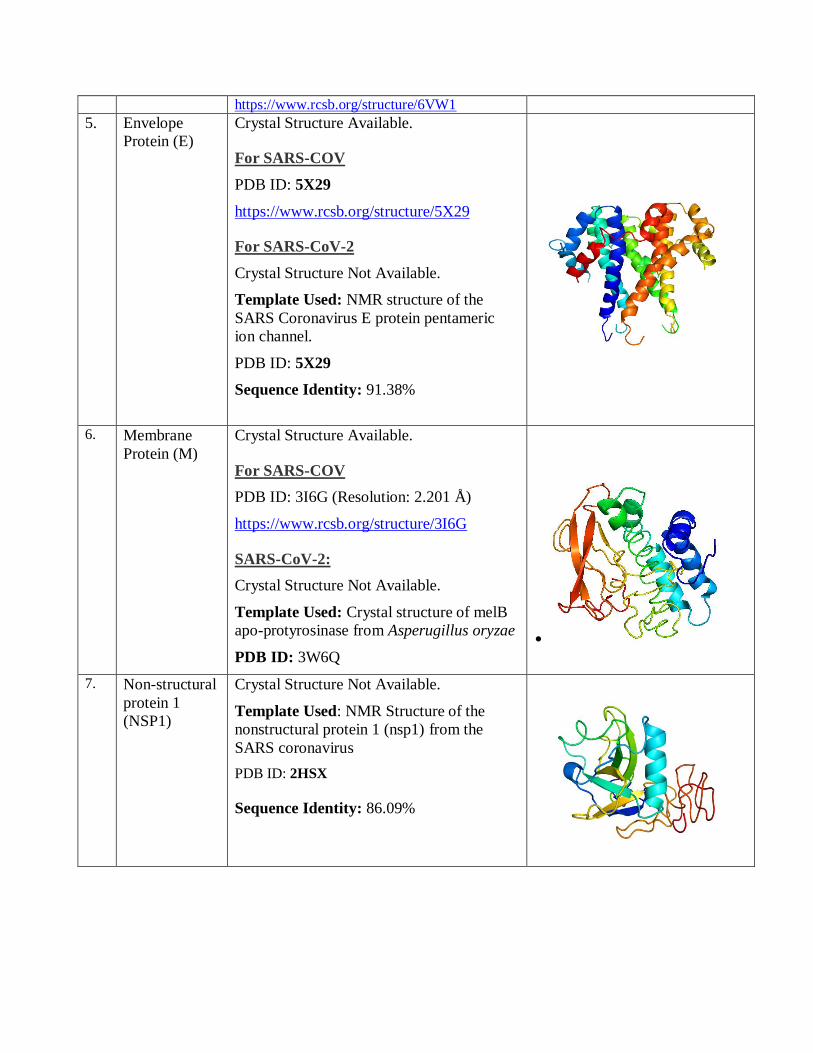
5. Envelope
Protein (E)
Crystal Structure Available.
For SARS-COV
PDB ID: 5X29
https://www.rcsb.org/structure/5X29
For SARS-CoV-2
Crystal Structure Not Available.
Template Used: NMR structure of the
SARS Coronavirus E protein pentameric
ion channel.
PDB ID: 5X29
Sequence Identity: 91.38%
6. Membrane
Protein (M)
Crystal Structure Available.
For SARS-COV
PDB ID: 3I6G (Resolution: 2.201 Å)
https://www.rcsb.org/structure/3I6G
SARS-CoV-2:
Crystal Structure Not Available.
Template Used: Crystal structure of melB
apo-protyrosinase from Asperugillus oryzae
PDB ID: 3W6Q
7. Non-structural
protein 1
(NSP1)
Crystal Structure Not Available.
Template Used: NMR Structure of the
nonstructural protein 1 (nsp1) from the
SARS coronavirus
PDB ID: 2HSX
Sequence Identity: 86.09%
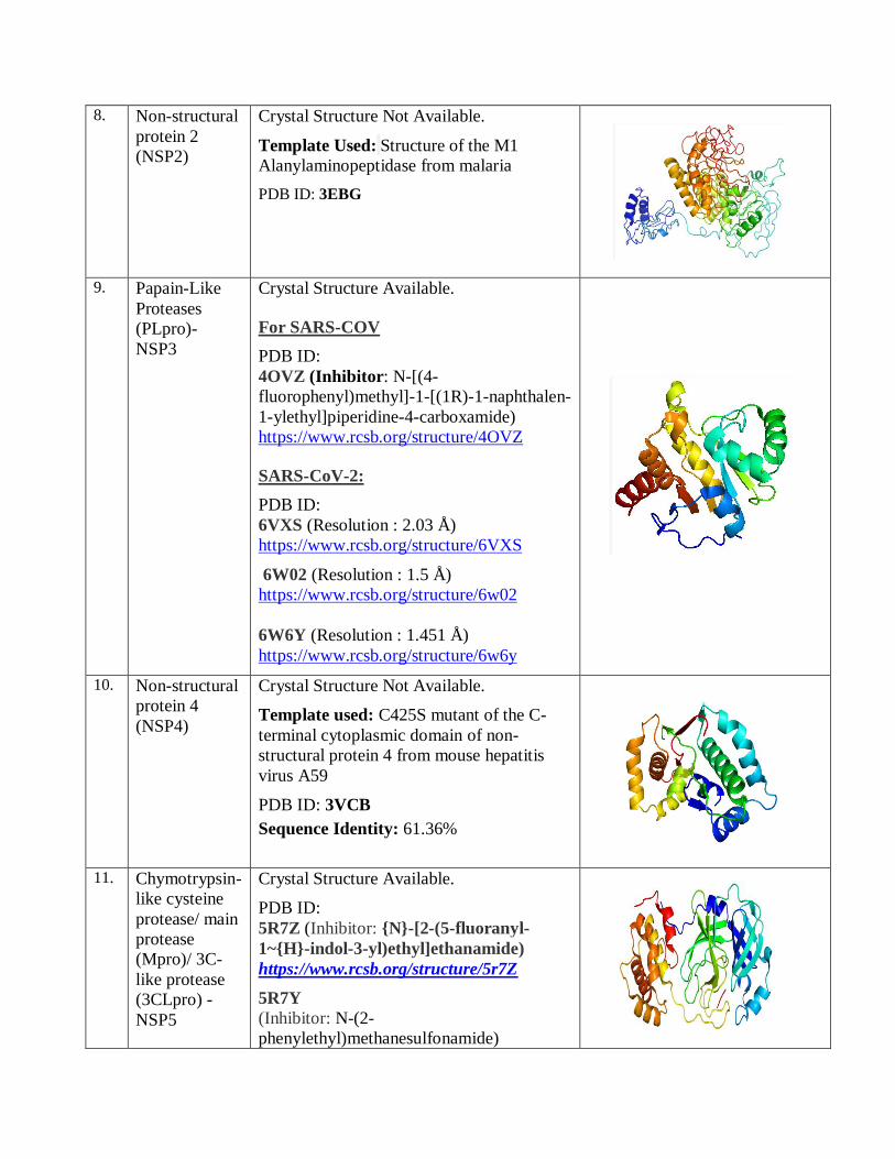
8. Non-structural
protein 2
(NSP2)
Crystal Structure Not Available.
Template Used: Structure of the M1
Alanylaminopeptidase from malaria
PDB ID: 3EBG
9. Papain-Like
Proteases
(PLpro)-
NSP3
Crystal Structure Available.
For SARS-COV
PDB ID:
4OVZ (Inhibitor: N-[(4-
fluorophenyl)methyl]-1-[(1R)-1-naphthalen-
1-ylethyl]piperidine-4-carboxamide)
https://www.rcsb.org/structure/4OVZ
SARS-CoV-2:
PDB ID:
6VXS (Resolution : 2.03 Å)
https://www.rcsb.org/structure/6VXS
6W02 (Resolution : 1.5 Å)
https://www.rcsb.org/structure/6w02
6W6Y (Resolution : 1.451 Å)
https://www.rcsb.org/structure/6w6y
10. Non-structural
protein 4
(NSP4)
Crystal Structure Not Available.
Template used: C425S mutant of the C-
terminal cytoplasmic domain of non-
structural protein 4 from mouse hepatitis
virus A59
PDB ID: 3VCB
Sequence Identity: 61.36%
11. Chymotrypsin-
like cysteine
protease/ main
protease
(Mpro)/ 3C-
like protease
(3CLpro) -
NSP5
Crystal Structure Available.
PDB ID:
5R7Z (Inhibitor: {N}-[2-(5-fluoranyl-
1~{H}-indol-3-yl)ethyl]ethanamide)
https://www.rcsb.org/structure/5r7Z
5R7Y
(Inhibitor: N-(2-
phenylethyl)methanesulfonamide)
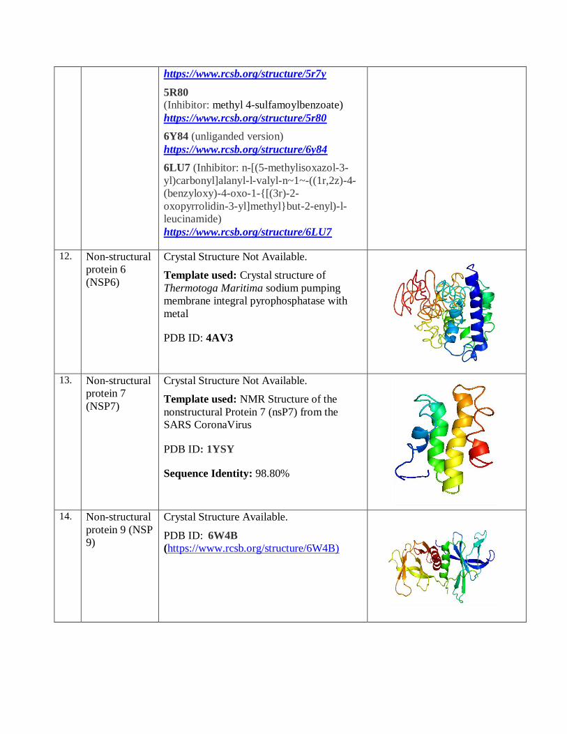
https://www.rcsb.org/structure/5r7y
5R80
(Inhibitor: methyl 4-sulfamoylbenzoate)
https://www.rcsb.org/structure/5r80
6Y84 (unliganded version)
https://www.rcsb.org/structure/6y84
6LU7 (Inhibitor: n-[(5-methylisoxazol-3-
yl)carbonyl]alanyl-l-valyl-n~1~-((1r,2z)-4-
(benzyloxy)-4-oxo-1-{[(3r)-2-
oxopyrrolidin-3-yl]methyl}but-2-enyl)-l-
leucinamide)
https://www.rcsb.org/structure/6LU7
12. Non-structural
protein 6
(NSP6)
Crystal Structure Not Available.
Template used: Crystal structure of
Thermotoga Maritima sodium pumping
membrane integral pyrophosphatase with
metal
PDB ID: 4AV3
13. Non-structural
protein 7
(NSP7)
Crystal Structure Not Available.
Template used: NMR Structure of the
nonstructural Protein 7 (nsP7) from the
SARS CoronaVirus
PDB ID: 1YSY
Sequence Identity: 98.80%
14. Non-structural
protein 9 (NSP
9)
Crystal Structure Available.
PDB ID: 6W4B
(https://www.rcsb.org/structure/6W4B)
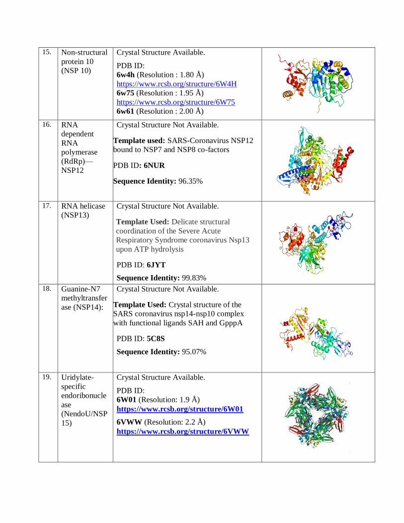
15. Non-structural
protein 10
(NSP 10)
Crystal Structure Available.
PDB ID:
6w4h (Resolution : 1.80 Å)
https://www.rcsb.org/structure/6W4H
6w75 (Resolution : 1.95 Å)
https://www.rcsb.org/structure/6W75
6w61 (Resolution : 2.00 Å)
16. RNA
dependent
RNA
polymerase
(RdRp)—
NSP12
Crystal Structure Not Available.
Template used: SARS-Coronavirus NSP12
bound to NSP7 and NSP8 co-factors
PDB ID: 6NUR
Sequence Identity: 96.35%
17. RNA helicase
(NSP13)
Crystal Structure Not Available.
Template Used: Delicate structural
coordination of the Severe Acute
Respiratory Syndrome coronavirus Nsp13
upon ATP hydrolysis
PDB ID: 6JYT
Sequence Identity: 99.83%
18. Guanine-N7
methyltransfer
ase (NSP14):
Crystal Structure Not Available.
Template Used: Crystal structure of the
SARS coronavirus nsp14-nsp10 complex
with functional ligands SAH and GpppA
PDB ID: 5C8S
Sequence Identity: 95.07%
19. Uridylate-
specific
endoribonucle
ase
(NendoU/NSP
15)
Crystal Structure Available.
PDB ID:
6W01 (Resolution: 1.9 Å)
https://www.rcsb.org/structure/6W01 6VWW (Resolution: 2.2 Å)
https://www.rcsb.org/structure/6VWW
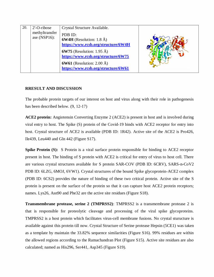
20. 2'-O-ribose
methyltransfer
ase (NSP16):
Crystal Structure Available.
PDB ID:
6W4H (Resolution: 1.8 Å)
https://www.rcsb.org/structure/6W4H 6W75 (Resolution: 1.95 Å)
https://www.rcsb.org/structure/6W75
6W61 (Resolution: 2.00 Å)
https://www.rcsb.org/structure/6W61
RRESULT AND DISCUSSION
The probable protein targets of our interest on host and virus along with their role in pathogenesis
has been described below. (9, 12-17)
ACE2 protein: Angiotensin Converting Enzyme 2 (ACE2) is present in host and is involved during
viral entry to host. The Spike (S) protein of the Covid-19 binds with ACE2 receptor for entry into
host. Crystal structure of ACE2 is available (PDB ID: 1R42). Active site of the ACE2 is Pro426,
Ile439, Leu440 and Gln 442 (Figure S17).
Spike Protein (S): S Protein is a viral surface protein responsible for binding to ACE2 receptor
present in host. The binding of S protein with ACE2 is critical for entry of virus to host cell. There
are various crystal structures available for S protein SAR-COV (PDB ID: 6CRV), SARS-n-CoV2
PDB ID: 6LZG, 6MOJ, 6VW1). Crystal structures of the bound Spike glycoprotein-ACE2 complex
(PDB ID: 6CS2) provides the nature of binding of these two critical protein. Active site of the S
protein is present on the surface of the protein so that it can capture host ACE2 protein receptors;
names. Lys26, Asn90 and Phe32 are the active site residues (Figure S18).
Transmembrane protease, serine 2 (TMPRSS2): TMPRSS2 is a transmembrane protease 2 is
that is responsible for proteolytic cleavage and processing of the viral spike glycoproteins.
TMPRSS2 is a host protein which facilitates virus-cell membrane fusions. No crystal sturucture is
available against this protein till now. Crystal Structure of Serine protease Hepsin (5CE1) was taken
as a template by maintain the 33.82% sequence similarities (Figure S16). 99% residues are within
the allowed regions according to the Ramachandran Plot (Figure S15). Active site residues are also
calculated; named as His296, Ser441, Asp345 (Figure S19).
Envelope (E) protein: Envelope (E) protein is an integral membrane protein involved in the
assembly and formation of envelop which is important for COVID-19 pathogenesis. Homology
model of the membrane protein was builded by taking the SAS-CoV E protein (PDB ID: 5X29)
(Table 2). Overall 98% residues are in the allowed regions (Figure S21). Active site of the protein
is Ala32, Thr35, Tyr42 and Tyr57 which is present on the surface of the protein (Figure S20).
Membrane (M) protein: Membrane (M) protein is the central organizer that determines the shape
of viral envelops and interacts with the E protein in the budding compartment of the host cell. The
crystal structure of SARS-COV (PDB ID: 3I6G) is present but not for COVID-19. Active Sites of
M protein is Lys50, Leu51, Leu54, Leu93 and Ala98 (Figure S22).
Non-structural protein 1 (NSP1): NSP1 is a membrane-associated protein which is responsible
for anchoring the replication complex to the cellular membranes. In the infected cell through
interaction with the 40S ribosomal subunit, NSP1 suppresses host gene expression which facilitates
viral gene expression by avoiding host immune response. Crystal structure of Novel corona virus is
still not discovered. So, homology model of NSP1 has been builded by taking SARS-CoV NSP1
(PDB ID: 2HSX) as a template structure. The Sequence identity is 86.09% (Figure S2). Overall
96% residues are in the allowed regions (Figure S1). The active sites region is built by Val 35,
Glu36, Pro 67 and Tyr 68 (Figure S23).
Non-structural protein 2 (NSP2): NSP2 plays a role in the host cell survival signaling pathway.
Homology model of this protein has been built with the M1 Alanyl aminopeptidase protein with
50.09% sequence similarities (Figure S4). 84% residues are on the allowed regions on
Ramachandran plot [Figure S3]. Active site of NSP2 protein is depicted on supplementary figure
24.
Papain-Like Proteases: A viral Papain-Like Proteases (PLpro) is critical for virus replication and
packaging within the host cells through cleavage of viral peptides into functional proteins. The
PLpro cleaves polyproteins (PPs) to generate NSP1, NSP2 and NSP3. The crystal structures for
SARS-COV (PDB ID: 4OVZ) and SARS-CoV-2 (PDB ID: 6VXS, 6WO2 AND 6W6Y) can be
used to design future antivirals. Asp22, Ile23, Val49, Ile131 and Leu160 residues are present in the
active site of PLpro (Figure S25).
Non-structural protein 4 (NSP4): NSP4 is necessary for viral replication. Due to unavailability of
nCov2 NSP4 protein, homology structure has been built by maintain the 61.36% sequence identity
with the template protein (3VCB) (Figure S6). Whole protein and along with the active sites are
depicted on the supplementary figure 26.
3-Chymotrypsin-like cysteine protease (3-CLpro)/main protease (Mpro): 3CLpro cleaves viral
peptides into functional proteins for virus replication and packaging within the host cells. It can
cleave PPs to release 16 NSPs and produce mature protein that anchors the replication/transcription.
There are many crystal structures (PDB ID: 5R7Z, 5R7Y, 5R80, 6Y84, 6LU7) for Mpro protein is
available. In their crystal structure three domains are present. Residues 8-99 are in domain I, 100-
183 are in the domain II and 183 to 200 are the loop regions and lastly 200 to 306 are the domain
III. His41 and Cys 145 are the active residues (Figure 2).
.
Figure 2: Structural alignment of NSP4 protein. Active site residues are marked with blue
and orange elemental colored ball and sticks representations
Non-structural protein 6 (NSP6): NSP6 is a viral replicase protein capable of inducing autophagy.
Homology structure has been made for this protein. Ramachandran plot and structure and sequence
alignment has incorporated on supplementary figure S7 and S8 respectively. His11, Leu15, Ile 18
and Phe70 are the active sites residues (Figure S27).
Non-structural protein 7 (NSP7): NSP7 is a primase may participate in viral replication. Due to
unavailability of the crystal structure it is built by homology modeling with SARS NSP7 template
protein (PDB ID: 1YSY). Sequence alignment and Ramachandran plot is on the figure S9 and
figure S10. Val16, Cys32 and Leu55 are active site residues (Figure S28).
NSP7 and NSP8 can form hexadecamer. NSP9 is a RNA binding protein of COVID-19 and
interacts with NSP8 for its functions. NSP9 is a replicase protein that participates in viral
replication. The crystal structure (PDB ID: 6W4B) of NSP9 is available. Active site of this protein
is depicted on figure S29.
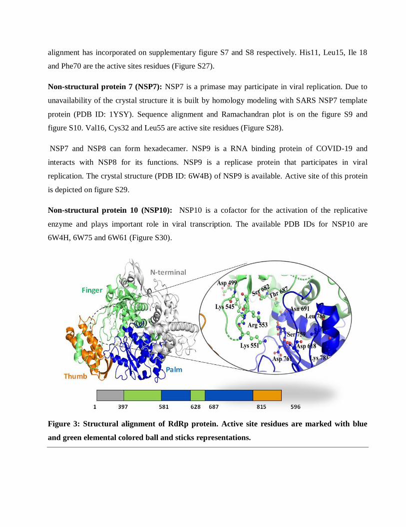
Non-structural protein 10 (NSP10): NSP10 is a cofactor for the activation of the replicative
enzyme and plays important role in viral transcription. The available PDB IDs for NSP10 are
6W4H, 6W75 and 6W61 (Figure S30).
Figure 3: Structural alignment of RdRp protein. Active site residues are marked with blue
and green elemental colored ball and sticks representations.
RNA dependent RNA polymerase (RdRp): RNA dependent RNA polymerase (RdRp)/NSP12 is a
RNA dependent RNA polymerase. RdRp is the main protein for RNA replication and transcription
of viral RNA genome. This protein is a cluster of several domains. 1-397 is N-terminal domain,
(397-581) and (629-687) is finger domain, (582-628) and (688-815) is the palm region and (816-
596) is the thumb region (Figure 3).
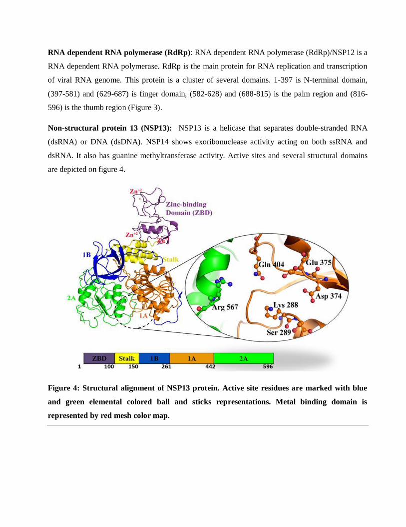
Non-structural protein 13 (NSP13): NSP13 is a helicase that separates double-stranded RNA
(dsRNA) or DNA (dsDNA). NSP14 shows exoribonuclease activity acting on both ssRNA and
dsRNA. It also has guanine methyltransferase activity. Active sites and several structural domains
are depicted on figure 4.
Figure 4: Structural alignment of NSP13 protein. Active site residues are marked with blue
and green elemental colored ball and sticks representations. Metal binding domain is
represented by red mesh color map.
Uridylate-specific endoribonuclease (NendoU/NSP15): NSP15 shows endoribonuclease activity.
The crystal structure (PDB ID: 6W01, 6VWW) for NSP15 is available His235, Gln245, Gly248,
Gln294 THr341 are the active sites residues (Figure S32).
2'-O-Ribose methyltransferase (NSP16): NSP16 has methyltransferase activity plays vital role in
viral mRNAs cap methylation which is important toevade immune system. Structural domain and
active sites are depicted on figure S33. All these target proteinsmentioned above have critical role
in the virus binding to human cell receptors, replication and transcription.
Table 2: Protein Analysis Table
Protein
Name
Ramachandran Plot Statistics Sequence
alignment
Ramachandran
Plot Figure
Number
Sequence
alignment
Fig. Number
Non-
structural
protein 1
(NSP1)
Residues in most favored regions: 77.1%
Residues in additional allowed regions :
18.8% Residues in generously allowed regions:
3.1%
Residues in disallowed regions: 1.0%
Sequence
Identity:
86.09%
Figure S1
Figure S2
Non-
structural
protein 2
(NSP2)
Residues in most favoured regions: 62.5% Residues in additional allowed regions:
25.4%
Residues in generously allowed regions: 7.2%
Residues in disallowed regions: 4.8%
Sequence Identity:
50.09%
Figure S3 Figure S4
Non-
structural
protein 4
(NSP4)
Residues in most favoured regions: 94.5%
Residues in additional allowed regions : 4.9%
Residues in generously allowed regions :
0.6% Residues in disallowed regions: 0.0%
Sequence
Identity: 61.36%
Figure S5 Figure S6
Non-
structural
protein 6
(NSP6)
Residues in most favoured regions: 33.5%
Residues in additional allowed regions:
35.0% Residues in generously allowed regions:
18.4%
Residues in disallowed regions: 13.2%
Sequence
Identity:
66%
Figure S7 Figure S8
Non-
structural
protein 7
(NSP7)
Residues in most favoured regions: 72.5%
Residues in additional allowed regions: 18
22.5%
Residues in generously allowed regions: 3.8%
Residues in disallowed regions: 1.2%
Sequence
Identity:
98.80%
Figure S9 Figure S10
RNA
helicase
Residues in most favoured regions: 80.3% Residues in additional allowed regions: 79
Sequence Identity:
Figure S11 Figure S12
(NSP13) 14.8%
Residues in generously allowed regions: 3.6%
Residues in disallowed regions: 1.3%
99.83%
Guanine-N7
methyltrans
ferase
(NSP14):
Residues in most favoured regions: 91.4%
Residues in additional allowed regions: 8.1%
Residues in generously allowed regions:
0.4%
Residues in disallowed regions: 0.0%
Sequence
Identity: 95.07%
Figure S13 Figure S14
Transmemb
rane
protease,
serine 2
(TMPRSS2)
Residues in most favoured regions 84.6%
Residues in additional allowed regions:
14.7% Residues in generously allowed regions:
0.3%
Residues in disallowed regions:0.3%
Sequence
Identity:
33.82%
Figure S15 Figure S16
Envelope
(E) protein
Residues in most favoured regions: 84.4% Residues in additional allowed regions:
14.1%
Residues in generously allowed regions: 1.5%
Residues in disallowed regions 0 0.0%
Sequence Identity:
91.38%
Figure S20
In summary, the compiled list of 20 probable drug targets on host and virus along with their role in
pathogenesis will provide a snap shot for the drug discovery scientists. They present an attractive
target for new antiviral design. We have prepared homology model of TMPRSS2, Envelope Protein
(E), Membrane Protein (M), NSP1, NSP2, NSP4, NSP6, NSP7, NSP14, that have critical role in the
virus recognition to human cell receptors, replication and transcription. The generated models were
validated followed by Ramachandran plot along with their sequence and structural alignment (table
2). The residues present in the active site of all the protein models were calculated by utilizing
COACH meta-server and subsequently cross verified with the CASTp webservers. All the active
sites of the homology build proteins were evaluated after superimposition of the closely related X-
ray crystallized structure bound with the co-crystal ligands. The computational scientists and
medicinal chemistry researchers can use these homology models and the active site analysis
described in the manuscript for rational designing and docking studies for novel antivirals as well as
can perform docking studying of the FDA approved drugs for repurposing against COVID-19.
Author Contribution
SP and AT have contributed to the concept of the study, preparation of model and writing of the
manuscript. Both the authors have approved the final version of the article.
Acknowledgments
SP has received Senior Research Fellowship from Indian Council of Medical Research
Disclosure Summary
The authors declare no conflict of interest
References
1. World Health Organization. Coronavirus disease (COVID-19) pandemic.
https://www.who.int/emergencies/diseases/novel-coronavirus-2019 Date of accessed: March
31, 2020.
2. Zhu N, Zhang D, Wang W, Li X, Yang B, Song J, Zhao X, Huang B, Shi W, Lu R, Niu P,
Zhan F, Ma X, Wang D, Xu W, Wu G, Gao GF, Tan W. A Novel Coronavirus from Patients
with Pneumonia in China, 2019. N Engl J Med. 2020;382(8):727-733. doi:
10.1056/NEJMoa2001017.
3. Chen Y, Liu Q, Guo D, Emerging coronaviruses: Genome structure, replication, and
pathogenesis. J Med Virol. 2020;92(4):418-423. doi: 10.1002/jmv.25681.
4. Canrong Wu, Yang Liu, Yueying Yang, Peng Zhang, Wu Zhong, Yali Wang, Qiqi Wang,
Yang Xu, Mingxue Li, Xingzhou Li, Mengzhu Zheng, Lixia Chen, Hua Li, Analysis of
therapeutic targets for SARS-CoV-2 and discovery of potential drugs by computational
methods. Acta Pharmaceutica Sinica B, 2020; 2211-3835.
doi.org/10.1016/j.apsb.2020.02.008.
5. Yang H, Bartlam M, Rao Z. Drug design targeting the main protease, the Achilles' heel of
coronaviruses. Curr Pharm Des. 2006;12(35):4573-90. doi: 10.2174/138161206779010369.
6. Paules CI, Marston HD, Fauci AS. Coronavirus Infections-More than Just the Common
Cold. JAMA. 2020. doi: 10.1001/jama.2020.0757.
7. Canrong Wu, Yang Liu, Yueying Yang, Peng Zhang, Wu Zhong, Yali Wang, Qiqi
Wang, Yang Xu, Mingxue Li, Xingzhou Li, Mengzhu Zheng, Lixia Chen, Hua Li Analysis
of therapeutic targets for SARS-CoV-2 and discovery of potential drugs by computational
methods Acta Pharmaceutica Sinica B. doi: https://doi.org/10.1016/j.apsb.2020.02.008.
8. Senanayake, SL. Drug repurposing strategies for COVID-19. Future Drug. Discov. eISSN
2631-3316. doi: 10.4155/fdd-2020-0010.
9. Prajapat M, Sarma P, Shekhar N, Avti P, Sinha S, Kaur H, Kumar S, Bhattacharyya A,
Kumar H, Bansal S, Medhi B. Drug targets for corona virus: A systematic review. Indian J
Pharmacol. 2020;52(1):56–65. doi: 10.4103/ijp.IJP_115_20.
10. Johnson M, Zaretskaya I, Raytselis Y, Merezhuk Y, McGinnis S, Madden TL. NCBI
BLAST: a better web interface. Nucleic Acids Res. 2008;36(Web Server issue):W5–W9.
doi: 10.1093/nar/gkn201
11. Yang J, Roy, A, Zhang Y. Protein–ligand binding site recognition using complementary
binding-specific substructure comparison and sequence profile alignment. 2013:29(20):
2588–2595.
12. te Velthuis AJ, van den Worm SH, Snijder EJ. The SARS-coronavirus nsp7+nsp8 complex
is a unique multimeric RNA polymerase capable of both de novo initiation and primer
extension. Nucleic Acids Res. 2012;40:1737–47.
13. Stobart CC, Sexton NR, Munjal H, Lu X, Molland KL, Tomar S, et al. Chimeric exchange
of coronavirus nsp5 proteases (3CLpro) identifies common and divergent regulatory
determinants of protease activity. J Virol. 2013;87:12611–8.
14. Wang H, Xue S, Yang H, Chen C. Recent progress in the discovery of inhibitors targeting
coronavirus proteases. Virol Sin. 2016;31:24–30.
15. Egloff MP, Ferron F, Campanacci V, Longhi S, Rancurel C, Dutartre H, et al. The severe
acute respiratory syndrome-coronavirus replicative protein nsp9 is a single-stranded RNA-
binding subunit unique in the RNA virus world. Proc Natl Acad Sci U S A. 2004;101:3792–
6.
16. Hu T, Chen C, Li H, Dou Y, Zhou M, Lu D, et al. Structural basis for dimerization and
RNA binding of avian infectious bronchitis virus nsp9. Protein Sci. 2017;26:1037–48
17. Bouvet M, Lugari A, Posthuma CC, Zevenhoven JC, Bernard S, Betzi S, et al. Coronavirus
Nsp10, a critical co-factor for activation of multiple replicative enzymes. J Biol Chem.
2014;289:25783–96.
download fileview on ChemRxivManuscript_Compilation of Potential Protein Targets for S... (1.15 MiB)
Compilation of Potential Protein Targets for SARS-CoV-2: Preparation of Homology
Model and Active Site Determination for Future Rational Antiviral Design
Sourav Pal1,2
and Arindam Talukdar
1,2*
1Department of Organic and Medicinal Chemistry, CSIR-Indian Institute of Chemical Biology, 4
Raja S. C. Mullick Road, Kolkata 700032, WB (India).
2 Academy of Innovative and Scientific Research, Ghaziabad-201002, India
Supplementary Information:
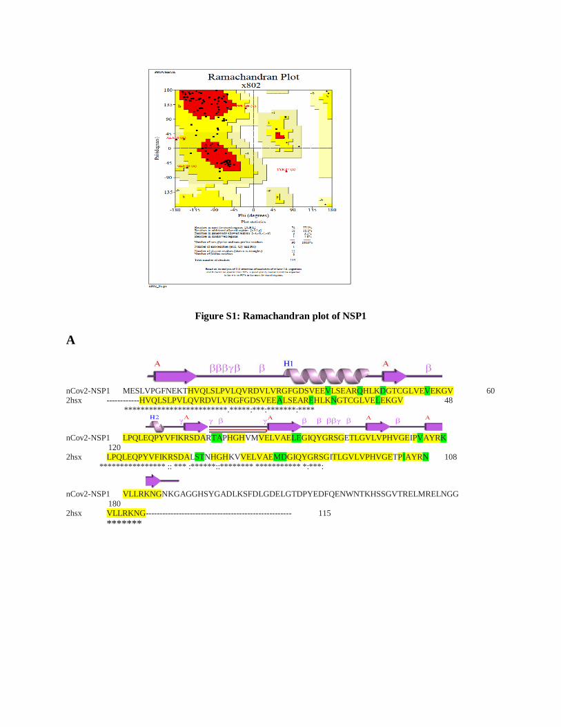
Figure S1: Ramachandran plot of NSP1
A
nCov2-NSP1 MESLVPGFNEKTHVQLSLPVLQVRDVLVRGFGDSVEEVLSEARQHLKDGTCGLVEVEKGV 60 2hsx ------------HVQLSLPVLQVRDVLVRGFGDSVEEALSEAREHLKNGTCGLVELEKGV 48 *************************.*****:***:*******:****
nCov2-NSP1 LPQLEQPYVFIKRSDARTAPHGHVMVELVAELEGIQYGRSGETLGVLVPHVGEIPVAYRK
120 2hsx LPQLEQPYVFIKRSDALSTNHGHKVVELVAEMDGIQYGRSGITLGVLVPHVGETPIAYRN 108 **************** :: *** :******::******** *********** *:***:
nCov2-NSP1 VLLRKNGNKGAGGHSYGADLKSFDLGDELGTDPYEDFQENWNTKHSSGVTRELMRELNGG
180 2hsx VLLRKNG----------------------------------------------------- 115
*******
B
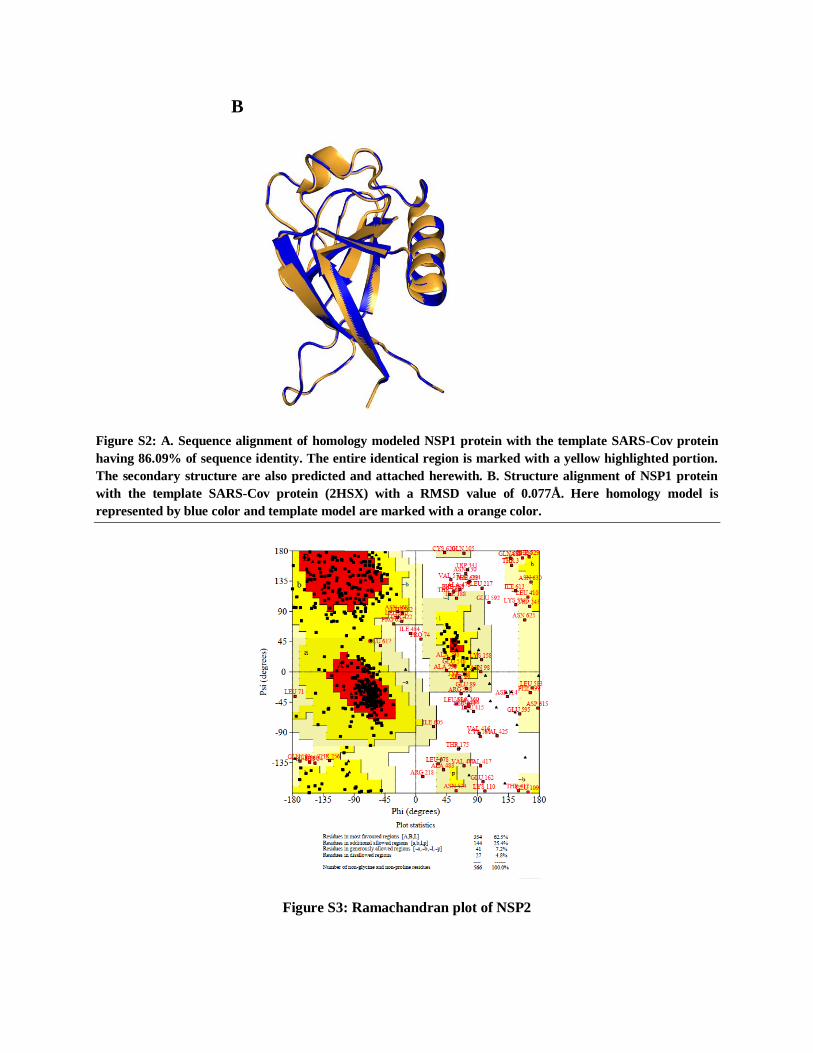
Figure S2: A. Sequence alignment of homology modeled NSP1 protein with the template SARS-Cov protein
having 86.09% of sequence identity. The entire identical region is marked with a yellow highlighted portion.
The secondary structure are also predicted and attached herewith. B. Structure alignment of NSP1 protein
with the template SARS-Cov protein (2HSX) with a RMSD value of 0.077Å. Here homology model is
represented by blue color and template model are marked with a orange color.
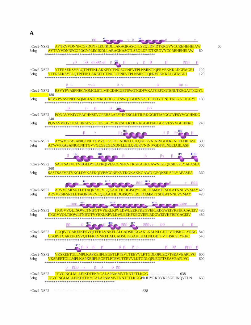
Figure S3: Ramachandran plot of NSP2
A
nCov2-NSP2 AYTRYVDNNFCGPDGYPLECIKDLLARAGKASCTLSEQLDFIDTKRGVYCCREHEHEIAW 60 3ebg AYTRYVDNNFCGPDGYPLECIKDLLARAGKASCTLSEQLDFIDTKRGVYCCREHEHEIAW 60 ************************************************************
nCov2-NSP2 YTERSEKSYELQTPFEIKLAKKFDTFNGECPNFVFPLNSIIKTIQPRVEKKKLDGFMGRI 120 3ebg YTERSEKSYELQTPFEIKLAKKFDTFNGECPNFVFPLNSIIKTIQPRVEKKKLDGFMGRI 120 ************************************************************
nCov2-NSP2 RSVYPVASPNECNQMCLSTLMKCDHCGETSWQTGDFVKATCEFCGTENLTKEGATTCGYL 180 3ebg RSVYPVASPNECNQMCLSTLMKCDHCGETSWQTGDFVKATCEFCGTENLTKEGATTCGYL 180
************************************************************
nCov2-NSP2 PQNAVVKIYCPACHNSEVGPEHSLAEYHNESGLKTILRKGGRTIAFGGCVFSYVGCHNKC 240 3ebg PQNAVVKIYCPACHNSEVGPEHSLAEYHNESGLKTILRKGGRTIAFGGCVFSYVGCHNKC 240 ************************************************************
nCov2-NSP2 AYWVPRASANIGCNHTGVVGEGSEGLNDNLLEILQKEKVNINIVGDFKLNEEIAIILASF 300 3ebg AYWVPRASANIGCNHTGVVGEGSEGLNDNLLEILQKEKVNINIVGDFKLNEEIAIILASF 300 ************************************************************
nCov2-NSP2 SASTSAFVETVKGLDYKAFKQIVESCGNFKVTKGKAKKGAWNIGEQKSILSPLYAFASEA
360 3ebg SASTSAFVETVKGLDYKAFKQIVESCGNFKVTKGKAKKGAWNIGEQKSILSPLYAFASEA 360 ************************************************************
nCov2-NSP2 ARVVRSIFSRTLETAQNSVRVLQKAAITILDGISQYSLRLIDAMMFTSDLATNNLVVMAY 420 3ebg ARVVRSIFSRTLETAQNSVRVLQKAAITILDGISQYSLRLIDAMMFTSDLATNNLVVMAY 420 ************************************************************
nCov2-NSP2 ITGGVVQLTSQWLTNIFGTVYEKLKPVLDWLEEKFKEGVEFLRDGWEIVKFISTCACEIV 480 3ebg ITGGVVQLTSQWLTNIFGTVYEKLKPVLDWLEEKFKEGVEFLRDGWEIVKFISTCACEIV 480 ************************************************************
nCov2-NSP2 GGQIVTCAKEIKESVQTFFKLVNKFLALCADSIIIGGAKLKALNLGETFVTHSKGLYRKC 540
3ebg GGQIVTCAKEIKESVQTFFKLVNKFLALCADSIIIGGAKLKALNLGETFVTHSKGLYRKC 540 ************************************************************
nCov2-NSP2 VKSREETGLLMPLKAPKEIIFLEGETLPTEVLTEEVVLKTGDLQPLEQPTSEAVEAPLVG 600 3ebg VKSREETGLLMPLKAPKEIIFLEGETLPTEVLTEEVVLKTGDLQPLEQPTSEAVEAPLVG 600 ************************************************************
nCov2-NSP2 TPVCINGLMLLEIKDTEKYCALAPNMMVTNNTFTLKGG---------------------- 638 3ebg TPVCINGLMLLEIKDTEKYCALAPNMMVTNNTFTLKGGPKIHYRKDYKPSGFIINQVTLN 660 ************************************** nCov2-NSP2 ------------------------------------------------------------ 638
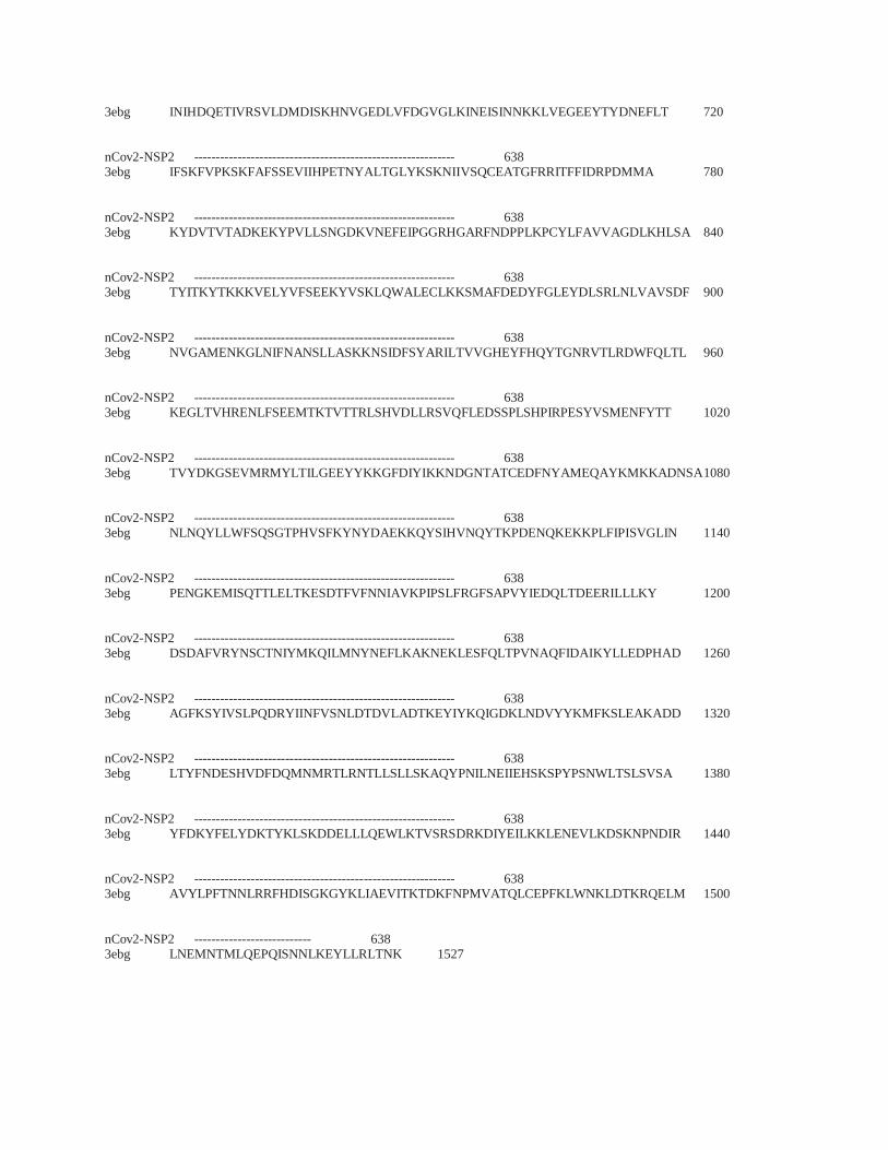
3ebg INIHDQETIVRSVLDMDISKHNVGEDLVFDGVGLKINEISINNKKLVEGEEYTYDNEFLT 720 nCov2-NSP2 ------------------------------------------------------------ 638 3ebg IFSKFVPKSKFAFSSEVIIHPETNYALTGLYKSKNIIVSQCEATGFRRITFFIDRPDMMA 780
nCov2-NSP2 ------------------------------------------------------------ 638 3ebg KYDVTVTADKEKYPVLLSNGDKVNEFEIPGGRHGARFNDPPLKPCYLFAVVAGDLKHLSA 840 nCov2-NSP2 ------------------------------------------------------------ 638 3ebg TYITKYTKKKVELYVFSEEKYVSKLQWALECLKKSMAFDEDYFGLEYDLSRLNLVAVSDF 900
nCov2-NSP2 ------------------------------------------------------------ 638 3ebg NVGAMENKGLNIFNANSLLASKKNSIDFSYARILTVVGHEYFHQYTGNRVTLRDWFQLTL 960 nCov2-NSP2 ------------------------------------------------------------ 638 3ebg KEGLTVHRENLFSEEMTKTVTTRLSHVDLLRSVQFLEDSSPLSHPIRPESYVSMENFYTT 1020
nCov2-NSP2 ------------------------------------------------------------ 638 3ebg TVYDKGSEVMRMYLTILGEEYYKKGFDIYIKKNDGNTATCEDFNYAMEQAYKMKKADNSA 1080 nCov2-NSP2 ------------------------------------------------------------ 638 3ebg NLNQYLLWFSQSGTPHVSFKYNYDAEKKQYSIHVNQYTKPDENQKEKKPLFIPISVGLIN 1140
nCov2-NSP2 ------------------------------------------------------------ 638 3ebg PENGKEMISQTTLELTKESDTFVFNNIAVKPIPSLFRGFSAPVYIEDQLTDEERILLLKY 1200 nCov2-NSP2 ------------------------------------------------------------ 638 3ebg DSDAFVRYNSCTNIYMKQILMNYNEFLKAKNEKLESFQLTPVNAQFIDAIKYLLEDPHAD 1260
nCov2-NSP2 ------------------------------------------------------------ 638 3ebg AGFKSYIVSLPQDRYIINFVSNLDTDVLADTKEYIYKQIGDKLNDVYYKMFKSLEAKADD 1320 nCov2-NSP2 ------------------------------------------------------------ 638 3ebg LTYFNDESHVDFDQMNMRTLRNTLLSLLSKAQYPNILNEIIEHSKSPYPSNWLTSLSVSA 1380
nCov2-NSP2 ------------------------------------------------------------ 638 3ebg YFDKYFELYDKTYKLSKDDELLLQEWLKTVSRSDRKDIYEILKKLENEVLKDSKNPNDIR 1440 nCov2-NSP2 ------------------------------------------------------------ 638 3ebg AVYLPFTNNLRRFHDISGKGYKLIAEVITKTDKFNPMVATQLCEPFKLWNKLDTKRQELM 1500
nCov2-NSP2 --------------------------- 638 3ebg LNEMNTMLQEPQISNNLKEYLLRLTNK 1527
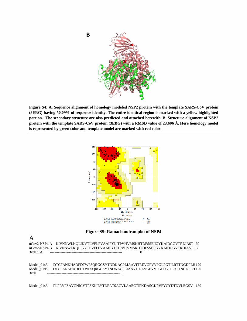
Figure S4: A. Sequence alignment of homology modeled NSP2 protein with the template SARS-CoV protein
(3EBG) having 50.09% of sequence identity. The entire identical region is marked with a yellow highlighted
portion. The secondary structure are also predicted and attached herewith. B. Structure alignment of NSP2
protein with the template SARS-CoV protein (3EBG) with a RMSD value of 23.606 Å. Here homology model
is represented by green color and template model are marked with red color.
Figure S5: Ramachandran plot of NSP4
A nCov2-NSP4:A KIVNNWLKQLIKVTLVFLFVAAIFYLITPVHVMSKHTDFSSEIIGYKAIDGGVTRDIAST 60
nCov2-NSP4:B KIVNNWLKQLIKVTLVFLFVAAIFYLITPVHVMSKHTDFSSEIIGYKAIDGGVTRDIAST 60 3vcb.1.A ------------------------------------------------------------ 0 Model_01:A DTCFANKHADFDTWFSQRGGSYTNDKACPLIAAVITREVGFVVPGLPGTILRTTNGDFLH 120 Model_01:B DTCFANKHADFDTWFSQRGGSYTNDKACPLIAAVITREVGFVVPGLPGTILRTTNGDFLH 120 3vcb ------------------------------------------------------------ 0
Model_01:A FLPRVFSAVGNICYTPSKLIEYTDFATSACVLAAECTIFKDASGKPVPYCYDTNVLEGSV 180
B
Model_01:B FLPRVFSAVGNICYTPSKLIEYTDFATSACVLAAECTIFKDASGKPVPYCYDTNVLEGSV 180 3vcb ------------------------------------------------------------ 0 Model_01:A AYESLRPDTRYVLMDGSIIQFPNTYLEGSVRVVTTFDSEYCRHGTCERSEAGVCVSTSGR 240
Model_01:B AYESLRPDTRYVLMDGSIIQFPNTYLEGSVRVVTTFDSEYCRHGTCERSEAGVCVSTSGR 240 3vcb ------------------------------------------------------------ 0 Model_01:A WVLNNDYYRSLPGVFCGVDAVNLLTNMFTPLIQPIGALDISASIVAGGIVAIVVTCLAYY 300 Model_01:B WVLNNDYYRSLPGVFCGVDAVNLLTNMFTPLIQPIGALDISASIVAGGIVAIVVTCLAYY 300 3vcb.1.A ------------------------------------------------------------ 0
Model_01:A FMRFRRAFGEYSHVVAFNTLLFLMSFTVLCLTPVYSFLPGVYSVIYLYLTFYLTNDVSFL 360 Model_01:B FMRFRRAFGEYSHVVAFNTLLFLMSFTVLCLTPVYSFLPGVYSVIYLYLTFYLTNDVSFL 360 3vcb ------------------------------------------------------------ 0
Model_01:A AHIQWMVMFTPLVPFWITIAYIICISTKHFYWFFSNYLKRRVVFNGVSFSTFEEAALCTF 420 Model_01:B AHIQWMVMFTPLVPFWITIAYIICISTKHFYWFFSNYLKRRVVFNGVSFSTFEEAALCTF 420 3vcb --------------------------------------------------TFEEMALTTF 10 **** ** **
Model_01:A LLNKEMYLKLRSDVLLPLTQYNRYLALYNKYKYFSGAMDTTSYREAACCHLAKALNDFS- 479 Model_01:B LLNKEMYLKLRSDVLLPLTQYNRYLALYNKYKYFSGAMDTTSYREAACCHLAKALNDFS- 479
3vcb MITKESYSKLKNSV--SDVAFNRYLSLYNKYRYFSGKMDTAAYREAACSQLAKAMETFNH 68 ::.** * **:..* . :****:*****:**** ***::******.:****:: *.
Model_01:A NSGSDVLYQPPQTSITSAVLQ 500 Model_01:B NSGSDVLYQPPQTSITSAVLQ 500 3vcb NNGNDVLYQPPTASVTTSFLQ 89 *.*.******* :*:*::.**
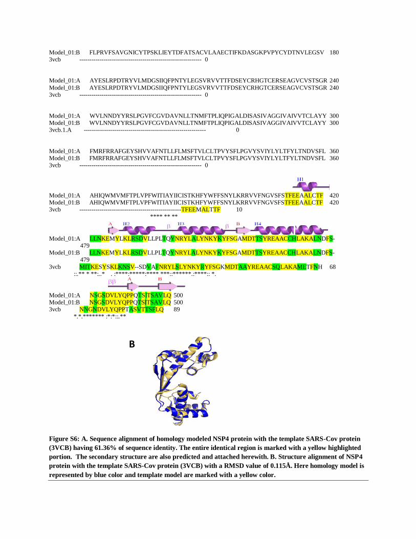
Figure S6: A. Sequence alignment of homology modeled NSP4 protein with the template SARS-Cov protein
(3VCB) having 61.36% of sequence identity. The entire identical region is marked with a yellow highlighted
portion. The secondary structure are also predicted and attached herewith. B. Structure alignment of NSP4
protein with the template SARS-Cov protein (3VCB) with a RMSD value of 0.115Å. Here homology model is
represented by blue color and template model are marked with a yellow color.
B
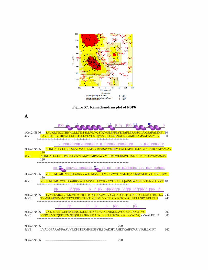
Figure S7: Ramachandran plot of NSP6
A
nCov2-NSP6 SAVKRTIKGTHHWLLLTILTSLLVLVQSTQWSLFFFLYENAFLPFAMGIIAMSAFAMMFV 60 4aV3 SAVKRTIKGTHHWLLLTILTSLLVLVQSTQWSLFFFLYENAFLPFAMGIIAMSAFAMMFV 60 ************************************************************
nCov2-NSP6 KHKHAFLCLFLLPSLATVAYFNMVYMPASWVMRIMTWLDMVDTSLSGFKLKDCVMYASAV 120 4aV3 KHKHAFLCLFLLPSLATVAYFNMVYMPASWVMRIMTWLDMVDTSLSGFKLKDCVMYASAV 120 ************************************************************
nCov2-NSP6 VLLILMTARTVYDDGARRVWTLMNVLTLVYKVYYGNALDQAISMWALIISVTSNYSGVVT 180 4aV3 VLLILMTARTVYDDGARRVWTLMNVLTLVYKVYYGNALDQAISMWALIISVTSNYSGVVT 180 ************************************************************
nCov2-NSP6 TVMFLARGIVFMCVEYCPIFFITGNTLQCIMLVYCFLGYFCTCYFGLFCLLNRYFRLTLG 240 4aV3 TVMFLARGIVFMCVEYCPIFFITGNTLQCIMLVYCFLGYFCTCYFGLFCLLNRYFRLTLG 240 ************************************************************
nCov2-NSP6 VYDYLVSTQEFRYMNSQGLLPPKNSIDAFKLNIKLLGVGGKPCIKVATVQ---------- 290 4aV3 VYDYLVSTQEFRYMNSQGLLPPKNSIDAFKLNIKLLGVGGKPCIKVATVQYVAALFFLIP 300 ************************************************** nCov2-NSP6 ------------------------------------------------------------ 290
4aV3 LVALGFAAANFAAVVRKPETERMKEISSYIRSGADSFLAHETKAIFKVAIVIAILLMIFT 360 nCov2-NSP6 ------------------------------------------------------------ 290
4aV3 TWQTGVAFLLGAVMSASAGIVGMKMATRANVRVAEAARTTKKIGPALKVAYQGGSVMGLS 420 nCov2-NSP6 ------------------------------------------------------------ 290
4aV3 VGGFALLGLVLVYLIFGKWMGQVDNLNIYTNWLGINFVPFAMTVSGYALGCSIIAMFDRV 480 nCov2-NSP6 ------------------------------------------------------------ 290 4aV3 GGGVYTKAADMAADLVGKNPATIADNVGDNVGDVAGLGADLLESFVGAIVSSIILASYMF 540 nCov2-NSP6 ------------------------------------------------------------ 290
4aV3 PIYVQKIGENLVHQVPKETIQALISYPIFFALVGLGCSMLGILYVIVKKPSDNPQRELNI 600 nCov2-NSP6 ------------------------------------------------------------ 290 4aV3 SLWTSALLTVVLTAFLTYFYLKDLQGLDVLGFRFGAISPWFSAIIGIFSGILIGFWAEYY 660 nCov2-NSP6 ------------------------------------------------------------ 290
4aV3 TSYRYKPTQFLGKSSIEGTGMVISNGLSLGMKSVFPPTLTLVLGILFADYFAGLYGVAIA 720 nCov2-NSP6 ------------------------------------------------------------ 290 4aV3 ALGMLSFVATSVSVDSYGPIADNAGGISEMCELDPEVRKITDHLDAVGNTTAAIGKGFAI 780 nCov2-NSP6 ------------------------------------------------------------ 290
4aV3 GSAIFAALSLFASYMFSQISPSDIGKPPSLVLLLNMLDARVIAGALLGAAITYYFSGYLI 840 nCov2-NSP6 ------------------------------------------------------------ 290 4aV3 SAVTKAAMKMVDEYNRCIEITSDNALKQMGYPAFIAILTPLVTGFLLGAEFVGGVLIGTV 900 nCov2-NSP6 ------------------------------------------------------------ 290 4aV3 LSGAMLAILTANSGGAWDNAKKYLEAGNLEGYGKGSEPHKALVIGDTVGDPLKDTVGPSL 960
nCov2-NSP6 ------------------------ 290 4aV3 DILIKIMSVVSVIAVSIFKHVHLF 984
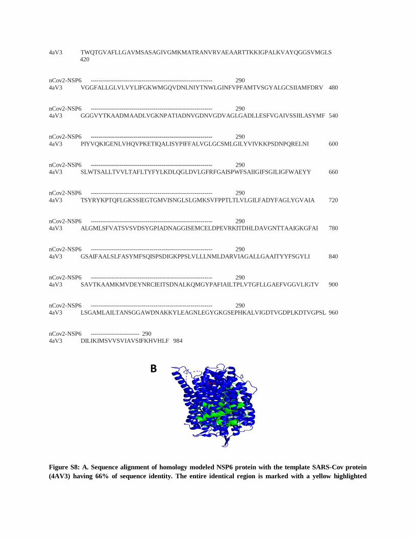
Figure S8: A. Sequence alignment of homology modeled NSP6 protein with the template SARS-Cov protein
(4AV3) having 66% of sequence identity. The entire identical region is marked with a yellow highlighted
B
portion. The secondary structure are also predicted and attached herewith. B. Structure alignment of NSP6
protein with the template SARS-Cov protein (3VCB) with a RMSD value of 16.675Å. Here homology model is
represented by green color and template model are marked with a blue color.
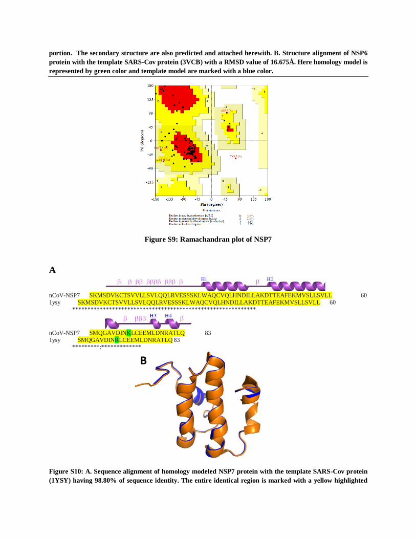
Figure S9: Ramachandran plot of NSP7
A
nCoV-NSP7 SKMSDVKCTSVVLLSVLQQLRVESSSKLWAQCVQLHNDILLAKDTTEAFEKMVSLLSVLL 60 1ysy SKMSDVKCTSVVLLSVLQQLRVESSSKLWAQCVQLHNDILLAKDTTEAFEKMVSLLSVLL 60 ************************************************************
nCoV-NSP7 SMQGAVDINKLCEEMLDNRATLQ 83 1ysy SMQGAVDINRLCEEMLDNRATLQ 83 *********:*************
Figure S10: A. Sequence alignment of homology modeled NSP7 protein with the template SARS-Cov protein
(1YSY) having 98.80% of sequence identity. The entire identical region is marked with a yellow highlighted
B
portion. The secondary structure are also predicted and attached herewith. B. Structure alignment of NSP7
protein with the template SARS-Cov protein (1YSY) with a RMSD value of 0.455Å. Here homology model is
represented by orange color and template model are marked with a blue color.
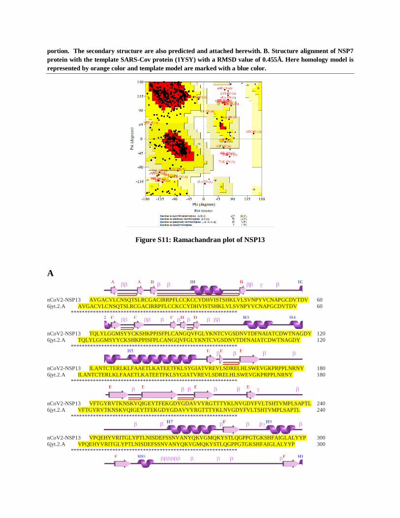
Figure S11: Ramachandran plot of NSP13
A
nCoV2-NSP13 AVGACVLCNSQTSLRCGACIRRPFLCCKCCYDHVISTSHKLVLSVNPYVCNAPGCDVTDV 60
6jyt.2.A AVGACVLCNSQTSLRCGACIRRPFLCCKCCYDHVISTSHKLVLSVNPYVCNAPGCDVTDV 60
************************************************************
nCoV2-NSP13 TQLYLGGMSYYCKSHKPPISFPLCANGQVFGLYKNTCVGSDNVTDFNAIATCDWTNAGDY 120
6jyt.2.A TQLYLGGMSYYCKSHKPPISFPLCANGQVFGLYKNTCVGSDNVTDFNAIATCDWTNAGDY 120
************************************************************
nCoV2-NSP13 ILANTCTERLKLFAAETLKATEETFKLSYGIATVREVLSDRELHLSWEVGKPRPPLNRNY 180
6jyt.2.A ILANTCTERLKLFAAETLKATEETFKLSYGIATVREVLSDRELHLSWEVGKPRPPLNRNY 180
************************************************************
nCoV2-NSP13 VFTGYRVTKNSKVQIGEYTFEKGDYGDAVVYRGTTTYKLNVGDYFVLTSHTVMPLSAPTL 240
6jyt.2.A VFTGYRVTKNSKVQIGEYTFEKGDYGDAVVYRGTTTYKLNVGDYFVLTSHTVMPLSAPTL 240
************************************************************
nCoV2-NSP13 VPQEHYVRITGLYPTLNISDEFSSNVANYQKVGMQKYSTLQGPPGTGKSHFAIGLALYYP 300
6jyt.2.A VPQEHYVRITGLYPTLNISDEFSSNVANYQKVGMQKYSTLQGPPGTGKSHFAIGLALYYP 300
************************************************************
nCoV2-NSP13 SARIVYTACSHAAVDALCEKALKYLPIDKCSRIIPARARVECFDKFKVNSTLEQYVFCTV 360
6jyt.2.A SARIVYTACSHAAVDALCEKALKYLPIDKCSRIIPARARVECFDKFKVNSTLEQYVFCTV 360
************************************************************
nCoV2-NSP13 NALPETTADIVVFDEISMATNYDLSVVNARLRAKHYVYIGDPAQLPAPRTLLTKGTLEPE 420
6jyt.2.A NALPETTADIVVFDEISMATNYDLSVVNARLRAKHYVYIGDPAQLPAPRTLLTKGTLEPE 420
************************************************************
nCoV2-NSP13 YFNSVCRLMKTIGPDMFLGTCRRCPAEIVDTVSALVYDNKLKAHKDKSAQCFKMFYKGVI 480
6jyt.2.A YFNSVCRLMKTIGPDMFLGTCRRCPAEIVDTVSALVYDNKLKAHKDKSAQCFKMFYKGVI 480
************************************************************
nCoV2-NSP13 THDVSSAINRPQIGVVREFLTRNPAWRKAVFISPYNSQNAVASKILGLPTQTVDSSQGSE 540
6jyt.2.A THDVSSAINRPQIGVVREFLTRNPAWRKAVFISPYNSQNAVASKILGLPTQTVDSSQGSE 540
************************************************************
nCoV2-NSP13 YDYVIFTQTTETAHSCNVNRFNVAITRAKVGILCIMSDRDLYDKLQFTSLEIPRRNVATL 600
6jyt.2.A YDYVIFTQTTETAHSCNVNRFNVAITRAKIGILCIMSDRDLYDKLQFTSLEIPRRNVATL 600
*****************************:******************************
nCoV2-NSP13 Q 601
6jyt.2.A Q 601
*
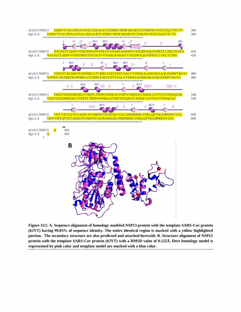
Figure S12: A. Sequence alignment of homology modeled NSP13 protein with the template SARS-Cov protein
(6JYT) having 99.83% of sequence identity. The entire identical region is marked with a yellow highlighted
portion. The secondary structure are also predicted and attached herewith. B. Structure alignment of NSP13
protein with the template SARS-Cov protein (6JYT) with a RMSD value of 0.125Å. Here homology model is
represented by pink color and template model are marked with a blue color.
B
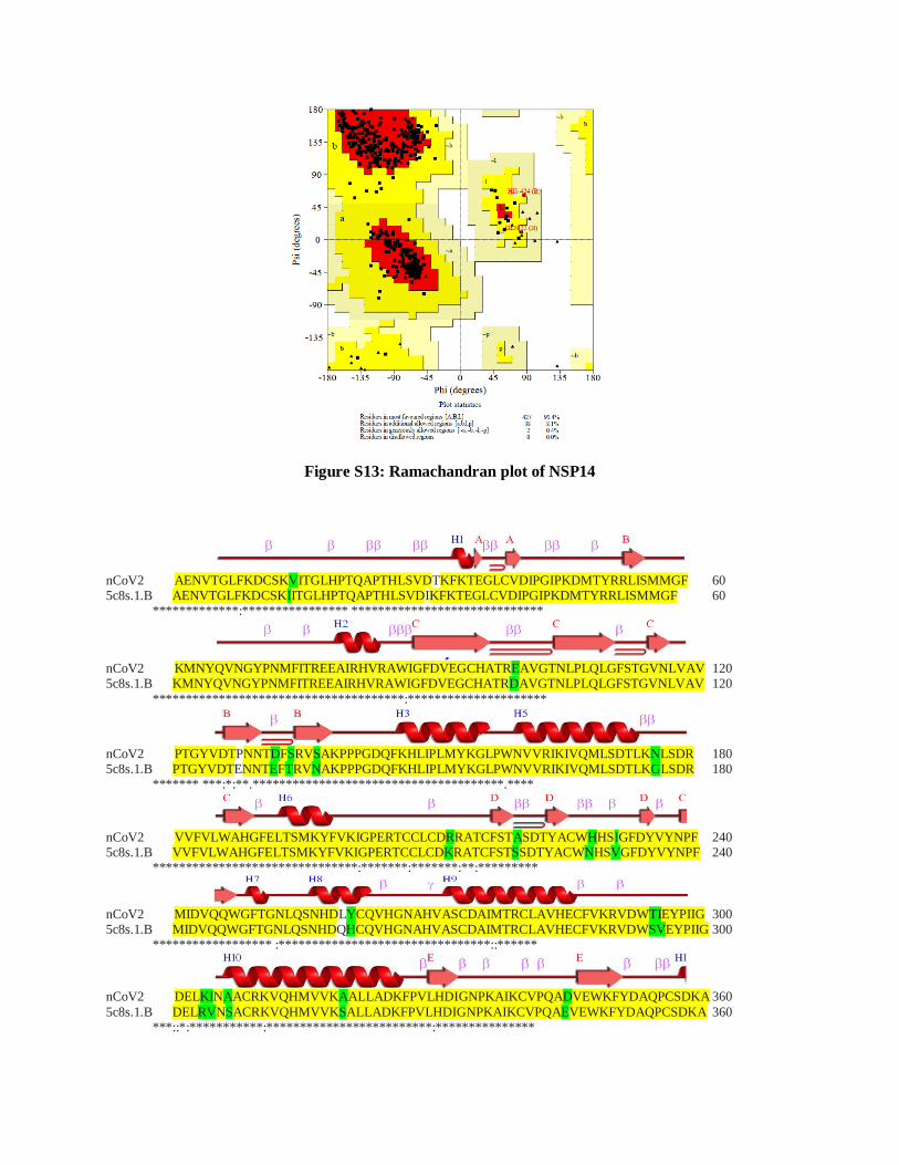
Figure S13: Ramachandran plot of NSP14
nCoV2 AENVTGLFKDCSKVITGLHPTQAPTHLSVDTKFKTEGLCVDIPGIPKDMTYRRLISMMGF 60 5c8s.1.B AENVTGLFKDCSKIITGLHPTQAPTHLSVDIKFKTEGLCVDIPGIPKDMTYRRLISMMGF 60 *************:**************** *****************************
nCoV2 KMNYQVNGYPNMFITREEAIRHVRAWIGFDVEGCHATREAVGTNLPLQLGFSTGVNLVAV 120 5c8s.1.B KMNYQVNGYPNMFITREEAIRHVRAWIGFDVEGCHATRDAVGTNLPLQLGFSTGVNLVAV 120
**************************************:*********************
nCoV2 PTGYVDTPNNTDFSRVSAKPPPGDQFKHLIPLMYKGLPWNVVRIKIVQMLSDTLKNLSDR 180 5c8s.1.B PTGYVDTENNTEFTRVNAKPPPGDQFKHLIPLMYKGLPWNVVRIKIVQMLSDTLKGLSDR 180 ******* ***:*:**.**************************************.****
nCoV2 VVFVLWAHGFELTSMKYFVKIGPERTCCLCDRRATCFSTASDTYACWHHSIGFDYVYNPF 240 5c8s.1.B VVFVLWAHGFELTSMKYFVKIGPERTCCLCDKRATCFSTSSDTYACWNHSVGFDYVYNPF 240 *******************************:*******:*******:**:*********
nCoV2 MIDVQQWGFTGNLQSNHDLYCQVHGNAHVASCDAIMTRCLAVHECFVKRVDWTIEYPIIG 300 5c8s.1.B MIDVQQWGFTGNLQSNHDQHCQVHGNAHVASCDAIMTRCLAVHECFVKRVDWSVEYPIIG 300 ****************** :********************************::******
nCoV2 DELKINAACRKVQHMVVKAALLADKFPVLHDIGNPKAIKCVPQADVEWKFYDAQPCSDKA 360
5c8s.1.B DELRVNSACRKVQHMVVKSALLADKFPVLHDIGNPKAIKCVPQAEVEWKFYDAQPCSDKA 360 ***::*:***********:*************************:***************
nCoV2 YKIEELFYSYATHSDKFTDGVCLFWNCNVDRYPANSIVCRFDTRVLSNLNLPGCDGGSLY 420 5c8s.1.B YKIEELFYSYATHHDKFTDGVCLFWNCNVDRYPANAIVCRFDTRVLSNLNLPGCDGGSLY 420 ************* *********************:************************
nCoV2 VNKHAFHTPAFDKSAFVNLKQLPFFYYSDSPCESHGKQVVSDIDYVPLKSATCITRCNLG 480 5c8s.1.B VNKHAFHTPAFDKSAFTNLKQLPFFYYSDSPCESHGKQVVSDIDYVPLKSATCITRCNLG 480 ****************.*******************************************
nCoV2 GAVCRHHANEYRLYLDAYNMMISAGFSLWVYKQFDTYNLWNTFTRLQ 527 5c8s.1.B GAVCRHHANEYRQYLDAYNMMISAGFSLWIYKQFDTYNLWNTFTRLQ 527 ************ ****************:*****************
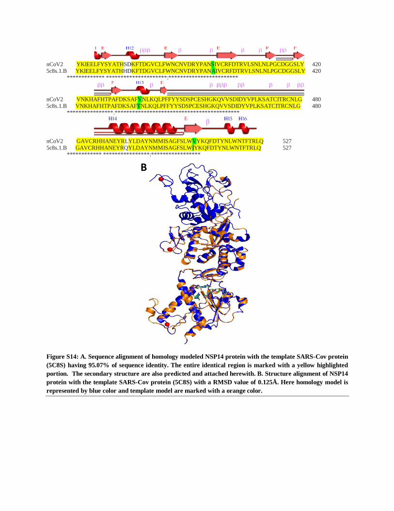
Figure S14: A. Sequence alignment of homology modeled NSP14 protein with the template SARS-Cov protein
(5C8S) having 95.07% of sequence identity. The entire identical region is marked with a yellow highlighted
portion. The secondary structure are also predicted and attached herewith. B. Structure alignment of NSP14
protein with the template SARS-Cov protein (5C8S) with a RMSD value of 0.125Å. Here homology model is
represented by blue color and template model are marked with a orange color.
B
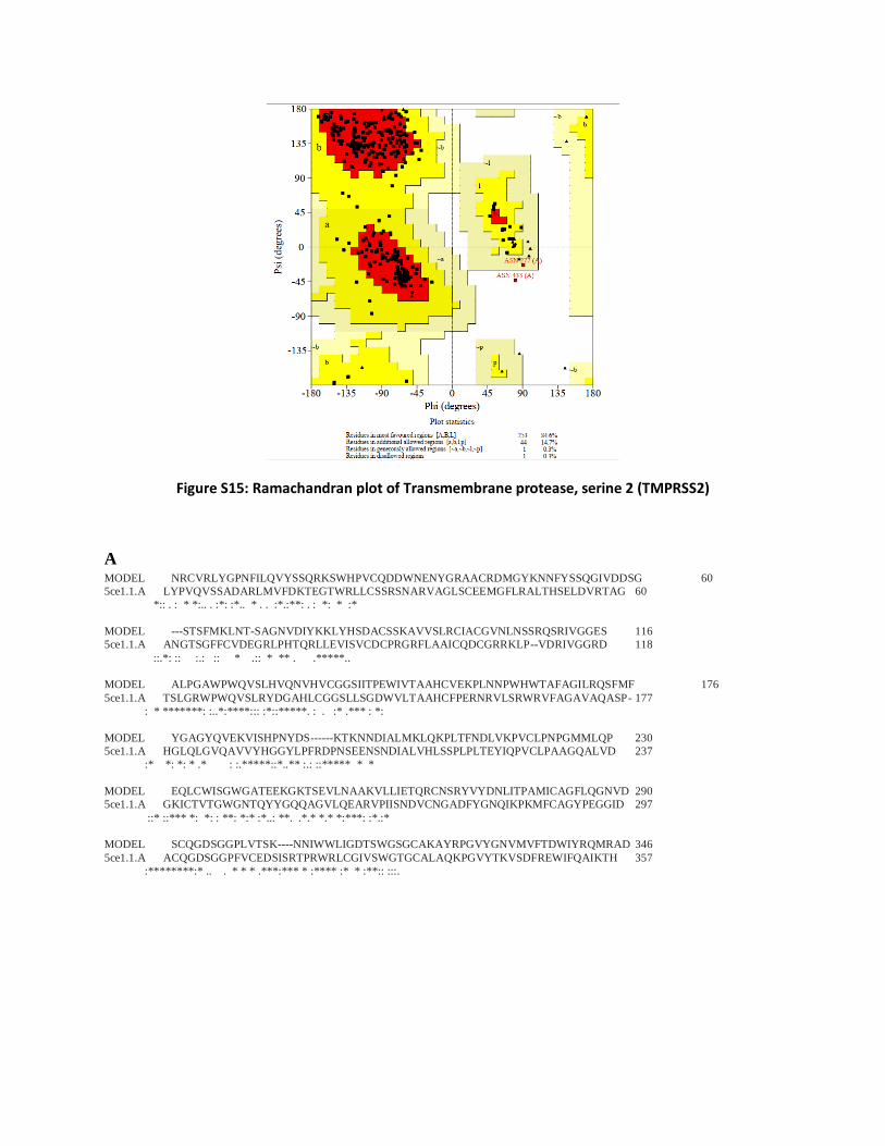
Figure S15: Ramachandran plot of Transmembrane protease, serine 2 (TMPRSS2)
A MODEL NRCVRLYGPNFILQVYSSQRKSWHPVCQDDWNENYGRAACRDMGYKNNFYSSQGIVDDSG 60
5ce1.1.A LYPVQVSSADARLMVFDKTEGTWRLLCSSRSNARVAGLSCEEMGFLRALTHSELDVRTAG 60
*:: . : * *:.. . :*: :*.. * . . :*.:**: . : *: * :*
MODEL ---STSFMKLNT-SAGNVDIYKKLYHSDACSSKAVVSLRCIACGVNLNSSRQSRIVGGES 116
5ce1.1.A ANGTSGFFCVDEGRLPHTQRLLEVISVCDCPRGRFLAAICQDCGRRKLP--VDRIVGGRD 118
::.*: :: :.: :: * .:: * ** . .*****..
MODEL ALPGAWPWQVSLHVQNVHVCGGSIITPEWIVTAAHCVEKPLNNPWHWTAFAGILRQSFMF 176
5ce1.1.A TSLGRWPWQVSLRYDGAHLCGGSLLSGDWVLTAAHCFPERNRVLSRWRVFAGAVAQASP- 177
: * *******: :..*:****::: :*::*****. : . :* .*** : *:
MODEL YGAGYQVEKVISHPNYDS------KTKNNDIALMKLQKPLTFNDLVKPVCLPNPGMMLQP 230
5ce1.1.A HGLQLGVQAVVYHGGYLPFRDPNSEENSNDIALVHLSSPLPLTEYIQPVCLPAAGQALVD 237
:* *: *: * .* : :.*****::*..** :.: ::***** * *
MODEL EQLCWISGWGATEEKGKTSEVLNAAKVLLIETQRCNSRYVYDNLITPAMICAGFLQGNVD 290
5ce1.1.A GKICTVTGWGNTQYYGQQAGVLQEARVPIISNDVCNGADFYGNQIKPKMFCAGYPEGGID 297
::* ::*** *: *: : **: *:* :*..: **. .*.* *.* *:***: :*.:*
MODEL SCQGDSGGPLVTSK----NNIWWLIGDTSWGSGCAKAYRPGVYGNVMVFTDWIYRQMRAD 346
5ce1.1.A ACQGDSGGPFVCEDSISRTPRWRLCGIVSWGTGCALAQKPGVYTKVSDFREWIFQAIKTH 357
:********:* .. . * * * .***:*** * :**** :* * :**:: :::.
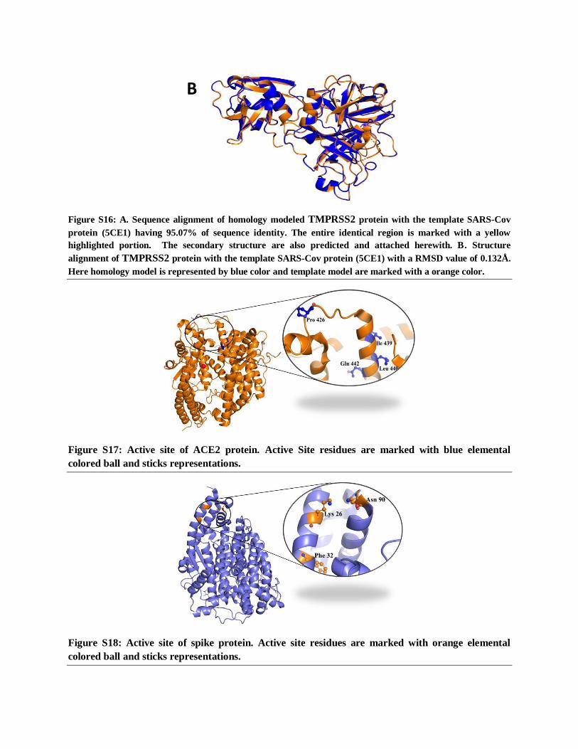
Figure S16: A. Sequence alignment of homology modeled TMPRSS2 protein with the template SARS-Cov
protein (5CE1) having 95.07% of sequence identity. The entire identical region is marked with a yellow
highlighted portion. The secondary structure are also predicted and attached herewith. B. Structure
alignment of TMPRSS2 protein with the template SARS-Cov protein (5CE1) with a RMSD value of 0.132Å.
Here homology model is represented by blue color and template model are marked with a orange color.
Figure S17: Active site of ACE2 protein. Active Site residues are marked with blue elemental
colored ball and sticks representations.
Figure S18: Active site of spike protein. Active site residues are marked with orange elemental
colored ball and sticks representations.
B
Figure S19: Active site of TMPRSS2 protein. Active site residues are marked with green elemental
colored ball and sticks representations.
Figure S20: Active site of envelope (E) protein. Active site residues are marked with blue elemental
colored ball and sticks representations.
Figure S21: Ramachandran Plot of Envelop protein.
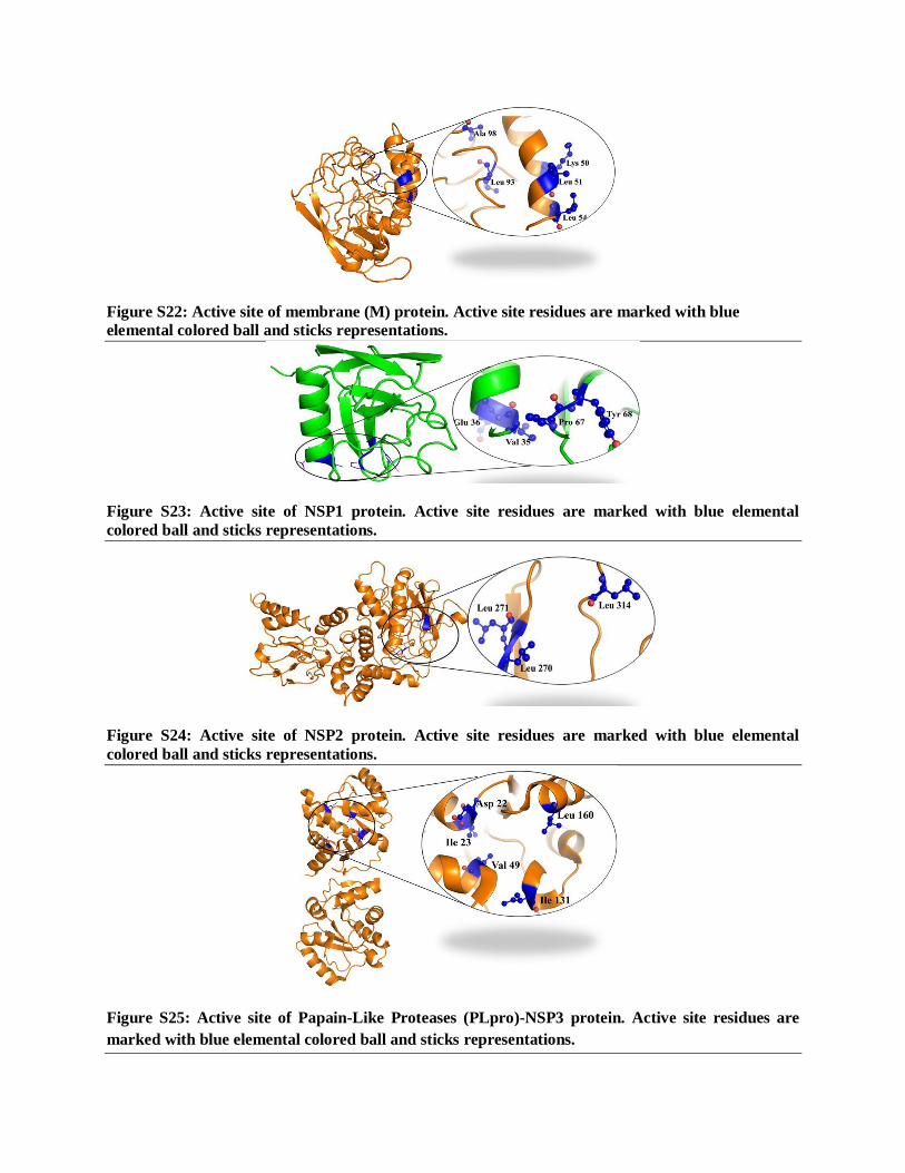
Figure S22: Active site of membrane (M) protein. Active site residues are marked with blue
elemental colored ball and sticks representations.
Figure S23: Active site of NSP1 protein. Active site residues are marked with blue elemental
colored ball and sticks representations.
Figure S24: Active site of NSP2 protein. Active site residues are marked with blue elemental
colored ball and sticks representations.
Figure S25: Active site of Papain-Like Proteases (PLpro)-NSP3 protein. Active site residues are
marked with blue elemental colored ball and sticks representations.
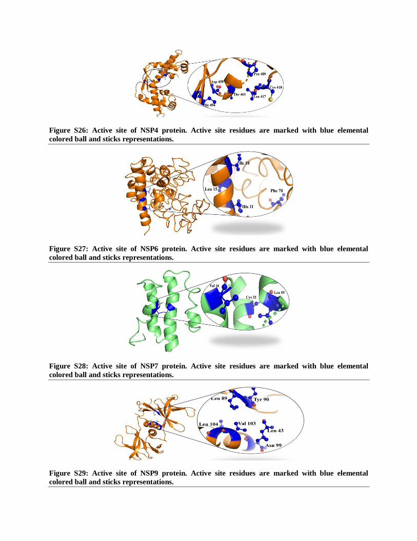
Figure S26: Active site of NSP4 protein. Active site residues are marked with blue elemental
colored ball and sticks representations.
Figure S27: Active site of NSP6 protein. Active site residues are marked with blue elemental
colored ball and sticks representations.
Figure S28: Active site of NSP7 protein. Active site residues are marked with blue elemental
colored ball and sticks representations.
Figure S29: Active site of NSP9 protein. Active site residues are marked with blue elemental
colored ball and sticks representations.
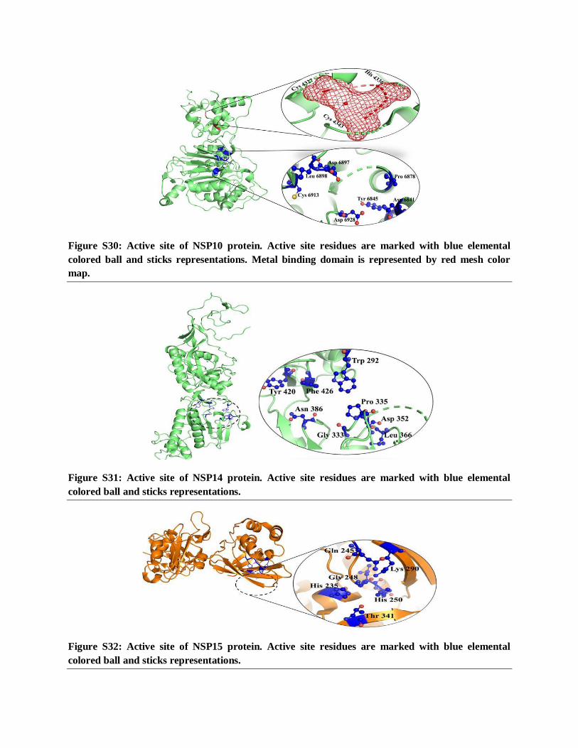
Figure S30: Active site of NSP10 protein. Active site residues are marked with blue elemental
colored ball and sticks representations. Metal binding domain is represented by red mesh color
map.
Figure S31: Active site of NSP14 protein. Active site residues are marked with blue elemental
colored ball and sticks representations.
Figure S32: Active site of NSP15 protein. Active site residues are marked with blue elemental
colored ball and sticks representations.
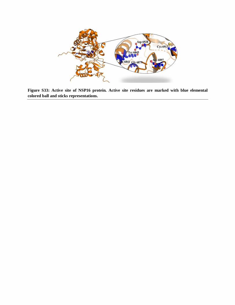
Figure S33: Active site of NSP16 protein. Active site residues are marked with blue elemental
colored ball and sticks representations.
download fileview on ChemRxivSupplementary_Compilation of Potential Protein Targets fo... (2.42 MiB)