Embed Size (px)
Citation preview
Competency Assessed SDLP:
UWSD Page 1 of 54 Feb 2009
COMPETENCY ASSESSED
SELF DIRECTED LEARNING PACKAGES
UNDERWATER SEAL DRAINAGE
NAME________________________________________
HEALTH SERVICE / DEPARTMENT________________________
FEBRUARY 2009, updated August 2013
Approved by the Gippsland Region Nurse Educators Group MARCH 2009
Acknowledgements to Gippsland Health Service Consortium Members for input
GRCE Points 3
for package + 1 point for competency assessment TOTAL 3 POINTS
GIPPSLAND HEALTH SERVICES CONSORTIUM
http://www.blebinfo.co.uk/phpBB2/viewtopic.php?t=10
Competency Assessed SDLP:
UWSD Page 2 of 54 Feb 2009
Under Water Seal Drainage
Table of Contents
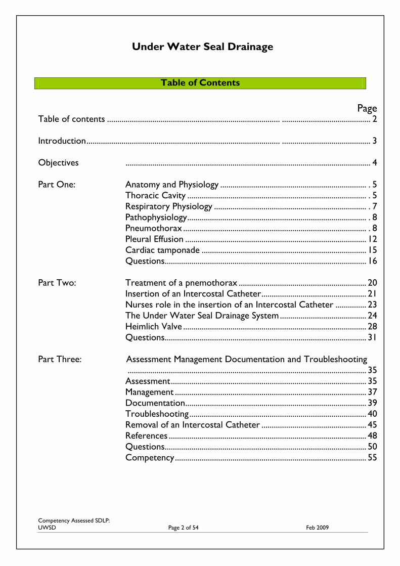
Page Table of contents .................................................................................... ........................................... 2
Introduction .............................................................................................. ........................................... 3
Objectives ....................................................................................................................... 4 4
Part One: Anatomy and Physiology ....................................................................... . 5
Thoracic Cavity ....................................................................................... . 5
Respiratory Physiology .......................................................................... . 7
Pathophysiology ....................................................................................... . 8
Pneumothorax ......................................................................................... . 8
Pleural Effusion ........................................................................................ 12
Cardiac tamponade ................................................................................ 15
Questions .................................................................................................. 16
Part Two: Treatment of a pnemothorax .............................................................. 20
Insertion of an Intercostal Catheter ................................................... 21
Nurses role in the insertion of an Intercostal Catheter ............... 23
The Under Water Seal Drainage System .......................................... 24
Heimlich Valve ......................................................................................... 28
Questions .................................................................................................. 31
Part Three: Assessment Management Documentation and Troubleshooting
.................................................................................................................... 35
Assessment ............................................................................................... 35
Management ............................................................................................. 37
Documentation ........................................................................................ 39
Troubleshooting ...................................................................................... 40
Removal of an Intercostal Catheter ................................................... 45
References ................................................................................................ 48
Questions .................................................................................................. 50
Competency ............................................................................................. 55
Competency Assessed SDLP:
UWSD Page 3 of 54 Feb 2009
INTRODUCTION
Congratulations on deciding to take on the challenge of a Self Directed Learning Package.
This package has been created to increase awareness and understanding of the concepts and related
nursing care associated with the insertion of an Intercostal Catheter (ICC) and the management of
an Underwater Seal Drainage (UWSD) system or other chest drainage devices.
The package addresses review of basic anatomy and physiology of the thoracic cavity in
concurrence with physiological conditions that require intervention. ICC insertion and UWSD
system management will be discussed in detail with both theoretical and practical information
included.
Assessment within the package includes both short answer and multiple choice questions at the end
of the three chapters. At the end of each chapter, questions may be submitted for marking and
feedback before progression onto the next chapter. The registered nurse is then required to
achieve a merit of clinical competency which is discussed in detail at the end of part 3 of the
learning package. On successful completion, a certificate will be awarded to you for the practical
component.
Proceed through the package at your own pace reviewing the information provided and responding
to the assessment questions. For further knowledge, go to the suggested reading or links to
websites for in depth explanations regarding the information provided. It is expected that this
package should take approximately 4 hours including suggested readings.
READ
This symbol indicates further reading to consolidate information or to further
your knowledge. It may be a book or a journal article
LINK
This symbol indicates a link with a website
REFER This symbol indicates to refer to your workplace policy
Competency Assessed SDLP:
UWSD Page 4 of 54 Feb 2009
OBJECTIVES
The nurse, upon completion of this package, should be able to:
1. Describe the thoracic anatomy, physiology and pathophysiology with particular reference to
conditions that may require chest drainage.
2. Outline the procedure for insertion of an Inter Costal Catheter
3. Discuss the functions of chest drainage
4. Outline the specific nursing care when managing a patient with an Under Water Seal
Drainage system
5. Document assessments performed on both the patient and Under Water Seal Drainage
system in an accurate and concise manner
6. Identify and discuss the potential hazards and emergencies that may occur during treatment
with an Under Water Seal Drainage system
7. Demonstrate increased knowledge of the concepts associated with Under Water Seal
Drainage by successfully completing the self-test at the end of each module.
8. Demonstrate clinical competency in the management of an Under Water Seal Drainage
system
Competency Assessed SDLP:
UWSD Page 5 of 54 Feb 2009
PART ONE
ANATOMY AND PHYSIOLOGY
One of the main difficulties in the understanding of the concept of UWSD lies in the knowledge
base of the nurse regarding normal anatomy and physiology of the thorax with emphasis on the
physiology of respiration. Once these concepts are grasped, it enables us to understand what can
go wrong in the structure and function of the chest and how these problems are treated.
The chest wall (known as the thoracic cavity) is made up of bones and muscles. The bones,
primarily ribs, sternum and vertebrae, form a protective cage for the internal structures of the
thorax. The main muscles of the chest wall, the external and internal intercostals, extend from one
rib to the next. [3]
THORACIC CAVITY
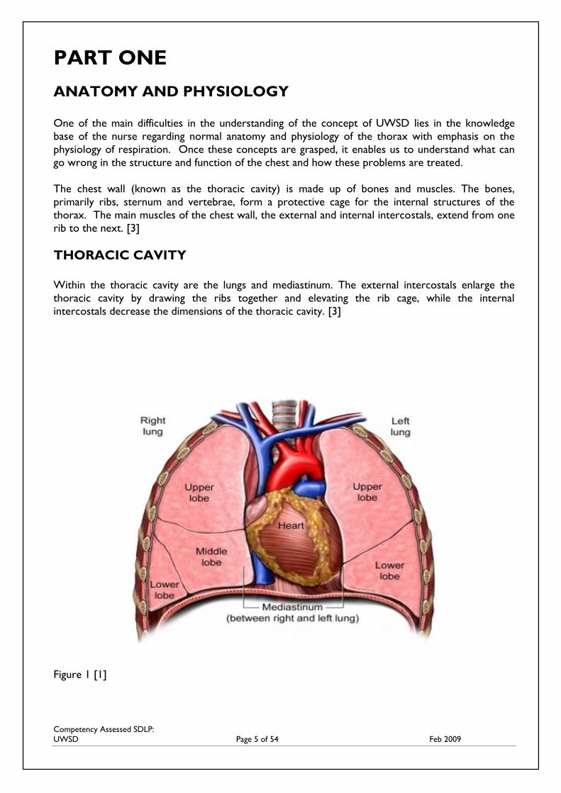
Within the thoracic cavity are the lungs and mediastinum. The external intercostals enlarge the
thoracic cavity by drawing the ribs together and elevating the rib cage, while the internal
intercostals decrease the dimensions of the thoracic cavity. [3]
Figure 1 [1]
Competency Assessed SDLP:
UWSD Page 6 of 54 Feb 2009
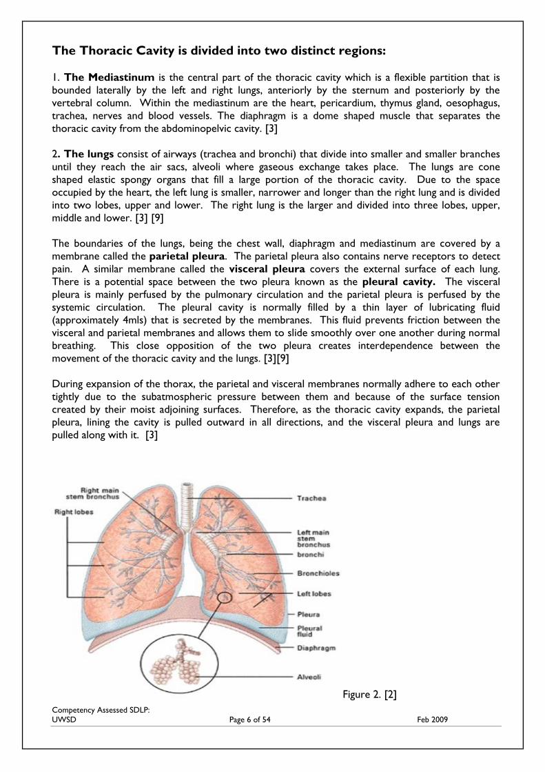
The Thoracic Cavity is divided into two distinct regions:
1. The Mediastinum is the central part of the thoracic cavity which is a flexible partition that is
bounded laterally by the left and right lungs, anteriorly by the sternum and posteriorly by the
vertebral column. Within the mediastinum are the heart, pericardium, thymus gland, oesophagus,
trachea, nerves and blood vessels. The diaphragm is a dome shaped muscle that separates the
thoracic cavity from the abdominopelvic cavity. [3]
2. The lungs consist of airways (trachea and bronchi) that divide into smaller and smaller branches
until they reach the air sacs, alveoli where gaseous exchange takes place. The lungs are cone
shaped elastic spongy organs that fill a large portion of the thoracic cavity. Due to the space
occupied by the heart, the left lung is smaller, narrower and longer than the right lung and is divided
into two lobes, upper and lower. The right lung is the larger and divided into three lobes, upper,
middle and lower. [3] [9]
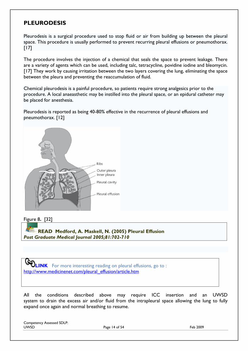
The boundaries of the lungs, being the chest wall, diaphragm and mediastinum are covered by a
membrane called the parietal pleura. The parietal pleura also contains nerve receptors to detect
pain. A similar membrane called the visceral pleura covers the external surface of each lung.
There is a potential space between the two pleura known as the pleural cavity. The visceral
pleura is mainly perfused by the pulmonary circulation and the parietal pleura is perfused by the
systemic circulation. The pleural cavity is normally filled by a thin layer of lubricating fluid
(approximately 4mls) that is secreted by the membranes. This fluid prevents friction between the
visceral and parietal membranes and allows them to slide smoothly over one another during normal
breathing. This close opposition of the two pleura creates interdependence between the
movement of the thoracic cavity and the lungs. [3][9]
During expansion of the thorax, the parietal and visceral membranes normally adhere to each other
tightly due to the subatmospheric pressure between them and because of the surface tension
created by their moist adjoining surfaces. Therefore, as the thoracic cavity expands, the parietal
pleura, lining the cavity is pulled outward in all directions, and the visceral pleura and lungs are
pulled along with it. [3]
Figure 2. [2]
Competency Assessed SDLP:
UWSD Page 7 of 54 Feb 2009
RESPIRATORY PHYSIOLOGY Respiration is an involuntary activity. [12] Air moves in and out of the thorax due to pressure
changes. Pulmonary Ventilation (breathing) is the process of gaseous exchange between the
atmosphere and lung alveoli. Air moves into the lungs when the pressure inside the lungs is less
than the air pressure in the atmosphere. Air moves out of the lungs when the pressure inside the
lungs is greater than the pressure in the atmosphere.
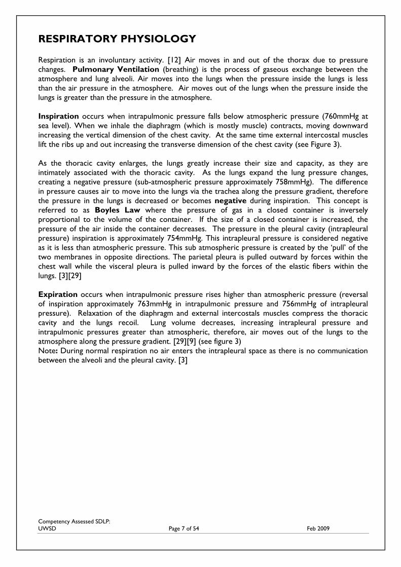
Inspiration occurs when intrapulmonic pressure falls below atmospheric pressure (760mmHg at
sea level). When we inhale the diaphragm (which is mostly muscle) contracts, moving downward
increasing the vertical dimension of the chest cavity. At the same time external intercostal muscles
lift the ribs up and out increasing the transverse dimension of the chest cavity (see Figure 3).
As the thoracic cavity enlarges, the lungs greatly increase their size and capacity, as they are
intimately associated with the thoracic cavity. As the lungs expand the lung pressure changes,
creating a negative pressure (sub-atmospheric pressure approximately 758mmHg). The difference
in pressure causes air to move into the lungs via the trachea along the pressure gradient, therefore
the pressure in the lungs is decreased or becomes negative during inspiration. This concept is
referred to as Boyles Law where the pressure of gas in a closed container is inversely
proportional to the volume of the container. If the size of a closed container is increased, the
pressure of the air inside the container decreases. The pressure in the pleural cavity (intrapleural
pressure) inspiration is approximately 754mmHg. This intrapleural pressure is considered negative
as it is less than atmospheric pressure. This sub atmospheric pressure is created by the ‘pull’ of the
two membranes in opposite directions. The parietal pleura is pulled outward by forces within the
chest wall while the visceral pleura is pulled inward by the forces of the elastic fibers within the
lungs. [3][29]
Expiration occurs when intrapulmonic pressure rises higher than atmospheric pressure (reversal
of inspiration approximately 763mmHg in intrapulmonic pressure and 756mmHg of intrapleural
pressure). Relaxation of the diaphragm and external intercostals muscles compress the thoracic
cavity and the lungs recoil. Lung volume decreases, increasing intrapleural pressure and
intrapulmonic pressures greater than atmospheric, therefore, air moves out of the lungs to the
atmosphere along the pressure gradient. [29][9] (see figure 3)
Note: During normal respiration no air enters the intrapleural space as there is no communication
between the alveoli and the pleural cavity. [3]
Competency Assessed SDLP:
UWSD Page 8 of 54 Feb 2009
Figure 3. [4]
PATHOPHYSIOLOGY
All gases travel from high pressure to low pressure in an attempt to equalize pressure. It is the
negative pressure present during normal respiration which prevents the visceral and parietal pleurae
from separating and therefore must be maintained at all times. [3]
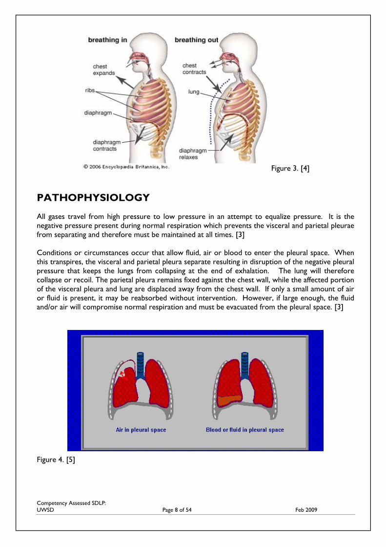
Conditions or circumstances occur that allow fluid, air or blood to enter the pleural space. When
this transpires, the visceral and parietal pleura separate resulting in disruption of the negative pleural
pressure that keeps the lungs from collapsing at the end of exhalation. The lung will therefore
collapse or recoil. The parietal pleura remains fixed against the chest wall, while the affected portion
of the visceral pleura and lung are displaced away from the chest wall. If only a small amount of air
or fluid is present, it may be reabsorbed without intervention. However, if large enough, the fluid
and/or air will compromise normal respiration and must be evacuated from the pleural space. [3]
Figure 4. [5]
Competency Assessed SDLP:
UWSD Page 9 of 54 Feb 2009
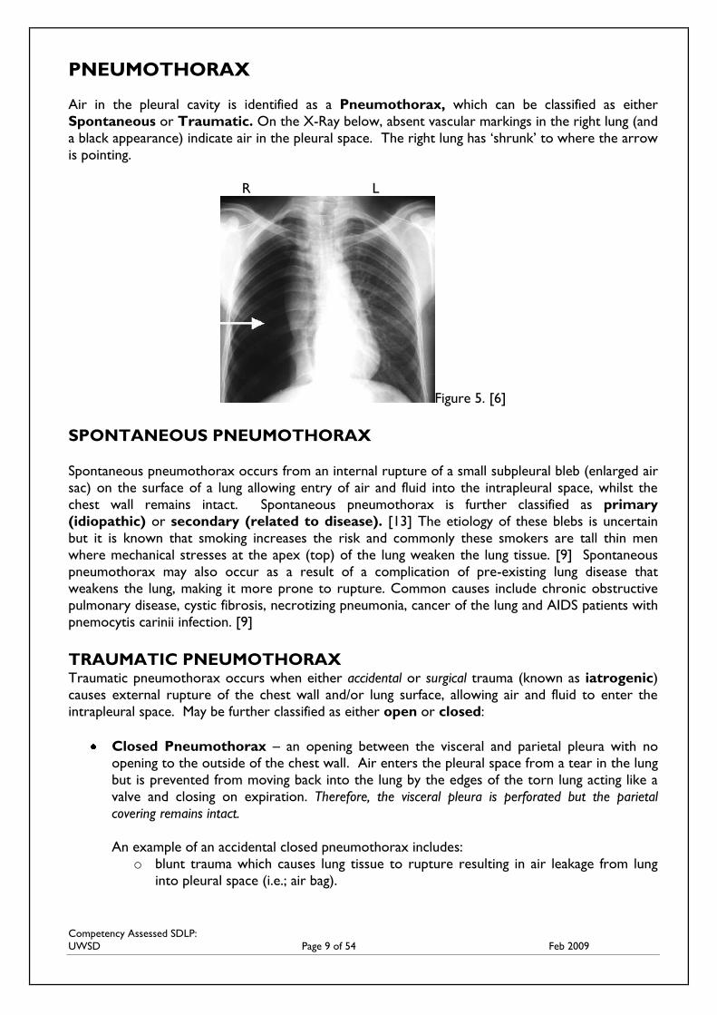
PNEUMOTHORAX
Air in the pleural cavity is identified as a Pneumothorax, which can be classified as either
Spontaneous or Traumatic. On the X-Ray below, absent vascular markings in the right lung (and
a black appearance) indicate air in the pleural space. The right lung has ‘shrunk’ to where the arrow
is pointing.
R L
Figure 5. [6]
SPONTANEOUS PNEUMOTHORAX
Spontaneous pneumothorax occurs from an internal rupture of a small subpleural bleb (enlarged air
sac) on the surface of a lung allowing entry of air and fluid into the intrapleural space, whilst the
chest wall remains intact. Spontaneous pneumothorax is further classified as primary
(idiopathic) or secondary (related to disease). [13] The etiology of these blebs is uncertain
but it is known that smoking increases the risk and commonly these smokers are tall thin men
where mechanical stresses at the apex (top) of the lung weaken the lung tissue. [9] Spontaneous
pneumothorax may also occur as a result of a complication of pre-existing lung disease that
weakens the lung, making it more prone to rupture. Common causes include chronic obstructive
pulmonary disease, cystic fibrosis, necrotizing pneumonia, cancer of the lung and AIDS patients with
pnemocytis carinii infection. [9]
TRAUMATIC PNEUMOTHORAX Traumatic pneumothorax occurs when either accidental or surgical trauma (known as iatrogenic)
causes external rupture of the chest wall and/or lung surface, allowing air and fluid to enter the
intrapleural space. May be further classified as either open or closed:
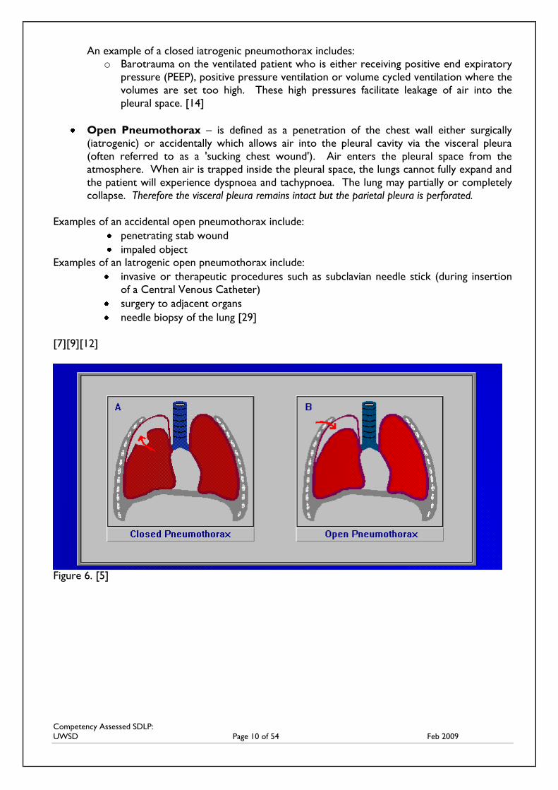
Closed Pneumothorax – an opening between the visceral and parietal pleura with no
opening to the outside of the chest wall. Air enters the pleural space from a tear in the lung
but is prevented from moving back into the lung by the edges of the torn lung acting like a
valve and closing on expiration. Therefore, the visceral pleura is perforated but the parietal
covering remains intact.
An example of an accidental closed pneumothorax includes:
o blunt trauma which causes lung tissue to rupture resulting in air leakage from lung
into pleural space (i.e.; air bag).
Competency Assessed SDLP:
UWSD Page 10 of 54 Feb 2009
An example of a closed iatrogenic pneumothorax includes:
o Barotrauma on the ventilated patient who is either receiving positive end expiratory
pressure (PEEP), positive pressure ventilation or volume cycled ventilation where the
volumes are set too high. These high pressures facilitate leakage of air into the
pleural space. [14]
Open Pneumothorax – is defined as a penetration of the chest wall either surgically
(iatrogenic) or accidentally which allows air into the pleural cavity via the visceral pleura
(often referred to as a 'sucking chest wound'). Air enters the pleural space from the
atmosphere. When air is trapped inside the pleural space, the lungs cannot fully expand and
the patient will experience dyspnoea and tachypnoea. The lung may partially or completely
collapse. Therefore the visceral pleura remains intact but the parietal pleura is perforated.
Examples of an accidental open pneumothorax include:
penetrating stab wound
impaled object
Examples of an Iatrogenic open pneumothorax include:
invasive or therapeutic procedures such as subclavian needle stick (during insertion
of a Central Venous Catheter)
surgery to adjacent organs
needle biopsy of the lung [29]
[7][9][12]
Figure 6. [5]
Competency Assessed SDLP:
UWSD Page 11 of 54 Feb 2009
TENSION PNEUMOTHORAX
Tension Pneumothorax occurs when air continuously enters the intrapleural space with no means
of escape causing the pressure to build within the space more rapidly than it can be evacuated. This
condition is very serious and potentially life threatening. Tension pneumothorax may develop from
spontaneous or traumatic pneumothorax but is more likely to be seen with a traumatic. For
example, a knife wound tends to seal itself in the chest wall thus trapping air leaked from the lung
injury in the chest cavity. Tension pneumothorax may also develop from a closed iatrogenic
pneumothorax as seen in barotrauma in the ventilated patient. [14]
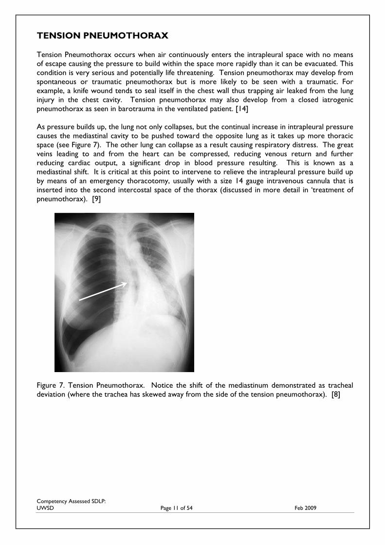
As pressure builds up, the lung not only collapses, but the continual increase in intrapleural pressure
causes the mediastinal cavity to be pushed toward the opposite lung as it takes up more thoracic
space (see Figure 7). The other lung can collapse as a result causing respiratory distress. The great
veins leading to and from the heart can be compressed, reducing venous return and further
reducing cardiac output, a significant drop in blood pressure resulting. This is known as a
mediastinal shift. It is critical at this point to intervene to relieve the intrapleural pressure build up
by means of an emergency thoracotomy, usually with a size 14 gauge intravenous cannula that is
inserted into the second intercostal space of the thorax (discussed in more detail in ‘treatment of
pneumothorax). [9]
Figure 7. Tension Pneumothorax. Notice the shift of the mediastinum demonstrated as tracheal
deviation (where the trachea has skewed away from the side of the tension pneumothorax). [8]
Competency Assessed SDLP:
UWSD Page 12 of 54 Feb 2009
HAEMOTHORAX
A condition caused by blood (‘haem’) collecting in the intrapleural space following trauma or
surgery which also causes lung collapse.
HAEMOPNEUMOTHORAX
The collection of blood and air in the intrapleural space. [12]
EMPYEMA
The accumulation of pus in the pleural space caused by infection (pneumonia), lung abscess, injury
or iatrogenic contamination. If extensive, a thoracotomy and drainage of the pleural cavity may be
required. [19]
CYLOTHORAX
The presence of lymph fluid within the intrapleural space. Rupture of a thoracic duct (part of the
lymphatic system) can occur during thoracic surgery (particularly during oesphagogastrectomy) or
as a result of erosion from the presence of an ICC. As a result, lymphatic fluid drains into the
thoracic cavity causing pressure on the lungs. This leads to basal collapse or chylothorax. [19]
The collection of fluid in the intrapleural space causes the same disruption to the intrapleural
pressure that leads to decreased lung expansion as a pneumothorax, although tension
pneumothorax is extremely rare. [19]
PLEURAL EFFUSION
Pleural Effusion is defined as a collection of fluid that has entered the pleural space. Anything that
affects the balance between the fluid filtration by the parietal pleura and fluid absorption by the
visceral pleura can cause a pleural effusion. Pleural effusions can develop from;
increased capillary pressure e.g.; left ventricular failure
reduced plasma oncotic pressure e.g.; cirrhosis of the liver
increased capillary permeability e.g.; pleural metastases
obstruction of lymphatic drainage by tumors or obstruction of the superior vena cava
[12 ]
Etiology of Pleural Effusions
When the presence of a pleural effusion is suspected by physical examination, confirmation with a
Chest X-Ray is necessary. To establish the aetiology of a pleural effusion, a thoracentesis usually
needs to be performed. [15] Thoracentesis involves inserting a catheter into the pleural space
through the thoracic cavity to extract fluid. It may also be referred to as a pleural biopsy or a
pleural tap. Thoracentesis may be performed while simultaneously using ultrasound guidance to
extract 50-100ml of fluid for analysis.
Competency Assessed SDLP:
UWSD Page 13 of 54 Feb 2009
Not every pleural effusion needs to be tapped, but when there is no obvious clinical cause for the
effusion, the patient is febrile, or has respiratory compromise, fluid should be removed. Extraction
of fluid may also be performed for the relief of symptoms caused by the pleural effusion. [15] It is
recommended that a pleural effusion be drained slowly to avoid the risk of re-expansion pulmonary
oedema. Laws et al (2003) suggests that no more than 1500mls should be drained at one time and
subsequent drainage should be limited to 500mls per hour. [20] Chest CT is performed to
distinguish between parenchymal and pleural disease and may demonstrate pleural thickening,
pleural calcification, a pleural based mass, or collections of fluid. [10]
The purpose of the pleural fluid analysis is to determine if the fluid is a transudate or an exudate (i.e.
where the fluid is coming from). Transudative (watery) effusions are associated with problems
outside the lungs, which disrupt protein production and decrease oncotic pressure. These effusions
are usually bilateral and are often caused by abnormal lung pressure. Congestive heart failure is
usually the cause. Exudative pleural effusions are protein rich effusions, which usually signal
infection or inflammation of the pleura as a result of inflammatory disease, cardiac surgery, drugs or
pleural malignancy. Blood stained fluid is suggestive of severe inflammation, infarction or malignancy
and is unilateral. [10]
Once an effusion is categorized as transudative or exudative, identifying the cause narrows.
Additional pleural fluid studies that help to establish a diagnosis include glucose, amylase, white
blood cell counts with differential and cytologic and microbiologic examination. [15]
Treatment of a Pleural Effusion
As previously mentioned therapeutic drainage of a pleural effusion may be done if the fluid
collection is large and causing pressure with associated shortness of breath. Pleural effusions caused
by congestive heart failure are treated with diuretics and other medications that treat heart failure.
Pleural effusions caused by infection are treated with antibiotics specific to the causative organism.
In patients with cancer or infections, the effusion is often treated by inserting an ICC to drain the
fluid or a smaller catheter known as a pigtail catheter, pleurax or pleurocath catheter. The choice of
catheter to be inserted is dependant on the type of fluid to be drained, for example empyema may
require a thicker catheter and dependant on the amount of drainage will decide how long the
catheter remains insitu. Chemotherapy or radiotherapy, or instilling medication within the chest
(referred to as a pleurodesis) that prevents re-accumulation of fluid after drainage may also be used
in some cases. [11][15]
Treatment for Chronic Pleural Effusions
The development of the PleurX Catheter system has now been introduced for patients whose
pleural effusion is chronic and at times irreversible. This entails a catheter being inserted into the
patient which remains in place and clamped off. When required, the patient or home nurse (or as
an inpatient) can attach the drainage system to the patient when they become symptomatic (i.e: in
the case of pleural effusion = shortness of breath) and drain fluid off. On completion of drainage,
the catheter is reclamped and the disposable system is simply discarded. This means that the
patient dose not have to endure repeated insertions of a pig tail catheter for treatment of a chronic
condition (Carefusion, 2010).
Competency Assessed SDLP:
UWSD Page 14 of 54 Feb 2009
PLEURODESIS
Pleurodesis is a surgical procedure used to stop fluid or air from building up between the pleural
space. This procedure is usually performed to prevent recurring pleural effusions or pneumothorax.
[17]
The procedure involves the injection of a chemical that seals the space to prevent leakage. There
are a variety of agents which can be used, including talc, tetracycline, povidine iodine and bleomycin.
[17] They work by causing irritation between the two layers covering the lung, eliminating the space
between the pleura and preventing the reaccumulation of fluid.
Chemical pleurodesis is a painful procedure, so patients require strong analgesics prior to the
procedure. A local anaeasthetic may be instilled into the pleural space, or an epidural catheter may
be placed for anesthesia.
Pleurodesis is reported as being 40-80% effective in the recurrence of pleural effusions and
pneumothorax. [12]
Figure 8. [32]
READ Medford, A. Maskell, N. (2005) Pleural Effusion
Post Graduate Medical Journal 2005;81:702-710
LINK For more interesting reading on pleural effusions, go to :
http://www.medicinenet.com/pleural_effusion/article.htm
All the conditions described above may require ICC insertion and an UWSD
system to drain the excess air and/or fluid from the intrapleural space allowing the lung to fully
expand once again and normal breathing to resume.
Competency Assessed SDLP:
UWSD Page 15 of 54 Feb 2009
Signs and Symptoms of a Pneumothorax
Whether traumatic or spontaneous and dependant on the health status of the individual,
pneumothorax may cause these cardinal signs and symptoms:
Sudden, sharp, pleuritic pain exacerbated by chest movement, breathing and coughing
Asymmetrical chest wall movement
Cyanosis
Shortness of breath
Hyper resonance or tympany heard on percussion
Respiratory distress
tachycardia
Absent or muffled breath sounds on affected side
Hypertension
Hypoxia with associated restlessness/confusion
Crackling beneath the skin on palpation indicating subcutaneous emphysema (air in the
subcutaneous tissue)
Referred pain to one or both shoulder tips
Advanced signs and symptoms indicating a Tension Pneumothorax has
developed may include: Chest rigidity on affected side
Cyanosis
hypoxemia
Tracheal deviation and hypotension signifying a mediastinal shift has occurred
Cardiac Arrest [8] [13]
CARDIAC TAMPONADE
All patients following cardiac surgery (such as valve replacement or coronary artery bypass graft
(CABG) surgery) will require chest drainage. Chest tubes must be placed in the pericardium
postoperatively to remove residual blood from the mediastinum.
Open thoracic surgery or trauma causes blood to pool in the mediastinal cavity therefore the
insertion of a chest tube post operatively is common. The most serious complication of mediastinal
drainage is known as cardiac tamponade. This occurs when blood or other fluids collect in the
pericardial sac, compressing the heart and preventing it from expanding to accept venous return.
This blood collection clots and compresses the heart, reducing cardiac contractility, cardiac output
and may produce a life threatening cardiopulmonary crisis. [19]
COMPLETE SHORT ANSWER AND MULTIPLE CHOICE QUESTIONS FOR PART
ONE AND SUBMIT TO YOUR NURSE EDUCATOR
Competency Assessed SDLP:
UWSD Page 16 of 54 Feb 2009
NAME: _________________________________DEPARTMENT: __________________ QUESTIONS: PART ONE
1. What 2 distinct regions is the thoracic cavity divided into?
___________________________________________________________________
___________________________________________________________________
___________________________________________________________________
___________________________________________________________________
___________________________________________________________________
___________________________________________________________________
2. Describe the two types of pleural membranes involved in respiration and the
organs/structures that they encase.
___________________________________________________________________
___________________________________________________________________
___________________________________________________________________
___________________________________________________________________
3. What causes destruction of the pleural space and what happens to both the parietal and
visceral pleura?
___________________________________________________________________
___________________________________________________________________
___________________________________________________________________
___________________________________________________________________
4. Describe the difference between the two types of traumatic pneumothorax?
___________________________________________________________________
___________________________________________________________________
___________________________________________________________________
___________________________________________________________________
Competency Assessed SDLP:
UWSD Page 17 of 54 Feb 2009
5. What is the difference between a tension pneumothorax and cardiac tamponade? What can
they both potentiate?
___________________________________________________________________
___________________________________________________________________
___________________________________________________________________
___________________________________________________________________
___________________________________________________________________
MULTIPLE CHOICE QUESTIONS
1. The organs encased in the mediastinum are:
a. heart, pericardium, thymus gland, oesophagus, trachea, nerves and blood vessels
b. heart, pericardium, thymus gland, oesophagus, trachea, lungs and blood vessels
c. heart, pericardium, intercostal muscles, thoracic vertebrae
d. heart, pericardium, lungs, thymus gland, pleural cavity
2. Pulmonary Ventilation works on the theory of Boyles Law where the volume of gas in a
closed container is inversely proportional to the volume of the container.
a. True
b. False
3. The visceral and parietal pleura normally rub smoothly against each other
because of:
a. The vacuum that separates them
b. Changes in thoracic cage size
c. Phrenic nerve stimulation
d. A thin layer of lubricating fluid
4. Mr Smith has been dropped off at your emergency department by a friend with a stab
wound to the right side of the chest. Clinical assessment indicates that he has incurred a
traumatic pneumothorax. Traumatic Pneumothorax is further divided into classifications.
What type of pneumothorax does he have?
a. Open pneumothorax
b. Closed pneumothorax
c. Iatrogenic pneumothorax
5. Signs and symptoms of a tension pneumothorax occurring include:
a. Hypotension, tracheal deviation, hypothermia
b. Hypotension, tracheal deviation, hypoxemia, hyperglycemia
c. Mediastinal shift, hypoglycemia, tachycardia, decreased respiratory rate
d. Hypotension, tracheal deviation, hypoxemia, dyspnoea
Competency Assessed SDLP:
UWSD Page 18 of 54 Feb 2009
6. A haemopneumothorax includes the collection of any type of fluid
a. True
b. False
7. A Pleural Effusion may be caused by:
a. Increased capillary pressure
b. Reduced plasma oncotic pressure
c. Increased capillary permeability
d. Obstruction of lymphatic drainage by tumors
e. All of the above
8. Transudative exudate aspirated from a pleural effusion is usually indicative of:
a. malignancy
b. Left ventricular failure
c. Infection
d. Trauma
9. Under normal conditions, pleural pressure is:
a. Zero
b. Positive
c. Negative
d. Equal to atmospheric pressure
10. Signs and symptoms of a pneumothorax include:
a. Shortness of breath, dyspnoea, tachycardia, abdominal pain
b. Shortness of breath, tracheal deviation, hypotension
c. Shortness of breath, tachycardia, dyspnoea, +/- shoulder tip pain
d. Shortness of breath, visual disturbance, absent breath sounds on affected side
Competency Assessed SDLP:
UWSD Page 19 of 54 Feb 2009
PART TWO
TREATMENT OF A PNEUMOTHORAX
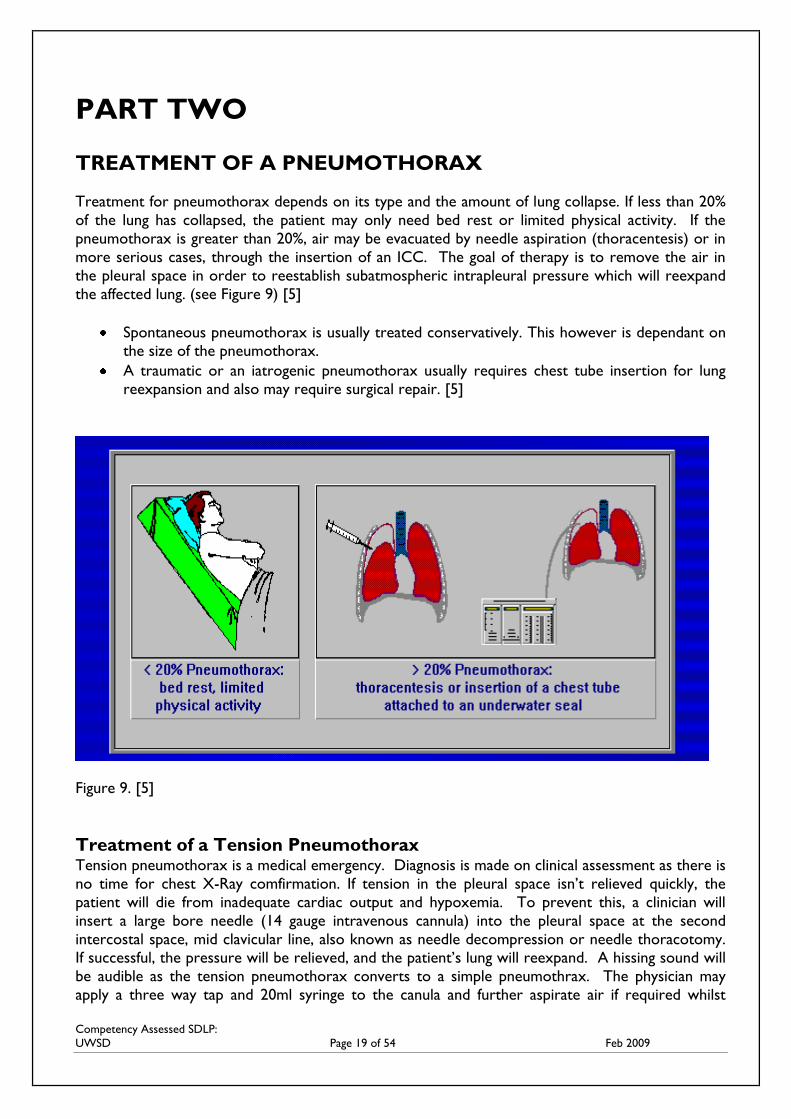
Treatment for pneumothorax depends on its type and the amount of lung collapse. If less than 20%
of the lung has collapsed, the patient may only need bed rest or limited physical activity. If the
pneumothorax is greater than 20%, air may be evacuated by needle aspiration (thoracentesis) or in
more serious cases, through the insertion of an ICC. The goal of therapy is to remove the air in
the pleural space in order to reestablish subatmospheric intrapleural pressure which will reexpand
the affected lung. (see Figure 9) [5]
Spontaneous pneumothorax is usually treated conservatively. This however is dependant on
the size of the pneumothorax.
A traumatic or an iatrogenic pneumothorax usually requires chest tube insertion for lung
reexpansion and also may require surgical repair. [5]
Figure 9. [5]
Treatment of a Tension Pneumothorax Tension pneumothorax is a medical emergency. Diagnosis is made on clinical assessment as there is
no time for chest X-Ray comfirmation. If tension in the pleural space isn’t relieved quickly, the
patient will die from inadequate cardiac output and hypoxemia. To prevent this, a clinician will
insert a large bore needle (14 gauge intravenous cannula) into the pleural space at the second
intercostal space, mid clavicular line, also known as needle decompression or needle thoracotomy.
If successful, the pressure will be relieved, and the patient’s lung will reexpand. A hissing sound will
be audible as the tension pneumothorax converts to a simple pneumothrax. The physician may
apply a three way tap and 20ml syringe to the canula and further aspirate air if required whilst
Competency Assessed SDLP:
UWSD Page 20 of 54 Feb 2009
inserting the ICC for the resulting pneumothorax, connecting the ICC to an UWSD system or valve
(known as a Heimlich Valve). [29]
INSERTION OF AN INTER COSTAL CATHETER
Also referred to as a thoracostomy tube, thoracic catheter, pleural drain tube, or a chest tube
Size and Structure
The intercostal catheter (ICC) is generally 16 to 20 inches in length and made of medical grade clear
polyvinyl chloride (PVC) with 3 to 6 drainage holes at its proximal end. There is a radiopaque line
along its length to allow the catheter to be visualized on x-ray, ensuring proper placement.
There are two types of ICCs:
1. Flexible straight or right angle catheters designed for insertion through an open chest during
surgery.
2. Trocar type catheters which are flexible straight transparent PVC which are packaged with a
rigid, pointed stylet through its centre,
Catheter Insertion
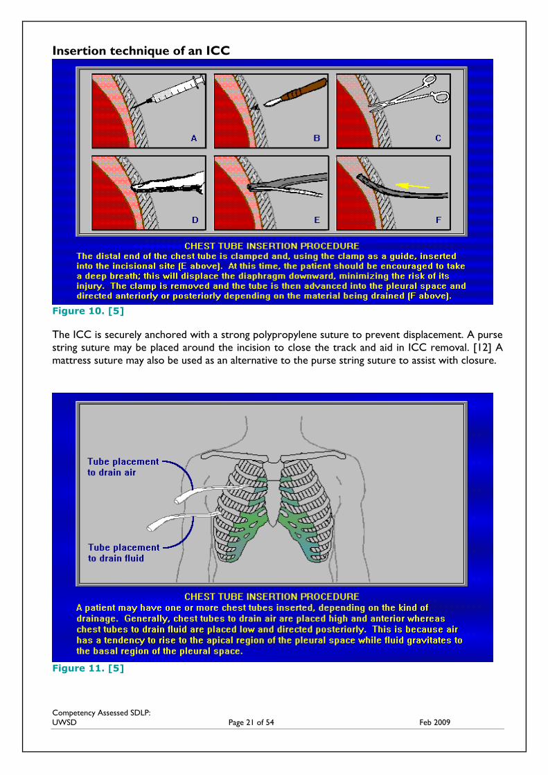
A patient may have one or more ICC’s inserted depending on the kind of drainage. If fluid (blood,
pleural effusions) is to be drained, the ICC is placed lower in the chest. Maintaining an upright
position will increase drainage by gravity as fluid sinks to the lowest point.
Placement:
o Pneumothorax
If the pneumothorax is large and the patient unstable, placement is directed
to the 4th to 5th inter costal space mid axillary line using a size 24-28 French
ICC
If the pneumothorax is large but the patient is stable, placement is the same
but a smaller french catheter may be used: 16-22 French
NB; 2nd intercostal space, mid clavicular line may be used for apical
pneumthoraces, but is not routinely recommended due to discomfort and
cosmesis (p. 5, 2012)
o Haemothorax
5th to 6th inter costal space mid axillary line using an ICC >28 French [24]
(See figure 11)
Patients who present with pleural effusions may have a smaller thoracic catheter inserted (usually an
8 to 14 French) and connected to a sealed drainage bag on free flow instead of an UWSD system to
evacuate the fluid. (e.g.: Pigtail Catheter or Pleurocath) This usually follows pleural aspiration and is
left in place for 24-48 hours until drainage ceases. These catheters are usually placed in the
posterior axillary line and may be inserted under fluoroscopy or ultrasound guidance. [15] Or as
mentioned earlier, the insertion of a long term catheter known as the Pleurex catheter.
Competency Assessed SDLP:
UWSD Page 21 of 54 Feb 2009
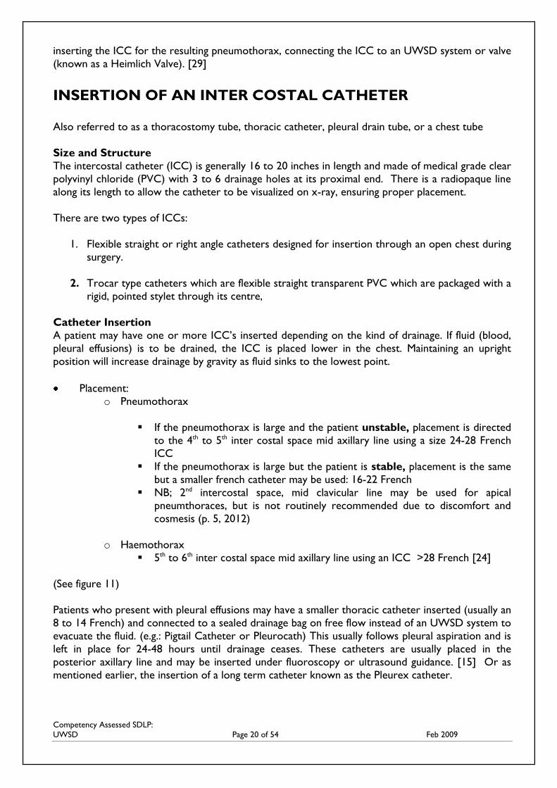
Insertion technique of an ICC
Figure 10. [5]
The ICC is securely anchored with a strong polypropylene suture to prevent displacement. A purse
string suture may be placed around the incision to close the track and aid in ICC removal. [12] A
mattress suture may also be used as an alternative to the purse string suture to assist with closure.
Figure 11. [5]
Competency Assessed SDLP:
UWSD Page 22 of 54 Feb 2009
NURSES ROLE IN THE INSERTION OF AN INTER COSTAL
CATHETER
The nurse’s role in insertion of an ICC is to:
Administer strong analgesia to the patient prior to the procedure and observe for side
effects
Set up equipment for the procedure, sterile gloves, mask and gown
Set up the UWSD system according to instructions (usually all is required is filling the water
seal to the indicated level and clamping all lines and adding sterile water if using a wet
suction system)
The position of the patient will depend on the position of the site of insertion but generally
the patient should be upright to encourage maximal lung expansion (the patient may rest
over a table supported with a pillow). The patient may also be asked to lift the arm up
around their head on the insertion side to aid visualization for the inserter
Connect patient to a cardiac and continuous oxygen saturation; monitor, observe and
report any changes to baselines observations to the medical officer throughout the insertion
procedure
Ensure emergency equipment is operational and emergency medication available (some
facilities administer a premedication of Atropine 600mcg IV or S/C to avoid a vasovagal
episode during insertion of the tube)
Communication with the patient during the procedure is essential and should include
adequate explanations
Once an ICC is inserted it is connected to a length of connecting tubing that leads to a drainage
collection chamber. The tubing should be of a length to allow the person to move and turn, and
reduce the chance of drainage being drawn back into the chest should a deep breath be taken. The
collection chamber, known as an UWSD system, is placed below the person’s chest so gravity
promotes the flow of drainage.
Dependant upon the UWSD system used the nurse will:
Connect the ICC to the patient and secure connection with tape ensuring connections
remain sterile. All connections should be secured in such a way that they can still be directly
observed [18]
Release all clamps and observe the UWSD for tidaling, bubbling, type and amount of
drainage and most importantly, the patient’s respiratory status.
REFER Check your local hospital policy and procedures regarding the current
UWSD system that you use and how the system is to be set up and secured
LINK For viewing of an ICC insertion go to:
http://www.youtube.com/watch?V=BOwGmWn8Ubs
Competency Assessed SDLP:
UWSD Page 23 of 54 Feb 2009
THE UNDER WATER SEAL DRAINAGE SYSTEM
In 1967, Deknatel introduced the first integrated disposable chest drainage unit based on the three
bottle system. Until about 25 years ago, this type of chest drainage systems consisted of three glass
bottles and 16 separate pieces. [9] Then a single, disposable plastic unit with three integrated
chambers was developed that could be used as a one, two or three bottle system depending on the
type and severity of the pneumothorax. This creation has reduced the amount of dead space which
was problematic with the traditional system. [9] Although more compact, easier to use, and less
prone to contamination than the traditional three-bottle system, the principles of modern chest
drain units are the same. UWSD systems come in a variety of forms dependant upon the product
supplied to your hospital. This tends to be the main issue that impedes nurses from confidently
managing UWSD. However, when a thorough understanding of the concepts behind UWSD and
respiratory pathophysiology are achieved, it enables the nurse to competently and confidently care
for a patient with any kind of UWSD system.
HOW AN UNDER WATER SEAL DRAINAGE SYSTEM WORKS
As previously mentioned, the Under Water Seal Drainage system has three major components:
1. Fluid Collection Chamber
2. Water Seal
3. Suction
Traditional UWSD systems could either function as a single, two, three or four bottle system
dependant on the requirements of the pneumothorax. For example, a straight forward
pneumothorax with very minimal drainage could function on a single bottle system utilizing the
concept of gravity drainage alone to evacuate air from the pleural space. In comparison, a
heamopneumothorax may have required a three bottle system, the first for drainage of blood, the
second for the water seal and the third for the application for suction. The three in one systems
are supplied with the three major components incorporated into the system. This means that the
disposable unit may be used for either a straight forward pneumothorax on gravity drainage alone
to a heavily exudating heamopneumothorax that requires the application of suction.
THREE IN ONE SYSTEM
1. Fluid Collection Chamber
The ICC directly connects to the tubing from the UWSD system. Drainage from the pleural cavity
flows through the tubing into the collection chamber. Drainage occurs on the concept of gravity by
where the UWSD system is placed below the level of the patient’s chest to evacuate air and or fluid
from the pleural cavity. The fluid will remain in the fluid collection chamber and the air will
continue to flow into the water seal chamber.
Competency Assessed SDLP:
UWSD Page 24 of 54 Feb 2009
2. The water seal
Traditionally, the simplest closed chest drainage previously used one bottle. The connecting tubing
attached directly to the chest tube into a single sterile bottle, which served as both a collection
chamber and a water seal; the single bottle having a dual purpose. A rigid tube that was positioned
inside the glass bottle was submerged under 2cm of sterile water so that air could escape into the
bottle during expiration but was unable to re-enter the tube with inspiration; the water acting as a
one-way valve.
An air vent in the top of the bottle allowed the pleural air to escape from the chamber into the
atmosphere. If this vent was capped, pressure would build up preventing further pleural air from
draining.
The bottle would also act as a collection chamber for any drainage. As the fluid in the bottle
increased, however, so did the positive pressure, therefore, the greater the workload for the
patient to expel air. This proved to be a disadvantage of this system because as fluid accumulated in
the bottle, so to would be the force required to overcome the increasing water seal [12]. To solve
this problem, a second drainage bottle could be applied, the first bottle would then become the fluid
collection chamber and the second the water seal.
One end of a short tube was inserted into the first bottle and the other end was inserted into the
water seal bottle extending almost to the bottom. Another short tube inserted in the water seal
bottle which provided the air vent or for the application of suction. [12] Those patients who had
significant volumes of fluid draining were best treated with a two-bottle system to avoid frequent
bottle changes.
In the modern disposable three in one systems, the second chamber, called the water seal contains
also contains about 2cm of water (which the nurse fills when setting up an UWSD system). The
water provides a barrier between the atmospheric pressure and subatmospheric (negative) intra-
pleural pressure. As air and/or fluid drain, there is an increase in negative intrapleural pressure and
re-expansion of the lung. Put simply the water seal prevents air being ‘sucked’ back into the pleural
space.
On inspiration, fluid in the water seal should tidal upwards as seen in the manometer of the water
seal (the manometer being a U shaped tube within the water seal that measures the amount of
negative pressure within the pleural cavity. This measurement is visualized as numbers on the
outside of the water seal compartment). [22] This directly reflects decreased intrapleural pressure
and increased suction between the visceral and parietal pleura. This is observed when the patient
inspires and the water in the water seal rises and then falls on exhalation. [9] The air then exits the
water seal (which is seen as bubbling in the water seal) and enters the suction control chamber,
which regulates the maximum amount of negative pressure that can be applied to the pleural space.
If suction is not being applied, this port should remain open so that air can escape from the system
(see Figure 12).
Note: The pattern of tidaling seen with a ventilated patient is the opposite of that observed in a
spontaneously breathing patient. This will be discussed in more detail further on.
In summary, the main purpose of the water seal is to allow air to exit from the pleural space on
exhalation and prevent air from reentering the pleural cavity on inhalation.
In the even more modern UWSD units, a one way valve replaces the traditional water seal. No
water is required to establish the one-way seal. Once connected to the patients tube and clamps
are released, the patient seal is established for patient protection. [9]
Competency Assessed SDLP:
UWSD Page 25 of 54 Feb 2009
High Negativity Relief Valve
The modern three in one units have been installed with a High Negativity Relief Valve. This is
indicated by rising water in the small arm of the water seal chamber when the water rises beyond
20cm. The relief chamber automatically vents excessive negative pressures, thus preventing
respiratory compromise from accumulated negativity. In instances of highly imposed negative
pressures such as stripping chest tubes, excessive coughing or crying, manual negative relief valves
may be pressed to relief these excessive pressures. Suction MUST be operational if using this valve
otherwise the negative pressure in the system may be reduced to zero (atmospheric pressure)
which may reaccumulate the pneumothorax. [9]
Air Leak Meter
Air leak meters have been included on some of the three in one systems. These are a part of the
water seal. The patient air leak meter indicates the approximate degree of air leak from the chest
cavity. The meter is made up of numbered columns from low to high i.e.; 1-7. The higher the
number the greater the degree of air leak and the size of the pneumothorax. By regularly
documenting the number, the nurse can monitor an air leak increase or decrease from the pleural
space. [9]
REFER Check the instructions on your current UWSD system and find out what
mechanism it has for the relief of excessive negative pressure and whether it has an
air leak meter
3. Suction
Traditionally, levels of suction were always established by filling a chamber with an ordered amount
of sterile fluid known as “wet” suction. Newer systems have been developed using “dry” suction.
There continue to be three in one systems that operate using wet suction but most newer systems
use the dry form for ease of operation.
Suction is applied to encourage reinflation of the lung by facilitating greater pressures to drain either
fluid or air. Suction increases the pressure difference between the pleural space and the UWSD
system by pulling air from the UWSD system and therefore lowering the pressure in the bottle and
enhancing drainage. Usually between 10-20cm of suction may be applied if:
gravity drainage is not enough
drainage of empyema or viscous pleural effusions
the patient has a weak respiratory effort or poor cough causing a reduction in pulmonary
compliance
air is leaking into the pleural space faster than it can be removed by a simple gravity drainage
system
to speed up drainage [9]
Competency Assessed SDLP:
UWSD Page 26 of 54 Feb 2009
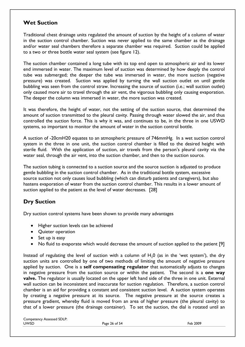
Wet Suction
Traditional chest drainage units regulated the amount of suction by the height of a column of water
in the suction control chamber. Suction was never applied to the same chamber as the drainage
and/or water seal chambers therefore a separate chamber was required. Suction could be applied
to a two or three bottle water seal system (see figure 12).
The suction chamber contained a long tube with its top end open to atmospheric air and its lower
end immersed in water. The maximum level of suction was determined by how deeply the control
tube was submerged; the deeper the tube was immersed in water, the more suction (negative
pressure) was created. Suction was applied by turning the wall suction outlet on until gentle
bubbling was seen from the control straw. Increasing the source of suction (i.e.; wall suction outlet)
only caused more air to travel through the air vent, the vigorous bubbling only causing evaporation.
The deeper the column was immersed in water, the more suction was created.
It was therefore, the height of water, not the setting of the suction source, that determined the
amount of suction transmitted to the pleural cavity. Passing through water slowed the air, and thus
controlled the suction force. This is why it was, and continues to be, in the three in one USWD
systems, so important to monitor the amount of water in the suction control bottle.
A suction of -20cmH20 equates to an atmospheric pressure of 746mmHg. In a wet suction control
system in the three in one unit, the suction control chamber is filled to the desired height with
sterile fluid. With the application of suction, air travels from the person’s pleural cavity via the
water seal, through the air vent, into the suction chamber, and then to the suction source.
The suction tubing is connected to a suction source and the source suction is adjusted to produce
gentle bubbling in the suction control chamber. As in the traditional bottle system, excessive
source suction not only causes loud bubbling (which can disturb patients and caregivers), but also
hastens evaporation of water from the suction control chamber. This results in a lower amount of
suction applied to the patient as the level of water decreases. [28]
Dry Suction
Dry suction control systems have been shown to provide many advantages
Higher suction levels can be achieved
Quieter operation
Set up is easy
No fluid to evaporate which would decrease the amount of suction applied to the patient [9]
Instead of regulating the level of suction with a column of H20 (as in the ‘wet system’), the dry
suction units are controlled by one of two methods of limiting the amount of negative pressure
applied by suction. One is a self compensating regulator that automatically adjusts to changes
in negative pressure from the suction source or within the patient. The second is a one way
valve. The regulator is usually located on the upper left hand side of the three in one unit. External
wall suction can be inconsistent and inaccurate for suction regulation. Therefore, a suction control
chamber is an aid for providing a constant and consistent suction level. A suction system operates
by creating a negative pressure at its source. The negative pressure at the source creates a
pressure gradient, whereby fluid is moved from an area of higher pressure (the pleural cavity) to
that of a lower pressure (the drainage container). To set the suction, the dial is rotated until an
Competency Assessed SDLP:
UWSD Page 27 of 54 Feb 2009
indicator of some sort (i.e.: an orange float appears in a window on the PLEUROVAC system) to
indicate that suction has been achieved (thus enough suction may created by as little as 5cmH20).
The suction control level can be changed at any time if prescribed by a medical officer by simply
rotating the dial to the new suction setting. [9]
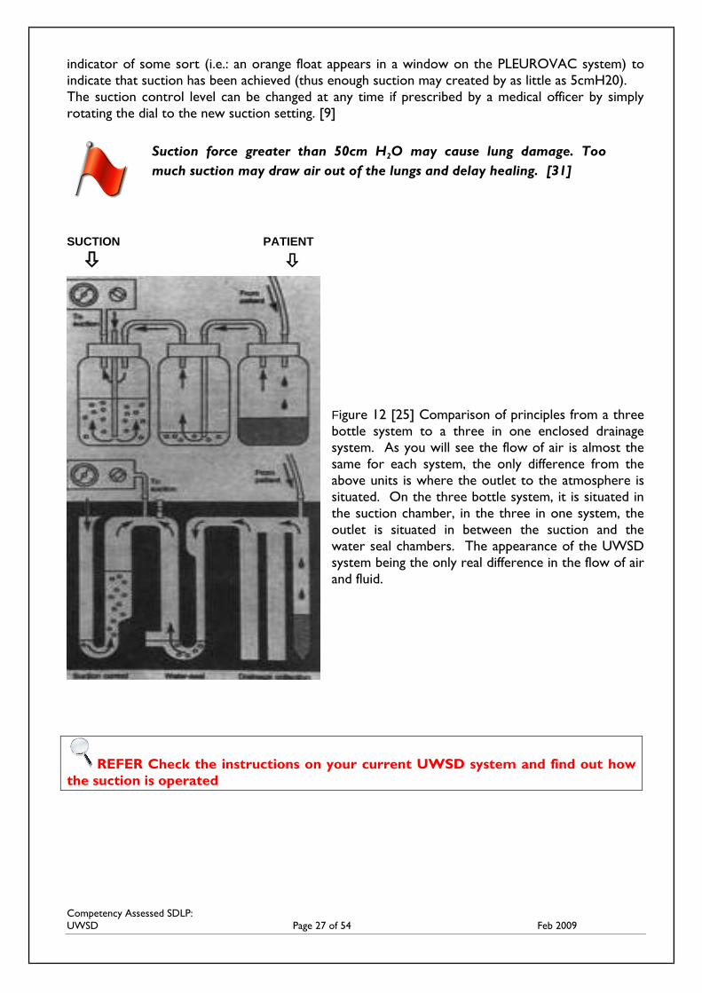
SUCTION PATIENT
Figure 12 [25] Comparison of principles from a three
bottle system to a three in one enclosed drainage
system. As you will see the flow of air is almost the
same for each system, the only difference from the
above units is where the outlet to the atmosphere is
situated. On the three bottle system, it is situated in
the suction chamber, in the three in one system, the
outlet is situated in between the suction and the
water seal chambers. The appearance of the UWSD
system being the only real difference in the flow of air
and fluid.
REFER Check the instructions on your current UWSD system and find out how
the suction is operated
Suction force greater than 50cm H2O may cause lung damage. Too
much suction may draw air out of the lungs and delay healing. [31]
Competency Assessed SDLP:
UWSD Page 28 of 54 Feb 2009
HEIMLICH VALVE
An alternative to UWSD was first devised in World War One when medical corpsmen created
makeshift one-way valves as the glass bottle systems were cumbersome and impractical to use on
the front. Then in the mid-1960s, Dr Henry Heimlich introduced the Heimlich valve which was
utilized during the Vietnam War. [21]
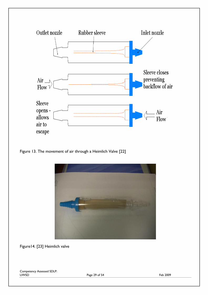
The Heimlich valve is attached directly to an ICC. This valve contains a piece of rubber tubing
which is flattened at one end to act as a flutter valve by allowing air to pass in only one direction.
The rubber tubing is enclosed in a hard, clear plastic case to prevent the tubing being compressed
and therefore occluded. The Heimlich flutter valve allows trapped intrapleural air and drainage to
escape during expiration without letting additional air to enter during inspiration. [21][22]
The Heimlich valve is best suited for an uncomplicated pneumothorax that requires little or no
drainage collection or suction. It is not recommended for a patient with large amounts of chest
drainage (more than 50 mls) or many blood clots. [21] The Heimlich Valve is still used today in
patients with small, slowly resolving pneumothoraces or those requiring transport, particularly in
emergency situations. [21] Patients with long term air leaks and minimal drainage can also be
discharged home with a Heimlich Valve with close support from healthcare professionals. [12]
Advantages
inexpensive
lightweight and portable – mobilization is easy
can function in any position; doesn’t need to be below the level of the patients chest
simple to use – requires very little assembly
reduces the amount of transport equipment (especially patients being retrieved)
Disadvantages
easily obstructed by fluid – unsuitable for use if fluid is to be drained, eg, post thoracic
surgery, haemothorax, haemopnuemothorax
detection of persistent air leaks or the formation of new air collections is difficult
education of staff required for safe use [21][22][24]
Competency Assessed SDLP:
UWSD Page 29 of 54 Feb 2009
Figure 13. The movement of air through a Heimlich Valve [22]
Figure14. [23] Heimlich valve

Competency Assessed SDLP:
UWSD Page 30 of 54 Feb 2009
COMPLETE SHORT ANSWER AND MULTIPLE CHOICE QUESTIONS FOR PART
TWO AND SUBMIT TO YOUR NURSE EDUCATOR
NAME: _____________________________ DEPARTMENT: __________________
1. Where is an ICC inserted for a
a) pneumothorax and
b) a haemothorax and what is the rationale for placement for both?
_________________________________________________________________
_________________________________________________________________
_________________________________________________________________
_________________________________________________________________
_________________________________________________________________
_________________________________________________________________
2. How is treatment decided for a Pneumothorax?
_________________________________________________________________
_________________________________________________________________
_________________________________________________________________
_________________________________________________________________
_________________________________________________________________
3. Summarize the nurse’s role in the insertion of an ICC.
_________________________________________________________________
_________________________________________________________________
_________________________________________________________________
_________________________________________________________________
_________________________________________________________________
_________________________________________________________________
_________________________________________________________________
_________________________________________________________________
QUESTIONS: PART 2
Competency Assessed SDLP:
UWSD Page 31 of 54 Feb 2009
_________________________________________________________________
_________________________________________________________________
_________________________________________________________________
_________________________________________________________________
_________________________________________________________________
_________________________________________________________________
_________________________________________________________________
4. Describe the movement of air and/or fluid as it leaves the pleural cavity through the
disposable three in one UWSD system (assuming suction has been attached).
_________________________________________________________________
_________________________________________________________________
_________________________________________________________________
_________________________________________________________________
_________________________________________________________________
_________________________________________________________________
_________________________________________________________________
5. Describe the differences between wet suction and dry suction.
_________________________________________________________________
_________________________________________________________________
_________________________________________________________________
_________________________________________________________________
_________________________________________________________________
_________________________________________________________________
Competency Assessed SDLP:
UWSD Page 32 of 54 Feb 2009
MULTIPLE CHOICE QUESTIONS
1. An ICC inserted in the midclavicular line, high in the anterior chest wall is to drain air only
a. True
b. False
2. The three in one UWSD systems were developed because of:
a. Increased risk of contamination
b. Ease of transportation
c. Decreased amount of dead space
d. All of the above
3. In a single bottle system (on the traditional ‘old’ UWSD system), moderate drainage (>150-
200mls) would prevent further drainage of air or fluid thus a second bottle could be attached
a. True
b. False
4. The air leak meter on the 3 in one system detects:
a. The amount of drainage
b. The amount of air being evacuated from the pleural space
c. When the system should be changed
d. How much pain the patient is in
5. When performing post insertion observations of the UWSD system, the nurse observes for:
a. Presence of tidaling in the water seal
b. The amount and type of drainage
c. the presence/absence of air/bubbling in the water seal on exhalation
d. all of the above
6. Shallow breathing would demonstrate less tidaling in the water seal and more in heavy
breathing as seen on the manometer
a. True
b. False
7. The new three in one systems suction control chamber that uses dry suction requires H20
to measure the amount of suction being applied to the pleural cavity
True
False
Competency Assessed SDLP:
UWSD Page 33 of 54 Feb 2009
8. Measurement of the amount of suction used in the ‘wet’ system is demonstrated by:
a. The amount of vigorous bubbling
b. The amount of suction dialed up at the suction outlet
c. the depth in which the control tube is submerged in the H20
d. all of the above
9. To observe the desired level of suction dialed up on the suction control dial on an UWSD
system using ‘dry’ suction being achieved, an indicator would be shown as either a symbol or
a float device
a. True
b. False
10. The Heimlich Valve may be used for transporting a patient to a metropolitan hospital. With
which type of pneumothorax would it be appropriate to remove the UWSD unit and
replace with the valve?
a. Haemothorax
b. Pneumothorax with moderate surgical emphysema (persistent air leak)
c. Spontaneous Pneumothorax with minimal drainage
d. haemopneumothorax
Competency Assessed SDLP:
UWSD Page 34 of 54 Feb 2009
PART THREE
ASSESSMENT
MANAGEMENT
DOCUMENTATION and
TROUBLESHOOTING
of the patient with an Under Water Seal Drainage System
ASSESSMENT
When any patient has an UWSD system or drainage device from the pleural cavity in place, it is imperative
that the patient is always assessed first.
PATIENT ASSESSMENT
When observing the drainage system, we must initially observe the patient. It is only when both the
patient and the UWSD system are assessed that any conclusions or trouble shooting can be
determined. The following assessments need to be performed on the patient with an UWSD
system:
Haemodynamic state: vital signs initially hourly including blood pressure, heart rate, skin
color and peripheral perfusion
Neurological assessment
Pain assessment – having an ICC insitu is very uncomfortable and painful for patients
therefore encourage analgesia prior to physiotherapy and on a regular basis to encourage
normal respirations compared to shallow respirations. Does the patient require education
regardingpain relief and does the patient understand the purpose of the UWSD?
Respiratory assessment (rate, depth, pattern). Auscultate to determine altered breath
sounds.
Palpate the insertion site and surrounding tissue for subcutaneous emphysema (free air
passing into the subcutaneous tissue). Subcutaneous emphysema can occur if one or more
drainage holes are situated outside of the pleural space or if the tubing is blocked or kinked.
[12]
Physiotherapy: is the patient performing deep breathing and coughing to facilitate the exit of
air from the pleural space. Coughing raises intrapleural pressure which facilitates emptying of
an accumulation in the pleural space of air and/or fluid
Frequent position changes to promote drainage [12]
Note any improvement and report and deterioration in any area. Always remember to document
observations and your actions in response to changes in observations.
Competency Assessed SDLP:
UWSD Page 35 of 54 Feb 2009
SYSTEM ASSESSMENT
Drainage – amount, color
Presence or absence of tidaling – (also referred to as ‘swinging’ or ‘oscillating’)
Presence or absence of bubbling
Maintenance of water seal level
Maintenance of water level in suction control chamber if using a ‘wet’ suction system
Degree of air leak
Checking of the insertion site and of all tubing and clamps
Drainage
It is important to monitor the blood or serous loss from the intercostal catheter. Initially, drainage
should be assessed hourly. What is the character of the drainage; is it bloody, straw-colored or
purulent? What is the rate of drainage? Any there any changes in color or the amount of drainage?
Excess of 100mls in one hour should be reported to the medical officer. The literature suggests that
under normal circumstances, ICC insertion in contraindicated in patients who are either
anticoagulated or those with a predisposition to bleeding or abnormal clotting profiles. [16]
Tidaling
Remembering that the UWSD is in a sense an extension of the pleural space, tidaling is a direct
reflection of respiration and the negative pressure in the pleural space. Tidaling is characterized by
the fluid column in the water seal chamber moving towards the patient on inspiration (due to
decreased intrapleural pressure) and away from the patient on exhalation. Tidaling demonstrates
the changing of intra pleural pressures in the ventilatory cycle. It is important to note that tidaling
will be affected by suction therefore it should be disconnected prior to UWSD observations. [18]
When removing suction do not clamp the UWSD tubing, simply turn off at the suction source and
remove suction tubing (so as air can escape from the system). Replace tubing and turn suction on to
previous setting. [18]
*If a patient is mechanically ventilated on Positive Pressure Ventilation the fluctuations are reversed
as the respiratory pressures are reversed. [24] This occurs due to the pressure within the thorax
becoming positive during inspiration which forces the fluid in the manometer of the water seal
down.
Bubbling
An air leak is characterized by bubbling in the water seal chamber of the UWSD system and
demonstrates air being expelled from the pleural space into the UWSD system on expiration, or
when the patient coughs (as long as the pneumothorax still exists). If bubbling is observed in the
UWSD unit, the chest drain should never be clamped as this is allowing air to escape. If the UWSD
unit is clamped, the air cannot escape and a pneumothorax may develop (Thorn, p. 183, 2006). The
only exception being in patients who are receiving positive end expiratory pressure (PEEP) from a
ventilator. In these cases, while a pneumothorax exists continuous bubbling is most likely observed.
[19] Bubbling will disappear as the lung re-expands. If continuous bubbling is observed in the
Competency Assessed SDLP:
UWSD Page 36 of 54 Feb 2009
absence of suction, this indicates that the seal in some area between the insertion site and the unit
has been broken.
Note – It is important to know when caring for a patient whether bubbling has been present so that
any changes can be noticed and acted on as necessary.
REFER Check your local policy regarding the removal of suction temporarily for
assessing tidaling and bubbling from the UWSD system
Suction
When suction is applied to the patient with an UWSD system, there is usually continuous bubbling
visible in the UWSD system (for the ‘dry’ system bubbling will be visible in the water seal).
Remembering that suction is controlled by the UWSD and not the wall suction outlet, suction is
turned on only until:
wet suction systems – gentle bubbling is observed in the suction control chamber
dry suction systems – titrated until a symbol or device appears in a window indicating
suction is present
To apply the suction for the ‘wet’ three in one UWSD system: place sterile water in suction control chamber up to level as prescribed by physician, usually
20cmH20
attach one end of suction tubing onto suction port on UWSD system and the other end to
the source suction
turn on wall suction until gentle bubbling appears in the suction control chamber
Note – some units have a suction control regulator. If this is present, turn the regulator to
“closed” and then turn on the source suction. Once source suction is on, start to open the suction
control regulator until gentle bubbling appears in the suction control chamber. [28]
To apply suction in the ‘dry’ UWSD system: Turn suction dial to prescribed level
Apply suction tubing onto suction port on UWSD unit
Turn on wall suction only until indicator appears (i.e. an orange float appears in the
Pleurovac UWSD whilst a green float appears in the Altitude UWSD system when the
suction level is achieved) [9]
Observe patient response when suction is applied
MANAGEMENT
The UWSD unit must always be below the level of the patient’s chest to avoid siphoning of
fluid back into the pleural cavity.
Position system as far below patient as possible to facilitate drainage
If on floor rotate footstand for added stability (if available on specific unit)
Competency Assessed SDLP:
UWSD Page 37 of 54 Feb 2009
Avoid dependant loops in tubing. Drained fluid from the pleural cavity may pool in lengths
of the tubing that is not tunneling directly into the UWSD system. This fluid may impede
further drainage of air thus increasing the pressure in the tubing and the accumulation of air
in the pleural space that is unable to be evacuated. [13]
Suction may be temporarily ceased if a patient requires transfer to X-Ray only if the patient
condition allows (this needs to be decided by the treating Physician). If the patient is
deemed unstable to have suction stopped then the patient will either require a portable
suction unit to travel with them to the X-Ray department or a portable chest X-Ray will be
required. Ensure when suction is stopped that the tubing is removed to allow the escape of
air from the UWSD unit
Never clamp tubing for transport alone.
Routine checks and observations of the UWSD system
At the commencement of each shift (following initial patient assessment) the nurse should assess
the UWSD system for the following:
Presence of swing. NB: swing may be affected by suction therefore may need to be briefly
disconnected prior to observation by an experienced RN
Presence or absence of bubbling
Measurement of air leak meter (i.e.; to indicate size of pneumothorax)
Amount, color and consistency of drainage
The amount of drainage in the UWSD over a 24 period should be marked on the UWSD
unit and charted on both the fluid balance chart and the fluid balance summary.
Correct taping of connections. There is ongoing debate about the type of tape to secure
connections. Some physicians advocate that taping the connection secures it to avoid
potential disconnection, but others argue that taped tubes may disconnect without being
seen. [12] Generally, all connections should be secured in a way that they can be directly
observed i.e.; connection should be able to be seen, not wholly covered by non-transparent
tapes [18]
Correct amount of water (H20) in water seal
Correct amount of H20 in suction chamber (if wet suction only)
That the amount of suction that has been ordered corresponds with the amount of suction
dialed up on the regulator (in dry suction units only)
Appropriate air vents are open (in the case of simple gravity drainage the air vent must
remain open so that pressure does not build up in the drainage bottle or could worsen
pneumothorax and potentiate a tension pneumothorax)
Insertion site: check dressing and ensure firmly fixed to prevent kinking or friction. Are any
eyelets of the ICC exposed? Change as required according to local hospital policy. Is there
any crepitus around the insertion site?
That two rubber tubing clamps are clearly visible at the beside in case the drainage bottle
has to be raised to or above chest level for any reason (see ‘Clamp or not to clamp’ below
regarding clamping of ICC’s)
That the tubing of UWSD has no dependent fluid filled loops in tubing
That there is a second unit on the ward they are working on and where it is located. This
ensures that if there is an accidental disconnection, then minimal time is wasted setting up
the second unit for connection
Competency Assessed SDLP:
UWSD Page 38 of 54 Feb 2009
To clamp or not to clamp?
The literature suggests that an ICC must never be clamped for greater than 60 seconds (except
when re-expansion of the lung is confirmed) as this may cause an accumulation of positive pressure
and if left clamped for an extended period of time in the lung that has not reinflated, a tension
pneumothorax. NEVER LEAVE A PATIENT UNATTENDED WHILE AN ICC IS CLAMPED AND
NEVER CLAMP FOR TRANSPORT! (the only exception is when the medical officer orders the ICC
to be clamped prior to removal)
Dependant on the organization within which you work clamping may be advocated in the event of
an accidental disconnection of the tubing from the UWSD system while a second system is quickly
prepared. [12] Other guidelines suggest placing the ICC on a sterile towel unclamped while a new
UWSD system is set up and to contact a medical officer immediately. [26] Other alternatives
include the placement of the ICC in approximately 2cm sterile water whilst a second UWSD unit is
prepared or the application of a Heimlich valve whilst the new UWSD unit is being prepared.
Exceptions (again dependant on the organization):
changing UWSD system – prepare everything first (i.e.: add correct amount of
H20 to water seal, remove tapes on connection, prepare new tapes specific
to your organization)
when it is necessary to lift bottle above the level of the bed
briefly clamping to locate an air leak (only by an experienced staff member or
under direct supervision of an experienced staff member)
REFER As evident from the above suggestions on clamping ICC’s, it is imperative
that you are aware what your local hospital policy is regarding the clamping of ICC’s.
Go to your policies and procedures and check what your local policy is regarding
clamping of ICC’s.
DOCUMENTATION
Following insertion of an ICC and connection to the UWSD unit, 15 minutely vital signs
should be performed until the patient is stable and thereon 1-2 hourly unless otherwise
indicated. Documentation should also include the presence or absence of;
bubbling, tidaling and the amount and type of drainage
Time and date chest drainage began
Suction level being applied (if applicable)
Patient’s respiratory status
Vital signs
Neurological status
Ongoing documentation should also include:
Frequency of patient and drainage system assessments.
Dressing changes
Patient education
Presence or absence of complications i.e.; episodes of respiratory distress, causes and
interventions
Pain management administered and effectiveness of medication given
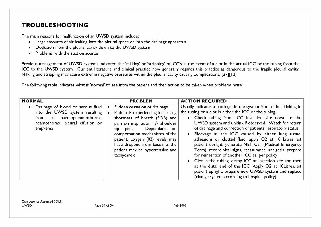
Competency Assessed SDLP:
UWSD Page 39 of 54 Feb 2009
TROUBLESHOOTING
The main reasons for malfunction of an UWSD system include:
Large amounts of air leaking into the pleural space or into the drainage apparatus
Occlusion from the pleural cavity down to the UWSD system
Problems with the suction source
Previous management of UWSD systems indicated the ‘milking’ or ‘stripping’ of ICC’s in the event of a clot in the actual ICC or the tubing from the
ICC to the UWSD system. Current literature and clinical practice now generally regards this practice as dangerous to the fragile pleural cavity.
Milking and stripping may cause extreme negative pressures within the pleural cavity causing complications. [27][12]
The following table indicates what is 'normal' to see from the patient and then action to be taken when problems arise
NORMAL PROBLEM ACTION REQUIRED
Drainage of blood or serous fluid
into the UWSD system resulting
from a haemopneumothorax,
heamothorax, pleural effusion or
empyema
Sudden cessation of drainage
Patient is experiencing increasing
shortness of breath (SOB) and
pain on inspiration +/- shoulder
tip pain. Dependant on
compensation mechanisms of the
patient, oxygen (02) levels may
have dropped from baseline, the
patient may be hypertensive and
tachycardic
Usually indicates a blockage in the system from either kinking in
the tubing or a clot in either the ICC or the tubing.
Check tubing from ICC insertion site down to the
UWSD system and unkink if observed. Watch for return
of drainage and correction of patients respiratory status
Blockage in the ICC caused by either lung tissue,
adhesions or clotted fluid: apply O2 at 10 Litres, sit
patient upright, generate MET Call (Medical Emergency
Team), record vital signs, reassurance, analgesia, prepare
for reinsertion of another ICC as per policy
Clot in the tubing: clamp ICC at insertion site and then
at the distal end of the ICC. Apply O2 at 10Litres, sit
patient upright, prepare new UWSD system and replace
(change system according to hospital policy)
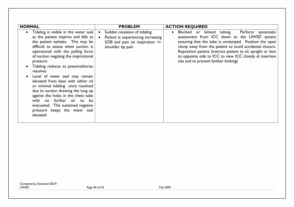
Competency Assessed SDLP:
UWSD Page 40 of 54 Feb 2009
NORMAL PROBLEM ACTION REQUIRED
Tidaling is visible in the water seal
as the patient inspires and falls as
the patient exhales. This may be
difficult to assess when suction is
operational with the pulling force
of suction negating the inspirational
pressure.
Tidaling reduces as pneumothorax
resolves
Level of water seal may remain
elevated from base with either nil
or minimal tidaling once resolved
due to suction drawing the lung up
against the holes in the chest tube
with no further air to be
evacuated. This sustained negative
pressure keeps the water seal
elevated
Sudden cessation of tidaling
Patient is experiencing increasing
SOB and pain on inspiration +/-
shoulder tip pain
Blocked or kinked tubing. Perform systematic
assessment from ICC down to the UWSD system
ensuring that the tube is unclamped. Position the open
clamp away from the patient to avoid accidental closure.
Reposition patient (instruct patient to sit upright or lean
to opposite side to ICC to view ICC closely at insertion
site and to prevent further kinking)
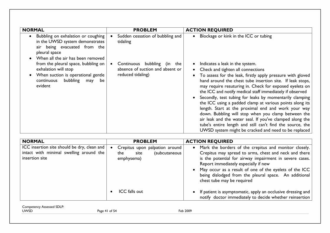
Competency Assessed SDLP:
UWSD Page 41 of 54 Feb 2009
NORMAL PROBLEM ACTION REQUIRED
Bubbling on exhalation or coughing
in the UWSD system demonstrates
air being evacuated from the
pleural space
When all the air has been removed
from the pleural space, bubbling on
exhalation will stop
When suction is operational gentle
continuous bubbling may be
evident
Sudden cessation of bubbling and
tidaling
Continuous bubbling (in the
absence of suction and absent or
reduced tidaling)
Blockage or kink in the ICC or tubing
Indicates a leak in the system.
Check and tighten all connections
To assess for the leak, firstly apply pressure with gloved
hand around the chest tube insertion site. If leak stops,
may require resuturing in. Check for exposed eyelets on
the ICC and notify medical staff immediately if observed
Secondly, test tubing for leaks by momentarily clamping
the ICC using a padded clamp at various points along its
length. Start at the proximal end and work your way
down. Bubbling will stop when you clamp between the
air leak and the water seal. If you've clamped along the
tube's entire length and still can't find the source, the
UWSD system might be cracked and need to be replaced
NORMAL PROBLEM ACTION REQUIRED
ICC insertion site should be dry, clean and
intact with minimal swelling around the
insertion site
Crepitus upon palpation around
the site (subcutaneous
emphysema)
ICC falls out
Mark the borders of the crepitus and monitor closely.
Crepitus may spread to arms, chest and neck and there
is the potential for airway impairment in severe cases.
Report immediately especially if new
May occur as a result of one of the eyelets of the ICC
being dislodged from the pleural space. An additional
chest tube may be required
If patient is asymptomatic, apply an occlusive dressing and
notify doctor immediately to decide whether reinsertion
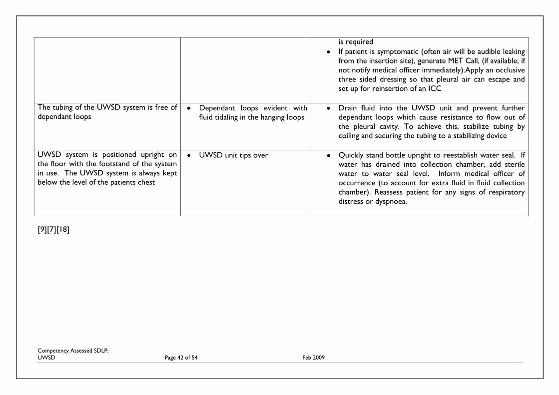
Competency Assessed SDLP:
UWSD Page 42 of 54 Feb 2009
is required
If patient is symptomatic (often air will be audible leaking
from the insertion site), generate MET Call, (if available; if
not notify medical officer immediately).Apply an occlusive
three sided dressing so that pleural air can escape and
set up for reinsertion of an ICC
The tubing of the UWSD system is free of
dependant loops Dependant loops evident with
fluid tidaling in the hanging loops
Drain fluid into the UWSD unit and prevent further
dependant loops which cause resistance to flow out of
the pleural cavity. To achieve this, stabilize tubing by
coiling and securing the tubing to a stabilizing device
UWSD system is positioned upright on
the floor with the footstand of the system
in use. The UWSD system is always kept
below the level of the patients chest
UWSD unit tips over Quickly stand bottle upright to reestablish water seal. If
water has drained into collection chamber, add sterile
water to water seal level. Inform medical officer of
occurrence (to account for extra fluid in fluid collection
chamber). Reassess patient for any signs of respiratory
distress or dyspnoea.
[9][7][18]
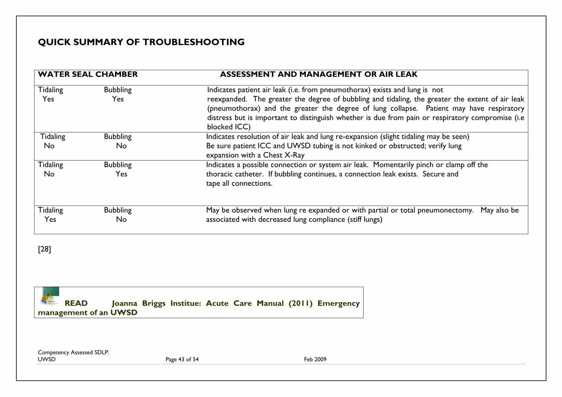
Competency Assessed SDLP:
UWSD Page 43 of 54 Feb 2009
QUICK SUMMARY OF TROUBLESHOOTING
WATER SEAL CHAMBER ASSESSMENT AND MANAGEMENT OR AIR LEAK
Tidaling Bubbling
Yes Yes
Indicates patient air leak (i.e. from pneumothorax) exists and lung is not
reexpanded. The greater the degree of bubbling and tidaling, the greater the extent of air leak
(pneumothorax) and the greater the degree of lung collapse. Patient may have respiratory
distress but is important to distinguish whether is due from pain or respiratory compromise (i.e
blocked ICC)
Tidaling Bubbling Indicates resolution of air leak and lung re-expansion (slight tidaling may be seen)
No No Be sure patient ICC and UWSD tubing is not kinked or obstructed; verify lung
expansion with a Chest X-Ray
Tidaling Bubbling Indicates a possible connection or system air leak. Momentarily pinch or clamp off the
No Yes thoracic catheter. If bubbling continues, a connection leak exists. Secure and
tape all connections.
Tidaling Bubbling May be observed when lung re expanded or with partial or total pneumonectomy. May also be
Yes No associated with decreased lung compliance (stiff lungs)
[28]
READ Joanna Briggs Institue: Acute Care Manual (2011) Emergency
management of an UWSD
Competency Assessed SDLP:
UWSD Page 44 of 54 Feb 2009
REMOVAL OF INTERCOSTAL CATHETER
The decision to remove the ICC is usually made by the attending physician. However, there are
several signs that nurses can communicate to the physician to assist in their decision making.
These include:
Scant or no drainage
Tidling has ceased or is minimal
Air/bubbling in water seal chamber has ceased for 24 hours
Equal air entry bibasilly on auscultation
Lung is fully inflated on chest x-ray with no fluid observable in the pleura [12]
The nurse may be instructed to clamp the ICC for 24 hours prior to removal to ensure the
pneumothorax does not recur.
Procedure: Verify written medical orders for removal
Two staff members are required to perform the procedure of removal of the ICC, one to
remove the drain and one to tie the purse string or mattress suture or in the absence of
either suture, to apply an occlusive dressing (only perform if within scope of practice and
according to local hospital policy)
Analgesia should be administered prior to the procedure and given time to take effect
Full explanation regarding breathing technique on removal should be given to the patient to
ensure co-operation and reduce the risk of complications (see Valsavla Maneuver below)
Check for air leakage in the water seal chamber of the UWSD system by asking the patient
to cough. If an air leak is detected notify the medical officer before proceeding to remove
Ensure that the purse string suture is viable before removable otherwise a second purse
string suture may be inserted by the doctor. Please check local protocol regarding the removal
of the ICC and who is permitted to perform.
Valsalva Maneuver
One of the main complications associated with removal of ICC’s is a recurrent pneumothorax
caused by movement of atmospheric air into the pleural cavity. This is more likely to occur if the
patient breaths in while the ICC is removed. [12] The Valsalva Maneuver was created in an effort
to equalize intrapulmonary and atmospheric pressure. This maneuver is performed by breathing
out against a closed glottis i.e.: a forced expiratory effort (strain) against a closed airway which
Avery (2000) and Cambell (1993) suggest. Mallett and Dougherty (2000) suggest to instruct the
patient to take three deep breaths and then hold their breath while the drain is removed. [12]
Step by step removal
Following analgesia, explanation and practicing of the valsalva maneuver, the patient is
placed in a comfortable position (this may be leaning over a bed side table slightly tilted to
the unaffected side. Ensure access to the ICC insertion site is achievable by both
nurses/medical staff performing removal)
Ensure the medical team are onsite and aware that removal of the ICC is about to occur
The old dressing is removed, the site is cleaned and an airtight dressing is prepared
Remove skin suture with suture cutter, ensuring the purse string suture remains intact
Competency Assessed SDLP:
UWSD Page 45 of 54 Feb 2009
Ensure ICC is clamped. Instruct patient to perform valsalva maneuvre. At same time,
smoothly remove ICC while the second person pulls the purse string or mattress suture
closed
Avoid the attempt at removal if extreme resistance on the ICC is felt
Apply an occlusive dressing, monitor patients respiratory status closely following removal
A chest X-ray should be ordered post removal to ensure that removal has not caused a
pneumothorax
Should air be heard escaping from the site, apply a three sided dressing and generate MET
Call (development of a pneumothorax caused by removal of the ICC) if available,
otherwise notify Medical officer immediately.
The nurse is also required to observe the wound closure site for signs of bleeding (initially)
and ongoing symptoms of infection. If the purse string suture is tied to tightly there is a
potential for tissue necrosis, thus direct visual observation of the wound post removal is
required. The purse string suture may be removed from 4-7 days dependant on physician
request [12]
COMPLETE SHORT ANSWER AND MULTIPLE CHOICE QUESTIONS FOR PART
THREE AND SUBMIT TO YOUR NURSE EDUCATOR ALONG WITH YOUR
EVALUATION FORM
ORGANISE WITH YOUR NURSE EDUCATOR TO PERFORM THE CLINICAL
COMPETENCY REGARDING DEMONSTRATION OF THE ABILITY TO ASSESS
AND MANAGE A PATIENT WITH AN UWSD SYSTEM
ON SUCCESSFUL COMPLETION, YOU SHALL BE AWARDED A CERTIFICATE
Competency Assessed SDLP:
UWSD Page 46 of 54 Feb 2009
REFERENCES
[1] http://services.epnet.com/getimage.aspx/?imageiid=658
[2] http://www.freewebs.com/soaring_sphincter_travel_agency/excretorysystem.htm
[3] Tortora, G. Anagnostakos, N. 2005. Principles of Anatomy and Physiology 12th Ed. Harper
and Row Publishers, New York
[4] http://media-2.web.britannica.com/eb-media/36/92936-034-8881E781.jpg
[5] http://www.cssolutions.biz/cts.html
[6] http://flashpointe.blogspot.com/2007/12/love-is-spontaneous-pneumothorax.html
[7] www.cancerbackup.org.uk/.../Pleural effusion
[8] www.unboundedmedicine.com/.../
[9] Pleur-evac Understanding Chest Drainage, 2005. Teleflex Medical, USA. Product handbook
[10] www.umm.edu/ency/article/000086.htm
[11] www.healthscout.com/ency/1/000086trt.html
[12] Allibone, Liz. (2003) Nursing Management of Chest Drains.
Nursing Standard. February 12/vol7/no22/2003pp45-54
[13] Understanding Pneumothorax.
Nursing 2002, Volume 32, Number 11, page 74-75
Sourced from:
www.nursingcentre.com
[14] Elton, Donald MD. (2006) Pneumothorax.
Sourced from:
http://www.lexingtonpulmonary.com/education/pneumothorax/pneumo.html
[15] National Lung Health Education Program (2000)
Frontline Assessment of Common Pulmonary Presentations. Monograph. Frontline Series
Sourced from: www.nlhep.org/books/pul_Pre/pleural-effusion.html
[16] Shelly P. Nascimiento, B. Simone, C. Chien, V. Chest tube insertion (2007)
` The New England Journal of Medicine. 357;15.October 11, 2007.
Sourced from: www.NEJMORG search Chest tubes
[17] www.en.wikipedia.org/wiki/Pleurodesis
[18] Central Gippsland Health Service (2007) Clinical Guideline. Underwater Seal Drainage.
Competency Assessed SDLP:
UWSD Page 47 of 54 Feb 2009
[19] Carroll, Patricia RN, C, CEN, RRT. Managing Chest Tubes: what is based on research, and
what is not? RN. Volume 58 (12), December 1995, pp46-56
Sourced from:
www.http:/ovidsp.tx.ovid.com/spb/ovidweb.cgi?&S
[20] Allibone, Liz. (2005) Principles for inserting and managing chest drains.
Nursing Times. 18 October 2005 Vol 101 No 42.pp45-49
[21] Lazzara, Deborah. (1996) Why is the Heimlich chest drain valve making a comeback?
Nursing, Dec 1996.pp1-5
sourced from:
http://findarticles.com/p/articles/mi_qa3689/is_199612/ai_n8756742
[22] http://en.wikipedia.org/wiki/Flutter_valve
[23] www.picable.com ‘search’ Heimlich valve
[24] Lazzara, Deborah. (2002) Eliminate the air of mystery from chest tubes.
Nursing. June 2002
sourced from:
http://findarticles.com/p/articles/mi_qa3689/is_200206/ai_n9128685
[25] Proehl, J. (1993) Adult Emergency Nursing Procedures. Jones and Bartlett Publishers
sourced from:
http://books.google.com.au/books?id=RxNryfowpd0C&pg=PA138&lpg=PA138&dq=heimlic
h+valve&source=web&ots=9KBnwYja_f&sig=xiFS8lkAU7fTQva0tYseUw8j6WY&hl=en&sa
=X&oi=book_result&resnum=9&ct=result
[26] Joanna Briggs Institute Acute Care Practice Manual (2004) The Joanna Briggs Institute
[27] Graham, K. (2006). Evidence Summary: Under Water Seal Drain: Management. The Joanna
Briggs Institute.
Sourced from:
www.jbiconnect.org/connect/docs/cis/es_html_viewer.php?SID=5214&lang=en&.
[28] Argyle Aqua-Seal All Purpose Autotransfusion Chest Drainage Unit (1998) Sherwood
Medical Company
[29] Urden, L. Stacy, K. Lough, M. (2006) Thelan’s Critical Care Nursing 5th Ed. Mosby Inc.
St.Louis Missouri
http://www.medicinenet.com/pleural_effusion/article.htm
Kas, Peter. Chest tube insertion on resus.com.au. Retrieved from
http://www.youtube.com/watch?v=B0wGmWn8Ubs
How to remove a chest tube. Retrieved from: http://www.youtube.com/watch?v=Cs_-
eSoSX7g
http://www.youtube.com/watch?v=Cs_-eSoSX7g

Competency Assessed SDLP:
UWSD Page 48 of 54 Feb 2009
NAME: _________________________________DEPARTMENT: __________________
1. What are the other references to tidaling commonly used and what does tidaling reflect in
the UWSD unit?
___________________________________________________________________
___________________________________________________________________
___________________________________________________________________
___________________________________________________________________
2. What does bubbling reflect? What is normal to see in the UWSD system if the system is
functioning properly and the patient continues to have an air leak (i.e. continues to have a
pneumothorax)?
___________________________________________________________________
___________________________________________________________________
___________________________________________________________________
___________________________________________________________________
3. A patient has continuous bubbling in the UWSD system as seen in the water seal. How do
you check for an air leak? What indications would there be if the air leak was from:
a. around the insertion site
b. the connections in the tubing?
___________________________________________________________________
___________________________________________________________________
___________________________________________________________________
___________________________________________________________________
___________________________________________________________________
___________________________________________________________________
___________________________________________________________________
___________________________________________________________________
UWSD QUESTIONS: PART THREE
Competency Assessed SDLP:
UWSD Page 49 of 54 Feb 2009
4. Why is it important that the tubing of the UWSD system be free of dependant loops?
___________________________________________________________________
___________________________________________________________________
___________________________________________________________________
__________________________________________________________________
5. Who orders the application and removal of suction? Can the nurse turn the suction up if
the UWSD system is indicating that there is not enough suction? How do you assess
tidaling and bubbling when the suction is operational?
___________________________________________________________________
___________________________________________________________________
___________________________________________________________________
___________________________________________________________________
___________________________________________________________________
___________________________________________________________________
___________________________________________________________________
Competency Assessed SDLP:
UWSD Page 50 of 54 Feb 2009
MULTIPLE CHOICE QUESTIONS
1. Mr. Jones who has an UWSD unit system functioning, has started to complain of increased
SOB and pain on inspiration radiating to his left shoulder. What could this indicate?
a. Kinking in the tubing
b. Dislodgement of the ICC
c. A clot in the ICC
d. A clot in the tubing of the UWSD
e. All of the above
2. The main reasons for malfunction of an UWSD system include:
a. Large amounts of air leaking into the pleural space or into the drainage apparatus
b. Occlusion from the pleural cavity down to the UWSD system
c. Problems with the suction source
d. All of the above
3. You observe minimal tidaling in the water seal of the UWSD system and the absence of
bubbling at the end of exhalation. The patient is asymptomatic. What could this indicate?
a. The unit is not functioning correctly
b. The pneumothorax has resolved
c. The pneumothorax has not resolved
d. Both a and c
4. You find the UWSD tipped over on the floor by the patient’s bed side and notice that
some of the water from the water seal has travelled into the drainage collection chambers.
What do you do?
a. Stand it back up quickly and make sure the stand in the unit has been set up correctly
b. Stand it back up quickly, observe the patient and secure to the top of the cot side on
the bed
c. Stand it back up quickly making sure the stand is operational, correct the water seal
with sterile H20 and observe the patient
d. Stand it back up quickly and prepare to set up a new UWSD unit whilst clamping the
ICC at the insertion site and end of the catheter
5. The physician is doing his morning round. He is assessing your patient who has an UWSD
system connected to an ICC. There has been no bubbling in the last 6 hours and very
minimal tidaling. The patient is asymptomatic. Suction has not been required. He asks the
patient to cough while he observes the UWSD system. What is the main reason he would
do so?
a. To see if the patient is performing his chest physiotherapy correctly
b. To see if there is any air left in the pleural cavity
c. To make sure his pain relief is effective
d. To make sure it is alright for him to have a shower
Competency Assessed SDLP:
UWSD Page 51 of 54 Feb 2009
6. As the nurse looking after the patient with a functioning UWSD system, education for the
patient includes:
a. The purpose of the specific tube(s)
b. The importance of not pulling/kinking the tube
c. Importance of adequate pain relief
d. How to manage the tube e.g.; showering, dressing
e. All of the above
7. You are about to remove an ICC. In order of importance, what would you ensure first?
a. That the ICC has been clamped
b. That the lung has fully inflated on Chest X-Ray
c. There are documented orders to remove
d. That the patient is neurologically alert
8. Clamping on an ICC should be avoided except:
1. When the patient requires to travel to the X-Ray department
2. When the patient is having a shower 3. When changing the UWSD system 4. When the physician orders you to do so
a. 1 and 3
b. 2 and 4
c. 2 and 3
d. 3 and 4
9. You find your patient in respiratory distress, confused and observe that she has pulled out
her ICC. What would you do?
a. Generate a MET call ( or call Medical Officer ) and run to greet the team or doctor
b. Generate a MET call ( or call Medical Officer) and apply an occlusive dressing
c. Generate a MET call (or call Medical officer) and apply a three sided occlusive dressing
d. Generate a MET call (or call Medical officer) and stick your finger in the insertion site
10. Prior to removal of an ICC, it is MOST important to:
a. Provide analgesia and educate the patient
b. Ensure there is documented instructions to remove
c. Allow the patient to shower prior
d. Ensure the suction has been turned off at the suction source
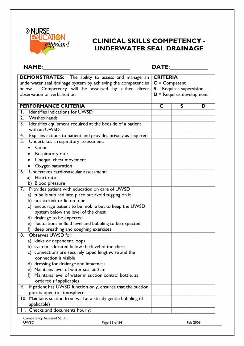
Competency Assessed SDLP:
UWSD Page 52 of 54 Feb 2009
NAME:________________________________ DATE:______________
DEMONSTRATES: The ability to assess and manage an
underwater seal drainage system by achieving the competencies
below. Competency will be assessed by either direct
observation or verbalisation
CRITERIA
C = Competent
S = Requires supervision
D = Requires development
PERFORMANCE CRITERIA C S D
1. Identifies indications for UWSD
2. Washes hands
3. Identifies equipment required at the bedside of a patient
with an UWSD.
4. Explains actions to patient and provides privacy as required
5. Undertakes a respiratory assessment:
Color
Respiratory rate
Unequal chest movement
Oxygen saturation
6. Undertakes cardiovascular assessment:
a) Heart rate
b) Blood pressure
7. Provides patient with education on care of UWSD
a) tube is sutured into place but avoid tugging on it
b) not to kink or lie on tube
c) encourage patient to be mobile but to keep the UWSD
system below the level of the chest
d) drainage to be expected
e) fluctuations in fluid level and bubbling to be expected
f) deep breathing and coughing exercises
8. Observes UWSD for:
a) kinks or dependent loops
b) system is located below the level of the chest
c) connections are securely taped lengthwise and the
connection is visible
d) dressing for drainage and intactness
e) Maintains level of water seal at 2cm
f) Maintains level of water in suction control bottle, as
ordered (if applicable)
9. If patient has UWSD function only, ensures that the suction
port is open to atmosphere
10. Maintains suction from wall at a steady gentle bubbling (if
applicable)
11. Checks and documents hourly:
CLINICAL SKILLS COMPETENCY -
UNDERWATER SEAL DRAINAGE
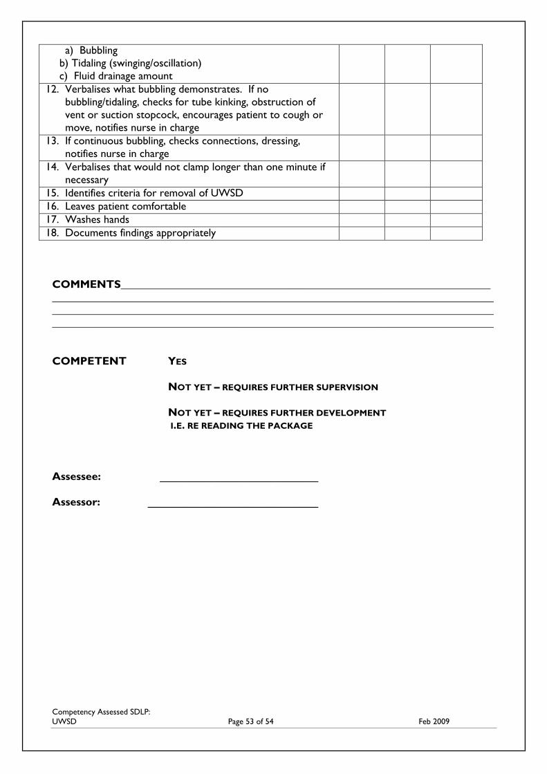
Competency Assessed SDLP:
UWSD Page 53 of 54 Feb 2009
a) Bubbling
b) Tidaling (swinging/oscillation)
c) Fluid drainage amount
12. Verbalises what bubbling demonstrates. If no
bubbling/tidaling, checks for tube kinking, obstruction of
vent or suction stopcock, encourages patient to cough or
move, notifies nurse in charge
13. If continuous bubbling, checks connections, dressing,
notifies nurse in charge
14. Verbalises that would not clamp longer than one minute if
necessary
15. Identifies criteria for removal of UWSD
16. Leaves patient comfortable
17. Washes hands
18. Documents findings appropriately
COMMENTS___________________________________________________________________
________________________________________________________________________________
________________________________________________________________________________
________________________________________________________________________________
COMPETENT YES
NOT YET – REQUIRES FURTHER SUPERVISION
NOT YET – REQUIRES FURTHER DEVELOPMENT
I.E. RE READING THE PACKAGE
Assessee: __________________________
Assessor: ____________________________
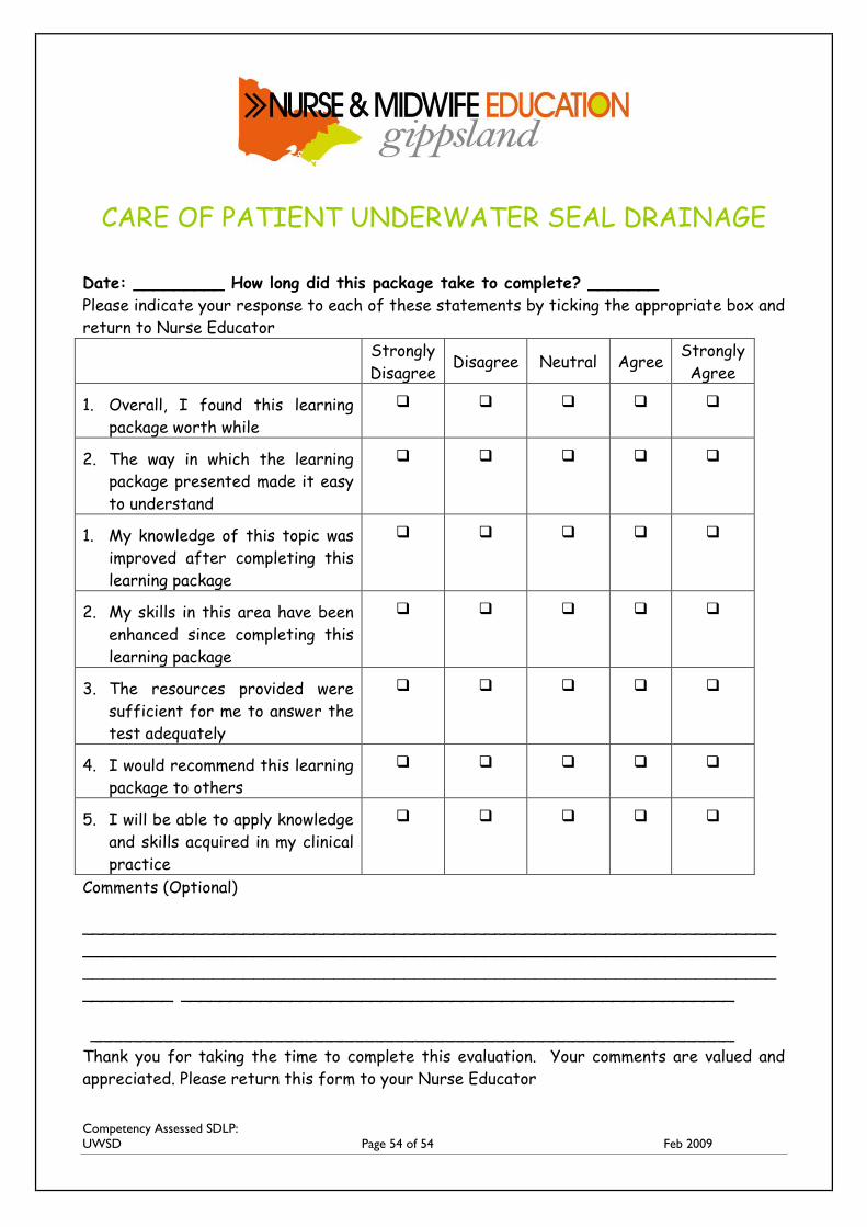
Competency Assessed SDLP:
UWSD Page 54 of 54 Feb 2009
CARE OF PATIENT UNDERWATER SEAL DRAINAGE
Date: _________ How long did this package take to complete? _______
Please indicate your response to each of these statements by ticking the appropriate box and
return to Nurse Educator
Strongly
Disagree Disagree Neutral Agree
Strongly
Agree
1. Overall, I found this learning
package worth while
2. The way in which the learning
package presented made it easy
to understand
1. My knowledge of this topic was
improved after completing this
learning package
2. My skills in this area have been
enhanced since completing this
learning package
3. The resources provided were
sufficient for me to answer the
test adequately
4. I would recommend this learning
package to others
5. I will be able to apply knowledge
and skills acquired in my clinical
practice
Comments (Optional)
_____________________________________________________________________
_____________________________________________________________________
_____________________________________________________________________
_________ _______________________________________________________
________________________________________________________________
Thank you for taking the time to complete this evaluation. Your comments are valued and
appreciated. Please return this form to your Nurse Educator