Embed Size (px)
Citation preview
[CANCER RESEARCH 42, 59-64, January 1982]
Comparison of the in Vitro Metabolism of N-Nitrosohexamethyleneimineby Rat Liver and Lung Microsomal Fractions1
Lanny l. Hecker and Gary A. McClusky
Chemical Carcinogenesis Program, NCI-Frederick Cancer Research Facility, Frederick, Maryland 21701
ABSTRACT
The in vitro metabolism of N-nitrosohexamethyleneimine by
lung and liver microsomes and cytosol from uninduced maleFischer rats is described. Metabolites produced by both organsappeared to be identical. The liver subcellular fractions had alower Km(0.6 mw) than did lung fractions (3 rriM) and metabolized 2.5 to 5 times as much nitrosamine per mg protein. Ourresults, together with those from our earlier studies, indicatethat, as the size of the carbon ring increases from nitrosopyr-
rolidine to nitrosohexamethyleneimine, lung microsomes hadan increased affinity for the cyclic nitrosamines; there was onlya small effect with liver enzymes. This suggests that microsomalenzymes that metabolize cyclic nitrosamines in rat livers andlungs are not the same. The first stable a-hydroxylation product, 6-hydroxyhexanal, was not detected in reactions involving
microsomes alone. Apparently, this compound is rapidly converted to 1,6-hexanediol by liver or lung microsomes. The
presence of cytosol was needed for the full conversion of thesemetabolites to e-hydroxycaproate and maximal a-hydroxylationactivity, i -Ammocaproate was always found in direct proportion
to the hydroxyacid, suggesting that both acids arise from thesame o-hydroxylation event by different breakdown mechanisms, ß-and •y-hydroxynitrosohexamethyleneimine were not
metabolized significantly by rat liver enzymes and thus, in thisspecies, may be "detoxification products" of A/-nitrosohexa-
methyleneimine.
INTRODUCTION
One of the more interesting aspects of nitrosamine carcino-
genesis is the difference in organ specificities of structurallysimilar compounds. We have been engaged in studies of themetabolism of cyclic nitrosamines by both "target" and "non-target" organs (7-9,18) in the hope that metabolic differences
we observe may be related to the varying carcinogenic effects.In this paper, we report on the metabolism of NO-HEX2 which
produces predominantly liver and esophageal tumors in 3different strains of rat (4,12,13). A/-Nitrosoheptamethyleneim-
ine which has only one more carbon atom in its ring causestumors in both lung and esophagus (14, 20). By comparing themetabolism of these 2 compounds by subcellular fractions fromuninduced rat liver and lung and incorporating the data wehave obtained with NO-PYR, we hope to gain some insights on
1This work was supported by Contract N01-CO-75380 with the National
Cancer Institute, NIH, Bethesda, Md. 20205.2 The abbreviations used are: NO-HEX, W-nitrosohexamethyleneimine; NO-
PYR, W-nitrosopyrrolidme; j8-hydroxy-NO-HEX, /f-rtydrnxy-W-nitrusohoxamethy-lenelmine; -y-hydroxy-NO-HEX, y-hydroxy-N-nitrosohexamethyleneimine; HPLC.high-pressure liquid chromatography; OPT, o-pthalaldehyde; GC, gas chroma-tography; a-hydroxy-NO-HEX, a-hydroxy-N-nitrosohexamethyleneimine; 2-oxy-NO-HEX. 2-oxy-W-nitrosohexametriyleneimine.
Received July 20, 1981 ; accepted October 13. 1981.
the initial events of nitrosamine Carcinogenesis.Information about the metabolism of NO-HEX can be found
in several reports. Neunhoeffer ef al. (16) demonstrated thatacetone extracts of rat livers could produce small amounts ofe-aminocaprohydroxamic acid from NO-HEX. Grandjean (5)detected 14CO2plus 11 different radioactive peaks in the urineof animals given [14C]NO-HEX by gavage. Four of these prod
ucts were tentatively identified as hexamethyleneimine, e-ami-nocaproic acid, e-caprolactam, and 6-aminocaprohydroxamic
acid. Ross and Mirvish (17) gave convincing evidence that atleast one of the adducts bound to RNA and possibly DNA afteradministration of labeled NO-HEX most probably resulted fromactive intermediates formed by decomposition of a-hydroxyl-ated NO-HEX. The observation of Snyder ef al. (18) confirmedthe fact that, at low dose levels (~12 mg/kg), large percentages (43%) of NO-HEX labeled in the a-carbon atom were
given off as CO2 within 24 hr; at higher dose levels, thepercentage given off as CO2 was greatly reduced. Hecker andSaavedra (9) have reported on the occurrence of 0-hydroxy-NO-HEX and y-hydroxy-NO-HEX which account for one-third
or more of the radioactive metabolites formed during the metabolism of NO-HEX by uninduced rat liver microsomes and
cytosol (postmicrosomal supernatant).
MATERIALS AND METHODS
Instrumentation. Mass spectra were obtained from a VG MicromassZAB-2F mass spectrometer equipped with a VG2035 data system. GasChromatographie analyses were done on a Shimadzu Model 4-BM gasChromatograph equipped with a Hewlett-Packard 18652 A/D convertercoupled to the recorder of a flame ionization detector or a Perkin-Elmer
Model Sigma 3 gas Chromatograph coupled to the mass spectrometer.The columns used in the Shimadzu gas Chromatograph were 2 m x 2.6mm and contained either Tenax G/C (Alltech Associates, Inc.), Deer-field, II.) or 3% SE-30 on Chromasorb W (Pierce Chemical Co., Rock-ford, II.). The silanized glass columns used on the Perkin-Elmer gas
Chromatograph were 6 ft x 2 mm and contained Tenax G/C, 3% SESO on Chromosorb W, or Ultrabond 20M (RFR Corp., Hope, R. I.).HPLC was done on a Waters Associates, Inc., system using either 4.6mm x 25 cm or 9.4 mm x 25-cm Whatman, Inc. (Clifton, N. J.) Partisil-10 ODS-2 columns. Radioactivity was monitored with a Packard Model
3225 liquid scintillation counter (8). Amino acid analysis was done ona Durrum DC-4A amino acid analyzer with a Ourrum D500 column (48
cm x 1.75 mm) and Durrum Pico buffers for single column analyses(Dionex Corp., Sunnyvale, Calif.).
Chemicals. e-Caprolactone, 1,6-hexanediol, and organic reagentswere obtained from Aldrich Chemical Co. (Milwaukee, Wis.). e-Amino-
caproic acid was purchased from Sigma Chemical Co. (St. Louis, Mo.).6-Hydroxyhexanal was kindly supplied by Dr. Saavedra of our laboratory. i-Caprolactone was converted to sodium e-hydroxycaproate by
the methods of Marvel and Birk himer (15). Diazomethane was generated by the procedures of deBaer and Backer (2). Ninhydrin-dimethylsulfoxide reagent was obtained from Pierce Chemical Co. O-p-Nitro-benzyl-N.W-diisopropylisourea and OPT were obtained from RegisChemical Co. (Morton Grove, II.). [MC]NO-HEX labeled in the a-carbon
JANUARY 1982 59
Research. on January 2, 2019. © 1982 American Association for Cancercancerres.aacrjournals.org Downloaded from
L. l. Hecker and G. A. McClusky
atoms was prepared as described previously (9, 18). Unlabeled NO-HEX was made by the procedure of Goodall ef al. (4).
O-p-Nitrobenzyl-W,N'-diisopropylisourea derivatives of acids were
made according to the instructions on p. 76 of the Regis catalog.Benzoyl chloride derivatives of metabolites were made by dissolving 2to 20 jug of compound in 50 to 100 /il acetonitrile, adding 1 »Iofbenzoyl chloride and 3 /¿Iof pyridine and then heating overnight at 60°
in a sealed vial. After evaporation, the residue was resuspended in 0.5to 1 ml of 2% sodium bicarbonate and extracted 3 times with ethylacetate. The organic phase was washed twice with 0.1 NHCI, concentrated, and resuspended in ethanol. Standards were made in much thesame manner except with higher concentrations of reactants and on alarger scale.
OPT derivatives of amino acids were made by modifying publishedmethods (10). We have made OPT derivatives without using anaerobicconditions and /?-mercaptoethanol. A stock solution of 1 mg OPT perml was prepared in 0.4 M boric acid-KOH buffer (pH 10.4). Twovolumes were mixed with one volume of amino acid solution (~1 mg/ml), and the reaction was allowed to continue for ~15 min at roomtemperature. The solution was then acidified with HCI to pH 1 to 2 andquickly extracted 2 or 3 times with ethyl acetate. The ethyl acetate isevaporated to dryness, and the derivative is dissolved in -100 ;iimethanol and then reacted with an excess amount of diazomethane.The solution is evaporated again, and the final derivatized product maybe dissolved in methanol, purified and/or analyzed by HPLC by monitoring absorbance at 254 nm, and analyzed by GC after purification.
Metabolism. Liver and lung microsomes and cytosol were preparedfrom male Fischer rats as described previously (7, 8). Optimal condi-
100-
80-
60-
40-
20^
80-
60-
40-
20-
1ml
r20
17 1»
60
m/z100
"'I'1'140
Chart 1. Mass spectra of the methyl ester of c-hydroxycaproic acid. A. standard; B, metabolite.
tions for the metabolism of NO-HEX by liver microsomes have beenpublished (9). Reactions with lung microsomes contained the followingconcentrations of reactants: [14C]NO-HEX(~1 »Ci/ml;0.1 to 3 mM);rat lung microsomes (-1.1 mg/ml); rat lung cytosol (~6. mg/ml); 50mM Tris (pH 8.0); 1 mM EDTA; 5 mM glucose-6-phosphate; 1 mMNADP;0.5 mMNAOPH;and glucose-6-phosphate dehydrogenase (0.2units/ml). Reactions (usually 100 /d)were assayed by either méthylènechloride extraction (8) (this method was used to determine optimalconditions and K,,,s)or BaSO4precipitation (19) followed by analysison HPLC (7, 8). HPLC analyses were done on a Whatman ODS-2column (9.4 mm x 25 cm) run at 4.5 ml/min in 18% methanol-0.01 Msodium acetate buffer, pH 5.5.
Large-scale reactions from which metabolites could be isolatedcontaining either liver microsomes or liver microsomes + cytosol wereprocessed as described previously (9). After chromatography on Seph-adex G-10,3 major fractions were isolated. The earliest eluting fraction(220 to 240 ml) contained both c-hydroxycaproate and e-aminocapro-ate, the next major fraction eluting between 270 and 360 ml contained1,6-hexanediol and 6-hydroxyhexanal, and hydroxylated nitrosamineseluted between 360 and 430 ml. e-Aminocaproate and e-hydroxy-caproate were separated by Sephadex LH-20 chromatography (8). Theamino acid was further purified on a Sephasorb HP column (1.8 mm x96 cm) run in 2 mMsodium acetate buffer (pH 4.0) and finally on a 9.4mm x 26 cm Whatman ODS-2 column on HPLC.The position of elutionof all compounds on these C,„columns may be determined by extrapolating the elution times shown in Chart 5 to other methanol concentrations. After LH-20 chromatography, e-hydroxycaproate was purified bychromatography on ODS-2 columns in acetate buffer (pH 5.5) and 8%methanol, followed by chromatography in 18% methanol-1 mM HCI. Inthe HCI solution, the e-hydroxycaproic acid eluted at 13 to 15 min witha flow rate of 4.5 ml/min. The methyl ester of e-hydroxycaproic acidwas purified by Ci,, chromatography (elution time, 9 min) using a 35%methanol-water solution run at 4.5 ml/mm on a 9-mm x 25-cm ODS-2 column. 1,6-Hexanediol and 6-hydroxyhexanal were separated bychromatography on an ODS-2 column in 18% methanol-sodium acetate, pH 5.5. The separated peaks were collected and then chromato-graphed under the same conditions using water instead of buffer.
RESULTS
Identification of Metabolites. '"Ola beIed metabolites of NO-
HEX were purified from 30- or 60-ml reactions after precipita
tion with BaSO4 and then ethanol. As described previously (7,19), the concentrated extracts were chromatographed on aSephadex G-10 column, which separated yS-hydroxy-NO-HEXand -y-hydroxy-NO-HEX from the other metabolites. The re
maining metabolites were further purified by chromatographyon Sephadex LH-20, Sephasorb HP, and/or Ci8 HPLC col
umns.(-Hydroxycaproate (Chart 4, Peak III). After purification,
this metabolite was converted to the methylester with diazomethane. The mass spectra of the methylated metabolite andstandard were also identical (Chart 1), but the molecular ion ofm/z (146) was not apparent. Confirmation of identity wasaccomplished by making derivatives of the acid with O-p-nitro-benzyl-A/,A/'-diisopropylurea; derivatives of both the metaboliteand standard coeluted at 10.5 min on Whatman ODS-2 columns (4.6 mm x 25 cm, 60% methanol-water, 1.5 ml/min).
Upon acetylation of the hydroxyl group with acetyl chloride,both the radioactive metabolite and the standard cochromato-graphed at 9.7 min when they were run on an ODS-2 columnin 70% methanol-water. i -Hydroxycaproate was identified in in
vitro reactions containing either microsomes or microsomes+ cytosol.
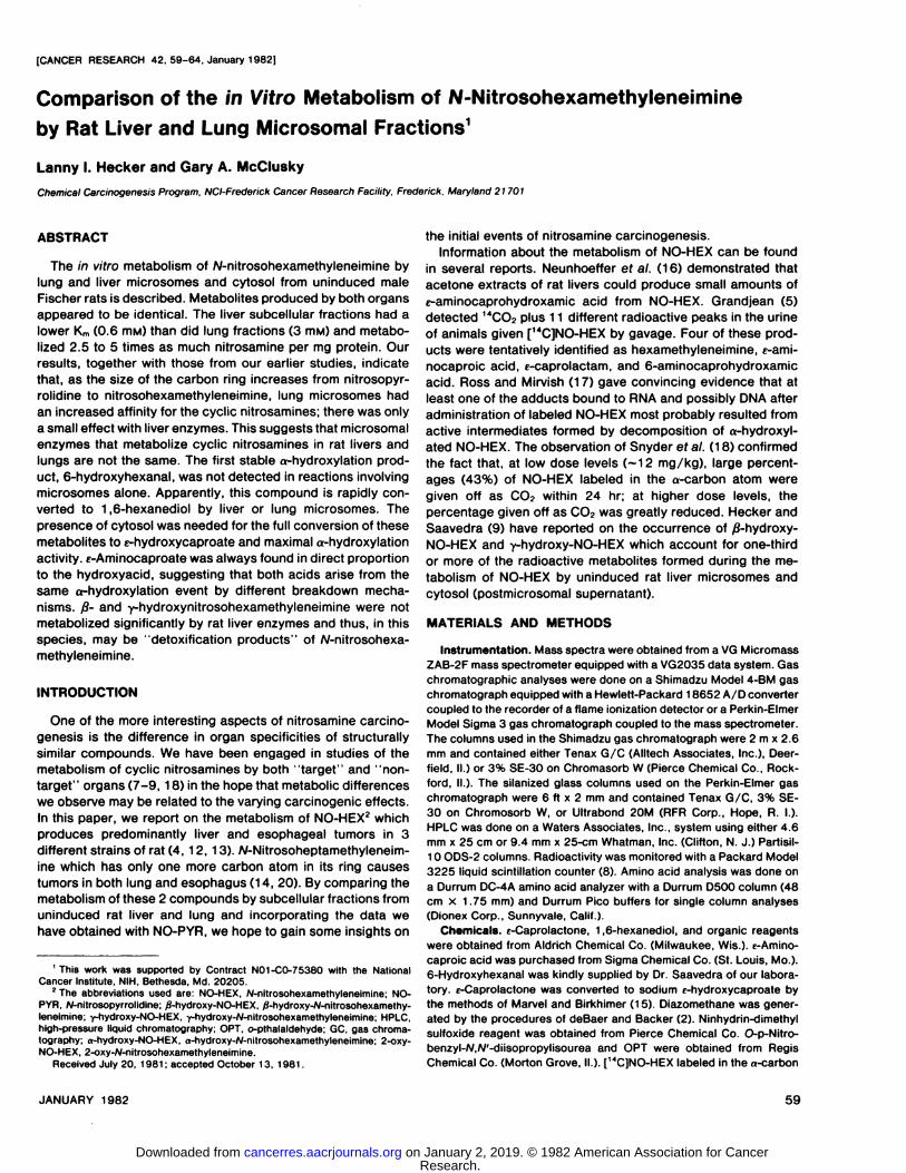
<-Aminocaproic Acid (Chart 4, Peak II). The purified metab-
60 CANCER RESEARCH VOL. 42
Research. on January 2, 2019. © 1982 American Association for Cancercancerres.aacrjournals.org Downloaded from
Comparison of NO-HEX Metabolism by Rat Liver and Lung
olite was analyzed on a Durrum D500 column for amino acidanalysis using a 115-min program. Both the metabolite andcommercially supplied e-aminocaproic acid eluted within 1 secof each other at 65.6 min in an area of the chromatogramwhere none of the common «-aminoacids eluted. This still didnot prove that the radioactivity in this sample was associatedwith the amino acid. OPT derivatives of the metabolite andstandard were purified by HPLC (see "Materials and Methods"). The peak of absorbance at 254 nm and the 14Cradioactivity of the OPT metabolites obtained from either liver micro-somal or microsome + cytosol reactions and the absorbanceof the e-aminocaproic acid derivative cochromatographed at10.7 min on Whatman ODS-2 columns (4.6 mm x 25 cm, 60%methanol-water, 1.5 ml/min). Metabolite and known derivativesalso coeluted at 9 min on SE-30 (2 m x 2.6 mm, 190°,60 ml
helium per min). Chart 2 indicates these OPT derivatives haveidentical mass spectra consistant with the structure shown inChart 2A.
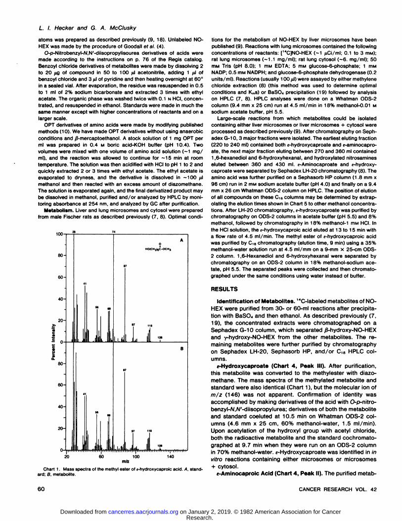
6-Hydroxyhexanal (Chart 4, Peak VIII). The identification ofthis metabolite is tentative. Both the metabolite and standardcompound elute at 5.8 min on Tenax G/C (2.5 m x 2.6 mm, 60ml helium per min, 200°). Although the mass spectra are
somewhat similar, the small amount of material used and thelack of a molecular ion are not sufficient for identification. Boththe radioactivity and absorbance at 254 nm of the benzoylchloride derivative of the metabolite and the absorbance of thestandard compound have retention times of approximately 7.5
100-
80-
60-
40-
20-
- 0-
2I
80-
60-
40-
20-
0-"i
II I '" Ifi l T a .»
OCM,
J174 I
, 1 ... 1 1
,„ 112 I 17«l...,,,,,; T 'i Ti J,., T. ,,T i r T i l i i i iP T—i i r i i i r
50 150m/z
250
min on ODS-2 columns (4.6 mm x 25 cm, 1 .5 ml/min, 70%methanol-H2O). Furthermore, the GC retention times of thederivitized metabolite and standard on SE-30 (6 ft x 2 mm, 20m helium per min, 190°)are similar as shown in Chart 3. Thus,this metabolite is most likely 6-hydroxyhexanal.
1,6-Hexanediol. After purification, this metabolite (Chart 4,Peak IX) eluted at 8 min on Tenax G/C (2.5 m x 2.5 mm, 60 mlhelium per min, 200°)as was the case for a sample of 1,6-
hexanediol. Mass spectra of the standard and metabolite weresimilar, but no molecular ion was apparent. The benzoyl chloride derivatives of the metabolite and known cochromato-graphed at 8 min on Whatman ODS-2 columns (87% methanol-water, 1.5 min ml/min) and on GC at 6.5 min using SE-30columns (2 m x 2.6 mm, 60 ml helium per min, 225°).Identity
was confirmed by high resolution mass spectra of both themetabolite and 1,6-hexanediol derivatives. The molecular ionshad indistinguishable exact mass measurements (326.1510versus 326.1508) with an elemental composition (C..,,H....O.,)indicative of the dibenzoyl derivative of 1,6-hexanediol. Theseanalyses were identical for metabolites obtained from reactionswith either microsomes alone or microsomes + cytosol.
Metabolism. The activity of liver and lung subcellular fractions on [<4C]NO-HEXwas initially assayed by the méthylène
chloride extraction method we have described previously forNO-PYR (8). After méthylènechloride extraction, most metabolites remain in the aqueous phase; these are the non-methylene chloride extraction products. In the case of NO-HEX, up to 20% of the less polar metabolites may be extractedinto méthylènechloride along with NO-HEX. This does notaffect the validity of the assay because a constant proportionof all the radioactive metabolites always remains in the aqueousphase. As is the case for NO-PYR, the cytosol (postmicrosomalsupernatant) itself metabolizes NO-HEX to only a small extent.Optimal results are observed only with combinations of microsomes + cytosol.
Reactions using liver subcellular fractions are optimal at pH7.5 in the presence of Mgz+ and Mn2+, while the optimum
activity for lung fractions is obtained at pH 8 in the absence ofdivalent cations. Using those conditions, the K,,,for the liverfractions was ~0.6 IHM as opposed to ~3 mM for the lungreactions. At NO-HEX concentrations of between 0.5 and 4mM, liver subcellular fractions metabolize 2.5 to 3 times the
:H21,OM
2345678Minutes
Chart 3. GC of the benzoyl chloride derivative of a metabolically produced 6-hydroxyhexanal. The top tracing is the HPLC purified benzoyl chloride derivativeof the metabolite. The bottom tracing is of benzoyl chloride derivatives of 6-
Chart 2. Mass spectra of the OPT derivative of e-aminocaproic acid methyl hydroxyhexanal and 1,6-hexanediol standards. Chromatography was done on aester. A. standard; 8. metabolite. 6-ft x 2-mm, 3% SE-30 column at 190°and 20 ml helium flow per mm.
JANUARY 1982 61
Research. on January 2, 2019. © 1982 American Association for Cancercancerres.aacrjournals.org Downloaded from
L. /. Hecker and G. A. McCIusky
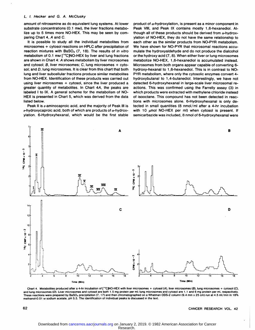
amount of nitrosamine as do equivalent lung systems. At lowersubstrate concentrations (0.1 mw), the liver fractions metabolize up to 5 times more NO-HEX. This may be seen by com
paring Chart 4, A and C.It is possible to study all the individual metabolites from
microsomes + cytosol reactions on HPLC after precipitation ofreaction mixtures with BaSO., (7, 18). The results of in vitrometabolism of 0.1 mM [14C]NO-HEX by liver and lung fractions
are shown in Chart 4: A shows metabolism by liver microsomesand cytosol; B, liver microsomes; C, lung microsomes + cytosol; and D, lung microsomes. It is clear from this chart that bothlung and liver subcellular fractions produce similar metabolitesfrom NO-HEX. Identification of these products was carried out
using liver microsomes + cytosol, since the liver produced agreater quantity of metabolites. In Chart 4/4. the peaks arelabeled I to IX. A general scheme for the metabolism of NO-
HEX is presented in Chart 5, which was derived from the datalisted below.
Peak II is c-aminocaproic acid, and the majority of Peak III ise-hydroxycaproic acid, both of which are products of a-hydrox-ylation. 6-Hydroxyhexanal, which would be the first stable
product of o-hydroxylation, is present as a minor component inPeak VIII, and Peak IX contains mostly 1,6-hexanediol. Although all of these products should be derived from a-hydrox-ylation of NO-HEX, they do not have the same relationship toeach other as the similar products from NO-PYR metabolism.We have shown for NO-PYR that microsomal reactions accu
mulate the hydroxyaldehyde and do not produce the dialcoholor the hydroxy acid (7,8). When either liver or lung microsomesmetabolize NO-HEX, 1,6-hexanediol is accumulated instead.Microsomes from both organs appear capable of converting 6-hydroxy-hexanal to 1,6-hexanediol. This is in contrast to NO-PYR metabolism, where only the cytosolic enzymes convert 4-hydroxybutanal to 1,4-butanediol. Interestingly, we have notdetected 6-hydroxyhexanal in large-scale liver microsomal re
actions. This was confirmed using the Farrelly assay (3) inwhich products were extracted with méthylènechloride insteadof isooctane. This compound has not been detected in reactions with microsomes alone. 6-Hydroxyhexanal is only detected in small quantities (8 nmol/ml after a 4-hr incubationwith 10 fimol NO-HEX per ml) when cytosol is present. Ifsemicarbazide was included, 8 nmol of 6-hydroxyhexanal were
nr
10
S 8
TUT
IX
10 12
Time (Min)
18
Q-o
iü
8 10 12
Time (Min)
Chart 4. Metabolites produced after a 4-hr incubation of [MCJNO-HEX with liver microsomes + cytosol (A), liver microsomes (B), lung microsomes + cytosol (C),
and lung microsomes (D). Liver microsomes and cytosol are both 1.5 mg protein per ml; lung microsomes and cytosol are 1.1 and 6 mg protein per ml, respectively.These reactions were prepared by BaSO., precipitation (7, 17) and then chromatographed on a Whatman ODS-2 column (9.4 mm x 25 cm) run at 4.5 ml/min in 18%methanol-0.01 M sodium acetate. pH 5.5. The identification of individual peaks is discussed in the text.
62 CANCER RESEARCH VOL. 42
Research. on January 2, 2019. © 1982 American Association for Cancercancerres.aacrjournals.org Downloaded from

Comparison of NO-HEX Metabolism by Rat Liver and Lung
fl-hydroxy NO-HEX ,-hydroxy NO-HEXC5"
f -aminocaproate
droxylation products (Peak II + III + IX) increase. This suggeststhat enzymes that carry out «-and y-hydroxylations may bedifferent. Peak VIII contains the frans conformer of /6-hydroxy-NO-HEX (9), although as noted previously, a small amount of6-hydroxyhexanal is also present. Peak IX contains some ofthe cis conformer of yS-hydroxy-NO-HEX. In liver reactions, theZ conformer of /?-hydroxy-NO-HEX should be no more than
50% of the E conformer (9). Peaks VI and VII have not as yetbeen identified, but it should be noted that Peak VI is apparentlyformed only in the presence of cytosolic enzymes.
Peak I has not as yet been identified, but it is the product ofmetabolism of either /?-hydroxy-NO-HEX and/or y-hydroxy-NO-HEX. Neither j8-hydroxy-NO-HEX nor y-hydroxy-NO-HEXis metabolized significantly by uninduced or induced rat sub-
cellular fractions. While liver microsomes + cytosol metabolize40 to 50% of a 0.1 mw NO-HEX solution in 4 hr, only 1 to 2%of an equivalent concentration of y-hydroxy-NO-HEX is metabolized within the same time period. The results with /8-hydroxy-NO-HEX are similar.
HO
VW6- hydroxytwxanal
Liver Of Lung IMicrosomti 1
xanai
/Llnr or LI Liver M
<-hydroxycaproate DISCUSSION
ung Cytoso!Microsomea
OH1.6-lMUMdiOl
Chart 5. A general scheme for the metabolism of NO-HEX by uninduced ratliver and lung subcellular fractions.
detected with microsomes alone, but —15nmol were foundwith microsomes + cytosol. In large-scale reactions, 6-hydrox
yhexanal has been isolated only from microsome + cytosolreactions and was present only in amounts one-third that of1,6-hexanediol. The relative accumulation of the diol is greater
with lung microsomes and thus coincides with their lack ofsignificant accumulation of 6-amino- and 6-hydroxycaproic
acids (Chart 4D). In contrast, these acids do accumulate tosome extent in liver microsomal reactions (Chart 46). As wouldbe expected, when either liver or lung cytosol is present aswell, only a small amount of 1,6-hexanediol remained, and
Peaks II and III increase (Chart 4, A and C). For both liver andlung reactions, the presence of cytosol increases the amountof a-hydroxylation products (Peaks II, III, and IX) 55 to 65%.
The above data suggest that 1,6-hexanediol may be con
verted to the hydroxy acid. To test this hypothesis, 0.5 HIM[1*C]NO-HEX was incubated with liver microsomes for 4 hr,
and then the extracted reaction mixture was chromatographedon HPLC as in Chart 4. Peak IX eluting at ~16 min was pooled,
concentrated, and incubated with liver cytosol. Peak IX wasconverted to Peak III with time by liver cytosol. Liver microsomes also possessed this enzymatic activity. Therefore, liverand lung cytosols and liver microsomes are able to convert1,6-hexandiol to e-hydroxycaproate. The presursors of e-ami-nocaproate and the reason for its parallel occurrence with e-hydroxycaproate are unknown and will be discussed below.
Peaks IV and V are the frans (E) and cis (Z) conformers ofy-hydroxy-NO-HEX (9). As we have noted previously, the ratio
of conformers formed by liver and lung reactions differs, suggesting that, in the liver and lung, different enzymes performsimilar functions. Upon addition of cytosol, the amount of y-hydroxy-NO-HEX remains relatively constant, while the a-hy-
Microsome + cytosol reactions from either uninduced ratlivers or lungs produce similar products from NO-HEX, as isthe case for NO-PYR (7). Although the Kmsof liver microsomes+ cytosol for NO-HEX and NO-PYR (0.6 mw versus 0.34 mM)
are approximately the same, lung microsomes have a muchlower Kmfor NO-HEX (3 mw) than for NO-PYR (20 mM). As the
size of the ring increases, lung microsomes apparently havemore affinity for the substrate. Our results indicate that the rateof metabolism by liver microsomes + cytosol is up to 5 timesgreater than lung per mg of protein, which is consistent withthe differences in Kms. This supports our previous suggestionsthat the affinity of microsomes for nitrosamines as well as thetotal amount of hydroxylation are initially important in determining if a nitrosamine will be able to produce tumors in a giventissue (7, 8). The different Kmsfor cyclic nitrosamines exhibitedby liver and lung enzymes strongly suggest that hydroxylationof cyclic nitrosamines in these organs is carried out by eitherdifferent proteins or similar proteins in different membraneenvironments.
Reactions containing microsomes + cytosol gave a higherrate of metabolism than those containing microsomes alone;similar results have been obtained with NO-PYR (8). FromChart 4, it may be determined that, in the absence of cytosol,ß-and y-hydroxylation products form a greater percentage of
total metabolites. These data are consistent with the supposition that microsomal enzymes involved in ß-and y-hydroxyl-ations are different than those which a-hydroxylate NO-HEX in
liver and lung.Liver and lung microsomal reactions with NO-PYR accumu
late 4-hydroxybutanal (6, 8), but the 6-carbon hydroxyalde-hyde from NO-HEX microsomal metabolism is only found ifcytosol is included in the reaction mixture. This is most probably due to the fact that, as soon as 6-hydroxyhexanal isformed by microsomes, it is immediately reduced to 1,6-hexanediol by either liver or lung microsomes; this activity is notfound for the 4-carbon compound (7, 8). Since 6-hydroxyhex
anal has been found only in reactions including cytosol, it isconceivable that it may be an intermediate in the conversion of1,6-hexanediol to the hydroxy acid. Such aldehyde intermedi-
JANUARY 1982 63
Research. on January 2, 2019. © 1982 American Association for Cancercancerres.aacrjournals.org Downloaded from
L. /. Hecker and G. A. McCIusky
ates may, for the most part, be enzyme bound, allowing onlysmall amounts to leak away from the active sites of the enzyme(see Ref. 11). It is apparent from our results that liver micro-somes are capable of converting 1,6-hexanediol and/or 6-hydroxyhexanal to the hydroxy acid. Lung microsomes onlyhave a trace of this activity and thus accumulate a greaterrelative amount of 1,6-hexanediol. In both cases (particularlythe lung), the presence of cytosol is necessary for maximumproduction of c-hydroxycaproate and e-aminocaproate.
The occurrence of e-aminocaproate, a possible detoxificationpathway for a-hydroxylated nitrosamines, raises some interesting questions. The amount of amino acid formed is alwaysin a constant proportion (50 to 67%) to the amount of hydroxyacid detected. This is true for both liver and lung reactionswhether cytosol is present or not. This is also true for liverreactions with microsomes induced with phénobarbitalor Aro-clor 1254. Therefore, the appearance of the amino acid mustbe related to the formation of the hydroxy acid. Both metabolites must either arise from the same chemical intermediates orbe produced by different mechanisms by the same enzyme.Grandjean (5) has pointed out that a-hydroxy-NO-HEX mayeither break down to the hydroxyaldehyde or it might formeither 2-oxy-NO-HEX or e-caprolactam instead, which wouldgive rise to the amino acid by hydrolysis. Based on our data, itwould appear that both the amino acid and the hydroxy acid(not detected by Grandjean) may be products of the a-hydroxylated nitrosamine, regardless of its enzymatic origin. Experiments are currently underway to try to determine the mode offormation of e-aminocaproate.
Although Grandjean (5) has detected e-caprolactam in largeamounts in the urine of animals fed NO-HEX and Cottrell et al.(1) have found low levels of pyrrolidine-2-one after administration of NO-PYRto rats, we have not yet identified E-caprolactamin our reactions.
Neither /?-hydroxy-NO-HEX nor -y-hydroxy-NO-HEX is metabolized to more than a small extent by rat liver subcellularfractions, and they may tentatively be classified as detoxification products in rat liver. This lack of metabolism is reflectedby a lack of mutagenicity in the Ames assay of these 2 stablehydroxylated nitrosamines using rat liver S9 fractions.3
3 L. l. Hecker, J. E. Saavedra, and A. W. Andrews, unpublished data.
ACKNOWLEDGMENTS
We would like to thank Dr. J. E. Saavedra and Dr. J. G. Farrelly for their adviceand help throughout this project. We would also like to thank Gary Smythers forrunning the amino acid analysis of one of our metabolites.
REFERENCES
1. Cottrell, R. C., Walters. 0. G.. Young, P. J., Phillips, J. C., Lake, B. G., andGangolli, S. D. Studies of the urinary metabolites of M-nitrosopyrrolidine inthe rat. Toxicol. Appi. Pharmacol., 54: 368-376, 1980.
2. deBaer, T. J., and Backer, H. J. Diazomethane. Org. Synth. Collog., IV250-253, 1963.
3. Farrelly, J. G. A new assay for microsomal metabolism of nitrosamines.Cancer Res., 40: 3241-3244, 1980.
4. Goodall, C. M., Lijinsky. W., and Tomatis, L. Tumorigenicity of N-nitroso-hexamethyleneimine. Cancer Res., 28 1217-1222, 1968.
5. Grandjean, C. J. Metabolism of W-nitrosohexamethyleneimine. J. Nati. Cancer Inst.. 57; 181-185, 1976.
6. Hecht, S. S., Chen, C. B., and Hoffmann, D. Evidence for metabolic o-hydroxylation of W-nltrosopyrrolidine. Cancer Res.. 38: 215-218, 1978.
7. Hecker, L. I. In vitro metabolism of N-nitrosopyrrolidine by rat lung subcellular fractions: o-hydroxylation in a non-target tissue. Chem.-Biol. Interact.,30:57-65. 1980.
8. Hecker, L. I.. Farrelly, J. G., Smith. J. H., Saavedra, J. E., and Lyon, P. A.Metabolism of the liver carcinogen N-nitrosopyrrolidine by rat liver microsomes. Cancer Res., 39: 2679-2686. 1979.
9. Hecker, L. I., and Saavedra, J. E. in vitro formation and properties of ß-and}~hydroxy-W-nitrosohexamethyleneimine. Carcinogenesis, 1: 1017-1025,1980.
10. Hodgin, J. C. The separation of pre-column o-pthalaldehyde derivitizedamino acids by high performance liquid chromatography. J. Liquid Chro-matogr., 2: 1047-1059, 1979.
11. Lee, T. C. Characterization of fatty alcohol:NAD+ oxidoreductase from ratliver. J. Biol. Chem., 254. 2892-2896. 1979.
12. Lijinsky, W., and Reuber, M. 0. Carcinogenic effect of nitrosopyrrolidine,nitrosopiperidine, and nitrosohexamethyleneimine in Fischer rats. CancerLett., 12: 99-103. 1981.
13. Lijinsky. W., and Taylor, H. W. Carcmogenicity of methylated derivatives ofAf-nitrosodiethylamine and related compounds In Sprague-Dawley rats. J.Nati. Cancer Inst., 62. 407-410, 1979.
14. Lijinsky. W., Tomatis, L., and Wenyon. C. E. M. Lung tumors in rats treatedwith W-nitrosoheptamethyleneimine and Af-nltrosooctramethyleneimine.Proc. Soc. Exp. Biol. Med.. 730. 945-949, 1969.
15. Marvel, C. S . and Birkhimer. E. R. The preparation of the sodium salts ofomega-hydroxybutyric, -valeric, and caproic acids. J. Am. Chem. Soc., 57:260-265. 1929.
16. Neunhoeffer, O.. Wllhem, G., and Lehmann, G. Eine enzymatische Umlagerung cancerogener Nitrosamine. Z. Naturforsch. Sect. C Biogcl., 25:302-307. 1970.
17. Ross, A. E.. and Mirvish, S. S. Metabolism of N-nitrosohexamethyleneimineto give 1,6-hexanediol bound to rat liver nucleic acids. J. Nati. Cancer Inst..58:651-655, 1977.
18. Snyder, C. M.. Farrelly. J. G , and Lijinsky. W. Metabolism of three cyclicnitrosamines in Sprague-Dawley rats. Cancer Res., 37: 3530-3532, 1977.
19. Somogyi, M. Determination of blood sugar. J. Biol. Chem., 760: 69-75,1945.
20. Taylor, H. W., and Nettesheim, P. Influence of administration route anddosage schedule on tumor response to nitrosoheptamethyleneimine in rats.Int. J. Cancer. 75: 301-307, 1975.
64 CANCER RESEARCH VOL. 42
Research. on January 2, 2019. © 1982 American Association for Cancercancerres.aacrjournals.org Downloaded from
1982;42:59-64. Cancer Res Lanny I. Hecker and Gary A. McClusky Fractions-Nitrosohexamethyleneimine by Rat Liver and Lung Microsomal
N Metabolism of in VitroComparison of the
Updated version
http://cancerres.aacrjournals.org/content/42/1/59
Access the most recent version of this article at:
E-mail alerts related to this article or journal.Sign up to receive free email-alerts
Subscriptions
Reprints and
To order reprints of this article or to subscribe to the journal, contact the AACR Publications
Permissions
Rightslink site. Click on "Request Permissions" which will take you to the Copyright Clearance Center's (CCC)
.http://cancerres.aacrjournals.org/content/42/1/59To request permission to re-use all or part of this article, use this link
Research. on January 2, 2019. © 1982 American Association for Cancercancerres.aacrjournals.org Downloaded from