Embed Size (px)
Citation preview
326
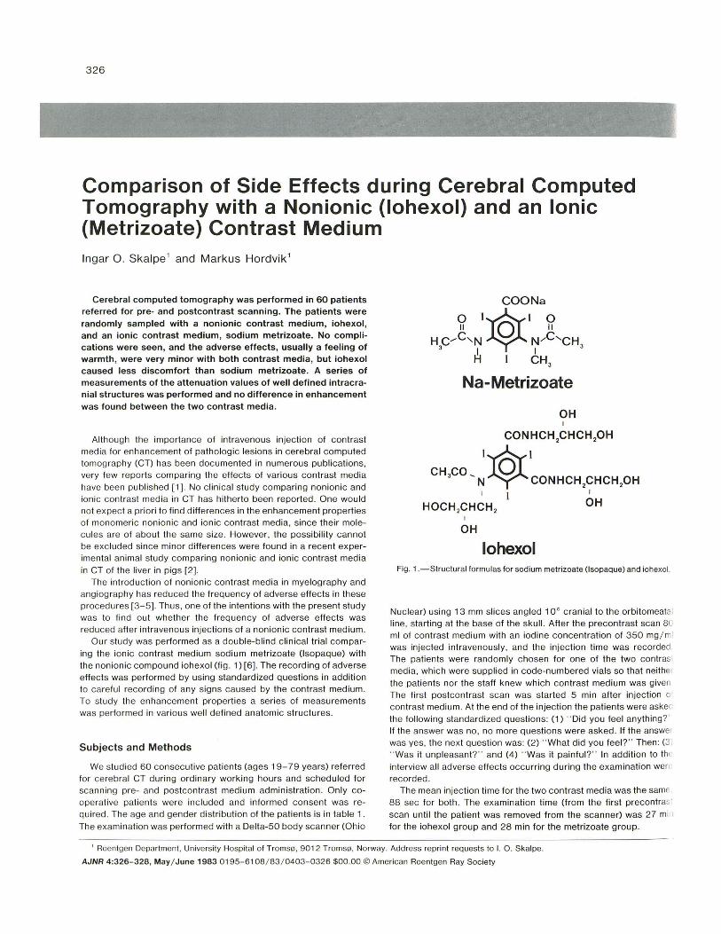
Comparison of Side Effects during Cerebral Computed Tomography with a Nonionic (Iohexol) and an Ionic (Metrizoate) Contrast Medium Ingar O. Skalpe 1 and Markus Hordvik 1
Cerebral computed tomography was performed in 60 patients referred for pre- and postcontrast scanning. The patients were randomly sampled with a non ionic contrast medium, iohexol, and an ionic contrast medium, sodium metrizoate. No complications were seen , and the adverse effects, usually a feeling of warmth, were very minor with both contrast media, but iohexol caused less discomfort than sodium metrizoate. A series of measurements of the attenuation values of well defined intracranial structures was performed and no difference in enhancement was found between the two contrast media.
Although the importance of intravenous injection of contrast med ia for enhancement of patholog ic lesions in cerebral computed tomography (CT) has been documented in numerous publications, very few reports comparing the effects of various contrast media have been published (1). No c linical study comparing non ionic and ion ic con trast med ia in CT has hitherto been reported. One would not expect a priori to find differences in the enhancement propert ies of monomeric non ionic and ionic contrast media, since their molecules are of about the same size. However, the possibility cannot be excluded since minor differences were found in a recent experimental animal study comparing non ionic and ionic contrast media in CT of the liver in pigs (2).
The introduction of non ionic contrast media in myelog raphy and angiog raphy has reduced the frequency of adverse effects in these procedures [3-5). Thus, one of the intentions with the present stud y was to find out wheth er the freq uency of adverse effec ts was reduced after intravenous injections of a non ionic contrast med ium .
Our study was performed as a double-blind c linical trial comparing the ionic contrast medium sodium metrizoate (Isopaque) with the non ionic compound iohexol (fig . 1) (6). Th e recording of adverse effects was performed by using standardized questions in addition to careful recording of any signs caused by the contrast medium. To study the enhancement properties a series of measurements was performed in various well defined anatomic structures.
Subjects and Methods
We studied 60 consecutive patients (ages 19- 79 years) referred for cerebral CT during ordinary working hours and scheduled for scanning pre- and postcontrast med ium administration. Only cooperative patients were inc luded and informed consent was required. The age and gender distr ibution of the patients is in table 1. Th e examination was performed with a Delta-50 body scanner (Ohio
COONa
~ 1*1 ~ H C ..... C"N W,C'CH
3 I I 3
H 1 CH3
Na-Metrizoate
OH
OH
lohexol Fig. 1.- Structural formul as for sodium metrizoate (l sopaque) and iohexol.
Nuclear) using 13 mm slices angled 10° cranial to the orbitomeatai line, starting at the base of the skull. After the precontrast scan Be ml of contrast medium with an iodine concentration of 350 mg/ ml
was injected intravenously, and the injection time was recorded The patients were randomly chosen for one of the two contrasl media, which were supplied in code-numbered vials so that neither the patients nor the staff knew which contrast medium was given The first postcontrast scan was started 5 min after injection 0
contrast medium. At the end of the injection the patients were asket the following standardized questions: (1) " Did you feel anything?' If the answer was no, no more questions were asked . If the answer was yes, the next question was : (2) " Wh at d id you feel?" Then: (3 " Was it un pleasant?" and (4) " Was it painful?" In addition to th( interview all adverse effec ts occurring during the examination wen recorded.
The mean injection time for the two contrast media was the samE' 88 sec for both . The examination time (from the first precontras: scan until the patient was removed from the scanner) was 27 mi I for the iohexol group and 28 min for the metrizoate group.
, Roentgen Department, University Hospital of Troms(i} , 9012 Troms(i}, Norway. Address reprin t requests to I. O. Skalpe.
AJNR 4:326- 328, May / June 1983 0 195-6108/ 83 / 0403-0326 $00.00 © Ameri can Roentgen Ray Society

AJNR:4 , May / June 1983 NEW CONTRAST AGENTS 327
TABLE 1: Gender and Age Distribution of Patients Studied with lohexol and Metrizoate
Contrast Medium Men Women <50 Years >50 Years
lohexol 18 12 14 16 Metrizoate .. . . .. . . . 15 15 20 10
Totals 33 27 34 26
% ~ ISOPAQUE DIOHEXOL
100 97 97
50
10
Yes Heat Nausea Unpleasa nt
Fig. 2. -Adverse effects after intravenous injecti on of 80 ml contrast medium. Paired columns 1-4 represent percentages of patients who: (1 ) answered yes when asked , " Did you feel anything?," (2) and (3) answered heat, nausea, or both when asked , " What did you feel?," (4) answered yes when asked , " Was it unpleasant?" The differences in columns 1 and 2 are statistically sign ificant ( p < 0.01); the difference in co lumn 3 is not sign ificant, in column 4 significant at p < 0.05.
Attenuation measurements were performed in the following anatomic reg ions: (1) white matter in the centrum semiovale, (2) gray matter in the cortex away from the skull , (3) head of th e caudate nucleus, (4) straight sinus in the confluens sinuum , and (5) falx cerebri. Measurements 1 - 3 were performed in a 52 pixel area and 4 and 5 in a 4 pixe l area. The chi-square test was used for the statistical evaluations.
Results
No com plications were seen and all the adverse effec ts were minor and would probably have escaped notice without the routine questioning of th e patients. The results of th e quest ioning appear in figure 2, which shows the kinds of reactions in those patients who felt sensations during the injec tion. In addition to these reactions one patient had a short period of dyspnea and one had palpitations, both after injection of metrizoate. Local pain at the site of injection occurred in another patient during injection of metrizoate and the injection was therefore stopped after 40 ml.
An abnormal CT scan was found in 10 patients, but the adverse effects in this group did not differ from those with a negati ve CT examination . The resu lts of the attenuation measurements appear in table 2. No difference was found between iohexol and metrizoate.
Discussion
The adverse effects were very minor with both iohexol and metrizoate, but they were somewhat more prevalent with metrizoate. The clinical implication of these findings is not very impressive,
TABLE 2: Mean Attenuation Values before and after Intravenous Injection of 80 ml Contrast Medium with an Iodine Concentration of 350 mg / ml
Mean Hounsfield Units Area: Time
lohexol Melrizoate Tolals
White matter: Before . . . . . . . . . . . . 28 28 28 After 28 29 28
Gray matter: Before ......... . .. 33 34 34 After ............ . . 35 35 35
Caudate nuc leus: Before ......... . . .. ... 32 32 32 After 33 33 33
Straight sinus: Before 34 35 35 After 54 52 53
Falx cerebri: Before 36 37 37 After 47 48 48
Note.- There were 30 patients in each group.
since the discomfort experienced by the pat ients after injec tion of the ionic contrast med ium was so minor that it would probably not justify the increased costs of a non ioni c contrast med iu m. However, we do not answer the quest ion whether non ion ic contrast med ia might red uce the frequency of serious and fatal reactions. Such reactions are so rare, about one death / 40,000 intravenous injections [7], that larger, multicenter studies will be necessary to solve this problem.
No differences in the frequency of adverse effects were seen between patients with a normal and a patholog ic cerebral CT examination, but th e number of patients was too small to d raw any conclusions from this. The known reduction in tox ic ity in the nonionic compounds may eventually prove to be of importance , since it has been shown that the prognosis of cerebral infarction is poorer in patients in wh om postcontrast scans are obtained with a contrast medium (ionic) than in patients examined without contrast med ium [8, 9].
No difference in attenuation measurements was found between ionic and non ionic contrast media. These resul ts ag ree with those of Gado et al. [ 1 0], who found no signifi can t enhancement of normal brain tissue and marked enhancement of the straight sinus after intravenous injec tion of 100 ml meg lumine iothalamate 60% using a first-generation EMI scanner. It is unlikely that scanners more modern than the second-generation uni t used in our stu dy will alter our conclusions, since contrast resolution, which is the important quality factor in the present study, has not been improved with the third-generat ion scanners.
In conclusion, very minor adverse effects were recorded with both iohexol and metrizoate, but iohexol caused discomfort much less often then did metrizoate. No differences in enhancement of normal intracranial structures were found .
REFERENCES
1. Norman 0 , Enzman DR, Newton TH. Comparat ive eff icacy of contrast agents in computed tomography scanning of the brain. J Comput Assist Tomogr 1978;2 :319-321
2. Jensen LI , Golman K, Nyman U, Dean P. CT-enhancement av leveren efter injection af et non-ion isk og et ionisk kontrastmid-
328 NEW CONTRAST AGENTS AJNR:4 , May / June 198 J
del i grise. Presented at the Nordiska Kongressen f~r Med icinsk Radiologi, Abo , Sweden, August 1981
3 . Amundsen P, Dugstad G, Sletteb~ M. Clinical testing of Amipaque for cerebral angiography. Neuroradiology 1978; 1 5: 89-93
4. Skalpe 10. Adverse effects of water-soluble contrast media in myelography, c isternography and ventriculography . A review with special reference to metrizamide. Acta Radiol {Suppl] (Stockh) 1977;3 55 : 359-370
5. Skalpe 10, Lundervold A, Tj~rstad K. Complications of cerebral angiog raphy. Comparing metrizamide (Amipaque) and meglumine metrizoate (lsopaque Cerebral). Neuroradiology 1975;19:67-71
6. Lindgren E. lohexol. A non-ionic contrast medium. Pharmacol-
ogy and toxocology. Ac ta Radiol {Suppl] (Stockh ) 1980;362 7. Ansell G. Adverse reactions to contrast agents. Scope of
problem. Invest Radio/1970;5 :374 8. Kendall BE, Pullicino P. Intravascular contrast injection ill
ischemic lesions. II. Effect on prognosis . Neuroradiology 1980;19 :241-243
9. Pullicino P, Kendall BE. Contrast enhancement in ischemic lesions. I. Relationship to prognosis. Neuroradiolog/ 1975;19: 235-239
10. Gada MH , Phelps ME, Coleman RE. An extravascular com·· ponent of contrast enhancement in cranial computed tomoGraphy. Part I: the tissue-blood ratio of contrast enhancement Radiology 1975;11 7 : 589-593