Embed Size (px)
Citation preview

Comparison of Secretory Protein Profiles in Developing Rat
Pancreatic Rudiments and Rat Acinar Tumor Cells
VICTORIA IWANIJ and JAMES D. JAMIESON Section of Cell Biology, Yale University School of Medicine, New Haven, Connecticut 06510. Dr. Iwanij's present address is the Department of Genetics and Cell Biology, University of Minnesota, St. Paul, Minnesota 55108.
ABSTRACT We have previously established that secretory proteins from a rat acinar cell tumor lack two forms of procarboxypeptidase B, are deficient in a major lipase species, and possess markedly reduced amounts of the basic proteins proelastase, basic chymotrypsinogen, basic trypsinogen and ribonuclease (Iwanij, V., and J. D. Jamieson, J. Cell Biol., 95:734-741). Because secretory proteins are markers for acinar cell differentiation, we sought to establish whether the secretory protein profile of the acinar cell tumor is unique to the transformed cell or whether it resembles that of a stage of normal pancreatic development. To this end, we compared the secretory protein pattern from acinar tumor cells with that of rat pancreatic rudiments at days 19-22 of gestation and through day 21 of the postnatal period. Two- dimensional IEF-SDS gel electrophoresis coupled with biosynthetic labeling and fluorography indicates a time-dependent appearance of individual secretory proteins with basic polypep- tides, except for amylase, appearing in the terminal stages of differentiation. In comparison, the secretory protein pattern of the acinar tumor cells most closely resembles that of day-19 embryonic pancreatic rudiments. We propose that the cells of the acinar cell tumor may, in part, mirror a stage of normal pancreatic development.
A number of reports have demonstrated biochemical similari- ties between developing and neoplastic tissue (1-4). For ex- ample, in the liver (1, 3) it is now evident that a-fetoprotein appears early in development, is virtually absent in adult tissue, but reappears at high levels in transformed hepatocytes. We have demonstrated previously (5) that secretory proteins pro- duced by a rat acinar cell tumor are quantitatively and quali- tatively different from those of normal adult rat pancreatic secretion with absence or reduction of several basic polypep- tides and, recently, Van Nest et al. (6) have reported that basic pancreatic secretory proteins appear only at late stages of embryonic development. In view of our observations on the altered pattern of secretory proteins of the acinar cell tumor and of those of Van Nest et al. (6), it was of interest to determine whether the phenotypic expression of the genes for secretory proteins in the acinar cell tumor resembles gene expression occurring during development of embryonic pan- creatic rudiments. In this paper, we compare secretory proteins from the developing rat pancreas to those synthesized by the acinar cell tumor. The results presented here support the hypothesis that the rat acinar cell tumor resembles day-19 normal rat acinar cell differentiation with respect to the phe-
742
notypic expression of secretory proteins. Portions of this study have appeared in abstract form (7).
MATERIALS AND METHODS
R u d i m e n t P repara t ion
Sprague-Dawley rats with timed pregnancies were obtained from Carom Research Lab Animals, Wayne, NJ. Animals were sacrificed by decapitation, the uterus was excised immediately, and embryos were placed in Dulbecco's modified minimal essential medium (Gibco Laboratories, Grand Island Biological Co., Grand Island, NY). Pancreatic rudiments were removed under a dissecting microscope as described before (8). Pancreatic lobules were mechanically pre- pared (5) from the glands of animals 10 or 21 d old. Pups were kept with their mothers until use and were not deprived of food. 10-12 rudiments or mechanically prepared pancreatic lobules were rinsed twice in the same medium, then trans- ferred to the well of a Linbro plate (Flow Laboratories, Inc., Rockville, MD) containing 0.5-1 ml of Dulbecco's medium, 50 U/ml Trasylol (FBA Pharmaceu- ticals, New York, NY), and 50 #Ci/ml of [S:~]methionine (Amersham Corp., Arlington Heights, IL). Rudiments or lobules were allowed to incorporate radio- active precursor for 3 h at 37°C in a tissue culture incubator under an air atmosphere. The pH of the medium was monitored throughout the experiment and adjusted to pH 7.4 with 1 N NaOH if acidification was noted. After labeling, rudiments or lobules were transferred into fresh Dulbecco's medium and rinsed twice by decantation.
THE JOURNAL OF CELL BIOLOGY - VOLUME 95 DeCEmBEr 1982 742-746
© T h e Rockefel ler Univers i ty Press • 0021-9525/82/12/0742/05 $1,00
Extraction Procedure In order to compare soluble proteins from the developing pancreas to those of
the tumor, an efficient procedure for extraction of secretory proteins from the tissues was required as basic polypeptides tend to adsorb avidly to pancreatic membranes (9). In preliminary studies we employed 0,1 M Na~CO:~, pH 11.2 because of its efficacy in the extraction of basic pancreatic polypeptides (10). However, with this extraction protocol, two-dimensional gel analysis revealed up to 16 amylase spots with a shift of isoelectric points toward the acidic region. This indicated that amylase was most probably undergoing deamidation in the high pH medium. To avoid this problem, yet also achieve efficient extraction of proteins, we used a pH 9. I buffer with the addition of a low concentration of Triton X-100.
The final procedure was carried out as follows: rudiments or lobules were dispersed by sonication in 0.5-1.0 ml of a solution composed of 50 mM Tris-HCl, pH 9.1, 50 mM KCL 0.1% Triton X-100, 1 mM phenylmethylsutfonyl fluoride (Sigma Chemical Co., St. Louis, MO) and 50 U/ml Trasylol. The microtip of a Branson sonifier (Branson Sonic Power Co., Danbury, CT) was used at a setting of 5 for 4-5 half-minute periods at 4°C. The homogenates were then centrifuged at 100,000 g~,,~ for 30 rain (Beckman No. 50 rotor, Beckman Instruments, Fullerton, CA). The supernatant was removed, additional phenylmethylsulfonyl fluoride was added (1 mM final concentration), and aliquots of the supernatant were frozen in liquid Ne for storage. Proteins were separated by two-dimensional gel electrophoresis as described in the previous paper (5) and analyzed by Coomassie-Blue staining patterns and by fluorography as described by Bonner and Laskey ( l I).
Pancreatic tumors grown subcutaneously were dissociated into clumps of cells as described before (5), and their secretory proteins were biosynthetically labeled with [:~S]methionine, harvested, and prepared for two-dimensional gel analysis in a manner identical to that used for soluble proteins from rudiments.
RESULTS
Development of Secretory Proteins in the Embryonic and Postnatal Rat Pancreas
Secretory proteins represent a class of pancreatic gene prod- ucts that reflect the stages of differentiation of acinar ceils. As previously reported by Rutter and co-workers (12-14), digestive enzyme and proenzyme production proceeds in two phases during development of the embryonic rat pancreas: during days 11-14 of development (the protodifferentiated state), low but detectable levels of exocrine secretory proteins are synthe- sized, whereas passage into the secondary transition phase (days 14-19) is characterized by the exponential accumulation of secretory proteins, which levels off as the differentiated state is approached at day 19 gestation. Van Nest et al. (6) have recently reported in detail on the pattern of appearance of secretory proteins in developing rat pancreas through day 18 of gestation, using two-dimensional nonequilibrium isoelectric focusing SDS gel electrophoresis. In this study, we report on the changes occurring from day 19 of gestation through birth (day 22) and through day-21 postpartum when adult patterns of secretory proteins are acquired.
Because the embryonic pancreas does not discharge secretory proteins in response to hormones until the time of birth (15, 16), we have used soluble, biosynthetically labeled proteins from homogenates of rat pancreatic rudiments or lobules (see Materials and Methods) at all ages in order to insure reliable sampling of these proteins independent of discharge capability. The analysis of secretory protein profiles at various ages was carried out by fluorography and by Coomassie Blue staining of proteins separated on two-dimensional isoelectric focusing- SDS gels. In the preceding paper (5), we analyzed by two- dimensional gel electrophoresis the profile of secretory proteins discharged from normal adult rat pancreas and obtained a complete pattern of enzymatically identifiable polypeptides, which is used here as a reference map.
We first determined whether the appearance of secretory proteins during days 19-22 of gestation and through day-21
IWANll AN[) JAMIESON
postpartum follows a defined temporal sequence. The results are shown in Fig. 1, in which we analyze soluble, extractable proteins by Coomassie Blue staining of two-dimensional gels.
At day 19 of development (D-19), the only secretory proteins detectable included the amylase complex, acidic chymotrypsin- ogen, procarboxypeptidase B, and a pair of polypeptides whose identity is unknown (spots 12 and 13). The results are consistent with the data of Van Nest et al. (6) who showed that chymo- trypsinogen, amylase, and procarboxypeptidase B appear be- fore day 19 of gestation in the embryonic rat pancreas.
On fetal days 19-22, five additional proenzymes and en- zymes appear, all of which are basic in isoelectric point: day 20, ribonuclease (polypeptide 1); D-21, basic chymotrypsino- gen and proelastase (polypeptides 6 and 7); and D-22, the basic trypsinogens (polypeptides 3 and 4). The results clearly show that at the time of birth the adult pattern of secretory proteins is not yet established.
Developmental changes in secretory protein pattern continue postnatally with the appearance of acidic trypsinogen (poly- peptide 2) at postpartum day 10 (P-10) and the major lipase species (polypeptide 11) at P-21 when the adult pattern of secretory proteins is acquired. Late postpartum appearance of the major form oflipase in the rat pancreas has also been noted by Bradshaw and Rutter (13). These developmental data are summarized in Table I.
Comparison of Acinar Tumor Cell Secretory Proteins with Those of Developing Pancreas
As we (5) and others (6) have reported, the major protein biosynthetic activity of cytodifferentiated pancreatic acinar cells is directed to the production of digestive enzymes and proenzymes. Consequently, to insure sensitive and selective conditions for comparison of acinar tumor cell secretory pro- teins with those of the developing pancreas, prenatal pancreatic rudiments, lobules from postnatal glands, or suspensions of acinar tumor cells were allowed to incorporate ['~SS]methionine into proteins. Soluble extracts, prepared as described in Mate- rials and Methods, were separated by two-dimensional gel electrophoresis and analyzed by fluorography.
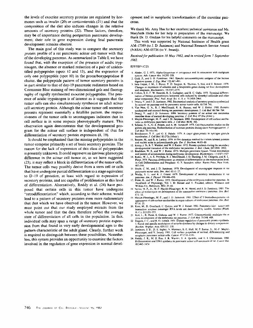
Although we compared fluorograms of secretory (soluble) proteins separated on two-dimensional gels for all develop- mental ages with those from acinar tumor cells, only fluoro- grams of D-19 and P-21 rudiments and postnatal glands, respectively, are shown in Fig. 2, for ease of comparison with tumor secretory proteins.
As illustrated in Fig. 2, the secretory protein pattern of acinar tumor cells is reminiscent of that of D-19 pancreatic rudiments. Thus, although amylase, acidic chymotrypsinogen, and one of the species of procarboxypeptidase B (polypeptide 10) are present, the basic polypeptides ribonuclease, proelastase, and the cationic forms of chymotrypsinogen and trypsinogen which appear between D-20 and D-22 of development are virtually absent. Similarly, the major form of lipase (polypeptide 1 I) is absent in both tumor and D-19 secretory proteins and is first detected by our techniques in the pancreas 21 d after birth (P- 21). The major difference between tumor secretory proteins and those of prenatal pancreatic rudiments is the presence of acidic trypsinogen (polypeptide 2), a protein not normally detected in the gland until 10 d after birth (P-10). Finally, two out of three species of procarboxypeptidase B are absent in tumor secretory proteins but all three are present as early as D-19 in the developing pancreas. The complete profile of changes in pancreatic secretory proteins and the secretory
Ill. Pancreatic Secretory Protein~ in Oevetopment and Neoplasia 743

FIGURE 1 Coomassie Blue staining pattern of two-dimensional IEF-SDS polyacrylamide gel electrophoretograms of soluble (secretory) proteins from developing pancreas. D - 19-22 indicate patterns obtained from embryonic pancreas of the gestational ages 19-22; P- 10 and P-21 are days 10 and 21 after birth, respectively. The circles enclose polypept ide spots of interest in this study. Numbers adjacent to the circles in the D-20 panel refer to identif ied enzymatic activities (see Table I). Solid arrowheads point to polypeptide spots which appear as a function of development. The asterisk indicates the position of the amylase complex; diagonal arrows indicate the position of unidentif ied polypeptides (spots 12 and 13) (a) and of procarboxypeptidase B (b). Molecular weights of the identif ied polypeptides ate given in Fig. 4 and Table II of the preceding paper (5). Variations in apparent IEF and Mr between panels are due to the fact that each experiment was run under slightly different electrophoretic conditions. Partial activation of procarboxypeptidases may account for variations in the polypeptide patterns in the acidic region of the gels. Results shown are representative of three to four determinations for each age.
protein profile of acinar tumor secretion is summarized in Table I. These data are based both on patterns obtained by fluorography or Coomassie Blue staining; the results were identical with either method of analysis.
D I S C U S S I O N
Development of the rat pancreas appears to be divided into three relatively distinct phases (17): the predifferentiated state (day 11) when the pancreatic anlage appears as a diverticulum of the embryonic foregut; the protodifferentiated state (days 11-14) during which time expression of genes for secretory proteins is initiated; and the secondary transition that begins at
744 THe Journat Oe CELt 81OtOGV - VOLUME 95, 1982
day 14-15 when rudiments enter the final differentiated state characterized by onset of rapid cell proliferation, active trans- lation of genes for exportable proteins (12, 14), and concomi- tant appearance of organeUes involved in secretory protein processing. These changes continue through the period of birth and into the postnatal stage.
Studies from our laboratory have also shown that the sec- ondary transition stage is accompanied by the selective ap- pearance of cell surface glycoconjugates that are characteristic of acinar, centroacinar, and endocrine cells (8). Additionally, we have demonstrated that the developmental programs for differentiation of intraceUular organelles and for expression of cell surface glycoconjugates and acquisition of secretagogue
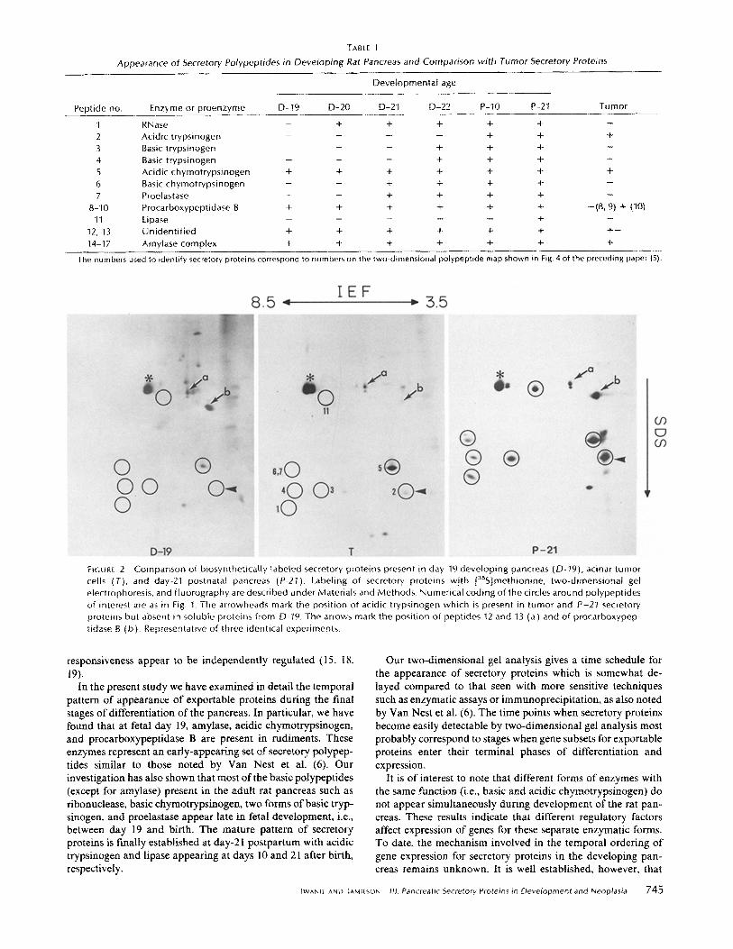
TABLE I
Appearance of Secretory Polypeptides in Developing Rat Pancreas and Comparison with Tumor Secretory Proteins
Deve lopmenta l age
Peptide no. Enzyme or p roenzyme D-19 D-20 D-21 D-22 P-10 P-21 Tumor
I RNase - + + + + + -- 2 Acidic t rypsinogen . . . . + + + 3 Basic t rypsinogen - - - + + + - 4 Basic t rypsinogen - - - + + + - 5 Acidic chymot ryps inogen + + + + + + + 6 Basic chymot ryps inogen - - + + + + -
7 Proelastase - - + + + + - 8 - I 0 Procarboxypept idase B + + + + + + - ( 8 , 9) + (10)
11 Lipase . . . . . + - 12, 13 Un ident i f ied + + + + + + + -
14-17 Amylase complex + + + + + + +
The numbers used to identify secretory proteins correspond to numbers on the two-dimensional polypeptide map shown in Fig. 4 of the preceding paper (5).
F~GUR[ 2 Compar ison of b iosynthet ical ly labeled secretory proteins present in day-19 deve lop ing pancreas (O-19), acinar tumor cells (T), and day-21 postnatal pancreas (P-21). Labeling of secretory proteins w i th [3SS]methionine, two-d imens iona l gel electrophoresis, and f luorography are described under Materials and Methods. Numerical coding of the circles around polypept ides
of interest are as in Fig. I . The arrowheads mark the posi t ion of acidic t rypsinogen which is present in tumor and P-21 secretory proteins but absent in soluble proteins from D-79. The arrows mark the posi t ion of pept ides 12 and 13 (a) and of procarboxypep-
tidase B (b) . Representative of three identical experiments.
responsiveness appear to be independently regulated (15, 18, 19).
In the present study we have examined in detail the temporal pattern of appearance of exportable proteins during the final stages of differentiation of the pancreas. In particular, we have found that at fetal day 19, amylase, acidic chymotrypsinogen, and procarboxypeptidase B are present in rudiments. These enzymes represent an early-appearing set of secretory polypep- tides similar to those noted by Van Nest et al. (6). Our investigation has also shown that most of the basic polypeptides (except for amylase) present in the adult rat pancreas such as ribonuclease, basic chymotrypsinogen, two forms of basic tryp- sinogen, and proelastase appear late in fetal development, i.e., between day 19 and birth. The mature pattern of secretory proteins is finally established at day-21 postpartum with acidic trypsinogen and lipase appearing at days 10 and 21 after birth, respectively.
Our two-dimensional gel analysis gives a time schedule for the appearance of secretory proteins which is somewhat de- layed compared to that seen with more sensitive techniques such as enzymatic assays or immunoprecipitation, as also noted by Van Nest et al. (6). The time points when secretory proteins become easily detectable by two-dimensional gel analysis most probably correspond to stages when gene subsets for exportable proteins enter their terminal phases of differentiation and expression.
It is of interest to note that different forms of enzymes with the same function (i.e., basic and acidic chymotrypsinogen) do not appear simultaneously during development of the rat pan- creas. These results indicate that different regulatory factors affect expression of genes for these separate enzymatic forms. To date, the mechanism involved in the temporal ordering of gene expression for secretory proteins in the developing pan- creas remains unknown. It is well established, however, that
tWAN~ AND IAMf~SON Ill. Pancreatic Secretory Proteins in Development and Neoplasia 7 4 5
the levels of exocrine secretory proteins are regulated by hor- mones such as insulin (20) or corticosteroids (21) and that the composition of the diet also induces changes in the relative amounts of secretory proteins (22). These factors, therefore, may be of importance during postpartum pancreatic develop- ment; their role in gene expression during fetal pancreatic development remains obscure.
The main goal of this study was to compare the secretory protein profile of a rat pancreatic acinar cell tumor with that of the developing pancreas. As summarized in Table I, we have found that, with the exception of the presence of acidic tryp- sinogen, the absence or marked reduction of a pair of uniden- tified polypeptides (spots 12 and 13), and the expression of only one polypeptide (spot 10) in the procarboxypeptidase B cluster, the polypeptide pattern of tumor secretory proteins is in part similar to that of day-19 pancreatic rudiments based on Coomassie Blue staining of two-dimensional gels and tluorog- raphy of rapidly synthesized exocrine polypeptides. The pres- ence of acidic trypsinogen is of interest as it suggests that the tumor cells can also simultaneously synthesize an adult acinar cell secretory protein. Although the acinar tumor cell secretory proteins represent essentially a fetallike pattern, the respon- siveness of the tumor cells to secretagogues indicates that its cell surface is in some respects phenotypically mature. This observation again demonstrates that the developmental pro- gram for the acinar cell surface is independent of that for differentiation of secretory protein expression (8, 19).
It should be emphasized that the missing polypeptides in the tumor comprise primarily a set of basic secretory proteins. The reason for the lack of expression of this class of polypeptides is presently unknown. It could represent an intrinsic phenotypic difference in the acinar cell tumor or, as we have suggested (23), it may reflect a block in differentiation of the tumor cells. The tumor cells may possibly derive from a stem cell or cells that have undergone partial differentiation to a stage equivalent to D-19 of gestation, at least with regard to expression of secretory proteins, and are capable of proliferation at this level of differentiation. Alternatively, Reddy et al. (24) have pro- posed that certain cells in this tumor have undergone "retrodifferentiation" which, according to their scheme, would lead to a pattern of secretory proteins even more rudimentary that that which we have observed in the tumor. However, we must point out that our study employed extracts from the whole tumor and that the data therefore reflect the average state of differentiation of all cells in the population. In fact, individual cells may span a range of secretory protein expres- sion from that found in very early developmental ages to the pattern characteristic of the adult gland. Clearly, further work is required to distinguish between these possibilities. Nonethe- less, this system provides an opportunity to examine the factors involved in the regulation of gene expression in normal devel-
opment and in neoplastic transformation of the exocrine pan- creas.
We thank Ms. Amy Hsu for her excellent technical assistance and Ms. Marybeth Hicks for her help in preparation of the manuscript. We thank Dr. D. Giulian for his helpful comments on the manuscript.
This work was supported by National Institutes of Health grant AM-17389 (to J. D. Jamieson) and National Research Service Award (NRSA) AM-05736 (to V. lwanij).
Received for publication 18 May 1982, and in revised form 7 September 1982.
REFERENCES
I. Abelev, O. L 1971. Alpha-fetoprotein in ontogenesis and its association with malignant tumors. Adv. Cancer Res. 14:295-358.
2. Gold, P., and S. O. Freedman. 1965. Specific carcinoembryonic antigens of the human digestive system. J. Exp. Med. 122:467481.
3. Sala-Trepat, J. M., J. Dever, T. D. Sargent, K. Thomas, S. Sell, and J. Bonner. 1979. Changes in expression of albumin and a-fetoprotein genes during rat liver development and neoplasia. Biochemistry. 18:2167-2178.
4. Collins, S. J., F. W. Ruscetti, R. E. Gallagher, and R. C. Gallo. 1978. Terminal differen- tiation of human promyelocytic leukemia cells induced by dimethyl sulfoxide and other polar compounds. Proc. Natl. Acad. Sci. U. S. A. 75:2458-2468.
5. Iwaulj, V., and J. D. Jamieson. 1982. Biochemical analysis of secretory proteins synthesized by normal rat pancreas and by pancreatic acinar tumor cells. 95:734-741.
6. Van Nest, G. A., R. L MacDonald, R. K. Raman, and W. J. Rutter. 1980. Proteins synthesized and secreted during rat pancreatic development. J. Cell BioL 86:784-794.
7. lwanij, V,, and J. D. Jami©son. 1980. Secretory proteins of rat acinar cell carcinoma resemble those of normal developing pancreas. J. Cell BioL 87:28a (Abstr.).
8. Maylid-Pfennirtgar, M.-F., and J. D. Jamieson. 1980. Development of cell surface saccha- rides on embryonic pancreatic cells..L Cell BioL 86:96-103.
9. Scheale, G. A., G. E. Pulade, and A. M. Tartakoff. 1978. Cell fractionation studies on the guinea pig pancreas. Redistribution of exocrine proteins during tissue homogenization. Z Cell Biol. 78:110-130.
10. Brockmeyer, T. F., and G. E. Palade. 1979. A major glycoprotein in zymogen granule membranes. J. Cell Biol. 83:272a (Abstr.).
I 1. Bonner, M., and R. A. Laskey. 1974. A film detection method for tritium-labeled proteins and nucleic acids in polyacrylamid¢ gels. Fur. J. Biochem. 46:83-88.
12. Kemp, J. D., B. T. WaRher, and W. J. Rutter. 1972. Protein synthesis during the secondary developmental transition of the embryonic rat pancreas. 3". BioL Chem. 247:3941 3952.
13. Bradshaw, W. S., and W. J. Rutter. 1972. Multiple pancreatic lipases. Tissue distribution and pattern of accumulation during embryonic development. Biochemistry. l 1:1517-1527.
14. Rutter, W. J., A. E. Przybyla, R. J. MacDonald, J. D. Harding, J. M. Chirgwin, and R. L. Pictet. 1978. Pancreas development: an analysis of differentiation at the transcription level. In Cell Differentiation and Neoplasia. G. F. Sauoders, editor. Raven Press. New York. 487-508.
15. Doyle, C. M., and J, D. Jamieson. 1978. Development of secretagogue response in rat pancreatic acinar cells. Day. BioL 65:11-27.
16. Werlin, S. L., and R. J. Grand. 1979. Development of secretory mechanisms in rat pancreas. Am. J. Physiol. 236:446-450.
17. Pictet, R., and W. J. Rutter. 1972. Development of the embryonic endocrine pancreas. In Handbook of Physiology, Vol. 1. D Steiner and N. Freink¢l, editors. Williams and WiLkins Co., Baltimore, MD. 25-66.
18. Sarras, M. P., Jr., M.-F. Maylid-Pferminger, R. M. Manzi, and J. D. Jamieson. 1981. The effect of tunicamycin on development of the mammalian embryonic pancreas. Dev. BioL 87:1-t5.
19. Mayli6-Pfenninger, M.-F., and J. D. Jamieson. 1981. Effect of 5-bromodeoxyuridine on appearance of cell-surface saccharides in organ cultures of embryonic pancreas. Day BioL 87:16-23.
20. Korc, M., D. Owerbach, C. Quinto, and W. J. Rutter. 1981. Pancreatic islet acinar cell interaction: amylase messenger RNA levels are determined by insulin. Science I Wash. D. C). 213:351-353.
21. Roll, L., R. Pictet, S. Githens, and W. J. Rutter. 1977. Glucocorticoids modulate the in vitro development of the embryonic rat pancreas. J. Cell BioL 75:398~K~9.
22. Dagorn, J. C., and R. G. Lahaie. 1981. Dietary regulation of pancreatic protein synthesis. I. Rapid and specific modulation of enzyme synthesis by changes in dietary composition. Biochim. Biophys. Acta. 654:111 118.
23. Jamieson, J. D., D. E. Ingber, V. Muresan, B. E. Hull, M. P. Sarras, Jr., M.-F Maylie- Pfenninger, and V. lwanij. 1981. Cell surface properties of normal, differentiating and neoplastic pancreatic acinar cells. Cancer. 47:1516 1525.
24. Reddy, J. K., M. S. Rao, J. R. Warren, S. A. Qureshi, and E. I. Christensen. 1980. Differentiation and DNA synthesis in pancreatic acinar cell carcinoma of rat. Cancer Res. 40:3443-3454.
746 T i l t IOURNA[ Or Ct tL BtOLOG¥ - VOLUME 95, 1982