Embed Size (px)
Citation preview
Research ArticleComparison of Rheological, Drug Release, and MucoadhesiveCharacteristics upon Storage between Hydrogels with Unmodifiedor Beta-Glycerophosphate-Crosslinked Chitosan
Emilia Szymańska , Anna Czajkowska-Kośnik , and Katarzyna Winnicka
Department of Pharmaceutical Technology, Faculty of Pharmacy, Medical University of Białystok, Mickiewicza 2c,Białystok 15-222, Poland
Correspondence should be addressed to Emilia Szymańska; [email protected]
Received 19 April 2018; Revised 25 July 2018; Accepted 15 August 2018; Published 19 September 2018
Academic Editor: Mingqiang Li
Copyright © 2018 Emilia Szymańska et al. This is an open access article distributed under the Creative Commons AttributionLicense, which permits unrestricted use, distribution, and reproduction in any medium, provided the original work isproperly cited.
The physicochemical characteristics of beta-glycerophosphate-crosslinked chitosan hydrogels were investigated upon long-termstorage at ambient, accelerated, and refrigerated conditions and compared to unmodified chitosan formulations. Additionally,the impact of chitosan modification on the ex vivo mucoadhesive performance in contact with porcine vaginal mucosa and onthe drug release profile from hydrogels was evaluated. Viscosity and mechanical properties of formulations with unmodifiedchitosan decreased significantly upon storage regardless of tested conditions as a result of hydrolytic depolymerization.Introduction of ion crosslinker exerted stabilizing effect on physicochemical performance of chitosan hydrogels but only uponstorage at refrigerated conditions. Beta-glycerophosphate-modified chitosan formulations preserved organoleptic, rheologicalbehavior, and hydrogel structure up to 3-month storage at 4± 2°C. Viscosity variations upon storage influenced markedlymucoadhesive properties and drug release rate from hydrogels.
1. Introduction
Application of multifunctional polymers as effective excipi-ents in drug delivery systems is a current and attractiveconcept in pharmaceutical technology [1]. Among variouspolymer materials, chitosan (CS)—an abundantly accessiblepolysaccharide, owing to its biodegradability and biocompat-ibility is extensively investigated for a number of pharmaceu-tical and medical applications, comprising prolonged drugrelease delivery systems [2], wound dressings [3], cartilageand bone tissue engineering scaffolds [4, 5], nanocarriersfor gene delivery [6], or vaccine adjuvants [7].
CS is commercially possessed by deacetylation of chitinderived mainly from the exoskeleton of crustaceans, thoughalternative production methods by fungal fermentation havebeen also explored to provide CS with more defined charac-teristics [8]. Due to polycationic behavior, CS structure canbe certainly modified and utilized into diverse semisolid or
solid vehicles. Hence, CS may be regarded to be a promisingtool not only in pharmaceutical [9] but also in cosmetic [10]or food industry [11].
CS displays mucoadhesive potential enabling interactionwith mucin via electrostatic or hydrogen bonding. Mucoad-hesiveness, providing an intimate contact between CS-baseddosage form and mucosal tissue, appears to be beneficial,especially in terms of CS application in technology of drugcarriers for local: nasal [12], ocular [2], buccal [13], or vaginaldelivery [14]. Additionally, with regard to capability of swell-ing and creating hydrogel matrix, CS may be useful in thedevelopment of modified release delivery systems [15, 16].In recent years, particular attention has been directed to theuse of CS as an antimicrobial adjunctive [17, 18]. This capa-bility of increasing the pharmacological action of an antimi-crobial agent provides the opportunity for combinationtherapy in which CS acts as the effective excipient and simul-taneously as an active part of the treatment.
HindawiInternational Journal of Polymer ScienceVolume 2018, Article ID 3592843, 12 pageshttps://doi.org/10.1155/2018/3592843
Regarding abovementioned advantages of CS, experi-mental research was conducted to investigate CS usabilityas pharmaceutical excipient in technology of vaginal hydro-gels for the treatment of mycotic vaginosis. Designed semi-solid platforms displayed favorable ability to interact withporcine vaginal mucosa confirmed in ex vivo studies followedwith enhanced anti-Candida activity compared to commer-cially available preparation with clotrimazole [19, 20]. None-theless, because of fast and uncontrollable disintegration inan acidic environment and poor rheological behavior withregard to vaginal administration, further improvement ofCS structure was needed. Previously presented data showedthat CS crosslinked with beta-glycerophosphate disodium(bGP) demonstrated a promising potential for the develop-ment of mucoadhesive hydrogels with feasible rheologicaland mechanical properties [20].
Even though CS is regarded as a multifunctional com-pound in the technology of drug carriers, only a limited num-ber of pharmaceutical products with CS (apart fromhemostatic dressings, products for wound healing, or nutra-ceuticals) are commercially available. One of the main draw-backs, limiting the application of CS in pharmaceuticaltechnology, is its susceptibility to environmental conditions(especially with regard to humidity and temperature) as wellas processing factors, which in consequence might impact thephysicochemical and pharmaceutical performance of poly-mer’s dosage form upon storage [21]. According to the liter-ature, modification of CS structure via grafting, chemical, orionic crosslinking may overcome the problems of reducedlong-term stability of polymer-based drug delivery systems[22–24]. Since, to our best knowledge, only limited numberof studies have been devoted to the issue of long-term stabil-ity data of CS-based dosage forms, therefore, the goal of thecurrent paper was to examine the influence of crosslinkingwith bGP on the physicochemical performance of CS hydro-gels with a model antifungal agent upon storage. Designedsemisolid platforms with bGP/CS were characterized byorganoleptic, rheological, mucoadhesive properties and com-pared to unmodified CS-based hydrogels. The impact of CSmodification on the drug release profile from hydrogels uponstorage was also examined.
2. Materials and Methods
2.1. Materials. High-quality medium molecular weight CS(200–400 kDa; viscosity 370–390mPa·s at 25°C, 1% (w/w)in 1% (v/v) acetic acid) with deacetylation degree 80% wasobtained from Heppe Medical CS GmbH (Haale, Germany).Clotrimazole was a gift from Ziaja Ltd. (Gdańsk, Poland).bGP disodium salt hydrate and Cremophor EL wereobtained from Sigma–Aldrich (Steinheim, Germany). Aceticacid 80%, glycerolum 85%, disodium hydrogen phosphate,potassium dihydrogen phosphate, sodium acetate, andsodium hydroxide were purchased from Chempur (PiekaryŚląskie, Poland). Methanol (HPLC grade) was purchasedfrom Merck (Darmstadt, Germany). Vaginal mucoadhesivegel Replens™ was acquired from APC Instytut Sp. z o.o.(Warsaw, Poland). Water for HPLC was distilled and passed
through a reverse osmosis system Milli Q Reagent WaterSystem (Billerica, MA, USA).
2.2. Experimental Procedure
2.2.1. Hydrogels with Unmodified or bGP-Crosslinked CS.Hydrogels with clotrimazole were prepared using mechanicalstirrer according to the previously described technique [20].Briefly, CS base with concentration of 3% or 4% (w/w) wasobtained by dissolving proper amount of polymer in a gentlyheated acetic acid solution (the weight ratio CS : glacial aceticacid was 0.6 : 1.0) [25] and stirred until homogenous mix-tures appeared. To prepare bGP-modified CS hydrogels, theappropriate amount (necessary to simulate vaginal pH 4.5)of cold 45% (w/w) aqueous solution of bGP (the weight ratioCS to bGP 1 : 0.63) was added dropwise to the cold CS basewith continuous agitation. Subsequently, suitable amountof unmodified CS or bGP/CS base was carefully added to amixture of humectant, preservative, and clotrimazole solubi-lized in Cremophor EL to obtain uniform dispersion of drugin the hydrogel matrix. Hydrogels’ composition is displayedin Table 1.
2.2.2. Long-Term Evaluation of Hydrogels.Hydrogel formula-tions were subjected to long-term stability studies underdefined conditions of humidity and temperature accordingto ICH guideline [26]. Briefly, hydrogels closed in sealed poly-ethylene containers and additionally in cardboard packagesto prevent from light exposure were kept in humidity cham-bers (CTC 256 Memmert GmbH, Schwabach, Germany)maintained at 25± 2°C/60± 5% relative humidity (RH) and40± 2°C/70± 5% RH as well as in the refrigerator at 4± 2°C.At specific time intervals (displayed in Table 2), macroscopicperformance, pH and viscosity values, drug particle’s diame-ter, and drug content uniformity followed with mechanicaland mucoadhesive performance were evaluated. Formula-tions were maintained for at least 1 h at ambient temperatureprior analysis.
(1) Macroscopic andMicroscopic Analysis. Organoleptic anal-ysis in terms of consistency, odor, and color was performedin accordance to the European Pharmacopoeia (Eur. Ph.)by placing hydrogel in a thin layer on a glass slide [27].
Particles size analysis was accomplished using an opticalmicroscope Motic BA 400 (Moticon, Wetzlar, Germany)with magnifications 100x and 400x. Concisely, 0.5mg of eachformulation (corresponding to 10μg of clotrimazole) waslocated on a glass slide and the longest dimension from edgeto edge of drug particles was defined for particles present inat least three areas of observation [28].
(2) Viscosity Measurement. The apparent viscosity was mea-sured according to Eur. Ph. [27] using a digital rotationalBrookfield RVDV-III ULTRA Viscometer (Brookfield Engi-neering Laboratories, Germany) at ambient temperature.Each hydrogel sample (0.5 g) was placed in the thermostatedsampler holder of the viscometer, allowed to equilibrate for10min at 25± 1°C, and then C-52 spindle was lowered into
2 International Journal of Polymer Science
the sample. The shear rate was 6/s. Each experiment was per-formed at least three times.
(3) pH Determination. The pH of hydrogels was estimated bya glass electrode of the pH meter Orion-3 Star (Thermo Sci-entific, Waltham, MA, USA) in triplicate.
(4) Drug Content Uniformity. Drug content uniformity wasevaluated after the extraction of hydrogel samples with abso-lute ethanol, centrifugation at 4000 rpm for 20min, and filtra-tion through 0.45μmnylon filters. Samples were next suitablydiluted with mobile phase and determined for clotrimazolecontent byHPLCmethod according to [19]. Separations wereperformed at ambient temperature on 5μm Zorbax EclipseXDB–C18 (4.6mm× 150mm) column (Agilent, Waldbronn,Germany). The mobile phase consisted of methanol-phosphate buffer pH7.4 (4 : 1, v/v), the flow rate was1.0mL/min, and the retention time was 5.3min. The detec-tor was set to 210nm. The standard calibration curve waslinear over the range of 1 to 100μg/mL (R2 = 0 995).
(5) Mechanical Properties. Mechanical properties were eval-uated using Texture Analyser TA.XT Plus (Stable MicroSystem, UK) equipped with a backward extrusion measur-ing system A/BE (25mm diameter). Each preparation(30.0± 0.2 g) was located in a measuring vessel, and thena disc was compressed with a speed of 2mm/s into thesample to a defined depth of 5mm. The experimentalparameters were established during preliminary study. Allmeasurements were carried out at 25± 2°C. From theforce-time curves, the values of consistency were calculatedand the hardness parameter was recorded using Texture
Exponent 32 software. Each experiment was carried out atleast three times.
(6) Ex Vivo Mucoadhesive Properties. Ex vivo mucoadhesivebehavior of hydrogels in contact with porcine vaginal mucosawas evaluated according to a method defined previously [15]with using Texture Analyser TA.XT. Plus (Stable Micro Sys-tems, Godalming, UK) equipped with the measuring systemA/GMP. Briefly, freshly excised porcine vaginal mucosa wasadhered by double-sided tape to the platform below the tex-ture analyzer probe whilst hydrogel sample (1.5mL) was seton the upper probe and secured with the attached supportcollar. The experiments were conducted at 37± 1°C. Priorto analysis, the support collar was removed and the probewas lowered onto the surface of the porcine vaginal mucosawith a speed of 0.5mm/s. After keeping a contact time for180 s with contact force 0.5N, the surfaces were separatedat a constant rate of 0.1mm/s. The maximum detachmentforce (Fmax) as a function of displacement was recordeddirectly from Texture Exponent 32 software, and the workof adhesion (Wad) was calculated from the area under theforce vs. distance curve. Each experiment was performedsix times.
(7) In Vitro Drug Release Studies. Due to unfavorable organ-oleptic and rheological changes observed for hydrogels upontheir storage at ambient and accelerated conditions, in vitrodissolution studies were performed only for formulationswith preserved rheological and mechanical behavior upon6-month storage at 4± 2°C. Drug release was measuredthrough natural cellulose membrane (Cuprophan withMWCO 10000Da, Medicell, London, UK) using anEnhancer cell (Agilent Technologies, Cary, USA) [29] andUSP dissolution Apparatus II (Agilent 708-DS, Agilent Tech-nologies, Cary, USA). Each formulation (1.0 g) was placed inthe drug reservoir on the top of the membrane makingcertain that no entrapped air was present at the interfaceof the dosage form and the membrane (diffusion area3.80 cm2). The dissolution medium was 100mL acetic buffer(pH4.5) with addition of 1% surfactant to retain sink condi-tions. The temperature of medium was maintained at 37± 1°C, and a stirring rate was 75 rpm. Samples (2mL) werewithdrawn at the predetermined time points, filtered through0.45 μm CA paper filters, diluted with mobile phase, and
Table 1: Composition of hydrogels with unmodified CS (F1, F2) or bGP-crosslinked CS (F3, F4).
Ingredient (g) CategoryHydrogel with
Unmodified CS bGP-modified CSF1 F2 F3 F4
CS Gelling agent 3.0 4.0 3.0 4.0
bGP 45% (w/w) Ion crosslinker — — 4.2 5.6
Clotrimazole API 2.0 2.0 2.0 2.0
Glycerolum Humectant 5.0 5.0 5.0 5.0
Cremophor EL Solubilizer 6.0 6.0 6.0 6.0
D-Glucono-1,5-lactone and sodium benzoate Preservative 1.0 1.0 1.0 1.0
Acetic acid Vehicle Up to 100.0 Up to 100.0 Up to 100.0 Up to 100.0
Table 2: Time intervals applied for stability studies [26].
Stabilitystudies
Storageconditions
The minimumperiod of storage
Time intervals(months)
Long term25± 2°C/60± 5% RH
12 months 1, 3, 6, 12
4± 2°C 6 months 1, 3, 6
Accelerated40± 2°C/75± 5% RH
6 months 1, 3, 6
3International Journal of Polymer Science
quantified using HPLC method. Withdrawn samples werereplaced with equal volume of fresh medium. All releaseexperiments were carried out in triplicate.
2.3. Statistical Analysis.Quantitative variables were expressedas the mean± standard deviation (SD) or the median byusing MS Excel software. A statistical analysis of mucoadhe-sive measurements was accomplished using a nonparametricKruskal–Wallis test, with the Statistica 12.0 software. Mea-surements were considered significant at p < 0 05.
3. Results and Discussion
3.1. Characterization of Hydrogels upon Storage. Stabilityevaluation provides the critical characteristics of the drugdosage form in terms of its physicochemical or biopharma-ceutical properties, and thus it is crucial for the full assess-ment of product quality during preformulation analysis.Basically, for drug products, a comparative stability data inboth long term (at 25°C/60% RH for 12 months) and accel-erated conditions (40°C/75% RH) is required to define anacceptable product shelf-life [26]. Given these premises,the effect of ion crosslinking on CS hydrogel physicochemi-cal characteristics upon storage at 25°C/60% RH and 40°C/75% RH was studied. Regarding the fact that CS is recom-mended to store at low temperatures (2–8°C) [27], addi-tional examination of formulations upon storage at 4± 2°Cwas also provided.
Directly after preparation, all hydrogels possessedsmooth, uniform consistency and off-white color resultingfrom the presence of suspended clotrimazole (Figure S1). Sig-nificant organoleptic differences were noticed upon firstmonths of storage at ambient conditions. Hydrogels withunmodified CS (F1, F2) became yellowish within one monthat 25°C/60% RH, and the color alterations were deepenedover time. After 3-month storage, loss of gel structure andhomogeneity of formulations F1 and F2 was observed. Itshould be noted that hydrogels with bGP-modified CS (F3and F4) remained stable in terms of organoleptic propertieswithin 3-month storage. Nonetheless, upon 6-month storageat 25°C/60% RH, a substantial liquidation with visible drugparticle sedimentation and simultaneous color changesoccurred in all examined hydrogels (Figure S2).
The most profound and particularly fast alterations inhydrogel performance appeared upon their storage at 40°C/75% RH. When hydrogels with unmodified chitosan (F1and F2) were subjected to accelerated stability studies, lossof homogeneity follow with color changes and drug particlesedimentation occurred just upon 1-month storage period.Regarding formulations with bGP/CS (F3 and F4), a signifi-cant enhancement in the structure stiffness was observedover time and after 3 months at 40°C/75% RH, hydro-gels transformed into brown solid state formulations(Figure S3). The observed time-delayed sol/gel transitionwas probably attributed to thermosensitivity of CS in thepresence of ion crosslinker, which affected the time and thetemperature of CS gelling [30].
Notably, all hydrogel formulations remained organolep-tically stable up to 3 months at refrigerated conditions. Upon
their 6-month storage at 4± 2°C, no alterations in bGP/CShydrogels’ appearance were observed whereas substantialchanges in consistency with simultaneous drug particle sedi-mentation in hydrogels with unmodified CS (F1 and F2)occurred (Figure S4).
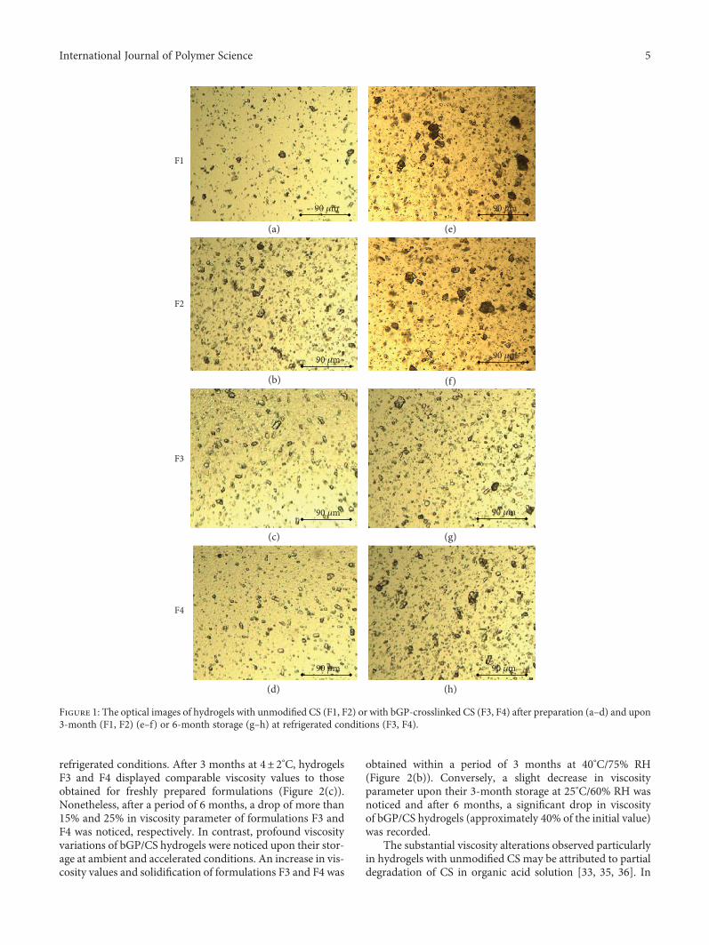
The microscopic analysis of all investigated formulationsrevealed no considerable change in average particle size ofsuspended clotrimazole upon storage at refrigerated condi-tions although the presence of few aggregates of drug parti-cles was observed in hydrogels F1 and F2 (Figure 1).
Due to the presence of undesirable changes in hydrogels’organoleptic properties followed with loss of their homoge-neity upon 3-month and 6-month storage at accelerated orambient conditions (Figures S2 and S3), respectively, formu-lations F1–F4 were excluded from further long-term analy-sis. Evaluation of drug content uniformity was performedfor these hydrogel formulations, which preserved their vis-cosity and organoleptic properties upon storage. As it is pre-sented in Table 3, clotrimazole was found to be chemicallystable in hydrogels F1 and F2 upon 3-month storage atambient and refrigerated conditions and upon 1-month at40°C/75% RH. Regarding bGP/CS hydrogels, the drug con-tent was found to be uniform and was within acceptableUSP limit 90%–110% after storage in all investigated storageconditions [31].
A slight increase in pH values of hydrogels F1 and F2 wasobserved after 3-month storage at 40°C/75% RH and 6-month at 25°C/60% RH in contrast to formulations F3 andF4, which displayed decreasing pH trend upon storageregardless of temperature conditions (Table 3). Nonetheless,at the end of the stability studies, all hydrogels displayed thepH values below 4.5, which was maintained within the phys-iological range 3.5–4.9 [32].
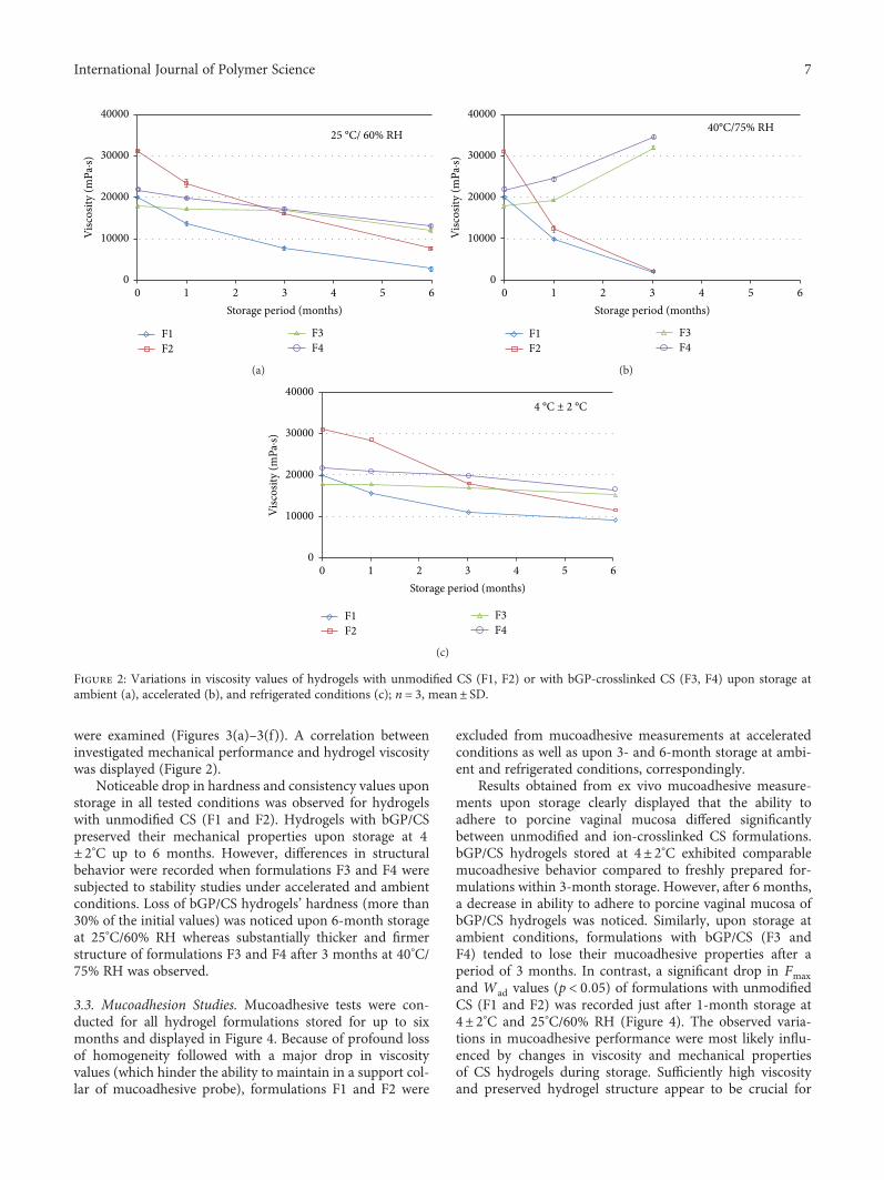
3.2. Viscosity and Mechanical Measurements. To assess theinfluence of ion crosslinking on the rheological behavior ofCS hydrogels upon storage, the viscosity measurements wereperformed. The intrinsic viscosity of CS defines the ability toform viscous solution and is proportional to the polymeraverage molecular weight [33]. Figure 2 displays the viscosityvariations upon storage at different conditions.
Freshly prepared hydrogels F3 and F4 displayed lowerviscosities compared to unmodified CS formulations (F1and F2) most likely as a consequence of greater ionic strengthresulted from the introduction of bGP. All formulationsexhibited non-Newtonian pseudoplastic behavior, and thepresence of bGP improved thixotropic properties of CShydrogels [20].
It can be seen that viscosity of hydrogels with unmodifiedCS (F1 and F2) decreased significantly over time regardless oftested conditions. Formulation F2, with higher concentrationof CS, displayed more profound viscosity variations and lossof more than 40%, 50%, and 90% of the initial value after 3-month storage at 4± 2°C, 25°C/60%RH, and 40°C/75% RHwas observed, respectively. Similarly, Chattopadhyay andInamdar observed the impact of CS concentration on the vis-cosity of polymer solution over storage [34]. Interestingly,the presence of ion crosslinker exerted stabilizing effecton CS hydrogels’ viscosity but only upon their storage at
4 International Journal of Polymer Science
refrigerated conditions. After 3 months at 4± 2°C, hydrogelsF3 and F4 displayed comparable viscosity values to thoseobtained for freshly prepared formulations (Figure 2(c)).Nonetheless, after a period of 6 months, a drop of more than15% and 25% in viscosity parameter of formulations F3 andF4 was noticed, respectively. In contrast, profound viscosityvariations of bGP/CS hydrogels were noticed upon their stor-age at ambient and accelerated conditions. An increase in vis-cosity values and solidification of formulations F3 and F4 was
obtained within a period of 3 months at 40°C/75% RH(Figure 2(b)). Conversely, a slight decrease in viscosityparameter upon their 3-month storage at 25°C/60% RH wasnoticed and after 6 months, a significant drop in viscosityof bGP/CS hydrogels (approximately 40% of the initial value)was recorded.
The substantial viscosity alterations observed particularlyin hydrogels with unmodified CS may be attributed to partialdegradation of CS in organic acid solution [33, 35, 36]. In
90 �휇m
(a) (e)
(f)
(g)
(h)
(b)
(c)
(d)
90 �휇m
90 �휇m
90 �휇m
90 �휇m90 �휇m
90 �휇m
90 �휇m
F1
F2
F3
F4
Figure 1: The optical images of hydrogels with unmodified CS (F1, F2) or with bGP-crosslinked CS (F3, F4) after preparation (a–d) and upon3-month (F1, F2) (e–f) or 6-month storage (g–h) at refrigerated conditions (F3, F4).
5International Journal of Polymer Science
overall, storage temperature and CS concentration werefound to accelerate the rate of hydrolysis of unmodifiedCS [33, 34]. Our findings from stability analysis at refriger-ated conditions are in the agreement with studies accom-plished by Nguyen et al., which revealed that the ratio ofCS decomposition in acetic acid environment could bereduced upon storage at 4± 2°C [35]. In contrast, Ruel-Gariépy et al. observed poor stability of bGP/CS thermogel-ling solutions below room temperature as gelation of thesystems occurred upon 3-month storage at refrigeratedconditions [37]. Similarly, Supper et al. indicated thatthe CS/bGP thermogelling solutions intended for parenteraladministration possessed reduced physicochemical stability
both under ambient and refrigerated conditions [38]. None-theless, in the present studies, much higher CS concentrationwas used for hydrogel preparation (3-4%) as compared tothe abovementioned studies, and it could be regarded asone of the factor improving hydrogel stability. In addition,the ability of bGP to balance the effect of the presence ofacetic acid solution might have slowed down the rate of CSdegradation and in consequence maintained the viscosityparameters over storage.
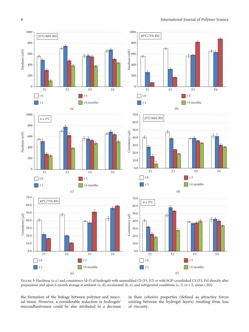
To assess deeper insight into the internal structure of pre-pared hydrogels upon their storage, the mechanical proper-ties: hardness and consistency—parameters reflecting thedegree of difficulty in loss of initial formulations’ structure,
Table 3: Changes in organoleptic properties, clotrimazole content, and drug particle size in hydrogels with unmodified CS (F1, F2) or withbGP-crosslinked CS (F3, F4) upon storage compared to values obtained to freshly prepared formulations.
Storage time (months) ParameterHydrogel with
Unmodified CS bGP-crosslinked CSF1 F2 F3 F4
0
pH 4.06± 0.02 4.08± 0.01 4.54± 0.04 4.56± 0.03Particle size range (μm) 6.0–49.5 6.0–47.0 4.0–40.0 4.0–41.5
Drug content (%)∗ 95.6± 4.9 98.2± 3.5 97.3± 4.1 97.4± 3.325°C/60% RH
1
pH∗ 4.16± 0.05 4.23± 0.05 4.51± 0.04 4.53± 0.02Particle size range (μm) 7.5–52.5 7.5–57.0 6.0–29.5 5.0–48.5
Drug content (%)∗ 94.8± 3.3 96.1± 4.2 96.5± 3.5 97.1± 3.9
3
pH∗ 4.40± 0.02 4.38± 0.03 4.35± 0.02 4.27± 0.03Particle size range (μm) 7.0–58.0 6.0–54.5 4.0–41.0 4.0–34.5
Drug content (%)∗ 94.6± 5.9 92.4± 5.1 98.1± 4.1 95.2± 4.5
6
pH∗ 4.31± 0.02 4.39± 0.01 4.26± 0.02 4.23± 0.02Particle size range (μm) n.d. n.d. Sedimentation Sedimentation
Drug content (%)∗ n.d. n.d. n.d. n.d.
40°C/75% RH
1
pH∗ 4.20± 0.03 4.24± 0.05 4.56± 0.04 4.51± 0.02Particle size diameter (μm) Sedimentation Sedimentation 10.0–53.0 5.0–61.0
Drug content (%)∗ n.d. n.d. 96.5± 3.5 97.1± 3.9
3
pH∗ 4.40± 0.02 4.48± 0.02 4.36± 0.01 4.29± 0.03Particle size range (μm) n.d. n.d. 5.0–44.5 4.5–37.0
Drug content (%)∗ n.d. n.d. 93.1± 7.1 94.2± 6.84± 2°C
1
pH∗ 4.10± 0.05 4.03± 0.05 4.56± 0.04 4.53± 0.02Particle size range (μm) 12.0–58.5 7.5–65.0 8.0–59.0 10.5–45.0
Drug content (%)∗ 94.3± 3.8 96.1± 4.2 96.5± 3.5 97.1± 3.9
3
pH∗ 4.10± 0.02 4.12± 0.02 4.31± 0.02 4.37± 0.03Particle size range (μm) Sedimentation 6.0–59.5 11.0–40.0 5.0–45.5
Drug content (%)∗ n.d. 95.1± 8.4 98.1± 4.7 95.2± 4.5
6
pH∗ 4.23± 0.03 4.29± 0.03 4.28± 0.03 4.37± 0.01Particle size range (μm) n.d. n.d. 6.5–46.0 5.0–40.0
Drug content (%)∗ n.d. n.d. 94.1± 4.9 96.0± 3.7∗n = 3, mean ± SD; n.d.: not determined.
6 International Journal of Polymer Science
were examined (Figures 3(a)–3(f)). A correlation betweeninvestigated mechanical performance and hydrogel viscositywas displayed (Figure 2).
Noticeable drop in hardness and consistency values uponstorage in all tested conditions was observed for hydrogelswith unmodified CS (F1 and F2). Hydrogels with bGP/CSpreserved their mechanical properties upon storage at 4± 2°C up to 6 months. However, differences in structuralbehavior were recorded when formulations F3 and F4 weresubjected to stability studies under accelerated and ambientconditions. Loss of bGP/CS hydrogels’ hardness (more than30% of the initial values) was noticed upon 6-month storageat 25°C/60% RH whereas substantially thicker and firmerstructure of formulations F3 and F4 after 3 months at 40°C/75% RH was observed.
3.3. Mucoadhesion Studies. Mucoadhesive tests were con-ducted for all hydrogel formulations stored for up to sixmonths and displayed in Figure 4. Because of profound lossof homogeneity followed with a major drop in viscosityvalues (which hinder the ability to maintain in a support col-lar of mucoadhesive probe), formulations F1 and F2 were
excluded from mucoadhesive measurements at acceleratedconditions as well as upon 3- and 6-month storage at ambi-ent and refrigerated conditions, correspondingly.
Results obtained from ex vivo mucoadhesive measure-ments upon storage clearly displayed that the ability toadhere to porcine vaginal mucosa differed significantlybetween unmodified and ion-crosslinked CS formulations.bGP/CS hydrogels stored at 4± 2°C exhibited comparablemucoadhesive behavior compared to freshly prepared for-mulations within 3-month storage. However, after 6 months,a decrease in ability to adhere to porcine vaginal mucosa ofbGP/CS hydrogels was noticed. Similarly, upon storage atambient conditions, formulations with bGP/CS (F3 andF4) tended to lose their mucoadhesive properties after aperiod of 3 months. In contrast, a significant drop in Fmaxand Wad values (p < 0 05) of formulations with unmodifiedCS (F1 and F2) was recorded just after 1-month storage at4± 2°C and 25°C/60% RH (Figure 4). The observed varia-tions in mucoadhesive performance were most likely influ-enced by changes in viscosity and mechanical propertiesof CS hydrogels during storage. Sufficiently high viscosityand preserved hydrogel structure appear to be crucial for
25 °C/ 60% RH
F1F2
F3F4
0
10000
20000
30000
40000
Visc
osity
(mPa
·s)
1 2 3 4 5 60Storage period (months)
(a)
40°C/75% RH
F1F2
F3F4
0
10000
20000
30000
40000
Visc
osity
(mPa
·s)
1 2 3 4 5 60Storage period (months)
(b)
F1F2
F3F4
4 °C ± 2 °C
0
10000
20000
30000
40000
Visc
osity
(mPa
·s)
1 2 3 4 5 60Storage period (months)
(c)
Figure 2: Variations in viscosity values of hydrogels with unmodified CS (F1, F2) or with bGP-crosslinked CS (F3, F4) upon storage atambient (a), accelerated (b), and refrigerated conditions (c); n = 3, mean± SD.
7International Journal of Polymer Science
the formation of the linkage between polymer and muco-sal tissue. However, a considerable reduction in hydrogels’mucoadhesiveness could be also attributed to a decrease
in their cohesive properties (defined as attractive forcesexisting between the hydrogel layers) resulting from lossof viscosity.
25°C/60% RH
F1 F2 F3 F4
t 1
t 3
t 6 months
t 0
0
200
400
600
800
1000
Har
dnes
s (m
N)
(a)
0
200
400
600
800
1000
Har
dnes
s (m
N)
40°C/75% RH
F1 F2 F3 F4
t 1
t 3
t 6 months
t 0
(b)
4 ± 2°C
F1 F2 F3 F4
t 1
t 3
t 6 months
t 0
0
200
400
600
800
1000
Har
dnes
s (m
N)
(c)
25°C/60% RH
F1 F2 F3 F4
t 1
t 3
t 6 months
t 0
0.0
10.0
20.0
30.0
40.0
50.0
60.0
70.0
Con
siste
ncy
(�휇J)
(d)
40°C/75% RH
F1 F2 F3 F4
t 1
t 3
t 6 months
t 0
0.0
10.0
20.0
30.0
40.0
50.0
60.0
70.0
Con
siste
ncy
(�휇J)
(e)
4 ± 2°C
F1 F2 F3 F4
t 1
t 3
t 6 months
t 0
0.0
10.0
20.0
30.0
40.0
50.0
60.0
70.0
Con
siste
ncy
(�휇J)
(f)
Figure 3: Hardness (a–c) and consistency (d–f) of hydrogels with unmodified CS (F1, F2) or with bGP-crosslinked CS (F3, F4) directly afterpreparation and upon 6-month storage at ambient (a, d), accelerated (b, e), and refrigerated conditions (c, f) (n = 3, mean± SD).
8 International Journal of Polymer Science
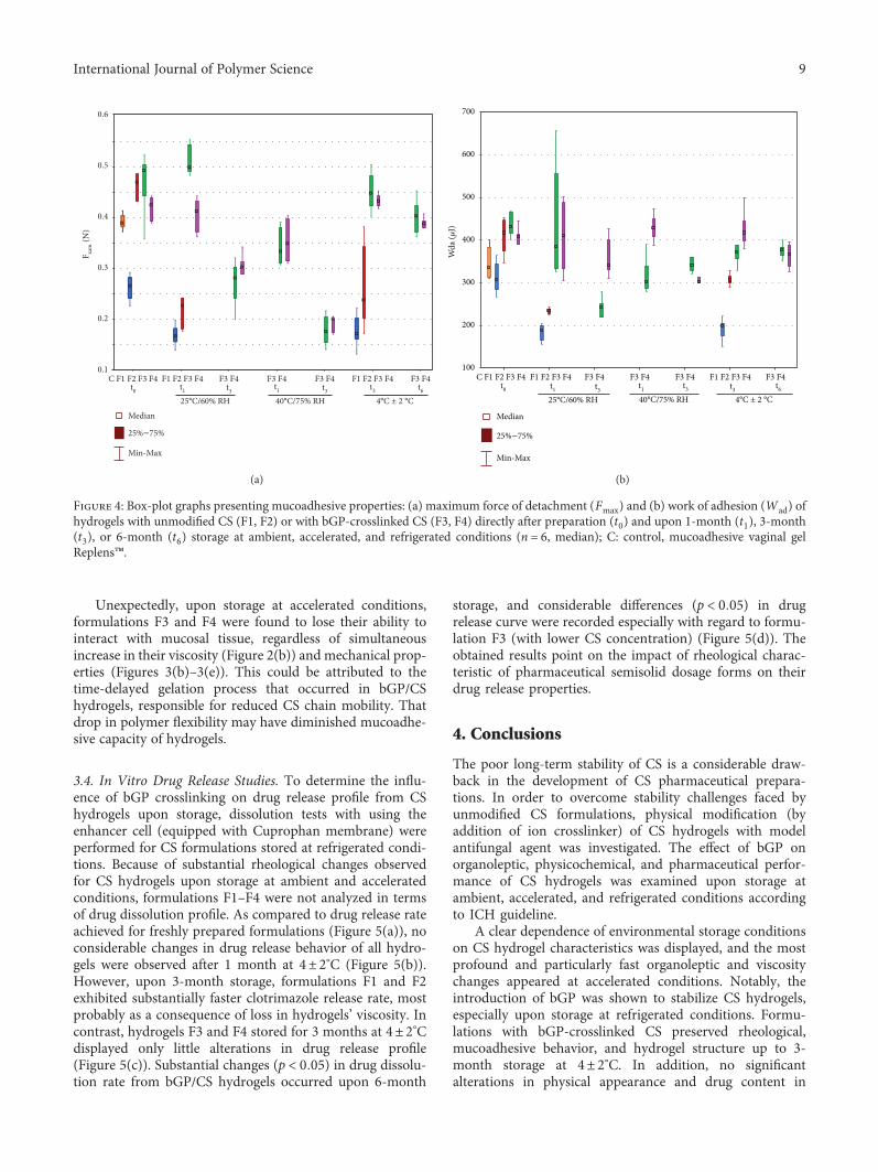
Unexpectedly, upon storage at accelerated conditions,formulations F3 and F4 were found to lose their ability tointeract with mucosal tissue, regardless of simultaneousincrease in their viscosity (Figure 2(b)) and mechanical prop-erties (Figures 3(b)–3(e)). This could be attributed to thetime-delayed gelation process that occurred in bGP/CShydrogels, responsible for reduced CS chain mobility. Thatdrop in polymer flexibility may have diminished mucoadhe-sive capacity of hydrogels.
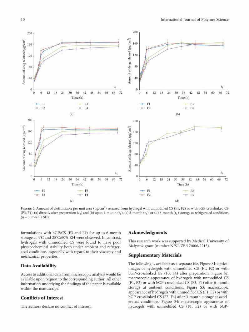
3.4. In Vitro Drug Release Studies. To determine the influ-ence of bGP crosslinking on drug release profile from CShydrogels upon storage, dissolution tests with using theenhancer cell (equipped with Cuprophan membrane) wereperformed for CS formulations stored at refrigerated condi-tions. Because of substantial rheological changes observedfor CS hydrogels upon storage at ambient and acceleratedconditions, formulations F1–F4 were not analyzed in termsof drug dissolution profile. As compared to drug release rateachieved for freshly prepared formulations (Figure 5(a)), noconsiderable changes in drug release behavior of all hydro-gels were observed after 1 month at 4± 2°C (Figure 5(b)).However, upon 3-month storage, formulations F1 and F2exhibited substantially faster clotrimazole release rate, mostprobably as a consequence of loss in hydrogels’ viscosity. Incontrast, hydrogels F3 and F4 stored for 3 months at 4± 2°Cdisplayed only little alterations in drug release profile(Figure 5(c)). Substantial changes (p < 0 05) in drug dissolu-tion rate from bGP/CS hydrogels occurred upon 6-month
storage, and considerable differences (p < 0 05) in drugrelease curve were recorded especially with regard to formu-lation F3 (with lower CS concentration) (Figure 5(d)). Theobtained results point on the impact of rheological charac-teristic of pharmaceutical semisolid dosage forms on theirdrug release properties.
4. Conclusions
The poor long-term stability of CS is a considerable draw-back in the development of CS pharmaceutical prepara-tions. In order to overcome stability challenges faced byunmodified CS formulations, physical modification (byaddition of ion crosslinker) of CS hydrogels with modelantifungal agent was investigated. The effect of bGP onorganoleptic, physicochemical, and pharmaceutical perfor-mance of CS hydrogels was examined upon storage atambient, accelerated, and refrigerated conditions accordingto ICH guideline.
A clear dependence of environmental storage conditionson CS hydrogel characteristics was displayed, and the mostprofound and particularly fast organoleptic and viscositychanges appeared at accelerated conditions. Notably, theintroduction of bGP was shown to stabilize CS hydrogels,especially upon storage at refrigerated conditions. Formu-lations with bGP-crosslinked CS preserved rheological,mucoadhesive behavior, and hydrogel structure up to 3-month storage at 4± 2°C. In addition, no significantalterations in physical appearance and drug content in
C F1 F2 F3 F4 F1 F2 F3 F4t0
25°C/60% RH 4°C ± 2 °C t1 t3 t3 t6
40°C/75% RHt1 t3
Median
Min-Max
25%–75%
F3 F4 F3 F4 F3 F4 F1 F2 F3 F4 F3 F40.1
0.2
0.3
0.4
0.5
0.6
F xam
(N)
(a)
t0 t1 t3 t3 t6t1 t3
25°C/60% RH 40°C/75% RH 4°C ± 2 °C
C F1 F2 F3 F4 F1 F2 F3 F4 F3 F4 F3 F4 F3 F4 F3 F4F1 F2 F3 F4
Median
Min-Max
25%–75%
100
200
300
400
500
600
700
Wda
(�휇J)
(b)
Figure 4: Box-plot graphs presenting mucoadhesive properties: (a) maximum force of detachment (Fmax) and (b) work of adhesion (Wad) ofhydrogels with unmodified CS (F1, F2) or with bGP-crosslinked CS (F3, F4) directly after preparation (t0) and upon 1-month (t1), 3-month(t3), or 6-month (t6) storage at ambient, accelerated, and refrigerated conditions (n = 6, median); C: control, mucoadhesive vaginal gelReplens™.
9International Journal of Polymer Science
formulations with bGP/CS (F3 and F4) for up to 6-monthstorage at 4°C and 25°C/60% RH were observed. In contrast,hydrogels with unmodified CS were found to have poorphysicochemical stability both under ambient and refriger-ated conditions, especially with regard to their viscosity andmechanical properties.
Data Availability
Access to additional data frommicroscopic analysis would beavailable upon request to the corresponding author. All otherinformation underlying the findings of the paper is availablewithin the manuscript.
Conflicts of Interest
The authors declare no conflict of interest.
Acknowledgments
This research work was supported by Medical University ofBiałystok grant (number N/ST/ZB/17/006/2215).
Supplementary Materials
The following is available as a separate file. Figure S1: opticalimages of hydrogels with unmodified CS (F1, F2) or withbGP-crosslinked CS (F3, F4) after preparation. Figure S2:macroscopic appearance of hydrogels with unmodified CS(F1, F2) or with bGP-crosslinked CS (F3, F4) after 6-monthstorage at ambient conditions. Figure S3: macroscopicappearance of hydrogels with unmodified CS (F1, F2) or withbGP-crosslinked CS (F3, F4) after 3-month storage at accel-erated conditions. Figure S4: macroscopic appearance ofhydrogels with unmodified CS (F1, F2) or with bGP-
t0
F1F2
F3F4
0
40
80
120
160
200
Am
ount
of d
rug
rele
ased
(�휇g/
cm2 )
6 12 18 24 30 36 42 48 54 60 66 720Time (h)
(a)
t1
F1F2
F3F4
0
40
80
120
160
200
Am
ount
of d
rug
rele
ased
(�휇g/
cm2 )
6 12 18 24 30 36 42 48 54 60 66 720Time (h)
(b)
F1F2
F3F4
t30
40
80
120
160
200
Am
ount
of d
rug
rele
ased
(�휇g/
cm2 )
6 12 18 24 30 36 42 48 54 60 66 720Time (h)
(c)
F1F2
F3F4
t6
6 12 18 24 30 36 42 48 54 60 66 720Time (h)
0
40
80
120
160
200
Am
ount
of d
rug
rele
ased
(�휇g/
cm2 )
(d)
Figure 5: Amount of clotrimazole per unit area (μg/cm2) released from hydrogel with unmodified CS (F1, F2) or with bGP-crosslinked CS(F3, F4): (a) directly after preparation (t0) and (b) upon 1-month (t1), (c) 3-month (t3), or (d) 6-month (t6) storage at refrigerated conditions(n = 3, mean± SD).
10 International Journal of Polymer Science
crosslinked CS (F3, F4) after 6-month storage at refrigeratedconditions. (Supplementary Materials)
References
[1] A. Srivastava, T. Yadav, S. Sharma, A. Nayak, A. AkankshaKumari, and N. Mishra, “Polymers in drug delivery,” Journalof Biosciences and Medicines, vol. 4, no. 1, pp. 69–84, 2016.
[2] J. R. Franca, G. Foureaux, L. L. Fuscaldi et al., “Bimatoprost-loaded ocular inserts as sustained release drug delivery systemsfor glaucoma treatment: in vitro and in vivo evaluation,” PLoSOne, vol. 9, no. 4, pp. e95461–e95472, 2014.
[3] F. Saporito, G. Sandri, S. Rossi et al., “Freeze dried chitosanacetate dressings with glycosaminoglycans and traxenamicacid,” Carbohydrate Polymers, vol. 184, pp. 408–417, 2018.
[4] J. Venkatesan, I. Bhatnagar, and S. K. Kim, “Chitosan-alginatebiocomposite containing fucoidan for bone tissue engineer-ing,” Marine Drugs, vol. 12, no. 1, pp. 300–316, 2014.
[5] B. Choi, S. Kim, B. Lin, B. M. Wu, and M. Lee, “Cartilaginousextracellular matrix-modified chitosan hydrogels for cartilagetissue engineering,” ACS Applied Materials & Interfaces,vol. 6, no. 22, pp. 20110–20121, 2014.
[6] Y. H. Lee, H. I. Park, and J. S. Choi, “Novel glycol chitosan-based polymeric gene carrier synthesized by a Michael addi-tion reaction with low molecular weight polyethylenimine,”Carbohydrate Polymers, vol. 137, pp. 669–677, 2016.
[7] A. Smith, M. Perelman, and M. Hinchcliffe, “Chitosan: apromising safe and immune-enhancing adjuvant for intranasalvaccines,” Human Vaccines & Immunotherapeutics, vol. 10,no. 3, pp. 797–807, 2014.
[8] P. N. Vaingankar and A. R. Juvekar, “Fermentative productionof mycelial chitosan from Zygomycetes: media optimizationand physico-chemical characterization,” Advances in Biosci-ence and Biotechnology, vol. 05, no. 12, pp. 940–956, 2014.
[9] T. A. Ahmed and B. M. Aljaeid, “Preparation, characterization,and potential application of chitosan, chitosan derivatives, andchitosan metal nanoparticles in pharmaceutical drug delivery,”Drug Design, Development and Therapy, vol. 10, pp. 483–507,2016.
[10] I. Aranaz, N. Acosta, C. Civera et al., “Cosmetics and cosme-ceutical applications of chitin, chitosan and their derivatives,”Polymer, vol. 10, no. 2, p. 213, 2018.
[11] T. T. Nguyen, A. R. Barber, K. Corbin, and W. Zhang,“Lobster processing by-products as valuable bioresource ofmarine functional ingredients, nutraceuticals, and pharma-ceuticals,” Bioresources and Bioprocessing, vol. 4, no. 1,pp. 27–46, 2017.
[12] S. Baltzley, A. Mohammad, A. H. Malkawi, and A. M. Al-Ghananeem, “Intranasal drug delivery of olanzapine-loadedchitosan nanoparticles,” AAPS PharmSciTech, vol. 15, no. 6,pp. 1598–1602, 2014.
[13] G. Tejada, M. G. Barrera, G. N. Piccirilli et al., “Developmentand evaluation of buccal films based on chitosan for the poten-tial treatment of oral candidiasis,” AAPS PharmSciTech,vol. 18, no. 4, pp. 936–946, 2017.
[14] A. Almomen, S. Cho, C. H. Yang et al., “Thermosensitiveprogesterone hydrogel: a safe and effective new formulationfor vaginal application,” Pharmaceutical Research, vol. 32,no. 7, pp. 2266–2279, 2015.
[15] E. Szymańska, K. Winnicka, A. Amelian, and U. Cwalina,“Vaginal chitosan tablets with clotrimazole—design and
evaluation of mucoadhesive properties using porcine vaginalmucosa, mucin and gelatine,” Chemical and PharmaceuticalBulletin, vol. 62, no. 2, pp. 160–167, 2014.
[16] F. Notario-Pérez, A. Martín-Illana, R. Cazorla-Luna et al.,“Influence of chitosan swelling behaviour on controlled releaseof tenofovir from mucoadhesive vaginal systems for preven-tion of sexual transmission of HIV,” Marine Drugs, vol. 15,no. 2, pp. 50–66, 2017.
[17] I. Younes, S. Sellimi, M. Rinaudo, K. Jellouli, and M. Nasri,“Influence of acetylation degree and molecular weight ofhomogeneous chitosans on antibacterial and antifungal activ-ities,” International Journal of Food Microbiology, vol. 185,pp. 57–63, 2014.
[18] M. Egusa, R. Iwamoto, H. Izawa et al., “Characterization ofchitosan nanofiber sheets for antifungal application,” Interna-tional Journal of Molecular Sciences, vol. 16, no. 11, pp. 26202–26210, 2015.
[19] E. Szymańska, K. Winnicka, P. Wieczorek, P. Sacha, andE. Tryniszewska, “Influence of unmodified and β-glycerophosphate cross-linked chitosan on anti-Candidaactivity of clotrimazole in semi-solid delivery systems,” Inter-national Journal of Molecular Sciences, vol. 15, no. 10,pp. 17765–17777, 2014.
[20] E. Szymańska, K. Sosnowska, W. Miltyk, M. Rusak, A. Basa,and K. Winnicka, “The effect of β-glycerophosphate crosslink-ing on chitosan cytotoxicity and properties of hydrogels forvaginal application,” Polymer, vol. 7, no. 11, pp. 2223–2244,2015.
[21] E. Szymańska and K. Winnicka, “Stability of chitosan—achallenge for pharmaceutical and biomedical applications,”Marine Drugs, vol. 13, no. 4, pp. 1819–1846, 2015.
[22] M. Fernandes, I. C. Gonçalves, S. Nardecchia, I. F. Amaral,M. A. Barbosa, and M. C. L. Martins, “Modulation of stabilityand mucoadhesive properties of chitosan microspheres fortherapeutic gastric application,” International Journal of Phar-maceutics, vol. 454, no. 1, pp. 116–124, 2013.
[23] C. Liu, E. Thormann, P. M. Claesson, and E. Tyrode, “Sur-face grafted chitosan gels. Part II. Gel formation and char-acterization,” Langmuir, vol. 30, no. 29, pp. 8878–8888,2014.
[24] A. N. Allam, V. F. Naggar, and S. S. El Gamal, “Formulationand physicochemical characterization of chitosan/acyclovirco-crystals,” Pharmaceutical Development and Technology,vol. 18, no. 4, pp. 856–865, 2013.
[25] M. Rinaudo, G. Pavlov, and J. Desbrières, “Influence of aceticacid concentration on the solubilization of chitosan,” Polymer,vol. 40, no. 25, pp. 7029–7032, 1999.
[26] “International conference on harmonisation of technicalrequirements for registration of pharmaceuticals for humanuse, ICH harmonised tripartite guideline, evaluation for sta-bility data Q1E,” April 2018, http://www.ich.org/fileadmin/Public.../Guidelines/.../Q1E_Guideline.pdf.
[27] TheEuropeanPharmacopoeia7.0,Council ofEurope, Strasbourg,France, 2011.
[28] EMA, ICH Topic Q4B Annex 12 Analytical Sieving GeneralChapterMarch 2018, http://www.ema.europa.eu/docs/en_GB/document_library/Scientific_guideline/2010/01/WC500044305.pdf.
[29] “In vitro release testing and in vivo bioequivalence docu-mentation,” in Guidance for Industry: SUPAC-SS Non-SterileSemisolid Dosage Forms. Scale-up and Postapproval Changes:
11International Journal of Polymer Science
Chemistry, Manufacturing and Controls, pp. 19–24, FDA-SUPAC-SS, Rockville, MD, USA, 1999.
[30] A. Chenite, M. Buschmann, D. Wang, C. Chaput, andN. Kandani, “Rheological characterisation of thermogellingchitosan/glycerol-phosphate solutions,” Carbohydrate Poly-mers, vol. 46, no. 1, pp. 39–47, 2001.
[31] The United States Pharmacopeia USP 34-NF 29, The UnitedStates Pharmacopeial Convention, Rockville, MD, USA, 2011.
[32] W. Y. Chien, “Drug delivery: vaginal route,” in Encyclopedia ofPharmaceutical Technology, vol. 2, J. Swarbrick, Ed., pp. 1339–1361, Informa Healthcare, New York, NY, USA, 3rd edition,2007.
[33] S. E. Harding, “Some observations on the effects of bioproces-sing on biopolymer stability,” Journal of Drug Targeting,vol. 18, no. 10, pp. 732–740, 2010.
[34] D. P. Chattopadhyay and M. S. Inamdar, “Aqueous behaviourof chitosan,” International Journal of Polymer Science,vol. 2010, Article ID 939536, 7 pages, 2010.
[35] T. T. B. Nguyen, S. Hein, C. H. Ng, andW. F. Stevens, “Molec-ular stability of chitosan in acid solutions stored at variousconditions,” Journal of Applied Polymer Science, vol. 107,no. 4, pp. 2588–2593, 2008.
[36] B. Martini, S. Dimida, E. de Benedetto, M. Madaghiele, andC. Demitri, “Study on the degradation of chitosan slurries,”Results in Physics, vol. 6, pp. 728-729, 2016.
[37] E. Ruel-Gariépy, A. Chenite, C. Chaput, S. Guirguis, andJ.-C. Leroux, “Characterization of thermosensitive chitosangels for the sustained delivery of drugs,” International Journalof Pharmaceutics, vol. 203, no. 1-2, pp. 89–98, 2000.
[38] S. Supper, N. Anton, N. Seidel, M. Riemenschnitter,C. Schoch, and T. Vandamme, “Rheological study of chito-san/polyol-phosphate systems: influence of the polyol parton the thermo-induced gelation mechanism,” Langmuir,vol. 29, no. 32, pp. 10229–10237, 2013.
12 International Journal of Polymer Science

CorrosionInternational Journal of
Hindawiwww.hindawi.com Volume 2018
Advances in
Materials Science and EngineeringHindawiwww.hindawi.com Volume 2018
Hindawiwww.hindawi.com Volume 2018
Journal of
Chemistry
Analytical ChemistryInternational Journal of
Hindawiwww.hindawi.com Volume 2018
Scienti�caHindawiwww.hindawi.com Volume 2018
Polymer ScienceInternational Journal of
Hindawiwww.hindawi.com Volume 2018
Hindawiwww.hindawi.com Volume 2018
Advances in Condensed Matter Physics
Hindawiwww.hindawi.com Volume 2018
International Journal of
BiomaterialsHindawiwww.hindawi.com
Journal ofEngineeringVolume 2018
Applied ChemistryJournal of
Hindawiwww.hindawi.com Volume 2018
NanotechnologyHindawiwww.hindawi.com Volume 2018
Journal of
Hindawiwww.hindawi.com Volume 2018
High Energy PhysicsAdvances in
Hindawi Publishing Corporation http://www.hindawi.com Volume 2013Hindawiwww.hindawi.com
The Scientific World Journal
Volume 2018
TribologyAdvances in
Hindawiwww.hindawi.com Volume 2018
Hindawiwww.hindawi.com Volume 2018
ChemistryAdvances in
Hindawiwww.hindawi.com Volume 2018
Advances inPhysical Chemistry
Hindawiwww.hindawi.com Volume 2018
BioMed Research InternationalMaterials
Journal of
Hindawiwww.hindawi.com Volume 2018
Na
nom
ate
ria
ls
Hindawiwww.hindawi.com Volume 2018
Journal ofNanomaterials
Submit your manuscripts atwww.hindawi.com