Embed Size (px)
Citation preview

Comparison of Magnetic Resonance Imaging Findings inAnterior Cruciate Ligament Grafts With and Without
Autologous Platelet-Derived Growth Factors
Fernando Radice, M.D., Roberto Yanez, M.D., Vicente Gutierrez, M.D., Julio Resales, M.D.,Miguel Pinedo, M.D., and Sebastian Coda, M.D.
t
Purpose: To determine whether the use of platelet-rich plasma gel (PRPG) affects magneticresonance imaging (MRI) findings in the anterior cruciate ligament (ACL) graft during the first yearafter reconstruction. Methods: A prospective single-blinded study of 50 ACL reconstructions in 50patients was performed. In group A (study group) PRPG was added to the graft with a standardizedtechnique, and in group B (control group) no PRPG was added. An MRI study was performedpostoperatively between 3 and 9 months in group A and between 3 and 12 months in group B. Theimaging analysis was performed in a blind protocol by the same radiologist. Results: The meanheterogeneity score value at the time of MRI, assigned by the radiologist, was 1.14 in group A and3.25 in group B. Both groups were comparable in terms of sex and age (P < .05). The mean timeto obtain a completely homogeneous intra-articular segment in group A (PRPG added) was 177 daysafter surgery, and it was 369 days in group B. Using the quadratic predictive model, these findingsshow that group A (PRPG added) needed inly 48% of the time group B required to achieve the sameMRI image (P < .001). Conclusions: ACL reconstruction with the use of PRPG achieves completehomogeneous grafts assessed by MRI, in 179 days compared with 369 days for ACL reconstructionwithout PRPG. This represents a time shortening of 48% with respect to ACL reconstruction withoutPRPG. Level of Evidence: Level Hl/case-conrrol study.
for the athlete; thus methods have been sought toshorten the biological time required for the graft toacquire biomechanical properties similar to theoriginal ACL.
The clinical results of ACL reconstruction and timeto return to sports could be improved if the grafthealing process is enhanced. In a classic publicationon this topic in 1982, Arnoczky and Tarvin1 describedthe behavior of the graft used in ACL reconstructionin dogs, describing 3 stages in the process of graftmetaplasia: incorporation, neoligament formation, andremodeling.2-3
Various authors have tried to study the behavior ofthe graft in clinical trials, with histology or imagingstudies, which experienced a significant boost with theappearance of magnetic resonance imaging (MRI).4-10
In 1995, in a prospective clinical study that reliedon second-look arthroscopy to perform a histologicand.MRI assessment of the graft at 6, 9, and 12months of postoperative evolution,11 we described
Rupture of the anterior cruciate ligament (ACL)is an injury commonly observed in sports med-
icine. Return to professional sports occurs at around6 to 7 months, depending on the sport practiced. Insports medicine this time period is often very long
From the Department of Orthopedics and Spans Medicine,Clinica Las Condes (F.R., V.G., M.P.), and Departments of SportsMedicine (R.Y., S.C.) and Radiology (J.R.), Clinica MEDS: Medi-cina, Ejercicio, Deports y Salud, Santiago, Chile.
Presented at the Biannual Meeting of the Sociedad Latinoameri-cana de Artroscopia Rodilla y Traumatologia Deportiva, Cancun,Mexico, June 5-7, 2008.
The authors report no conflict of interest.Received August 24, 2008; accepted June 30, 2009.Address correspondence and reprint requests to Fernando Ra-
dice, M.D., Department of Orthopedics and Sports Medicine,Clinica Las Condes, Lo Fontecilla 441, Santiago 6772610, Chile.E-mail: fradice @ clc. cl
© 2010 by the Arthroscopy Association of North America0749-8063/10/2601 -8486$36.00/0doi:l 0.1016/j. arthro.2009.06.030
50 Arthroscopy: The Journal of Arthroscopic and Related Surgery, Vol 26, No 1 (January), 2010: pp 50-57

OUTCOMES OF ARTHROSCOPIC ACL RECONSTRUCTION 49
36. Jackson DW, Grood BS, Goldstein JD, et al. A comparison of 38. Zaffagnini S, De Pasquale V, Marchesini Reggiani L, et al.patellar tendon autograft and allograft used for anterior cruci- Neoligamentization process of BPTB used for ACL graft:ate ligament reconstruction in the goat model. Am J Sports Histological evaluation from 6 months to 10 years. KneeMed 1993:21:176-185. 2007; 14:87-93.
37. Shino K, Inoue M, Horibe S, Nakata K, Maeda A, Ono K. 39. Ahn JH, Yoo JC, Yang HS, Kim JH, Wang JH. Second-lookSurface blood flow and histology of human anterior cruciate arthroscopic findings of 208 patients after ACL reconstruction,ligament allografts. Arthroscopy 1991;7:171-176. Knee Surg Sports Traumatol Arthrosc 2007; 15:242-248.

ACL GRAFT AND AUTOLOGOUS PDGF 51
t
how the patellar tendon graft used in human ACLreconstruction is incorporated. We concluded that thegraft maturation takes a long time: 12 months toachieve histology similar to a normal ACL. At 12months, the MRI study of the graft was homogeneousand hyperintense, without swelling in the bone tun-nels. The correlation of the histology with MRI was ofgreat help in establishing a reliable imaging pattern,which allowed us to noninvasively verify the grafthealing process.
Weiler et al.12 report correlations between biome-chanical properties and vascularity of an ACL graftand MRI in a sheep model.
Clinical applications of autologous platelet-richplasma gel (PRPG) include maxillofacial surgery,treatment of bone fractures, and tendon repair, report-ing excellent outcomes.13"16 Platelets contain differentgrowth factors that facilitate healing. PRPG is a frac-tion of plasma volume with a platelet concentrationabove baseline (whole blood). Platelet concentratescontain an enormous amount of activated platelet-derived growth factors (PDGFs).17-23
Platelets contain PDGFs, transforming growth fac-tors (TGFs), insulin-like growth factors, epidermalgrowth factors, vascular endothelial growth factors,and fibroblast growth factors. These factors are in-volved in the majority of biological remodeling pro-cesses in the body. In the specific case of ACL graft,PDGFs, fibroblast growth factor 1, and the varioustypes of TGF-/3 are responsible for accelerating thehealing process, as well as increasing the tensilestrength of the graft.24-30
Only 2 articles have shown an enhancing effect oftreatment with PRPG on the tendon or ligament inhumans. In a human study Orrego et al.31 showed anenhancing effect over the graft maturation process asevaluated by MRI signal intensity, without showing asignificant effect on the osteoligamentous interface ortunnel widening evolution. In human tenocyte cul-tures, de Mos et al.32 showed that PRPG stimulatescell proliferation and collagen production.
Currently, it is practically impossible to performhuman clinical trials of biomechanical or histologicassessments of the graft's behavior in ACL recon-struction. For this reason, we decided to practice anindirect and noninvasive assessment in our patients,using MRI. The purpose of our investigation was tostudy MRI findings in the. ACL graft when PRPG wasadded during surgery, thus allowing future studiescorrelating MRI findings with histology and ultimateload and strength. We hypothesized that PRPG has apositive effect on cell proliferation and collagen pro-
duction in the human tendon and plays a key role inthe remodeling and repair processes of the graft usedin ACL reconstruction.
METHODS
Study Design
This is a prospective and single-blinded study per-formed between lune 2005 and December 2006. Theinclusion criteria were sport athletes of both genderbetween 18 and 35 years old with an isolated ACL tearshown by MRI. Exclusion criteria were previous ACLrevision surgery, chronic or systemic disease undertreatment, and previous or current treatment for ma-lignant disease. These pathologies can modify thebiologic behavior of the graft. Fifty consecutive pa-tients met the inclusion criteria.
The type of graft used was determined according toour institution's protocol, and it depended on the typeof sports the patient practiced. Bone-patellar tendon-bone (BPTB) autograft was used in rugby and soccerplayers, whether hamstring autograft was used inplayers who practiced skiing, hockey, tae kwon do,and volleyball. One of the surgeons (R. Y.) did not usePRPG, and the other (F.R.) did. Two groups wereestablished: Group A included 25 patients (18 menand 7 women), with a mean age of 30 years (range, 18to 33 years), with ACL reconstruction plus PRPG; 15of these patients underwent reconstruction withBPTB. Group B included 25 patients (21 men and 4women), with a mean age of 32 years (range, 18 to 35years), with ACL reconstruction without PRPG; 10 ofthese patients underwent reconstruction with ham-string autografts. Both groups (A and B) followed thesame rehabilitation protocol.
Surgical Technique
In the case of BPTB autograft, fixation was per-formed with metallic interference screws. Hamstringautograft fixation was performed with metallic or bio-absorbable cross-pin femoral fixation using the Trans-Fix technique (Arthrex, Naples, EL) in the distal fe-mur and a Delta-type bioabsorbable screw with ametallic staple in the proximal tibia (Arthrex).
In group A PRPG was administered by an applica-tion technique developed to allow standardization ofthe dose of concentrate used and avoidance of its losswhen the graft goes through the bone tunnels.33-34 Theautologous platelet concentrate was obtained from theGPS system of Biomet (Warsaw, IN). This procedureis done aseptically in the same operating room. Pre-

52 F. RADICE ET AL.
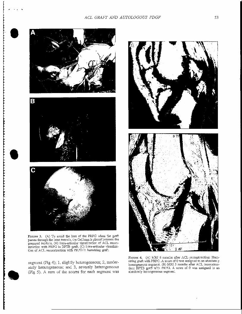
operatively, 60 mL of autologous blood is obtainedand centrifuged at 3,200 rpm for 15 minutes. In thecase of BPTB graft, after adaptation of the bone plugsfor them to fit through the tunnels, the femoral bonesegment and the intra-articular segment are wrappedwith a bioabsorbable synthetic gelatin called Gelfoam(Pfizer, New York, NY) and secured to it with a No.3-0 Vicryl suture (Ethicon, Somerville, NJ). In the caseof hamstring tendon graft, it is prepared in the usualmanner with removal of the remnant muscle tissue. Ateach end, a 3-cm-long braid with FiberWire (Arthrex) ismade, and the tendon's thickness and length are mea-sured. Under moderate tension, a piece of Gelfoam isplaced between the portion of the tendons that will belocated in the femoral tunnel and the intra-articularsegment. This is sutured to the adjacent tendon withNo. 3-0 Vicryl. The Gelfoam acts as a sponge thatmaintains the platelet concentrate dose in direct con-tact with the graft used (Fig 1). A total volume of 5mL of platelet-rich plasma, activated at the moment ofinoculation on the graft, is added homogeneously soas to completely cover the graft, waiting until it formsa clot (Fig 2). The dose administered was determinedbased only on our criteria. We do not know the idealdose, and at the time of this study, nothing about theideal dose had been published. The formed clot ad-heres to the graft, because of the presence of thesutured and compressed Gelfoam. This allows thegraft to hold a precise amount of PRPG and, evenmore importantly, avoids the loss of PRPG when thegraft goes through the bone tunnels (Fig 3).
t
FIGURE 1. Hamstring graft preparation with Gelfoam and PRPG.
FIGURE 2. The Gelfoam acts as a sponge that maintains the PRPGdose in direct contact with the graft.
Imaging Assessment
The imaging protocol was standardized and similarin both groups. Included were a series of MRI scansfocused to study the intra-articular segment of thegraft. The study of the femoral and tibia! parts of thegraft was not considered because its maturation pro-cess occurs first, compared with the intra-articularpart. This was performed with a Tl and T2 sequence(repetition time, 4,020 milliseconds; echo time, 105milliseconds) with a 1.5-T Siemens Magnetom MRIScanner (Siemens AG, Erlangen, Germany). Slices of2 mm in thickness, in the oblique parasagittal view,between 10° and 15°, centered on the intercondylarregion, with the knee flexed at 9° to 10°,n wereobtained. Patients in group A had MRI performed at 3,4, 5, 6, 7, 8, and 9 months postoperatively so as tobuild a homogenization curve of the graft, accordingto the statistic quadratic predictive model, and sup-ported by this study's hypothesis that the use of PRPGaccelerates the graft homogenization time. The controlgroup had MRI performed at 6, 7, 8, 9, 10, 11, and 12months, with the assumption that before 6 months,homogenization is not present.
The imaging analysis was done by the same ra-diologist, experienced in musculoskeletal studies,blinded to the time of reconstruction and to PRPGapplication to the graft.
The radiologist divided the intra-articular segmentof the graft into 3 segments: proximal, medial, anddistal. To each segment, he assigned a score accordingto the degree of heterogeneity observed. Therefore ascore of 0 was assigned to an absolutely homogeneous
ACL GRAFT AND AUTOLOGOUS PDGF 53
t
FIGURE 3. (A) To avoid the loss of the PRPG when the graftpasses through the bone runnels, the Gelfoam is placed between theprepared tendons. (B) Intra-articular visualization of ACL recon-struction with PRPG in BPTB graft. (C) Intra-articular visualiza-tion of ACL reconstruction with PRPG in hamstring graft.
segment (Fig 4); 1, slightly heterogeneous; 2, moder-ately heterogeneous; and 3, severely heterogeneous(Fig 5). A sum of the scores for each segment was
FIGURE 4. (A) MRI 6 months after ACL reconstruction: Ham-string graft with PRPG. A score of 0 was assigned to an absolutelyhomogeneous segment. (B) MRI 5 months after ACL reconstruc-tion: BPTB graft with PRPG. A score of 0 was assigned to anabsolutely homogeneous segment.

54 F. RADICE ET AL.
FIGURE 5. (A) MRI 6 months after ACL reconstruction: Ham-string graft without PRPG. A score of 3 was assigned to a severelyheterogeneous segment. (B) MRI 6 months after ACL reconstruc-tion: BPTB graft without PRPG. A score of 3 was assigned to aseverely heterogeneous segment.
obtained for each patient, which was compared statis-tically between the 2 groups and correlated with thetime at which the MRI study was done.
Statistic Analysis
For the statistic analysis, data were analyzed withthe SPSS data analysis program (SPSS, Chicago, EL).This program was used to work with quadraticstatistics, graphs, data, and descriptive indicators. Todetermine whether the 2 groups were comparable interms of number, age, and sex, an F test and Studentt test were used.
The quadratic predictive model was used for dataanalysis to determine, through a linear relation, theextrapolated midpoint that predicted the time whenboth groups had completely homogeneous grafts.
RESULTS
The mean heterogeneity score value at the time ofMRI, assigned by the radiologist, was 1.14 in group Aand 3.25 in group B. Both groups were comparable interms of sex and age (P < .05). The mean time toobtain a completely homogeneous intra-articular seg-ment in group A (PRPG added) was 177 days aftersurgery, and it was 369 days in group B. Using thequadratic predictive model, the percentage of time thatgroup A (PRPG added) needed to achieve the sameMRI aspect as group B was 48% (Fig 6). This fact iseven more evident when we compared only the BPTBgraft cases in both groups: a homogeneous graft wasobtained in 109 days in patients with PRPG versus363 days in the control group, that is, one third thetime as that for control group (Fig 7). In the compar-ative analysis of those patients in whom BPTB graftwas used, we observed an even shorter time requiredfor the graft's homogenization when PRPG was used,
t
Days
1 year
FIGURE 6. Homogenization of graft, by use of quadratic predic-tive model, in group A (with PRPG) versus group B (control).

ACL GRAFT AND AUTOLOGOUS PDGF 55
t
400-
350-
300-
250-
200-
ISO-
100-
50-
o-
DAwithPKPG
OB without PKPG
//
/
/
/
//
/
A m
/ —
ST
r3f
^K
-G
n>9
m BI
^- —
Bl13
2^B
CB)9',3
•r
FIGURE 7. Homogenization of graft comparing BPTB and ham-string in group A (with PRPG) versus group B (control). (BPTBand hamstring without PRPG.)
but this can only be considered a trend, because thesample's number was too small to draw conclusionswith statistical significance (/3 type error).
The latter fact, despite the large difference betweenthe groups, merely shows a statistical trend, because itlacks statistical significance. On the other hand, tocertify the findings of the quadratic model, in bothgroups only the patients who fully completed therequirements of a return to sports without restrictions,with normal functional sport-specific and isokineticevaluations, were selected. The mean time to obtain anMRI score of 0, that is, a completely homogeneousgraft, was compared between the groups (12 in thePRPG group and 6 in the control group). It wasdetermined that the mean time in days to obtain ahomogeneous graft was 179 days and 362 days in thePRPG and control groups, respectively (Fig 8). Thisfinding indicates a decrease by half of the time(49.4%) in the group with PRPG versus the controlgroup (P < .001). Once again, modifying the dataanalysis, the homogenization time of the intra-articu-lar segment of the ACL graft evaluated with specificMRI slices is halved when growth factors, obtainedthrough a standardized autologous platelet concentratemethod, are used.
DISCUSSION
For elite athletes, recovery from ACL injury mustreach a level close to normal and occur in the shortesttime possible so as not to affect the future athleticperformance. In the last decade great advances haveoccurred in ACL reconstruction surgery, considerablyimproving the outcomes. This is because of the devel-opment of more anatomic reconstruction techniques,
stronger and more stable methods of attachment overtime, accelerated rehabilitation protocols, better tech-nical training, and increased expertise of surgicalteams. However, reinjury in these athletes, attributedto trauma in early periods of sports reintegration,clearly indicates that the biological period of matura-tion and metaplasia needed by the graft used in thereconstruction is not affected by the described ad-vances. For the patellar tendon graft, this period is onaverage 9 to 12 months.2-1 U4 Therefore, during thelast years the focus of research has been on advance-ments in the basic sciences relating to the study offunction and capacity for repair, as well as develop-ment of growth factors and tissue. Yasuda et al.,28
analyzing the effect of growth factors applied to graftsin dog models, indicated that TGF-/3 and epidermalgrowth factor act by increasing the collagen and fi-broblast synthesis by 40% in the graft. Anderson etal.14 indicated that the presence of TGF-/31, TGF-/32,TGF-|33, and TGF-1 growth factors directly influ-ences the graft by improving the scarring rate andincreasing the tensile force resistance by 65%. Inanother interesting experimental study, Weiler et al.27
indicate that the application of autologous PDGFsapplied to the graft during surgery was capable ofchanging its natural evolution, improving tensilestrength and resistance, increasing the maturation rate,and improving collagen quality. The results in ourstudy showed a significant shortening of the biologicalmaturation time of the graft, by at least 48%. Ourresults show that when PRPG is used, the time re-quired by the graft to achieve complete homogeniza-tion, as assessed by MRI, is statistically shortened.
P-valueO.OOO
Score 0
FIGURE 8. Comparison of only grafts with absolutely homoge-neous segment in group A (with PRPG) versus group B (control).

56 F. RADICE ET AL
This is very important for the graft's biological mat-uration. This means that the graft used with PRPGcould undergo its complete process in half the time itnaturally requires. We are performing a follow-up ofall of our operated athletes to see what happenedregarding reinjury, but the follow-up time is still short.
The use of the gelatin (Gelfoam) could affect themagnetic resonance image or analysis, but because itis absorbable, it may already be biodegraded at thetime of imaging assessment. There are no studies inthe literature describing local changes related to boneor tendon grafts. The early changes seen in the intra-articular segment of the graft with the use of PRPGsuggest some effect on it. There are no publishedstudies that relate the quality of the MRI signal inten-sity with histology or strength of grafts in humanmodels. However, investigations by Weiler et al.12 insheep models showed that there is a correlation amongthe homogeneity of the graft on MRI, maturation, andstrength, similar to the native ACL.
Much field to cover still remains. The current ap-plication of autologous PDGFs22-25 does not allow usto specifically isolate the factors related to the process.We are most likely applying a mixture of factors thatapparently do not participate in or influence the heal-ing process of these tissues.24-26-30'35'36 It is also notclear to us whether isolated application at the time ofsurgery is enough or whether it would be even moreeffective to repeat application of these factors duringthe postoperative recovery and rehabilitation process.Which are the growth factors that are actually neededin ACL reconstruction? Is the quantity we are apply-ing adequate? Is it important to maintain the interac-tion and balance between all of the growth factorspresent in the platelet concentrate? How long doestheir effect last? We still do not have the answers tothese questions, and further studies are required.
CONCLUSIONS
ACL reconstruction with the use of PRPG achievescompletely homogeneous grafts, assessed by MRI, in179 days compared with 369 days for ACL recon-struction without PRPG. This represents a time short-ening of 48% with respect to ACL reconstructionwithout PRPG.
REFERENCES
1. Arnoczky SP, Tarvin GB. Anterior cruciate ligament replace-ment using patellar tendon: An evaluation of graft revascular-ization in the dog. J Bone Joint Surg Am 1982;64:217-224.
2. Falconiero RP, DiStefano VJ, Cook TM. Revascularizationand ligamentization of autogenous anterior cruciate ligamentgrafts in humans. Arthroscopy 1998;14:197-205.
3. Kleiner JB, Amiel D, Harwood PL, Akeson WH. Early histo-logic, metabolic, and vascular assessment of anterior cruciateligament autografts. J Orthop Res 1989;7:235-242.
4. Abe S, Kurosaka M, Iguchi T, Yoshiya S, Hirohata K. Lightand electron microscopic study of remodeling and maturationprocess in autogenous graft for anterior cruciate ligamentreconstruction. Arthroscopy 1993;9:394-405.
5. Gr0nrvedt T, Engebretsen L, Rossvoll I, Smevik O, Nilsen G.Comparison between magnetic resonance imaging findingsand knee stability: Measurements after anterior cruciate liga-ment repair with and without augmentation. A five- to seven-year follow-up of 52 patients. Am J Sports Med 1996;23:729-735.
6. Howell SM, Clark JA, Blasier RD. Serial magnetic resonanceimaging of hamstring anterior cruciate ligament autograftsduring the first year of implantation. Am J Spans Med 1991;19:42-47.
7. Maywood RM, Murphy BJ, Uribe JW, et al. Evaluation ofarthroscopic anterior cruciate ligament reconstruction usingmagnetic resonance imaging. Am J Sports Med 1993;21:523-527.
8. Rougraff BT, Shelbourne KD. Early histologic appearance ofhuman patellar tendon autografts used for anterior cruciateligament reconstruction. Knee Surg Spans Traumatol Arthrosc1999;7:9-14.
9. Unterhauser FN, Bail HJ, Hoher J, Haas NP, Weiler A. En-doligamentous revascularization of an anterior cruciate liga-ment graft. Clin Onhop Relat Res 2003:276-288.
10. Yoshikawa T, Tohyama H, Enomoto H, Matsumoto H,Toyama Y, Yasuda K. Temporal changes in relationshipsbetween fibroblast repopulation, VEGF expression, and angio-genesis in the patellar tendon graft after ACL reconstruction.Trans Onhop Res Sac 2003;29:236.
11. Radice F, Gutierrez V, Ibarra A. Arthroscopic, histologic andMRI correlation in the maturation process of the graft in ACLreconstruction in humans. Arthroscopy 1998;14:S20 (Suppl 1).Abstracts presented at the First Biennial Congress of ISAKOS.
12. Weiler A, Peters G, Maurer J, Unterhauser FN, Siidkamp NP.Biomechanical properties and vascularity of an anterior cruci-ate ligament graft can be predicted by contrast-enhanced mag-netic resonance imaging. A two-year study in sheep. Am JSports Med 2001;29:751-761.
13. Hildebrand KA, Woo SL-Y, Smith DW, et al. The effects ofplatelet-derived growth factor-BB on healing of the rabbitmedial collateral ligament. Am J Sports Med 1998;26:549-554.
14. Anderson K, Senevirarne AM, Izawa K, Atkinson BL, PotterHG, Rodeo SA. Augmentation of tendon healing in an inrra-articular bone tunnel with use of a bone growth factor. Am JSports Med 2001;29:689-698.
15. Jenner JM, van Eijk F, Saris DB, Willems WJ, Dhert WJ,Creemers LB. Effect of transforming growth factor-beta andgrowth differentiation factor-5 on proliferation and matrixproduction by human bone marrow stromal cells cultured onbraided poly lactic-coglycolic acid scaffolds for ligament tis-sue engineering. Tissue Eng 2007;13:1573-1582.
16. Kondo E, Yasuda K, Yamanaka M, Minami A, Tohyama H.Effects of administration of exogenous growth factors on bio-mechanical properties of the elongation-type anterior cruciateligament injury with partial laceration. Am J Sports Med 2005;33:188-196.
17. Lee J, Green MH, Amiel D. Synergistic effect of growthfactors on cell outgrowth from explants of rabbit anteriorcruciate and medial collateral ligaments. J Orthop Res 1995;13:435-441.
18. Letson AK, Dahners LE. The effect of combinations of growth
t
t

ACL GRAFT AND AUTOLOGOUS PDGF 57
t
factors on ligament healing. Clin Orthop Relat Res1994:207-212.
19. Marui T, Niyibizi C, Georgescu HI, et al. Effects of growthfactors on matrix synthesis by ligament flbroblasts. J OrthopRes 1997;15:18-23.
20. Murray MM, Spindler KP, Abreu E, et al. Collagen-plateletrich plasma hydrogel enhances primary repair of the porcineanterior cruciate ligament. / Orthop Res 2007;25:81-91.
21. Pierce GF, Mustoe TA, Lingelbach J, et al. Platelet-derivedgrowth factor and transforming growth factor-/? enhance tis-sue repair activities by unique mechanisms. J Cell Biol 2000;109:429-440.
22. Sanchez M, Anitua E, Azofra J, Andia I, Padilla S, Mujika I.Comparison of surgically repaired achilles tendon tears usingplatelet-rich fibrin matrices. Am J Sports Med 2007;35:245-251.
23. Sanchez M, Azofra J, Aizpurua B, Elorriaga R, Anitua E,Andia I. Application in arthroscopic surgery of autologousplasma rich in growth factors. CworfArtrasc2003;10:12-19 (inSpanish).
24. Anitua E, Andia I, Sanchez M, et al. Autologous preparationsrich in growth factors promote proliferation and induce VEGFand HGF production by human tendon cells in culture. / Or-thop Res 2005;23:281-286.
25. Azuma H, Yasuda K, Tohyama H, et al. Timing of adminis-tration of transforming growth factor-beta and epidermalgrowth factor influences the effect on material properties of thein situ frozen-thawed anterior cruciate ligament. / Biomech2003:36:373-381.
26. Sakai T, Yasuda K, Tohyama H, et al. Effects of combinedadministration of transforming growth factor-beta 1 and epi-dermal growth factor on properties of the in situ frozen ACLin rabbits. J Orthop Res 2002;20:1345-1351.
27. Weiler A, Forster C, Hunt P, et al. The influence of locallyapplied platelet-derived growth factor-BB on free tendon graftremodeling after anterior cruciate ligament reconstruction.Am J Sports Med 2004;32:881-891.
28. Yasuda K, Tomita F, Yamazaki S, Minami A, Tohyama H.The effect of growth factors on biomechanical properties ofthe bone-patellar tendon-bone graft after anterior cruciateligament reconstruction: A canine model study. Am J SpansMed 2004;32:870-880.
29. Yamazaki S, Yasuda K, Tomita F, Tohyama H, Minami A.The effect of transforming growth factor-1 on intraosseoushealing of flexor tendon autograft replacement of ACL in dogs.Anhroscopy 2005;21:1034-1041.
30. Steiner ME, Murray MM, Rodeo SA. Strategies to improveanterior cruciate ligament healing and graft placement. Am JSports Med 2008;36:176-189.
31. Orrego M, Larrain C, Resales J, et al. Effects of plateletconcentrate and bone plug on the healing of hamstring tendonsin bone tunnel. Anhroscopy 2008;24:1373-1380.
32. de Mos M, van der Windt A, Jahr H, et al. Can platelet-richplasma enhance tendon repair? A cell culture study. Am JSpans Med 2008;36:1171-1178.
33. Radice F, Yafiez R, Gutierrez V, Pinedo M. Application ofplatelet-derived growth factors in ACL reconstruction. Practi-cal advice to quantify dosage and to avoid its loss whenpassing through the bone tunnels. NotiSLARD 2007;2:7-8.Available from: www.slard.org.
34. Radice F. Preparation of the graft in ACL reconstruction,applying platelet-derived growth factors. Video 10. November2005. Available from: www.socht.cl.
35. Ju YJ, Tohyama H, Kondo E, et al. Effects of local adminis-tration of vascular endothelial growth factor on properties ofthe in situ frozen thawed anterior cruciate ligament in rabbits.Am J Spans Med 2006;34:84-91.
36. Yoshikawa T, Tohyama H, Katsura T, et al. Effects of localadministration of vascular endothelial growth factor on me-chanical characteristics of the semitendinosus tendon graftafter anterior cruciate ligament reconstruction in sheep. Am JSports Med 2006;36:1918-1925.
Grafted Tendon Healing in Tibial Tunnel Is Inferior to Healingin Femoral Tunnel After Anterior Cruciate Ligament
Reconstruction: A Histomorphometric Study in Rabbits
t
Chun-Yi Wen, Ph.D., Ling Qin, Ph.D., Kwong-Man Lee, Ph.D.,Margaret Wan-Nar Wong, M.D., and Kai-Ming Chan, M.D.
Purpose: This study aimed to test whether graft healing in the tibial tunnel was inferior to that in thefemoral tunnel after anterior cruciate ligament (ACL) reconstruction in rabbits. Methods: Surgicalreconstruction by use of the digital extensor tendon in the bone tunnel was performed in 18 rabbits.The rabbits were killed at weeks 2, 6, and 12 postoperatively, with 6 at each time point, for histologicexamination. .Results: The transiently formed cartilaginous interface was gradually mineralizedduring re-establishment of direct tendon-to-bone integration, which was observed significantly less inthe tibial tunnel than in the femoral tunnel (P < .05). The cell density of the graft was significantlylower in the tibial tunnel than that in the femoral tunnel at weeks 2 and 6 postoperatively (P < .05for both). An increase in the immature type III collagen content was accompanied by a decrease ingraft collagen fiber organization, with healing over time in both the femoral and tibial tunnels. Thecollagen fiber organization of the graft was significantly poorer in the tibial tunnel than that in thefemoral tunnel at week 12 after surgery (P < .05). Conclusions: Grafted tendon healing in the tibialtunnel was inferior to that in the femoral tunnel at the tendon-to-bone interface and with regard to thegrafted tendon within the bone tunnel after ACL reconstruction in rabbits. Clinical Relevance:Future biopsy study is desirable to test whether this observation was valid clinically, which mightprovide a scientific basis for therapeutic targets to improve the outcome of ACL surgery.
Athroscopic reconstruction of grafted tendon in thefemoral and tibial tunnels is a common surgical
procedure to replace a torn anterior cruciate ligament(ACL) in patients. Yet 11% to 32% of patients showed
From the Department of Orthopaedics and Traumatology(C.-Y.W., L.Q., M.W.-N.W., K.-M.C.) and The Hong Kong JockeyClub Spans Medicine and Health Sciences Centre (C.-Y.W., L.Q.,K.-M.C.), Faculty of Medicine, and Lee-Hysan Clinical ResearchLaboratory (K.-M.L.), The Chinese University of Hong Kong,Hong Kong, China.
Supported by the Research Grant Council Earmarked Grants06-07 (CUHK4497/06M), Hong Kong, China. The authors reportno conflict of interest.
Received November 18, 2008; accepted June 29, 2009.Address correspondence and reprint requests to Kai-Ming Chan,
Department of Orthopaedics and Traumatology, The Chinese Uni-versity of Hong Kong, 5/F, General Office, Hong Kong SAR,China. E-mail: [email protected]
© 2010 by the Arthroscopy Association of North America0749-8063/10/2601-8650$36.00/0doi:10.1016/j.arthro.2009.06.025
an unsatisfactory prognosis, and up to 10% requiredsurgical revision.1"3 Unsecure or failed graft healing inthe bone tunnel was one of the major causes of sur-gical revision.4-5 Graft healing included graft incorpo-ration to the surrounding bone and intraosseous graftremodeling.4'5 Rodeo, et al.6 described that graft incor-poration relied on bony ingrowth at the tendon-to-bone (T-B) healing interface for T-B collagen fiberreconnection. An immunohistochemical study showedthat the process of such bony ingrowth during T-Bhealing resembled endochondral ossification.7 Graftremodeling in the bone tunnel required host cell re-population and subsequent matrix deposition,8 whichwas slower and later than that for graft incorporation.7
It has commonly been considered that graft fixationis more problematic in the tibial tunnel than in thefemoral tunnel, in association with a lower bone massin the tibial tunnel.9-10 However, the influence of suchdisparity in the osseous environment on graft healing
58 Arthroscopy: The Journal of Arthroscopic and Related Surgery, Vol 26, No 1 (January), 2010: pp 58-66