Embed Size (px)
Citation preview
1Gut Month 2021 Vol 0 No 0
Comparison between non- pulmonary and pulmonary immune responses in a HIV decedent who succumbed to COVID-19
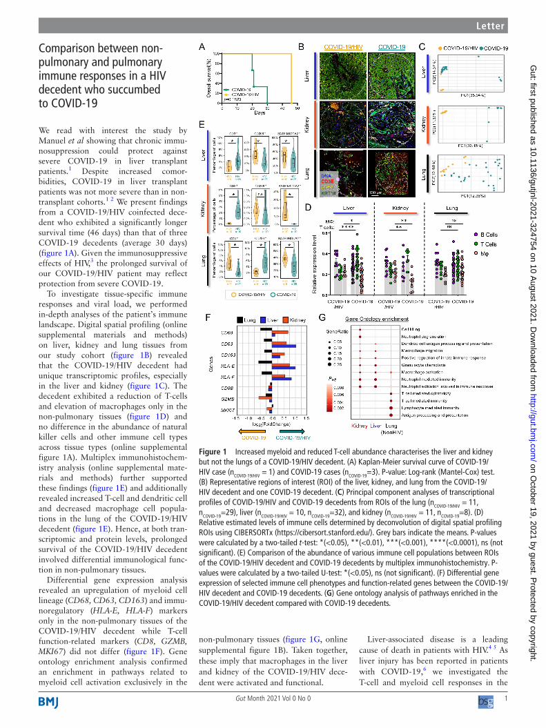
We read with interest the study by Manuel et al showing that chronic immu-nosuppression could protect against severe COVID-19 in liver transplant patients.1 Despite increased comor-bidities, COVID-19 in liver transplant patients was not more severe than in non- transplant cohorts.1 2 We present findings from a COVID-19/HIV coinfected dece-dent who exhibited a significantly longer survival time (46 days) than that of three COVID-19 decedents (average 30 days) (figure 1A). Given the immunosuppressive effects of HIV,3 the prolonged survival of our COVID-19/HIV patient may reflect protection from severe COVID-19.
To investigate tissue- specific immune responses and viral load, we performed in- depth analyses of the patient’s immune landscape. Digital spatial profiling (online supplemental materials and methods) on liver, kidney and lung tissues from our study cohort (figure 1B) revealed that the COVID-19/HIV decedent had unique transcriptomic profiles, especially in the liver and kidney (figure 1C). The decedent exhibited a reduction of T- cells and elevation of macrophages only in the non- pulmonary tissues (figure 1D) and no difference in the abundance of natural killer cells and other immune cell types across tissue types (online supplemental figure 1A). Multiplex immunohistochem-istry analysis (online supplemental mate-rials and methods) further supported these findings (figure 1E) and additionally revealed increased T- cell and dendritic cell and decreased macrophage cell popula-tions in the lung of the COVID-19/HIV decedent (figure 1E). Hence, at both tran-scriptomic and protein levels, prolonged survival of the COVID-19/HIV decedent involved differential immunological func-tion in non- pulmonary tissues.
Differential gene expression analysis revealed an upregulation of myeloid cell lineage (CD68, CD63, CD163) and immu-noregulatory (HLA- E, HLA- F) markers only in the non- pulmonary tissues of the COVID-19/HIV decedent while T- cell function- related markers (CD8, GZMB, MKI67) did not differ (figure 1F). Gene ontology enrichment analysis confirmed an enrichment in pathways related to myeloid cell activation exclusively in the
non- pulmonary tissues (figure 1G, online supplemental figure 1B). Taken together, these imply that macrophages in the liver and kidney of the COVID-19/HIV dece-dent were activated and functional.
Liver- associated disease is a leading cause of death in patients with HIV.4 5 As liver injury has been reported in patients with COVID-19,6 we investigated the T- cell and myeloid cell responses in the
Letter
Figure 1 Increased myeloid and reduced T- cell abundance characterises the liver and kidney but not the lungs of a COVID-19/HIV decedent. (A) Kaplan- Meier survival curve of COVID-19/HIV case (nCOVID-19/HIV = 1) and COVID-19 cases (nCOVID-19=3). P- value: Log- rank (Mantel- Cox) test. (B) Representative regions of interest (ROI) of the liver, kidney, and lung from the COVID-19/HIV decedent and one COVID-19 decedent. (C) Principal component analyses of transcriptional profiles of COVID-19/HIV and COVID-19 decedents from ROIs of the lung (nCOVID-19/HIV = 11, nCOVID-19=29), liver (nCOVID-19/HIV = 10, nCOVID-19=32), and kidney (nCOVID-19/HIV = 11, nCOVID-19=8). (D) Relative estimated levels of immune cells determined by deconvolution of digital spatial profiling ROIs using CIBERSORTx (https://cibersort.stanford.edu/). Grey bars indicate the means. P- values were calculated by a two- tailed t- test: *(<0.05), **(<0.01), ***(<0.001), ****(<0.0001), ns (not significant). (E) Comparison of the abundance of various immune cell populations between ROIs of the COVID-19/HIV decedent and COVID-19 decedents by multiplex immunohistochemistry. P- values were calculated by a two- tailed U- test: *(<0.05), ns (not significant). (F) Differential gene expression of selected immune cell phenotypes and function- related genes between the COVID-19/HIV decedent and COVID-19 decedents. (G) Gene ontology analysis of pathways enriched in the COVID-19/HIV decedent compared with COVID-19 decedents.
on October 19, 2021 by guest. P
rotected by copyright.http://gut.bm
j.com/
Gut: first published as 10.1136/gutjnl-2021-324754 on 10 A
ugust 2021. Dow
nloaded from
2 Gut Month 2021 Vol 0 No 0
Letter
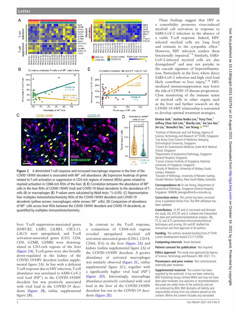
liver. T- cell suppression- associated genes (HAVCR2, LAIR1, LILRB1, CXCL13, LAG3) were upregulated, and T- cell activation- associated genes (CD3, CD4, CD8, GZMB, GZMK) were downreg-ulated in CD3- rich regions of the liver (figure 2A). T- cell genes were also broadly down- regulated in the kidney of the COVID-19/HIV decedent (online supple-mental figure 2A). In line with a deficient T- cell response due to HIV infection, T- cell abundance was unrelated to SARS- CoV-2 viral load (NP+) in the COVID-19/HIV decedent but was positively associated with viral load in the COVID-19 dece-dents (figure 2B, online supplemental figure 2B).
In contrast to the T- cell response, a comparison of CD68- rich regions revealed upregulated myeloid cell activation- associated genes (CD63, CD14, CD68, IL6) in the liver (figure 2A) and kidney (online supplemental figure 2A) of the COVID-19/HIV decedent. A greater abundance of activated macrophages was similarly observed (figure 2C, online supplemental figure 2C), together with a significantly higher viral load (NP+) (figure 2D). Interestingly, macrophage abundance positively correlated with viral load in the liver of the COVID-19/HIV decedent but not in the COVID-19 dece-dents (figure 2E).
These findings suggest that HIV as a comorbidity promotes virus- induced myeloid cell activation in response to SARS- CoV-2 infection in the absence of a viable T- cell response. Indeed, HIV- infected myeloid cells are long lived and resistant to the cytopathic effect.7 However, HIV infection renders them functionally impaired.7 8 Similarly, SARS- CoV-2- infected myeloid cells are also dysregulated9 and may not partake in the cascade signature of hyperinflamma-tion. Particularly in the liver, where direct SARS- CoV-2 infection and high viral load likely contribute to liver injury,6 10 HIV- mediated immunosuppression may lower the risk of COVID-19 disease progression. Close monitoring of the immune status of myeloid cells in other organs such as the liver and further research on the COVID-19–HIV relationship is warranted to develop optimal treatment strategies.
Denise Goh,1 Justina Nadia Lee,1 Tracy Tien,1 Jeffrey Chun Tatt Lim,1 Sherlly Lim,1 An Sen Tan,2 Jin Liu,3 Benedict Tan,1 Joe Yeong 4,5,6,7
1Institute of Molecular and Cell Biology, Agency of Science, Technology and Research (A*STAR), Singapore2Lee Kong Chian School of Medicine, Nanyang Technological University, Singapore3Centre for Quantitative Medicine, Duke- NUS Medical School, Singapore4Department of Anatomical Pathology, Singapore General Hospital, Singapore5Cancer Science Institute of Singapore, National University of Singapore, Singapore6Faculty of Medicine, University of Malaya, Kuala Lumpur, Malaysia7Disciple of Pathology, University of Western Sydney, Greater Western Sydney, New South Wales, Australia
Correspondence to Dr Joe Yeong, Department of Anatomical Pathology, Singapore General Hospital, Singapore 169608; yeongps@ imcb. a- star. edu. sg
Correction notice This article has been corrected since it published Online First. The fifth affiliation has been updated.
Contributors JY, BT and JL conceived and directed the study. DG, AST, BT and JL collated and interpreted the data and performed biostatistical analysis. JNL, TT, SL and JCTL performed immunohistochemical techniques and scoring. DG and BT drafted the manuscript and final approval of all authors.
Funding The authors received funding from A*STAR Career Development Award (C21112056).
Competing interests None declared.
Patient consent for publication Not required.
Ethics approval This study was approved by Agency of Science, Technology and Research, IRB: 2021 112.
Provenance and peer review Not commissioned; externally peer reviewed.
Supplemental material This content has been supplied by the author(s). It has not been vetted by BMJ Publishing Group Limited (BMJ) and may not have been peer- reviewed. Any opinions or recommendations discussed are solely those of the author(s) and are not endorsed by BMJ. BMJ disclaims all liability and responsibility arising from any reliance placed on the content. Where the content includes any translated
Figure 2 A diminished T- cell response and increased macrophage response in the liver of the COVID-19/HIV decedent is associated with NP+ cell abundance. (A) Expression heatmap of genes related to T- cell activation or suppression in CD3- rich regions of interest (ROIs) genes related to myeloid activation in CD68- rich ROIs of the liver. (B, E) Correlation between the abundance of NP+ cells in the liver ROIs of COVID-19/HIV (red) and COVID-19 (blue) decedents to the abundance of T- cells (B) or macrophages (E). P- values were calculated by Wald tests: *(<0.05). (C) Representative liver multiplex immunohistochemistry ROIs of the COVID-19/HIV decedent and COVID-19 decedents (yellow arrows: macrophages; white arrows: NP+ cells). (D) Comparison of abundance of NP+ cells across liver ROIs between the COVID-19/HIV decedent and COVID-19 decedents, as quantified by multiplex immunohistochemistry.
on October 19, 2021 by guest. P
rotected by copyright.http://gut.bm
j.com/
Gut: first published as 10.1136/gutjnl-2021-324754 on 10 A
ugust 2021. Dow
nloaded from
3Gut Month 2021 Vol 0 No 0
Letter
material, BMJ does not warrant the accuracy and reliability of the translations (including but not limited to local regulations, clinical guidelines, terminology, drug names and drug dosages), and is not responsible for any error and/or omissions arising from translation and adaptation or otherwise.
This article is made freely available for use in accordance with BMJ’s website terms and conditions for the duration of the covid-19 pandemic or until otherwise determined by BMJ. You may use, download and print the article for any lawful, non- commercial purpose (including text and data mining) provided that all copyright notices and trade marks are retained.
© Author(s) (or their employer(s)) 2021. No commercial re- use. See rights and permissions. Published by BMJ.
► Additional supplemental material is published online only. To view, please visit the journal online (http:// dx. doi. org/ 10. 1136/ gutjnl- 2021- 324754).
DG, JNL, TT and JCTL contributed equally.
DG, JNL, TT and JCTL are joint first authors.
JL, BT and JY are joint senior authors.
To cite Goh D, Lee JN, Tien T, et al. Gut Epub ahead of print: [please include Day Month Year]. doi:10.1136/gutjnl-2021-324754
Received 24 March 2021Accepted 30 July 2021
Gut 2021;0:1–3. doi:10.1136/gutjnl-2021-324754
ORCID iDJoe Yeong http:// orcid. org/ 0000- 0002- 6674- 7153
REFERENCES 1 Rodriguez- Peralvarez M, Salcedo M, Colmenero J, et al.
Modulating immunosuppression in liver transplant patients with COVID-19. Gut 2021;70:1412–4.
2 Becchetti C, Zambelli MF, Pasulo L. COVID-19 in an international European liver transplant recipient cohort. Gut 1832;2020:69.
3 McMichael AJ, Rowland- Jones SL. Cellular immune responses to HIV. Nature 2001;410:980–7.
4 Chamroonkul N, Bansal MB. Hiv and the liver. Nat Rev Gastroenterol Hepatol 2019;16:1–2.
5 Kaspar MB, Sterling RK. Mechanisms of liver disease in patients infected with HIV. BMJ Open Gastroenterol 2017;4:e000166.
6 Weber S, Hellmuth JC, Scherer C, et al. Liver function test abnormalities at hospital admission are associated with severe course of SARS- CoV-2 infection: a prospective cohort study. Gut 2021:gutjnl-2020-323800.
7 Wong ME, Jaworowski A, Hearps AC. The HIV reservoir in monocytes and macrophages. Front Immunol 2019;10.
8 Galvão- Lima LJ, Espíndola MS, Soares LS, et al. Classical and alternative macrophages have impaired function during acute and chronic HIV-1 infection. Braz J Infect Dis 2017;21:42–50.
9 Schulte- Schrepping J, Reusch N, Paclik D, et al. Severe COVID-19 is marked by a dysregulated myeloid cell compartment. Cell 2020;182:1419–40.
10 Morgan K, Samuel K, Vandeputte M, et al. SARS- CoV-2 infection and the liver. Pathogens 2020;9:430.
on October 19, 2021 by guest. P
rotected by copyright.http://gut.bm
j.com/
Gut: first published as 10.1136/gutjnl-2021-324754 on 10 A
ugust 2021. Dow
nloaded from