Embed Size (px)
Citation preview
fmicb-08-02372 November 27, 2017 Time: 15:56 # 1
ORIGINAL RESEARCHpublished: 29 November 2017
doi: 10.3389/fmicb.2017.02372
Edited by:Phil B. Pope,
Norwegian University of Life Sciences,Norway
Reviewed by:Antonis Chatzinotas,
Helmholtz-Zentrum fürUmweltforschung (UFZ), Germany
Shaillay Kumar Dogra,Vishuo Biomedical Pte Ltd.,
SingaporeBart Keijser,
Netherlands Organisation for AppliedScientific Research (TNO),
NetherlandsChristine Bassis,
University of Michigan, United States
*Correspondence:Sarah Lebeer
Specialty section:This article was submitted to
Systems Microbiology,a section of the journal
Frontiers in Microbiology
Received: 06 July 2017Accepted: 16 November 2017Published: 29 November 2017
Citation:De Boeck I, Wittouck S, Wuyts S,
Oerlemans EFM,van den Broek MFL,
Vandenheuvel D, Vanderveken O andLebeer S (2017) Comparing
the Healthy Nose and NasopharynxMicrobiota Reveals Continuity As Well
As Niche-Specificity.Front. Microbiol. 8:2372.
doi: 10.3389/fmicb.2017.02372
Comparing the Healthy Nose andNasopharynx Microbiota RevealsContinuity As Well AsNiche-SpecificityIlke De Boeck1, Stijn Wittouck1, Sander Wuyts1, Eline F. M. Oerlemans1,Marianne F. L. van den Broek1, Dieter Vandenheuvel1, Olivier Vanderveken2,3 andSarah Lebeer1*
1 Department of Bioscience Engineering, University of Antwerp, Antwerp, Belgium, 2 Department of TranslationalNeurosciences, Faculty of Medicine and Health Sciences, University of Antwerp, Antwerp, Belgium, 3 Department of ENT,Head and Neck Surgery, Antwerp University Hospital, Antwerp, Belgium
To improve our understanding of upper respiratory tract (URT) diseases and theunderlying microbial pathogenesis, a better characterization of the healthy URTmicrobiome is crucial. In this first large-scale study, we obtained more insight in the URTmicrobiome of healthy adults. Hereto, we collected paired nasal and nasopharyngealswabs from 100 healthy participants in a citizen-science project. High-throughput 16SrRNA gene V4 amplicon sequencing was performed and samples were processedusing the Divisive Amplicon Denoising Algorithm 2 (DADA2) algorithm. This allowedus to identify the bacterial richness and diversity of the samples in terms of ampliconsequence variants (ASVs), with special attention to intragenus variation. We foundboth niches to have a low overall species richness and uneven distribution. Moreover,based on hierarchical clustering, nasopharyngeal samples could be grouped into somebacterial community types at genus level, of which four were supported to someextent by prediction strength evaluation: one intermixed type with a higher bacterialdiversity where Staphylococcus, Corynebacterium, and Dolosigranulum appeared mainbacterial members in different relative abundances, and three types dominated byeither Moraxella, Streptococcus, or Fusobacterium. Some of these bacterial communitytypes such as Streptococcus and Fusobacterium were nasopharynx-specific and neveroccurred in the nose. No clear association between the nasopharyngeal bacterialprofiles at genus level and the variables age, gender, blood type, season of sampling, orcommon respiratory allergies was found in this study population, except for smokingshowing a positive association with Corynebacterium and Staphylococcus. Basedon the fine-scale resolution of the ASVs, both known commensal and potentialpathogenic bacteria were found within several genera – particularly in Streptococcusand Moraxella – in our healthy study population. Of interest, the nasopharynx hostedmore potential pathogenic species than the nose. To our knowledge, this is thefirst large-scale study using the DADA2 algorithm to investigate the microbiota inthe “healthy” adult nose and nasopharynx. These results contribute to a better
Frontiers in Microbiology | www.frontiersin.org 1 November 2017 | Volume 8 | Article 2372
fmicb-08-02372 November 27, 2017 Time: 15:56 # 2
De Boeck et al. Healthy Nose and Nasopharynx Microbiota
understanding of the composition and diversity of the healthy microbiome in the URTand the differences between these important URT niches.Trial Registration: Ethical Committee of Antwerp University Hospital,B300201524257, registered 23 March 2015, ClinicalTrials.gov Identifier: NCT02933983.
Keywords: microbiome, amplicon sequence variants, upper respiratory tract, bacterial community types,nasopharynx, next-generation sequencing
INTRODUCTION
Respiratory tract infections, including acute and chronic otitismedia in children and chronic rhinosinusitis in adults, are themost commonly treated health issues in primary care (Franciset al., 2009). The respiratory tract can be divided in the lowerand upper respiratory tract (URT), where the latter comprisesthe anterior nares, the nasal passages, the paranasal sinuses,the naso- and oropharynx, and finally the larynx above thevocal cords (reviewed in Man et al., 2017). In Europe, URTinfections account for 57% of all prescribed antibiotics, havinga significant impact on the emerging problem of antibioticresistance (van der Velden et al., 2013). Without evidence fora clear causative role for specific bacterial species, culture-based sampling has linked various opportunistic pathogensto chronic rhinosinusitis and otitis media. However, thesebacterial species seem also to be present in healthy individuals(Lemon, 2010; Stearns et al., 2015). To better elucidate thecontribution of the microbiota to URT diseases and to designtargeted (anti-)microbial approaches, a better understanding ofthe composition and diversity of the “healthy URT microbiota” isessential.
Due to major advances in sequencing techniques andlarge-scale sequencing projects such as the NIH HumanMicrobiome Project, our understanding of the compositionand functional properties of the human microbiota hasimproved greatly (Turnbaugh et al., 2007). While manystudies have previously focused on the microbiota ofthe gastrointestinal tract, in recent years, interest in theresident microbial communities of other human bodyniches, such as the respiratory tract, has clearly beenexpanding.
The nose and nasopharynx are key niches of the URT.Both niches host commensals and potential pathogenic speciesthat may cause airway infections under certain conditions(reviewed in Man et al., 2017). While the nasal microbialcommunity is being mapped in large initiatives like the NIHHuman Microbiome Project (Turnbaugh et al., 2007) andother more recent studies (Bassis et al., 2014; Camarinha-Silva et al., 2014; Biswas et al., 2015), the nasopharynxis less explored. Some larger studies (including 50 up to234 participants) have profiled the nasopharyngeal microbiotain children with next-generation sequencing (NGS) (Bogaertet al., 2011; Biesbroek et al., 2014; Stearns et al., 2015; Teoet al., 2015; Bosch et al., 2017; Chonmaitree et al., 2017),but only a few studies have investigated the healthy adultnasopharyngeal microbiota (Ling et al., 2013; Cremers et al.,
2014; Stearns et al., 2015). These studies in adults were small-scale (including less than 40 participants), with different agegroups and populations from different geographical locations.Furthermore, with the exception of the study of Stearns et al.(2015), these studies used 16S rRNA gene pyrosequencing,which has some limitations over the more in-depth IlluminaMiSeq sequencing. Main limitations are sequencing errorsin homopolymeric regions and lower sequencing depth. Inaddition, all these studies have used a clustering approachwhere sequences were clustered into operational taxonomicunits (OTUs). While this approach is most often used, itcurrently underutilizes the power of high-quality sequencesproduced by modern sequencing technologies, such as IlluminaMiSeq (reviewed in Hugerth and Andersson, 2017). Therefore,alternative algorithms that detect more fine-scale variationslike MED (Eren et al., 2014, 2016) and Divisive AmpliconDenoising Algorithm 2 (DADA2) (Callahan et al., 2016)have recently emerged, resulting in improved precision ofdiversity and dissimilarity measures. Since both the nose andthe nasopharynx are low-complexity (in terms of observedrichness or total number of bacterial genera present) and low-biomass (in terms of total amount of bacterial cells) microbialniches (Biesbroek et al., 2012), an accurate discriminationbetween these biological variants is essential. The recentlydeveloped DADA2 algorithm in combination with IlluminaMiSeq sequencing has the potential to improve sensitivity,specificity, and reproducibility compared to OTU-pickingmethods. This algorithm infers unique biological variantscalled “amplicon sequence variants” or ASVs (Callahan et al.,2017) by correcting sequencing errors in the reads. TheASV concept is an alternative to the classical concept of anOTU: OTU-based strategies perform clustering based on afixed percentage identity threshold (e.g., 97%), while ASVsare the result of a denoising procedure only. The DADA2denoising strategy is based on the quality scores of allreads as well as the abundance distribution of the uniquesequences.
By the implementation of this DADA2 pipeline, weexplored here the diversity and main bacterial members ofthe nose and nasopharynx of 100 healthy participants inorder to obtain more insight in the commensal and potentialpathogenic bacteria colonizing these URT niches. The resultingbacterial profiles were mined for associations to the dataavailable from our healthy volunteers, such as age, gender,blood type, smoking, season, and blood analyses for totalimmunoglobulin E (IgE) and IgE levels against commonrespiratory allergies.
Frontiers in Microbiology | www.frontiersin.org 2 November 2017 | Volume 8 | Article 2372
fmicb-08-02372 November 27, 2017 Time: 15:56 # 3
De Boeck et al. Healthy Nose and Nasopharynx Microbiota
MATERIALS AND METHODS
Study Design and Sample CollectionParticipants between 18 and 65 years old without acute or chronicURT diseases were recruited between July 2015 and October2016 via the University of Antwerp and a Belgian–Dutch citizen-science platform1, after approval of the study by the EthicalCommittee of the Antwerp University Hospital/Universityof Antwerp (registration number B300201524257, registered23 March 2015, ClinicalTrials.gov Identifier: NCT02933983).A written informed consent was obtained from all participants,as well as a blood sample and a questionnaire with generalinformation on their medical history and additional informationsuch as smoking behavior. Participants who received antibiotics(self-reported) in the past year or suffered from acute orchronic airway infections were excluded from the study. Intotal, 90 nasal and 100 nasopharynx samples were collected ina standardized way by the responsible ear, nose, and throat(ENT) specialist with flocked swabs (Copan, 503CS01) at thelevel of the anterior nasal cavity, and nasopharynx. All sampleswere immediately suspended in 750 µl MoBio bead solution(PowerFecal R© DNA Isolation Kit; MO BIO Laboratories Inc.,Carlsbad, CA, United States) and placed on ice prior to DNAextraction. DNA extraction took place within 4 h after samplecollection. DNA samples were stored at−20◦C until further use.
Blood Analysis for Total and Specific IgEA serum sample was collected from all participants in orderto investigate the total IgE level in their blood, as well assome specific IgEs for respiratory allergies (tree pollen, grasspollen, and house dust mite). Blood samples were collectedat the Antwerp University Hospital by a responsible nurse.Total and specific IgEs were quantified by an ImmunoCAPSystem (Thermo Fisher Scientific, Uppsala, Sweden). Allassays were performed and results interpreted according tothe manufacturers’ recommendation. Total IgE counts below114 kU/l were considered as non-allergic. For specific IgE counts,values below 0.35 kUA/l were considered as non-allergic.
DNA ExtractionThe PowerFecal R© DNA Isolation Kit (with Inhibitor RemovalTechnology R©) was used according to the instructions of themanufacturer. DNA concentrations were measured with aQubit R© 3.0 Fluorometer (Life Technologies, Ledeberg, Belgium).DNA extractions were performed in a laboratory roomdedicated for DNA/RNA extraction, physically separated fromthe microbiology room to minimize contamination.
Illumina MiSeq 16S rRNA Gene AmpliconSequencingThe primers used for Illumina MiSeq sequencing were basedon the previously described 515F-806R primers (Caporasoet al., 2010) and altered for dual-index paired-end sequencing,as earlier described (Kozich et al., 2013). Briefly, each DNA
1www.IedereenWetenschapper.be
sample was subjected to dual barcoded PCR, amplifying the V4region of the 16S rRNA gene using Phusion High-Fidelity DNApolymerase (New England Biolabs, United States). PCR productswere purified by the Agencourt AMPure XP Magnetic BeadCapture Kit (Beckman Coulter, Suarlee, Belgium), and quantifiedusing the Qubit R© 3.0 Fluorometer. The library was prepared bypooling all PCR samples in equimolar concentration and loadedonto a 0.8% agarose gel to remove remaining primer dimersfrom the product. The product was purified by gel extractionusing the NucleoSpin R© Gel and PCR clean-up (Macherey-Nagel). The final library concentration was determined withthe Qubit R© 3.0 Fluorometer. The library was denatured with0.2 N NaOH (Illumina), diluted to 7 pM and spiked with 10%PhiX control DNA (Illumina). The library was loaded ontothe flow cell of the v2 Chemistry MiSeq Reagent Kit (paired-end dual indexing sequencing; 2 × 250 bp kit; Illumina, SanDiego, CA, United States) on the MiSeq Desktop Sequencer(M00984, Illumina) at the Centre of Medical Genetics, Universityof Antwerp, Belgium. The sequencing data were deposited inENA under accession number PRJEB23057.
Sequence Processing and QualityControlProcessing and quality control of reads was performed usingthe R package dada2, version 1.4.0 (Callahan et al., 2016).After inspection of quality control profiles, the first 35 basesof all reverse reads were trimmed since they frequentlycontained uncalled bases. Next, all reads containing remaininguncalled bases or more than three expected errors wereremoved. Afterward, the parameters of the DADA2 errormodel were learned from a random subset of 1 million reads.This error model was then used to denoise all sequences;i.e., to infer the ASVs. Denoised reads (ASVs) were thenmerged and read pairs with one or more conflicting basesbetween the forward and reverse read were removed. Chimericsequences were then detected and removed using the function“removeBimeraDenovo.” Finally, reads (ASVs) were classifiedfrom the kingdom to the genus level using the Silva reference 16SrRNA gene database, version 123 resulting in the construction ofan ASV table with read counts of all ASVs in all samples.
In the next phase, quality control was performed on the levelof the ASVs and samples. ASVs longer than 251 bases wereremoved, as well as ASVs classified as Archaea, chloroplasts, ormitochondria. The PCR and DNA extraction negative controlswere inspected, and ASVs classified as known contaminantsand/or that were overrepresented in the negative blank controls(when compared to the samples) were removed. Finally, sampleswere subjected to quality control based on total read count andread count per sample volume pooled. Samples were requiredto contain at least five times more reads per volume than thenegative controls, as well as more than 1000 total reads.
Biostatistical AnalysisProcessing of the ASV table, ASV annotations (e.g.,classification), and sample annotations (metadata) wereperformed using the in-house R package “tidyamplicons,”
Frontiers in Microbiology | www.frontiersin.org 3 November 2017 | Volume 8 | Article 2372
fmicb-08-02372 November 27, 2017 Time: 15:56 # 4
De Boeck et al. Healthy Nose and Nasopharynx Microbiota
publicly available at github.com/SWittouck/tidyamplicons. Forthe analyses on the genus level, ASV read counts were aggregatedon the genus level or, if unavailable, on the most specific levelat which taxonomic annotation was available. Alpha-diversitywas explored at the genus level using two different metrics: thenumber of observed genera and the inverse Simpson metric(defined as the inverse probability that two random reads belongto the same taxon). Differences in these two metrics between thenose and nasopharynx samples were tested using a Wilcoxonrank-sum test. Correlation of the alpha-diversity metricsbetween the nose and nasopharynx was assessed using Pearsoncorrelation and the corresponding significance test implementedin the cor.test function in R. For beta-diversity analysis, the Bray–Curtis distance was used, defined as the summed differences inread counts for all taxa, divided by the total read counts in bothsamples. The Bray–Curtis beta-diversity matrix was exploredvisually using principal coordinates analysis (PCoA). In orderto test the bacterial profiles for clustering structure, we madeuse of the prediction strength metric (Tibshirani and Walther,2005). First, all samples (from both nose and nasopharynx) wereclustered in seven clusters using hierarchical clustering with theunweighted pair group method with arithmetic mean (UPGMA)on the Bray–Curtis distance matrix. Clusters containing onlyone or two samples were considered outliers and were removed,since those cannot be evaluated using prediction strength. Next,four distance matrices were calculated: Bray–Curtis (on relativeabundances, as usual), Bray–Curtis on the presence/absencelevel, Jensen–Shannon divergence, and Jensen–Shannon distance(equal to the square root of the Jensen–Shannon divergence).Two clustering techniques were then performed on thosematrices: UPGMA and partitioning around medoids (PAM).Each distance metric – clustering algorithm combination wasperformed for different numbers of clusters (2 up until 10).This approach to evaluate clustering largely follows Koren et al.(2013), except that we added the UPGMA clustering method andthe presence/absence Bray–Curtis distance metric.
The association of the nasopharyngeal microbiota withparticipant metadata was performed for all metadata variablesthat had more than six participants in at least two categories.These variables were gender, age, blood type, smoking, season ofsampling, total IgE level, and specific IgE levels for house dustmite, grass pollen, and tree pollen. For each of these variables, theassociation with the microbiota was tested using a permanova testimplemented in the function “adonis” of the R package “vegan.”Specifically, the adonis function tests whether the Bray–Curtisdistances within groups of samples are smaller than the distancesbetween groups; significance is assessed using a permutationstrategy. The association between the variable “smoking” andthe genera Corynebacterium, Dolosigranulum, and Staphylococcuswas tested with a Wilcoxon rank-sum test.
For the analyses on the ASV level, only samples were retainedof participants that had both a nose and a nasopharynx samplepassing quality control. To test whether ASV presence wascorrelated between both niches, the following strategy was usedfor each ASV. First, a two-way frequency table was constructed,where each cell contained a count of participants and thevariables were “presence of the ASV in the nose” and “presence
of the ASV in the nasopharynx.” Next, association between thetwo variables was tested using a Fisher’s exact test (implementedin the base R function fisher.test). All ASVs were then visualizedin a scatterplot with on the x-axis the expected proportion ofco-occurrence under the assumption of no correlation and on they-axis the observed proportion of co-occurrence. The expectedproportion of co-occurrence was calculated by multiplying theoccurrence proportion in the nasopharynx with the occurrenceproportion in the nose. To test preference of ASVs for one of theniches of the other, a similar approach was followed. First, a two-way frequency table was constructed where each cell containeda count of samples and the variables were “sample type (noseor nasopharynx)” and “presence/absence.” Preference for thenose or nasopharynx was then assessed by testing the associationbetween these two variables using a Fisher’s exact test. Finally,the ASVs were visualized in a scatterplot with on the x-axisthe occurrence proportion in the nose and on the y-axis theoccurrence proportion in the nasopharynx.
Quality control, biostatistical analysis, and visualization wereperformed in R version 3.4.1. All visualizations were createdusing ggplot2 version 2.2.1 (Wickham, 2009). Vegan version 2.4.3(Oksanen et al., 2016) was used for alpha- and beta-diversityanalyses.
RESULTS
The Adult Nasopharynx Is Dominated byat Least Four Bacterial CommunityTypes at the Genus LevelSamples from the “healthy” nose and nasopharynx of participantswith no signs of URT infections, recruited in collaborationwith a Belgian–Dutch citizen-science platform, were collected.In total, we collected 90 nasal samples and 100 nasopharyngealsamples, of which, respectively, 84 and 92 remained after qualitycontrol. Supplementary Table S1 presents the different steps inthe quality control with the remaining amount of reads after eachstep.
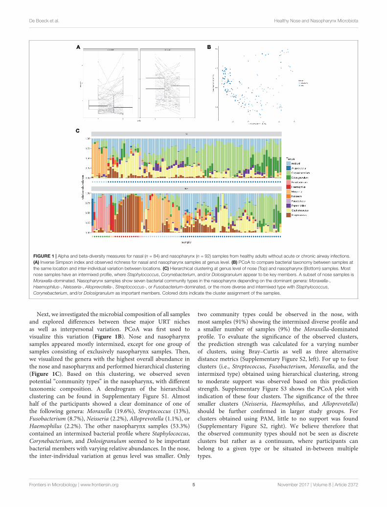
Alpha and beta-diversity measures of both nose andnasopharynx swabs were calculated to estimate the bacterial16S rRNA gene diversity in these adult URT niches. Figure 1Apresents the overall observed richness and the inverse Simpsonindex in each participant for both the nose and the nasopharynx.Both niches contain a rather low number of observed genera(on average 31 in the nose and 25 in the nasopharynx) with asignificantly higher observed richness in the nose than in thenasopharynx (p = 0.002). Additionally, the low inverse Simpsonindex (on average 4.1 for the nose and 4.3 for the nasopharynx)suggests an uneven distribution of the abundance of this limitedamount of genera, indicating that both the adult nose and theadult nasopharynx are low-diversity niches where only a limitednumber of bacterial genera are dominant. Finally, the correlationof alpha-diversities between the nose and nasopharynx wascalculated to be 0.19 and 0.21 for observed richness and inverseSimpson, respectively, meaning that the amount of genera in thenose is only weakly informative for the amount of genera in thenasopharynx and vice versa.
Frontiers in Microbiology | www.frontiersin.org 4 November 2017 | Volume 8 | Article 2372
fmicb-08-02372 November 27, 2017 Time: 15:56 # 5
De Boeck et al. Healthy Nose and Nasopharynx Microbiota
FIGURE 1 | Alpha and beta-diversity measures for nasal (n = 84) and nasopharynx (n = 92) samples from healthy adults without acute or chronic airway infections.(A) Inverse Simpson index and observed richness for nasal and nasopharynx samples at genus level. (B) PCoA to compare bacterial taxonomy between samples atthe same location and inter-individual variation between locations. (C) Hierarchical clustering at genus level of nose (Top) and nasopharynx (Bottom) samples. Mostnose samples have an intermixed profile, where Staphylococcus, Corynebacterium, and/or Dolosigranulum appear to be key members. A subset of nose samples isMoraxella-dominated. Nasopharynx samples show seven bacterial community types in the nasopharynx depending on the dominant genera: Moraxella-,Haemophilus-, Neisseria-, Alloprevotella-, Streptococcus-, or Fusobacterium-dominated, or the more diverse and intermixed type with Staphylococcus,Corynebacterium, and/or Dolosigranulum as important members. Colored dots indicate the cluster assignment of the samples.
Next, we investigated the microbial composition of all samplesand explored differences between these major URT nichesas well as interpersonal variation. PCoA was first used tovisualize this variation (Figure 1B). Nose and nasopharynxsamples appeared mostly intermixed, except for one group ofsamples consisting of exclusively nasopharynx samples. Then,we visualized the genera with the highest overall abundance inthe nose and nasopharynx and performed hierarchical clustering(Figure 1C). Based on this clustering, we observed sevenpotential “community types” in the nasopharynx, with differenttaxonomic composition. A dendrogram of the hierarchicalclustering can be found in Supplementary Figure S1. Almosthalf of the participants showed a clear dominance of one ofthe following genera: Moraxella (19.6%), Streptococcus (13%),Fusobacterium (8.7%), Neisseria (2.2%), Alloprevotella (1.1%), orHaemophilus (2.2%). The other nasopharynx samples (53.3%)contained an intermixed bacterial profile where Staphylococcus,Corynebacterium, and Dolosigranulum seemed to be importantbacterial members with varying relative abundances. In the nose,the inter-individual variation at genus level was smaller. Only
two community types could be observed in the nose, withmost samples (91%) showing the intermixed diverse profile anda smaller number of samples (9%) the Moraxella-dominatedprofile. To evaluate the significance of the observed clusters,the prediction strength was calculated for a varying numberof clusters, using Bray–Curtis as well as three alternativedistance metrics (Supplementary Figure S2, left). For up to fourclusters (i.e., Streptococcus, Fusobacterium, Moraxella, and theintermixed type) obtained using hierarchical clustering, strongto moderate support was observed based on this predictionstrength. Supplementary Figure S3 shows the PCoA plot withindication of these four clusters. The significance of the threesmaller clusters (Neisseria, Haemophilus, and Alloprevotella)should be further confirmed in larger study groups. Forclusters obtained using PAM, little to no support was found(Supplementary Figure S2, right). We believe therefore thatthe observed community types should not be seen as discreteclusters but rather as a continuum, where participants canbelong to a given type or be situated in-between multipletypes.
Frontiers in Microbiology | www.frontiersin.org 5 November 2017 | Volume 8 | Article 2372
fmicb-08-02372 November 27, 2017 Time: 15:56 # 6
De Boeck et al. Healthy Nose and Nasopharynx Microbiota
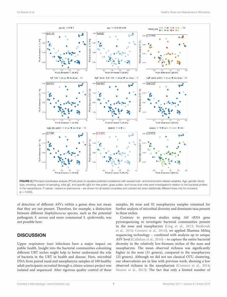
The Nasopharyngeal Bacterial“Community Types” Show anAssociation with Smoking Behavior, ButNot with Gender, Age, Blood Type,Season of Sampling, and CommonRespiratory AllergiesWe recorded several variables for each participating volunteer:age, gender, blood type, smoking and season (sampling date)to investigate possible associations with the nasopharyngealmicrobial profiles (Supplementary Table S2). Each of thevariables was visualized on a PCoA plot to look for potentialassociations with the bacterial composition of the samples, usingpermanova for statistical analysis (Figure 2). We divided ourstudy population (mean age = 34.78, SD = 11.2, range = 18–65)in two age categories, 18–45 years (84% of the study population)and 45–65 years (16%) but could not demonstrate an associationwith these age classes and the bacterial profiles in our – quiteyoung – study population. Also gender (34 males and 58 females),blood type, and season were not found to be associated with thenasopharyngeal microbiota. Smoking behavior, however, showedan association with the bacterial profiles in the nasopharynx(p = 0.002). Participants who smoke or used to smoke(17% of the study population) seemed to almost all have anintermixed bacterial profile with high relative abundances ofStaphylococcus, Corynebacterium, and Dolosigranulum, with theexception of one participant in the Haemophilus-dominatedgroup. Because smoking behavior showed a positive associationwith the intermixed nasopharyngeal “community type,” weinvestigated this association further at genus level. We found apositive association of smoking with Corynebacterium (Wilcoxonrank-sum test, p = 0.002) and Staphylococcus (p = 0.02),while Dolosigranulum showed no association (SupplementaryFigure S4).
In addition to these more descriptive variables, a blood sampleof each participant was analyzed for total blood IgE and someIgEs specific for respiratory allergens, such as house dust mite,grass pollen, and tree pollen (Supplementary Table S1). Subjectswith total IgE and specific IgE counts above 114 kU/l and0.35 kUA/l, respectively, were considered as “allergic.” In ourstudy population, 15% of our participants was considered allergicbased on total IgE and 25, 25, and 14% were allergic for housedust mite, grass pollen, and tree pollen, respectively. For allparticipants, no clear association was found with the testedrespiratory allergies and the genus-abundance profiles of theirnasopharyngeal microbiota.
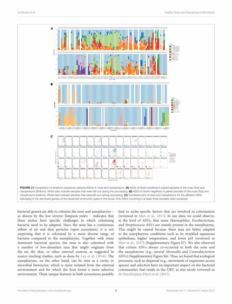
Intragenus-Information onCo-occurrence of ASVs in the Nose andNasopharynxIn order to be able to distinguish different variants withinone genus, we applied the DADA2 pipeline and found formany of the abundant genera multiple ASVs in both nose andnasopharynx. ASVs were then aligned to the SILVA database(version123) to get an overview of all species with V4 sequencesidentical to the ASV. This gives a general idea of the classification
of the ASV at the sub-genus level. In order to be able tovisualize more ASVs, we divided Gram-positive (Figure 3A)and Gram-negative (Figure 3B) ASVs. For Corynebacterium,the most abundant ASVs found in the nose could be classifiedas Corynebacterium accolens/macginleyi (Corynebacterium1)and Corynebacterium propinquum/pseudodiphtheriticum(Corynebacterium2). Interestingly, these variants were alsopresent in the nasopharynx, although less frequent (Figure 3A).For Moraxella, three abundant variants were detected in boththe nose and nasopharynx: Moraxella porci (Moraxella1),Moraxella catarrhalis/nonliquefaciens (Moraxella2), andMoraxella bovoculi/lacunata/equi (Moraxella3). The Moraxella2variant, M. catarrhalis/nonliquefaciens, was most abundantin different samples and almost never co-occurred with theother Moraxella variants in the same sample. Of interest,the two persons hosting the third Moraxella variant inthe nasopharynx also had this variant in their nose.Furthermore, some Streptococcus ASVs could be discriminatedin the nasopharynx of which Streptococcus1 was furtherclassified as Streptococcus pneumoniae/pseudopneumoniaeand Streptococcus3 as Streptococcus dentisani/tigurinus/oralis/oligofermentans/mitis/infantis/gordonii. Interestingly,the latter variant was present in 1–15% abundance in a largepart of our nasopharynx samples of the healthy adults, while theASV classified as S. pneumoniae/pseudopneumoniae, which isdescribed in literature as a common URT pathogen, dominatedthe samples if present. The Streptococcus2 ASV was found toco-occur with Streptococcus1 and in addition, their sequencesdiffered in only one nucleotide. Therefore, it is likely that theyoriginate from two different copies of the 16S rRNA gene of thesame strain. Only one abundant Haemophilus ASV was found(Haemophilus haemolyticus/influenzae) in the nasopharynx. ForDolosigranulum, also only one abundant ASV was identified,which we could classify as D. pigrum, and this ASV seemedto be more abundant in the nose than the nasopharynx.Finally, three different Fusobacterium variants were observedin the nasopharynx samples (Fusobacterium1; Fusobacteriumnucleatum/canifenilum, Fusobacterium2; F. nucleatum, andFusobacterium3; F. nucleatum/naviforme). In addition to themost abundant ASVs discussed above (Figures 3A,B), otherASVs showing a lower abundance within the genera of the“community types” were detected, of which some were onlypresent in the nasopharynx, such as Haemophilus1 and 3,Fusobacterium2, and Streptococcus1,4, and 6 (Figure 3C).Supplementary Figures S5a–h give a more detailed comparisonof paired nose and nasopharynx samples at ASV level for each ofthe dominant genera of the bacterial community types, showingthe unique niche-specificity of some ASVs, while other ASVsare less niche-related and show continuity between both niches.The co-occurrence and niche-specificity for other ASVs was alsovisualized and was statistically tested using a Fisher’s exact test(Supplementary Figures S6, S7). For example, Streptococcus1and Fusobacterium2 only appeared in nasopharynx samplesstudied, while Corynebacterium2 and Moraxella2 occurred inboth niches. It should be noted that ASVs presented here aredetermined based on the amount of variation present in theV4 region of the 16S rRNA gene. This implicates that absence
Frontiers in Microbiology | www.frontiersin.org 6 November 2017 | Volume 8 | Article 2372
fmicb-08-02372 November 27, 2017 Time: 15:56 # 7
De Boeck et al. Healthy Nose and Nasopharynx Microbiota
FIGURE 2 | Principal coordinates analysis (PCoA) plots to visualize potential correlations with several host- and environment-related variables. Age, gender, bloodtype, smoking, season of sampling, total IgE, and specific IgEs for tree pollen, grass pollen, and house dust mite were investigated in relation to the bacterial profilesin the nasopharynx. P-values – based on permanova – are shown for all tested covariates and colored red when statistically different (here only for smokers)(p = 0.002).
of detection of different ASVs within a genus does not meanthat they are not present. Therefore, for example, a distinctionbetween different Staphylococcus species, such as the potentialpathogenic S. aureus and more commensal S. epidermidis, wasnot possible here.
DISCUSSION
Upper respiratory tract infections have a major impact onpublic health. Insight into the bacterial communities colonizingdifferent URT niches might help to better understand the roleof bacteria in the URT in health and disease. Here, microbialDNA from paired nasal and nasopharynx samples of 100 healthyadult participants recruited through a citizen-science project wasisolated and sequenced. After rigorous quality control of these
samples, 84 nose and 92 nasopharynx samples remained forfurther analysis of microbial diversity and dominant taxa presentin these niches.
Contrary to previous studies using 16S rRNA genepyrosequencing to investigate bacterial communities presentin the nose and nasopharynx (Ling et al., 2013; Biesbroeket al., 2014; Cremers et al., 2014), we applied Illumina MiSeqsequencing technology – combined with analysis up to uniqueASV level (Callahan et al., 2016) – to capture the entire bacterialdiversity in the relatively low-biomass niches of the nose andnasopharynx. The mean observed richness was significantlyhigher in the nose (31 genera), compared to the nasopharynx(25 genera). Although we did not use classical OTU clustering,our observations are in line with previous work, showing a lowobserved richness in the nasopharynx (Cremers et al., 2014;Stearns et al., 2015). The fact that only a limited number of
Frontiers in Microbiology | www.frontiersin.org 7 November 2017 | Volume 8 | Article 2372
fmicb-08-02372 November 27, 2017 Time: 15:56 # 8
De Boeck et al. Healthy Nose and Nasopharynx Microbiota
FIGURE 3 | Comparison of amplicon sequence variants (ASVs) in nose and nasopharynx. (A) ASVs of Gram-positives in paired samples of the nose (Top) andnasopharynx (Bottom). White bars indicate samples that were left-out during the processing. (B) ASVs of Gram-negatives in paired samples of the nose (Top) andnasopharynx (bottom). White bars indicate samples that were left-out during processing. (C) Combined plot of nose and nasopharynx for the different ASVsbelonging to the dominant genera of the observed community types in this study. Only ASVs occurring in at least three samples were visualized.
bacterial genera are able to colonize the nose and nasopharynx –as shown by the low inverse Simpson index – indicates thatthese niches have specific challenges to which colonizingbacteria need to be adapted. Since the nose has a continuousinflow of air and dust particles (open ecosystem), it is notsurprising that it is colonized by a more diverse range ofbacteria compared to the nasopharynx. Together with somedominant bacterial species, the nose is also colonized witha number of low-abundant taxa that might originate fromthe air, the skin, or other external sources, as suggested insource-tracking studies, such as done by Lax et al. (2014). Thenasopharynx, on the other hand, can be seen as a cavity ormicrobial bioreactor, which is more isolated from the externalenvironment and for which the host forms a more selectiveenvironment. These unique features in both ecosystems possibly
lead to niche-specific factors that are involved in colonization(reviewed in Man et al., 2017). In our data, we could observe,at the level of ASVs, that some Haemophilus, Fusobacterium,and Streptococcus ASVs are mainly present in the nasopharynx.This might be caused because these taxa are better adaptedto the nasopharynx conditions such as its stratified squamousepithelium, higher temperature, and lower pH (reviewed inMan et al., 2017) (Supplementary Figure S7). We also observedthat certain ASVs always co-occurred in both the nose andthe nasopharynx (e.g., several Moraxella and CorynebacteriumASVs) (Supplementary Figure S6). Thus, we found that ecologicalprocesses, such as dispersal (e.g., movement of organisms acrossspaces) and selection have an important impact on the bacterialcommunities that reside in the URT, as also nicely reviewed inde Steenhuijsen Piters et al. (2015).
Frontiers in Microbiology | www.frontiersin.org 8 November 2017 | Volume 8 | Article 2372
fmicb-08-02372 November 27, 2017 Time: 15:56 # 9
De Boeck et al. Healthy Nose and Nasopharynx Microbiota
In addition, we also aimed to explore the dominanceof bacterial taxa and the possible occurrence of communitytypes, similarly as previously done in the gastrointestinaltract (referred to as “enterotypes”) (Arumugam et al., 2011),the vaginal tract (reviewed in Petrova et al., 2015), andthe nasopharynx of newborns (Biesbroek et al., 2014). Basedon hierarchical clustering, our samples could be groupedby the dominant genera, resulting in at least four bacterial“community types” for the nasopharynx samples analyzed here:Moraxella-, Fusobacterium-, or Streptococcus-dominated, or onemore intermixed type showing a higher bacterial diversity whereCorynebacterium, Staphylococcus, and/or Dolosigranulum are themain members of the bacterial community. Some smaller clusters(Haemophilus, Alloprevotella, and Neisseria) were also observed,but their significance should be confirmed in larger studies. Sincesome (albeit exceptional) samples show a mixture of generafrom different clusters, these profiles should not be interpretedas discrete community types but rather as a continuum. Incontrast, nose samples mainly showed the diverse type withagain Corynebacterium, Staphylococcus, and Dolosigranulum asthe main members. This last observation is in agreement withprevious studies demonstrating that these genera are highlyabundant in the anterior nares (Bassis et al., 2014; Camarinha-Silva et al., 2014; Biswas et al., 2015). A minority of thenose samples (9%) was dominated by Moraxella. Of note, thebiological relevance of such discrete community types in otherhuman body niches, such as the vagina and gastrointestinal tract,is still under debate and needs further substantiation (Koren et al.,2013). This will certainly also be the case for the nasopharynx, forwhich our present study should merely be seen as a starting point,since the clusters observed in this study were only confirmed tosome extent for hierarchical clustering on a Bray–Curtis distancemetric.
The nasopharyngeal microbiota in healthy adults was – tothe best of our knowledge – not yet investigated in largestudy cohorts. However, Biesbroek et al. (2014) demonstratedthe bacterial succession of the nasopharynx microbiota inDutch children. They found the infant nasopharynx to bemostly dominated by Moraxella, Haemophilus, or Streptococcus.Interestingly, our data show that these three genera arealso maintained in the nasopharynx of adults, both in theage class 18–45 and 45–65 years. In young children, aStreptococcus-dominated bacterial profile was found to beassociated with a less-stable nasopharyngeal microbiota, therebypotentially increasing the risk of URT infections (Biesbroeket al., 2014). The same study suggested that Moraxella- orDolosigranulum/Corynebacterium-dominated bacterial profilesmight be beneficial for respiratory health, which was alsosuggested before (Laufer et al., 2011). In contrast, Santee et al.(2016) found the enrichment of Moraxella in the nasopharynx ofchildren, in particular Moraxella nonliquefaciens, to be associatedwith acute sinusitis.
The differences observed between studies such as byBiesbroek et al. (2014) and Santee et al. (2016) might becaused by the difference in molecular techniques used (e.g.,phylogenetic microarray vs. pyrosequencing, respectively) andby the limitation of sequencing techniques to distinguish
between discrete species within a genus. Therefore, we used therecently described DADA2 pipeline that is able to distinguishsequence variants differing by as little as one nucleotide(Callahan et al., 2016) to investigate intragenus variation,described as unique ASVs. We could further classify themost abundant Moraxella and Streptococcus ASVs in ourhealthy study group as Moraxella nonliquefaciens/catarrhalis andS. pneumoniae/pseudopneumoniae, two species well documentedin the literature to have potential as URT pathogens (de Vrieset al., 2009; Goldstein et al., 2009; van der Poll and Opal, 2009).Future studies need to investigate whether the presence andabundance of these ASVs is linked to susceptibility for airwaydiseases, such as chronic rhinosinusitis. In general, potentialpathogenic ASVs such as S. pneumoniae/pseudopneumoniae (vander Poll and Opal, 2009) and H. haemolyticus/influenzae (Duellet al., 2016) were more present in the nasopharynx compared tothe nose. On the other hand, some of the ASVs that we found,such as Streptococcus3 (with hits in the SILVA database to amongothers S. oralis and S. mitis) and Dolosigranulum, have shownpotential as probiotics for the URT in other studies (Roos et al.,2001; Tano et al., 2002; Laufer et al., 2011; Biesbroek et al., 2014).
The possible existence of nasopharyngeal community typesraises the question which host and environmental factors areassociated with these community types. Several available variablesobtained from our healthy study population were analyzedhere, including age, gender, blood type, smoking, commonrespiratory allergies, and season. We could not observe anassociation between our tested variables and the microbiota,except for the variable smoking, where we observed a positivecorrelation between smoking and the nasopharyngeal dominantgenera (p = 0.002). The nasopharyngeal microbiota of smokersor ex-smokers appeared to be associated with the genusCorynebacterium (p = 0.002) and Staphylococcus (p = 0.02).Although some studies suggest a possible link between cigarettesmoke and the URT microbiota (Charlson et al., 2010; Yuet al., 2017), this link still remains unclear and needs furtherresearch. We should note, however, that the identification ofmicrobiome-associated variables is extremely challenging andlarge study-cohorts are probably necessary to identify suchassociations, as nicely shown for the gut microbiome (Falconyet al., 2016).
CONCLUSION
Our findings indicate that the healthy adult nasopharynxcan be grouped into some bacterial community types, eachdominated by different genera. For up to four clusters,their significance was supported with prediction strengthevaluation: Moraxella-dominated, Streptococcus-dominated,Fusobacterium-dominated, or a more intermixed diverse typewhere Corynebacterium, Staphylococcus, and/or Dolosigranulumappeared to be key bacterial members. Of these types, somewere highly nasopharynx-specific, and never dominant inthe nose, for instance the Fusobacterium- and Streptococcus-dominated type. By using the DADA2 pipeline, we couldobserve intragenus variation in the nose and nasopharynx and
Frontiers in Microbiology | www.frontiersin.org 9 November 2017 | Volume 8 | Article 2372
fmicb-08-02372 November 27, 2017 Time: 15:56 # 10
De Boeck et al. Healthy Nose and Nasopharynx Microbiota
found both commensal as well as potential pathogenic bacteriapresent in the “healthy” URT. Several variables that couldpossibly influence these bacterial profiles were investigated, buta positive association could only be found between smokingand the occurrence of Corynebacterium and Staphylococcus inour study population. Future studies should be performed todetermine how stable these bacterial profiles are and whetherthey are associated with susceptibility to the development of URTdiseases.
AUTHOR CONTRIBUTIONS
SL conceived the study. IDB, StW, DV, OV, and SL designeddifferent aspects of the study protocols. OV was the responsibleENT specialist. Laboratory work was performed by IDB andEO. Bioinformatic analyses were done by StW and SaW. Theanalysis and interpretation of the results was carried out byIDB, StW, SaW, EO, MvdB, and SL. IDB, StW, and SL draftedthe manuscript, and all authors read and approved the finalmanuscript.
FUNDING
This research was funded by a Ph.D. grant and Research Grant ofthe Research Foundation Flanders (FWO; grant numbers 7103,
11A0618N, and KaN 1507114N), by a research grant of theUniversity of Antwerp (FFB150344), and by grants from theFlanders Innovation and Entrepreneurship Agency [IWT-SBOProCure project (IWT/50052) and IWT-SB 141198].
ACKNOWLEDGMENTS
The authors want to thank the entire research group ENdEMICof the University of Antwerp, in particular Camille Allonsius,Ingmar Claes, and the lab technicians Ines Tuyaerts andLeen Van Ham. They also want to thank the entire ENTdepartment of the Antwerp University Hospital, the Centreof Medical Genetics (in particular Charlotte Claes), Prof.Peter Hellings (KU Leuven), and the other partners ofthe IWT-SBO ProCure Project and the Eos portal CitizenScience for their help with the recruitment of volunteers.They also would like to thank the lab of Prof. DidierEbo (University of Antwerp), for the analyses of the bloodsamples.
SUPPLEMENTARY MATERIAL
The Supplementary Material for this article can be foundonline at: https://www.frontiersin.org/articles/10.3389/fmicb.2017.02372/full#supplementary-material
REFERENCESArumugam, M., Raes, J., Pelletier, E., Le Paslier, D., Yamada, T., Mende, D. R.,
et al. (2011). Enterotypes of the human gut microbiome. Nature 473, 174–180.doi: 10.1038/nature09944
Bassis, C. M., Tang, A. L., Young, V. B., and Pynnonen, M. A. (2014). Thenasal cavity microbiota of healthy adults. Microbiome 2:27. doi: 10.1186/2049-2618-2-27
Biesbroek, G., Sanders, E. A. M., Roeselers, G., Wang, X., Caspers, M. P. M.,Trzciñski, K., et al. (2012). Deep sequencing analyses of low density microbialcommunities: working at the boundary of accurate microbiota detection. PLOSONE 7:e32942. doi: 10.1371/journal.pone.0032942
Biesbroek, G., Tsivtsivadze, E., Sanders, E. A. M., Montijn, R., Veenhoven,R. H., Keijser, B. J. F., et al. (2014). Early respiratory microbiota compositiondetermines bacterial succession patterns and respiratory health in children. Am.J. Respir. Crit. Care Med. 190, 1283–1292. doi: 10.1164/rccm.201407-1240OC
Biswas, K., Hoggard, M., Jain, R., Taylor, M. W., and Douglas, R. G. (2015). Thenasal microbiota in health and disease: variation within and between subjects.Front. Microbiol. 9:134. doi: 10.3389/fmicb.2015.00134
Bogaert, D., Keijser, B., Huse, S., Rossen, J., Veenhoven, R., van Gils, E., et al.(2011). Variability and diversity of nasopharyngeal microbiota in children: ametagenomic analysis. PLOS ONE 6:e17035. doi: 10.1371/journal.pone.0017035
Bosch, A. A., de Steenhuijsen Piters, W. A., van Houten, M. A., Chu, M. L. J. N.,Biesbroek, G., Kool, J., et al. (2017). Maturation of the infant respiratorymicrobiota, environmental drivers and health consequences: a prospectivecohort study. Am. J. Respir. Crit. Care Med. doi: 10.1164/rccm.201703-0554OC[Epub ahead of print].
Callahan, B. J., McMurdie, P. J., and Holmes, S. P. (2017). Exact sequence variantsshould replace operational taxonomic units in marker-gene data analysis. ISMEJ. doi: 10.1038/ismej.2017.119 [Epub ahead of print].
Callahan, B. J., Mcmurdie, P. J., Rosen, M. J., Han, A. W., Johnson, A. J.,and Holmes, S. P. (2016). DADA2: high-resolution sample inference fromIllumina amplicon data. Nat. Methods 13, 581–583. doi: 10.1038/nMeth.3869
Camarinha-Silva, A., Jáuregui, R., Chaves-Moreno, D., Oxley, A. P. A.,Schaumburg, F., Becker, K., et al. (2014). Comparing the anterior nare bacterialcommunity of two discrete human populations using Illumina ampliconsequencing. Environ. Microbiol. 16, 2939–2952. doi: 10.1111/1462-2920.12362
Caporaso, J. G., Lauber, C. L., Walters, W. A., Berg-lyons, D., Lozupone, C. A.,Turnbaugh, P. J., et al. (2010). Global patterns of 16S rRNA diversity at a depthof millions of sequences per sample. Proc. Natl. Acad. Sci. U.S.A. 108(Suppl. 1),4516–4522. doi: 10.1073/pnas.1000080107
Charlson, E. S., Chen, J., Custers-Allen, R., Bittinger, K., Li, H., Sinha, R., et al.(2010). Disordered microbial communities in the upper respiratory tract ofcigarette smokers. PLOS ONE 5:e15216. doi: 10.1371/journal.pone.0015216
Chonmaitree, T., Jennings, K., Golovko, G., Khanipov, K., Pimenova, M., Patel,J. A., et al. (2017). Nasopharyngeal microbiota in infants and changes duringviral upper respiratory tract infection and acute otitis media. PLOS ONE12:e0180630. doi: 10.1371/journal.pone.0180630
Cremers, A. J., Zomer, A. L., Gritzfeld, J. F., Ferwerda, G., van Hijum, S. A., Ferreira,D. M., et al. (2014). The adult nasopharyngeal microbiome as a determinant ofpneumococcal acquisition. Microbiome 2:44. doi: 10.1186/2049-2618-2-44
de Steenhuijsen Piters, W. A. A., Sanders, E. A. M., and Bogaert, D. (2015). The roleof the local microbial ecosystem in respiratory health and disease. Philos. Trans.R. Soc. B Biol. Sci. 370:20140294. doi: 10.1098/rstb.2014.0294
de Vries, S. P. W., Bootsma, H. J., Hays, J. P., and Hermans, P. W. M. (2009).Molecular aspects of Moraxella catarrhalis pathogenesis. Microbiol. Mol. Biol.Rev. 73, 389–406. doi: 10.1128/MMBR.00007-09
Duell, B. L., Su, Y. C., and Riesbeck, K. (2016). Host–pathogen interactions ofnontypeable Haemophilus influenzae: from commensal to pathogen. FEBS Lett.590, 3840–3853. doi: 10.1002/1873-3468.12351
Eren, A. M., Morrison, H. G., Lescault, P. J., Reveillaud, J., Vineis, J. H., and Sogin,M. L. (2014). Minimum entropy decomposition: unsupervised oligotyping forsensitive partitioning of high-throughput marker gene sequences. ISME J. 9,968–979. doi: 10.1038/ismej.2014.195
Eren, A. M., Sogin, M. L., and Maignien, L. (2016). Editorial: new insights intomicrobial ecology through subtle nucleotide variation. Front. Microbiol. 7:1318doi: 10.3389/fmicb.2016.01318
Frontiers in Microbiology | www.frontiersin.org 10 November 2017 | Volume 8 | Article 2372
fmicb-08-02372 November 27, 2017 Time: 15:56 # 11
De Boeck et al. Healthy Nose and Nasopharynx Microbiota
Falcony, G., Joossens, M., Vieira-Silva, S., Wang, J., Darzi, Y., Faust, K., et al. (2016).Population-level analysis of gut microbiome variation. Science 352, 560–564.doi: 10.1126/science.aad3503
Francis, N. A., Butler, C. C., Hood, K., Simpson, S., Wood, F., and Nuttall, J.(2009). Effect of using an interactive booklet about childhood respiratorytract infections in primary care consultations on reconsulting and antibioticprescribing: a cluster randomised controlled trial. BMJ 339:b2885. doi: 10.1136/bmj.b2885
Goldstein, E. J. C., Murphy, T. F., and Parameswaran, G. I. (2009). Moraxellacatarrhalis, a human respiratory tract pathogen. Clin. Infect. Dis. 49, 124–131.doi: 10.1086/599375
Hugerth, L. W., and Andersson, A. F. (2017). Analysing microbial communitycomposition through amplicon sequencing: from sampling to hypothesistesting. Front. Microbiol. 8:1561. doi: 10.3389/fmicb.2017.01561
Koren, O., Knights, D., Gonzalez, A., Waldron, L., Segata, N., Knight, R.,et al. (2013). A guide to enterotypes across the human body: meta-analysisof microbial community structures in human microbiome datasets. PLOSComput. Biol. 9:e1002863. doi: 10.1371/journal.pcbi.1002863
Kozich, J. J., Westcott, S. L., Baxter, N. T., Highlander, S. K., and Schloss, P. D.(2013). Development of a dual-index sequencing strategy and curation pipelinefor analyzing amplicon sequence data on the MiSeq Illumina sequencingplatform. Appl. Environ. Microbiol. 79, 5112–5120. doi: 10.1128/AEM.01043-13
Laufer, A. S., Metlay, J. P., Gent, J. F., Fennie, K. P., Kong, Y., and Pettigrew, M. M.(2011). Microbial communities of the upper respiratory tract and otitis mediain children. mBio 2:e00245-10. doi: 10.1128/mBio.00245-10
Lax, S., Smith, D. P., Hampton-Marcell, J., Owens, S. M., Handley, K. M., Scott,N. M., et al. (2014). Longitudinal analysis of microbial interaction betweenhumans and the indoor environment. Science 345, 1048–1052. doi: 10.1126/science.1254529
Lemon, K. (2010). Comparative analyses of the bacterial microbiota of the humannostril and oropharynx. mBio 1:e00129-10. doi: 10.1128/mBio.00129-10
Ling, Z., Liu, X., Luo, Y., Yuan, L., Nelson, K. E., Wang, Y., et al. (2013).Pyrosequencing analysis of the human microbiota of healthy Chineseundergraduates. BMC Genomics 14:390. doi: 10.1186/1471-2164-14-390
Man, W. H., de Steenhuijsen Piters, W. A. A., and Bogaert, D. (2017). Themicrobiota of the respiratory tract: gatekeeper to respiratory health. Nat. Rev.Microbiol. 15, 259–270. doi: 10.1038/nrmicro.2017.14
Oksanen, J., Blanchet, F., Kindt, R., Legendre, P., and O’Hara, R. (2016). Vegan:Community Ecology Package. R Package 2.3-3. doi: 10.4135/9781412971874.n145
Petrova, M. I., Lievens, E., Malik, S., Imholz, N., and Lebeer, S. (2015). Lactobacillusspecies as biomarkers and agents that can promote various aspects of vaginalhealth. Front. Physiol. 6:81. doi: 10.3389/fphys.2015.00081
Roos, K., Håkansson, E. G., and Holm, S. (2001). Effect of recolonisation with“interfering” α streptococci on recurrences of acute and secretory otitis media inchildren: randomised placebo controlled trial. BMJ 322, 210–212. doi: 10.1136/bmj.322.7280.210
Santee, C. A., Nagalingam, N. A., Faruqi, A. A., DeMuri, G. P., Gern, J. E.,Wald, E. R., et al. (2016). Nasopharyngeal microbiota compositionof children is related to the frequency of upper respiratory infectionand acute sinusitis. Microbiome 4, 34. doi: 10.1186/s40168-016-0179-9
Stearns, J. C., Davidson, C. J., McKeon, S., Whelan, F. J., Fontes,M. E., Schryvers, A. B., et al. (2015). Culture and molecular-basedprofiles show shifts in bacterial communities of the upper respiratorytract that occur with age. ISME J. 9, 1246–1259. doi: 10.1038/ismej.2014.250
Tano, K., Grahn Håkansson, E., Holm, S. E., and Hellström, S. (2002). A nasal spraywith alpha-haemolytic streptococci as long term prophylaxis against recurrentotitis media. Int. J. Pediatr. Otorhinolaryngol. 62, 17–23. doi: 10.1016/S0165-5876(01)00581-X
Teo, S. M., Mok, D., Pham, K., Kusel, M., Serralha, M., Troy, N., et al. (2015).The infant nasopharyngeal microbiome impacts severity of lower respiratoryinfection and risk of asthma development. Cell Host Microbe 17, 704–715.doi: 10.1016/j.chom.2015.03.008
Tibshirani, R., and Walther, G. (2005). Cluster validation by prediction strength.J. Comput. Graph. Stat. 14, 511–528. doi: 10.1198/106186005X59243
Turnbaugh, P. J., Ley, R. E., Hamady, M., Fraser-Liggett, C. M., Knight, R., andGordon, J. I. (2007). The human microbiome project. Nature 449, 804–810.doi: 10.1038/nature06244
van der Poll, T., and Opal, S. M. (2009). Pathogenesis, treatment, and preventionof pneumococcal pneumonia. Lancet 374, 1543–1556. doi: 10.1016/S0140-6736(09)61114-4
van der Velden, A., Duerden, M., Bell, J., Oxford, J., Altiner, A., Kozlov, R., et al.(2013). Prescriber and patient responsibilities in treatment of acute respiratorytract infections — essential for conservation of antibiotics. Antibiotics 2,316–327. doi: 10.3390/antibiotics2020316
Wickham, H. (2009). ggplot2: Elegant Graphics for Data Analysis. New York, NY:Springer.
Yu, G., Phillips, S., Gail, M. H., Goedert, J. J., Humphrys, M. S., Ravel, J., et al.(2017). The effect of cigarette smoking on the oral and nasal microbiota.Microbiome 5, 3. doi: 10.1186/s40168-016-0226-6
Conflict of Interest Statement: The authors declare that the research wasconducted in the absence of any commercial or financial relationships that couldbe construed as a potential conflict of interest.
Copyright © 2017 De Boeck, Wittouck, Wuyts, Oerlemans, van den Broek,Vandenheuvel, Vanderveken and Lebeer. This is an open-access article distributedunder the terms of the Creative Commons Attribution License (CC BY). The use,distribution or reproduction in other forums is permitted, provided the originalauthor(s) or licensor are credited and that the original publication in this journalis cited, in accordance with accepted academic practice. No use, distribution orreproduction is permitted which does not comply with these terms.
Frontiers in Microbiology | www.frontiersin.org 11 November 2017 | Volume 8 | Article 2372