Embed Size (px)
Citation preview

Eze et al. World Journal of Pharmaceutical and Medical Research
www.wjpmr.com
8
COMPARING THE EFFECT OF TOCOPHEROL AND INULIN ON FREE RADICALS
PRODUCTION IN VITRO
Eze Vivian Uju1, Obeagu Emmanuel Ifeanyi*
2, Dr. Ghali Lucy
1, Prof. Ezimah Anthony C.U.
3,Ochei
Kingsley Chinedum4 , Uchegbu-Ibezim Udochukwu Anurika
5 and Iwegbulam, C.Pauline
6
1Department of Natural Sciences, School of Science and Technology, Middlesex University, London.
2Diagnostic Laboartory Unit, Health Services Department, Michael Okpara University of Agriculture, Umudike, Abia
State, Nigeria. 3Department of Physiology, Faculty Basic Medical Sciences, Federal University Ndufu-Alike Ikwo, PMB 1010,
Abakaliki. Ebonyi State Nigeria. 4Laboratory Services & Health System Strengthening Department, FHI 360 country Office, Abuja Nigeria.
5Department of Medical Laboratory Science, Medical and Health Service Division, Federal Polytechnic Nekede,
Owerri, Nigeria.
6Department of Health Services, Michael Okpara University of Agriculture, Umudike, Abia State, Nigeria.
Article Received on 09/01/2016 Article Revised on 01/02/2016 Article Accepted on 23/02/2016
INTRODUCTION
The human biological system uses a variety of
mechanisms to maintain a stable environment for cells to
function normally, a general term known as homeostasis.
A disruption in this balance will lead to deleterious effect
on biological system as cells lose their capacity to
function normally (Droge, 2002). One of these
mechanisms is that involved in maintaining a stable
redox state where free radicals and antioxidants are
available in the right proportions to efficiently carry out
their roles (Dröge, 2002). Studies have shown that free
radicals (e.g superoxide and nitric oxide) carry out vital
functions such as necessitating cell renewal and cell
proliferation (cell signalling), apoptosis (gene regulation)
and combating pathogens (immune surveillance) (Blaise
et al., 2005; Davies, 2005). A situation where free
radicals produced becomes more than the antioxidants
(e.g superoxide dismutase and reduced glutathione), the
free radicals can induce the cells adapting in response to
proportions slightly above normal as seen in aging and in
extreme pathological conditions stimulate uncontrolled
cell proliferation, protein mutagenesis, evasion of
apoptosis, uncontrolled inflammatory response to
infective agents and cell death (Droge, 2002). Studies
have shown that free radicals imbalance in the gastro
intestinal tract has been linked to inflammatory bowel
diseases such as Crohn’s disease, ulcerative colitis and
has also been implicated in the aetiology of colon cancer
(Wasilewski et al., 2015). However the mechanism by
which they contribute to these diseases is yet to be fully
understood.
In addition to the antioxidant mechanism which is the
first line of maintaining redox homeostasis by which free
radicals are evened out, the gastrointestinal tract also
contributes to maintaining its homeostasis through the
presence of microflora also referred to as beneficial
bacteria or microbiota in the tract (Meyer, 2009). These
bacteria’s are able to contribute to the redox homeostasis
through various ways which include adherence to the
gastrointestinal tract (GIT) wall, hence, preventing the
adherence of foreign bacteria’s, and production of
antimicrobial substances (Apajalahti, 2005).
wjpmr, 2016, 2(2), 08-19 SJIF Impact Factor: 3.535
Research Article
ISSN 2455-3301
WJPMR
WORLD JOURNAL OF PHARMACEUTICAL
AND MEDICAL RESEARCH www.wjpmr.com
*Correspondence for Author: Obeagu Emmanuel Ifeanyi
Diagnostic Laboartory Unit, Health Services Department, Michael Okpara University of Agriculture, Umudike, Abia State, Nigeria.
ABSTRACT
Free radicals such as superoxide and nitric oxide in moderate proportions are able to carry out vital functions such
as necessitating cell renewal and cell proliferation, apoptosis and combating pathogens.Excess production could
overwhelm the antioxidant mechanism used to maintain a stable redox state. This leads to pathological conditions
such as inflammatory bowel diseases and colon cancer. The use of exogenous sources of antioxidant is being
explored. α-tocopherol is a known antioxidant. However, the use of inulin (functional food) is an area of recent
interest with little publications confirming its antioxidant ability. This project compares the antioxidant ability of α-
tocopherol to inulin. Data was analysed using one-way ANOVA in the Minitab 17. Student two sample t.test was
used for two-treatment groups treatment. Data observed from this project is not enough to confirm the antioxidant
ability of inulin.
KEYWORD: Inulin, Free radicals, Antioxidant, α-tocopherol, Superoxide, Nitric oxide.

Eze et al. World Journal of Pharmaceutical and Medical Research
www.wjpmr.com
9
Also, it has been frequently observed that several
disorders associated with gastro intestinal tract motility
disturbance and oxidative stress production has been
attributed to lipopolysaccharide (LPS),an endotoxin
present in the cell wall of gram negative bacteria
(Fujihara et al., 2003 ; De Plaen et al., 2006). This LPS
is a major virulent factor capable of mediating
multisystem organ failure and has been reported to cause
significant impairment of smooth muscle contractility in
animal models (Niki, 2009).This impairment was caused
by LPS activating macrophages capable of secreting
various mediators such as hydrogen peroxide (H2O2),
prostaglandins (PGs), cytokines and nitric oxide; most of
these mediators are known to influence smooth muscle
cells (SMCs) motility(Niki, 2009).Recently obtained
results from an experimental model where human
colonic mucosa was exposed to LPS showed that LPS is
able to affect smooth muscle cell contractility due to its
translocation throughout the mucosa and submucosa
which subsequently suppresses muscle cell contractility
through oxidative stress production (Niki, 2009).The
progressive advancement in health awareness globally
has propelled individuals as well as health professionals
to adopt preventive approach to diseases rather than
curative. This has led to intense studies of probiotics,
prebiotics and symbiotics which aim to maintain or
stimulate the growth of beneficial bacteria in the gut
(Wasilewski et al., 2015).
This project focuses on the use of inulin (a known
example of prebiotic), but we will give a brief
explanation of probiotics, prebiotics, and
symbiotics.Probiotics are usually gram positive
bacteria’s of human origin (Wasilewski et al., 2015) that
are ingested to increase the population of beneficial
bacteria in the gut and have been reported to have anti-
inflammatory effects which has been employed in
treating acute gastro intestinal infections (Wasilewski et
al.,2015). Its modulation of the microflora population in
turn prevents gastro intestinal diseases and has also been
reported to alleviate symptoms of lactose intolerance in
humans (Wasilewskiet al., 2015).Examples of bacteria
ingested as probiotics include Lactobacillus spp and
Bifidobacterium spp (Meyer, 2009).Prebiotics on the
other hand are usually non-digestible ingredients of plant
origin that are resistant to digestive enzymes in the
gastrointestinal tract (Wasilewski et al., 2015 )until it
reaches the colon where it undergoes fermentation
resulting in the alteration and promotion of the growth of
beneficial microflora present here (Van den Ende et al.,
2011). Examples of prebiotics are galacto-
oligosaccharides (lactulose), fructo-oligosaccharide
(FOS) (oligofructose and inulin) and gluco and xylo-
oligosaccharide (all of them being derived from
oligosaccharides) (Wasilewski et al., 2015). The most
common example is fructo-oligosaccharides (FOS)
(oligofructose and inulin). Symbiotics employs the
resistance of prebiotic bacteria to digestive enzymes in
the upper GIT in combination with a probiotic to give a
more efficient result. This project explores free radical
production and compares the effectiveness of α-
tocopherol (a known antioxidant) to inulin (a prebiotic)
in balancing out the free radicals produced. To give us a
clearer insight into this project, it is imperative to have a
general understanding of the physiological relevance of
free radicals, antioxidants and inulin as a prebiotic to the
biological system.
Aim
The main aim of this project is to investigate the ability
of inulin in inhibiting free radical production and
compare it to tocopherol a known antioxidant in vitro.
Objectives:
1. To stimulate the production of free radicals by bile
acids using human keratinocyte (HK) cells
2. To investigate the inhibition of free radicals by the
use of tocopherol and inulin
3. To study the tocopherol and inulin effects on free
radical production on human epithelial cells in vitro
Hypothesis
Null hypothesis (H0) states that inulin and tocopherol
exhibit the same level of antioxidant effect on free
radicals produced by human keratinocyte (HK) cells
invitro. Alternative hypothesis (H1) states that there is a
significant difference between the antioxidant effect
exhibited by inulin and tocopherol on free radicals
produced by human keratinocyte (HK) cells invitro.
MATERIALS AND METHODOLOGY
Chemicals
Deoxycholic acid (CalBiochem, UK), α-Tocopherol
(Sigma-Aldrich, UK), potassium hydroxide (Sigma-
Aldrich, UK). Alcohol (ThermoFisher Scientific, UK),
Benzo-a-pyrene (Sigma-Aldrich, UK). Inulin (Sigma-
Aldrich, UK),dimethyl sulfoxide(DMSO)(ThermoFisher
Scientific, UK, diphenyleneiodonium (DPI) (Sigma-
Aldrich, UK ) and phorbol myristate acetate (PMA)
(Sigma-Aldrich, UK). Superoxide dismutase (SOD)
(Sigma-Aldrich, UK).Metabolic test reagents was
purchased from (Invitrogen, UK).. Griess reagent
(Sigma-Aldrich UK)and Nitroblue tetrazolium test kit
(Millipore, UK).
Cell culture
All tissue culture work was conducted under a class-2
laminar flow hood. Normal human keratinocytes (HK),
neonatal (HEKn), catalog number: C-001-5C (Life
technology, UK). Standard keratinocyte culture medium,
keratinocyte-STF (1X) (GIBCO by life technologies TM
).
The cell culture flask containing cell lines were
incubated in HK cell culture media treated with
Penicillin- Streptomycin to prevent contamination of
cells at 37C with 5% carbon dioxide (CO2). Ethical was
obtained from the Middlesex University London.
Eze et al. World Journal of Pharmaceutical and Medical Research
www.wjpmr.com
10
METHODOLOGY
Nitroblue tetrazolium (NBT)
Principles of Nitroblue tetrazolium (NBT) Assay
The conventional NBT assay is a quantitative test used to
estimate the superoxide anion (O2−) production by
phagocytic cells.This microscopic assay is conducted by
counting the cells containing blue NBT formazan
deposits, which are formed by reduction of the
membrane permeable, water-soluble, yellow-colored,
nitroblue tetrazolium (Y-NBT) by (O2−) (NLM, 2015).
Cytokines are produced by normal human epithelial cells
when the gut is exposed to pathogens. During this
process, the phagocytes release superoxide among other
free radicals which will be detected in the project using
the modified NBT assay (Sim Choi et al., 2006). It has
been shown that themodified NBT assay is able to
override the limitations of the conventional assay by
being more quantitative, more sensitive and easier to
carry out (Sim Choi et al., 2006).
Nitroblue tetrazolium test (NBT assay)Procedure
Cells were seeded at 1x 10⁴/ well cells were seeded in
total volume of 500μl in a 24well plate and allowed to
adhere for 2-3 hours in the presence or absence of 30%
inulin/tocopherol in the incubator at 37degree in 5%
CO2. Thereafter, cells were treated with 50-150 μm
deoxycholic acid and NBT reagent and further incubated
for 20mins. After which, it was transferred to a 96 well
plate and read at 620 Griess test.
Principles of Greiss Assay
The Griess reagent is most commonly used to measure
the metabolites of nitric oxide more specifically nitrite
(non-volatile and more stable) (Tsikas, 2007). This
principle is based on the ability of nitrate to react with
the amino group of sulphanilamide under acidic
conditions to form the diazonium cation, which couples
to N-(1-naphthyl)ethylenediamine in para-position to
form the corresponding azo dye that is then
measured(Tsikas, 2007).
Greiss Assay Procedure
Cells were seeded at 1 x10⁴ / well in totalvolume of
500μl in 24 well plates in the appropriate media and
allowed to adhere for 2-3 hours in the presence or
absence of 10μM DPI, 30% inulin and/ tocopherol in the
incubator at 370 C, 5%carbon dioxide CO2. Thereafter,
cells were treated with 0.4 μM of BAP and 50 μM-
150μM of deoxycholic acid and further incubated for 20-
30 mins. Afterwards, 100 μl of supernatant of each
designated well were pipetted in triplicate into a 96 well
plate.This was followed by addition of 100 μl of Griess
reagent then left to stand in the incubator for 10-15
minutes and read at 540nm (protected from light all
through experiment).
Metabolic (MTT) assay
Principle of MTT assay
Cell proliferation and activity was measured using the
metabolic test assay (MTT cell proliferation assay). The
metabolic test assay is a widely accepted and highly
reliable way of examining invitro cell proliferation, the
yellow tetrazolium MTT (3-(4, 5-dimethylthiazolyl-2)-
2,5-diphenyltetrazolium bromide is reduced to a purple
formazan in the presence of metabolic active cells.
MTT Assay Procedure
Cells were seeded at 5 x 10⁴
/ well in total volume of 200
µl in a 96 well plateand allowed to adhere for 24 and 48
hours in the presence or absence of 10µm DPI, 50-150
µm deoxycholic acid and 30% inulin / tocopherol in the
incubator at 37degree in 5% CO2.Afterwhich, 100 µl of
supernatant was aspirated and MTT reagent added and
further incubated for 4 hours and read at 570 nm.
Statistical analysis
Statistical analysis was performed using one way
ANOVA for analysis of more than two treatment groups,
based on 95% confidence interval and Turkey analysis.
Data are average of three reproducible results, using the
Minitab 17. Student two sample t.test was used for two-
treatment groups treatment.
RESULTS
Inulin and Nitroblue Tetrazolium (NBT) Assay
Effect of Deoxycholic acid on Superoxide production
by Normal Human Keratinocytes (HK) in the
presence of Inulin.
The result in Fig 1 shows the superoxide production in
normal human keratinocytes in the presence of
deoxycholic acid (50-150 µm) and / absence of 30%
inulin after 20mins. It was observed that superoxide
production increased in cells treated with only 30%
inulin by approximately 120% relative to the negative
control (untreated cells). A progressive increase in
superoxide production was observed in cells treated with
only DC with increase in DC concentration and relative
to the negative control (untreated cells), DC 50 µm
showed about 72% increase in superoxide production,
DC 100µm showed about 90% increase in superoxide
production and DC 150µm approximately 92% increase
in superoxide production. Superoxide production in the
cells treated with DC + INU was seen to progressively
decrease with increase in DC concentration with DC 50
µm + INU showing approximately 100% increase in
nitric oxide production relative to the negative control
(untreated cells), 85% increase in DC 100 µm + INU
relative to negative control and about 75% increase in
DC 150 µm + INU relative to negative control.

Eze et al. World Journal of Pharmaceutical and Medical Research
www.wjpmr.com
11
Figure 1: Effect of Deoxycholic acid on Superoxide
production in Normal Human Keratinocytes (HK) in
the presence of inulin. Cells were seeded at 1 x 10⁴ in
the presence of deoxycholic acid and/ 30% inulin for
20mins, Nitric oxide production was measured using
NBT assay. Data are analysed as mean ± SE using
One –Way ANOVA (Control vs treated groups).
Table 1: Showing p-values of different two sample t
tests (P-value ≤ 0.05 is considered significant).
Two sample t- tests p-value
(50µm) Dc – Dc+Inu 0.717
(100µm) Dc – Dc+Inu 0.999
(150µm) Dc – Dc+Inu 0.717
The null hypothesis (H0) here states that there is no
difference in superoxide production between the cells
treated with only Dc (50-150 µm) and those treated with
a combination of tocopherol and DC (50-150µm).
The alternative hypothesis (H1) states that there is a
difference between them.
The p-values obtained from the two sample t tests, 0.717,
0.999 and 0.717 are higher than 0.05 hence the
difference in superoxide production between cells treated
with only DC (50-150µm) and cells treated with a
combination of DC (50-150µm) + INU are not
considered statistically significant. We fail to reject the
null hypothesis.
Figure 2: Effect of Deoxycholic acid on Superoxide
production in Normal Human Keratinocytes (HK) in
the presence of inulin. Cells were seeded at 1 x 10⁴ in
the presence of deoxycholic acid and/ 30% inulin for
30mins, Nitric oxide production was measured using
NBT assay. Data are expressed as mean ± SE using
One –Way ANOVA (Control vs treated groups).
The result in Fig 2 shows the superoxide production
innormal human keratinocytes in the presence of
deoxycholic acid (50-150 µm) and / absence of 30%
inulin after 30mins. It was observed that superoxide
production reduced in cells treated with only 30% inulin
by approximately 42% relative to the negative control
(untreated cells). A progressive decrease in superoxide
production was observed in cells treated with only DC
with increase in DC concentration and relative to the
negative control (untreated cells), DC 50 µm showed
about 16% decrease in superoxide production, DC
100µm showed about 30% decrease in superoxide
production and DC 150µm approximately 40% decrease
in superoxide production. Superoxide production in the
cells treated with DC 50 µm + INU showed about 30%
in reduction in superoxide production relative to the
negative control (untreated cells) and 27% reduction for
cells treated with DC 100 - 150µm + INU relative to
negative control.
Table 1: Showing p-values of different two sample t
tests (P-value ≤ 0.05 is considered significant).
Two sample t- tests p-value
(50µm) Dc – Dc+Inu 0.858
(100µm) Dc – Dc+Inu 0.997
(150µm) Dc – Dc+Inu 0.760
The null hypothesis (H0) here states that there is no
difference in superoxide production between the cells
treated with only Dc (50-150 µm) and those treated with
acombination of tocopherol and DC (50-150µm).
The alternative hypothesis (H1) states that there is a
difference between them.
The p-values from the two sample t tests are not
considered statistically significant. We fail to reject the
null hypothesis.
Inulin and Griess Test
Effect of Deoxycholic acid on Nitric oxide production
by Normal Human Keratinocytes (HK) in the
presence of Inulin.
The result in Fig 3 shows the nitric oxide production
innormal human keratinocytes in the presence of
deoxycholic acid (50-150 µm) and / absence of 30%
inulin after 20mins. It was observed that nitric oxide
production increased in cells treated with only 30%
inulin by approximately 16% relative to the negative
control (untreated cells). A progressive increase in nitric
oxide production was observed in cells treated with only
DC with increase in DC concentration but relative to the
negative control (untreated cells) DC 50 µm showed
about 8% reduction in nitric oxide production while DC
100µm showed about 5% increase in nitric oxide
production relative to negative control and DC 150µm
approximately 10% increase in nitric oxide production

Eze et al. World Journal of Pharmaceutical and Medical Research
www.wjpmr.com
12
relative to the negative control. Nitric oxide production
in the cells treated with DC + INU was seen to
progressively increase with increase in DC concentration
with DC 50 µm + INU showing approximately 19%
increase in nitric oxide production relative to the
negative control (untreated cells), 37% increase in DC
100 µm + INU relative to negative control and about
40% increase in DC 150 µm + INU relative to negative
control.
Figure 3: Effect of Deoxycholic acid on Nitric Oxide
production in Normal Human Keratinocytes (HK) in
the presence of inulin. Cells were seeded at 1 x 10⁴ in
the presence of deoxycholic acid and/ 30% inulin for
20mins, Nitric oxide production was measured using
Griess assay. Data are analysed as mean ± SE using
One –Way ANOVA (Control vs treated groups).
Table 3: Showing p-values of different two sample t
tests (P-value ≤ 0.05 is considered significant).
Two samplet-tests p-value
(50µm) Dc – Dc+Inu 0.000
(100µm) Dc – Dc+Inu 0.000
(150µm) Dc – Dc+Inu 0.000
The null hypothesis (H0) here states that there is no
difference in nitric oxide production between the cells
treated with only Dc (50-150 µm) and those treated with
a combination of inulin and DC (50-150µm).
The alternative hypothesis (H1) states that there is a
difference between them.
The P-values for the 3 paired tests are 0.000, a value
lower than 0.05, hence we can say there is a significant
difference between the cells treated with only DC (50-
150 µm) and the cells treated with a combination of
tocopherol and DC (50-150 µm). Therefore, we fail to
accept the null hypothesis.
Figure 4: Effect of Deoxycholic acid on Nitric Oxide
production in Normal Human Keratinocytes (HK) in
the presence of inulin. Cells were seeded at 1 x 10⁴ in
the presence of deoxycholic acid and/ 30% inulin for
30mins, Nitric Oxide production was measured using
Griess assay. Data are analysed as mean ± SE using
One –Way ANOVA (Control vs treated groups).
The result in Fig 4 shows the nitric oxide production
innormal human keratinocytes in the presence of
deoxycholic acid (50-150 µm) and / absence of 30%
inulin after 30mins. It was observed that nitric oxide
production was slightly reduced in the presence of 30%
inulin by approximately 2% relative to the negative
control (untreated cell). Nitric oxide production in cells
treated with only deoxycholic acid was observed to be
further reduced relative to negative control with
approximately 12% in DC 50 µm, 24% in DC 100 µm
and 8% in DC 150 µm. In contrast, nitric oxide
production was seen to increase progressively in cells
treated with DC + INU showing about 20% increase in
DC 50 µm + INU, 30% increase in DC 100 µm + INU
and about 50% increase in DC 150 µm + INU.
Table 4: Showing p-values of different two sample
ttests (P-value ≤ 0.05 is considered significant).
Two samplet-tests p-value
(50µm) Dc – Dc+Inu 0.030
(100µm) Dc – Dc+Inu 0.001
(150µm) Dc – Dc+Inu 0.000
The null hypothesis (H0) here states that there is no
difference in nitric oxide production between the cells
treated with only Dc (50-150 µm) and those treated with
acombination of inulin and DC (50-150µm).
The alternative hypothesis (H1) states that there is a
difference between them.
The P-values of the paired tests, 0.030, 0.001 and 0.000
are lower than 0.05 hence statistically significant. In this
case we fail to accept the null hypothesis.
Inulin and Metabolic test (MTT) assay
Effect of Deoxycholic acid on Metabolism of Normal
Human Keratinocytes (HK) in the presence of Inulin
The result in Fig. 5 shows the metabolic activity of
normal human keratinocytes (HK) in the presence

Eze et al. World Journal of Pharmaceutical and Medical Research
www.wjpmr.com
13
deoxycholic acid (50µM-150µM) and / absence of 30%
inulin after 24 hrs. It was observed that metabolism of
normal human keratinocytes was reduced relative to
negative control (untreated cells) with approximately
42% in the presence of 30% inulin, 60% in the presence
of 10µM diphenyleneiodonium chloride (DPI), 42% in
the presence of 30% inulin + DPI (10µM), 30% in the
presence 50µM-100µM deoxycholicacid (DC), and 42%
in the presence of DC 150µM. Further reduction in
metabolic activity of HK was observed in the presence of
both 30% inulin and deoxycholic acid, and deoxycholic
acid (DC) + DPI (relative to deoxycholic acid alone
treated cells). DC 50 µM + inulin showed approximately
38% metabolic reduction, same with DC100 µM +
inulin, while no reduction of metabolic activity was
observed in the presence of both DC 150 µM and inulin.
Metabolic activity in the presence of DPI +DC 50 µM -
100 µM were observed to be reduced further relative to
deoxycholic acid alone or DC +INU.
Figure 5: Effect of Deoxycholic acid on Metabolism of
Normal Human Keratinocytes (HK) in the presence
of Inulin. Cells were seeded at 5 x 10⁴ in the presence
of deoxycholic acid and/ 30% inulin for 24 hrs, cell
metabolism was measured using MTT assay. Data are
analysed as mean ± SE using One –Way ANOVA
(Control vs treated groups).
Table 5: Showing p-values of different two sample t
tests (P-value ≤ 0.05 is considered significant).
Two samplet-tests p-value
(50µm) Dc – Dc+Inu 1.000
(100µm) Dc – Dc+Inu 1.000
(150µm) Dc – Dc+Inu 1.000
The null hypothesis (H0) here states that there is no
difference in the metabolism of cells treated with only
DC(50-150µm) and the metabolism of cells treated with
a combination of inulin and DC (50-150µm).
The alternative hypothesis (H1) here states that there is a
difference in the metabolism of the cells.
Despite the difference observed from the chart, the p-
values which are 1.000 are considered to have no
significant difference because the values are greater than
0.05. So we fail to reject the null hypothesis.
Figure 6: Effect of Deoxycholic acid on Metabolism of
Normal Human Keratinocytes (HK) in the presence
of Inulin. Cells were seeded at 5 x 10⁴ in the presence
of deoxycholic acid and/ 30% inulin for 48 hrs, cell
metabolism was measured using MTT assay. Data
are analysed as mean ± SE using One –Way ANOVA
(Control vs treated groups).
The result in Fig. 6 shows the metabolic activity of
normal human keratinocytes (HK) in the presence
deoxycholic acid (50µM-150µM) and / absence of 30%
inulin after 48 hrs. It was observed that metabolism of
normal human keratinocytes was reduced relative to
negative control (untreated cells) with approximately
74% in the presence of 30% inulin, 85% in the presence
of 10µM diphenyleneiodonium chloride (DPI), 74% in
the presence of 30% inulin + DPI (10µM), 78% in the
presence 50µM-100µM deoxycholicacid (DC), and 77%
in the presence of DC 150µM. Further reduction in
metabolic activity of HK was observed in the presence of
both 30% inulin and deoxycholic acid, and deoxycholic
acid (DC) + DPI (relative to cells treated with only
deoxycholic acid). DC 50 µM + inulin showed
approximately 73% metabolic reduction, same with
DC100 µM + inulin, with further reduction of metabolic
activity observed in the presence of both DC 150 µM
and inulin by approximately 85% relative to negative
control. Metabolic activity in the presence of DPI +DC
50 µM - 100 µM were observed to be reduced further
relative to deoxycholic acid alone or DC +INU.
Table 6: Showing p-values of different two sample t
tests (P-value ≤ 0.05 is considered significant).
Two sample t-tests p-value
(50µm) Dc – Dc+Inu 1.000
(100µm) Dc – Dc+Inu 1.000
(150µm) Dc – Dc+Inu 0.918
The null hypothesis (H0) here states that there is no
difference in the metabolism of cells treated with only
DC (50-150µm) and the metabolism of cells treated with
a combination of inulin and DC (50-150µm).
The alternative hypothesis (H1) here states that there is a
difference in the metabolism of the cells.
The p-values obtained which are 1.000, 1.000 and 0.918
are considered to have no significant difference because
the values are greater than 0.05. So we fail to reject the
null hypothesis. Hence there was no difference in the

Eze et al. World Journal of Pharmaceutical and Medical Research
www.wjpmr.com
14
effect of DC (50-150µm) and DC (50-150µm) +INU on
the cell metabolism.
Tocopherol and Nitroblue Tetrazolium (NBT) Assay
Effect of Deoxycholic acid on Superoxide production
by Normal Human Keratinocytes (HK) in the
presence of Tocopherol.
The result in Fig 7 shows the superoxide production in
normal human keratinocytes in the presence of
deoxycholic acid (50-150 µm) and / absence of 30%
tocopherol after 20mins. It was observed that superoxide
production reduced in cells treated with only tocopherol
by approximately 20% relative to the negative control
(untreated cells). Further reduction in superoxide
production was observed in cells treated with only DC
(50-150 µm) relative to negative control where DC 50
µm showed about 26% reduction in superoxide
production, DC 100µm about 22% increase in
superoxide production and DC 150µm approximately
10% increase in superoxide production. Superoxide
production in the cells treated with DC + TOC was seen
to progressively decrease with increase in DC
concentration with DC 50 µm + TOC showing
approximately 22% decrease in nitric oxide production
relative to the negative control (untreated cells), 30%
increase in DC 100 µm + TOC relative to negative
control and about 32% increase in DC 150 µm + TOC
relative to negative control. Superoxide production was
seen to be further reduced in cells treated with DC
150µm + DPI.
Figure 7: Effect of Deoxycholic acid on Superoxide
production in Normal Human Keratinocytes (HK) in
the presence of tocopherol. Cells were seeded at 1 x
104 in the presence of deoxycholic acid and/
tocopherol for 20mins, superoxide production was
measured using NBT assay. Data are analysed as
mean ± SE using One –Way ANOVA (Control vs
treated groups).
Table 7: Showing p-values of different two sample
ttests (P-value ≤ 0.05 is considered significant).
Two sample t-tests p-value
(50µm) Dc – Dc+Toc 1.000
(100µm) Dc – Dc+Toc 1.000
(150µm) Dc – Dc+Toc 0.932
The null hypothesis (H0) here states that there is no
difference in superoxide production between the cells
treated with only Dc (50-150 µm) and those treated with
acombination of tocopherol and DC (50-150µm).
The alternative hypothesis (H1) states that there is a
difference between them.
It was observed that the p-values of the two sample t
tests, 1.000, 1.000 and 0.932 are greater than 0.05 hence
are not statistically significant. We therefore fail to reject
the null hypothesis
Figure 8: Effect of Deoxycholic acid on Superoxide
production in Normal Human Keratinocytes (HK) in
the presence of tocopherol. Cells were seeded at 1 x
104 in the presence of deoxycholic acid and/
tocopherol for 30mins, Superoxide production was
measured using NBT assay. Data are analysed as
mean ± SE using One –Way ANOVA (Control vs
treated groups).
The result in Fig 8 shows the superoxide production in
normal human keratinocytes in the presence of
deoxycholic acid (50-150 µm) and / absence of
tocopherol after 30 mins. It was observed that superoxide
production reduced in cells treated with only 30%
tocopherol by approximately 11% relative to the negative
control (untreated cells). Further reduction in superoxide
production was observed in cells treated with only DC
(50-150 µm) relative to negative control where DC 50
µm showed about 30% reduction in superoxide
production, DC 100µm about 18% increase in
superoxide production and DC 150µm approximately
10% increase in superoxide production. Superoxide
production in the cells treated with DC + TOC was also
seen to decrease relative to negative control (untreated
cells) with DC 50 µm + TOC showing approximately
32% decrease in superoxide production, DC 100 µm +
TOC about 29% and DC 150 µm + TOC by
approximately 34%. Superoxide production was seen to
be further reduced in cells treated with DC 150µm +
DPI.
Eze et al. World Journal of Pharmaceutical and Medical Research
www.wjpmr.com
15
Table 8: Showing p-values of different two sample t
tests (P-value ≤ 0.05 is considered significant).
Two samplet-tests p-value
(50µm) Dc – Dc+Toc 1.000
(100µm) Dc – Dc+Toc 0.998
(150µm) Dc – Dc+Toc 0.905
The null hypothesis (H0) here states that there is no
difference in superoxide production between the cells
treated with only Dc (50-150 µm) and those treated with
acombination of tocopherol and DC (50-150µm).
The alternative hypothesis (H1) states that there is a
difference between them.
The p-values of the two sample t tests obtained, 1.000,
0.998, and 0.905 are higher than 0.05 hence, are not
statistically significant. We fail to reject the null
hypothesis
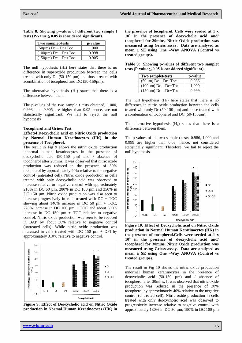
Tocopherol and Griess Test
Effectof Deoxycholic acid on Nitric Oxide production
by Normal Human Keratinocytes (HK) in the
presence of Tocopherol.
The result in Fig 9 shows the nitric oxide production
innormal human keratinocytes in the presence of
deoxycholic acid (50-150 µm) and / absence of
tocopherol after 20mins. It was observed that nitric oxide
production was reduced in the presence of 30%
tocopherol by approximately 40% relative to the negative
control (untreated cell). Nitric oxide production in cells
treated with only deoxycholic acid was observed to
increase relative to negative control with approximately
210% in DC 50 µm, 280% in DC 100 µm and 350% in
DC 150 µm. Nitric oxide production was also seen to
increase progressively in cells treated with DC + TOC
showing about 140% increase in DC 50 µm + TOC,
220% increase in DC 100 µm + TOC and about 300%
increase in DC 150 µm + TOC relative to negative
control. Nitric oxide production was seen to be reduced
in BAP by about 30% relative to negative control
(untreated cells). While nitric oxide production was
increased in cells treated with DC 150 µm + DPI by
approximately 310% relative to negative control.
Figure 9: Effect of Deoxycholic acid on Nitric Oxide
production in Normal Human Keratinocytes (HK) in
the presence of tocopherol. Cells were seeded at 1 x
104 in the presence of deoxycholic acid and/
tocopherol for 20mins, Nitric Oxide production was
measured using Griess assay. Data are analysed as
mean ± SE using One –Way ANOVA (Control vs
treated groups).
Table 9: Showing p-values of different two samplet
tests (P-value ≤ 0.05 is considered significant).
Two samplet-tests p-value
(50µm) Dc – Dc+Toc 0.986
(100µm) Dc – Dc+Toc 1.000
(150µm) Dc – Dc+Toc 0.999
The null hypothesis (H0) here states that there is no
difference in nitric oxide production between the cells
treated with only Dc (50-150 µm) and those treated with
a combination of tocopherol and DC (50-150µm).
The alternative hypothesis (H1) states that there is a
difference between them.
The p-values of the two sample t tests, 0.986, 1.000 and
0.999 are higher than 0.05, hence, not considered
statistically significant. Therefore, we fail to reject the
null hypothesis.
Figure 10; Effect of Deoxycholic acid on Nitric Oxide
production in Normal Human Keratinocytes (HK) in
the presence of tocopherol.Cells were seeded at 1 x
104 in the presence of deoxycholic acid and/
tocopherol for 30mins, Nitric Oxide production was
measured using Griess assay. Data are analysed as
mean ± SE using One –Way ANOVA (Control vs
treated groups).
The result in Fig 10 shows the nitric oxide production
innormal human keratinocytes in the presence of
deoxycholic acid (50-150 µm) and / absence of
tocopherol after 30mins. It was observed that nitric oxide
production was reduced in the presence of 30%
tocopherol by approximately 40% relative to the negative
control (untreated cell). Nitric oxide production in cells
treated with only deoxycholic acid was observed to
progressively increase relative to negative control with
approximately 130% in DC 50 µm, 190% in DC 100 µm

Eze et al. World Journal of Pharmaceutical and Medical Research
www.wjpmr.com
16
and 255% in DC 150 µm. Nitric oxide production was
also seen to increase progressively in cells treated with
DC + TOC showing about 80% increase in DC 50 µm +
TOC, 160% increase in DC 100 µm + TOC and about
220% increase in DC 150 µm + TOC relative to negative
control. Nitric oxide production was seen to be reduced
in BAP by about 60% relative to negative control
(untreated cells). While nitric oxide production was
increasedin cells treated with DC 150 µm + DPI by
approximately by approximately 235% relative to
negative control.
Table 10: Showing p-values of different two sample t
tests (P-value ≤ 0.05 is considered significant).
Two samplet-tests p-value
(50µm) Dc – Dc+Toc 0.998
(100µm) Dc – Dc+Toc 1.000
(150µm) Dc – Dc+Toc 0.999
The null hypothesis (H0) here states that there is no
difference in nitric oxide production between the cells
treated with only Dc (50-150 µm) and those treated with
a combination of tocopherol and DC (50-150µm).
The alternative hypothesis (H1) states that there is a
difference between them.
The p-values of the two sample t tests, 0.998, 1.000 and
0.999 are higher than 0.05, hence, not considered
statistically significant. Therefore, we fail to reject the
null hypothesis.
Tocopherol and Metabolic Test (MTT) Assay
Effectof Deoxycholic acid on the metabolism of
Normal Human Keratinocytes (HK) in the presence
of Tocopherol (1mg/ml).
The result in Fig. 11 shows the metabolic activity of
normal human keratinocytes (HK) in the
presencedeoxycholic acid (50µM-150µM) and / absence
of tocopherol after 24 hrs. It was observed that
metabolism of normal human keratinocytes was
reduced relative to negative control (untreated cells)
with approximately 52% in the presence of tocopherol,
51% in the presence of 10µM diphenyleneiodonium
chloride (DPI), 39% in the presence 50 µM
deoxycholicacid (DC), 22% in the presence of 100 µM
and 38% in the presence of DC 150µM. Further
reduction in metabolic activity of HK was observed in
the presence of both tocopherol and deoxycholic acid,
and deoxycholic acid (DC) + DPI (relative to untreated
cells).DC 50 µM + inulin showed approximately 73%
metabolic reduction, same with DC100 µM + inulin,
with further reduction of metabolic activity observed in
the presence of both DC 150 µM and inulin by
approximately 85% relative to negative control.
Metabolic activity in the presence of DPI +DC 50 µM -
100 µM were observed to be reduced further relative to
deoxycholic acid alone or DC +INU.
Figure 11: Effect of Deoxycholic acid on Metabolism
of Normal Human Keratinocytes (HK) in the
presence of tocopherol. Cells were seeded at 5 x 10⁴ in
the presence of deoxycholic acid and/ tocopherol for
24 hrs, cell metabolism was measured using MTT
assay. Data are analysed as mean ± SE using One –
Way ANOVA (Control vs treated groups).
Table 11: Showing p-values of different paired tests
(P-value ≤ 0.05 is considered significant).
Two samplet-tests p-value
(50µm) Dc – Dc+Toc 0.998
(100µm) Dc – Dc+Toc 0.993
(150µm) Dc – Dc+Toc 0.925
The null hypothesis (H0) here states that there is no
difference in the metabolism of cells treated with only
DC (50-150µm) and the metabolism of cells treated with
a combination of tocopherol and DC (50-150µm).
The alternative hypothesis (H1) here states that there is a
difference in the metabolism of the cells.
The p-values of the two sample t tests, 0.998, 0.993 and
0.925 are higher than 0.05 hence are not considered
statistically significant. We fail to reject the null
hypothesis.
Figure 12: Effect of Deoxycholic acid on Metabolism
of Normal Human Keratinocytes (HK) in the
presence of tocopherol. Cells were seeded at 5 x 10⁴ in
the presence of deoxycholic acid and/ tocopherol for
48 hrs, cell metabolism was measured using MTT
assay. Data are analysed as mean ± SE using One –
Way ANOVA (Control vs treated groups).

Eze et al. World Journal of Pharmaceutical and Medical Research
www.wjpmr.com
17
The result in Fig. 12 shows the metabolic activity of
normal human keratinocytes (HK) in the
presencedeoxycholic acid (50µM-150µM) and / absence
of tocopherol after 48 hrs. It was observed that
metabolism of normal human keratinocytes was
reduced relative to negative control (untreated cells)
with approximately 42% in the presence of tocopherol,
42% in the presence of 10µM diphenyleneiodonium
chloride (DPI), 45% in the presence 50 µM (DC), 48% in
the presence of 100 µM and 42% in the presence of DC
150µM. Further reduction in metabolic activity of HK
was observed in the presence of both 30% tocopherol
and deoxycholic acid, and deoxycholic acid (DC) + DPI
(relative to untreatedcells). DC 50 µM + tocopherol
showed approximately 30% metabolic reduction, 50%in
DC100 µM + tocopherol and about 29% in DC 150 µM
and tocopherol relative to negative control. Metabolic
activity in the presence of DPI +DC 50 µM - 100 µM
was observed to be reduced further relative to
deoxycholic acid alone or DC +TOC.
Table 12: showing p-values of different paired tests
(P-value ≤ 0.05 is considered significant).
Two samplet-tests p-value
(50µm) Dc – Dc+Toc 0.999
(100µm) Dc – Dc+Toc 1.000
(150µm) Dc – Dc+Toc 1.000
The null hypothesis (H0) here states that there is no
difference in the metabolism of cells treated with only
DC (50-150µm) and the metabolism of cells treated with
a combination of tocopherol and DC (50-150µm).
The alternative hypothesis (H1) here states that there is a
difference in the metabolism of the cells.
The p-values of the two sample t tests, 0.999, 1.000 and
1.000 are higher than 0.05 hence are not considered
statistically significant. We fail to reject the null
hypothesis.
DISCUSSION
Free radicals such as superoxide and nitric oxide which
moderately produced by the cell are known to promote
cell signalling that necessitate cell proliferation, cell
renewal, induce apoptosis by gene regulation and
enhance immune regulatory functions in response to
invading pathogens (Victor et al., 2003). However, when
produced in excess leads to harmful effects on cells
rather than the intended protective function.This study
focused on the impact of increased and sustained
production of superoxide and nitric oxide; It also
measured the cell metabolism which is reported to
increase with a corresponding increase in free radical
activity (Perez, 2009).
Nitric oxide (NO) is released by different types of cells
which include denritic cells, natural killer cells (NK),
phagocytes, mast cells, monocytes, macrophages,
neutrophils, vascular smooth muscle cells, fibroblasts,
Schwan cells, keratinocytes, chondrocytes, hepatocytes,
eosinophils (Victor et al., 2004). NO release is generated
by specific nitric oxide synthases which have different
isoforms, neuronal NOS (nNOS), Inducible NOS (iNOS)
and endothelial NOS (eNOS). While nNOS and eNOS
are constitutively expressed, iNOS is inducibly expressed
in macrophages following stimulation by cytokines, LPS
and other agents. NO carries out different physiologic
functions such as blood pressure regulation, smooth
muscle relaxation and inhibits the adhesion of platelets
and leukocytes to the endothelium. NO also down-
regulates endothelial adhesion of different adhesion
molecule families (VCAM-1, ICAM-1, E-selectin),
however, the extent to which it modulates them is
variable. iNOS also regulates leukocyte recruitment
during inflammatory responses. Induced NO production
mechanism has been reported to be of great significance
in macrophage cytotoxicity for tumour cells and
bacterias (Victor et al., 2004).
It has been reported that reactive nitrogen species
activates transcription nuclear factor κΒ (NF- κΒ) while
antioxidants tends to reduce its expression. Also
lipopolysaccharide (LPS) induced monocyte and
macrophages activation lead to the expression of
numerous mediators and inflammatory cytokines through
transcription factors such as activator protein-1 (AP-1)
and nuclear factor κΒ (NF- κΒ). (Fulihara et al.,2003).
Inducable nitric oxide (iNOS) expression is one of the
results of macrophage activation by LPS and
subsequently increases the transformation of L-arginine
to NO which is able to combine with superoxide (O2.-) to
form peroxynitrite (ONOO-) (Cals-Grierson and
Ormerod, 2004). Peroxynitrite is a cytotoxic oxidant,
which NO toxicity is predominantly linked to and is
capable of causing DNA fragmentation and lipid
oxidation (Victor et al., 2004). Lipid peroxidation leads
to oxidation of DNA and protein along the membrane
leading to change in membrane permeability and
modification of protein structure. Oxidative damage to
mitochondrial membrane can also occur leading to
membrane depolarization and uncoupling of oxidative
phosphorylation with corresponding change in cellular
respiration. This can result in the mitochondrial damage
accompanied with the release of cytochrome c, the
activation of caspases and eventual apoptosis (Victor et
al., 2004). NO dependent apoptosis has also been
reported to be associated with decrease in cardiolipin
concentration, reduced mitochondrial chain activity and
release of mitochondrial cytochrome c into the cytosol.
Increased resistance to NO-induced apoptosis has been
reported to be associated high level of intracellular
glutathione (Droge. 2002).
As earlier mentioned, antioxidants are pivotal in the
maintenance of redox balance and function by either
eliminating free radical precursors or by preventing
catalysis, e.g., glutathione peroxidise or reacting with the
reactive oxygen produced either to remove them or

Eze et al. World Journal of Pharmaceutical and Medical Research
www.wjpmr.com
18
inhibit them, e.g., vitamin E. This experiment employed
the use of α-Tocopherol (fat soluble vitamin E), whose
antioxidant ability has been reasonably established by
different studies (Perez, 2009; Perse, 2013) in
comparison to inulin whose antioxidant ability is
currently widely investigated. Fat soluble vitamin E (α-
tocopherol) is the most important antioxidant in the
hydrophobic lipid interior of cell membranes as they help
to prevent membrane polyunsaturated fatty acids from
undergoing lipid peroxidation which leads to the loss of
cell membrane integrity (Bostick, 2015 ; Victor et al.,
2004). Studies have shown that α-tocopherol is able to
inhibit activation of NF- κΒ (associated with IBD)
produced by LPS which could lead to subsequent
decrease in TNFα. In the cause of neutrophil activation
where reactive oxygen species are seen to be present in
the extracellular matrix, the plasma and red cell
components of the blood act as antioxidants; superoxide
is inactivated by the copper-zinc SOD-dependent
pathway present in the red blood cell.Apotransferrin,
lactoferrin and ceruloplasmin are examples of metal-
binding plasma proteins that function as important
antioxidants in addition to their transport roles (Victor et
al.,2004). Inulin is reported to have the ability to release
short chain fatty acids (SCFAs) that in turn reduce the
luminal pH thereby inhibiting the growth and adhesion
of pathogen. Via this pathway, inulin is able to prevent
the activation of the host immune response that would
have led to the increase in protein expressions through
the resulting adhesion molecules (Fujihara et al., 2003).
The p-values from Table 3.1.1 showed there was a
significant difference in superoxide production between
the cells treated with only DC 50 -150 µm and the cells
treated with DC 50 -150 µm + INU after 20 mins
incubation. This confirmed that deoxycholic acid which
is a well established free radical stimulator (Perez, 2009)
was able to stimulate superoxide production in the
normal human keratinocytes used in amounts higher
relative to the negative control. However the inhibitory
activity of inulin in the cells treated with DC 50 µm +
INU is questioned (Fig 3.1.1) as superoxide production is
seen to be more in the cells treated with DC 50 µm +
INU than the cells treated with DC 50 µm alone. It can
be said that inulin inhibited superoxide production in
cells treated with DC 100 – 150 µm (Fig 3.1.1).
However, further analysis would have to be carried out
to confirm this. In contrast, after 30 mins incubation, the
superoxide production was seen to be lower relative to
the negative control (untreated cells) (Fig 3.1.2) and the
superoxide production was seen to reduce progressively
in cells treated with only deoxycholic acid as the
concentration increases. The p-values (Table 3.1.2)
showed that there was a significant difference in
superoxide production in cells treated with only DC 50 -
150 µm and the cells treated with both DC 50 -150 µm +
INU. The decrease in superoxide production in cells
treated with only DC 50 -150 µm contradicts the result
obtained in Fig 3.1.1. More so, because it is lower
relative to the negative control it can be said that it was
not a positive significance. Data from the results showed
that there was a significant difference in nitric oxide
production between the cells treated with only DC 100 –
150 µm and the cells treated with a combination of DC
100 – 150 µm + INU (Table 3.2.1 and Table 3.2.2).
However the cells treated with a combination of DC 100
– 150 µm + INU were observed to have more nitric
oxide production, hence, inferring reduced inhibitory
activity of inulin. The data from this experiment also
showed that there was no significant metabolic activity
between the cells treated with only DC 100 – 150 µm
and the cells treated with a combination of DC 100 – 150
µm + INU (Table 3.3.1 and Table 3.3.2).Again, this
inconsistency is to be questioned because it has been
reported that cell metabolism increases with increase in
deoxycholic concentration but Fig 3.3.1 and Fig 3.3.1
depict otherwise (Monte, 2009 ; Ajouz and Shamseddine,
2014).
Data resulting from the experiments using tocopherol as
the antioxidant showed no significant difference in
superoxide production, nitric oxide production and cell
metabolism between the cells treated with only DC 100 –
150 µm and the cells treated with a combination of DC
100 – 150 µm + TOC (Table 3.2.1 and Table 3.2.2).
These results infers that there was an error during this
experiment because a wide range of journals have
established the antioxidant ability of α-tocopherol hence
a significant difference was meant to be observed using
α-tocopherol.
CONCLUSION
The data resulting from this experiment is not enough to
compare the antioxidant activity of inulin and α-
tocopherol. Various reasons could have resulted to the
inconsistent data, such as the amount (3mg/ml) of inulin
and tocopherol used (1mg/ml). Perhaps, a higher
concentration could have resulted in more significant
result. Also, there is a possibility that the timing of the
incubation for the NBT and griess assays could have
yielded better data if they were incubated longer. ELISA PLUS
assay would have also helped to quantitatively
detect the presence of any apoptotic cell death. In future,
western blot can be used to analyse proteins that are
upregulated during ROS activities such as E-cadherin
(upregulated in colon cancer), since the expression of
cell adhesion molecules have been correlated with cell
growth, cell differentiation and wound repair and are said
to be stimulated by tissue necrotic factor (TNF), LPS,
ROS, and interleukin-1α. Also, ROS activity can further
be determined by measuring the proteolytic degradation
of NF-κB inhibitor IκB after ROS exposure. The LPS
receptor complex plays a vital role in sensoring and
mediating the response to LPS. Advance knowledge has
been made in identifying the molecules of the LPS
receptor complex which is composed of three proteins –
CD14,Toll-like receptor 4 (TLR4), and myeloid
differentiation protein-2.The need for establishment of
biomarkers to measure oxidative stress invivo cannot be
overlooked. This would immensely assist in early

Eze et al. World Journal of Pharmaceutical and Medical Research
www.wjpmr.com
19
discovery of disease, diagnose the progression of disease,
and evaluate the effectiveness of treatment with
antioxidants or functional foods. Lipid peroxidation
products may serve as useful biomarkers, since the major
reaction caused by oxidative stress is lipid peroxidation
and is assumed to play a pivotal role in the pathogenesis
and progression of many diseases despite the fact that
they are non specific biomarkers.Furthermore, some lipid
peroxidation products are chemically reactive and react
with proteins, sugars and DNA to form stable adducts
that are biologically specific to various protein.
REFERENCES
1. Ajouz, H., Mukherji, D. and Shamseddine, A.
Secondary bile acids: an underrecognized cause of
colon cancer. World. J. Surg. Onc, 2014; 12(1): 164.
2. Apajalahti, J. Comparative Gut Microflora,
Metabolic Challenges, and Potential Opportunities.
The Journal of Applied Poultry Research, 2005;
14(2): 444-453.
3. Blaise, G., Gauvin, D., Gangal, M. and Authier, S.
Nitric oxide, cell signaling and cell death.
Toxicology, 2005; 208(2): 177-192.
4. Bostick, RM, (2015). Reduced risk of colon cancer
with high intake of vitamin E: the Iowa Women's
Health Study. - PubMed - NCBI. [online]
Ncbi.nlm.nih.gov. Available at:
http://www.ncbi.nlm.nih.gov/pubmed/8364919
5. Cals-Grierson, M. and Ormerod, A. Nitric oxide
function in the skin', Nitric Oxide, 2004; 10(4): 179-
193.
6. Davies, M. The oxidative environment and protein
damage.Biochimica et Biophysica Acta (BBA) -
Proteins and Proteomics, 2005; 1703(2): 93-109.
7. De Plaen, I., Han, X., Liu, X., Hsueh, W., Ghosh, S.
and May, M. Lipopolysaccharide induces
CXCL2/macrophage inflammatory protein-2 gene
expression in enterocytes via NF-kappaB activation:
independence from endogenous TNF-alpha and
platelet-activating factor. Immunology, 2006;
118(2): 153-163.
8. Dröge, W. Free Radicals in the Physiological
Control of Cell Function'. Physiol Rev, 2002; 82(1):
47-95.
9. Fujihara, M., Muroi, M., Tanamoto, K., Suzuki, T.,
Azuma, H. and Ikeda, H. Molecular mechanisms of
macrophage activation and deactivation by
lipopolysaccharide: roles of the receptor complex',
Pharmacology & Therapeutics, 2003; 100(2): 171-
194.
10. Meyer, D. and Stasse-Wolthuis, M. The bifidogenic
effect of inulin and oligofructose and its
consequences for gut health.European Journal of
Clinical Nutrition, 2009; 63(11): 1277-1289.
11. Monte, M. Bile acids: Chemistry, physiology, and
pathophysiology. World Journal of
Gastroenterology, 2009; 15(7): 804.
12. Niki, E. Lipid peroxidation: Physiological levels and
dual biological effects', Free Radical Biology and
Medicine, 2009; 47(5): 469-484.
13. Perez, M. Bile-acid-induced cell injury and
protection. World Journal of Gastroenterology,
2009; 15(14): 1677.
14. Perše, M. Oxidative Stress in the Pathogenesis of
Colorectal Cancer: Cause or Consequence? BioMed
Research International, 2013: 1-9.
15. Sim Choi, H., Woo Kim, J., Cha, Y. and Kim, C. A
Quantitative Nitroblue Tetrazolium Assay for
Determining Intracellular Superoxide Anion
Production in Phagocytic Cells. Journal of
Immunoassay and Immunochemistry, 2006; 27(1):
31-44.
16. Tsikas, D. Analysis of nitrite and nitrate in
biological fluids by assays based on the Griess
reaction: Appraisal of the Griess reaction in the l-
arginine/nitric oxide area of research. Journal of
Chromatography B, 2007; 851(1-2): 51-70.
17. Van den Ende, W., Peshev, D. and De Gara, L.
Disease prevention by natural antioxidants and
prebiotics acting as ROS scavengers in the
gastrointestinal tract', Trends in Food Science &
Technology, 2011; 22(12): 689-697.
18. Velayutham, M., Hemann, C. and Zweier, J.
'Removal of H2O2 and generation of superoxide
radical: Role of cytochrome c and NADH', Free
Radical Biology and Medicine, 2011; 51(1): 160-
170. doi: 10.1016/j.freeradbiomed.2011.04.007.
19. Victor, V., Rocha, M. and De la Fuente, M. Immune
cells: free radicals and antioxidants in sepsis',
International Immunopharmacology, 2004; 4(3):
327-347.
20. Wasilewski, A., Zielińska, M., Storr, M. and Fichna,
J. Beneficial Effects of Probiotics, Prebiotics,
Synbiotics, and Psychobiotics in Inflammatory
Bowel Disease', Inflammatory Bowel Diseases,
2015; 21(7): 1674-1682.