Embed Size (px)
Citation preview

COMPARATIVE STUDY ON ESR SPECTRA OF CARBONATES
E. D. SELEÞCHI1, O. G. DULIU2
1 University of Bucharest, Faculty of Physics, Romania2 University of Bucharest, Department of Atomic and Nuclear Physics, Mãgurele,
P.O. Box, MG-11, RO-077125, Bucharest, Romania
Received September 12, 2006
This paper is a comparative study on Electron Spin Resonance (ESR) spectraof carbonates such as mollusk shells collected from the Black Sea Coast (Romania),Mediterranean Coast (Cyprus), Tyrrhenian Sea Coast (Italy) and Atlantic Coast(Mauritania). Additionally we have been analyzed the ESR spectra of limestone,marbles, stalactites and corals. Some of the carbonate samples revealed the samecharacteristic Mn2+ spectrum with an additional free radical peak in the centre ofsextet, at a g value in the range of 2.0006–2.0062. The differences into the ESR spectraof carbonates have been observed depending on their species, geographical origin,harvesting year, geologically age and dose. The gamma-irradiated samples exhibited
a complex spectrum consisting of 2CO ,− 33CO ,− 3CO ,− 2SO− and 3SO− species.
1. INTRODUCTION
Electron Spin Resonance (ESR) represent a specialized spectroscopic methodof investigation restricted only to atomic and molecular paramagnetic centers(PC) that contains at last one unpaired electron. Beside different kind of PC,transition elements ions or radiation induced free radicals are relatively frequentlyfound in environmental samples. Concerning the last ones, it is worth to mentionthat their concentration monotonously increases with absorbed dose, and, if theyare trapped in insulators, their lifetime could reach thousands of years.
Among an astonishing variety of compounds able to retain for a long timeradiation free radicals, natural carbonates play a significant role mainly due totheir occurrence in multitude varieties, beginning with natural limestone andmarble and finishing with invertebrate exo-skeleton. For this reason and for arelative simplicity of their crystalline lattices, natural carbonates were intensivelyinvestigated by means of ESR.
Some ESR spectra of unirradiated carbonates consists of six, double linesplus ten single ones between the doublets, the other samples show the same
Paper presented at the 7th International Balkan Workshop on Applied Physics, 5–7 July2006, Constanþa, Romania.
Rom. Journ. Phys., Vol. 52, Nos. 5–7 , P. 657–666, Bucharest, 2007
658 E. D. Seleţchi, O. G. Duliu 2
characteristic Mn2+ spectrum with an additional free radical peak in the centre ofsextet, whereas a lot of specimens exhibit a single line. Some differences into theESR spectra obtained before and after irradiation processing of CaCO3 con-taining hard tissues have been observed depending on their species, geographicalorigin, harvesting year, geologically age, dose, etc. [4, 23, 24].
Elementary defects induced by ionizing radiation: an “electron center”33CO − with 25 electrons and a “hole center”, 3CO ,− with 23 electrons are stable
only at low temperatures. Another “electron center” more stable than 33CO − and
also formed by radiation is the 2CO− molecular ion. For natural calcites, the
“electron center”, 2SO− (g = 2.0057) and the “hole center”, 3SO− (g = 2.0036)from impurities result in additional signals [8].
Many studies show that the ESR amplitude of free radicals enhancesexponentially with the additive dose:
00
( ) 1 expSDI D I ID
⎡ ⎤⎛ ⎞= + − −⎜ ⎟⎢ ⎥⎝ ⎠⎣ ⎦ (1)
where I0 and IS are the intensities corresponding to the unirradiated spectrum andto the saturation level, D is the artificial radiation dose and D0 is the dosecorresponding to the IS(1 – e–1) value (0.63 of the maximum intensity), howeversome experimental indicates a linearly increase of ESR signal intensity with theadded dose [5, 9, 14].
A wide range of carbonates including bivalve and gastropod shells collec-ted from the Black Sea Coast (Romania), Tyrrhenian Sea Coast (Italy), Mediter-ranean Coast (Cyprus) and Atlantic Coast (Mauritania), corals, limestone,stalactites and marbles were subjected to an ESR study in order to analyze theirspectral feature.
2. EXPERIMENTAL
The ESR spectra of carbonate samples were performed at ambientconditions by using an X-band JEOL JES MX 3X spectrometer provided with aTE011 cylindrical resonant cavity and 100 kHz modulation frequency. Thesamples in powder form were placed separately into quartz tubes with a 3 mminner tube diameter. The position of tube in the cavity was not changed duringthe whole experiment in order to ensure the uniform and reproduciblemeasurements of the sample. The optimal conditions of ESR measurement were:microwave frequency 9.50 GHz, modulation amplitude 1–2 G, microwave power4 mW, sweep time 5 min and response time 0.3 s. The g factors of the observed

3 Comparative study on ESR spectra of carbonates 659
signals have been determined by using a 2.2-diphenyl-1-picryl-hydrazil (DPPH)as well as CaO:Mn2+.
3. ESR STUDY OF MOLLUSK SHELLS
Mollusk shells are composed mainly of 95–99 % crystalline form ofcalcium carbonate (CaCO3), mostly aragonite (orthorhombic, metastable) and/orcalcite (trigonal/ rhombohedric, stable) and an organic part of the protein called“conchiolin”. The bivalve and gastropd shells are usually formed of threedistinct layers: the outermost thin layer (periostracum), the middle layer(ostracum or prismatic layer) is the thickest and the innermost layer (hypo-stracum, nacreous layer or mother-of-pearl) consisting of stacks of aragonite and/or amorphous CaCO3 tablets, approximately 0.5–1 μm thick, separated fromtheir neighbors by their organic membranes [21]. The presence of ESR signals inperiostracum and nacreous layers at g ≈ 2.0 and g ≈ 4.1 was attributed to Fe3+
ions [16]. Recent researches show the osteogenic properties of nacre, as a bio-material for bone implants.
Since the levels of carbon dioxide in the sea have increased, making thewater more acidic, the sea snails and the other marine organisms have found itharder to secrete CaCO3 in order to build their shells. As the pH of sea waterdecreases, it is not clear how it chemistry will change or how ecosystems willadapt. The effect of the petroleum pollution on the lipid composition of someorganisms is well studied. In gastropods the Diesel fuel treatment causedinsignificant effects in the fatty acid composition of lipids since these changesappear to be stronger in the evolutionary less advanced organisms [18]. It isgenerally assumed that the external coloration, pigmentation pattern andcomposition of fossil shells are determined by genetic mechanism and by thephysical/ chemical environment in which the mollusk lived [28]. Thepigmentation pattern is directly correlated to the mechanisms that control thegrowth of the shell in order to achieve the optimal shape [1]. The analysis ofnacre texture provides some information about the nature of shell growth. Fibertextures appear only in gastropods and cephalopods although the crystallo-graphic features of their nacre tablets are different [3]. The growth of shellcrystals is initiated from a mineralizing solution by the extra-cellular matrix(ECM). One of these theory reveals that the organic matrix (OM) may controlcrystal nucleation, polymorph selection and crystal orientation [12] and analternative assumption shows that a class of granulocytic hemocytes would bedirectly involved in shell crystal production [15, 20].
Acidic proteins control mineral formation and crystal growth [7]. Themajor role of phosphoproteins in the regulation of calcium carbonate biomine-ralization has been also studied [2]. The ferritin, the principal protein for iron

660 E. D. Seleţchi, O. G. Duliu 4
storage is also involved in shell formation [29]. The most acidic of all themollusk shell matrix proteins, Aspein is responsible for directed formation ofcalcite in the shell [27]. Another molecule involved in the calcification process,mucoperlin, is a protein of the nacreous layer, with some mucin-like charac-teristics, able to interact with Ca2+ and/or to interfere with CaCO3 precipitation[13]. Its primary role may be to finish the crystal growth and to combinecrystals. Lustrin isolated from the nacreous layer with a highly modularstructures with Gly-, Ser-, Cys- and Pro- rich domains protects the OM againstdegradations and ensures elasticity to the shell [22].
The analysis of chemical constituents of the shell (SiO2, Al2O3, Fe2O3,MgO, CaO, SO3, Cl, Na, K, Sr, etc.) indicates that the content of CaO for theshells is the same as for the limestone. However, the sea shells contain a largeamount of alkali metals and chlorine. The desulphurization characteristics of thesea shells have been studied by using a bubbling fluidized bed coal combustor[17]. In the CaCO3 crystal structure, the divalent trace elements (alkaline earthand transition elements) occupy regularly a lattice site and the monovalent traceelements (alkali metals) are non-selectively distributed into the shell asinclusions of carbonates and/or amorphous compounds [19].
Unirradiated shells from a large variety of bivalve and gastropod specieswere subjected to ESR measurements in order to emphasize the great differencesbetween their spectra (Fig. 1).
The ESR spectra of Mytilus galloprovincialis, Moerella tenuis andChamelea gallina bivalve shells consist mainly of a group of six lines of Mn2+
(Fig. 1a, b, d). An additional free radical peak identified as the radiation-induced
defects 2(CO ,− 33CO ,− 3CO− and 2SO )− stabilized by impurities at a g = 2.0059
was observed at the center of this group for Mytilus galloprovincialis specie. Forthe other samples, no characteristic manganese signals or weak intensity of Mn2+
hf lines were observed while the central signal presents a lot of possible forms:no line (Strombus sp. – Fig. 1e), a single one at a g = 2.0062 (Cardium sp. –Fig. 1c), doublets at g1 = 2.0026 and g2 = 2.0053 (Conus sp. – Fig. 1f) dependingof the shell varieties, but also may be from their compositions. A very intensesignal characterized by gyromagnetic factors: g1 = 2.0006 and g2 = 2.0053 wasnoticed in the case of Murex sp.
The g spectroscopic splitting factor of unknown signal was determinedusing the standard signal with a known g0 factor:
( )00
1 Hg gHΔ= − (2)
The magnetic field separation ΔH between the third and fourth line and theresonance field H0 were considered 8.69 mT and 339 mT respectively at theX-band frequency.

5 Comparative study on ESR spectra of carbonates 661
Fig. 2 – ESR spectra of unirradiated samples: a) Mytilus galloprovincialis, b)Moerella tenuis, c) Cardium sp. d) Chamelea gallina, e) Strombus sp.,
f) Conus sp., g) Murex sp.
4. ESR STUDY OF CORALS
Coral reefs have the potential to provide information about coral growthand climate over the past several centuries. According to the von Bertalanffyfunction we can write:
( )1 e k ttL L −
∞= − (3)
Lt is individual length at age t, L∞ is asymptotic length (maximum expectedlength, K is a growth constant, and t is individual age [6].
662 E. D. Seleţchi, O. G. Duliu 6
Annual skeletal density bands and the growth rate of corals (in the range4–20 mm/year) are correlated with the light level and water temperature.
The high precision of the 230Th/234U method for dating a closed system(there are no other changes to isotopic ratios other than that caused byradioactive decay) such as young corals is well established. The activity ratio234U/238U and the concentration of uranium (2–4 ppm) in corals are very close tothe value typical of seawater in contrast to shells which contain a much reducedlevel of uranium in the range 0.001–0.6 ppm [26].
The determination of Sr/Ca, U/Ca and Mg/Ca ratios in fossils corals can alsoprovide information on past sea surface temperatures [10]. Stable oxygen isotoperatios 18δO provide a record of sea surface temperature and surface salinity [11].
The variation in 18δO of coral skeletal reflects variation in the growth ratewhile carbon isotope ratios 13δC showed significantly inverse correlation withlinear growth rates. In shallow faster-growing corals, oxygen and carbon isotopicratios reveal out-of-phase annual fluctuations, whereas in deep slower growingcorals oxygen and carbon isotope fluctuations are in phase [25].
The impurities-associated signals such as 2SO− and 3SO− are useful forassessing the historical environmental change and the ESR signal of g = 2.0007is the most suitable one for dating corals.
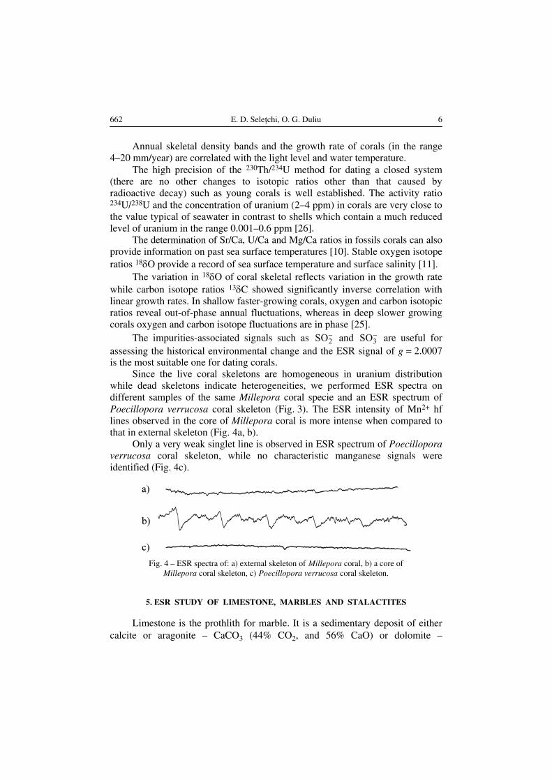
Since the live coral skeletons are homogeneous in uranium distributionwhile dead skeletons indicate heterogeneities, we performed ESR spectra ondifferent samples of the same Millepora coral specie and an ESR spectrum ofPoecillopora verrucosa coral skeleton (Fig. 3). The ESR intensity of Mn2+ hflines observed in the core of Millepora coral is more intense when compared tothat in external skeleton (Fig. 4a, b).
Only a very weak singlet line is observed in ESR spectrum of Poecilloporaverrucosa coral skeleton, while no characteristic manganese signals wereidentified (Fig. 4c).
Fig. 4 – ESR spectra of: a) external skeleton of Millepora coral, b) a core ofMillepora coral skeleton, c) Poecillopora verrucosa coral skeleton.
5. ESR STUDY OF LIMESTONE, MARBLES AND STALACTITES
Limestone is the prothlith for marble. It is a sedimentary deposit of eithercalcite or aragonite – CaCO3 (44% CO2, and 56% CaO) or dolomite –

7 Comparative study on ESR spectra of carbonates 663
CaMg(CO3)2 ( 47.9% CO2, 30.4% CaO and 21.7% MgO) or a mixture of both.The pure white marbles have none or only trace amounts of accessory mineralssuch as: iron oxides and sulphides, graphite, chlorite, etc.
The Mn2+ sextet is clearly visible in ESR spectra of limestone and marble.An additional line between the third and fourth Mn2+ lines at a g = 2.0060 isobserved, in the case of a limestone sample from Kouriou Temple – Cyprus(Fig. 5a, and Fig. 6a, b).
Fig. 6 – ESR spectra of a limestone sample from Kouriou Temple – Cyprus:(a) δB = 2G, (b) δB = 10G.
The ESR spectrum of marble from Ruşchiţa – Romania exhibits the wellknown Mn2+ peaks. It consists of six double lines plus 10 single ones betweenthe doublets. The absence of free radicals in the center of Mn2+ sextet indicatesthe purity of marble (Fig. 7).
Fig. 7 – ESR spectrum of marble (Ruºchiþa – Romania).
Stalactite is a type of speleothems form from the deposition of calciumcarbonate and other minerals, which is precipitated from mineralized watersolutions:
CaCO3 + H2O + CO2 ⇔ Ca(HCO3)2 (4)
The stalactites contain brown rings due to clay minerals. The red andorange coloration of speleothems is due to the iron oxide, the blue hue derivesfrom manganese and the dark brown is related to the limonite that is dissolvedfrom the overlaying rocks. The relatively transparent pure stalactite illustrated inFig. 5.b. reveals a high growth rate inside the cave. The amplitude of Mn2+ lines
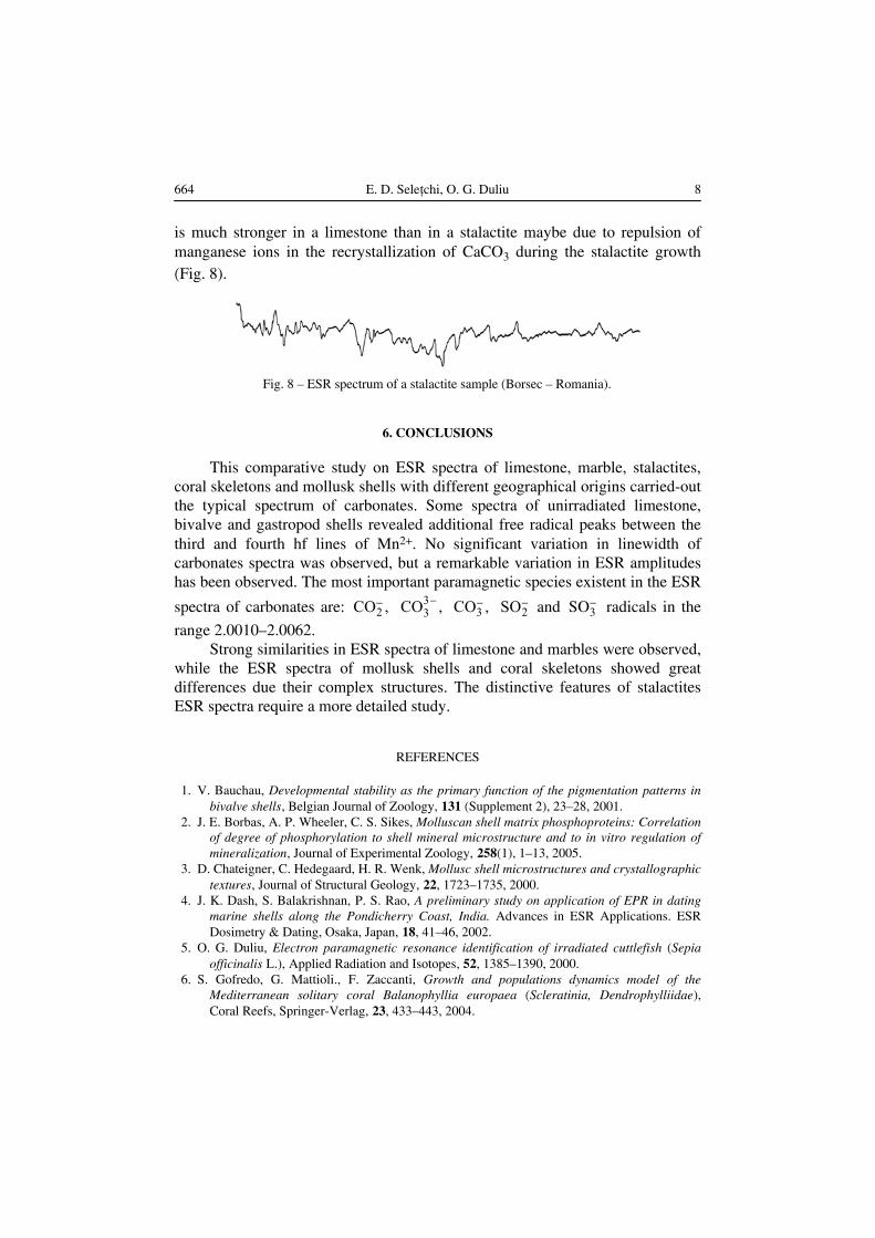
664 E. D. Seleţchi, O. G. Duliu 8
is much stronger in a limestone than in a stalactite maybe due to repulsion ofmanganese ions in the recrystallization of CaCO3 during the stalactite growth(Fig. 8).
Fig. 8 – ESR spectrum of a stalactite sample (Borsec – Romania).
6. CONCLUSIONS
This comparative study on ESR spectra of limestone, marble, stalactites,coral skeletons and mollusk shells with different geographical origins carried-outthe typical spectrum of carbonates. Some spectra of unirradiated limestone,bivalve and gastropod shells revealed additional free radical peaks between thethird and fourth hf lines of Mn2+. No significant variation in linewidth ofcarbonates spectra was observed, but a remarkable variation in ESR amplitudeshas been observed. The most important paramagnetic species existent in the ESR
spectra of carbonates are: 2CO ,− 33CO ,− 3CO ,− 2SO− and 3SO− radicals in the
range 2.0010–2.0062.Strong similarities in ESR spectra of limestone and marbles were observed,
while the ESR spectra of mollusk shells and coral skeletons showed greatdifferences due their complex structures. The distinctive features of stalactitesESR spectra require a more detailed study.
REFERENCES
1. V. Bauchau, Developmental stability as the primary function of the pigmentation patterns inbivalve shells, Belgian Journal of Zoology, 131 (Supplement 2), 23–28, 2001.
2. J. E. Borbas, A. P. Wheeler, C. S. Sikes, Molluscan shell matrix phosphoproteins: Correlationof degree of phosphorylation to shell mineral microstructure and to in vitro regulation ofmineralization, Journal of Experimental Zoology, 258(1), 1–13, 2005.
3. D. Chateigner, C. Hedegaard, H. R. Wenk, Mollusc shell microstructures and crystallographictextures, Journal of Structural Geology, 22, 1723–1735, 2000.
4. J. K. Dash, S. Balakrishnan, P. S. Rao, A preliminary study on application of EPR in datingmarine shells along the Pondicherry Coast, India. Advances in ESR Applications. ESRDosimetry & Dating, Osaka, Japan, 18, 41–46, 2002.
5. O. G. Duliu, Electron paramagnetic resonance identification of irradiated cuttlefish (Sepiaofficinalis L.), Applied Radiation and Isotopes, 52, 1385–1390, 2000.
6. S. Gofredo, G. Mattioli., F. Zaccanti, Growth and populations dynamics model of theMediterranean solitary coral Balanophyllia europaea (Scleratinia, Dendrophylliidae),Coral Reefs, Springer-Verlag, 23, 433–443, 2004.
9 Comparative study on ESR spectra of carbonates 665
7. B. A. Gotliv, L. Addadi, S. Weiner, Mollusk shell acidic proteins: In search of individualfunctions, Chem. BioChem., 4(6), 522–529, 2003.
8. M. Ikeya, New applications of Electron Spin Resonance – Dating, Dosimetry and Microscopy,World Scientific, Singapore, New Jersey, London, Hong Kong, 1–420, 1993.
9. A. Kinoshita, A. Brunetti, W. E. P. Avelar, F. L. M. Mantelatto, M. G. Simões, A. Fransozo,O. Baffa, Dating of sub fossil shell by ESR and K band spectrum for paramagnetic speciesassignment, Advances in ESR Applications, The Society of ESR Applied metrology, Osaka,Japan, 18, 27–29, 2002.
10. F. Le Cornec, T. Correge, Determination of Uranium to Calcium and Strontium to CalciumRatios in Corals by Inductively Coupled Plasma Mass Spectrometry, JAAS, 12, 969–973,1997.
11. B. K. Linsley, R. G. Messier, R. B. Dunbar, Assessing between-colony oxygen isotope varia-bility in the coral Porites lobata at Clipperton Atoll, Coral Reefs, Springer-Verlag, 18, 13–27, 1999.
12. F. Manoli, E. Dalas, Calcium carbonate crystallization on xiphoid of the cuttlefish, Journal ofCrystal Growth, 217, 422–428, 2000.
13. F. Marin, P. Corstjens, B. de Gaulejac, E. De-Vrind-De-Jong, P. Westbroek, Mucins andmolluscan calcification, J. Biol. Chem., 275(27), 20667–20675, 2000.
14. M. Miyahara, T. Nagasawa, S. Akiyama, Y. Kobayashi, T. Mashimizu, T. Maitani, ESRmethod for the detection of irradiated unboned meats and seafood, Journal of HealthScience, 50(5), 542–544, 2004.
15. A. S. Mount, A. P. Wheeler, R. P. Paradkar, D. Snider, Hemocyte-mediated shell minera-lization in the eastern oyster, Science, 304(5668), 297–300, 2004.
16. K. V. Narasimhulu, J. L. Rao, EPR and IR spectral studies of the sea water mussel Mytilusconradinus shells, Spectrochimica Acta A 56, 1345–1353, 1999.
17. I. Naruse, K. Saito, T. Murakami, Application of wasted sea-shell to desulfurizer in fluidizedbed coal combustion, Proceedings of the 15th International Conference on Fluidized BedCombustion, Savannah, Georgia. Paper No. FBC99–0088, 1999.
18. J. T. Nechev, S. V. Khotimchenko, A. P. Ivanova, K. L. Stefanov, S. D. Dimitrova-Konaklieva, S. Andreev, S. S. Popov, Effect of Diesel fuel pollution on the lipid compo-sition of some wide-spread Black Sea algae and invertebrates, Verlag der Zeitschrift fürNaturforschung, Tübingen, 57c, 339–343, 2002.
19. N. Onuma F. Masuda, M. Hirano, K. Wada, Crystal structure control on trace elementpartition in molluscan shell formation, Geochemical Journal, 13, 187–189, 1979.
20. L. Pramatarova E. Pecheva, R. Presker, M. T. Pham, M. F. Maitz, M. Stutzmann,Hydroxyapatite growth induced by native extracellular matrix deposition on solid surfaces,European Cells and Materials, 9, 9–12, 2005.
21. C. P. L. Prasuna, K. V. Narasimhulu, N. O. Gopal, J. L. Rao, T. V. R. K. Rao, Themicrostructures of biomineralized surfaces: a spectroscopic study on the exoskeletons offresh water (Apple) snail, Pila globosa, Spectrochimica Acta, A 60, 2305–2314, 2004.
22. T. Samata, Recent advances in studies on nacreous layer biomineralization. Molecular andcellular aspects, Thalassas, International Journal of Marine Sciences, 20(1), 25–44, 2004.
23. T. S. Shih, H. Sato, M. Ikeya, P. M. Liew, S. H. Chien, Conditions and new extrapolationmethod for ESR dating of aragonitic mollusk shells in Taiwan, Advances in ESRApplications, ESR Dosimetry & Dating, Osaka, Japan, 18, 31–39, 2002.
24. E. M. Stewart, D. J. Kilpatrick, An International Collaborative Blind Trial on Electron SpinResonance (ESR) Identification of Irradiated Crustacea, J. Sci. Food Agric., Great Britain,74, 473–484, 1997.
25. A. Suzuki, T. Omata, H. Kawahata, Intercolony variability of skeletal oxygen and carbonisotope ratios of corals: temperature-controlled tank experiment and field observation,AGU Fall Meet., 86(52), 2005.

666 E. D. Seleţchi, O. G. Duliu 10
26. P. Szeffer, S. Ostrowski, On the Occurrence of Uranium and Thorium in the Biosphere ofnatural waters, Part. 2, Uranium and Thorium in Corals, Molluscs and Fish, Oceanologia,13, 45–47, 1981.
27. D. Tsukamoto, I. Sarashina, K. Endo, Structure and expression of an unusually acidic matrixprotein of pearl oyster shells, Biochemical and Biophysical Research Communications,320(4), 1175–1180, 2004.
28. F. M. Winkler, B. F. Estevez, L. B. Jollan, J. P. Garrido, Inheritance of the general shell colorin the scallop Argopecten purpuratus (Bivalvia: Pectinidae), J. Hered. 92(6), 521–525, 2001.
29. Y. Zhang, Q. Meng, T. Jiang, H. Wang, L. Xie, R. Zhang, A novel ferritin subunit involved inshell formation from the pearl oyster (Pinctada fucata), Comp. Biochem. Physiol. B,Biochem. Mol. Biol. 135(1), 43–54, 2003.

Fig. 1 – Bivalve and gastropod shells collected from:
Black Sea Coast, Romania: a) , b) , c) sp.
Tyrrhenian Sea Coast, Italy: d) ,
Mediterranean Sea Coast, Cyprus: e) sp.
Atlantic Coast, Mauritania: f) sp., e) sp
Mytilus galloprovincialis Moerella tenuis Cardium
Chamelea gallina
Strombus
Conus Murex .

Fig. 3 – Coral skeletons – Philippines:
a) sp., b)Millepora Poecillopora verrucosa.

Fig. 5 – a) Limestone and marbles found at Kouriou Temple (Cyprus), b) A white stalactite collected
from Borsec (Romania).