Embed Size (px)
Citation preview
practices
t h i s a rt i c l e i s r e p r i n t e d f ro m t h e j o u r n a l o f wo u n d c a r e vo l 2 4 , n o 3 , m a r c h 2 0 1 5
© 2
01
5 M
A H
eA
lt
Hc
Ar
e l
td
Comparative study of two antimicrobial dressings in infected leg ulcers: a pilot study
leg ulcers; infection; bacterial load; antimicrobial dressing; efficacy; skin allograft
G. Mosti, md, head angiology department;a. Magliaro, md, consultant dermatologist;V. Mattaliano, md, consultant vascular surgeon;P. Picerni, md, senior registrar;n. angelotti, md, consultant dermatologist;all at angiology department, Barbantini hospital, lucca, italy via del calcio 2, 55100 lucca, italy
e-mail: [email protected]
l objective: the aim of the study was to compare the efficacy of a microorganism-binding (mB) dressing with a silver-containing hydrofiber (sch) dressing in controlling the bacterial loads of heavily colonised or locally infected chronic venous leg ulcers, before surgical management with homologous skin grafts.l Method: a randomised comparative single centre study recruited patients presenting with hard-to-heal critically colonised or locally infected leg ulcers, who could be treated with skin grafting. inclusion criteria included; ulcers of vascular aetiology, over 18 years old, a wound duration ≥6 months and ankle brachial index (aBpi) >0.6. patients were randomly assigned to treatment with sch dressings (aquacel ag) or mB dressing (cutimed sorbact). dressings were changed daily over a four-day observation period, after which they were taken for a skin grafting procedure. swab samples from ulcer beds were taken in order to quantify the bacterial load at inclusion (d0) and at the end of the observation period day 4 (d4). no antibiotics were administered before or during the evaluation period. l results: Both groups (n=20 sch, n=20 mB) were similar in gender, age, pathophysiology (both had 15 patients with venous leg ulcers and 5 with arterial leg ulcers), ulcer surface, ulcer duration, treatment-related pain and initial bacterial load. analysing bacterial load variation showed a significant reduction of bacterial burden at d4 in both groups. in the sch group, we found an average bacterial load reduction of 41.6%, with an average reduction of 73.1% in the mB group (p< 0.00001). no serious adverse events were reported.l conclusion: our evaluation confirmed that mB and sch dressings are effective in reducing the bacterial burden in critically colonised or locally infected chronic leg ulcers, without inducing adverse events, with mB dressings significantly more effective. l declaration of interest: there were no external sources of funding for this study. the authors have no conflicts of interest to declare.
Skin grafting failure due to infection was proposed in 1951 by Jackson.1 In 1967 Krizek et al. published data showing that on average 94% of grafts survived when ≤105CFU/g were present in the
tissue biopsies, whereas 19% survived when count exceeded 105CFU/g.2 Another study3 demonstrated the presence of Pseudomona aeruginosa and Staphylo-coccus aureus results in a significant probability of the skin graft failing to take. These finding were sup-ported by Hogsberg et al.,4 who concluded that a successful skin graft ‘take’ is less likely to occur with wounds containing more than 105 viable bacteria per gram of tissue.
Bacteria can secrete a large number of enzymes such as hyaluronidase, fibrinolysins, and proteases. In the case of skin grafting, these may damage the growth of capillaries through the fibrin layer between the granulation tissue and the graft.
Critical colonisation is used to describe the level of bacteria that inhibits wound healing but does not display classical signs of infections.5 The term, which has been part of the wound care vocabulary for a
long time, is frequently challenged6 but not yet dis-proved. Synonyms for critical colonisation include: silent infection, covert infection, occult infection, refractory wound, subclinical infection, indolent wound, stunned wound, subacute infection and recalcitrant wound.5 This means that clinical criteria are required to diagnose concealed infection.
Robson et al.7 defined infection as a level of >105 microorganisms/g of tissue, and using quantitative bacteriology, they found that wounds undergoing delayed closure with <10 CFU/g healed successfully, while those with >105CFU/g did not.
For ulcers with high bacterial loads, the correct choice of a dressing to reduce bioburden is impor-tant. Adequate delivery of bactericidal agents to an infected ulcer can be very difficult; the dressing must be able to effectively decrease the microor-ganism population (planktonic and biofilms), with a broad spectrum of action. The dressing must not be toxic or induce resistance. It is widely accepted that topical antibiotics should be avoid-ed owing to the risk of increasing bacterial resist-ance and contact dermatitis.8
JoWC_2015_24_3_Mosti.indd 121 18/03/2015 09:59
practice
t h i s a rt i c l e i s r e p r i n t e d f ro m t h e j o u r n a l o f wo u n d c a r e vo l 2 4 , n o 3 , m a r c h 2 0 1 5
© 2
01
5 M
A H
eA
lt
Hc
Ar
e l
td
Silver-containing dressings are used worldwide for the local management of colonised or infected leg ulcers. We routinely use a silver-containing Hydrofib-er dressing. (SCH: Aquacel Ag, ConvaTec, NJ, US). The dressing releases silver ions on the wound bed or inside the dressing, these need to come into contact with and get inside bacteria to exert their bactericidal action. Bacterial destruction may result in the release of substances capable of prolonging the inflamma-tory response. Silver ion release has to be slow in order to provide a long lasting antimicrobial effect.
Systemic uptake of silver ions with deposition in organs like liver and kidney has been demonstrated.9 Even if silver’s systemic toxicity seems very low, there is no clear evidence about the effects of long-term exposure to high levels.9 Another concern when using silver dressings is that silver at higher concen-trations may exert a local cytotoxic effect binding fibroblasts and keratinocytes resulting in delayed healing.10 Finally, the most important concern, is the onset of bacterial resistance to silver, which has been reported for Escherichia coli, Klebsiella pneumoniae, Enterobacter cloacae, Pseudomona aeruginosa, Proteus mirabilis and Citrobacter freundii.11 Silver resistance determinants are often located on mobile genetic ele-ments, facilitating their spread.12 Even if the risk of widespread resistance to silver in wound care seems low, it has to be carefully monitored.13
As concerns persist about silver’s potential toxicity, and the risk of bacterial resistance to silver,9 we want-ed to explore the clinical efficacy of a microorgan-ism-binding (MB) dressing (Cutimed Sorbact, BSN Medical; Hamburg Germany), available locally for the treatment of critically colonised or infected wounds. MB dressings have antimicrobial capabili-ties. The dressing, which is designed to be in contact with the wound bed, is coated with dialkylcarbamoyl chloride (DACC) making the dressing hydrophobic. Wound bacteria are largely hydrophobic in nature and when in proximity to the hydrophobic dressing become bound to the dressing and are removed from the wound bed with dressing change. The result is a reduced wound bacterial load.14 The antimicrobial properties of MB dressings are based on a physical effect; as a result, no bacterial resistance is expected or has been demonstrated.15
objectiveThe aim of our study was to evaluate the efficacy of MB versus SCH dressings, before surgical manage-ment with skin grafting, in controlling the bacterial load of heavily colonised or locally infected chronic leg ulcers.
Materials and methodsThis was a comparative, randomised, single centre pilot study. Patients with vascular leg ulcers (venous and arterial) and considered suitable for
wound management with skin grafting were recruited for the study.
Signed informed consent was obtained from patients. The study complied with the Helsinki Dec-laration and the rules of the local ethical committee.
Inclusion criteria Patients older than 18 years, of both genders, with critically colonised (multiplying bacteria causing delayed healing without sign of infection) or locally infected (multiplying bacteria with sign of local tis-sue damage) ulcers of vascular aetiology, duration ≥6 months and ankle brachial pressure index (ABPI)>0.6.
exclusion criteriaPatients were excluded if they were younger than 18 years, had ulcers without signs of critical colonisa-tion or infection, had ulcers of immunological or dia-betic origin, were receiving cortisone or immunosup-pressive treatment, had a ulcer duration <6 months, or had an ABPI<0.6.
Treatment protocolFollowing inclusion, patients were randomly, using List Randomizer, assigned to treatment with SCH (20 patients) or MB dressings (20 patients). After an observation period of 4 days, during which time dressings were changed daily, patients were taken to the operating room for a planned skin grafting proce-dure. In cases of an incomplete wound bed prepara-tion, with some areas of ulcer bed still covered by slough or necrotic tissue, sharp debridement was per-formed before skin grafting.
For the purpose of the present comparative study, the type of dressing was the only modification intro-duced to the management protocol. All products had the CE mark and were used according to the manu-facturers’ instructions.
Inelastic compression was used on all patients throughout the treatment period before and after the skin grafting. The level of compression was adapted individually depending on the ulcer aetiology and the peripheral vascular conditions. Patients with venous leg ulcers had compression up to 40mmHg,16 while patients with arterial leg ulcers had lower levels of compression. In no case did the compression level exceed 40mmHg.16
The primary outcome was the ulcer bacterial load. Secondary outcomes were: l Ease of dressing application and removall Treatment related pain variationl Adverse events.
Primary outcome bacterial quantificationAt inclusion (D0) and upon conclusion of the obser-vation period (D4) swab samples from ulcer beds were taken in order to quantify bacterial load. After cleansing of the ulcer bed with Ringer’s solution,
JoWC_2015_24_3_Mosti.indd 122 18/03/2015 09:59
practice
t h i s a rt i c l e i s r e p r i n t e d f ro m t h e j o u r n a l o f wo u n d c a r e vo l 2 4 , n o 3 , m a r c h 2 0 1 5
© 2
01
5 M
A H
eA
lt
Hc
Ar
e l
td
samples were taken from clinically chosen 1cm2 areas by pressing and rotating the swab tip uniformly. In some cases, marks were made on the periwound skin in order to identify the same area for further swab-bing procedures. Swabs were transferred to the labo-ratory and cultured for aerobic bacteria on Agar plates. The results were checked after 5 days. No anti-biotics were administered to any patients before or during the evaluation period.
Secondary outcomesUlcer-related pain was evaluated using a visual ana-logue scale (VAS) where 0 represented absence of pain and 10 represented agonising pain.
Two nurses and one doctor provided their opinion about the features of the dressing, its conformability and ease of use.
Statistical analysisGiven the exploratory nature of the study we did not establish or test any hypothesis. Data were ana-lysed using descriptive statistics and comparative tests including Student’s t-tests to analyse differenc-es between groups regarding demographic data, wound size, ulcer duration time, pain scores, bacte-rial loads at D0 and D4. ANOVA tests were used to analyse bioburden variation between D0 and D4 within and between groups, with p values <0.05 considered statistically significant.
ResultsThere were 20 patients allocated to each group with similar demographics in each, gender (16 male, 24 female) and age (69.5 ± 13.5 years). The aetiology of the lesions was also similar—in each group, 15 patients presented with venous leg
ulcers and five with arterial leg ulcers (Tables I and II).
All patients completed the study. Surgical sharp debridement was not required in any case.
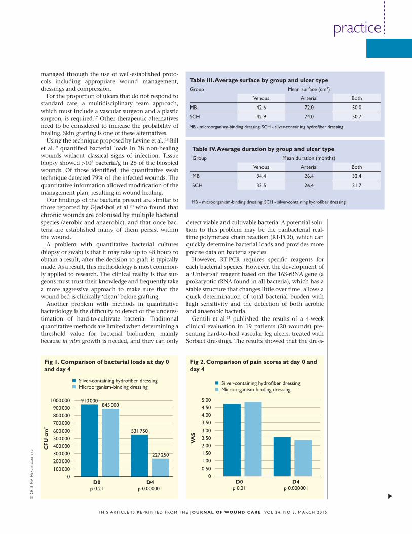
The statistical analysis found no significant differ-ence between groups regarding wound size (p=0.48), ulcer duration time (p=0.47) or bacterial load at D0 (p=0.21; Tables III, IV and Fig 1).
Staphylococcus aureus, methicillin-resistant Staphylo-coccus aureus (MRSA), Pseudomona aeruginosa, Entero-coccus faecalis, Escherichia coli, Klebsiella, Enterobacter cloacae, and Proteus mirabilis were most frequently found on the ulcer beds. In general we found a pol-ymicrobial burden with bacterial species equally dis-tributed in the two groups. The recorded data does not allow us to make any further comparison between bacteria species.
Primary outcome bacterial quantificationThe average bacterial load was similar in both groups at D0, that is, 9.1 x 10⁵ CFU/cm2 and 8.5 x 10⁵ CFU/cm2 in the SCH and MB groups, respectively. After analysing bacterial load within each group, the results showed a significant reduction of bacterial burden at D4 in both groups. In the SCH group, the average bac-terial load reduction was 41.6%, with a reduction of 73.1% in the MB group. When comparing bacterial load between groups at D4, the reduction was signifi-cantly higher in the MB group (p< 0.0001; Fig 1).
Secondary outcomes Dressing application and removal was found to be atraumatic and simple for both dressing types. Aver-age ulcer-related pain scores were 4.65 and 4.75 at D0 in the SCH and MB groups, respectively. Pain scores decreased in both groups, −35% in the SCH group and −38% in the MB group. The statistical analysis found no significant difference between groups at D0 (p=0.41) or at D4 (p=0.89; Fig 2).
Of the 40 patients, 20 (10 SCH and 10 MB) required analgesics before treatment. At D4, only 4 patients in the SCH group and 3 in the MB group still required analgesics.Only four patients in the SCH group and five in the MB group needed more than one piece of the dressing at each dressing change. Using more than one piece of the dressing did not have any effect on bacterial load reduction.
Two patients in the SCH group reported intense burning following the application of the dressing. The burning sensation lasted for a few hours, then disappeared without further problems and without the need for analgesics.
No serious adverse events related to the dressings were seen during the present study.
DiscussionClinical practice has demonstrated that the majority of leg ulcers heal within 4–6 months when correctly
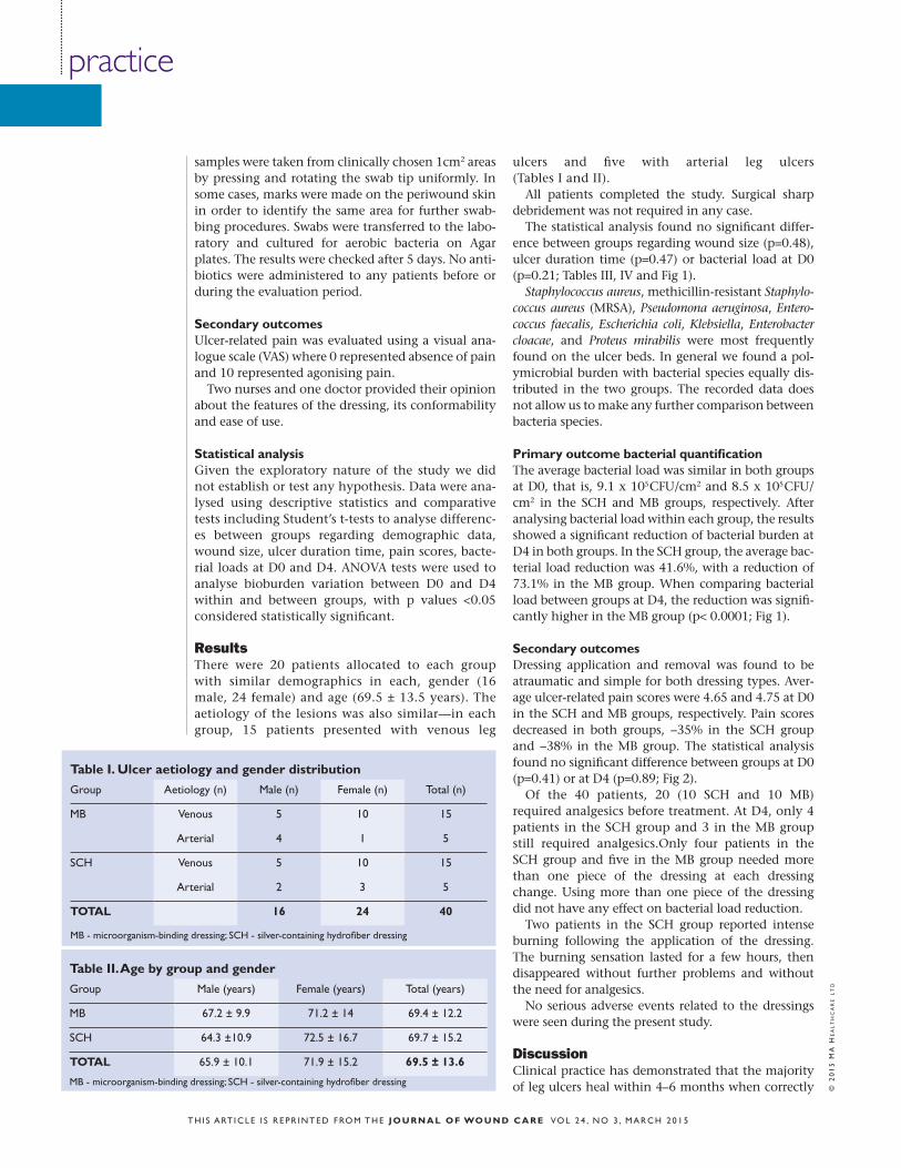
Table I. ulcer aetiology and gender distribution
Group aetiology (n) male (n) female (n) total (n)
mB venous 5 10 15
arterial 4 1 5
sch venous 5 10 15
arterial 2 3 5
ToTal 16 24 40
Table II. age by group and gender
Group male (years) female (years) total (years)
mB 67.2 ± 9.9 71.2 ± 14 69.4 ± 12.2
sch 64.3 ±10.9 72.5 ± 16.7 69.7 ± 15.2
ToTal 65.9 ± 10.1 71.9 ± 15.2 69.5 ± 13.6
mB - microorganism-binding dressing; sch - silver-containing hydrofiber dressing
mB - microorganism-binding dressing; sch - silver-containing hydrofiber dressing
JoWC_2015_24_3_Mosti.indd 124 18/03/2015 09:59
practices
t h i s a rt i c l e i s r e p r i n t e d f ro m t h e j o u r n a l o f wo u n d c a r e vo l 2 4 , n o 3 , m a r c h 2 0 1 5
© 2
01
5 M
A H
eA
lt
Hc
Ar
e l
td
managed through the use of well-established proto-cols including appropriate wound management, dressings and compression.
For the proportion of ulcers that do not respond to standard care, a multidisciplinary team approach, which must include a vascular surgeon and a plastic surgeon, is required.17 Other therapeutic alternatives need to be considered to increase the probability of healing. Skin grafting is one of these alternatives.
Using the technique proposed by Levine et al.,18 Bill et al.19 quantified bacterial loads in 38 non-healing wounds without classical signs of infection. Tissue biopsy showed >105 bacteria/g in 28 of the biospied wounds. Of those identified, the quantitative swab technique detected 79% of the infected wounds. The quantitative information allowed modification of the management plan, resulting in wound healing.
Our findings of the bacteria present are similar to those reported by Gjødsbøl et al.20 who found that chronic wounds are colonised by multiple bacterial species (aerobic and anaerobic), and that once bac-teria are established many of them persist within the wound.
A problem with quantitative bacterial cultures (biopsy or swab) is that it may take up to 48 hours to obtain a result, after the decision to graft is typically made. As a result, this methodology is most common-ly applied to research. The clinical reality is that sur-geons must trust their knowledge and frequently take a more aggressive approach to make sure that the wound bed is clinically ‘clean’ before grafting.
Another problem with methods in quantitative bacteriology is the difficulty to detect or the underes-timation of hard-to-cultivate bacteria. Traditional quantitative methods are limited when determining a threshold value for bacterial bioburden, mainly because in vitro growth is needed, and they can only
detect viable and cultivable bacteria. A potential solu-tion to this problem may be the panbacterial real-time polymerase chain reaction (RT-PCR), which can quickly determine bacterial loads and provides more precise data on bacteria species.
However, RT-PCR requires specific reagents for each bacterial species. However, the development of a ‘Universal’ reagent based on the 16S-rRNA gene (a prokaryotic rRNA found in all bacteria), which has a stable structure that changes little over time, allows a quick determination of total bacterial burden with high sensitivity and the detection of both aerobic and anaerobic bacteria.
Gentili et al.21 published the results of a 4-week clinical evaluation in 19 patients (20 wounds) pre-senting hard-to-heal vascular leg ulcers, treated with Sorbact dressings. The results showed that the dress-
fig 1. comparison of bacterial loads at day 0 and day 4
1 000 000900 000800 000700 000600 000500 000400 000300 000200 000100 000
0
cf
u c
m2
910 000845 000
531 750
227 250
d0p 0.21
d4p 0.000001
n silver-containing hydrofiber dressing n microorganism-binding dressing
Table III. average surface by group and ulcer type
Group mean surface (cm²)
venous arterial Both
mB 42.6 72.0 50.0
sch 42.9 74.0 50.7
mB - microorganism-binding dressing; sch - silver-containing hydrofiber dressing
Table IV. average duration by group and ulcer type
Group mean duration (months)
venous arterial Both
mB 34.4 26.4 32.4
sch 33.5 26.4 31.7
mB - microorganism-binding dressing; sch - silver-containing hydrofiber dressing
fig 2. comparison of pain scores at day 0 and day 4
5.004.504.003.503.002.502.001.501.000.50
0
Va
S
d0p 0.21
d4p 0.000001
n silver-containing hydrofiber dressing n microorganism-binding dressing
JoWC_2015_24_3_Mosti.indd 125 18/03/2015 09:59
practice
t h i s a rt i c l e i s r e p r i n t e d f ro m t h e j o u r n a l o f wo u n d c a r e vo l 2 4 , n o 3 , m a r c h 2 0 1 5
© 2
01
5 M
A H
eA
lt
Hc
Ar
e l
td
ing promoted healing in 7 patients and improve-ment in another 8. They used quantitative 16S RT-PCR to assess bacterial loads. The initial bacterial load was considerably different in the samples ranging from 4.38 x 10³–2.44 x 10⁸ bacterial genomes/mg of tissue. Nevertheless, the average of the total bacterial load before the treatment was 4.41 x 10⁷/mg of tis-sue, which decreased to 1.73 x 10⁵/mg of tissue, cor-responding to a significant 254-fold decrease in the total bacterial load in the healing wounds, whereas in the non-healing wounds they found only a non-significant 5.3-fold decrease of the total bacterial load. The results allowed them to confirm the suita-bility of 16S RT-PCR quantification of total bacterial load as a quick and sensitive parameter of wound evolution when performed on tissue biopsies.
When designing our pilot study protocol, we took into consideration the available experimental data about the technology on which the MB dress-ing is based.15,22–25 The dressings mechanism of action constitutes a paradox: microorganisms are trapped not destroyed, and eliminated from the wound at dressing change. The mesh structure allows conformability and ease of application. Because of their mechanism of action, it is unlikely that MB dressings will cause bacterial resistance or have systemic absorption and local or systemic tox-icity. As bacteria are removed intact, the release of bacterial endotoxins is prevented and the local inflammatory response is reduced. However, a change in the current assumptions about antimi-crobial dressings is required to accept that local antimicrobial activity is achieved without using more conventional antimicrobial substances.
In general, the frequency of dressing change depends on the quantity of wound exudate, the wound status and bioburden. For all our patients with these types of wounds, included or not in this study, we change the dressings every day. This is based on the following reasons: l To assess the wound on a daily basisl Our belief that a daily change could have a better impact on the preoperative preparation of the wound bed as it could provide a more intense antimicrobial effectl The fact that the preparation period is short l MB dressings are indicated to be changed daily.
It is important to highlight the observed pain reduction, which was probably due to a reduction of bacterial load and inflammation as a direct result of the dressings and compression. The presence of sil-ver could be seen as a cause for stronger pain in the SCH group; however, the results didn’t show any sig-nificant difference between the two groups.
limitations and future studiesBy definition, pilot studies are size-limited and our trial is not an exception. New technologies are not
always easy to assess, and in the absence of reliable evidence, pilot studies are a good way to obtain baseline data to assist the designing of further research. A further larger trial is necessary to con-firm our data.
Blinding of treatment does not apply to this study. Devices used during a comparative trial are expected to perform similar actions, but as both dressings are physically different, blinding is not possible. What could have been blinded here were the initial assessment of the wound and the assess-ment of outcomes by different expert clinicians. However, implementing this type of blinding dur-ing the present trial was logistically difficult because of the short observation period. A complete analy-sis about blinding in wound research can be found in a document published by the EWMA‘s Patients Outcome Group.26
This study may also be limited by the swabbing of the wound for bacteria. Although qualitative biop-sies are more reliable, they are also more invasive. Hence, we chose to swab the wound area carefully in the same place with the same method.
The observation period was limited to 4 days as a direct result of our protocol of care, which we have adapted for this type of patients. The four-day peri-od is intended to prepare the wound for surgery. Owing to the short duration of the study, we did not record data on ulcer development and healing rates. It is highly likely that by increasing the chance of the graft taking we are improving healing out-comes. However, we focused this pilot study on bacterial loads to investigate the dressings’ antimi-crobial efficacy.
There are limitations around the identification of the bacterial species, which are due to the tradition-al culture methods we used. As RT-PCR becomes increasinly accessible and widely used, more clinical evidence will be available, and it is highly likely that the bacteriological criteria for wound infection that we apply today will be challenged and modified in the future.
ConclusionsOur evaluation seems to confirm that, independ-ently from their mechanisms of action, MB dress-ings as well as SCH dressings are both effective in reducing bacterial burden in critically colonised or locally infected chronic venous leg ulcers without inducing adverse events.
In this pilot trial, MB dressings were significantly more effective in reducing bacterial numbers than SCH dressings. However, the size of the popula-tion, represents a challenge regarding comparative efficacy. A trial including a larger population, a longer follow up and the use of PCR techniques for quantitative bacteriology are required to confirm these results. n
JoWC_2015_24_3_Mosti.indd 126 18/03/2015 09:59
practice
t h i s a rt i c l e i s r e p r i n t e d f ro m t h e j o u r n a l o f wo u n d c a r e vo l 2 4 , n o 3 , m a r c h 2 0 1 5
references1 Jackson, d.m., lowbury, e.J., topley, e. pseudomonas pyocyanea in burns: its role as a pathogen and the value of local polymyxin therapy. lancet 1951; 2: 6674, 137–147.2 Krizek, t.J., robson, m.c., Kho, e. Bacterial growth and skin graft survival. surg forum 1967; 18: 518–519.3 Gilliland, e.l., nathwani, n., dore, c.J., lewis, J.d. Bacterial colonisation of leg ulcers and its effect on the success rate of skin grafting. ann r col surg engl 1988; 70: 2, 105–108.4 hogsberg, t., Bjarnsholt, t, thomsen, Js, Kirketerp-møller K. success rate of split-thickness skin grafting of chronic venous ulcers depends on the presence of pseudomona aeruginosa: a retrospective study. plos one 2011; 6: 5, e204925 White, r., cutting, K. critical colonization – the concept under scrutiny. ostomy Wound manage 2006; 52: 11, 50–56.6 Gottrup, f., apelqvist, J., Bjansholt, t., et al. eWma document: antimicrobials and non-healing Wounds – evidence, controversies and suggestions. the principal role of bioburden in wounds. J Wound care 2013; 22: (5 suppl), s10–s26.7 robson, m.c., lea, c.e,. dalton, J.B., heggers, J.p. Quantitative bacteriology and delayed wound closure. surg forum 1968; 19:
501–502.8 Grey, J.e., enoch, s., harding, K.G. venous and arterial leg ulcers. BmJ 2006; 332: 7537, 347–350.9 landsdown a.B. a pharmacological and toxicological profile of silver as an antimicrobial agent in medical devices, advances in pharmacological sciences, 2010; id910686, available at www.hindawi.com/journals/aps/2010/910686/ 10 Bard a., Kwok, c.h., hung, s.c., et al, a comparative study of the cytotoxicity of silver-based dressings in monolayer cell, tissue explant and animal models. Wound repair regen 2007; 15: 1, 94–104.11 hendry, a.t., stewart, i.o. silver-resistant enterobacteriaceae from hospital patients. can J microbiol 1979; 25: 8, 915–921.1212 mijnendonckx K., leys, n., mahillon, J., et al. antimicrobial silver: toxicity and potential for resistance. Biometals 2013: 26: 4, 609-621.13 percival s.l., Woods, e., nutekpor, m., et al. prevalence of silver resistance in bacteria isolated from diabetic foot ulcers and efficacy of silver-containing wound dressings. ostomy Wound manage 2008; 54: 3, 30–40.14 hampton s. an evaluation of the efficacy of cutimed® sorbact® in different types of
non-healing wounds.Wounds UK 2007; 3; 4, 113–119.15 ljungh, a., Yanagisawa, n., Wadström, t. Using the principle of hydrophobic interaction to bind and remove wound bacteria, J Wound care 2006; 15: 4, 175–180.16 mosti, G., iabichella, m.l., partsch, h. compression therapy in mixed ulcers increases venous output and arterial perfusion, J vasc surg 2012; 55: 1, 122–128.17 ino, K., Kiyokawa, K., akaiwa, K., et al. a team approach to the management of intractable leg ulcers. ann vasc dis 2013; 6: 1, 39–45.18 levine, n.s., lindberg, r.B., mason, a.d. Jr., pruitt, B.a. Jr. the quantitative swab culture and smear: a quick, simple method for determining the number of viable aerobic bacteria on open wounds. J trauma 1976; 16: 2, 89–9419 Bill, t.J., ratliff, c.r., donovan, a.m., et al. Quantitative swab culture versus tissue biopsy: a comparison in chronic wounds. ostomy Wound manage 2001; 47: 1, 34–37.20 Gjødsbøl K., christensen, J.J., Karlsmark, t., et al, multiple bacterial species reside in chronic wounds: a longitudinal study. int Wound J 2006; 3: 3, 225–231.21 Gentili, v., Gianesini, s., Balboni, p.G., et al. panbacterial real-time pcr to evaluate bacterial burden in chronic wounds treated with cutimedtm sorbacttm. eur J clin microbiol infect dis 2012; 31:
1523–1529.22 ljungh, a., Wadström, t., Growth conditions influence expression of cell surface hydrophobicity of staphylococci and other wound infection pathogens. microbiol immunol 1995; 39: 10, 753–757.23 Katsikogianni, m., missirlis, Y.f., concise review of mechanisms of bacterial adhesion to biomaterials and of techniques used in estimating bacteria-material interactions. eur cell mater 2004; 8: 37–57.24 malmsjö, m., lindstedt, s., ingemansson, r., Gustafsson, l. Bacteria and fungus binding mesh in negative pressure wound therapy – a review of the biological effects in the wound bed. eWma Journal 2012; 12: 3, 27–31.25 malmsjö, m., ingemansson, r., lindstedt, s., Gustafsson, l. comparison of bacteria and fungus-binding mesh, foam and gauze as fillers in negative pressure wound therapy – pressure transduction, wound edge contraction, microvascular blood flow and fluid retention, int Wound J 2013; 10: 5, 597–605.26 Gottrup, f., apelqvist, J. price, p. outcomes in controlled and comparative studies on non-healing wounds: recommendations to improve the quality of evidence in wound management. J Wound care 2010; 19: 6, 239–268.
© 2
01
5 M
A H
eA
lt
Hc
Ar
e l
td
JoWC_2015_24_3_Mosti.indd 127 18/03/2015 09:59
BSN medical Inc. 5825 Carnegie Blvd. Charlotte, NC 28209-4633Tel. (+1) 704 554 9933 Fax (+1) 704 358 4558 www.bsnmedical.comTo order toll-free: BSN medical (+1) 800 552 1157
62120 RN REV 04/15