Embed Size (px)
Citation preview
Journal of
Functional
Biomaterials
Article
Comparative Study of Technologies for Tubule Occlusion andTreatment of Dentin Hypersensitivity
Camilla Berg, Erik Unosson, Håkan Engqvist and Wei Xia *
�����������������
Citation: Berg, C.; Unosson, E.;
Engqvist, H.; Xia, W. Comparative
Study of Technologies for Tubule
Occlusion and Treatment of Dentin
Hypersensitivity. J. Funct. Biomater.
2021, 12, 27. https://doi.org/
10.3390/jfb12020027
Academic Editors: Gianrico Spagnuolo
and Håvard J. Haugen
Received: 10 February 2021
Accepted: 23 April 2021
Published: 27 April 2021
Publisher’s Note: MDPI stays neutral
with regard to jurisdictional claims in
published maps and institutional affil-
iations.
Copyright: © 2021 by the authors.
Licensee MDPI, Basel, Switzerland.
This article is an open access article
distributed under the terms and
conditions of the Creative Commons
Attribution (CC BY) license (https://
creativecommons.org/licenses/by/
4.0/).
Department of Materials Science and Engineering, Division of Applied Materials Science, UppsalaUniversity, 751 21 Uppsala, Sweden; [email protected] (C.B.); [email protected] (E.U.);[email protected] (H.E.)* Correspondence: [email protected]; Tel.: +46-184-717-961
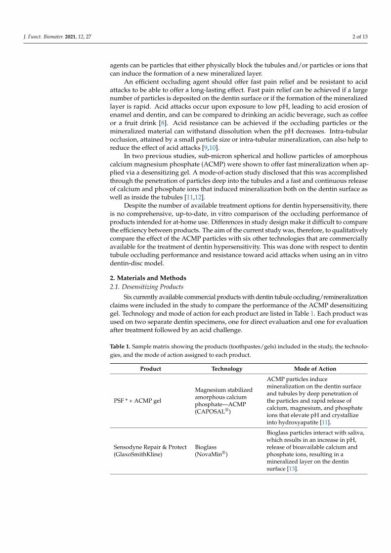
Abstract: This study aimed to evaluate the occluding/remineralization performance and resistanceto acid attacks of the mineralization layer formed by a tooth-desensitizing gel containing amorphouscalcium magnesium phosphate (ACMP) particles and compare it to six other desensitizing productsavailable on the market. Similar comprehensive studies are few and there is especially a lack ofstudies that are up to date. A dentin-disc model was used for in vitro evaluation of the desensitizingtoothpastes/gels. Application of the products was performed twice daily for seven days. Oneset of specimens were evaluated using scanning electron microscopy (SEM) directly after the finaltreatment and another set was evaluated after an acid challenge, exposing specimens to 2 wt%citric acid. The ACMP desensitizing gel was the only product resulting in complete occlusionby the formation of mineralized material on the dentin surface and inside the tubules. Particledeposition was dominant after treatment with the other desensitizing products, with little or nomineralization, resulting in partial occlusion only. Sensodyne Repair & Protect and Oral-B Pro-Expertshowed the highest resistance toward acid attacks. Material inside the tubules remained relativelyunaffected by acid attacks in all specimens. The results in this study indicated a great variabilityamong the occluding agents in terms of occlusion and acid resistance of the mineralization layer.The high degree of occlusion and intra-tubular mineralization that could mitigate the effect of acidsolubilization indicate that the ACMP desensitizing gel may be a superior option for the treatment ofdentin hypersensitivity.
Keywords: dentin hypersensitivity; occlusion; desensitizing agents; calcium phosphate; remineral-ization
1. Introduction
Dentin hypersensitivity is characterized by a short and sudden pain caused by expo-sure of dentin as a result of loss of enamel or cementum [1]. The number of individualswho experience dentin hypersensitivity varies across studies, due to differences in studydesigns. Most studies, however, conclude that up to 57% of adult individuals and 84.5% ofpatients after periodontal treatment suffer from dentin hypersensitivity [2].
The underlying cause for dentin hypersensitivity has been widely discussed but themost used and commonly accepted explanation is the hydrodynamic theory [3]. It statesthat pain is elicited from exposed dentin due to fluid movement within the dentin tubulesin response to stimuli, typically thermal, evaporative, tactile, osmotic, or chemical [4]. Themovement of fluid results in mechanical deformation of the nerve terminals in the pulp,which causes pain [5].
One way of treating dentin hypersensitivity is to hinder, or reduce, the movement offluids within the tubules. This can be achieved in several different ways ranging from inva-sive techniques such as laser etching of the dentin surface to non-invasive techniques thatcan be applications of a gel or toothpaste containing an occluding agent [6,7]. Occluding
J. Funct. Biomater. 2021, 12, 27. https://doi.org/10.3390/jfb12020027 https://www.mdpi.com/journal/jfb
J. Funct. Biomater. 2021, 12, 27 2 of 13
agents can be particles that either physically block the tubules and/or particles or ions thatcan induce the formation of a new mineralized layer.
An efficient occluding agent should offer fast pain relief and be resistant to acidattacks to be able to offer a long-lasting effect. Fast pain relief can be achieved if a largenumber of particles is deposited on the dentin surface or if the formation of the mineralizedlayer is rapid. Acid attacks occur upon exposure to low pH, leading to acid erosion ofenamel and dentin, and can be compared to drinking an acidic beverage, such as coffeeor a fruit drink [8]. Acid resistance can be achieved if the occluding particles or themineralized material can withstand dissolution when the pH decreases. Intra-tubularocclusion, attained by a small particle size or intra-tubular mineralization, can also help toreduce the effect of acid attacks [9,10].
In two previous studies, sub-micron spherical and hollow particles of amorphouscalcium magnesium phosphate (ACMP) were shown to offer fast mineralization when ap-plied via a desensitizing gel. A mode-of-action study disclosed that this was accomplishedthrough the penetration of particles deep into the tubules and a fast and continuous releaseof calcium and phosphate ions that induced mineralization both on the dentin surface aswell as inside the tubules [11,12].
Despite the number of available treatment options for dentin hypersensitivity, thereis no comprehensive, up-to-date, in vitro comparison of the occluding performance ofproducts intended for at-home use. Differences in study design make it difficult to comparethe efficiency between products. The aim of the current study was, therefore, to qualitativelycompare the effect of the ACMP particles with six other technologies that are commerciallyavailable for the treatment of dentin hypersensitivity. This was done with respect to dentintubule occluding performance and resistance toward acid attacks when using an in vitrodentin-disc model.
2. Materials and Methods2.1. Desensitizing Products
Six currently available commercial products with dentin tubule occluding/remineralizationclaims were included in the study to compare the performance of the ACMP desensitizinggel. Technology and mode of action for each product are listed in Table 1. Each product wasused on two separate dentin specimens, one for direct evaluation and one for evaluationafter treatment followed by an acid challenge.
Table 1. Sample matrix showing the products (toothpastes/gels) included in the study, the technolo-gies, and the mode of action assigned to each product.
Product Technology Mode of Action
PSF * + ACMP gel
Magnesium stabilizedamorphous calciumphosphate—ACMP(CAPOSAL®)
ACMP particles inducemineralization on the dentin surfaceand tubules by deep penetration ofthe particles and rapid release ofcalcium, magnesium, and phosphateions that elevate pH and crystallizeinto hydroxyapatite [11].
Sensodyne Repair & Protect(GlaxoSmithKline)
Bioglass(NovaMin®)
Bioglass particles interact with saliva,which results in an increase in pH,release of bioavailable calcium andphosphate ions, resulting in amineralized layer on the dentinsurface [13].
J. Funct. Biomater. 2021, 12, 27 3 of 13
Table 1. Cont.
Product Technology Mode of Action
Colgate SensitivePRO-Relief(Colgate-Palmolive)
Arginine(Pro-Argin®)
Arginine and calcium carbonate forma positively charged complex thatbinds to the negatively chargeddentin surface, which physicallyblocks the tubule openings [14,15].
Oral-B Pro-Expert(Procter&Gamble) Stannous fluoride
Deposition of stannous fluoridecomplexes on the dentin surface thatocclude exposed tubules [16].
MI Paste Plus (GCCorporation)
Casein phosphopeptideamorphous calciumphosphate—CPP-ACP(RECALDENT™)
CPP-ACP adheres to dentin andreleases bioavailable calcium andphosphate ions to aidremineralization [17,18].
GUM SensiVital+ (Sunstar) Hydroxyapatite—HA
HA particles occlude exposed tubulesand release calcium and phosphateions for remineralization of thedentin surface [19].
Enamelon PreventiveTreatment Gel (Premier)
Amorphous calciumphosphate—ACP
Stannous fluoride together withcalcium and phosphate salts formACP in situ that occludes andmineralizes exposed tubules [20,21].
* Pepsodent Super Fluor toothpaste.
2.2. Sample Preparation
Extracted human molars, free from caries and without anatomical defects, were usedin the study to compare the occlusion effects and resistance to acid attacks. The use ofhuman molars was performed in accordance with the guidelines from the Swedish EthicalReview Authority (2016/039). Specimens were prepared by cutting 1-mm-thin discs fromthe midsections of the coronal part of the molars using a low-speed saw (Buhler Isomet2000). The tubules were exposed and the smear layer removed by etching the specimens in35% phosphoric acid for 15 s, followed by thorough rinsing in deionized water.
2.3. Treatment Sequence
All specimens were treated twice daily for a total of seven days. Specimens weregently dried using a low-lint tissue, and approximately 0.6 g of the desensitizing productswere applied to the dentin surface with a soft-bristled toothbrush (GUM SensiVital), usinga circular motion and light hand pressure. Both sides of the specimens were brushedfor 30–45 s each, and excess material was removed using the toothbrush. The specimenstreated with MI Paste Plus were treated according to instructions from the manufacturer,i.e., application of the product using a gloved finger. The paste was applied evenly on bothsides and left undisturbed for 3 min after which excess gel was removed. All products,apart from the ACMP gel, contained fluoride (900–1450 ppm F), for which treatment witha standard fluoride toothpaste (Pepsodent Super Fluor, “PSF”, 1450 ppm F) was performedprior to each application of the ACMP gel. This was performed by applying approximately0.6 g of PSF on the dentin specimen and brushing both sides of the specimens as previouslydescribed. All specimens were stored in individual containers in 2 mL of complete artificialsaliva (T0300, Northeast Laboratory Services, Winslow, Maine, USA) at 37 ◦C on a rockingplatform in between treatments. The artificial saliva was exchanged daily, and the sampleswere vacuum dried at the end of the period of treatment (after seven days).
2.4. Acid Challenge
After the final application and storage in saliva at 37 ◦C overnight, one set of specimenswas subjected to an acid challenge by immersing the specimens in 2 wt% citric acid (pH 2)
J. Funct. Biomater. 2021, 12, 27 4 of 13
for 30 s to mimic the exposure to an acidic beverage. The specimens were then rinsedthoroughly in deionized water for 2 min and gently dried using a low-lint tissue followedby vacuum drying.
2.5. Characterization
The specimens were characterized with scanning electron microscopy (SEM; Zeiss,Leo 1530), imaging the samples with secondary electrons at 2 kV acceleration voltage. Toavoid charging and to allow for imaging, the specimens were sputtered with a conductiveAu/Pd layer prior to analysis. Cross sections of the dentin specimens were analyzed bymanually fracturing the dentin discs longitudinally.
3. Results3.1. Occlusion
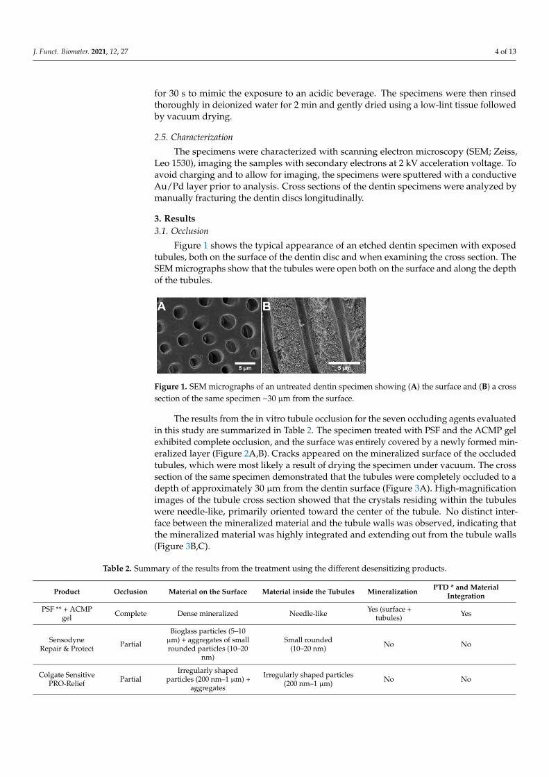
Figure 1 shows the typical appearance of an etched dentin specimen with exposedtubules, both on the surface of the dentin disc and when examining the cross section. TheSEM micrographs show that the tubules were open both on the surface and along the depthof the tubules.
Figure 1. SEM micrographs of an untreated dentin specimen showing (A) the surface and (B) a crosssection of the same specimen ~30 µm from the surface.
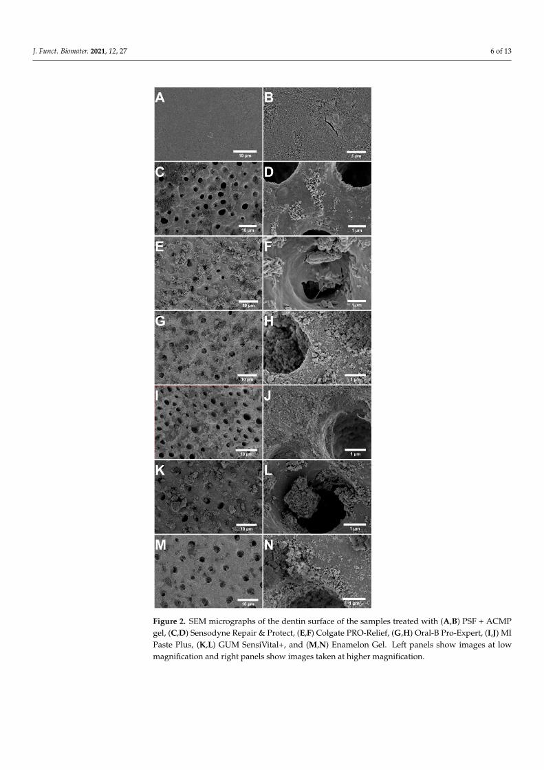
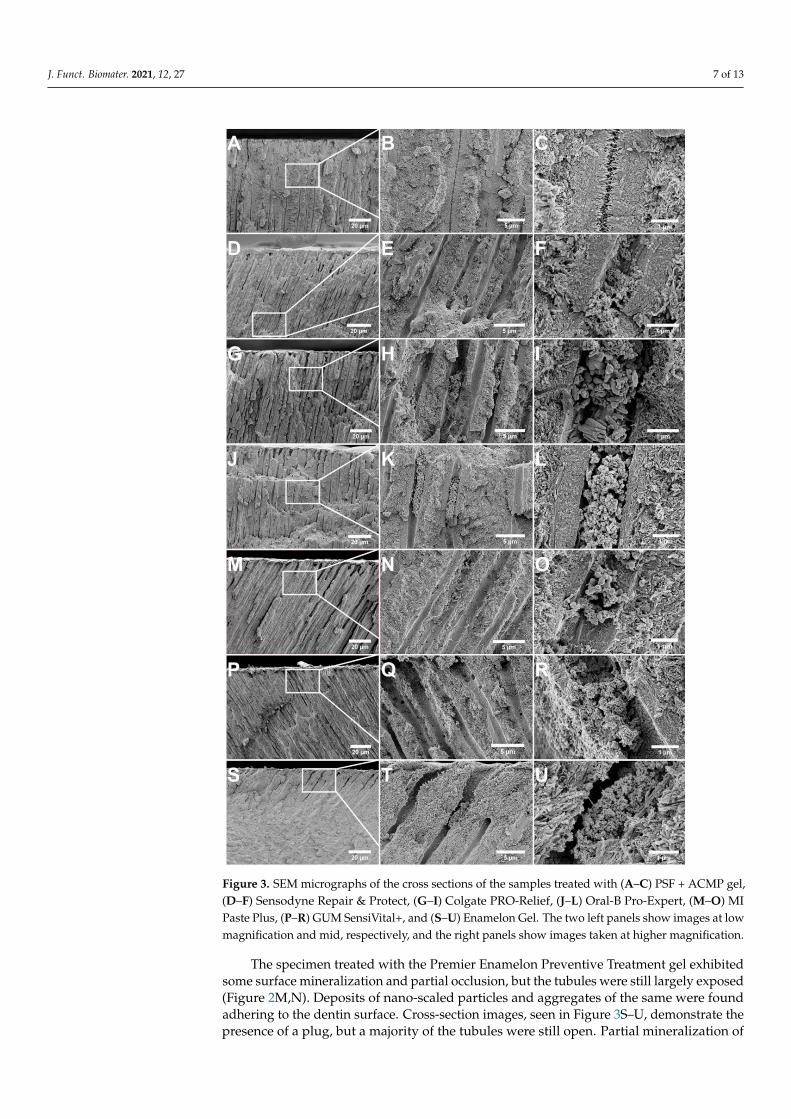
The results from the in vitro tubule occlusion for the seven occluding agents evaluatedin this study are summarized in Table 2. The specimen treated with PSF and the ACMP gelexhibited complete occlusion, and the surface was entirely covered by a newly formed min-eralized layer (Figure 2A,B). Cracks appeared on the mineralized surface of the occludedtubules, which were most likely a result of drying the specimen under vacuum. The crosssection of the same specimen demonstrated that the tubules were completely occluded to adepth of approximately 30 µm from the dentin surface (Figure 3A). High-magnificationimages of the tubule cross section showed that the crystals residing within the tubuleswere needle-like, primarily oriented toward the center of the tubule. No distinct inter-face between the mineralized material and the tubule walls was observed, indicating thatthe mineralized material was highly integrated and extending out from the tubule walls(Figure 3B,C).
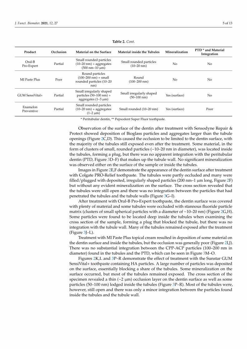
Table 2. Summary of the results from the treatment using the different desensitizing products.
Product Occlusion Material on the Surface Material inside the Tubules Mineralization PTD * and MaterialIntegration
PSF ** + ACMPgel Complete Dense mineralized Needle-like Yes (surface +
tubules) Yes
SensodyneRepair & Protect Partial
Bioglass particles (5–10µm) + aggregates of smallrounded particles (10–20
nm)
Small rounded(10–20 nm) No No
Colgate SensitivePRO-Relief Partial
Irregularly shapedparticles (200 nm–1 µm) +
aggregates
Irregularly shaped particles(200 nm–1 µm) No No
J. Funct. Biomater. 2021, 12, 27 5 of 13
Table 2. Cont.
Product Occlusion Material on the Surface Material inside the Tubules Mineralization PTD * and MaterialIntegration
Oral-BPro-Expert Partial
Small rounded particles(10–20 nm) + aggregates
(500 nm–10 µm)
Small rounded particles(10–20 nm) No No
MI Paste Plus Poor
Round particles(100–200 nm) + small
rounded particles (10–20nm)
Round(100–200 nm) No No
GUM SensiVital+ PartialSmall irregularly shapedparticles (50–100 nm) +
aggregates (1–3 µm)
Small irregularly shaped(50–100 nm) Yes (surface) No
EnamelonPreventive Partial
Small rounded particles(10–20 nm) + aggregates
(1–2 µm)Small rounded (10–20 nm) Yes (surface) Poor
* Peritubular dentin, ** Pepsodent Super Fluor toothpaste.
Observation of the surface of the dentin after treatment with Sensodyne Repair &Protect showed deposition of Bioglass particles and aggregates larger than the tubuleopenings (Figure 2C,D). This caused the occlusion to be limited to the dentin surface, withthe majority of the tubules still exposed even after the treatment. Some material, in theform of clusters of small, rounded particles (~10–20 nm in diameter), was located insidethe tubules, forming a plug, but there was no apparent integration with the peritubulardentin (PTD, Figure 3D–F) that makes up the tubule wall. No significant mineralizationwas observed either on the surface of the sample or inside the tubules.
Images in Figure 2E,F demonstrate the appearance of the dentin surface after treatmentwith Colgate PRO-Relief toothpaste. The tubules were partly occluded and many werefilled/plugged with deposited, irregularly shaped particles (200 nm–1 µm long, Figure 2F)but without any evident mineralization on the surface. The cross section revealed thatthe tubules were still open and there was no integration between the particles that hadpenetrated the tubules and the tubule walls (Figure 3G–I).
After treatment with Oral-B Pro-Expert toothpaste, the dentin surface was coveredwith plenty of material and some tubules were occluded with stannous fluoride particlematrix (clusters of small spherical particles with a diameter of ~10–20 nm) (Figure 2G,H).Some particles were found to be located deep inside the tubules when examining thecross section of the sample, forming a plug that blocked the tubule, but there was nointegration with the tubule wall. Many of the tubules remained exposed after the treatment(Figure 3J–L).
Treatment with MI Paste Plus topical cream resulted in deposition of some material onthe dentin surface and inside the tubules, but the occlusion was generally poor (Figure 2I,J).There was no substantial integration between the CPP-ACP particles (100–200 nm indiameter) found in the tubules and the PTD, which can be seen in Figure 3M–O.
Figures 2K,L and 3P–R demonstrate the effect of treatment with the Sunstar GUMSensiVital+ toothpaste containing HA particles. A large number of particles was depositedon the surface, essentially blocking a share of the tubules. Some mineralization on thesurface occurred, but most of the tubules remained exposed. The cross section of thespecimen revealed a thin (~2 µm) occlusion layer on the dentin surface as well as someparticles (50–100 nm) lodged inside the tubules (Figure 3P–R). Most of the tubules were,however, still open and there was only a minor integration between the particles foundinside the tubules and the tubule wall.
J. Funct. Biomater. 2021, 12, 27 6 of 13
Figure 2. SEM micrographs of the dentin surface of the samples treated with (A,B) PSF + ACMPgel, (C,D) Sensodyne Repair & Protect, (E,F) Colgate PRO-Relief, (G,H) Oral-B Pro-Expert, (I,J) MIPaste Plus, (K,L) GUM SensiVital+, and (M,N) Enamelon Gel. Left panels show images at lowmagnification and right panels show images taken at higher magnification.
J. Funct. Biomater. 2021, 12, 27 7 of 13
Figure 3. SEM micrographs of the cross sections of the samples treated with (A–C) PSF + ACMP gel,(D–F) Sensodyne Repair & Protect, (G–I) Colgate PRO-Relief, (J–L) Oral-B Pro-Expert, (M–O) MIPaste Plus, (P–R) GUM SensiVital+, and (S–U) Enamelon Gel. The two left panels show images at lowmagnification and mid, respectively, and the right panels show images taken at higher magnification.
The specimen treated with the Premier Enamelon Preventive Treatment gel exhibitedsome surface mineralization and partial occlusion, but the tubules were still largely exposed(Figure 2M,N). Deposits of nano-scaled particles and aggregates of the same were foundadhering to the dentin surface. Cross-section images, seen in Figure 3S–U, demonstrate thepresence of a plug, but a majority of the tubules were still open. Partial mineralization of
J. Funct. Biomater. 2021, 12, 27 8 of 13
the tubule walls was noted, but integration between the particle deposits and surroundingtissue was generally poor.
3.2. Acid Challenge
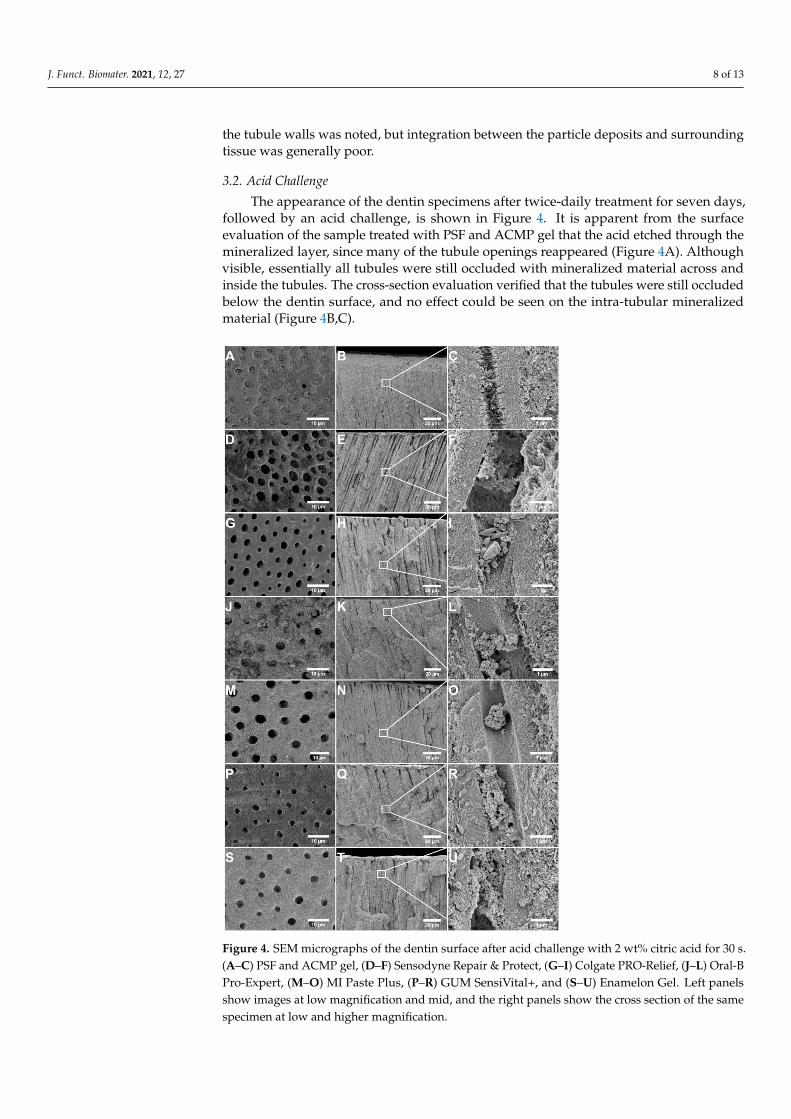
The appearance of the dentin specimens after twice-daily treatment for seven days,followed by an acid challenge, is shown in Figure 4. It is apparent from the surfaceevaluation of the sample treated with PSF and ACMP gel that the acid etched through themineralized layer, since many of the tubule openings reappeared (Figure 4A). Althoughvisible, essentially all tubules were still occluded with mineralized material across andinside the tubules. The cross-section evaluation verified that the tubules were still occludedbelow the dentin surface, and no effect could be seen on the intra-tubular mineralizedmaterial (Figure 4B,C).
Figure 4. SEM micrographs of the dentin surface after acid challenge with 2 wt% citric acid for 30 s.(A–C) PSF and ACMP gel, (D–F) Sensodyne Repair & Protect, (G–I) Colgate PRO-Relief, (J–L) Oral-BPro-Expert, (M–O) MI Paste Plus, (P–R) GUM SensiVital+, and (S–U) Enamelon Gel. Left panelsshow images at low magnification and mid, and the right panels show the cross section of the samespecimen at low and higher magnification.

J. Funct. Biomater. 2021, 12, 27 9 of 13
Figure 4D–F shows the appearance after the acid challenge of the sample treatedwith Sensodyne Repair & Protect toothpaste. Compared to the surface before the acidchallenge (Figure 2C,D), a larger share of the tubules was exposed, indicating that the acidhad dissolved some of the mineralized material on the surface. Particles presumed to beBioglass particles were still present on the surface.
The dentin specimen treated with Colgate PRO-Relief toothpaste was heavily affectedby the acid challenge, as shown in Figure 4G–I. Essentially all tubules were re-exposed, andnone of the deposited particles seen in Figure 2E,F remained on the surface. The particlesinside the tubules were less affected by the acid exposure, but the intra-tubular occlusionwas still poor since the amount of particles lodged inside the tubules after the treatmentwas small (Figure 3G–I).
The Oral-B Pro-Expert specimen fared comparably well in the acid challenge, withno significant changes in terms of tubule occlusion or particle deposits on the surface (seeFigure 4J–L compared to Figure 3G,H). The sample treated with GC MI Paste Plus did, incontrast to this, withstand the acid challenge poorly. The surface-adhered particles haddissolved and the tubules were again exposed (Figure 4M–O).
The specimen treated with GUM SensiVital+ that was subjected to acid challengeis shown in Figure 4P–R. Compared to the images shown in Figure 3K,L, there were noparticles on the surface, but a layer of mineralized material was still present. However, thismineralized layer did not occlude the tubules effectively.
The dentin surface and cross-section appearance after treatment with Enamelon Geland the acid challenge are shown in Figure 4S–U. Some nano-scaled particles still adheredto the surface, but all larger particle aggregates had dissolved or vanished, and the tubuleswere largely exposed.
4. Discussion
The hydrodynamic theory is widely accepted as the principal mechanism of actionfor the cause of dentin hypersensitivity [3]. Occlusion of exposed dentin tubules canreduce pain related to the condition by the hindrance of fluid movements within the dentintubules [5]. In this study, the occluding effect and resistance to acid attacks were evaluatedfor seven different desensitizing products after a twice-daily, seven-day treatment period.
Examining the results, in general, it was only the specimen treated with PSF andACMP gel that resulted in complete occlusion with a mineralized layer covering theentire dentin surface (Figure 2A,B). In our previous studies, we showed that the ACMPparticles alone result in this type of occlusion. Fluoride application does not affect thedegree of occlusion upon application, but it alters the mineralization characteristics by theformation of needle-like structures [11,12]. The sample treated with PSF and ACMP gelwas additionally the only sample that exhibited intra-tubular mineralization with goodadherence of the occluding material and the tubule walls (Figure 3A–C). As described byMarkowitz and Pashley, the key factor in reducing the hydraulic conductance, achievingpain-relief, is to reduce the anatomic tubule radius [22]. This indicates that the occlusionby mineralization after treatment with PSF and ACMP gel would offer a higher degree ofpain relief compared to the samples with less intra-tubular occlusion or poor integrationbetween the occluding particles and the tubule wall. As reported by Ryou et al. [23], there isa natural variation in tubule diameters related to age and the gradual formation of scleroticdentin, which reduces the tubular lumen diameter. The current study was based on alimited set of extracted molars obtained from separate individuals. However, the tubulediameters were generally in agreement across specimens when examining unaffected areasfurther from the treated surface in cross section (Figures 3 and 4). This supports the claimthat the mineralization and reduction of the anatomic tubule diameter was, in fact, a resultof the treatment with PSF and the ACMP gel.
The other treatment options that resulted in mineralization, at least on the dentinsurface, were GUM SensiVital+ and Premier Enamelon containing HA particles or ACP(Figure 2K–N). The probable reason for this is that they, in a similar manner as the ACMP
J. Funct. Biomater. 2021, 12, 27 10 of 13
particles, release calcium and phosphate ions to a concentration exceeding the supersatura-tion of saliva, triggering the nucleation of material on the dentin surface [19–21]. Previousstudies using NovaMin® Bioglass and CPP-ACP reported that the mineralization wascaused by the release of previously mentioned ions, which were not clearly observed in thisstudy [24–27]. Only particle deposition was observed in all three cases, with no significantsurface or intra-tubular mineralization. This could possibly be explained by inter-studyvariations in the design of the studies, such as the amount of gel applied on each brushing,storage conditions, or the number of times that the gel has been applied.
No mineralization was observed for the specimens treated with Colgate PRO-Relief(Figures 2E,F and 3G–I) or Oral-B Pro-Expert (Figures 3G,H and 4J–L), which can beexplained by the lack of phosphate ions in the toothpastes. Arginine is added to theColgate PRO-Relief toothpaste since the association of calcium carbonate, and the aminoacid is said to provide an alkaline environment that can encourage endogenous calciumand phosphate ions to deposit on the dentin surface [28]. The results in this study, however,indicated that this effect, if present at all, was much slower compared to direct delivery ofcalcium and phosphate ions. The stannous fluoride particles in Oral-B Pro-Expert are saidto offer pain relief by particle deposition on the dentin surface, so the lack of a mineralizedlayer was expected for this specimen [16].
The resistance toward acid attacks varied greatly when comparing the different treat-ment alternatives (Figure 4). This is most likely dependent on the solubility of the occludingmaterial, particularly at low pH. The occluding materials that were the least affected bythe acid challenge were the NovaMin® Bioglass particles in Sensodyne Repair & Protectand the stannous fluoride particles in the Oral-B Pro-Expert toothpaste (Figure 4D–F,J–L).These results were in accordance with previous studies using Bioglass and stannous flu-oride complexes (with hexametaphosphate) as occluding agents, where both materialswere shown to resist acid solubilization [13,29,30]. The HA particles included in the GUMSensiVital+ toothpaste also had fairly low solubility, at least compared to other calciumphosphate phases, which was indicated, in part, by the mineralized layer that remainedafter acid challenge (Figure 4P–R) [31].
Specimens treated with toothpastes containing, or claiming to form, ACP (i.e., MIPaste Plus and Premier Enamelon) had a high solubility and are, thus, susceptible to acidattacks [31]. This was clearly visible in Figure 4M–O,S–U. The Colgate Pro-Argin particles(arginine with calcium carbonate) appeared to be particularly sensitive to acid since allparticles that largely covered the dentin surface before the acid treatment (Figure 2E,F)were removed by the exposure to acid (Figure 4G–I). This can be explained by the solubilityof calcium carbonate that is higher compared to calcium phosphate, which could withstandacid attacks better. The solubility of calcium carbonate is pH-dependent due to the fact thatit reacts with the acid to form calcium ions, water, and carbon dioxide, even in dilute acidicsolutions [32]. Apatite materials, either in particle form or as a mineralized layer, were, incomparison, not as sensitive to acid exposure [33,34].
The particles in the ACMP gel are, like the other ACP materials, highly soluble inaqueous solutions, particularly at low pH. This was confirmed already prior to the acidchallenge where all particles had dissolved (Figure 2A,B). The high dissolution rate andconsequent high release of ions, combined with deep penetration inside the tubules, makefor a rapid crystallization process that occludes the tubules. Particle dissolution andrelease of phosphate ions will also raise pH, aiding the formation and stability of HA [31].The acid resistance of the ACMP gel treated specimen should, therefore, not be assignedto the resistance of the particles themselves but to the crystalline and firmly integratedmineral that is rapidly formed inside the tubules by the transformation of the particles. Theformation of needle-like structures (Figures 3C and 4C) indicated that the acid resistancemay have been further enhanced by the use of a fluoride toothpaste prior to the applicationof the ACMP particles. We showed in a previous study that fluoride incorporation in themineralized material could be recognized by the formation of needle-like structures, similarto what was observed in this study [12]. This would improve the resistance toward acid
J. Funct. Biomater. 2021, 12, 27 11 of 13
attacks since fluoride substitution in apatites is known to increase the stability through thereduction of strain in the crystal lattice [31,35].
It should be noted that this study only was performed in terms of evaluating thequalitative differences of the occluding agents, their effects on remineralization of dentin,and resistance to acid attacks after a single week’s daily application. For quantitativecomparisons, more tests have to be performed. Reeder et al. developed a setup formeasuring the hydraulic conductance, i.e., the ease with which a fluid can “filter” acrossdentin, which can be used to determine the occlusion in a quantitative manner [36]. Thistechnique has successfully been used in several other comparative studies of occludingagents [37–39].
5. Conclusions
Given the timeframe and design of the current in vitro study, particle deposition wasdominant for all evaluated toothpastes/gels except for the ACMP desensitizing gel thatinduced the significantly better formation of a mineralized layer occluding exposed dentintubules. The resistance toward acid attacks was highest for Sensodyne Repair & Protectand Oral-B Pro-Expert, but the risk of re-exposure of the dentin tubules could likely bemitigated by intra-tubular mineralization, as observed after application of the ACMP gel.Suggesting that the key factor for an efficient treatment is the formation of a mineralizedlayer, the results in this study indicate that the gel containing the ACMP particles maybe a promising alternative for the treatment of dentin hypersensitivity. This warrantsfurther investigations of the occluding agent, i.e., quantitative comparisons with competingproducts and clinical evaluation.
Author Contributions: Conceptualization, H.E.; methodology, C.B. and E.U.; investigation, C.B andE.U.; resources, E.U.; writing—original draft preparation, C.B.; writing—review and editing, C.B.,E.U., H.E., and W.X.; visualization, C.B.; supervision, H.E. and W.X.; funding acquisition, H.E. Allauthors have read and agreed to the published version of the manuscript.
Funding: The authors gratefully acknowledge financial support from the Swedish Research Council(grant number 2017-04728).
Institutional Review Board Statement: The use of extracted human molars was approved accordingto the guidelines from the Regional Ethics Review board in Uppsala (Sweden) (2016/039).
Informed Consent Statement: The study did not involve any interventions or handling of personaldata that are covered in the Swedish Ethics Review Act (2006:615). The use of would be discarded ex-tracted molars for scientific purposes was therefore waived on condition of anonymity and provisionsregarding personal consent did not apply to the use of human samples in the study
Data Availability Statement: The data presented in this study are available on request from thecorresponding author.
Conflicts of Interest: The authors declare the following financial interests/personal relationships,which may be considered as potential competing interests: E.U., W.X., and H.E. are connected toPsilox AB that has developed the ACMP material (CAPOSAL®) tested in the study. E.U. is employedby the company and W.X. and H.E. are shareholders. C.B. has no conflict of interest.
References1. West, N.X.; Lussi, A.; Seong, J.; Hellwig, E. Dentin hypersensitivity: Pain mechanisms and aetiology of exposed cervical dentin.
Clin. Oral Investig. 2012, 17, 9–19. [CrossRef] [PubMed]2. Splieth, C.H.; Tachou, A. Epidemiology of dentin hypersensitivity. Clin. Oral Investig. 2012, 17, 3–8. [CrossRef] [PubMed]3. Dababneh, R.H.; Khouri, A.T.; Addy, M. Dentine hypersensitivity—An enigma? A review of terminology, mechanisms, aetiology
and management. Br. Dent. J. 1999, 187, 606–611. [CrossRef]4. Tavares, M.; Stultz, J.; Newman, M.; Smith, V.; Kent, R.; Carpino, E.; Goodson, J.M. Light augments tooth whitening with peroxide.
J. Am. Dent. Assoc. 2003, 134, 167–175. [CrossRef]5. Brännström, M.; Åström, A. A Study on the Mechanism of Pain Elicited from the Dentin. J. Dent. Res. 1964, 43, 619–625. [CrossRef]
[PubMed]
J. Funct. Biomater. 2021, 12, 27 12 of 13
6. Renton-Harper, P.; Midda, M. NdYAG laser treatment of dentinal hypersensitivity. Br. Dent. J. 1992, 172, 13–16. [CrossRef][PubMed]
7. Orchardson, R.; Gillam, D.G. Managing dentin hypersensitivity. J. Am. Dent. Assoc. 2006, 137, 990–998. [CrossRef] [PubMed]8. Imfeld, T. Dental erosion. Definition, classification and links. Eur. J. Oral Sci. 1996, 104, 151–155. [CrossRef] [PubMed]9. Suge, T.; Ishikawa, K.; Kawasaki, A.; Yoshiyama, M.; Asaoka, K.; Ebisu, S. Effects of Fluoride on the Calcium Phosphate
Precipitation Method for Dentinal Tubule Occlusion. J. Dent. Res. 1995, 74, 1079–1085. [CrossRef]10. Kuroiwa, M.; Kodaka, T.; Kuroiwa, M.; Abe, M. Dentin Hypersensitivity. Occlusion of Dentinal Tubules by Brushing With and
Without an Abrasive Dentifrice. J. Periodontol. 1994, 65, 291–296. [CrossRef]11. Berg, C.; Unosson, E.; Engqvist, H.; Xia, W. Amorphous Calcium Magnesium Phosphate Particles for Treatment of Dentin
Hypersensitivity: A Mode of Action Study. ACS Biomater. Sci. Eng. 2020, 6, 3599–3607. [CrossRef]12. Berg, C.; Unosson, E.; Riekehr, L.; Xia, W.; Engqvist, H. Electron microscopy evaluation of mineralization on peritubular dentin
with amorphous calcium magnesium phosphate microspheres. Ceram. Int. 2020, 46, 19469–19475. [CrossRef]13. Burwell, A.; Jennings, D.; Muscle, D.; Greenspan, D.C. NovaMin and dentin hypersensitivity–in vitro evidence of efficacy. J. Clin.
Dent. 2010, 21, 66–71.14. Lavender, S.A.; Petrou, I.; Heu, R.; Stranick, M.A.; Cummins, D.; Kilpatrick-Liverman, L.; Santarpia, R.P., III. Mode of action
studies on a new desensitizing dentifrice containing 8.0% arginine, a high cleaning calcium carbonate system and 1450 ppmfluoride. Am. J. Dent. 2010, 23, 14–19.
15. Petrou, I.; Heu, R.; Stranick, M.; Lavender, S.; Zaidel, L.; Cummins, D.; Sullivan, R.J.; Hsueh, C.; Gimzewski, J.K. A breakthroughtherapy for dentin hypersensitivity: How dental products containing 8% arginine and calcium carbonate work to deliver effectiverelief of sensitive teeth. J. Clin. Dent. 2009, 20, 23–31. [PubMed]
16. Miller, S.; Truong, T.; Heu, R.; Stranick, M.; Bouchard, D.; Gaffar, A. Recent advances in stannous fluoride technology: Anti-bacterial efficacy and mechanism of action towards hypersensitivity. Int. Dent. J. 1994, 44, 83–98.
17. Reynolds, E.C. Remineralization of Enamel Subsurface Lesions by Casein Phosphopeptide-stabilized Calcium PhosphateSolutions. J. Dent. Res. 1997, 76, 1587–1595. [CrossRef] [PubMed]
18. Cochrane, N.; Reynolds, E. Calcium Phosphopeptides—Mechanisms of Action and Evidence for Clinical Efficacy. Adv. Dent. Res.2012, 24, 41–47. [CrossRef] [PubMed]
19. Amaechi, B.T.; Mathews, S.M.; Ramalingam, K.; Mensinkai, P.K. Evaluation of nanohydroxyapatite-containing toothpaste foroccluding dentin tubules. Am. J. Dent. 2015, 28, 33–39.
20. Kardos, S.; Shi, B.; Sipos, T. The in vitro demineralization potential of a sodium fluoride, calcium and phosphate ion-containingdentifrice under various experimental conditions. J. Clin. Dent. 1999, 10, 22–25.
21. Winston, A.; Usen, N. Processes for the Remineralization and Mineralization of Teeth. U.S. Patent 6,036,944, 14 March 2000.22. Markowitz, K.; Pashley, D.H. Discovering new treatments for sensitive teeth: The long path from biology to therapy. J. Oral
Rehabilitation 2008, 35, 300–315. [CrossRef]23. Ryou, H.; Romberg, E.; Pashley, D.H.; Tay, F.R.; Arola, D. Importance of Age on the Dynamic Mechanical Bahviour of In-tertubular
and Peritubular Dentin. J. Behav. Biomed. Mater. 2015, 42, 229–242. [CrossRef]24. Earl, J.S.; Topping, N.; Elle, J.; Langford, R.M.; Greenspan, D.C. Physical and chemical characterization of the surface layers
formed on dentin following treatment with a fluoridated toothpaste containing NovaMin. J. Clin. Dent. 2011, 22, 68–73.25. Da Cruz, L.P.D.; Hill, R.G.; Chen, X.; Gillam, D.G. Dentine Tubule Occlusion by Novel Bioactive Glass-Based Toothpastes. Int. J.
Dent. 2018, 2018, 1–10. [CrossRef]26. Poggio, C.; Lombardini, M.; Vigorelli, P.; Ceci, M. Analysis of dentin/enamel remineralization by a CPP-ACP paste: AFM and
SEM study. Scanning 2013, 35, 366–374. [CrossRef] [PubMed]27. Cao, Y.; Mei, M.L.; Xu, J.; Lo, E.C.; Li, Q.; Chu, C.H. Biomimetic mineralisation of phosphorylated dentine by CPP-ACP. J. Dent.
2013, 41, 818–825. [CrossRef]28. Kleinberg, I. SensiStat. A new saliva-based composition for simple and effective treatment of dentinal sensitivity pain. Dent.
Today 2002, 21, 42–47.29. Pereira, R.; Dg, G.; Shaikh, K.; Phad, S. Comparative Evaluation of Desensitizing Dentifrices containing BioMin ®, Novamin
®and Fluoride on Dentinal Tubule Occlusion before and after a Citric Acid Challenge—A scanning Electron Microscope in-vitroStudy. J. Odontol. 2018, 2, 1000105.
30. White, D.J.; Lawless, M.A.; Fatade, A.; Baig, A.; von Koppenfels, R.; Duschner, H.; Götz, H. Stannous fluoride/sodium hex-ametaphosphate dentifrice increases dentin resistance to tubule exposure in vitro. J. Clin. Dent. 2007, 18, 55–59.
31. Dorozhkin, S.V. Calcium orthophosphates. Biomatter 2011, 1, 121–164. [CrossRef]32. Roth-Bassell, H.A.; Clydesdale, F.M. In Vitro Solubility Characteristics of Six Calcium Salts. J. Food Prot. 1992, 55, 1003–1005.
[CrossRef] [PubMed]33. Chow, L.C.; Eanes, E.D. (Eds.) Octacalcium Phosphate; Karger Medical and Scientific Publishers: Basel, Switzerland, 2001;
Volume 18.34. Elliott, J.C. Structure and Chemistry of the Apatites and Other Calcium Orthophosphates; Elsevier: Amsterdam, The Netherlands, 1994.35. Rodríguez-Lorenzo, L.M.; Hart, J.N.; Gross, K.A. Structural and Chemical Analysis of Well-Crystallized Hydroxyfluorapatites. J.
Phys. Chem. B 2003, 107, 8316–8320. [CrossRef]
J. Funct. Biomater. 2021, 12, 27 13 of 13
36. Reeder, O.; Walton, R.; Livingston, M.; Pashley, D. Dentin Permeability: Determinants of Hydraulic Conductance. J. Dent. Res.1978, 57, 187–193. [CrossRef] [PubMed]
37. Wang, Z.; Sa, Y.; Sauro, S.; Chen, H.; Xing, W.; Ma, X.; Jiang, T.; Wang, Y. Effect of desensitising toothpastes on dentinal tubuleocclusion: A dentine permeability measurement and SEM in vitro study. J. Dent. 2010, 38, 400–410. [CrossRef] [PubMed]
38. Ishihata, H.; Kanehira, M.; Finger, W.J.; Takahashi, H.; Tomita, M.; Sasaki, K. Effect of two desensitizing agents on dentinpermeability in vitro. J. Appl. Oral Sci. 2017, 25, 34–41. [CrossRef]
39. Kim, S.Y.; Kim, E.J.; Kim, D.S.; Lee, I.B. The Evaluation of Dentinal Tubule Occlusion by Desensitizing Agents: A Real-timeMeasurement of Dentinal Fluid Flow Rate and Scanning Electron Microscopy. Oper. Dent. 2013, 38, 419–428. [CrossRef] [PubMed]