Embed Size (px)
Citation preview
Comparative study of notoungulate (Placentalia,Mammalia) bony labyrinths and new phylogeneticallyinformative inner ear charactersThomas E. Macrini,1,2 John J. Flynn,2,3 Xijun Ni,2,4 Darin A. Croft5 and Andr�e R. Wyss6
1Department of Biological Sciences, St Mary’s University, San Antonio, TX, USA2Department of Vertebrate Paleontology, Division of Paleontology, American Museum of Natural History, New York, NY, USA3Richard Gilder Graduate School, American Museum of Natural History, New York, NY, USA4Key Laboratory of Vertebrate Evolution and Human Origin, Institute of Vertebrate Paleontology and Paleoanthropology,
Beijing, China5Department of Anatomy, Case Western University School of Medicine, Cleveland, OH, USA6Department of Earth Science, University of California, Santa Barbara, CA, USA
Abstract
The phylogenetic relationships of notoungulates, an extinct group of predominantly South American herbivores,
remain poorly resolved with respect to both other placental mammals and among one another. Most previous
phylogenetic analyses of notoungulates have not included characters of the internal cranium, not least because
few such features, including the bony labyrinth, have been described for members of the group. Here we describe
the inner ears of the notoungulates Altitypotherium chucalensis (Mesotheriidae), Pachyrukhos moyani
(Hegetotheriidae) and Cochilius sp. (Interatheriidae) based on reconstructions of bony labyrinths obtained from
computed tomography imagery. Comparisons of the bony labyrinths of these taxa with the basally diverging
notoungulate Notostylops murinus (Notostylopidae), an isolated petrosal from Itabora�ı, Brazil, referred to
Notoungulata, and six therian outgroups, yielded an inner ear character matrix of 25 potentially phylogenetically
informative characters, 14 of them novel to this study. Two equivocally optimized character states potentially
support a pairing of Mesotheriidae and Hegetotheriidae, whereas four others may be diagnostic of Notoungulata.
Three additional characters are potentially informative for diagnosing more inclusive clades: one for crown
Placentalia; another for a clade containing Kulbeckia, Zalambdalestes, and Placentalia; and a third for Eutheria
(crown Placentalia plus stem taxa). Several other characters are apomorphic for at least one notoungulate in our
study and are of potential interest for broader taxonomic sampling within Notoungulata to clarify currently
enigmatic interrelationships. Measures of the semicircular canals were used to infer agility (e.g. capable of quick
movements vs. lethargic movements) of these taxa. Agility scores calculated from these data generally corroborate
interpretations based on postcranial remains of these or closely related species. We provide estimates of the low-
frequency hearing limits in notoungulates based on the ratio of radii of the apical and basal turns of the cochlea.
These limits range from 15 Hz in Notostylops to 149 Hz in Pachyrukhos, values comparable to the Asian elephant
(Elephas maximus) and the California sea lion (Zalophus californianus) when hearing in air, respectively.
Key words: cochlea; CT; Hegetotheriidae; Interatheriidae; Mesotheriidae; Notoungulata; petrosal; phylogenetic
characters; South America.
Introduction
Notoungulata was a taxonomically, morphologically and
ecologically diverse group of nearly exclusively South
American mammals that thrived during much of the
Cenozoic (Simpson, 1948, 1967, 1980; Patterson & Pascual,
1968). Notoungulates are characterized by a distinctive
‘crochet’ on the metaloph of the upper molars and an
expansive epitympanic sinus in the squamosal (Patterson,
1934b, 1936; Cifelli, 1993).
The relationship of notoungulates to other placental
mammals is debated (see Cifelli, 1993; Horovitz, 2004; Billet,
2010; Agnolin & Chimento, 2011; Billet & Martin, 2011;
O’Leary et al. 2013), as are many of the higher-level
Correspondence
Thomas E. Macrini, Department of Biological Sciences, One Camino
Santa Maria, St Mary’s University, San Antonio, TX 78228, USA.
T: +210 431 4304; F: +210 431 4363; E: [email protected]
Accepted for publication 12 August 2013
© 2013 Anatomical Society
J. Anat. (2013) doi: 10.1111/joa.12108
Journal of Anatomy
relationships within the group. Previous analyses of notoun-
gulate interrelationships have relied primarily on dental,
external cranial and limited postcranial characters (e.g. Cifelli,
1993; Madden, 1997; Shockey, 1997; Cerde~no & Bond, 1998;
Nasif et al. 2000; Croft et al. 2004; Flynn et al. 2005; Croft &
Anaya, 2006; Hitz et al. 2006; Billet et al. 2009; Billet, 2010,
2011; Shockey et al. 2012). Features of the skull interior
have been sampled sparsely in previous analyses of notoun-
gulates. Indeed, such characters have received scant atten-
tion in phylogenetic studies of mammals generally, for the
obvious reason that they are difficult to assess by traditional
comparative anatomical techniques, particularly in fossils.
Portions of the internal anatomy of the notoungulate
auditory region are well known, however (e.g. Patterson,
1932, 1934a, 1936; Simpson, 1936; Gabbert, 2004). These
earlier studies focused primarily on the anatomy of the
auditory bulla, epitympanic sinus, external anatomy of the
petrosal, middle ear cavity and auditory ossicles. A few char-
acters from these regions of the skull have been incorpo-
rated into recent phylogenetic analyses (e.g. Billet, 2010,
2011). A richer understanding of the notoungulate internal
cranial osteology began to emerge with the application of
high-resolution X-ray computed tomography (HRXCT;
Macrini et al. 2010) and, by extending this approach, the
present study contributes to a burgeoning body of new
information about internal anatomical characteristics of the
notoungulate skull. HRXCT also was applied recently to an
isolated presumptive notoungulate petrosal from Itabora�ı,
Brazil (Billet & de Muizon, 2013).
The bony labyrinth comprises the cochlear canal, vestibule
and semicircular canals (MacIntyre, 1972). These structures
house the cochlear duct, saccule plus utricle and semicircu-
lar ducts, respectively. The cochlea functions primarily in
hearing, whereas the other structures are associated with
spatial orientation and balance. The semicircular ducts
detect angular acceleration of the head, and aid in stabiliz-
ing vision during motion (summarized by Spoor, 2003;
Spoor et al. 2007; Cox & Jeffery, 2010).
The radius of curvature of the semicircular canals, scaled
for body mass, is correlated with agility in many extant
mammals (e.g. Spoor et al. 2007; Cox & Jeffery, 2010), and
thus can be used to infer agility capabilities in extinct mam-
mals (e.g. Silcox et al. 2009). Moreover, the bony labyrinth
has proven to include phylogenetically informative charac-
ter data in some groups of mammals (e.g. diprotodontian
marsupials, Schmelzle et al. 2007; strepsirrhine primates,
Lebrun et al. 2010).
The aim of the present paper is fourfold. First, we use
HRXCT to image and digitally reconstruct the bony laby-
rinths of representatives of three notoungulate clades, an
interathere (Cochilius), a hegetothere (Pachyrukhos) and a
mesothere (Altitypotherium). We describe these digitally
reconstructed inner ear endocasts and compare them with
the previously described bony labyrinth of Notostylops
murinus, a Paleogene notoungulate (Macrini et al. 2010),
and a newly described isolated petrosal from Itabora�ı, Brazil
(cf. Notoungulata; Billet & de Muizon, 2013).
Second, we present a matrix of potentially phylogeneti-
cally informative inner ear characters scored across all sam-
pled notoungulates in which they are known. Several
characters in our matrix have not been described in the lit-
erature previously. We emphasize that this study represents
a preliminary comparison of these characters among no-
toungulates and mammals in general; additional features
and taxa are being examined in our ongoing studies.
Third, we compare various measures of the bony laby-
rinth of the notoungulates described here with those of
selected extant mammals, to better understand the agility
and potentially locomotor habits of these extinct species.
Although Altitypotherium and Cochilius are known from
sparse or not yet analyzed postcranial remains, other mem-
bers of the clades to which they belong (Mesotheriidae and
Interatheriidae, respectively) have well-characterized postcr-
ania. Inner ears thus provide an independent source of data
against which to test postcranially based hypotheses of
locomotor agility.
Finally, we assess auditory capabilities of notoungulates
based on dimensions of the cochlear canal from inner ear
virtual endocasts. The ratio of the radii of the apical and
basal turns of the cochlea is examined with respect to low-
frequency (LF) hearing limits, following the methodology
of Manoussaki et al. (2008). Extant mammals with LF hear-
ing below the human limit (i.e. mammals capable of detect-
ing infrasound) utilize interaural time differences to
localize sound (Manoussaki et al. 2008; Grothe et al. 2010).
In some cases this capability is associated with LF communi-
cation (e.g. elephants) and long-distance hearing, particu-
larly in species living in open habitats (e.g. some desert
rodents), perhaps potentially related to predator avoidance
(Grothe et al. 2010).
Materials and methods
Institutional abbreviations
FMNH, The FieldMuseum, Chicago, IL, USA; MNHN-F-BRD, Brazil fos-
sil collections, Mus�eum national d’Histoire naturelle, Paris, France;
SGOPV, vertebrate paleontology collections, Museo Nacional de
Historia Natural, Santiago, Chile.
Specimens
Skulls of each of three groups of typothere notoungulates (Intera-
theriidae, Hegetotheriidae, Mesotheriidae) were analyzed using
HRXCT. The resulting images and virtual reconstructions deriving
therefrom were compared with the inner ear of Notostylops (Noto-
stylopidae), recently documented through similar means (Macrini
et al. 2010). Comparisons are also made to MNHN-F-BRD 23, a
recently described isolated petrosal tentatively referred to the Noto-
ungulata by Billet & de Muizon (2013). Specimens examined are
listed in Table 1.
© 2013 Anatomical Society
Inner ears of notoungulates, T. E. Macrini et al.2
The skull of Altitypotherium chucalensis investigated here
(SGOPV 4100) was originally described and illustrated by Croft et al.
(2004, fig. 9). This specimen is part of the Chucal Fauna, a high-
altitude site on the Altiplano of northern Chile. The fauna, ~18 Ma
(late early Miocene) in age, pertains to the Santacrucian South
American Land Mammal ‘Age’ (SALMA; Croft et al. 2004).
The skull of Cochilius sp. (SGOPV 3774) analyzed here is from a
fauna discovered near the Upeo River in east central Chile. This
fauna is one of more than a dozen uncovered in volcaniclastic hori-
zons of the Abanico Formation of the Andean Main Range in
recent years (Flynn et al. 2012). The enormously thick (2–3 km) and
geographically widespread Abanico Formation and its lateral equiv-
alents have proven challenging to date radioisotopically, but cur-
rently appear to span much of Paleogene and early Neogene time.
In the Upeo region fossiliferous horizons likely pertain to the Desea-
dan SALMA, although the fauna has yet to be fully analyzed, and
thus SGOPV 3774 is approximately 29–24 Ma in age (Flynn et al.
2012).
The skull of Pachyrukhos moyani (FMNH P13051) examined here,
representing a juvenile individual, was collected from Killik Aike
Norte (Felton’s Estancia), R�ıo Gallegos, Santa Cruz Province, Argen-
tina. Marshall (1976) provided a useful overview of this and other
Santa Cruz Formation localities, of which Vizca�ıno et al. (2012) pro-
vided a recent update. Patterson (1936) briefly described the bulla,
middle ear and other aspects of the auditory anatomy of a different
specimen of P. moyani, but the inner ear was not accessible to him.
CT scanning and digital endocast reconstruction
The three skulls were scanned in their entirety in the coronal plane
at the Center for Quantitative X-ray Imaging at Pennsylvania State
University (www.cqi.psu.edu) in University Park, PA, USA. Scan data
were reconstructed as 1024 9 1024 pixel, 16-bit TIFF slices (i.e.
images). Scanning parameters (e.g. slice thicknesses, pixel dimen-
sions) are given in Table 1. Digital endocasts were extracted using
the segmentation tools of Avizo 5.0 (2008, Visualization Sciences
Group, www.vsg3d.com) and VGStudio 1.2 (2004, Volume Graphics
GmbH, www.volumegraphics.com) following the protocols of
Macrini et al. (2010) and Ni et al. (2012), respectively.
Reference is made to specific CT slices in the descriptions that fol-
low. The prefix ‘C’ designates an image from the coronal plane
(transverse plane of some authors), for example, C0800 is the 800th
coronal slice, with slices being numbered from anterior to posterior.
Measurements
The inner ear dimensions described by Spoor & Zonneveld (1995)
were taken from the reconstructed three-dimensional (3-D) digital
models using the ‘3-D measure tool’ of Avizo following the protocol
described by Macrini et al. (2010). The measurement technique
employed here differs slightly from that of Spoor et al. (2007), but
there is no effect on calculated agility scores. The axes measured
are illustrated in Fig. 1. Linear dimensions reported here (Table 2)
are the mean of three replicate measurements. Following Spoor &
Zonneveld (1998), we calculated radius of curvature (R) of each
canal using the equation: R = ([H +W]/4) where H = height [dorso-
ventral dimension of the anterior semicircular canal (ASC) and pos-
terior semicircular canal (PSC); anteroposterior dimension or length
(L) of the lateral semicircular canal (LSC)] and W =width of canal.
Volumes of the digitally reconstructed endocasts were calculated in
Avizo (Table 2).
Inferring locomotor agility
Locomotor agility scores for notoungulates were inferred using
equations (Silcox et al. 2009; Table 3) derived from comparisons of
semicircular canal radii of curvature, body masses and agility scores
of 210 extant mammals from the work of Spoor et al. (2007). Spoor
et al. (2007) scored the agility of modern taxa on a scale of 1 (slug-
gish) to 6 (agile/quick moving) based on field observations and data
from the literature.
Body masses were taken from the literature and/or estimated
from preserved cranial remains using published regressions for
extant mammals (Tables 2 and 4). In all cases, body masses were
estimated using the specimens from which semicircular canal
dimensions were measured. Ranges of locomotor agility scores were
calculated to reflect a range of body mass estimates in the literature
(Table 5). Locomotor agility inferences are reported in Table 5.
Estimating auditory capabilities
The ratio of the radii of the innermost (apical) and outermost
(basal) turns of the cochlea is correlated with the limits of LF hear-
ing based on a study of extant marine and terrestrial mammals
(Manoussaki et al. 2008). The relationship between the limit of LF
hearing and the ratio of cochlear radii is described by the equation:
f = 1507 exp[�0.578 (p – 1)], where f = LF hearing limit; p = radii
ratio = Rbase/Rapex; Rapex = radius of curvature of apex of cochlea;
Rbase = radius of curvature of base of cochlea (Manoussaki et al.
2008). Rapex and Rbase were determined using the method of
Manoussaki et al. (2008, fig. 4). Following the approach of Orliac
et al. (2012b), we applied the above methodology to extinct taxa.
Character-taxon matrix
We scored 25morphological characters of the inner ear across several
taxa (Tables 6 and 7), including the three notoungulates described
here (Altitypotherium chucalensis, Cochilius sp., Pachyrukhosmoyani),
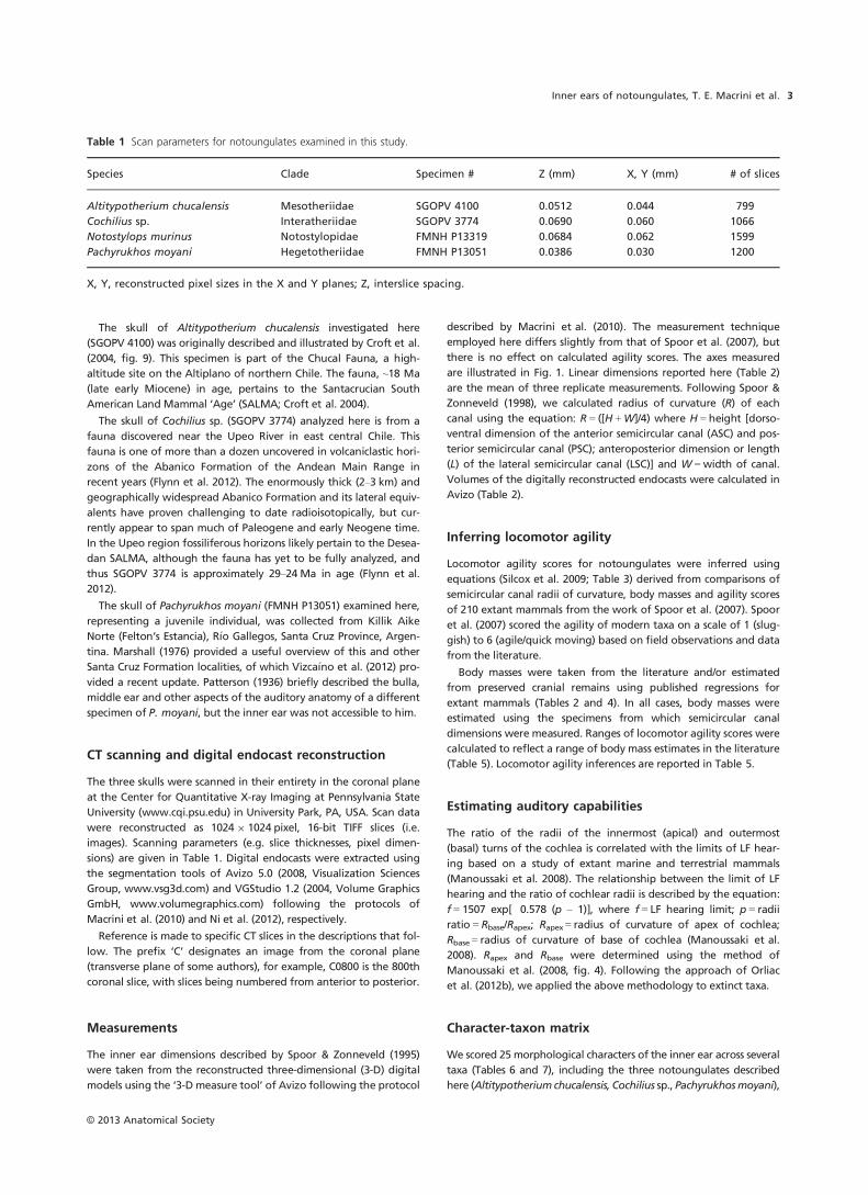
Table 1 Scan parameters for notoungulates examined in this study.
Species Clade Specimen # Z (mm) X, Y (mm) # of slices
Altitypotherium chucalensis Mesotheriidae SGOPV 4100 0.0512 0.044 799
Cochilius sp. Interatheriidae SGOPV 3774 0.0690 0.060 1066
Notostylops murinus Notostylopidae FMNH P13319 0.0684 0.062 1599
Pachyrukhos moyani Hegetotheriidae FMNH P13051 0.0386 0.030 1200
X, Y, reconstructed pixel sizes in the X and Y planes; Z, interslice spacing.
© 2013 Anatomical Society
Inner ears of notoungulates, T. E. Macrini et al. 3
Notostylops murinus (from Macrini et al. 2010), MNHN-F-BRD 23
(cf. Notoungulata) based on Billet & de Muizon (2013), and several
therian outgroups. Outgroups included the extinct primate Chilece-
bus carrascoensis (from Ni et al. 2010); Caluromys philander, the
woolly opossum (from S�anchez-Villagra & Schmelzle, 2007); the
‘condylarth’ Hyopsodus lepidus (AMNH 143783, from Orliac et al.
2012a; Benoit et al. 2013); and the stem placentals Ukhaatherium
gobiensis, Kulbeckia kulbecke and Zalambdalestes lecheyi (from
Wible et al. 2004, 2007; Ekdale & Rowe, 2011). We used several
outgroups because many of the characters are new, making their
polarities and the taxonomic level of their phylogenetic significance
uncertain.
We examined the distributions of these character states on a
pruned consensus topology of notoungulate relationships following
the analyses of Cifelli (1993), Billet (2011) and Shockey et al. (2012).
Relationships of the outgroups sampled in our analysis follow the
topology of Wible et al. (2007). One exception is the use of Chilece-
bus, an extinct primate with a published CT analysis of its inner ear
(Ni et al. 2010), which was substituted as an exemplar primate for
the more inclusive clade ‘Primates’ analyzed by Wible et al. (2007)
because the ancestral condition for inner ear characters is unknown
for the entire primate clade. Character states were examined using
the parsimony ancestral character state reconstruction option in
Mesquite (version 2.74; Maddison & Maddison, 2010), which utilizes
DELTRAN, delayed transformations, for missing character states.
Description
Below we describe 3-D reconstructions of the inner ears of
three notoungulates, comparing them with Notostylops
murinus (Macrini et al. 2010), MNHN-F-BRD 23 (cf.
Notoungulata) based on Billet & de Muizon (2013), and var-
ious other therians, fossil and extant. These comparisons
yielded the matrix of inner ear characters provided in
Tables 6 and 7.
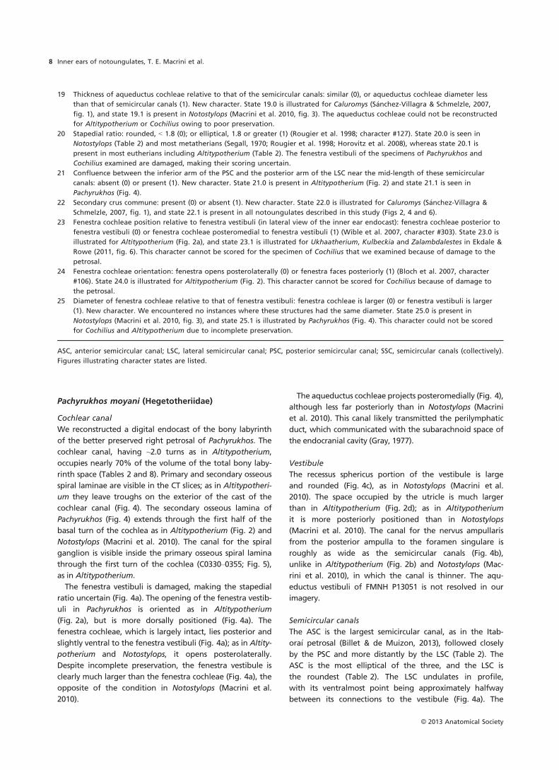
Altitypotherium chucalensis (Mesotheriidae)
Cochlear canal
We extracted a digital endocast of the bony labyrinth of
Altitypotherium from the left petrosal, the more complete
of the two petrosals. The great length of the cochlear canal
relative to the size of the entire endocast is the most note-
worthy feature of the inner ear of Altitypotherium (Fig. 2).
Despite its length, the cochlear canal occupies only about
64% of the total bony labyrinth volume (Table 2), compara-
ble to Notostylops (66%; Macrini et al. 2010). The cochlea
has 2.0 turns vs. the 2.25 turns of Notostylops (Table 8) and
2.75 turns in MNHN-F-BRD 23 (Billet & de Muizon, 2013).
The primary and secondary osseous spiral laminae are visi-
ble in the cochlear canal, as in Notostylops (Macrini et al.
2010) and therians generally (Meng & Fox, 1995; Luo et al.
2011). The primary osseous spiral lamina projects from the
meatal (inner) wall of the cochlear canal (e.g. C0342),
whereas the secondary osseous spiral lamina projects from
the radial (outer) wall (e.g. C0398). These structures leave
troughs on the external surface of the cochlear canal por-
tion of the inner ear endocast. The secondary osseous lam-
ina extends through the first half of the basal turn of the
cochlea, as in Notostylops (Macrini et al. 2010).
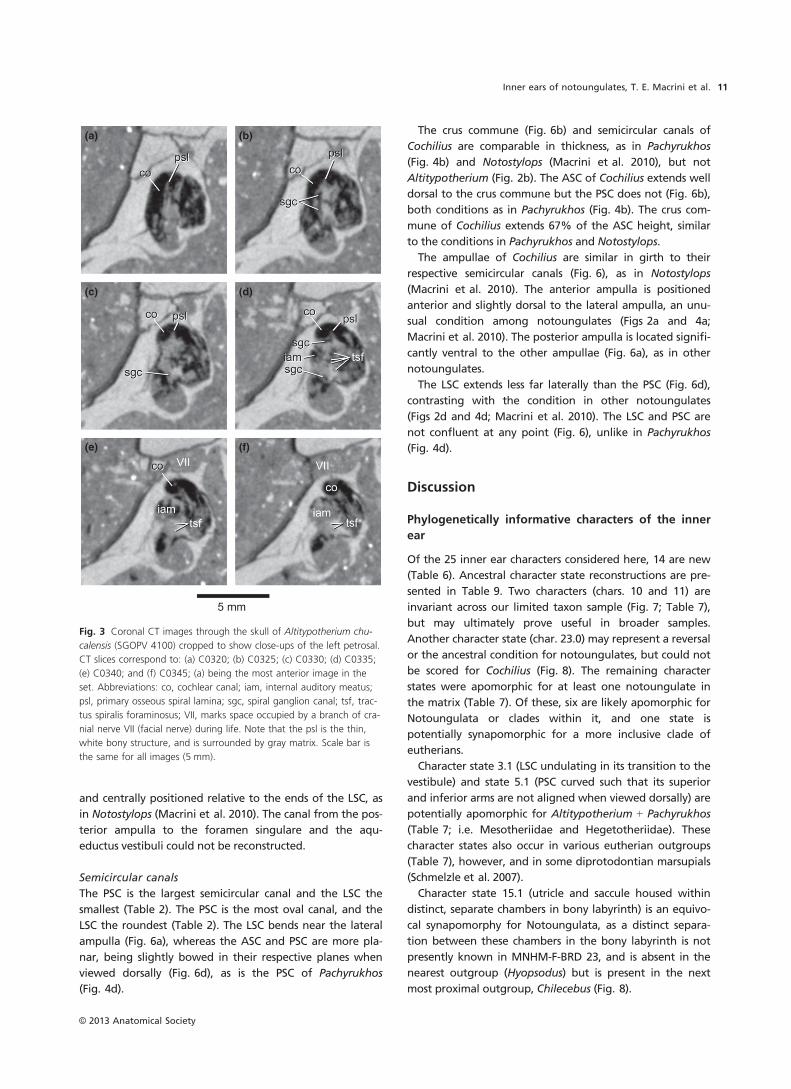
The canal housing the spiral ganglion of the cochlear
nerve is visible within the first turn of the primary osseous
spiral lamina (C0321-0338; Fig. 3). Cells of this ganglion syn-
apse with cochlear hair cells (Gray, 1977; Luo & Marsh,
1996). The tractus spiralis foraminosus, which connects the
(a)
(b)
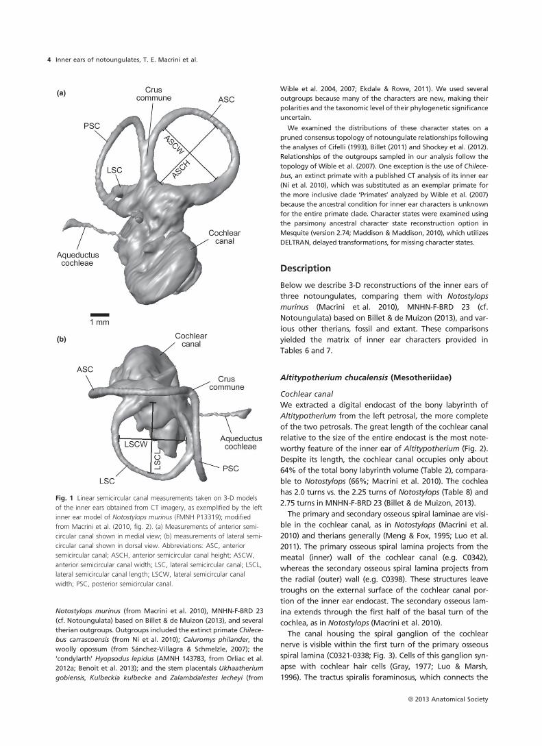
Fig. 1 Linear semicircular canal measurements taken on 3-D models
of the inner ears obtained from CT imagery, as exemplified by the left
inner ear model of Notostylops murinus (FMNH P13319); modified
from Macrini et al. (2010, fig. 2). (a) Measurements of anterior semi-
circular canal shown in medial view; (b) measurements of lateral semi-
circular canal shown in dorsal view. Abbreviations: ASC, anterior
semicircular canal; ASCH, anterior semicircular canal height; ASCW,
anterior semicircular canal width; LSC, lateral semicircular canal; LSCL,
lateral semicircular canal length; LSCW, lateral semicircular canal
width; PSC, posterior semicircular canal.
© 2013 Anatomical Society
Inner ears of notoungulates, T. E. Macrini et al.4
spiral ganglion canal to cranial nerve VIII within the internal
auditory meatus (Gray, 1977; Luo et al. 2011), is visible on
the CT images of Altitypotherium (Fig. 3d).
The fenestra vestibuli (fenestra ovalis) lies diagonal to the
plane of the LSC in an anteroventral to posterodorsal orien-
tation (Fig. 2a), as in Notostylops (Macrini et al. 2010). The
stapedial ratio (2.0), approximated from the outline of the
fenestra vestibuli because the stapes itself is missing,
contrasts with the ratios of 1.6 in Notostylops (Table 2) and
1.7 in the Itabora�ı petrosal (Billet & de Muizon, 2013).
Although the fenestra cochleae (fenestra rotunda) could
only be partially reconstructed due to damage, it appears
to face posterolaterally (Fig. 2a), as in Notostylops (Macrini
et al. 2010). Damage to the medial portion of the petrosal
precludes a clear reconstruction of the aqueductus cochleae
(cochlear aqueduct) from the CT imagery.
Vestibule
The recessus sphericus, which housed the saccule (sacculus),
is significantly smaller and less rounded (Fig. 2c) than in
Notostylops (Macrini et al. 2010). The utricular cavity is lar-
ger than the recessus sphericus (Fig. 2d). Nevertheless, the
utricule is much smaller relative to the rest of the vestibule,
and more posteriorly located, than in Notostylops (Macrini
et al. 2010).
The canal leading from the posterior ampulla to the fora-
men singulare (Fig. 2b), an opening in the internal auditory
meatus, is longer and thinner (relative to the thickness of
the semicircular canals) than in Notostylops (Macrini et al.
2010). This canal probably transmitted the nervus ampullaris
posterior, a branch of the vestibular nerve (Gray, 1977). The
Table 2 Measurements of notoungulate bony labyrinths.
Species Altitypotherium chucalensis Pachyrukhos moyani Notostylops murinus Cochilius sp.
Specimen number SGOPV 4100 FMNH P13051 FMNH P13319 SGOPV 3774
Body mass estimate (g) 8900–128003 275–5513 31194 1200–24003
Skull length 160.0 mm5 44.1 mm 99.6 mm 72.4 mm
Petrosal sampled Left Right Left Right
Cochlea volume1 122.4 mm3 17.8 mm3 28.0 mm3 21.7 mm3
Vestibule volume 37.4 mm3 4.5 mm3 8.8 mm3 14.0 mm3
SC volume2 31.7 mm3 3.3 mm3 5.4 mm3 5.2 mm3
Stapedial ratio 2.0 Damaged 1.6 Damaged
ASC height 5.03 mm 3.39 mm 4.28 mm 3.80 mm
ASC width 4.91 mm 3.85 mm 3.91 mm 3.27 mm
ASCR 2.49 mm 1.81 mm 2.05 mm 1.77 mm
LSC length 4.30 mm 2.97 mm 3.69 mm 2.93 mm
LSC width 4.80 mm 2.76 mm 3.51 mm 3.05 mm
LSCR 2.28 mm 1.43 mm 1.80 mm 1.50 mm
PSC height 4.66 mm 3.06 mm 4.00 mm 3.56 mm
PSC width 5.70 mm 3.36 mm 4.02 mm 4.35 mm
PSCR 2.59 mm 1.61 mm 2.01 mm 1.98 mm
SCR 2.45 mm 1.62 mm 1.95 mm 1.75 mm
Skull length measured from the anterior tip of the premaxillae to the end of the occipital condyles in Avizo based on the CT slices.
Stapedial ratio approximated from the outline of the fenestra vestibuli. Number of cochlear turns determined using the method of
West (1985). Radius of curvature (R) of semicircular canals calculated following Spoor & Zonneveld (1998). Measurements from Noto-
stylops murinus originally reported by Macrini et al. (2010) included for comparative purposes.1Includes aqueductus cochleae.2Includes crus commune and ampullae.3See Table 4 for explanation.4From Croft (2000).5Skull length estimated from the preserved part of the skull (Croft et al. 2004, fig. 9), and scaled to the full skull length of ‘Plesioty-
potherium’ minus of Cerdas, which has similar skull proportions (Townsend & Croft, 2010).
ASC, anterior semicircular canal; LSC, lateral semicircular canal; PSC, posterior semicircular canal; R, radius of curvature; SC, semicircular
canal; SCR, average semicircular canal radius of curvature.
Table 3 Locomotor agility equations derived by Silcox et al. (2009)
based on the dataset of Spoor et al. (2007).
Canal Equation
ASCR log10AGIL = 0.850–0.153(log10BM) + 0.706(log10ASCR)
PSCR log10AGIL = 0.881–0.151(log10BM) + 0.677(log10PSCR)
LSCR log10AGIL = 0.959–0.1670(log10BM) + 0.854(log10LSCR)
SCR log10AGIL = 0.948–0.188(log10BM) + 0.962(log10SCR)
AGIL, agility; ASCR, anterior semicircular canal radius of curva-
ture; BM, body mass in grams; LSCR, lateral semicircular canal
radius of curvature; PSCR, posterior semicircular canal radius of
curvature; SCR, average semicircular canal radius of curvature.
© 2013 Anatomical Society
Inner ears of notoungulates, T. E. Macrini et al. 5
cross-sectional diameter of the aqueductus vestibuli (vestib-
ular aqueduct), which housed the endolymphatic duct, is
much greater in Altitypotherium (Fig. 2) than in Noto-
stylops (Macrini et al. 2010).
Semicircular canals
The PSC has the largest radius of curvature of the three
semicircular canals, followed closely by the ASC, and then
more distantly by the LSC (Table 2). The PSC is the most
elliptical in outline, whereas the ASC is the most circular
(Table 2), the opposite of the condition in Notostylops
(Macrini et al. 2010). The ASC is planar (Fig. 2d) unlike the
other two canals, which undulate. The posterior arm of the
LSC bends immediately outside the vestibule (Fig. 2b), and
similarly the PSC deviates from a plane (Fig. 2d).
The crus commune is more robust compared with the
thickness of the individual semicircular canals (Fig. 2) than
in Notostylops (Macrini et al. 2010). The ASC and PSC of
Altitypotherium extend little beyond the dorsal margin of
the crus commune, unlike in Notostylops where they pro-
ject considerably farther (Macrini et al. 2010). The crus
commune extends 73% of the height of the ASC,
whereas in Notostylops the crus extends only 64% of this
height.
The anterior and posterior ampullae (Fig. 2) are more
pronounced than in Notostylops (Macrini et al. 2010), being
significantly more inflated than the semicircular canals. The
anterior and lateral ampullae attach to the vestibule in the
same horizontal plane as the LSC; the posterior ampulla,
however, is located posteroventral to the crus commune
and the entry of the posterior arm of the LSC into the
vestibule (Fig. 2a).
The LSC and PSC extend equally far laterally when viewed
dorsally (Fig. 2d). No secondary crus commune occurs
between these two canals unlike the condition in
MNHN-F-BRD 23 (Billet & de Muizon, 2013).
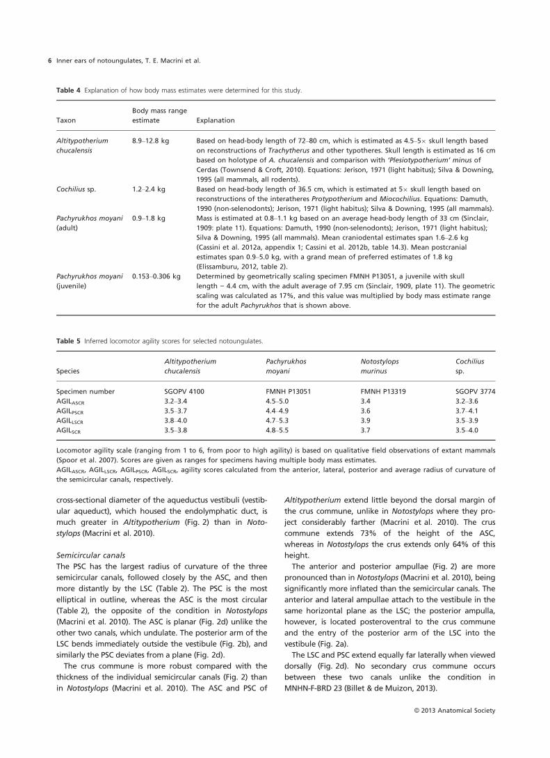
Table 5 Inferred locomotor agility scores for selected notoungulates.
Species
Altitypotherium
chucalensis
Pachyrukhos
moyani
Notostylops
murinus
Cochilius
sp.
Specimen number SGOPV 4100 FMNH P13051 FMNH P13319 SGOPV 3774
AGILASCR 3.2–3.4 4.5–5.0 3.4 3.2–3.6
AGILPSCR 3.5–3.7 4.4–4.9 3.6 3.7–4.1
AGILLSCR 3.8–4.0 4.7–5.3 3.9 3.5–3.9
AGILSCR 3.5–3.8 4.8–5.5 3.7 3.5–4.0
Locomotor agility scale (ranging from 1 to 6, from poor to high agility) is based on qualitative field observations of extant mammals
(Spoor et al. 2007). Scores are given as ranges for specimens having multiple body mass estimates.
AGILASCR, AGILLSCR, AGILPSCR, AGILSCR, agility scores calculated from the anterior, lateral, posterior and average radius of curvature of
the semicircular canals, respectively.
Table 4 Explanation of how body mass estimates were determined for this study.
Taxon
Body mass range
estimate Explanation
Altitypotherium
chucalensis
8.9–12.8 kg Based on head-body length of 72–80 cm, which is estimated as 4.5–59 skull length based
on reconstructions of Trachytherus and other typotheres. Skull length is estimated as 16 cm
based on holotype of A. chucalensis and comparison with ‘Plesiotypotherium’ minus of
Cerdas (Townsend & Croft, 2010). Equations: Jerison, 1971 (light habitus); Silva & Downing,
1995 (all mammals, all rodents).
Cochilius sp. 1.2–2.4 kg Based on head-body length of 36.5 cm, which is estimated at 59 skull length based on
reconstructions of the interatheres Protypotherium and Miocochilius. Equations: Damuth,
1990 (non-selenodonts); Jerison, 1971 (light habitus); Silva & Downing, 1995 (all mammals).
Pachyrukhos moyani
(adult)
0.9–1.8 kg Mass is estimated at 0.8–1.1 kg based on an average head-body length of 33 cm (Sinclair,
1909: plate 11). Equations: Damuth, 1990 (non-selenodonts); Jerison, 1971 (light habitus);
Silva & Downing, 1995 (all mammals). Mean craniodental estimates span 1.6–2.6 kg
(Cassini et al. 2012a, appendix 1; Cassini et al. 2012b, table 14.3). Mean postcranial
estimates span 0.9–5.0 kg, with a grand mean of preferred estimates of 1.8 kg
(Elissamburu, 2012, table 2).
Pachyrukhos moyani
(juvenile)
0.153–0.306 kg Determined by geometrically scaling specimen FMNH P13051, a juvenile with skull
length = 4.4 cm, with the adult average of 7.95 cm (Sinclair, 1909, plate 11). The geometric
scaling was calculated as 17%, and this value was multiplied by body mass estimate range
for the adult Pachyrukhos that is shown above.
© 2013 Anatomical Society
Inner ears of notoungulates, T. E. Macrini et al.6
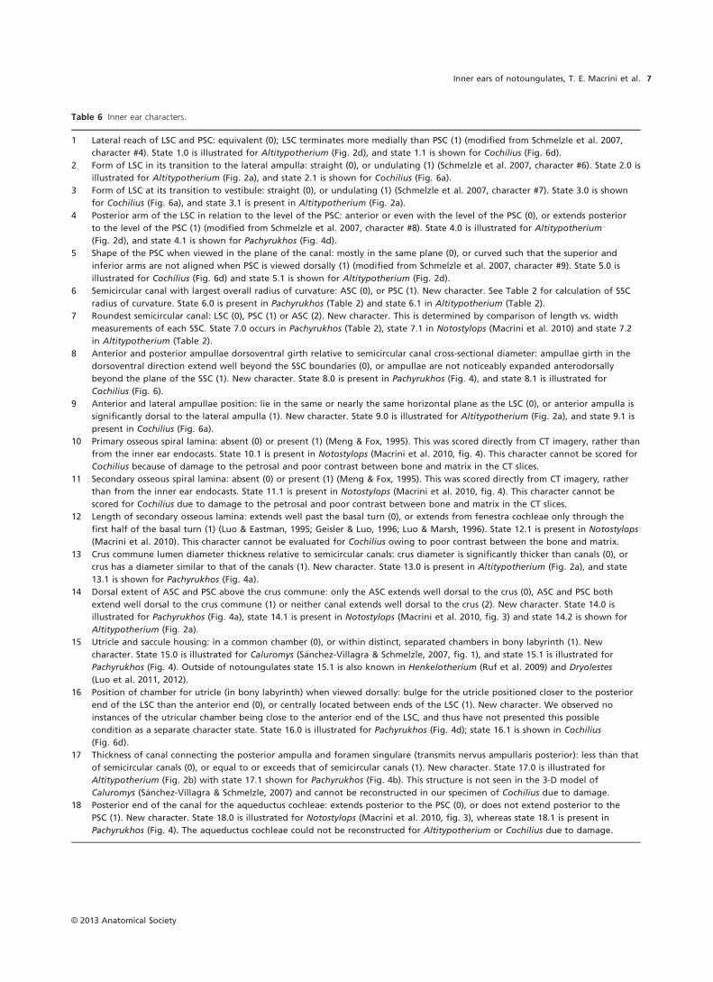
Table 6 Inner ear characters.
1 Lateral reach of LSC and PSC: equivalent (0); LSC terminates more medially than PSC (1) (modified from Schmelzle et al. 2007,
character #4). State 1.0 is illustrated for Altitypotherium (Fig. 2d), and state 1.1 is shown for Cochilius (Fig. 6d).
2 Form of LSC in its transition to the lateral ampulla: straight (0), or undulating (1) (Schmelzle et al. 2007, character #6). State 2.0 is
illustrated for Altitypotherium (Fig. 2a), and state 2.1 is shown for Cochilius (Fig. 6a).
3 Form of LSC at its transition to vestibule: straight (0), or undulating (1) (Schmelzle et al. 2007, character #7). State 3.0 is shown
for Cochilius (Fig. 6a), and state 3.1 is present in Altitypotherium (Fig. 2a).
4 Posterior arm of the LSC in relation to the level of the PSC: anterior or even with the level of the PSC (0), or extends posterior
to the level of the PSC (1) (modified from Schmelzle et al. 2007, character #8). State 4.0 is illustrated for Altitypotherium
(Fig. 2d), and state 4.1 is shown for Pachyrukhos (Fig. 4d).
5 Shape of the PSC when viewed in the plane of the canal: mostly in the same plane (0), or curved such that the superior and
inferior arms are not aligned when PSC is viewed dorsally (1) (modified from Schmelzle et al. 2007, character #9). State 5.0 is
illustrated for Cochilius (Fig. 6d) and state 5.1 is shown for Altitypotherium (Fig. 2d).
6 Semicircular canal with largest overall radius of curvature: ASC (0), or PSC (1). New character. See Table 2 for calculation of SSC
radius of curvature. State 6.0 is present in Pachyrukhos (Table 2) and state 6.1 in Altitypotherium (Table 2).
7 Roundest semicircular canal: LSC (0), PSC (1) or ASC (2). New character. This is determined by comparison of length vs. width
measurements of each SSC. State 7.0 occurs in Pachyrukhos (Table 2), state 7.1 in Notostylops (Macrini et al. 2010) and state 7.2
in Altitypotherium (Table 2).
8 Anterior and posterior ampullae dorsoventral girth relative to semicircular canal cross-sectional diameter: ampullae girth in the
dorsoventral direction extend well beyond the SSC boundaries (0), or ampullae are not noticeably expanded anterodorsally
beyond the plane of the SSC (1). New character. State 8.0 is present in Pachyrukhos (Fig. 4), and state 8.1 is illustrated for
Cochilius (Fig. 6).
9 Anterior and lateral ampullae position: lie in the same or nearly the same horizontal plane as the LSC (0), or anterior ampulla is
significantly dorsal to the lateral ampulla (1). New character. State 9.0 is illustrated for Altitypotherium (Fig. 2a), and state 9.1 is
present in Cochilius (Fig. 6a).
10 Primary osseous spiral lamina: absent (0) or present (1) (Meng & Fox, 1995). This was scored directly from CT imagery, rather than
from the inner ear endocasts. State 10.1 is present in Notostylops (Macrini et al. 2010, fig. 4). This character cannot be scored for
Cochilius because of damage to the petrosal and poor contrast between bone and matrix in the CT slices.
11 Secondary osseous spiral lamina: absent (0) or present (1) (Meng & Fox, 1995). This was scored directly from CT imagery, rather
than from the inner ear endocasts. State 11.1 is present in Notostylops (Macrini et al. 2010, fig. 4). This character cannot be
scored for Cochilius due to damage to the petrosal and poor contrast between bone and matrix in the CT slices.
12 Length of secondary osseous lamina: extends well past the basal turn (0), or extends from fenestra cochleae only through the
first half of the basal turn (1) (Luo & Eastman, 1995; Geisler & Luo, 1996; Luo & Marsh, 1996). State 12.1 is present in Notostylops
(Macrini et al. 2010). This character cannot be evaluated for Cochilius owing to poor contrast between the bone and matrix.
13 Crus commune lumen diameter thickness relative to semicircular canals: crus diameter is significantly thicker than canals (0), or
crus has a diameter similar to that of the canals (1). New character. State 13.0 is present in Altitypotherium (Fig. 2a), and state
13.1 is shown for Pachyrukhos (Fig. 4a).
14 Dorsal extent of ASC and PSC above the crus commune: only the ASC extends well dorsal to the crus (0), ASC and PSC both
extend well dorsal to the crus commune (1) or neither canal extends well dorsal to the crus (2). New character. State 14.0 is
illustrated for Pachyrukhos (Fig. 4a), state 14.1 is present in Notostylops (Macrini et al. 2010, fig. 3) and state 14.2 is shown for
Altitypotherium (Fig. 2a).
15 Utricle and saccule housing: in a common chamber (0), or within distinct, separated chambers in bony labyrinth (1). New
character. State 15.0 is illustrated for Caluromys (S�anchez-Villagra & Schmelzle, 2007, fig. 1), and state 15.1 is illustrated for
Pachyrukhos (Fig. 4). Outside of notoungulates state 15.1 is also known in Henkelotherium (Ruf et al. 2009) and Dryolestes
(Luo et al. 2011, 2012).
16 Position of chamber for utricle (in bony labyrinth) when viewed dorsally: bulge for the utricle positioned closer to the posterior
end of the LSC than the anterior end (0), or centrally located between ends of the LSC (1). New character. We observed no
instances of the utricular chamber being close to the anterior end of the LSC, and thus have not presented this possible
condition as a separate character state. State 16.0 is illustrated for Pachyrukhos (Fig. 4d); state 16.1 is shown in Cochilius
(Fig. 6d).
17 Thickness of canal connecting the posterior ampulla and foramen singulare (transmits nervus ampullaris posterior): less than that
of semicircular canals (0), or equal to or exceeds that of semicircular canals (1). New character. State 17.0 is illustrated for
Altitypotherium (Fig. 2b) with state 17.1 shown for Pachyrukhos (Fig. 4b). This structure is not seen in the 3-D model of
Caluromys (S�anchez-Villagra & Schmelzle, 2007) and cannot be reconstructed in our specimen of Cochilius due to damage.
18 Posterior end of the canal for the aqueductus cochleae: extends posterior to the PSC (0), or does not extend posterior to the
PSC (1). New character. State 18.0 is illustrated for Notostylops (Macrini et al. 2010, fig. 3), whereas state 18.1 is present in
Pachyrukhos (Fig. 4). The aqueductus cochleae could not be reconstructed for Altitypotherium or Cochilius due to damage.
© 2013 Anatomical Society
Inner ears of notoungulates, T. E. Macrini et al. 7
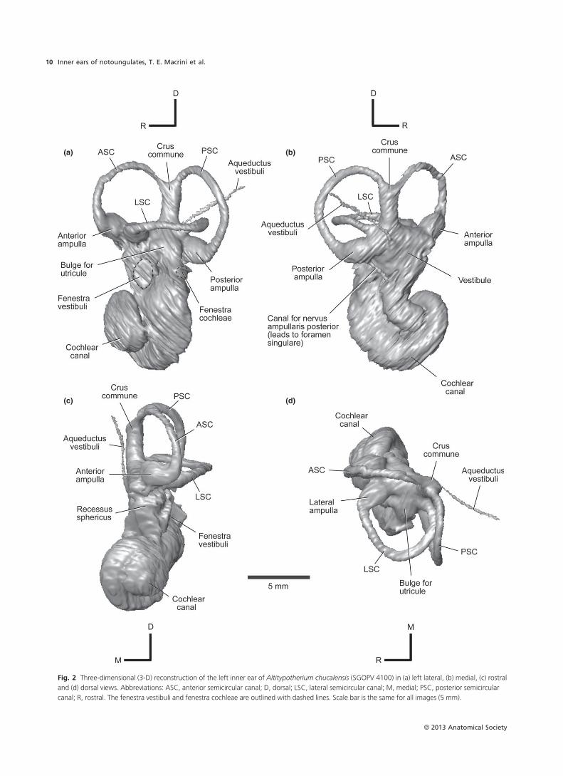
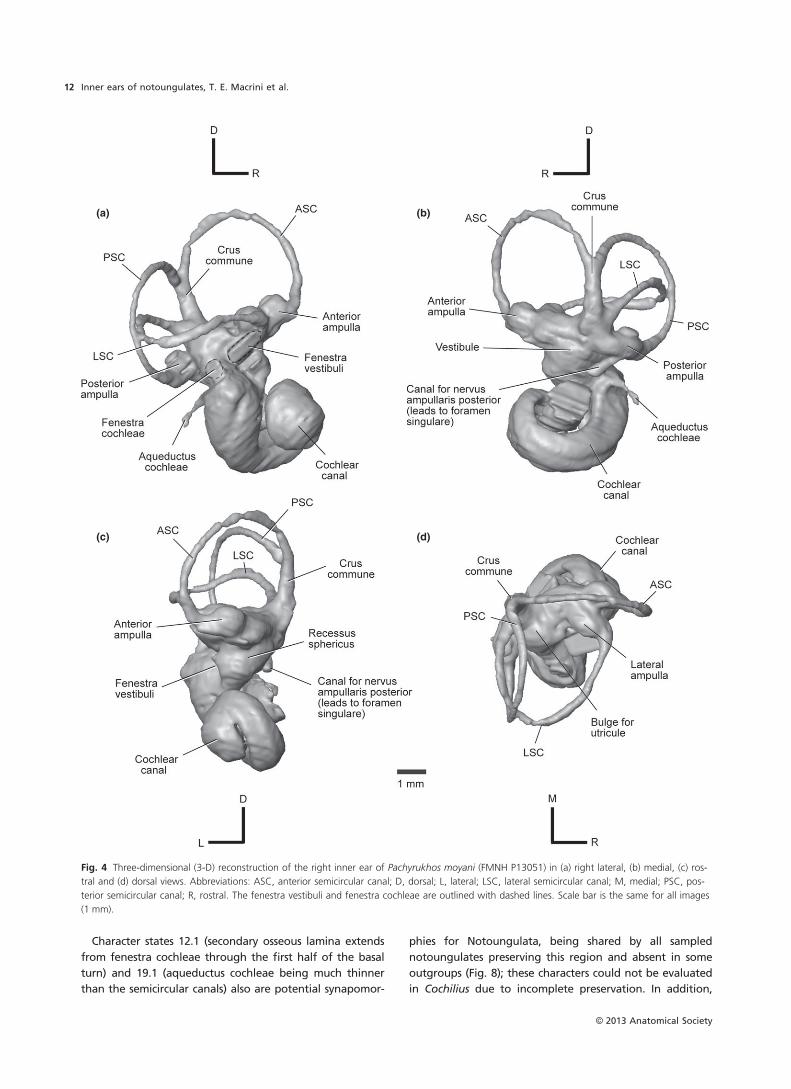
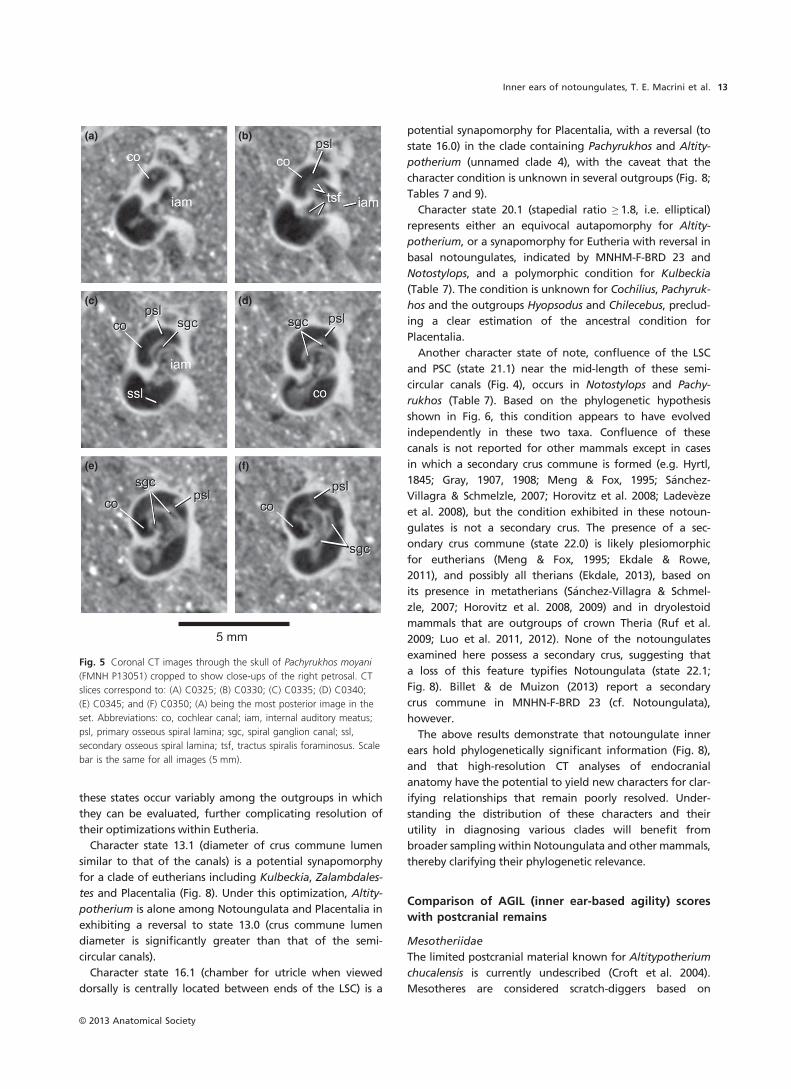
Pachyrukhos moyani (Hegetotheriidae)
Cochlear canal
We reconstructed a digital endocast of the bony labyrinth
of the better preserved right petrosal of Pachyrukhos. The
cochlear canal, having ~2.0 turns as in Altitypotherium,
occupies nearly 70% of the volume of the total bony laby-
rinth space (Tables 2 and 8). Primary and secondary osseous
spiral laminae are visible in the CT slices; as in Altitypotheri-
um they leave troughs on the exterior of the cast of the
cochlear canal (Fig. 4). The secondary osseous lamina of
Pachyrukhos (Fig. 4) extends through the first half of the
basal turn of the cochlea as in Altitypotherium (Fig. 2) and
Notostylops (Macrini et al. 2010). The canal for the spiral
ganglion is visible inside the primary osseous spiral lamina
through the first turn of the cochlea (C0330–0355; Fig. 5),
as in Altitypotherium.
The fenestra vestibuli is damaged, making the stapedial
ratio uncertain (Fig. 4a). The opening of the fenestra vestib-
uli in Pachyrukhos is oriented as in Altitypotherium
(Fig. 2a), but is more dorsally positioned (Fig. 4a). The
fenestra cochleae, which is largely intact, lies posterior and
slightly ventral to the fenestra vestibuli (Fig. 4a); as in Altity-
potherium and Notostylops, it opens posterolaterally.
Despite incomplete preservation, the fenestra vestibule is
clearly much larger than the fenestra cochleae (Fig. 4a), the
opposite of the condition in Notostylops (Macrini et al.
2010).
The aqueductus cochleae projects posteromedially (Fig. 4),
although less far posteriorly than in Notostylops (Macrini
et al. 2010). This canal likely transmitted the perilymphatic
duct, which communicated with the subarachnoid space of
the endocranial cavity (Gray, 1977).
Vestibule
The recessus sphericus portion of the vestibule is large
and rounded (Fig. 4c), as in Notostylops (Macrini et al.
2010). The space occupied by the utricle is much larger
than in Altitypotherium (Fig. 2d); as in Altitypotherium
it is more posteriorly positioned than in Notostylops
(Macrini et al. 2010). The canal for the nervus ampullaris
from the posterior ampulla to the foramen singulare is
roughly as wide as the semicircular canals (Fig. 4b),
unlike in Altitypotherium (Fig. 2b) and Notostylops (Mac-
rini et al. 2010), in which the canal is thinner. The aqu-
eductus vestibuli of FMNH P13051 is not resolved in our
imagery.
Semicircular canals
The ASC is the largest semicircular canal, as in the Itab-
ora�ı petrosal (Billet & de Muizon, 2013), followed closely
by the PSC and more distantly by the LSC (Table 2). The
ASC is the most elliptical of the three, and the LSC is
the roundest (Table 2). The LSC undulates in profile,
with its ventralmost point being approximately halfway
between its connections to the vestibule (Fig. 4a). The
19 Thickness of aqueductus cochleae relative to that of the semicircular canals: similar (0), or aqueductus cochleae diameter less
than that of semicircular canals (1). New character. State 19.0 is illustrated for Caluromys (S�anchez-Villagra & Schmelzle, 2007,
fig. 1), and state 19.1 is present in Notostylops (Macrini et al. 2010, fig. 3). The aqueductus cochleae could not be reconstructed
for Altitypotherium or Cochilius owing to poor preservation.
20 Stapedial ratio: rounded, < 1.8 (0); or elliptical, 1.8 or greater (1) (Rougier et al. 1998; character #127). State 20.0 is seen in
Notostylops (Table 2) and most metatherians (Segall, 1970; Rougier et al. 1998; Horovitz et al. 2008), whereas state 20.1 is
present in most eutherians including Altitypotherium (Table 2). The fenestra vestibuli of the specimens of Pachyrukhos and
Cochilius examined are damaged, making their scoring uncertain.
21 Confluence between the inferior arm of the PSC and the posterior arm of the LSC near the mid-length of these semicircular
canals: absent (0) or present (1). New character. State 21.0 is present in Altitypotherium (Fig. 2) and state 21.1 is seen in
Pachyrukhos (Fig. 4).
22 Secondary crus commune: present (0) or absent (1). New character. State 22.0 is illustrated for Caluromys (S�anchez-Villagra &
Schmelzle, 2007, fig. 1), and state 22.1 is present in all notoungulates described in this study (Figs 2, 4 and 6).
23 Fenestra cochleae position relative to fenestra vestibuli (in lateral view of the inner ear endocast): fenestra cochleae posterior to
fenestra vestibuli (0) or fenestra cochleae posteromedial to fenestra vestibuli (1) (Wible et al. 2007, character #303). State 23.0 is
illustrated for Altitypotherium (Fig. 2a), and state 23.1 is illustrated for Ukhaatherium, Kulbeckia and Zalambdalestes in Ekdale &
Rowe (2011, fig. 6). This character cannot be scored for the specimen of Cochilius that we examined because of damage to the
petrosal.
24 Fenestra cochleae orientation: fenestra opens posterolaterally (0) or fenestra faces posteriorly (1) (Bloch et al. 2007, character
#106). State 24.0 is illustrated for Altitypotherium (Fig. 2). This character cannot be scored for Cochilius because of damage to
the petrosal.
25 Diameter of fenestra cochleae relative to that of fenestra vestibuli: fenestra cochleae is larger (0) or fenestra vestibuli is larger
(1). New character. We encountered no instances where these structures had the same diameter. State 25.0 is present in
Notostylops (Macrini et al. 2010, fig. 3), and state 25.1 is illustrated by Pachyrukhos (Fig. 4). This character could not be scored
for Cochilius and Altitypotherium due to incomplete preservation.
ASC, anterior semicircular canal; LSC, lateral semicircular canal; PSC, posterior semicircular canal; SSC, semicircular canals (collectively).
Figures illustrating character states are listed.
© 2013 Anatomical Society
Inner ears of notoungulates, T. E. Macrini et al.8
ASC and PSC are planar, but the PSC is somewhat
bowed (when viewed dorsally) in the rostrocaudal
direction (Fig. 4d).
The crus commune (Fig. 4b) and semicircular canals are
similar in thickness, whereas in Altitypotherium the crus is
thicker (Fig. 2b). The ASC of Pachyrukhos extends well dor-
sal of the crus commune but the PSC does not (Fig. 4b). The
crus commune of Pachyrukhos extends 65% of the height
of the ASC, resembling Notostylops.
The maximum diameter of the anterior and posterior
ampullae in the dorsoventral axis (Fig. 4a) exceeds the
diameter of the semicircular canals, as in Altitypotherium
(Fig. 2a). The anterior and lateral ampullae occur in the
same horizontal plane as the LSC (as in Notostylops, Macrini
et al. 2010; and Altitypotherium, Fig. 2); by contrast, the
posterior ampulla lies ventral to this plane and posterior to
the crus commune (Fig. 4a).
The PSC and LSC extend equally far laterally (Fig. 4d), as
in Notostylops (Macrini et al. 2010) and Altitypotherium
(Fig. 2d). The posterior arm of the LSC and the inferior arm
of the PSC are confluent in Pachyrukhos (seen on both sides
of the skull; Fig. 4), resembling Notostylops (Macrini et al.
2010).
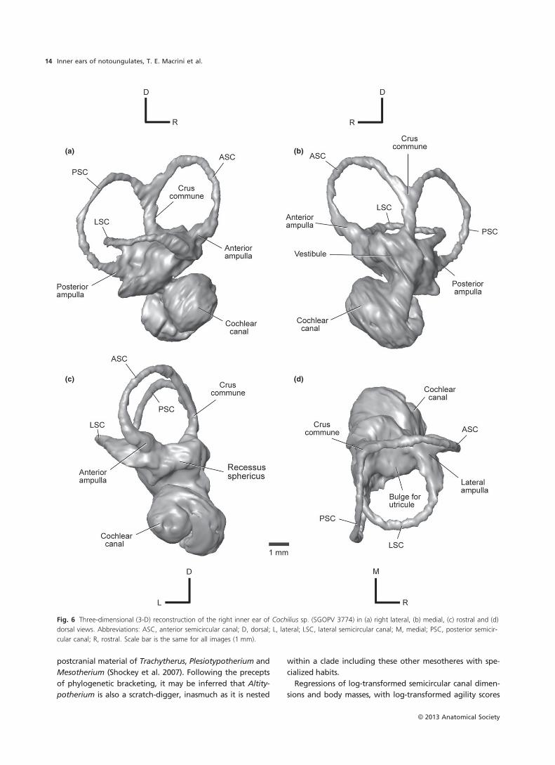
Cochilius sp. (Interatheriidae)
Cochlear canal
We analyzed the better preserved right petrosal of Cochilius
sp. (SGOPV 3774); this recently collected specimen has not
yet been identified to species, and indeed Cochilius as a
whole requires taxonomic revision. The cochlear canal
makes up ~53% of the volume of the bony labyrinth
(Table 2) and consists of 2.0 turns (Table 8). Damage to the
petrosals and poor density contrast between the fossil and
matrix of this specimen prevent reliable reconstructions of
the fenestra vestibuli, fenestra cochleae and aqueductus
cochleae. Similarly, it cannot be established whether
primary and osseous spiral laminae are present, nor can the
spiral ganglion canal be discerned.
Vestibule
The recessus sphericus portion of the vestibule for the sac-
cule and the chamber for the utricle are separated within
the bony labyrinth (Fig. 6), as in all other notoungulates
examined (Figs 2 and 4; Macrini et al. 2010). The recessus
sphericus of Cochilius is small (Fig. 6c), as in Altitypotherium
(Fig. 2c), but the chamber for the utricule is large (Fig. 6d)
Table 7 Taxon-inner ear character matrix.
Taxon 1 2 3 4 5 6 7 8 9 10 11 12 13 14 15 16 17 18 19 20 21 22 23 24 25
Caluromys 0 0 0 0 0 0 0 0 0 1 1 0 0 0 0 0 ? 0 0 0 0 0 0 0 0†Ukhaatherium 0 0 0 0 1 0 0 0 1 ? ? ? 0 0 ? ? ? ? ? 1 0 0 1 ? ?†Kulbeckia 0 0 0 0 1 0 1&2 0 0 1 1 0 1 0 ? ? ? ? ? 0&1 0 0 1 ? ?†Zalambdalestes 0 0 0 0 1 0 1 0 0 1 1 1 1 0 ? ? ? ? ? 1 0 0 1 0 ?†Chilecebus 0 0 1 0 0 0 0 0 0 ? ? ? 1 0 1 1 ? 0 1 ? 0 1 ? ? ?†Hyopsodus 0 1 0 0 0 0 0 0 0 ? 1 ? 1 0 0 ? ? 1 0 ? 0 0 ? 1 ?†MNHN-F-BRD 23 0 0 0 0 0 0 0 0 0 1 1 1 1 1 ? ? 1 0 1 0 0 0 0 0 0†Notostylops 0 0 0 0 0 0 1 1 0 1 1 1 1 1 1 1 0 0 1 0 1 1 0 0 0†Altitypotherium 0 0 1 0 1 1 2 0 0 1 1 1 0 2 1 0 0 ? ? 1 0 1 0 0 ?†Pachyrukhos 0 0 1 1 1 0 0 0 0 1 1 1 1 0 1 0 1 1 1 ? 1 1 0 0 1†Cochilius 1 1 0 0 0 1 0 1 1 ? ? ? 1 0 1 1 ? ? ? ? 0 1 ? ? ?
?, unknown. Character data sources: Caluromys (S�anchez-Villagra & Schmelzle, 2007); Chilecebus (Ni et al. 2010); Hyopsodus (AMNH
143783, inner ear endocast courtesy of Maeva Orliac; Benoit et al. 2013; Orliac et al. 2012a); Kulbeckia (Wible et al. 2004, 2007; Ekdale
& Rowe, 2011); MNHN-F-BRD 23 (Billet & de Muizon, 2013); Notostylops (Macrini et al. 2010); Ukhaatherium (Wible et al. 2007; Ekdale
& Rowe, 2011); Zalambdalestes (Wible et al. 2004, 2007; Ekdale & Rowe, 2011).
†, extinct taxon.
Table 8 LF limits and morphometric data for the inner ear of selected notoungulates.
Species
Altitypotherium
chucalensis
Pachyrukhos
moyani
Notostylops
murinus Cochilius sp.
Specimen number SGOPV 4100 FMNH P13051 FMNH P13319 SGOPV 3774
# cochlear turns 2.0 2.0 2.25 2.0
Radii ratio 5.8 5 9 6
60-dB LF limit (Hz) 92 149 15 84
Radii ratio = Rbase/Rapex (measured following the methods of Manoussaki et al. 2008); 60-dB LF limit calculated using equation of
Manoussaki et al. (2008).
LF, low-frequency hearing; Rapex, radius of apex of cochlear canal; Rbase, radius of base of cochlear canal.
© 2013 Anatomical Society
Inner ears of notoungulates, T. E. Macrini et al. 9
(a) (b)
(c) (d)
Fig. 2 Three-dimensional (3-D) reconstruction of the left inner ear of Altitypotherium chucalensis (SGOPV 4100) in (a) left lateral, (b) medial, (c) rostral
and (d) dorsal views. Abbreviations: ASC, anterior semicircular canal; D, dorsal; LSC, lateral semicircular canal; M, medial; PSC, posterior semicircular
canal; R, rostral. The fenestra vestibuli and fenestra cochleae are outlined with dashed lines. Scale bar is the same for all images (5 mm).
© 2013 Anatomical Society
Inner ears of notoungulates, T. E. Macrini et al.10
and centrally positioned relative to the ends of the LSC, as
in Notostylops (Macrini et al. 2010). The canal from the pos-
terior ampulla to the foramen singulare and the aqu-
eductus vestibuli could not be reconstructed.
Semicircular canals
The PSC is the largest semicircular canal and the LSC the
smallest (Table 2). The PSC is the most oval canal, and the
LSC the roundest (Table 2). The LSC bends near the lateral
ampulla (Fig. 6a), whereas the ASC and PSC are more pla-
nar, being slightly bowed in their respective planes when
viewed dorsally (Fig. 6d), as is the PSC of Pachyrukhos
(Fig. 4d).
The crus commune (Fig. 6b) and semicircular canals of
Cochilius are comparable in thickness, as in Pachyrukhos
(Fig. 4b) and Notostylops (Macrini et al. 2010), but not
Altitypotherium (Fig. 2b). The ASC of Cochilius extends well
dorsal to the crus commune but the PSC does not (Fig. 6b),
both conditions as in Pachyrukhos (Fig. 4b). The crus com-
mune of Cochilius extends 67% of the ASC height, similar
to the conditions in Pachyrukhos and Notostylops.
The ampullae of Cochilius are similar in girth to their
respective semicircular canals (Fig. 6), as in Notostylops
(Macrini et al. 2010). The anterior ampulla is positioned
anterior and slightly dorsal to the lateral ampulla, an unu-
sual condition among notoungulates (Figs 2a and 4a;
Macrini et al. 2010). The posterior ampulla is located signifi-
cantly ventral to the other ampullae (Fig. 6a), as in other
notoungulates.
The LSC extends less far laterally than the PSC (Fig. 6d),
contrasting with the condition in other notoungulates
(Figs 2d and 4d; Macrini et al. 2010). The LSC and PSC are
not confluent at any point (Fig. 6), unlike in Pachyrukhos
(Fig. 4d).
Discussion
Phylogenetically informative characters of the inner
ear
Of the 25 inner ear characters considered here, 14 are new
(Table 6). Ancestral character state reconstructions are pre-
sented in Table 9. Two characters (chars. 10 and 11) are
invariant across our limited taxon sample (Fig. 7; Table 7),
but may ultimately prove useful in broader samples.
Another character state (char. 23.0) may represent a reversal
or the ancestral condition for notoungulates, but could not
be scored for Cochilius (Fig. 8). The remaining character
states were apomorphic for at least one notoungulate in
the matrix (Table 7). Of these, six are likely apomorphic for
Notoungulata or clades within it, and one state is
potentially synapomorphic for a more inclusive clade of
eutherians.
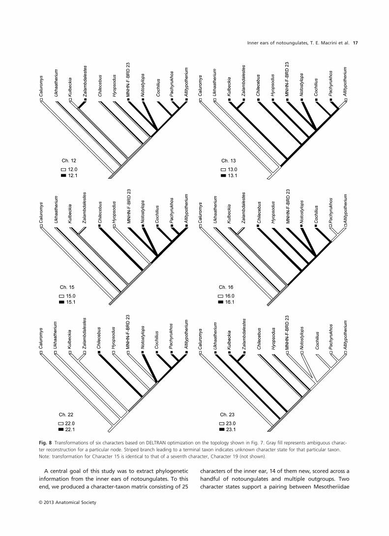
Character state 3.1 (LSC undulating in its transition to the
vestibule) and state 5.1 (PSC curved such that its superior
and inferior arms are not aligned when viewed dorsally) are
potentially apomorphic for Altitypotherium + Pachyrukhos
(Table 7; i.e. Mesotheriidae and Hegetotheriidae). These
character states also occur in various eutherian outgroups
(Table 7), however, and in some diprotodontian marsupials
(Schmelzle et al. 2007).
Character state 15.1 (utricle and saccule housed within
distinct, separate chambers in bony labyrinth) is an equivo-
cal synapomorphy for Notoungulata, as a distinct separa-
tion between these chambers in the bony labyrinth is not
presently known in MNHM-F-BRD 23, and is absent in the
nearest outgroup (Hyopsodus) but is present in the next
most proximal outgroup, Chilecebus (Fig. 8).
(a) (b)
(c) (d)
(e) (f)
Fig. 3 Coronal CT images through the skull of Altitypotherium chu-
calensis (SGOPV 4100) cropped to show close-ups of the left petrosal.
CT slices correspond to: (a) C0320; (b) C0325; (c) C0330; (d) C0335;
(e) C0340; and (f) C0345; (a) being the most anterior image in the
set. Abbreviations: co, cochlear canal; iam, internal auditory meatus;
psl, primary osseous spiral lamina; sgc, spiral ganglion canal; tsf, trac-
tus spiralis foraminosus; VII, marks space occupied by a branch of cra-
nial nerve VII (facial nerve) during life. Note that the psl is the thin,
white bony structure, and is surrounded by gray matrix. Scale bar is
the same for all images (5 mm).
© 2013 Anatomical Society
Inner ears of notoungulates, T. E. Macrini et al. 11
Character states 12.1 (secondary osseous lamina extends
from fenestra cochleae through the first half of the basal
turn) and 19.1 (aqueductus cochleae being much thinner
than the semicircular canals) also are potential synapomor-
phies for Notoungulata, being shared by all sampled
notoungulates preserving this region and absent in some
outgroups (Fig. 8); these characters could not be evaluated
in Cochilius due to incomplete preservation. In addition,
(a) (b)
(c) (d)
Fig. 4 Three-dimensional (3-D) reconstruction of the right inner ear of Pachyrukhos moyani (FMNH P13051) in (a) right lateral, (b) medial, (c) ros-
tral and (d) dorsal views. Abbreviations: ASC, anterior semicircular canal; D, dorsal; L, lateral; LSC, lateral semicircular canal; M, medial; PSC, pos-
terior semicircular canal; R, rostral. The fenestra vestibuli and fenestra cochleae are outlined with dashed lines. Scale bar is the same for all images
(1 mm).
© 2013 Anatomical Society
Inner ears of notoungulates, T. E. Macrini et al.12
these states occur variably among the outgroups in which
they can be evaluated, further complicating resolution of
their optimizations within Eutheria.
Character state 13.1 (diameter of crus commune lumen
similar to that of the canals) is a potential synapomorphy
for a clade of eutherians including Kulbeckia, Zalambdales-
tes and Placentalia (Fig. 8). Under this optimization, Altity-
potherium is alone among Notoungulata and Placentalia in
exhibiting a reversal to state 13.0 (crus commune lumen
diameter is significantly greater than that of the semi-
circular canals).
Character state 16.1 (chamber for utricle when viewed
dorsally is centrally located between ends of the LSC) is a
potential synapomorphy for Placentalia, with a reversal (to
state 16.0) in the clade containing Pachyrukhos and Altity-
potherium (unnamed clade 4), with the caveat that the
character condition is unknown in several outgroups (Fig. 8;
Tables 7 and 9).
Character state 20.1 (stapedial ratio ≥ 1.8, i.e. elliptical)
represents either an equivocal autapomorphy for Altity-
potherium, or a synapomorphy for Eutheria with reversal in
basal notoungulates, indicated by MNHM-F-BRD 23 and
Notostylops, and a polymorphic condition for Kulbeckia
(Table 7). The condition is unknown for Cochilius, Pachyruk-
hos and the outgroups Hyopsodus and Chilecebus, preclud-
ing a clear estimation of the ancestral condition for
Placentalia.
Another character state of note, confluence of the LSC
and PSC (state 21.1) near the mid-length of these semi-
circular canals (Fig. 4), occurs in Notostylops and Pachy-
rukhos (Table 7). Based on the phylogenetic hypothesis
shown in Fig. 6, this condition appears to have evolved
independently in these two taxa. Confluence of these
canals is not reported for other mammals except in cases
in which a secondary crus commune is formed (e.g. Hyrtl,
1845; Gray, 1907, 1908; Meng & Fox, 1995; S�anchez-
Villagra & Schmelzle, 2007; Horovitz et al. 2008; Ladev�eze
et al. 2008), but the condition exhibited in these notoun-
gulates is not a secondary crus. The presence of a sec-
ondary crus commune (state 22.0) is likely plesiomorphic
for eutherians (Meng & Fox, 1995; Ekdale & Rowe,
2011), and possibly all therians (Ekdale, 2013), based on
its presence in metatherians (S�anchez-Villagra & Schmel-
zle, 2007; Horovitz et al. 2008, 2009) and in dryolestoid
mammals that are outgroups of crown Theria (Ruf et al.
2009; Luo et al. 2011, 2012). None of the notoungulates
examined here possess a secondary crus, suggesting that
a loss of this feature typifies Notoungulata (state 22.1;
Fig. 8). Billet & de Muizon (2013) report a secondary
crus commune in MNHN-F-BRD 23 (cf. Notoungulata),
however.
The above results demonstrate that notoungulate inner
ears hold phylogenetically significant information (Fig. 8),
and that high-resolution CT analyses of endocranial
anatomy have the potential to yield new characters for clar-
ifying relationships that remain poorly resolved. Under-
standing the distribution of these characters and their
utility in diagnosing various clades will benefit from
broader samplingwithin Notoungulata and other mammals,
thereby clarifying their phylogenetic relevance.
Comparison of AGIL (inner ear-based agility) scores
with postcranial remains
Mesotheriidae
The limited postcranial material known for Altitypotherium
chucalensis is currently undescribed (Croft et al. 2004).
Mesotheres are considered scratch-diggers based on
(a) (b)
(c) (d)
(e) (f)
Fig. 5 Coronal CT images through the skull of Pachyrukhos moyani
(FMNH P13051) cropped to show close-ups of the right petrosal. CT
slices correspond to: (A) C0325; (B) C0330; (C) C0335; (D) C0340;
(E) C0345; and (F) C0350; (A) being the most posterior image in the
set. Abbreviations: co, cochlear canal; iam, internal auditory meatus;
psl, primary osseous spiral lamina; sgc, spiral ganglion canal; ssl,
secondary osseous spiral lamina; tsf, tractus spiralis foraminosus. Scale
bar is the same for all images (5 mm).
© 2013 Anatomical Society
Inner ears of notoungulates, T. E. Macrini et al. 13
postcranial material of Trachytherus, Plesiotypotherium and
Mesotherium (Shockey et al. 2007). Following the precepts
of phylogenetic bracketing, it may be inferred that Altity-
potherium is also a scratch-digger, inasmuch as it is nested
within a clade including these other mesotheres with spe-
cialized habits.
Regressions of log-transformed semicircular canal dimen-
sions and body masses, with log-transformed agility scores
(a) (b)
(c) (d)
Fig. 6 Three-dimensional (3-D) reconstruction of the right inner ear of Cochilius sp. (SGOPV 3774) in (a) right lateral, (b) medial, (c) rostral and (d)
dorsal views. Abbreviations: ASC, anterior semicircular canal; D, dorsal; L, lateral; LSC, lateral semicircular canal; M, medial; PSC, posterior semicir-
cular canal; R, rostral. Scale bar is the same for all images (1 mm).
© 2013 Anatomical Society
Inner ears of notoungulates, T. E. Macrini et al.14

in living mammals (Table 3), indicate scores of 3.2–4.0 (aver-
age 3.6; Table 5) for Altitypotherium. These are somewhat
higher than the 2–3 range ascribed to extant scratch-
diggers (Spoor et al. 2007), indicating that Altitypotherium
was a more generalized terrestrial mammal with fossorial
tendencies, or that this scratch-digging notoungulate pos-
sessed a slightly different inner ear architecture than
exemplars of extant mammals with similar specialization.
Interatheriidae
The postcranium of Protypotherium indicates that it was a
generalized terrestrial mammal with cursorial tendencies
(Croft & Anderson, 2008). Other interatheriids have been
interpreted as more cursorial or as fossorial (summarized by
Croft & Anderson, 2008; Shockey & Anaya, 2008; Cassini
et al. 2012b).
The agility scores of Cochilius range from 3.2 to 4.1, aver-
aging 3.7 (Table 5). Terrestrial artiodactyls and carnivorans
sampled by Spoor et al. (2007) scored between 3 and 4,
comparable to Cochilius. These scores, coupled with analy-
ses of the postcrania of various interatheriids, seemingly
suggest that Cochilius was a generalized terrestrial mammal
with cursorial tendencies. However, its range of agility
scores is also compatible with a wide variety of locomotor
styles (generalized terrestrial, cursorial, scansorial, arboreal,
semiaquatic or saltatorial) based on overlap with modern
forms (Spoor et al. 2007). Our interpretation that Cochilius
had generalized terrestrial locomotor capabilities with
cursorial tendencies is provisional, requiring testing with
postcranial data when they become available.
Hegetotheriidae
Pachyrukhos moyanoi may have been saltatory, judging
from the length of its hind limbs and inner digits (Reguero
et al. 2007; Cassini et al. 2012b), as well as from the pres-
ence of a long, slender, caudally projecting metacromion
process of the scapula (Seckel & Janis, 2008). The postcrani-
um of Paedotherium suggests cursorial and burrowing ten-
dencies (Elissamburu, 2004), whereas the hind limb of
Prohegetotherium is consistent with cursoriality (Shockey &
Anaya, 2008).
Agility scores for Pachyrukhos range from 4.4 to 5.5
(average 4.9; Table 5). The upper end of this range
approaches scores for extant saltatorial forms, which range
from 5 to 6 in the Spoor et al. (2007) dataset. On the other
hand, the lower end of this range and the average both
point toward more generalized terrestrial locomotion with
cursorial tendencies.
Locomotor patterns inferred from agility scores require
testing with analyses of postcranial material given uncer-
tainty inherent in AGIL calculations. Beyond variance about
the means of the regressions on which AGIL values are
Table 9 Parsimony ancestral state reconstructions.
Character Theria1 Eutheria
Unnamed
clade 1
Unnamed
clade 2 Placentalia
Unnamed
clade 3 Notoungulata Typotheria
Unnamed
clade 4
1 0 0 0 0 0 0 0 0 0
2 0 0 0 0 0 0 0 0 0
3 0 0 0 0 0 0 0 0 1
4 0 0 0 0 0 0 0 0 0
5 0/1 0/1 0/1 1 0 0 0 0 1
6 0 0 0 0 0 0 0 0/1 0/1
7 0 0 0 1 0 0 0 0 0
8 0 0 0 0 0 0 0 0 0
9 0 0 0 0 0 0 0 0 0
10 1 1 1 1 1 1 1 1 1
11 1 1 1 1 1 1 1 1 1
12 0/1 0/1 0/1 0/1 0/1 0/1 1 1 1
13 0 0 1 1 1 1 1 1 1
14 0 0 0 0 0 0 0/1 0 0
15 0/1 0/1 0/1 0/1 0/1 0/1 1 1 1
16 0/1 0/1 0/1 0/1 1 1 1 1 0
17 0/1 0/1 0/1 0/1 0/1 0/1 0/1 0/1 0/1
18 0 0 0 0 0 0 0 0/1 0/1
19 0/1 0/1 0/1 0/1 0/1 0/1 1 1 1
20 0/1 0/1 0/1 0/1 0/1 0/1 0/1 0/1 0/1
21 0 0 0 0 0 0 0 0 0
22 0 0 0 0 0/1 0/1 0/1 1 1
23 0/1 0/1 0/1 1 0/1 0/1 0 0 0
24 0 0 0 0 0 0 0 0 0
25 0 0 0 0 0 0 0 0/1 0/1
1Clade names correspond with those labeled in Fig. 7.
© 2013 Anatomical Society
Inner ears of notoungulates, T. E. Macrini et al. 15
based, sources of uncertainty include imprecision in estimat-
ing body mass for extinct taxa and the subjectivity of loco-
motor agility scores for extant mammals – given their basis
in qualitative field observations (Spoor et al. 2007). These
and other sources of error are considered more fully else-
where (Macrini et al. 2010).
Auditory capabilities
Notoungulate LF hearing limits, as estimated from the
equation of Manoussaki et al. (2008), are reported in
Table 8. The LF hearing limits range from 15 Hz in Notosty-
lops to 149 Hz in Pachyrukhos. The estimated LF hearing
limit of Notostylops is comparable to that of Elephas maxi-
mus, the Asian elephant (17 Hz), and Bos taurus, the cow
(23 Hz; based on the analysis of Manoussaki et al. 2008),
whereas that of Pachyrukhos is most similar to Tursiops
truncatus, the bottlenose dolphin (150 Hz in water), and
Zalophus californianus, the California sea lion (180 Hz in air;
Manoussaki et al. 2008). The LF hearing limit of Altitypothe-
rium (92 Hz) is comparable to those of Oryctolagus cunicu-
lus, the European rabbit (96 Hz), and Saimiri sciureus, the
squirrel monkey (100 Hz; Manoussaki et al. 2008), whereas
that for Cochilius (84 Hz; Table 8) lies between those of
Oryctolagus and Canis lupus familiaris, the domestic dog
(64 Hz; West, 1985).
Manoussaki et al. (2008) examined the relationship
between LF hearing and cochlear metrics for both marine
and terrestrial mammals, correcting for sound measurement
differences in air vs. water. The small sample (N = 13) of
their study included only taxa with ‘generalist ears’ (in their
words; Manoussaki et al. 2008, pp. 6163–6164). Thus, the LF
hearing estimates for notoungulates should be viewed in
light of these limitations, and the significance of the eco-
logical and habitat inferences drawn from these data
should not be overstated. These results are open to any
number of interpretations. For example, the extremely low
LF hearing estimate in Notostylops might imply adaptations
for hearing over long distances in open habitats, which is
consistent with predator avoidance strategies in some
extant desert rodents, or perhaps is associated with LF com-
munication similar to that utilized by extant elephants and
other large terrestrial mammals (Manoussaki et al. 2008;
Grothe et al. 2010). Given the paucity of data on extant ter-
restrial taxa tuned to LF hearing, it is difficult to make more
precise ecological and habitat inferences from the LF hear-
ing estimates of the other extinct notoungulates in this
study.
Conclusions
Anatomical descriptions of inner ears based on CT scans of
three extinct, native South American ungulates presented
above greatly augment our understanding of this region in
notoungulates as a whole, and add to a growing library of
anatomical data on the inner ears of mammals in general
(e.g. Hyrtl, 1845; Gray, 1907, 1908; MacIntyre, 1972;
Fleischer, 1973; Luo & Ketten, 1991; Meng & Fox, 1995;
S�anchez-Villagra & Schmelzle, 2007; Schmelzle et al. 2007;
Horovitz et al. 2008, 2009; Ladev�eze et al. 2008; Ruf et al.
2009; Ekdale, 2010, 2011, 2013; Ni et al. 2010; Ekdale &
Rowe, 2011; Luo et al. 2011; Billet & de Muizon, 2013).
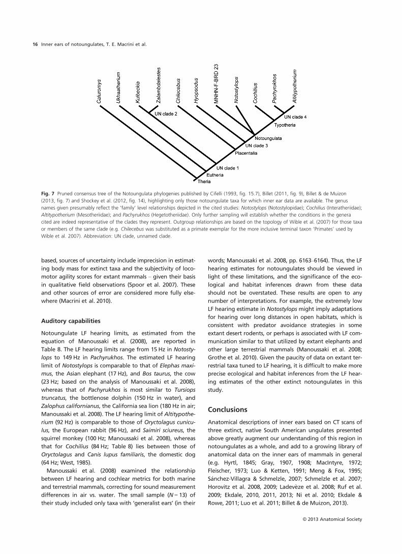
Fig. 7 Pruned consensus tree of the Notoungulata phylogenies published by Cifelli (1993, fig. 15.7), Billet (2011, fig. 9), Billet & de Muizon
(2013, fig. 7) and Shockey et al. (2012, fig. 14), highlighting only those notoungulate taxa for which inner ear data are available. The genus
names given presumably reflect the ‘family’ level relationships depicted in the cited studies: Notostylops (Notostylopidae); Cochilius (Interatheriidae);
Altitypotherium (Mesotheriidae); and Pachyrukhos (Hegetotheriidae). Only further sampling will establish whether the conditions in the genera
cited are indeed representative of the clades they represent. Outgroup relationships are based on the topology of Wible et al. (2007) for those taxa
or members of the same clade (e.g. Chilecebus was substituted as a primate exemplar for the more inclusive terminal taxon ‘Primates’ used by
Wible et al. 2007). Abbreviation: UN clade, unnamed clade.
© 2013 Anatomical Society
Inner ears of notoungulates, T. E. Macrini et al.16
A central goal of this study was to extract phylogenetic
information from the inner ears of notoungulates. To this
end, we produced a character-taxon matrix consisting of 25
characters of the inner ear, 14 of them new, scored across a
handful of notoungulates and multiple outgroups. Two
character states support a pairing between Mesotheriidae
Fig. 8 Transformations of six characters based on DELTRAN optimization on the topology shown in Fig. 7. Gray fill represents ambiguous charac-
ter reconstruction for a particular node. Striped branch leading to a terminal taxon indicates unknown character state for that particular taxon.
Note: transformation for Character 15 is identical to that of a seventh character, Character 19 (not shown).
© 2013 Anatomical Society
Inner ears of notoungulates, T. E. Macrini et al. 17
and Hegetotheriidae, and four are potential synapomor-
phies for Notoungulata. Three other characters represent
potential synapomorphies for Placentalia, Eutheria and a
clade of eutherians, including Kulbeckia, Zalambdalestes,
plus Placentalia. Twelve other characters exhibited apomor-
phic states for one or more notoungulates. Although our
taxonomic sampling in this initial analysis is limited, we will
continue to broaden our sampling of taxa and endocranial
characters, encouraging others to incorporate such features
in phylogenetic analyses of notoungulates and other mam-
mals. Only through such efforts will the potential phyloge-
netic significance of these characters be realized.
A second aim of this study, assessing the locomotor capa-
bilities of these extinct taxa, was accomplished by deriving
locomotor agility scores from morphometrics of the semicir-
cular canals, following the methods of Silcox et al. (2009).
Published analyses of the postcranial skeletons of these
taxa, or close relatives, provide an important point of com-
parison to the agility scores deduced from the semicircular
canal morphometrics. Agility scores and postcranial analyses
yield generally consistent estimates of locomotor style.
Minor discrepancies between results produced by the two
methods may be ascribed to uncertainties in determining
agility scores in living forms, or potentially to clade-specific
distinctions among taxa with similar locomotor styles.
Another issue is the fact that extant taxa with disparate
locomotor styles sometimes exhibit broad overlap in agility
scores.
Finally, we used data obtained from CT scan reconstruc-
tions to estimate auditory capabilities for the taxa surveyed.
The radii of the apical and basal turns of the cochlea corre-
late with LF hearing limits (Manoussaki et al. 2008). On this
basis, LF hearing limits for the sampled notoungulates ran-
ged from 15 Hz in Notostylops to 149 Hz in Pachyrukhos,
values comparable to the Asian elephant and cow for Noto-
stylops, and the bottlenose dolphin and California sea lion
for Pachyrukhos.
Acknowledgements
The authors thank Tim Ryan (Department of Anthropology;
Center for Quantitative X-ray Imaging at Penn State University)
for scanning the specimens. Maeva Orliac (Institut des sciences de
l’�evolution de Universit�e Montpellier 2, France) graciously made
images of a digital inner ear endocast of Hyopsodus (AMNH
143783) available for coding as an outgroup in our character
matrix. We are grateful to The Field Museum and the Museo
Nacional de Historia Natural (Santiago, Chile) for providing access
to the notoungulate specimens scanned and analyzed for this
study. Funding was provided by NSF DEB-0513476 to JJF and a
Frick Postdoctoral Fellowship (2008–2009) from the Department
of Vertebrate Paleontology at the AMNH to TEM. This work also
represents a contribution to the AToL-Mammal Morphology stud-
ies supported by BIO EF- 0629811 (to JFF, XN and colleagues).
We thank Zhe-Xi Luo and an anonymous reviewer for construc-
tive comments that improved this paper. The authors have no
conflicts of interest to declare.
References
Agnolin FL, Chimento NR (2011) Afrotherian affinities for
endemic South American ‘ungulates’. Mamm Biol 76, 101–
108.
Benoit J, Orliac M, Tabuce R (2013) The petrosal of Chambius
(Macroscelidea, Afrotheria) from the Eocene of Djebel Chambi
(Tunisia). J Syst Palaeontol, DOI: 10.1080/14772019.2012.713400.
Billet GB (2010) New observations on the skull of Pyrotherium
(Pyrotheria, Mammalia) and new phylogenetic hypotheses on
South American ungulates. J Mamm Evol 17, 21–59.
Billet GB (2011) Phylogeny of the Notoungulata (Mammalia)
based on cranial and dental characters. J Syst Palaeontol 9,
481–497.
Billet G, de Muizon C (2013) External and internal anatomy of a
petrosal from the Late Paleocene of Itabora�ı, Brazil, referred
to Notoungulata (Placentalia). J Vertebr Paleontol 33,
455–469.
Billet G, Martin T (2011) No evidence for an afrotherian-like
delayed dental eruption in South American notoungulates.
Naturwissenschaften 98, 509–517.
Billet G, Patterson B, de Muizon C (2009) Craniodental anatomy
of late Oligocene archaeohyracids (Notoungulata, Mammalia)
from Bolivia and Argentina and new phylogenetic hypotheses.
Zool J Linn Soc 155, 458–509.
Bloch JI, Silcox MT, Boyer DM, et al. (2007) New Paleocene skel-
etons and the relationship of plesiadapiforms to crown-clade
primates. Proc Natl Acad Sci USA 104, 1159–1164.
Cassini GH, Vizca�ıno SF, Bargo MS (2012a) Body mass estimation
in early Miocene native South American ungulates: a predic-
tive equation based on 3D landmarks. J Zool 287, 53–64.
Cassini GH, Cerde~no E, Villafa~ne AL, et al. (2012b) Paleobiology
of Santacrucian native ungulates (Meridiungulata: Astrapothe-
ria, Litopterna and Notoungulata). In: Early Miocene Paleobi-
ology in Patagonia: High-Latitude Paleocommunities of the
Santa Cruz Formation. (eds Vizca�ıno SF, Kay RF, Bargo MS),
pp. 243–286. New York: Cambridge University Press.
Cerde~no E, Bond M (1998) Taxonomic revision and phylogeny of
Paedotherium and Tremacyllus (Pachyrukhinae, Hegetotherii-
dae, Notoungulata) from the late Miocene to Pleistocene of
Argentina. J Vertebr Paleontol 18, 799–811.
Cifelli RL (1993) The phylogeny of the native South American
ungulates. In: Phylogeny Mammal Volume 2: Placentals. (eds
Szalay FS, Novacek MJ, McKenna MC), pp. 195–216. New York:
Springer.
Cox PG, Jeffery N (2010) Semicircular canals and agility: the
influence of size and shape measures. J Anat 216, 37–47.
Croft DA (2000) Archaeohyracidae (Mammalia: Notoungulata)
from the Tinguiririca Fauna, Central Chile, and the Evolution
and Paleoecology of South American Mammalian Herbivores.
Chicago, IL: PhD dissertation,University of Chicago.
Croft DA, Anaya F (2006) A new middle Miocene hegetotheriid
(Notoungulata: Typotheria) and a phylogeny of the Hegeto-
theriidae. J Vertebr Paleontol 26, 387–399.
Croft DA, Anderson LC (2008) Locomotion in the extinct
notoungulate Protypotherium. Palaeontol Electron 11, 1–20.
Croft DA, Flynn JJ, Wyss AR (2004) Notoungulata and Litopter-
na of the early Miocene Chucal Fauna, northern Chile. Fieldi-
ana: Geol, New Series 50, 1–52.
Damuth J (1990) Problems in estimating body masses of archaic
ungulates using dental measurements. In: Body Size in Mam-
malian Paleobiology: Estimation and Biological Implications
© 2013 Anatomical Society
Inner ears of notoungulates, T. E. Macrini et al.18
(eds Damuth J, MacFadden BJ), pp. 229–253. Cambridge: Cam-
bridge University Press.
Ekdale EG (2010) Ontogenetic variation in the bony labyrinth of
Monodelphis domestica (Mammalia: Marsupialia) following
ossification of the inner ear cavities. Anat Rec 293, 1896–1912.
Ekdale EG (2011) Morphological variation in the ear region of
Pleistocene Elephantimorpha (Mammalia, Proboscidea) from
Central Texas. J Morphol 272, 452–464.
Ekdale EG (2013) Comparative anatomy of the bony labyrinth
(inner ear) of placental mammals. PLoS ONE 8, e66624: 1–100.
Ekdale EG, Rowe T (2011) Morphology and variation within the
bony labyrinth of zhelestids (Mammalia, Eutheria) and other
therian mammals. J Vertebr Paleontol 31, 658–675.
Elissamburu A (2004) An�alisis morfom�etrico y morfofuncional
del esqueleto apendicular de Paedotherium (Mammalia, Noto-
ungulata). Ameghiniana 41, 363–380.
Elissamburu A (2012) Estimation of the body mass in the Noto-
ungulata order. Estudios Geologic�os 68, 91–111.
Fleischer G (1973) Studien am Skelett des Geh€ororgans der
S€augetiere, einschließlich des Menschen. S€augetierkd Mitt 21,
131–239.
Flynn JJ, Croft DA, Charrier R, et al. (2005) New Mesotheriidae
(Mammalia, Notoungulata, Typotheria), geochronology and
tectonics of the Caragua area, northernmost Chile. J South
Am Earth Sci 19, 55–74.
Flynn JJ, Charrier R, Croft DA, et al. (2012) Cenozoic Andean
faunas: shedding new light on South American mammal evo-
lution, biogeography, environments, and tectonics. In: Histori-
cal Biogeography of Neotropical Mammals (eds Patterson BD,
Costa LP), pp. 51–75. Chicago, IL: University of Chicago Press.
Gabbert SL (2004) The basicranium and posterior cranial anat-
omy of the families of the Toxodontia. Bull Am Mus Nat Hist
285, 177–190.
Geisler JH, Luo ZX (1996) The petrosal and inner ear of Herpe-
tocetus sp. (Mammalia: Cetacea) and their implications for the
phylogeny and hearing of archaic mysticetes. J Paleontol 70,
1045–1066.
Gray AA (1907) The Labyrinth of Animals: Including Mammals,
Birds, Reptiles and Amphibians, Vol. 1. London: J & A Chur-
chill.
Gray AA (1908) The Labyrinth of Animals: Including Mammals,
Birds, Reptiles and Amphibians, Vol. 2. London: J & A Chur-
chill.
Gray H (1977) Anatomy, Descriptive and Surgical. Classic collec-
tor’s edition reprint of revised American from 15th English
edn (published 1901). New York: Gramercy Books.
Grothe B, Pecka M, McAlpine D (2010) Mechanisms of sound
localization in mammals. Physiol Rev 90, 983–1012.
Hitz R, Flynn JJ, Wyss AR (2006) New basal Interatheriidae
(Typotheria, Notoungulata, Mammalia) from the Paleogene of
central Chile. Am Mus Novit 3520, 1–32.
Horovitz I (2004) Eutherian mammal systematics and the origins
of South American ungulates as based on postcranial osteol-
ogy. Bull Carnegie Mus Nat Hist 36, 63–79.
Horovitz I, Ladev�eze S, Argot C, et al. (2008) The anatomy of
Herpetotherium cf. fugax Cope, 1873, a metatherian from the
Oligocene of North America. Palaeontographica (A) 284,
109–141.
Horovitz I, Martin T, Bloch J, et al. (2009) Cranial anatomy of
the earliest marsupials and the origin of opossums. PLoS ONE
4, e8278: 1–9.
Hyrtl J (1845) Vergleichend-anatomische Untersuchungen €uber
das innere Geh€ororgan des Menschen und der S€augethiere.
Prague: Friedrich Ehrlich.
Jerison HJ (1971) Quantitative analysis of the evolution of the
camelid brain. Am Nat 105, 227–239.
Ladev�eze S, Asher RJ, S�anchez-Villagra MR (2008) Petrosal anat-
omy in the fossil mammal Necrolestes: evidence for metatheri-
an affinities and comparisons with the extant marsupial mole.
J Anat 213, 686–697.
Lebrun R, de Le�on MP, Tafforeau P, et al. (2010) Deep evolu-
tionary roots of strepsirrhine primate labyrinthine morphol-
ogy. J Anat 216, 368–380.
Luo ZX, Eastman ER (1995) Petrosal and inner ear of a squal-
odontoid whale: implications for evolution of hearing in
odontocetes. J Vertebr Paleontol 15, 431–442.
Luo ZX, Ketten DR (1991) CT scanning and computerized
reconstructions of the inner ear of multituberculate mammals.
J Vertebr Paleontol 11, 220–228.
Luo ZX, Marsh K (1996) Petrosal (periotic) and inner ear of a
Pliocene kogiine whale (Kogiinae, Odontoceti): implications
on relationships and hearing evolution of toothed whales.
J Vertebr Paleontol 16, 328–348.
Luo ZX, Ruf I, Schultz JA, et al. (2011) Fossil evidence on evolu-
tion of inner ear cochlea in Jurassic mammals. P Roy Soc
B- Biol Sci 278, 28–34.
Luo ZX, Ruf I, Martin T (2012) The petrosal and inner ear of the
Late Jurassic cladotherian mammal Dryolestes leiriensis and
implications for evolution of ear in therian mammals. Zool
J Linn Soc 166, 433–463.
MacIntyre GT (1972) The trisulcate petrosal pattern of mammals.
In: Evolutionary Biology, Vol. 6 (eds Dobzhansky T, Hecht MK,
Steere WC), pp. 275–303. New York: Appleton-Century-Crofts.
Macrini TE, Flynn JJ, Croft DA, et al. (2010) Inner ear of a no-
toungulate placental mammal: anatomical description and
examination of potentially phylogenetically informative char-
acters. J Anat 216, 600–610.
Madden RH (1997) A new toxodontid notoungulate. In: Verte-
brate Paleontology in the Neotropics: the Miocene Fauna of
La Venta, Colombia (eds Kay RF, Madden RH, Cifelli RL, Flynn
JJ), pp. 335–354. Washington, DC: Smithsonian Institution
Press.
Maddison WP, Maddison DR (2010) Mesquite: a modular system
for evolutionary analysis. Version 2.74 http://mesquiteproject.
org.
Manoussaki D, Chadwick RS, Ketten DR, et al. (2008) The influ-
ence of cochlear shape on low-frequency hearing. Proc Natl
Acad Sci USA 105, 6162–6166.
Marshall LG (1976) Fossil localities for Santacrucian (early Mio-
cene) mammals, Santa Cruz Province, southern Patagonia,
Argentina. J Paleontol 50, 1129–1142.
Meng J, Fox RC (1995) Osseous inner ear structures and hearing
in early marsupials and placentals. Zool J Linn Soc 115, 47–71.
Nasif NL, Musalem S, Cerde~no E (2000) A new toxodont from
the Late Miocene of Catamarca, Argentina, and a phyloge-
netic analysis of the Toxodontidae. J Vertebr Paleontol 20,
591–600.
Ni X, Flynn JJ, Wyss AR (2010) The bony labyrinth of the early
platyrrhine primate Chilecebus. J Hum Evol 59, 595–607.
Ni X, Flynn JJ, Wyss AR (2012) Imaging the inner ear in fossil
mammals: high-resolution CT scanning and 3-D virtual recon-
structions. Palaeontol Electron 15, 18A: 1–10.
© 2013 Anatomical Society
Inner ears of notoungulates, T. E. Macrini et al. 19
O’Leary M, Bloch J, Flynn J, et al. (2013) The placental mammal
ancestor and the post-K-Pg radiation of placentals. Science
339, 662–667.
Orliac MJ, Argot C, Gilissen E (2012a) Digital cranial endocast of
Hyopsodus (Mammalia, ‘Condylarthra’): a case of Paleogene
terrestrial echolocation? PLoS ONE 7, e30000: 1–10.
Orliac MJ, Benoit J, O’Leary MA (2012b) The inner ear of Dia-
codexis, the oldest artiodactyl mammal. J Anat 221, 417–426.
Patterson B (1932) The auditory region of the Toxodontia. Field
Mus Nat Hist, Geol Ser 6, 5–27.
Patterson B (1934a) The auditory region of an upper Pliocene
typotherid. Geol Ser, Field Mus Nat Hist 6, 83–89.
Patterson B (1934b) Upper premolar-molar structure in the Not-
oungulata with notes on taxonomy. Geol Ser, Field Mus Nat
Hist 6, 91–111.
Patterson B (1936) The internal structure of the ear in some
notoungulates. Geol Ser, Field Mus Nat Hist 6, 199–227.
Patterson B, Pascual R (1968) The fossil mammal fauna of South
America. Q Rev Biol 43, 409–451.
Reguero MA, Dozo MT, Cerde~no E (2007) A poorly known
rodentlike mammal (Pachyrukhinae, Hegetotheriidae, Noto-
ungulata) from the Deseadan (Late Oligocene) of Argentina.
Paleoecology, biogeography, and radiation of the rodentlike
ungulates in South America. J Paleontol 81, 1301–1307.
Rougier GW, Wible JR, Novacek MJ (1998) Implications of Delta-
theridium specimens for early marsupial history. Nature 396,
459–463.
Ruf I, Luo ZX, Wible JR, et al. (2009) Petrosal anatomy and inner
ear structures of the Late Jurassic Henkelotherium (Mammalia,
Cladotheria, Dryolestoidea): insight into the early evolution of
the ear region in cladotherian mammals. J Anat 214, 679–693.
S�anchez-Villagra MR, Schmelzle T (2007) Anatomy and develop-
ment of the bony inner ear in the woolly opossum, Caluromys
philander (Didelphimorphia, Marsupialia). Mastozool Neotrop
14, 53–60.
Schmelzle T, S�anchez-Villagra MR, Maier W (2007) Vestibular
labyrinth diversity in diprotodontian marsupial mammals.
Mammal Study 32, 83–97.
Seckel L, Janis C (2008) Convergences in scapula morphology
among small cursorial mammals: an osteological correlate for
locomotory specialization. J Mamm Evol 15, 261–279.
Segall W (1970) Morphological parallelisms of the bulla and
auditory ossicles in some insectivores and marsupials. Fieldiana
Zool 51, 169–205.
Shockey BJ (1997) Two new notoungulates (Family Notohippi-
dae) from the Salla Beds of Bolivia (Deseadan: late Oligocene):
systematics and functional morphology. J Vertebr Paleontol
17, 584–599.
Shockey BJ, Anaya F (2008) Postcranial osteology of mammals
of Salla, Bolivia (late Oligocene): form, function, and phyloge-
netic implications. In: Mammalian Evolutionary Morphology:
A Tribute to Frederick S. Szalay. (eds Sargis E, Dagosto M), pp.
135–157. Dordrecht: Springer.
Shockey BJ, Croft DA, Anaya F (2007) Analysis of function in the
absence of extant functional homologues: a case study using
mesotheriid notoungulates (Mammalia). Paleobiology 33,
227–247.
Shockey BJ, Flynn JJ, Croft DA, et al. (2012) New leontiniid Not-
oungulata (Mammalia) from Chile and Argentina: comparative
anatomy, character analysis, and phylogenetic hypotheses. Am
Mus Novit 3737, 1–64.
Silcox MT, Bloch JI, Boyer DM, et al. (2009) Semicircular canal
system in early primates. J Hum Evol 56, 315–327.
Silva M, Downing JA (1995) CRC Handbook of Mammalian Body
Masses. New York: CRC Press.
Simpson GG (1936) Structure of a primitive notoungulate
cranium. Am Mus Novit 824, 1–32.
Simpson GG (1948) The beginning of the age of mammals in
South America. Part 1. Bull Am Mus Nat Hist 91, 1–232.
Simpson GG (1967) The beginning of the age of mammals in
South America. Part 2. Bull Am Mus Nat Hist 137, 1–260.
Simpson GG (1980) Splendid Isolation: the Curious History of
South American Mammals. New Haven: Yale University
Press.
Sinclair WJ (1909) Mammalia of the Santa Cruz beds. Vol. VI.
Part I. Typotheria. In: Reports of the Princeton University
Expeditions to Patagonia, 1896–1899. (ed. Scott WB), pp. 1–
110. Lancaster, PA: Lancaster Press.
Spoor F (2003) The semicircular canal system and locomotor
behaviour, with special reference to hominin evolution. Cour
Forsch-Inst Senckenberg 243, 93–104.
Spoor F, Zonneveld F (1995) Morphometry of the primate bony
labyrinth: a new method based on high-resolution computed
tomography. J Anat 186, 271–286.
Spoor F, Zonneveld F (1998) Comparative review of the human
bony labyrinth. Yearbk Phys Anthropol 41, 211–251.
Spoor F, Garland T, Krovitz G, et al. (2007) The primate semicir-
cular canal system and locomotion. Proc Natl Acad Sci USA
104, 10808–10812.
Townsend B, Croft DA (2010) Middle Miocene mesotheriine
diversity at Cerdas, Bolivia, and a reconsideration of Plesioty-
potherium minus. Palaeontol Electron 13, 1A: 1–36.
Vizca�ıno SF, Kay RF, Bargo MS (2012) Background for a paleo-
ecological study of the Santa Cruz Formation (late Early
Miocene) on the Atlantic Coast of Patagonia. In: Early
Miocene Paleobiology in Patagonia: High-Latitude Paleocom-
munities of the Santa Cruz Formation (eds Vizca�ıno SF, Kay
RF, Bargo MS), pp. 1–22. Cambridge: Cambridge University
Press.
West CD (1985) The relationship of the spiral turns of the
cochlea and the length of the basilar membrane to the range
of audible frequencies in ground dwelling mammals. J Acoust
Soc Am 77, 1091–1101.
Wible JR, Novacek MJ, Rougier GW (2004) New data on the
skull and dentition in the Mongolian Late Cretaceous euthe-
rian mammal Zalambdalestes. Bull Am Mus Nat Hist 281,
1–144.
Wible JR, Rougier GW, Novacek MJ, et al. (2007) Cretaceous eu-
therians and Laurasian origin for placental mammals near the
K/T boundary. Nature 447, 1003–1006.
© 2013 Anatomical Society
Inner ears of notoungulates, T. E. Macrini et al.20