Embed Size (px)
Citation preview
i
COMPARATIVE STUDY OF DYDROGESTERONE DOSAGE OF THE DUPHASTON
40mg DAILY AND DUPHASTON 20mg DAILY IN THE OUTCOME OF PREGNANCY
WITH THREATENED MISCARRIAGE IN HUSM.
BY
DR RAHIMAH BT ABD RAHIM
DISSERTATION SUBMITTED IN PARTIAL FULFILMENT
OF THE REQUIREMENT FOR
THE DEGREE OF MASTER OF MEDICINE
(OBSTETRIC & GYNAECOLOGY)
UNIVERSITI SAINS MALAYSIA
MAY 2011
ii
ACKNOWLEDGEMENTS
First of all, I would like to thank my lovely husband for his support, patience and continual help,
and also provide me with daily inspiration. I would not be able to complete this dissertation
without his encouragement.
Highly appreciation and special thanks to my supervisor, Prof. Dr Nik Mohamed Zaki bin Nik
Mahmood, Senior consultant and Lecturer, Department of Obstetrics and Gynaecology, Hospital
USM; my statistician, Dr Wan Mohamad Zahiruddin bin Wan Mohamad, Consultant and
Lecturer from Department of Community Medicine for their ideas and guidance in completion of
this dissertation.
Finally, I would like to thank all my lecturers, my colleagues, staffs in casualty, ward 1 Utara
and O&G clinic Hospital USM, and not forgotten to all my patients for their kind cooperation
and assistance throughout the preparation of this book.

iii
TABLE OF CONTENTS
Page
Acknowledgements ii
Table of contents iii
List of tables v
List of figures vii
Abbreviations viii
Abstrak x
Abstract xii
1. INTRODUCTION 1
2. LITERATURE REVIEW 12
3. OBJECTIVE
3.1 General objectives 19
3.2 Specific objectives 19
4. METHODOLOGY
4.1 Research methodology 20
4.2 Inclusion criteria 20
iv
4.3 Exclusion criteria 21
4.4 Sample size calculation 22
4.5 Randomization And Consent 24
5. Results 28
Flow Chart of study sample (result) 29
6. Discussion 40
7. Conclusion 49
8. Limitations 50
9. Recommendations 51
References
Appendix A: Clinical Research Form
Appendix B: BORANG MAKLUMAT PROJEK PENYELIDIKAN DAN BORANG
KEIZINAN PESAKIT (VERSI BAHASA MALAYSIA)
Appendix C: PATIENT INFORMATION OF RESEARCH PROJECT AND CONSENT
FORM (ENGLISH VERSION)
Appendix D: ETHICAL APPROVAL
v
LIST OF TABLES
Table 1 Causes of spontaneous abortion 2
Page
Table 2 Racial distribution in between two groups 30
Table 3 Patients characteristic in between the two groups 31
Table 4 Age distribution in the two groups 32
Table 5 Gravida of patients in the two groups 33
Table 6 The outcome of pregnancy based on gestational age 34
Table 7 The outcome of pregnancy at 20weeks among those in
category < 14 weeks POA in both groups. 36

vi
Table 8 The outcome of pregnancy at 20weeks gestation in both groups 37
Table 9 Symptoms in between the two groups 39
vii
LIST OF FIGURES
Page
Figure 1 Study Flow Chart 27
Figure 2 Flow Chart of Study Sample 29
Figure 3 Total of various symptoms in both groups 38

viii
AFP Alpha Feto Protein
LIST OF ABBREVIATIONS
BPD Biparietal Diameter
CI Confidence Interval
cm centimeter
CRL Crown Rump Length
CTG Cardiotocogram
D&C Dilatation and curettage
ECG Electrocardigram
ERPOC Evacuation of retained product of conception
FBC Full Blood Count
FHR Fetal heart rate
g/dl Gram per deciliter
hCG Human Chorionic Gonadotrophin
HPE Histopatological examination
HPL Human placental Lactogen
IUD Intrauterine death
ix
IL Interleukin
IFN-g interferon-g
nmol nanomol
mg miligram
NK Natural Killer Cell
O&G Obstetric and Ginekology
PAPP-A Pregnancy Associated Plasma Protein A
PIBF Pregnancy Induced Blocking Factor
POC Product of conception
RR Relative risk
Th T helper
TNF tumour necrosis factor
TVS Transvaginal Sonography
USG Ultrasonography
vs versus
< equal or less than
> equal or more than
x
ABSTRAK
Objektif
Satu kajian untuk menentukan lebih keberkesanan dan kesan sampingan pengambilan ubat
Duphaston 40mg setiap hari (sehari) dalam menangani masalah wanita di ambang keguguran
berbanding dengan pengambilan ubat Duphaston 20mg.
Kaedah (Metodologi)
Kajian perbandingan secara prospektif dan rawak ini dilakukan di Hospital USM Kubang
Kerian bermula dari 1 Mac 2009 sehingga 30 Mac 2010. Seramai 130 orang pesakit yang
terlibat dalam kajian ini. Pemilihan secara rawak dibuat dan pesakit dibahagikan kepada dua
kumpulan, kumpulan A seramai 65 orang dan kumpulan B juga seramai 65 orang.
Kumpulan A akan menerima rawatan ubat Duphaston 20mg sehari manakala pesakit dalam
Kumpulan B akan menerima rawatan ubat Duphaston 40mg sehari. Pemerhatian terhadap
kesan sampingan ubat Duphaston juga dipantau dan dikaji di antara dua kumpulan tersebut.
Kejayaan kehamilan melepasi peringkat di ambang keguguran akan diambil kira sekiranya
usia kandungan dapat melepasi 20 minggu. Keputusan kajian dianalisa menggunakan kaedah
ujian Chi-Square dan Fisher’s exact. Ujian diambil kira sebagai relevan sekiranya nilai p <
0.05. Analisa penurunan logistik juga dibuat bagi mencari dan menentukan faktor
hubungkait yang mempengaruhi hasil kajian.
xi
Keputusan
Didapati Kumpulan B yang mendapat rawatan ubat Duphaston 40mg sehari mempunyai
peratusan yang lebih tinggi dalam kejayaan kehamilan berbanding dengan Kumpulan A yang
menerima ubat Duphaston 20mg sehari (86.7% berbanding dengan 81.7%).
Walaubagaimanapun, tiada perbezaan yang signifikan diantara kedua-dua dos Duphaston
tersebut di mana nilai p yang diperolehi ialah 0.50.
Kesan sampingan ubat yang dialami dalam kedua-dua kumpulan ini juga tidak banyak
menunjukan perbezaan yang ketara.
Kesimpulan
Kajian menunjukan pengambilan ubat Duphaston 40mg sehari tidak meningkatkan kadar
kejayaan kehamilan yang signifikan dikalangan wanita di ambang keguguran (p = 0.500 di
dalam analisa multivariate). Manakala kesan sampingan yang dialami di antara dua
kumpulan kajian tidak menunjukan sebarang perbezaan yang ketara.
xii
ABSTRACT
Objective
To evaluate the effectiveness and the adverse effect of Duphaston 40mg daily and Duphaston
20mg daily in threatened miscarriage.
Methodology
This is a prospective randomized controlled trial conducted at Hospital USM, Kubang Kerian
Kelantan from 1st of March 2009 until 30th March 2010. A total of 130 patients were studied, 65
patients in Group A for those who is taking Duphaston 20mg daily and the other 65 patients in
Group B on Duphaston 40mg daily. Besides the effectiveness, the side effect of the two different
dosage of Duphaston is also evaluated. The successful of the pregnancy is measured by
continuity of the pregnancy beyond 20weeks of gestation. Result was analysed with Chi-square
and Fisher’s Exact test to determine the statistical significant. The test considered significant if
p value < 0.05.
Results
There were higher successful pregnancy in Group B (Duphaston 40mg daily) compared to Group
A (Duphaston 20mg daily) (86.7% versus 81.7%). But this is not statistically significant as the p
value in multivariate analysis is 0.50 ( p> 0.05).
xiii
There were no significant differences in adverse effect of the two different dosage of Duphaston.
Conclusion
Duphaston 40mg daily was not associated with higher chances of successful pregnancy in
threatened miscarriage (p = 0.50 in multivariate analysis). There were also no significant
differences of adverse effect of the drugs in between the two groups.
COMPARATIVE STUDY OF DYDROGESTERONE DOSAGE OF THE DUPHASTON
40mg DAILY AND DUPHASTON 20mg DAILY IN THE OUTCOME OF PREGNANCY
WITH THREATENED MISCARRIAGE IN HUSM.
BY
DR RAHIMAH BT ABD RAHIM
DISSERTATION SUBMITTED IN PARTIAL FULFILMENT
OF THE REQUIREMENT FOR
THE DEGREE OF MASTER OF MEDICINE
(OBSTETRIC & GYNAECOLOGY)
UNIVERSITI SAINS MALAYSIA
MAY 2011

ACKNOWLEDGEMENTS
First of all, I would like to thank my lovely husband for his support, patience and continual help,
and also provide me with daily inspiration. I would not be able to complete this dissertation
without his encouragement.
Highly appreciation and special thanks to my supervisor, Prof. Dr Nik Mohamed Zaki bin Nik
Mahmood, Senior consultant and Lecturer, Department of Obstetrics and Gynaecology, Hospital
USM; my statistician, Dr Wan Mohamad Zahiruddin bin Wan Mohamad, Consultant and
Lecturer from Department of Community Medicine for their ideas and guidance in completion of
this dissertation.
Finally, I would like to thank all my lecturers, my colleagues, staffs in casualty, ward 1 Utara
and O&G clinic Hospital USM, and not forgotten to all my patients for their kind cooperation
and assistance throughout the preparation of this book.
TABLE OF CONTENTS
Page
Acknowledgements ii
Table of contents iii
List of tables v
List of figures vii
Abbreviations viii
Abstrak x
Abstract xii
1. INTRODUCTION
1.1 Introduction 1
2. LITERATURE REVIEW 12
3. OBJECTIVE
3.1 General objectives 19
3.2 Specific objectives 19
4. METHODOLOGY
4.1 Research methodology 20
4.2 Inclusion criteria 20
4.3 Exclusion criteria 21
4.4 Sample size calculation 22
4.5 Consent And Randomization 23
4.6 Study Flow Chart 25
5. Results 28
Flow Chart of study sample (result) 28
6. Discussion 36
7. Conclusion 43
8. Limitations 44
9. Recommendations 45
References
Appendix A: Clinical Research Form
Appendix B: BORANG MAKLUMAT PROJEK PENYELIDIKAN DAN BORANG
KEIZINAN PESAKIT (VERSI BAHASA MALAYSIA)
Appendix C: PATIENT INFORMATION OF RESEARCH PROJECT AND CONSENT
FORM (ENGLISH VERSION)
Appendix D: ETHICAL APPROVAL
LIST OF TABLES
Table 1 Causes of spontaneous abortion 2
Page
Table 2 Racial distribution in between two groups 30
Table 3 Patients characteristic in between the two groups 31
Table 4 Age distribution in the two groups 32
Table 5 Gravida of patients in the two groups 33
Table 6 The outcome of pregnancy based on gestational age 34
Table 7 The outcome of pregnancy at 20weeks among those in
category < 14 weeks POA in both groups. 36

Table 8 The outcome of pregnancy at 20weeks gestation in both groups 37
Table 9 Symptoms in between the two groups 39
LIST OF FIGURES
Page
Figure 1 Total of various symptoms in both groups 27
Figure 2 Flow Chart of Study Sample 29
Figure 3 Total of various symptoms in both groups 38

AFP Alpha Feto Protein
LIST OF ABBREVIATIONS
BPD Biparietal Diameter
CI Confidence Interval
cm centimeter
CRL Crown Rump Length
CTG Cardiotocogram
D&C Dilatation and curettage
ECG Electrocardigram
ERPOC Evacuation of retained product of conception
FBC Full Blood Count
FHR Fetal heart rate
g/dl Gram per deciliter
hCG Human Chorionic Gonadotrophin
HPE Histopatological examination
HPL Human placental Lactogen
IUD Intrauterine death
IL Interleukin
IFN-g interferon-g
nmol nanomol
mg miligram
NK Natural Killer Cell
O&G Obstetric and Ginekology
PAPP-A Pregnancy Associated Plasma Protein A
PIBF Pregnancy Induced Blocking Factor
POC Product of conception
RR Relative risk
Th T helper
TNF tumour necrosis factor
TVS Transvaginal Sonography
USG Ultrasonography
< equal or less than
> equal or more than

ABSTRAK
Objektif
Satu kajian untuk menentukan lebih keberkesanan dan kesan sampingan pengambilan ubat
Duphaston 40mg setiap hari (sehari) dalam menangani masalah wanita di ambang keguguran
berbanding dengan pengambilan ubat Duphaston 20mg.
Kaedah (Metodologi)
Kajian perbandingan secara prospektif dan rawak ini dilakukan di Hospital USM Kubang
Kerian bermula dari 1 Mac 2009 sehingga 30 Mac 2010. Seramai 130 orang pesakit yang
terlibat dalam kajian ini. Pemilihan secara rawak dibuat dan pesakit dibahagikan kepada dua
kumpulan, kumpulan A seramai 65 orang dan kumpulan B juga seramai 65 orang .
Kumpulan A akan menerima rawatan ubat Duphaston 20mg sehari manakala pesakit dalam
Kumpulan B akan menerima rawatan ubat Duphaston 40mg sehari. Pemerhatian terhadap
kesan sampingan ubat Duphaston juga dipantau dan dikaji di antara dua kumpulan tersebut.
Kejayaan kehamilan melepasi peringkat di ambang keguguran akan diambil kira sekiranya
usia kandungan dapat melepasi 20 minggu. Keputusan kajian dianalisa menggunakan kaedah
ujian Chi-Square dan Fisher’s exact. Ujian diambil kira sebagai relevan sekiranya nilai p <
0.05. Analisa penurunan logistik juga dibuat bagi mencari dan menentukan faktor
hubungkait yang mempengaruhi hasil kajian.
Keputusan
Didapati Kumpulan B yang mendapat rawatan ubat Duphaston 40mg sehari mempunyai
peratusan yang lebih tinggi dalam kejayaan kehamilan berbanding dengan Kumpulan A yang
menerima ubat Duphaston 20mg sehari (86.7% berbanding dengan 81.7%).
Walaubagaimanapun, tiada perbezaan yang signifikan diantara kedua-dua dos Duphaston
tersebut di mana nilai p yang diperolehi ialah 0.50.
Kesan sampingan ubat yang dialami dalam kedua-dua kumpulan ini juga tidak banyak
menunjukan perbezaan yang ketara.
Kesimpulan
Kajian menunjukan pengambilan ubat Duphaston 40mg sehari tidak meningkatkan kadar
kejayaan kehamilan yang signifikan dikalangan wanita di ambang keguguran (p = 0.500 di
dalam analisa multivariate). Manakala kesan sampingan yang dialami di antara dua
kumpulan kajian tidak menunjukan sebarang perbezaan yang ketara.
ABSTRACT
Objective
To evaluate the effectiveness and the adverse effect of Duphaston 40mg daily and Duphaston
20mg daily in threatened miscarriage.
Methodology
This is a prospective randomized controlled trial conducted at Hospital USM, Kubang Kerian
Kelantan from 1st of March 2009 until 30th March 2010. A total of 130 patients were studied, 65
patients in Group A for those who is taking Duphaston 20mg daily and the other 65 patients in
Group B on Duphaston 40mg daily. Besides the effectiveness, the side effect of the two different
dosage of Duphaston is also evaluated. The successful of the pregnancy is measured by
continuity of the pregnancy beyond 20weeks of gestation. Result was analysed with Chi-square
and Fisher’s Exact test to determine the statistical significant. The test considered significant if
p value < 0.05.
Results
There were higher successful pregnancy in Group B (Duphaston 40mg daily) compared to Group
A (Duphaston 20mg daily) (86.7% versus 81.7%). But this is not statistically significant as the p
value in multivariate analysis is 0.50 ( p> 0.05).
There were no significant differences in adverse effect of the two different dosage of Duphaston.
Conclusion
Duphaston 40mg daily was not associated with higher chances of successful pregnancy in
threatened miscarriage (p = 0.50 in multivariate analysis). There were also no significant
differences of adverse effect of the drugs in between the two groups.
1.1
INTRODUCTION
Miscarriage is the spontaneous loss of a fetus before it is capable of surviving outside the uterus;
this is generally defined as being before 24 completed weeks of gestation. The occurrence of
vaginal bleeding during this time is known as threatened miscarriage, provided the cervix is
closed and the fetus remains viable and inside the uterine cavity (CunninghamFG, 2005). This
bleeding may also be accompanied by abdominal cramps.
Threatened miscarriage is a common complication, occurring in about 20% of all clinically
recognized pregnancies (EverettC, 1997, Weiss JL, 2004). If bleeding occurs during pregnancy,
in the case of a viable fetus, the incidence of miscarriage can be around 20%, or even up to 30% ,
depending on the severity and risk factors(Al-Sebai MA, 1996).
Vaginal bleeding during the early stages of pregnancy could be due to a range of conditions
including ectopic pregnancy, cervical abnormalities such as polyps or cancer, infection, molar
pregnancy or vaginal trauma (Jauniaux E, 2005). A thorough evaluation is therefore essential to
establish the diagnosis.
Initial laboratory tests should include a complete blood count and blood typing. A pelvic
examination will determine whether the cervix is effaced or dilated, indicating imminent
miscarriage. Finally, transvaginal ultrasound is crucial to confirm whether or not the fetus is still
viable, and to diagnose an incomplete or missed abortion (Jauniaux E, 2005).
2. Risk of miscarriage
The risk of miscarriage is dependent on a variety of factors including demographic and clinical
characteristics, maternal serum biochemistry and ultrasound findings (Table 1).
2.1. Demographic and clinical characteristics.
It is well established that advancing maternal age is associated with an increased risk (Mbugua
Gitau G, 2009). One study of 182 women with threatened miscarriage found a significantly (p <
0.05) higher rate of miscarriage in those aged 31–40 years (27.1%) than in those aged 21–30
years (7.1%) (Basama FM, 2004).
Table 1: Causes of spontaneous miscarriage
Genetic Trisomy aneuploidy/polyploidy, translocations
Uterus Congenital uterine anomalies, leiomyoma, intrauterine adhesions or
synechiae (Asherman’s Syndrome)
Endocrine Progesterone deficiency (inadequate luteal phase), thyroid disease,
diabetes mellitus (uncontrolled), luteinizing hormone hypersecretion
Immunologic Antiphospholipid syndrome, systemic lupus erythematosus
Infections Toxoplasma gondii, Listeria monocytogens, Chlamydia trachomatis,
Ureaplasma urealyticum, Mycoplasma hominis, Borrelia burgdorferi,
Neisseria gonorrhoea
A history of previous miscarriages is also associated with an increased risk (Risch HA, 1988,
Regan L, 1989), as is the presence of poorly controlled systemic disease such as diabetes or
thyroid dysfunction (Basama FM, 2004, Greene, 1999, Roberts CP, 2000).
The timing and severity of vaginal bleeding are important prognostic factors in women with
threatened miscarriage, with both early and severe bleeding being associated with a higher risk
of miscarriage. Bleeding before 6 weeks gestation has been reported to result in a miscarriage
rate of 29% compared with 8.2% for bleeding during weeks 7th–12th and 5.6% for second
trimester bleeding (Basama FM, 2004).
The duration of bleeding was also shown to play a role in the risk of miscarriage amongst 200
women with symptoms of imminent miscarriage during weeks 5th–12th of gestation (Fiegler P,
2003). The rate of miscarriage in women with abdominal pain and bleeding for more than 3 days
(81%) was significantly (p < 0.001) greater than that in women with abdominal pain only (10%)
or abdominal pain and bleeding of <3 days (13%).
2.2. Maternal serum biochemistry
Amongst the maternal serum markers with prognostic value, progesterone and human chorionic
gonadothrophin (hCG) have been most widely investigated. Data from 3674 first trimester
pregnancies showed an increasing risk of miscarriage with declining serum progesterone
levels(McCord ML, 1996). Levels of less than 5 ng/ml were associated with a spontaneous
miscarriage in 86% of cases compared with only 8% at levels of 20–25 ng/ml.
As seen with progesterone, low levels of hCG also predict a higher risk of miscarriage. Amongst
398 women with bleeding and/or abdominal pain during the first 18 weeks of pregnancy and 156
control pregnancies, a cut-off value of 20 ng/ml was found to have 88% sensitivity and 83%
positive predictive value in differentiating between viable continuing pregnancies and those that
were not viable(Al-Sebai MA, 1996).
Further markers under investigation are the tumour marker CA-125, inhibin A, anandamide and
progesterone induced blocking factor (PIBF).
Elevated levels of CA-125, which can be associated with damage to the deciduous membrane,
have been proposed as an indicator of miscarriage risk. Sequential determinations of CA-125 in
women with bleeding during gestational weeks 6th–12th were able to distinguish between women
who subsequently miscarried and those who did not (Schmidt T, 2001). Other studies have
pointed to the prognostic value of single measurements of CA-125. One showed that a cut-off of
125 IU/ml had positive and negative predictive values of 93% and 92%, respectively (Leylek
OA, 1997), in women with bleeding between gestational weeks 6 and 12; mean values were 67.3
and 221.0 IU/ml (p < 0.05) in the continuing pregnancy and miscarriage groups, respectively.
Inhibin A levels were significantly (p < 0.05) lower in women who miscarried compared with
those who did not in a sample of 55 women with bleeding during early pregnancy (0.38 vs. 0.98
multiples of median) (Florio P, 2004). A cut-off value of 0.553 multiples of mean was the best
predictor of a failing pregnancy.
Anandamide (Maccarrone M, 2001) and PIBF (Kalinka and Radwan, 2006, Szekeres-Bartho J,
2008) are both biomarkers that are modulated by progesterone. Anandamide is an
endocannabinoid known to participate in reproductive processes. Oestradiol and progesterone
have been shown to regulate its production in the rat uterus (Ribeiro ML, 2009). In women with
successful pregnancy after in-vitro fertilisation (IVF), plasma anandamide was low during the
implantation phase but high in gestational weeks 4th and 5th, declining again in week 6th (El-
Talatini MR, 2009).
PIBF mediates the effect of progesterone on the immune system (Szekeres-Bartho J, 1995).
Progestogens like dydrogesterone increase its production in animal models (Joachim R, 2003)
and in women suffering from threatened miscarriage (Kalinka and Szekeres-Bartho, 2005).
2.3. Ultrasound findings
Ultrasonography is important in identifying prognostic factors for a poor outcome in women
with threatened miscarriage, such as small crown to rump length, an empty gestational sac or
fetal bradycardia (Dogra V, 2005).
A combination of several factors increases the risk of miscarriage is still under further studies.
For example, logistic regression analysis showed that the chance of miscarriage in women with
bleeding between weeks 5th and 12th of gestation was 84% in cases of fetal bradycardia plus
discrepancies between crown to rump length and the diameter of the gestational sac, and
menstrual and sonographic age; this risk was reduced to 6% if none of these factors were
apparent (Falco P, 1996).
Evaluation of the gestational sac can also give an indication of the viability of the pregnancy,
with empty sacs of >15–17mm diameter and sacs of ≥13mm without a visible yolk sac
suggesting a poor prognosis (Falco P, 2003). A study of 781 women who presented with
threatened miscarriage found that 211 (28%) had shown to constitute a viable pregnancy. A
mean sac diameter of ≥17mm that lacked an embryo or a mean diameter of ≥13mm without a
yolk sac both showed 100% specificity and 100% predictive value.
Most studies suggest that the risk of spontaneous miscarriage is only around 3–5% if fetal heart
activity is detected in women with vaginal bleeding (Scroggins KM, 2000, Tannirandorn Y,
2003). However, fetal bradycardia is a predictor of a poor outcome (Chittacharoen A, 2004).
Absence of fetal heart activity in embryos with a crown to rump length of >5mm indicates that
the pregnancy is no longer viable (Dogra V, 2005).
Retroplacental haematoma during the first trimester has been linked to an increased risk of
miscarriage (Nagy S, 2003). It is estimated that about one-fifth of women with threatened
miscarriage have a subchorionic haematoma (Pedersen JF, 1990). The size and situation of the
haematoma have prognostic value. Amongst 516 women with bleeding and subchorionic
haematoma in the first trimester, the miscarriage rate was about twice as high in those with a
large haematoma (18.8%) compared with small and medium haematomas (7.7% and 9.2%,
respectively) (Bennett GL, 1996). Bleeding near the cord has also been shown to be more likely
to result in placental separation and subsequent miscarriage than bleeding in other locations
(Jauniaux E, 2005).
3. Consequences of threatened miscarriage
Threatened miscarriage causes considerable stress and anxiety for a pregnant woman. It can
cause anxiety and depression, and may be experienced as a traumatic life event (Lok IH, 2007).
Although pregnancies advancing after a threatened miscarriage may be associated with more
complications such as preterm delivery and prelabour rupture of membranes than other
pregnancies, the evidence for the association between threatened abortion and birth defects is
limited and inconsistent (Jauniaux E, 2005).
A study in 16,506 pregnant women, of whom 2346 experienced first trimester bleeding, showed
that bleeding was an independent risk factor for preterm delivery, Caesarean delivery, pre-
eclampsia, placental abruption, intra-uterine growth restriction and preterm premature rupture of
the membranes (Weiss JL, 2004). The babies born to women reporting bleeding also had a
significantly (p < 0.05) lower birth weight and mean gestational age at delivery.
4. Treatment options
Miscarriage is a difficult and distressing event for a woman and her partner and can result in
depression, anxiety, anger and marital breakdown (Lok IH, 2007). There is therefore a clear
medical need to prevent miscarriage whenever possible. However, it is essential to ensure that
the pregnancy is viable before any treatment is considered.
This is best achieved with a combination of serum hCG levels and ultrasound, which have been
shown to provide an accurate diagnosis (Dogra V, 2005). The most widely used of the currently
available treatment options include bed rest and luteal support with progestogens or hCG.
4.1. Bed rest
Bed rest is conventionally the most commonly used management technique for threatened
miscarriage. Despite this, there is little evidence of its value. Physical activity is rarely associated
with an increased risk of miscarriage, and indeed a lack of activity can lead to a number of other
complications such as thromboembolic events, back pain, muscle atrophy and bone loss
(Promislow JH, 2004).
Evidence also suggests that women may experience emotional, familial and economic stress
during bed rest, as well as self-blame if they fail to comply and subsequently suffer a miscarriage
(Promislow JH, 2004, Ben-Haroush A, 2003). Very few studies have specifically assessed the
efficacy of bed rest.
A recent Cochrane review also came to the conclusion that there is insufficient evidence to
support a policy of bed rest to prevent miscarriage (Aleman A, 2005). A search of the Cochrane
Pregnancy and Childbirth Group trials register, the Cochrane Library, MEDLINE, POPLINE,
LILACS and EMBASE revealed only two trials, conducted in a total of 84 women, which
compared bed rest with alternative care or no intervention in women at high risk of miscarriage.
There were no statistically significant differences in the risk of miscarriage between the two
groups (relative risk 1.54; 95% CI 0.92–2.58).
4.2. Progesterone
Progesterone secreted by the corpus luteum is essential for the maintenance of early pregnancy
(Raghupathy R, 2005), and it has been proposed that corpus luteum deficiency may be
responsible for some cases of miscarriage. Direct supplementation with progestogens, or
exogenous administration of hCG, should therefore have beneficial effects in women with
threatened miscarriage.
Unfortunately, luteal phase defect is notoriously difficult to diagnose reliably (Medicine., 2008).
One diagnostic criterion is low serum progesterone, but levels vary widely during early
pregnancy and any later decline may be attributed to a dysfunctioning placenta. Other criteria
including a pre-ovulatory follicle diameter of <17mm and the absence of a post-ovulatory rise in
basal body temperature are imprecise, and the validity of endometrial histological diagnosis has
been called into question (Medicine., 2008). Nevertheless, luteal support is widely used for the
management of threatened miscarriage.
The clinical management and immunology of miscarriage has substantially advanced since most
of the early work to support the use of progesterone in early pregnancy was done. The role of
progesterone is likely to be far more complex than previously thought.
4.2.1. Dydrogesterone
Dydrogesterone is a synthetic progestogen that has a similar molecular structure and
pharmacological profile to natural progesterone. In contrast to natural progesterone, however, it
is orally active at low dosages. It is therefore not associated with hepatic side effects that have
been reported in some cases with the high doses of micronised progesterone necessary for oral
dosing (Schindler AE, 2003).
Dydrogesterone is highly selective for the progesterone receptor and differs from most other
synthetic progestogens in its lack of oestrogenic, androgenic, anabolic and corticoid properties. It
is considered particularly suitable for the management of women with threatened miscarriage
and other pregnancy-related disorders as it does not suppress the pituitary–gonadal-axis at
normal therapeutic doses (Schindler AE, 2003).
This means that it does not affect the normal secretory transformation of the endometrium nor
inhibiting formation of progesterone in the placenta during early pregnancy and does not cause
masculinisation of the female foetus.
4.3. Human chorionic gonadotrophin
The rationale for the use of hCG is its potential to stimulate progesterone production by the
corpus luteum and feto-placental unit. Initial early studies showed promise in women with early
threatened miscarriage (SuvonnakoteT, 1986), and the small randomized study conducted by
Harrison showed hCG to be significantly more effective than bed rest (p < 0.01) (HarrisonRF,
1993).
4.4. Uterine muscle relaxants
Uterine muscle relaxing drugs, which include beta-agonists and atropine-like antispasmodic
agents, are rarely used today. A recent search of the Cochrane Pregnancy and Childbirth Group
Trials register and Central Register of Controlled Trials confirmed that there is insufficient
evidence to support their use (Qureshi, 2009).
Miscarriage is a physically and mentally traumatic event that frequently has long-term
psychological consequences. Every effort should therefore be made to maintain viable
pregnancies in women with threatened miscarriage. Our understanding of pregnancy has
advanced considerably in recent years, and there is some evidence that luteal support with a
progestogen, such as progesterone and dydrogesterone, may help to prevent miscarriage in at
least a subpopulation of these women.
The wide spread use of progesterone and dydrogesterone over many years also suggests that
there are no safety problems with these treatments.
Although data from clinical studies suggest efficacy for luteal support with progesterone and
dydrogesterone, there is no study done earlier to compare the efficacy of different dosages and
regimens of progesterone. It is the intention of this study to try to address these important issues.
2.0 LITERATURE REVIEW
Progesterone is an essential hormone in the process of reproduction. It is involved in the
menstrual cycle and implantation, and is essential for pregnancy maintenance. The role of
progesterone in the maintenance of pregnancy is well accepted. It is known to induce secretory
changes in the lining of the uterus essential for successful implantation of a fertilised egg.
It has been suggested that a causative factor in many cases of miscarriage may be inadequate
secretion of progesterone. Therefore, progestogens have been used, beginning in the first
trimester of pregnancy, in an attempt to prevent spontaneous miscarriage. Progestogens have
been prescribed for over 30 years by clinician’s world-wide in the belief that they reduce the risk
of pregnancy failure, in particular first trimester miscarriage.
Although the pharmacokinetics and pharmacodynamics of progesterone have been well studied,
and since 1935 it has been synthesized and is available commercially, its use in the
pathophysiology of pregnancy remains controversial.
One relatively recently discovered mode of action is modulation of the maternal immune
response (Walch and Huber, 2008, Graham JD, 1997, Di Renzo, 2005, Al-Azzawi et al., 1999).
During normal pregnancy, there is a shift towards a protective T helper (Th)-2 dominated
cytokine balance (e.g. interleukin (IL-4 and IL-10) and away from Th-1 cytokines (e.g. IL-12
and interferon). This shift towards Th-2 cytokines is promoted by PIBF, which is synthesised by
activated lymphocytes in the presence of progesterone (Raghupathy R, 2000).
Other mechanisms by which PIBF prevents inflammatory and thrombotic reactions towards the
fetus include an increase of asymmetric non-cytotoxic blocking antibodies (Eblen AC, 2000) and
blockade of natural killer (NK) cell degranulation (CunninghamFG, 2005). Studies have
confirmed that PIBF levels fail to increase in pregnancies that end in miscarriage(EverettC,
1997).
Progestogens also have a direct pharmacological effect by reducing the synthesis of
prostaglandins, thereby relaxing uterine smooth musculature and preventing inappropriate
contractions that may result in miscarriage (Hidalgo A and B., 1996, Eskes TKAB, 1970).
A recent Cochrane review conducted to assess the efficacy and safety of progestogens in
threatened miscarriage identified only two studies that were suitable to include in a meta-
analysis, both of which compared progesterone with placebo (Wahabi HA, 2007). The Cochrane
Pregnancy and Childbirth Group’s Trials Register, Cochrane Central register of Controlled
Trials, MEDLINE, EMBASE and CINAHL were searched for randomised or quasi-randomised-
controlled trials comparing a progestogen with no treatment, placebo or any other treatment
regimen.
The two studies that met the inclusion criteria were double-blind and included a total of 84
women treated with vaginal progesterone or placebo. Although the meta-analysis suggested a
reduced risk of miscarriage with progesterone (relative risk 0.47), the small sample size meant
that the 95% confidence interval was too wide (0.17–1.30) to draw any conclusions. In addition,
the methodological quality of both studies was considered relatively poor and there was no data
on the safety of progesterone.
One of the studies included in the meta-analysis randomized 56 women with vaginal bleeding
during the first trimester to treatment with 25mg progesterone or placebo suppositories twice
daily until either miscarriage or 14 days after bleeding had stopped (Gerhard I, 1987). Of the 52
women included in the analysis, 3/26 (11%) given progesterone and 5/26 (19%) given placebo
had a miscarriage. However, only the 34 women with fetal viability confirmed by ultrasound
before treatment were included in the meta-analysis. There were no miscarriages in the
progesterone group and one in the placebo group, resulting in a relative risk of 0.33 (95% CI
0.01–7.65). Serum progesterone levels were significantly increased in women treated with
progesterone.
The other study evaluated 50 women with an ultrasound diagnosis of threatened miscarriage
between 6 and 12 weeks of gestation and a previous diagnosis of luteal phase dysfunction
(Palagiano A, 2004). They were randomised to receive 90mg progesterone or placebo vaginal gel
daily for 5 days. At the end of treatment, there was a significant reduction in pain and the number
of uterine contractions with progesterone. During a 60-day follow-up, significantly (p < 0.05)
fewer women miscarried in the progesterone group (4/25; 16%) than in the placebo group (8/25;
32%), resulting a relative risk of 0.50 (95% CI 0.17–1.45).
Dydrogesterone
Like progesterone, dydrogesterone is able to inhibit the production of Th-1 cytokines and up-
regulate production of Th-2 cytokines, thus shifting the balance towards a pregnancy protective
Th-2-dominated immune response (Raghupathy R, 2007, Blois SM, 2004).
For example, incubation of dydrogesterone with peripheral blood mononuclear cells from
women with unexplained recurrent abortion increased PIBF and inhibited the production of Th-
1-cytokines tumour necrosis factor and interferon whilst increasing that of the Th-2-cytokines
IL-4 and IL-6 (Raghupathy R, 2005).
In a mouse model, stress-induced miscarriage was associated with low levels of progesterone and
PIBF (Blois SM, 2004). Treatment with dydrogesterone before the stress reduced the number of
miscarriages, restored PIBF levels and decreased uterine levels of Th-1 cytokines.
An early uncontrolled study with dydrogesterone in 111 women showed favorable results, with
only 9 subsequent miscarriages(Radulesco, 1970). Dydrogesterone (2.5–20mg daily) was
frequently combined with synthetic oestrogens and treatment duration varied from a few weeks
to more than 6 months.
Amongst more recent studies, dydrogesterone was compared with conservative management in
154 women who had vaginal bleeding before week 13th of gestation(Omar et al., 2005). All
women received conservative management with bed rest and folic acid, whilst 74 were
randomised to receive oral dydrogesterone (40mg initial dose followed by 10mg twice daily)
until the bleeding stopped. During follow-up to 20 weeks gestation, the miscarriage rate was
significantly (p < 0.05) lower with dydrogesterone (3/74; 4.1%) than with conservative
management only (11/80; 13.8%). The odds ratio was 3.773 (95% CI 1.01–14.11).
A smaller study, which was published in 2007 and was therefore too recent to be included in the
meta-analysis, compared oral dydrogesterone with vaginal micronised progesterone (Czajkowski
K, 2007). This double-blind study randomised 53 women with threatened miscarriage at up to 12
weeks gestation to treatment with dydrogesterone 30mg or micronised progesterone 300mg daily
for 6 weeks. There were fewer miscarriages in the dydrogesterone group (2/24; 8.3%) than in the
progesterone group (4/29; 14%), although the difference was not statistically significant.
Another recent study randomised 191 women with vaginal bleeding up to week 16 of pregnancy
to treatment with dydrogesterone (40mg stat followed by 10mg twice daily) or conservative
management (Pandian, 2009). Dydrogesterone treatment resulted in significantly (p < 0.05)
fewer miscarriages up to 20th weeks of gestation than conservative management (12.5% versus
28.4%).
A significantly (p < 0.05) lower incidence of miscarriage with dydrogesterone was also observed
in a study of 146 women who presented with mild or moderate bleeding during the first trimester
of pregnancy (El-Zibdeh, 2009). All women received standard supportive care, whilst 86 were
randomised to additional treatment with dydrogesterone (10mg b.i.d.). The incidence of
miscarriage was 17.5% in the dydrogesterone group compared with 25.0% in the control group.
The effect of dydrogesterone on urinary PIBF and serum progesterone and cytokines has also
been evaluated in women with threatened miscarriage (Kalinka and Radwan, 2006, Ribeiro ML,
2009). A total of 27 women with threatened miscarriage were treated with dydrogesterone 30–
40mg daily for 10 days and the cytokine and PIBF levels compared with those in 16 women with
normal healthy pregnancies. There was no statistically significant difference between the treated
women with threatened miscarriage and the healthy controls with regard to pregnancy outcome
(missed miscarriage 2/27 vs. 1/16 and preterm delivery 2/27 vs. 0/16). At baseline, PIBF levels
were significantly lower in women with threatened miscarriage than in healthy controls (453
pg/ml vs. 1058 pg/ml; p < 0.01). After treatment with dydrogesterone, there was no statistically
significant difference between the threatened miscarriage and control group (1292 pg/ml vs.
1831 pg/ml, respectively). Women who subsequently had a miscarriage had lower PIBF and
progesterone levels than those who progressed to a successful pregnancy. Serum Th1 and Th2
cytokine levels did not differ significantly between women with threatened miscarriage and
healthy controls.
There is no evidence to suggest that progesterone supplementation during pregnancy has any
adverse consequences for the foetus (Medicine, 2008). Although a case–control study reported
an association between maternal exposure to progestogens and hypospadias (Zhang J, 1994,
Carmichael SL, 2005), the data was based on interviews with the mothers who often could not
specify the type or dose of progestogen. Moreover, the indication for the use of progesterone has
itself has been related to an increased risk of hypospadias. Other studies have found no link
between maternal progesterone exposure and defects of the external genitalia.
A recent review of birth defects reported between 1977 and 2005 following maternal use of
dydrogesterone during pregnancy found no link between dydrogesterone and birth defects
(Queisser-LuftA, 2009). It is estimated that, during this 28-year period, fetuses were exposed to
dydrogesterone in utero in more than 10 million pregnancies.
Unfortunately there are no studies done to compare the efficacy of different dosages and regimes
of progesterone in the prevention of miscarriages.

1.
OBJECTIVE
General objective:
To determine the effectiveness and the adverse effect of the Duphaston 40mg daily.
Specific objective:
I. To determine the effectiveness of Duphaston 40mg vs Duphaston 20mg in prolonging
a pregnancy.
II. To describe the adverse effect of the different dosages of Duphaston.
2.
RESEARCH METHODOLOGY
This study design was a randomized controlled trial between two different dosages of duphaston,
dose of 40mg daily and 20mg daily performed via alternate sampling limitation, from 9th March
2009 until 8th March 2010 (12 months).
The study setting was taken place at the Casualty Department, Gynaecology Ward (1 Utara) and
Obstetrics and Gynaecology (O&G) Clinic in Hospital Universiti Sains Malaysia (HUSM).
Consented women presented with clinical symptoms of threatened miscarriage, who fulfilled the
inclusion criteria as below were recruited into the study.
INCLUSION CRITERIA:
I. Only singleton pregnancy.
II. 1st episode of threatened miscarriage for current pregnancy.
III. Mild or moderate vaginal bleeding.
IV. No history of passing out product of conception.
V. Absence of systemic illness or fever
VI. Presence of fetal heart at 7 weeks
VII. Viable pregnancy at 7weeks up to 16weeks
However, patients with either one of the below criteria were excluded from the study.
EXCLUSION CRITERIA:
a) Empty sac of more than 26mm
b) History of recurrent miscarriage (3 or more consecutive miscarriages)
c) Patients with history of chronic disease; e.g. hypertension, diabetes, renal,
liver or heart disease.
d) Genital or reproductive anatomical abnormality.
e) Fetal abnormality
f) History of hypersensitivity to dydrogesterone.
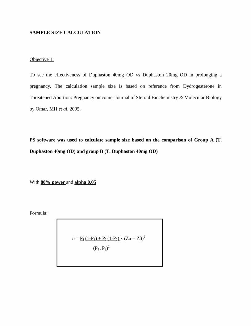
SAMPLE SIZE CALCULATION
To see the effectiveness of Duphaston 40mg OD vs Duphaston 20mg OD in prolonging a
pregnancy. The calculation sample size is based on reference from Dydrogesterone in
Threatened Abortion: Pregnancy outcome, Journal of Steroid Biochemistry & Molecular Biology
by Omar, MH et al, 2005.
Objective 1:
PS software was used to calculate sample size based on the comparison of Group A (T.
Duphaston 40mg OD) and group B (T. Duphaston 40mg OD)
With 80% power and
alpha 0.05
Formula:
n = P1 (1-P1) + P2 (1-P2)
(P1 - P2)2
x (Zα + Zβ)2
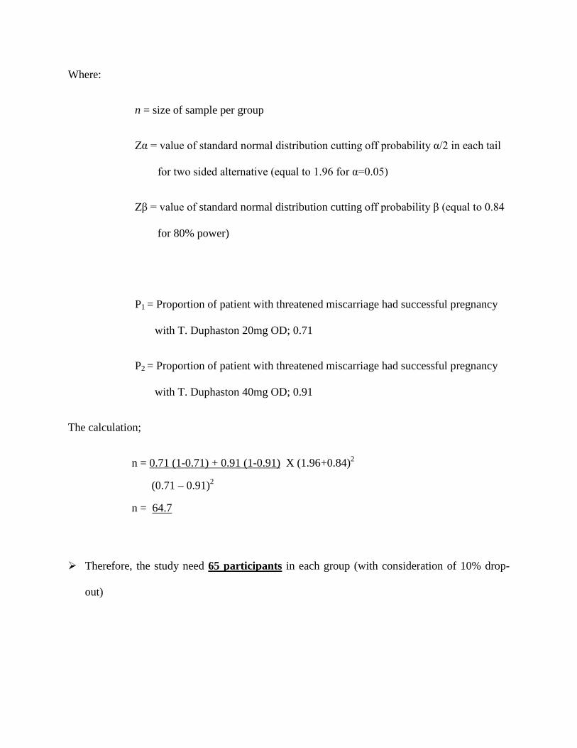
Where:
n = size of sample per group
Zα = value of standard normal distribution cutting off probability α/2 in each tail
for two sided alternative (equal to 1.96 for α=0.05)
Zβ = value of standard normal distribution cutting off probability β (equal to 0.84
for 80% power)
P1 = Proportion of patient with threatened miscarriage had successful pregnancy
with T. Duphaston 20mg OD; 0.71
P2 = Proportion of patient with threatened miscarriage had successful pregnancy
with T. Duphaston 40mg OD; 0.91
The calculation;
n = 0.71 (1-0.71) + 0.91 (1-0.91)
(0.71 – 0.91)2
X (1.96+0.84)2
n = 64.7
Therefore, the study need 65 participants
in each group (with consideration of 10% drop-
out)
RANDOMIZATION AND CONSENT
Eligible women who met the definition of threatened miscarriage, being confirmed viable
intrauterine pregnancy by transabdominal or transvaginal ultrasound and had per vaginal
bleeding while the os still closed, presented to Casualty Department, Ward 1 Utara and O&G
Clinic HUSM, will be fully assess.
Consented patient who fulfilled the inclusion criteria were recruited into the study.
Randomization was done by limited alternate sampling method whereby the patients were
divided into Group A and Group B alternately upon their presentation. GROUP A patients
received T. Duphaston total 20mg daily while GROUP B patients received T. Duphaston 40mg
daily. In both groups, the medication was taken until 20 weeks of gestation.
After completed assessment and given explanation by medical officer regarding research
information and also signed the consent form, they were prescribed with the T. Duphaston 20mg
daily for those in Group A and T. Duphaston 40mg daily in Group B. They were advised to take
their medication until their pregnancy reached 20 weeks of gestations.
They were followed up 2 weeks after the first presentation (first appointment), subsequently 3 or
4 weeks later, depends on their gestation on the first presentation (the second appointment), for
those who first presented after or at 15 weeks of gestations were skipped from their second
appointment and finally, they were reviewed at the 20 weeks of gestations to determine the
successful of the treatment. Those who are still in their viable pregnancy were considered
successful treatment whereas those who were aborted are unsuccessful.
During the followed up, if any of exclusion criteria identified e.g. diagnosed to have abnormal
fetus or patient developed hypersensitivity to Duphaston, they will be eliminated.
Patients advised for bed rest and avoid sexual intercourse for at least a few days after the
bleeding stop or to wait at least until the first assessment (the first appointment).
They also advised to come to the hospital as soon as possible if they developed excessive per
vaginal bleeding, increased abdominal pain, passing out any product of conception or developed
moderate to severe reaction towards tablet Duphaston.
They were recommended taking T. Folic Acid 5 mg daily along their pregnancy.

![TheSynthesis of Human Placental Lactogen Ribosomes …Cell-Free Synthesis of HumanPlacental Lactogen 1323 TABLE 1. Theincorporation of [tIS]methionine into proteins synthesized by](https://img.pdfslide.us/doc/110x75/60c70974e4f5290f2474e6dd/thesynthesis-of-human-placental-lactogen-ribosomes-cell-free-synthesis-of-humanplacental.jpg)


![ZZZ IUD]LHUFRPPHUFLDO FRP€¦ · zzz iud]lhufrpphufldo frp 'rq )ud]lhu giud]lhu#iud]lhufrpphufldo frp &roh )ud]lhu froh#iud]lhufrpphufldo frp x w á { w r x d x ä w r x x s x ï](https://img.pdfslide.us/doc/110x75/5f573fcc84aeba748f1c92d9/zzz-iudlhufrpphufldo-frp-zzz-iudlhufrpphufldo-frp-rq-udlhu-giudlhuiudlhufrpphufldo.jpg)


![ZZZ IUD]LHUFRPPHUFLDO FRP](https://img.pdfslide.us/doc/110x75/61977a993564fd557a21a871/zzz-iudlhufrpphufldo-frp.jpg)