Embed Size (px)
Citation preview
RESEARCH Open Access
Comparative studies on genital infections andantimicrobial susceptibility patterns of isolatesfrom camels (Camelus dromedarius) and cows(Bos indicus) in Maiduguri, north-eastern NigeriaGideon Dauda Mshelia1*, Godfrey Okpaje1, Yepmo Andre Casimir Voltaire1 and Godwin Onyeamaechi Egwu2
Abstract
A total of 160 genitalia of Camels and cows were investigated in Maiduguri, north-eastern Nigeria to compare bacterialisolates and the antibacterial susceptibilities of some of the isolates. Streptococcus (Str.) pyogenes (31%), Escherichia (E.)coli (24%) and Staphylococcus (S.) aureus (20%) were the most common vaginal bacterial isolates in camels; while E. coli(73%), Str. pyogenes (18%) and S. aureus (11%) were the most frequent isolates in the cows. Of the 78 uterine isolatesrecovered in this study, E. coli was the most prominent in camels (8%) and cows (54%). The overall weight of genitalinfection in all camels and cows examined was highest (P < 0.05) with E. coli (79%), but there was no difference(P > 0.05) between vaginal and uterine bacterial isolates from camels and cows in this study. The Relative Risk (RR) foran infection of the vagina with E coli (3.04, 95% Confidence Interval (CI): 2.104 to 4.398, P < 0.0001) is more in cowscompared to the camel, but the RR for vaginal infection with S. aureus and Str. pyogenes were lower in cows comparedto Camels. The E. coli and S. aureus isolates were highly susceptible to the antimicrobial agents tested. Thereforeeffective management of reproductive disorders associated with these pathogens can be achieved with proper use ofthese antimicrobial agents in these animal species
Keywords: Antimicrobial susceptibility; Bacteria; Camels; Cows; Genitalia; Nigeria
IntroductionThe camelidae family has been described previously(Mouchira 2009). The camelidae are resilient to adverseweather conditions and are able to stay long without waterand food, which have made them to become more import-ant as a source of meat and draught power in sub-SaharanAfrica (Mouchira 2009). Camel meat is daily being ac-cepted in northern Nigeria and they may likely replacecattle as the main animal protein source for humanpopulation in Nigeria (Srikandakumar et al. 2001).At the moment these two livestock resources provide
the meat consumed in northern Nigeria, so there is needto continue to intensify their production to meet up withthe ever increasing demand for this product in the coun-try. These animals are bred majorly using natural mating
which is characterized by low reproductive performance,mostly associated with puerperal infections of the genitaltracts (Sheldon et al. 2006). Therefore, it is important toroutinely assess the genital bacterial status of breedinganimals as part of custom reproductive improvementprogramme.The bacteria colonizing the genital tract of the female
camel (Camelus dromedarius) have been shown to bethe major causes of reproductive disorders in this spe-cies (Ali et al. 2010; Tibary et al. 2006; Wernery andKumar 1994). Evidence for the presence of a variety ofuterine bacterial isolates has been demonstrated fromstudies using slaughterhouse materials. Some were iso-lated from the uterus of barren camels, but their re-sponsibility as primary causes of uterine infections hasbeen doubted (Enany et al. 1990; Hussein et al. 2006).Postpartum infections in cattle are eliminated within 2–4 weeks of parturition (Hussain et al. 1990). However,some uterine pathogens persist to cause subclinical
* Correspondence: [email protected] of Veterinary Surgery and Theriogenology, Faculty of VeterinaryMedicine, University of Maiduguri, PMB 1069 Maiduguri, NigeriaFull list of author information is available at the end of the article
a SpringerOpen Journal
© 2014 Mshelia et al.; licensee Springer. This is an Open Access article distributed under the terms of the Creative CommonsAttribution License (http://creativecommons.org/licenses/by/2.0), which permits unrestricted use, distribution, and reproductionin any medium, provided the original work is properly credited.
Mshelia et al. SpringerPlus 2014, 3:91http://www.springerplus.com/content/3/1/91
endometritis in this species (Fourichon et al. 2000;Heuwieser et al. 2000).The disruption of the immune status during the peri-
parturient period in cattle renders the uterus vulnerableto ascending infections with opportunistic bacteria fromthe vagina and the animals’ environment. These infec-tions rise during this period compared with other stagesof the reproductive cycle (Sheldon et al. 2002; Singhet al. 2008) requiring antimicrobial treatment for clear-ance (Drillich 2006; Sheldon et al. 2009) and improve-ment in subsequent fertility in these species. However,the inappropriate use of antimicrobial agents for thetreatment of infective reproductive disorders in camelsand cows led to increased bacterial contaminations ofthe genital environment (Tibary and Anouassi 2001;Potter et al. 2010; Gani et al. 2008). The pattern in thepastoral husbandry system is changing particularly in thesemi-arid regions in Nigeria (Blench 1999), associatedwith the scarcity of food resources and drinking water atcertain times of the year. So, camels could often be seenbeing herded together with cattle, donkeys and othersmall ruminant species at some watering points andmarket places (El-Yuguda et al. 2010; Markemann andZarate 2010), which facilitates the transmission of infec-tions between these species. Unlike cattle, there is dearthof information regarding infective pathogens colonizingthe genital tracts in camels. This study was therefore de-signed to identify and compare the bacterial species col-onizing the vagina and uterus of camels and cows innorth-eastern Nigeria; and to determine their antimicro-bial susceptibilities for effective management of repro-ductive disorders in these species.
Materials and methodsAnimals and samples collectionThe study was conducted in Maiduguri, Borno State innorth eastern Nigeria. The parity and other reproductivehistories of the she-camels (Camelus dromedarius) andcows (Bos indicus) were unknown, but they were culledfrom pastoral herds using natural breeding. The size ofthe samples collected was determined according to theformula provided by Thrustfield (2005):
n ¼ 1:962 � Pexp 1−Pexpð Þd2
where n = required sample size, Pexp = expected preva-lence and d = desired precision. The calculation wasbased on 95% level of confidence (CL), 5% margin of error,and with the assumption that 50% of the genitalia will beinfected with bacteria, Accordingly, a total of 160 (80 eachfrom camels and cows) were collected in Maiduguri muni-cipal abattoir. The genital tracts were collected twiceweekly and transferred on ice in clean polyethelyne bags
to the diagnostic laboratory within two hrs of collection.This study was approved and carried out in accordancewith the ethical provisions of the faculty of VeterinaryMedicine, University of Maiduguri.
Bacterial culture and isolationBacteriological examination was carried out on the vaginaand uterus using standard protocols (Cheesbrough 2000).Swab samples were collected routinely from the vagina(Amin et al. 1996) and uterus (Azawi et al. 2010). All thesamples were inoculated onto blood and MacConkey’sagar plates, and incubated at 37°C for 24–48 hrs. Suspectcolonies were examined for colony morphology, Gramscharacteristics and motility. Gram negative bacilli andGram positive cocci were further subjected to catalase,oxidase and coagulase tests as well as standard biochem-ical tests (Cowan and Steel 1993; Koneman et al. 2005) toidentify the isolates.
Antimicrobial susceptibility testThe disk diffusion test was used to determine the anti-microbial susceptibility of the confirmed bacterial iso-lates against panels of antimicrobial agents. This testwas performed on the E. coli and S. aureus isolates re-covered in the present study. The antimicrobial agentstested were Amoxycillin (25 μg), Ampicillin (10 μg),Amoxycillin-Clavulanate (30 μg), Cephalexin (30 μg),Ciprofloxacin (10 μg), Clindamycin (10 μg) and Co-trimoxazole (25 μg). Others include Erythromycin (5 μg),Gentamycin (30 μg), Nalidixic acid (30 μg), Norfloxacin(10 μg), Ofloxacin (5 μg), Pefloxacin (5 μg) and Strepto-mycin (10 μg). The antimicrogram pattern was determinedaccording to the Kirby Bauer procedure described byDemissie (2011). Briefly, pure colonies of bacterial growthwere suspended in tubes containing 5mls of Brain Heartinfusion broth (Sigma-Aldrich, UK) and adjusted to 0.5McFarland turbidity standards. 10 μl of the diluted bacter-ial suspensions were transferred to Mueller Hinton agarplates (BBL®, Becton Dickinson, USA) using sterile cottonswab applicator sticks. Excess fluid was squeezed out byrotating the swabs against the sides of the tubes. Theplates were then inoculated uniformly by rubbing theswabs against the entire agar surfaces and allowed to dry.The impregnated antimicrobial discs (Optun LaboratoriesNig Ltd., Lagos, Nigeria) were applied to the surfaces ofthe inoculated plates using sterile forceps. All the discswere gently pressed with forceps to ensure complete con-tact with the agar surface. The discs were placed 1.5 cmaway from the edges of the plates and 3 cm away fromeach other with the guide of a template placed under thepetri-dish. The plates were then inverted and incubatedaerobically for 24 hr at 37°C. The zones of inhibition ofbacteria by the antimicrobial discs were measured in milli-meters using a caliper on the underside of the plates. The
Mshelia et al. SpringerPlus 2014, 3:91 Page 2 of 7http://www.springerplus.com/content/3/1/91
susceptibility of the bacteria was determined based on thebreakpoints recommended by the Clinical LaboratoryStandards Institute (CLSI 2011).
Data analysisThe data on the genital bacteria and their isolation rateswere analysed using descriptive statistics. The Chi-squaretest was used to test the differences in the percentages ofthe isolates, while the relative risks (RR) for an infectionwith bacteria were analysed and the significance testedwith the Fisher’s Exact Test using Graph Pad Prism Statis-tical Software version 5.04 (GraphPad Software 2010).Unless otherwise stated P value was considered significantat <0.05.
ResultsVaginal and uterine bacterial isolatesThe bacteria colonizing the vagina and uterus in camelsand cows are shown in Table 1. Out of a total of 75 bac-terial isolates from camels (n = 80), 66 of those were re-covered from the vagina and made up of 25 (31.3%) Str.pyogenes, 19 (23.8%) E. coli, 16 (20%) S. aureus, 5 (6.3%)Proteus spp and 1 (1.3%) Corynebacterium spp. Nine ofthe isolates were recovered from the uterus. Out of thesenumber, 6 (7.5%) were E. coli, 1 (1.25%) S. aureus, 1(1.25%) Str. pyogenes and 1 (1.25%) Corynebacteriumspp. There were four gravid camelidae uteri examinedfrom this specie, but no bacteria were isolated fromthem.In the cows (n = 80), there were 111 isolates recovered
from the vagina which include 14 (17.5%) Str. pyogenes, 9(11.3%) S. aureus, 58 (72.5%) E. coli, 11 (13.8%) Proteus
spp and 19 (23.3%) other Staphylococcal spp. The uterinecultures yielded 69 bacterial isolates, 43 (53.8%) of whichwere E. coli, 11 (13.8%) Str. pyogenes, 9 (11.3%) S. aureus,3 (3.8%) Proteus spp and 6 (7.5%) other Staphylococcalspp. Two gravid uteri were examined from this specie,from which, E coli and Staphylococcal spp were isolatedfrom 2/2 and 1/2 of the uteri. The overall weight of genitalinfection was calculated; and the highest infection ratewas associated with E. coli (79%) followed by Str. pyogenes(32%) and S. aureus (25%) in all the animals investigatedin the present study (Table 2).
Relative risk (RR) analysisThe risks of infection of the vagina and uterus with bac-teria were calculated for the cow and camels. The RR foran infection of the vagina with E. coli (3.04, 95% CI:2.104 to 4.398, P < 0.0001) is higher in cows comparedto camels, while the RR for S. aureus (0.55, 95% CI:0.2782 to 1.087, P = 0.1170) and Str. pyogenes (0.58, 95%CI: 0.3485 to 0.9674, P = 0.0479) were lower in cows com-pared to camels. However, the RR for uterine infectionwith Str. pyogenes (14.0, 95% CI: 1.875 to 104.51, P =0.0006) is higher in cows compared to camels, so also withE. coli and S. aureus (Table 1).
Antimicrobial susceptibility testThe antimicrobial susceptibility of E. coli was highest(100%) against Pefloxacine and Ofloxacin, and 96% againstAmoxycillin-clavulanate, Ciprofloxacin, Gentamycin andStreptomycin. This pathogen was susceptible to the otherantimicrobial drugs at rates ranging from 73–82%. Thelowest susceptibility was observed against Nalidixic acid
Table 1 Vaginal and uterine bacterial isolates from Cows and Camels slaughtered in north-eastern Nigeria
Isolates Cows (n = 80) Camels(n = 80) RR
n % n %
VAGINA
Streptococcus pyogenes 14 18 25 31 (0.58, 95% CI: 0.3485 to 0.9674, P = 0.0479),
Staphylococcus aureus 9 11 16 20 (0.55, 95% CI: 0.2782 to 1.087, P = 0.1170)
*Staphylococcus spp 19 23 NI
Escherichia coli 58 73 19 24 (3.04**, 95% CI: 2.104 to 4.398, P < 0.0001)
Proteus spp 11 14 5 6
Corynebacterium spp NI 1 1
UTERUS
Streptococcus pyogenes 11 14 1 1 (14.0**, 95% CI: 1.875 to 104.51, P = 0.0006)
Staphylococcus aureus 9 11 1 1 (11.0**, 95% CI: 1.446 to 83.652; P = 0.0050)
*Staphylococcus spp 6 8 NI
Escherichia coli 43 54 6 8 (6.750**, 95% CI: 3.389 to 13.444, P < 0.0001)
Proteus spp 3 4 NI
Corynebacterium spp NI 1 1
Abbreviations: NI, not isolated, *other Staphylococcus species, RR, relative risk for an infection with bacteria in cows compared to camels, CI, Confidence Interval,**within a column, values are statistically significant.
Mshelia et al. SpringerPlus 2014, 3:91 Page 3 of 7http://www.springerplus.com/content/3/1/91
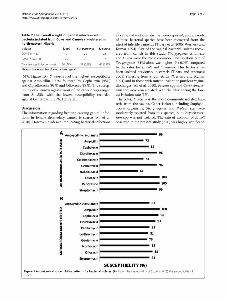
(64%; Figure 1A). S. aureus had the highest susceptibilityagainst Ampicillin 100%, followed by Cephalexin (98%)and Ciprofloxacin (93%) and Ofloxacin (88%). The suscep-tibility of S. aureus against most of the other drugs rangedfrom 81–83%, with the lowest susceptibility recordedagainst Gentamycin (79%; Figure 1B).
DiscussionThe information regarding bacteria causing genital infec-tions in female dromedary camels is scarce (Ali et al.2010). However, evidence implicating bacterial infections
as causes of endometritis has been reported, and a varietyof these bacterial species have been recovered from theuteri of infertile camelids (Tibary et al. 2006; Wernery andKumar 1994). Out of the vaginal bacterial isolates recov-ered from camels in this study, Str. pyogenes, S. aureusand E. coli were the most common. The isolation rate ofStr. pyogenes (31%) alone was higher (P < 0.0%) comparedto the rates for E. coli and S. aureus. This bacteria hasbeen isolated previously in camels (Tibary and Anouassi2001) suffering from endometritis (Wernery and Kumar1994) and in those with mucopurulent or purulent vaginaldischarges (Ali et al. 2010). Proteus spp and Corynebacter-ium spp were also isolated, with the later having the low-est isolation rate (1%).In cows, E. coli was the most commonly isolated bac-
teria from the vagina. Other isolates including Staphylo-coccal organisms, Str. pyogenes and Proteus spp weremoderately isolated from this species, but Corynebacter-ium spp was not isolated. The rate of isolation of E. coliobserved in the present study (73%) was highly significant,
Table 2 The overall weight of genital infection withbacteria isolated from Cows and Camels slaughtered innorth-eastern Nigeria
Isolates E. coli Str. pyogenes S. aureus
COWS (n = 80) 101 25 23
CAMELS (n = 80) 25 26 17
Total isolates (Infection rate) 126 (79%) 51 (32%) 40 (25%)
Abbreviation: n, number of animals investigated.
Figure 1 Antimicrobial susceptibility patterns for bacterial isolates. (A) Shows the susceptibility of E. coli, and (B) the susceptibility ofS. aureus.
Mshelia et al. SpringerPlus 2014, 3:91 Page 4 of 7http://www.springerplus.com/content/3/1/91
and accounted for more than all the other bacterial iso-lates put together. Previously, E. coli was thought to be anon-specific pathogen associated with endometritis inmares and cows (Arthur et al. 2000), but they were re-cently isolated in camels with purulent discharges (Aliet al. 2010). The presence of this bacterium at the presentlevel of infection in the vagina could likely lead to metritisas a consequence of ascending uterine infection duringbreeding. Specific strains of E. coli have recently beenshown to be pathogenic for the endometrium, causing Pel-vic Inflammatory Disease (PID) in cattle (Sheldon et al.2010). But the role of this bacterium in the pathogenesisof metritis especially in camels merits further studies.The uterine bacterial isolates observed in camels in
the present study are similar to those observed in the va-gina, and they concurred with the findings of Yagoub(2005) who reported S. aureus, E. coli, Klebsiella sp, Pro-teus sp, Corynebacterium sp and Streptococcus sp as themain bacterial isolates from several cases of uterine in-fections in camels. The most common isolate in thepresent study was E. coli with an isolation rate of 6%.The isolation rates of the uterine bacteria observed inthe present study were lower compared to those fromthe vagina, and more so in the camels than the cows.Unlike the cows, Proteus sp was not isolated in thecamelids uterus, but Str. pyogenes, S. aureus and Coryne-bacterium sp were isolated at a minimal rate of 1% fromall the camels examined. The result from the cows alsoshowed that E. coli was the major bacteria isolated fromthe uterus. The isolation rate of this bacterium (54%)accounted for more than all the rates for the other bac-teria isolated from the bovine uterus. Although uterineinfection with E. coli is significant in the present study,the rate is lower in the uterus compared to the vagina,which is likely due to the continuous clearance of bac-teria from the uterine lumen (Singh et al. 2008). Despitethis low isolation rate, it is important to note that theisolation of pathogenic bacteria such as E. coli in thepresent study portends a risk factor for lowered repro-ductive efficiency because of increased inflammatory re-actions and possible damages to the uterine tissues(Sheldon and Dobson 2004) by direct action of the bac-teria or its toxins.The data in the present study has shown that there
was no difference (p > 0.05) between the bacterial speciescolonizing the vagina and uterus in camels and cows.But on the overall, the weight of infection with E coli(79%) in all the animals examined was more than therates for Str. pyogenes and S. aureus put together.Generally the pre-breeding and peri-parturient periods
are known as the most critical for bacterial infection ofthe genital tract. This is due to the hormonal changesthat make the uterus susceptible for ascending infectionswith resident bacteria colonizing the vagina (Singh et al.
2008). During these periods, the vagina is constantly be-ing contaminated with bacteria from the environmentand from faecal droppings that smear the vagina duringbreeding seasons. These and other contaminants fromthe male genitalia are introduced into the female vaginaby stud bulls which can lead to uterine infections in vul-nerable animals (Singh et al. 2008; Tibary and Anouassi2001). Also, during the immediate period post partumthe cervix is dilated (Sheldon and Dobson 2004) which al-lows bacteria to ascend from the vagina into the uterus,causing infections in 90% of cows by 21 days post partum(Sheldon et al. 2006). This could possibly explain the simi-larity between the types of vaginal and uterine bacteriaisolated in the present study.The occurrence of ovulation prior to the expulsion of
exudates and debris from the uterus has been shown tofavor heavy growth of bacteria in the uterine environmentwhich leads to the retention of the corpus luteum (CL)and consequent impairment of the ability of the uterus tosecret PGF2α (Kaneko et al. 2013). Although, there is con-tinuous bacterial clearance and recontamination of theuterine lumen for up to 7 weeks postpartum (Singh et al.2008), some bacteria still persist in the uterus triggeringinflammatory responses and pathological changes. Thisdelays uterine involution (Williams et al. 2005) therebylowering fertility. The types of bacteria colonizing theuterus are likely to influence the severity of this condition(Singh et al. 2008) in countries where natural breeding iscommon practice.The result of the present study also showed that E. coli
and S. aureus were isolated from pregnant bovine uteri.This finding is interesting considering that the uterus isthought to be sterile during pregnancy (Singh et al. 2008),when the cervix is closed. Infections of the uterus carryinglife pregnancies are the common causes of repeat breedingoccasioned by conception failures (Ferreira et al. 2008;Gani et al. 2008). Furthermore, the isolation of E. coli froma gravid uterus is particularly important in this study be-cause the bacterium is most frequently associated withuterine disease in cattle (Sheldon et al. 2002; Williamset al. 2005). Infections in the pregnant female could leadto abortion, prenatal-neonatal loss and stillbirth (Tibaryet al. 2006). The persistence of such infections post par-tum is likely to contribute to the early demise of the CLwith decrease in secretion of progesterone (P4) in the af-fected animal (Williams et al. 2007) which could also leadto pregnancy failures.The number of bacteria colonizing the uterus and the
level of uterine immune response are important determi-nants of uterine infections (Azawi 2008; Singh et al.2008). When the immune status is lowered, the patho-genic bacteria adhere to the endometrial mucosa, get in-ternalized and penetrate the epithelium. Alternatively,the bacteria can also release toxins that cause uterine
Mshelia et al. SpringerPlus 2014, 3:91 Page 5 of 7http://www.springerplus.com/content/3/1/91
diseases (Azawi 2008). The findings in the present studyhave shown that the risk of an infection of the vaginawith E. coli is higher in the cows compared to thecamels, but inversely so with S. aureus and Str. pyogenes.Antimicrobial agents are commonly used in the man-
agement of reproductive failures in livestock (Drillich2006). In Nigeria, they are used concurrently with prophy-lactic de-worming regimen in beef bull fattening schemes,but there is no special attention given to antibiosis with re-placement heifers or breeding cows. The findings in thepresent study showed that S. aureus was highly susceptibleto most of the antimicrobial agents tested, among whichAmpicillin, Cephalexin, Ciprofloxacin and Ofloxacin werethe most effective. This finding concurs with the observa-tions made by Gani et al. (2008) who found Ciprofloxacinas one of the most effective antimicrobial agent againstStaphyloccocal uterine infections in dairy cows. In onestudy, Fazlani et al. (2011) showed that S. aureus isolatesfrom camels milk was moderately susceptible to amoxicil-lin, ampicillin, cephalexin and ofloxacin within the rangeof 64-86%. Serin et al. (2010) also demonstrated that S.aureus isolate from wrestling dromedary bulls was 100%susceptible to ciprofloxacin.For E. coli, the susceptibility pattern to most of the anti-
microbial agents tested was similar to what was observedwith the S. aureus isolates, but the susceptibility was highercompared to those observed with the S. aureus isolates.The most effective antimicrobial agents observed includeOfloxacin and Pefloxacin, also Amoxycillin-clavulanate,Ciprofloxacin, Gentamycin and Streptomycin. This findingis similar to previous reports with isolates associated withgenital infections in cattle and sheep (Gani et al. 2008;Goncuoglu et al. 2010), and from mastitic milk samplesof camels (Fazlani et al. 2011).Amongst the factors that have been reported to be con-
tributing to uterine infections in camelids are overbreed-ing (excessive matings during the period of receptivity),postpartum complications and unhygienic gynaecologicalexamination and manipulation (Tibary and Anouassi2001). It has been highlighted that antimicrobial treat-ments have some beneficial effects on reproductive per-formance in livestock (Drillich et al. 2003). Therefore, atthe current level of susceptibility of these bacteria to theantimicrobial agents tested in the present study, effectivetreatment could be achieved if these antimicrobial agentsare used appropriately.
ConclusionsThe bacteria colonizing the genital tract are similar incamels and cows reared in north-eastern Nigeria. E. coliand S. aureus were amongst the most prevalent bacteriaisolated, and they were found to be susceptible to theantimicrobial agents tested. It is advised that effectivegynaecological evaluations should precede the initiation
of antimicrobial treatments in order to minimize the de-velopment of antimicrobial resistant pathogenic strainsin these species.
Competing interestsThe authors have no any competing interest to disclose.
Authors’ contributionsThe study was conceived by GDM, samples were collected by GO and YACV.The manuscript was drafted by GDM, GOE read and corrected themanuscript. All authors also read and approved the final manuscript.
AcknowledgementsWe are grateful to Isa A. Gulani of the Department of Veterinary Medicine,University of Maiduguri, Nigeria, for technical assistance; and the staff of theMaiduguri municipal abattoir, for their support with sample collection.
Author details1Department of Veterinary Surgery and Theriogenology, Faculty of VeterinaryMedicine, University of Maiduguri, PMB 1069 Maiduguri, Nigeria.2Department of Veterinary Medicine, Faculty of Veterinary Medicine,University of Maiduguri, PMB 1069 Maiduguri, Nigeria.
Received: 20 December 2013 Accepted: 12 February 2014Published: 15 February 2014
ReferencesAli A, Hassanein KM, Al-Sobayil FA, Tharwat M, Al-Hawas A, Ahmed AF (2010)
Relationship between characters of vaginal discharges and uterine bacterialisolates recovered from repeat breeding female camels (Camelus dromedarius).J Agric Vet Sci 2:87–97
Amin JD, Zaria LT, Malgwi RW (1996) Vaginal aerobic bacterial flora of apparentlyhealthy cattle in various stages of reproductive cycle in the Sahel region ofNigeria. Bull Anim Hlth Prod Afr 44:15–18
Arthur GH, Pearson H, Noakes DE (2000) Veterinary Reproduction and Obstetrics.English Language Book Society and Bailliere. Tindall, London
Azawi OI (2008) Postpartum uterine infection in cattle. Anim Reprod Sci 105:187–208Azawi OI, Al-Abidy HF, Ali AJ (2010) Pathological and bacteriological studies of
hydrosalpinx in buffaloes. Reprod Domest Anim 45:416–420Blench R (1999) Traditional livestock breeds: Geographical distribution and
dynamics in relation to the ecology of West Africa., pp 44–50, Available fromhttp://www.odi.org.uk/publications/2041-traditional-livestock-breeds-geographical-distributiondynamics-relations-ecology-west-africa (Assessed31st January 2014)
Cheesbrough M (2000) District Laboratory Practice in Tropical Countries Part 2.Cambridge University Press, Cambridge, UK
CLSI (2011) Clinical and Laboratory Standards Institute, Performance StandardsFor Antimicrobial Disk Susceptibility Testing. 21st International SupplementCLSI Document. M100-S21, Wayne, PA
Cowan SJ, Steel KJ (1993) Manual of Identification of Medical Bacteria, 3rd edn.Cambridge University Press, Cambridge, UK
Demissie M (2011) Isolation and identification of aerobic, septicaemia bacteriafrom cattle in and around Sebeta town and antimicrobial susceptibilitytesting. Afr J Microbiol Res 5:87–92
Drillich M (2006) An update on uterine infections in dairy cattle. Slovakia Vet Res43:11–15
Drillich M, Pfützner A, Sabin HJ, Sabin M, Heuwieser W (2003) Comparison of twoprotocols for the treatment of retained foetal membranes in dairy cattle.Theriogenology 59:951–960
El-Yuguda AD, Abubakar MB, Baba SS, Ngangnou A (2010) Competitive ELISArinderpest virus antibody in slaughtered camels (Camelus dromedarius):implication for rinderpest virus elimination from Nigeria. Afr J Biomed Res13:83–85
Enany M, Hanafi MS, El-Ged AGF, El-Seedy FR, Khalid A (1990) Microbiologicalstudies on endometritis in she-camels in Egypt. J Egypt Vet Med Assoc50:229–243
Fazlani SA, Khan SA, Faraz S, Awan MS (2011) Antimicrobial susceptibility ofbacterial species identified from mastitic milk samples of camel. Afr JBiotechnol 10(15):2959–2964
Ferreira R, Oliveira JFC, Antoniazzi AQ, Pimentel CA, Moraes JCF, Henkes LE,Bordignon V, Gonçalves PB (2008) Relationship between clinical and
Mshelia et al. SpringerPlus 2014, 3:91 Page 6 of 7http://www.springerplus.com/content/3/1/91
postmortem evaluation in repeat breeder beef cows. Ciência Rural, SantaMaria 38:1056–1060
Fourichon C, Seegers H, Malher X (2000) Effect of disease on reproduction indairy cows: a meta-analysis. Theriogenology 53:1729–1759
Gani MO, Amin MM, Alam MGS, Kayesh MEH, Karim MR, Samad MA, Islam MR(2008) Bacterial flora associated with repeat breeding and uterine infectionsin dairy cows. Bangladesh J Vet Med 6:79–86
Goncuoglu M, Seda F, Ormanci B, Ayaz ND, Erol I (2010) Antibiotic resistance ofEscherichia coli O157:H7 isolated from cattle and sheep. Ann Microbiol60:489–494
GraphPad (2010) GraphPad Prism Version 5.04, GraphPad Software Inc.,www.graphpad.com
Heuwieser W, Tenhagen BA, Tischer M, Luhr J, Blum H (2000) Effect of threeprogrammes for the treatment of endometritis on the reproductiveperformance of a dairy herd. Vet Rec 146:338–341
Hussain AM, Daniel RCW, O’Boyle D (1990) Post-partum uterine flora followingnormal and abnormal puerperium in cows. Theriogenology 34:291–302
Hussein FM, El-Amrawi GA, El-Bawab IE, Metwally KK (2006) Bacteriological andhaematological studies in the camel genitalia. In: Proceedings of theInternational Scientific Conference on Camels, Qassim University 10–12 May.,pp 491–500
Kaneko K, Nakamura M, Reiichiro-Sato R (2013) Influence of Trueperella pyogenes inuterus on corpus luteum lifespan in cycling cows. Theriogenology 79:803–808
Koneman WK, Allen SD, Janda WM, Schreckenberger PC, Propcop GW, Woods GL,Winn WC Jr (2005) Color Atlas and Textbook of Diagnostic Microbiology, 6thedn. Lippincott-Raven Publisher, Philadelphia, USA
Markemann A, Zarate AV (2010) Traditional llama husbandry and breedingmanagement in the Ayopaya region, Bolivia. Trop Anim Health Pro 42:79–87
Mouchira MM (2009) Pathological Studies on Ascariasis in Dromedary (Camelusdromedarius) and Llama (Lama glama) Camelidae. Eur J Sci Res 38:159–171
Potter TJ, Guitian J, Fishwick J, Gordon PJ, Sheldon IM (2010) Risk factors forclinical endometritis in postpartum dairy cattle. Theriogenology 74:127–134
Serin I, Ceyland A, Kirklan S, Parin U (2010) Preputial bacterial flora and antibioticsusceptibility in wrestling dromedary bulls in Aydin region, Turkey. J AnimVet Adv 9(3):482–485
Sheldon IM, Dobson H (2004) Postpartum uterine health in cattle. Anim ReprodSci 82–83:295–306
Sheldon IM, Noakes DE, Rycroft AN, Pfeiffer DU, Dobson H (2002) Influence ofuterine bacterial contamination after parturition on ovarian dominant follicleselection and follicle growth and function in cattle. Reproduction 123:837–845
Sheldon IM, Lewis GS, LeBlanc S, Gilbert RO (2006) Defining post-partum uterinedisease in cattle. Theriogenology 65:1516–1530
Sheldon IM, Cronin J, Goetze L, Donofrio G, Schuberth HJ (2009) Definingpostpartum uterine disease and the mechanisms of infection and immunityin the female reproductive tract in cattle. Biol Reprod 81:1025–1032
Sheldon IM, Rycroft AN, Dogan B, Craven M, Bromfield JJ, Chandler A, RobertsMH, Price SB, Gilbert RO, Simpson KW (2010) Specific strains of Escherichiacoli are pathogenic for the endometrium of cattle and cause pelvicinflammatory disease in cattle and mice. PLoS One 5:e9192, doi:10.1371/journal.pone.0009192
Singh J, Murray RD, Mshelia G, Woldehiwet Z (2008) The immune status of thebovine uterus during the peri-partum period. Vet J 175:301–309
Srikandakumar A, Johnson EH, Mahgoub O, Kadim IT, Al-Ajmi DS (2001) Anatomyand histology of the female reproductive tract of the Arabian camel. EmiratesJ Agric Sci 13:23–26
Thrustfield M (2005) Veterinary Epidemiology, 3rd edn. Black Well Science Ltd,Cambridge, USA, pp 225–228
Tibary A, Anouassi A (2001) Uterine infections in Camelidae. Vet Sci Tomorrow,http://dspace.library.uu.nl:8080/handle/1874/28883
Tibary A, Fite C, Anouassi A, Sghiri A (2006) Infectious causes of reproductive lossin Camelids. Theriogenology 66:633–647
Wernery U, Kumar BN (1994) Reproductive disorders in dromedary camels due toinfectious causes and its treatment. J Camel Pract Res 1:85–87
Williams EJ, Fischer DP, Pfeiffer DU, England GCW, Noakes DE, Dobson H,Sheldon IM (2005) Clinical evaluation of postpartum vaginal mucus reflectsuterine bacterial infection and the immune response in cattle.Theriogenology 63:102–117
Williams EJ, Fischer DP, Noakes DE, England GCW, Rycroft A, Dobson H, Sheldon IM(2007) The relationship between uterine pathogen growth density and ovarianfunction in the postpartum dairy cow. Theriogenology 68:549–559
Yagoub SO (2005) Bacterial diseases of the reproductive system of camels(Camelus dromedaries) in Eastern Sudan. J Anim Vet Adv 4:642–644
doi:10.1186/2193-1801-3-91Cite this article as: Mshelia et al.: Comparative studies on genitalinfections and antimicrobial susceptibility patterns of isolates fromcamels (Camelus dromedarius) and cows (Bos indicus) in Maiduguri,north-eastern Nigeria. SpringerPlus 2014 3:91.
Submit your manuscript to a journal and benefi t from:
7 Convenient online submission
7 Rigorous peer review
7 Immediate publication on acceptance
7 Open access: articles freely available online
7 High visibility within the fi eld
7 Retaining the copyright to your article
Submit your next manuscript at 7 springeropen.com
Mshelia et al. SpringerPlus 2014, 3:91 Page 7 of 7http://www.springerplus.com/content/3/1/91