Embed Size (px)
Citation preview

Braz Dent J 15(1) 2004
SEM evaluation of adhesive systems 19Braz Dent J (2004) 15(1): 19-25
Comparative SEM Evaluation of Penetration ofAdhesive Systems in Human Dentin with a
Non-rinse Conditioner and a Self-etching Primer
Julio Orrico de Aragão Pedra e CAL-NETO1
Mauro Sayão de MIRANDA2
Katia Regina Hostílio Cervantes DIAS2
1Department of Orthodontics, Faculty of Dentistry, Rio de Janeiro State University, Rio de Janeiro, RJ, Brazil2Department of Restorative Dentistry, Faculty of Dentistry, Rio de Janeiro State University, Rio de Janeiro, RJ, Brazil
The purpose of this study was to compare the effect of a self-etching primer and a non-rinse conditioner with the effect of a conventionaladhesive system on the penetration depth in dentin of human teeth, using scanning electronic microscopy (SEM). Fifteen human thirdmolar teeth were sectioned into 2 pieces. All pieces were flattened with grade 600 to 1200 silicon carbide paper and divided into 6groups: group 1 - Prime & Bond NT (NT), negative control; group 2 - 37% phosphoric acid + Prime & Bond NT (PANT), positivecontrol; group 3 - Non-rinse Conditioner (NRC) + Prime & Bond NT (NRCNT); group 4 - NRC + Prime & Bond 2.1 (NRCPB); group5 - NRC + Scotchbond MP (NRCSB); group 6 - Prompt L-Pop (PLP). All teeth were covered with Dyract AP. The tooth fragments weredecalcified, and its resin replicas were evaluated by SEM by three previously standardized examiners. The penetration was scored from0 (no penetration) to 3 (maximum penetration). The Kruskal-Wallis and Mann-Whitney U tests (p<0.05) showed three statisticallyhomogeneous groups: {NT, NRCPB, NRCSB and PLP}, {NRCNT} and {PANT}. The authors concluded that the self-etching primerand the non-rinse conditioner provide a lower penetration depth in human tooth dentin than the conventional adhesive system.
Key Words: adhesive systems, dentin, resin-dentin interface, self-etching primers, scanning electron microscopy.
Correspondence: Dr. Julio Orrico de Aragão Pedra e Cal Neto, Rua Almirante Luis Belart 190/206, Jardim Guanabara, 21941-100 Rio de Janeiro,RJ, Brasil. Tel: + 55-21-2462-5239. e-mail: [email protected]
ISSN 0103-6440
INTRODUCTION
The acid etching technique, presented byBuonocore (1) in 1955, generated a revolution in Den-tistry, stimulating the development of several new prod-ucts. Recently, acid primers, single-bottle agents andself-etching bonding systems have become known in avery short period of time. However, these productsmust be evaluated and tested before they are used on alarge-scale basis. As several factors must be analyzed,scanning electron microscopy (SEM) has been used toevaluate the micromechanical bonding mechanism of-fered by the current adhesives. This research is donethrough observation of dentinal tissue samples or repli-cas in resin (2), which clearly reveals that bonding hasa biomechanical nature, based on hybridization of thedemineralized dentinal tissues, with resin tag and lat-eral branch formation (3-5).
Some of the adhesive systems have an etchingand primer combination that is applied in only one step(acid primers), in order to render their clinical use morepractical and fast. Others include a primer and adhesivein only one bottle (single-bottle agents); these are usedafter enamel and dentin etching procedures with phos-phoric acid have been performed (6,7). Although possi-bilities of error are minimized during their use, the totalapplication procedure is not simple because severallayers must be applied, therefore time saving proce-dures regarding conventional systems are not that sig-nificant (8,9).
Thus, efforts aiming to develop new materialsare being carried out, in order to simplify the total acidetching system and to prevent the phenomenon knownas “nanoleakage”, by avoiding a greater dentin demin-eralization where the total penetration of the adhesivemany times did not occur (10,11). Many products are

Braz Dent J 15(1) 2004
20 J.O.A.P. Cal Neto et al.
available on the market, such Non-rinse Conditioner(Dentsply), which is an acid that needs no rinsing, andPrompt L-Pop (ESPE), which is a new self-etchingprimer which, in fact, combines all the etching andbonding process in only one stage. However, the actionof these new materials on dentinal tissues is not yet wellknown and must be analyzed under different aspects, inorder to be properly used by professionals so that theycan make the best of their properties during clinicalapplication.
The purpose of this study was to evaluate theeffect of a non-rinse conditioner associated to adhe-sives and also to evaluate a self-etching primer, com-paring them with the effects of 37% phosphoric acid,regarding regularity and infiltration depth of adhesivesin human teeth dentin using SEM.
MATERIAL AND METHODS
Fifteen recently extracted erupted third humanmolars were selected for this study. These teeth had nocaries, nor previous restorations. They were stored indistilled water at room temperature for 3 months.
In order to evaluateadhesive infiltration in thedentin, the teeth were sec-tioned longitudinally fol-lowing the buccal-lingualgroove direction, obtaining2 halves. The occlusal sur-faces were removed 1 mmbelow the dentinal-enameljunction, which corre-sponded to the central groove.With a transversal section,their roots were also elimi-nated. They underwent cut-ting procedures under theocclusal dentin surface, soas to obtain 3-mm thickdiscs for a total of 30 speci-mens. These were cut witha double-faced diamonddisc and water was used forcooling. The dentin sampleswere flattened and polishedusing number 600 to 1200wet silicon carbide paper.
Teeth fragments were divided into 6 groups of 5dentin samples each (Table 1). The materials used andthe corresponding manufacturer and composition arepresented in Table 2.
The etching agents and adhesive systems wereapplied following manufacturer recommendations. Thedentin discs were coated with Dyract AP (Dentsply),making a cylinder of approximately 6 mm high with adiameter of 4 mm. In order to do this, 3 increments of 2mm each were superimposed, and each one wasphotopolymerized for 40 s with a QHL 75 Curing Lite
Table 1. Etching and adhesive agents of the 6 experimentalgroups.
Group Etching agent Adhesive agent Symbol
1 ——— Prime & Bond NT NT2 37% Phosphoric Acid Prime & Bond NT PANT3 Non-Rinse Conditioner Prime & Bond NT NRCNT4 Non Rinse Conditioner Prime & Bond 2.1 NRCPB5 Non Rinse Conditioner Scotch Bond MP NRCSB6 Prompt L-Pop PLP
Table 2. Materials used and their corresponding composition and manufacturers.
Material Composition Manufacturer
Non-Rinse Conditioner (NRC) Itaconic acid, Maleic ácid, Dentsply/De TreyWater, Solvent Konstanz, Germany
Prime & Bond NT Di and trimethacrylate resins, Dentsply/De TreyFunctionalized amorphous silica, Konstanz, GermanyPENTA, Photoinitiators, Stabilizers,Cetylamine hydrofluoride, Acetone
Prime & Bond 2.1 Elastomeric dimethacrylate resins, Dentsply/De TreyPENTA, Photoinitiators, Stabilizers, Konstanz, GermanyCetylamine hydrofluoride, Acetone
Scotchbond Multi-Purpose Primer: HEMA, 3M Dental Productsphoto-activated polymer, water St. Paul, MN, USAAdhesive: BIS-GMA, HEMA,Photoinitiators
Prompt L-Pop Liquid 1(red blister): Methacrylated ESPE Dental AGphosphoric esters, Initiators, Seefeld, GermanyStabilizersLiquid 2 (yellow blister): Water,Fluoride complex, Stabilizers

Braz Dent J 15(1) 2004
SEM evaluation of adhesive systems 21
equipment (Dentsply; intensity>500 mW/cm2). Allsamples were stored for one week in distilled water atroom temperature.
In order to observe the adhesive penetrationmicromorphology, specimens were submitted to cyclesof demineralization, which led to complete decalcifica-tion and dissolution of the dentinal structures. Approxi-mately 5 consecutive cycles were performed consistingof a 10% hydrochloric acid bath for 5 h, and a 5%sodium hypochlorite bath for 1 h; baths and cycles were
intercalated with a 5-min distilled water rinse. After all dentinal tissues had been completely
eliminated, resin replicas were then coated with a goldlayer (Sputer equipment) and examined with a scanningelectron microscope (DSM 960 - Zeiss, Germany).
Photomicrographs were made at 1000X and3000X, for each sample obtained in the dentin, near thepulp area. These photomicrographs were evaluated bythree specialized professionals who attributed com-parative scores, according to the following penetration
Figure 1. Group 1. Left: Resin replica of the dentinal surface. Only some areas presented resin tags (1000X). Right: Highermagnification revealing an irregular shape of the resin tags (3000X).
Figure 2. Group 2. Left: Resin replica of the dentin where a regular tag distribution can be observed, with deep penetration (1000X). Right:Higher magnification revealing long tags, with an inverted cone shape and a large quantity of lateral bond branches (3000X).
Braz Dent J 15(1) 2004
22 J.O.A.P. Cal Neto et al.
Prime & Bond NT was applied, no acceptable adhesiveinfiltration was revealed. In many areas there was noevidence of tag formation and when tags were present,they were of irregular shape (Figure 1).
Group 2: Good penetration was verified all overthe sample. The resin tags were long and presented aninverted cone shape, showing an adequate tubular seal-ing. The presence of several lateral adhesive brancheswas also observed. (Figure 2).
Group 3: Variable penetration was observed andtags were generally short, presenting an irregular shape
Figure 3. Group 3. Left: Resin replica of the dentinal surface where an irregular penetration was observed (1000X). Right: Highermagnification revealing short tags, irregularly shaped, in the entrance of tubular orifices (3000X).
Figure 4. Group 4. Left: An irregular etch pattern was observed in the dentin, with small island formation with good penetration(1000X). Right: Higher magnification revealing irregular shaped resin tags (3000X).
scale: 0 = without penetration; 1 = minimum penetra-tion; 2 = intermediate penetration and 3 = maximumpenetration. Characteristic photomicrographs of eachscore were used for calibration among evaluators.
Data obtained was statistically analyzed byKruskal-Wallis and Mann-Whitney U non-parametrictests (p<0.05).
RESULTS
Group 1: On the dentin samples where only
Braz Dent J 15(1) 2004
SEM evaluation of adhesive systems 23
at the entrance of the tubular orifice. This might indi-cate imperfect sealing. (Figure 3).
Group 4: When NRC was applied associated toPrime & Bond 2.1 on the dentinal surface, an irregularetch pattern was observed with some areas with goodpenetration, and others without resin tag formation(Figure 4).
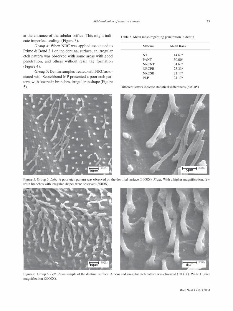
Group 5: Dentin samples treated with NRC asso-ciated with Scotchbond MP presented a poor etch pat-tern, with few resin branches, irregular in shape (Figure5).
Figure 5. Group 5. Left: A poor etch pattern was observed on the dentinal surface (1000X). Right: With a higher magnification, fewresin branches with irregular shapes were observed (3000X).
Figure 6. Group 6. Left: Resin sample of the dentinal surface. A poor and irregular etch pattern was observed (1000X). Right: Highermagnification (3000X).
Table 3. Mean ranks regarding penetration in dentin.
Material Mean Rank
NT 14.67a
PANT 50.00c
NRCNT 34.67b
NRCPB 23.33a
NRCSB 21.17a
PLP 21.17a
Different letters indicate statistical differences (p<0.05)

Braz Dent J 15(1) 2004
24 J.O.A.P. Cal Neto et al.
Group 6: The Prompt L-Pop self-etching systempresented a poor etch pattern that was quite irregularwith areas with no tags and other areas with tag forma-tion, although these tags were short (Figure 6).
Statistical analysis revealed homogeneous groupsfor dentin penetration. The decreasing penetration or-der was as follows: {PANT}, {NRCNT} and {NT,NRCPB, NRCSB and PLP} (Table 3).
DISCUSSION
The method used in this study, SEM of totaldental tissues decalcification, is extremely simple anduseful for evaluation of characteristics of resin tags andalso for evaluation of their lateral branches and thedentinal surface etch pattern (12). When the dentinaltubule orifices are totally sealed, the initial parts of thetags present a reverse cone shape, generally havingadhesive lateral branches, indicating a very good etchpattern. Some studies (3,5) have suggested that theselateral branches or anastomoses between the dentinaltubules may contribute to the micromechanical reten-tion mechanism, resulting in a positive influence on thejunction between the dentinal tissue and the compositeresin.
According to Ferrari et al. (2), lower SEM mag-nifications allow dentinal enamel and dentin etch pat-tern verification, where tag density and depth can alsobe observed, whereas higher magnifications reveal mor-phological characteristics of resin tags penetrating bothsubstrates.
In studies regarding “nanoleakage”, Sano et al.(11) suggested that adhesive systems should be devel-oped using the least possible demineralized dentin thick-ness, as well as monomers having a great affinity withcollagen and with hydroxyapatite, because this wouldenable a more even penetration and full polymeriza-tion. Thus, more careful investigations on acid solutionconcentrations were carried out in order to avoid den-tinal decalcification in such magnitude that the adhe-sive would not be able to penetrate as deeply, leaving acollagen basal portion unprotected. This could in thefuture involve running risks regarding bond strength(7,10). Self-etching systems may be a solution for thisproblem, because they provide less dentinal deminerali-zation, simultaneous to resin monomer penetration (13).
When Prime & Bond NT was associated with37% phosphoric acid (PANT) on the dentinal surface,
an even etch pattern with long tags was observed, aswell as a large number of adhesive lateral branches.This process clearly revealed the micromechanical bond-ing mechanism. Currently, it is believed that tag lengthis not as important for bond strength as was oncebelieved; however, the importance of its shape, as wellas the presence of lateral branches, must be noted(10,14,15).
When NRC was associated with Prime & BondNT, a lesser tag density was observed; in fact, these tagswere much shorter. When NRC was associated withPrime & Bond 2.1 or Scotchbond MP, in addition toshorter, less dense tags, their distribution was quiteirregular, indicating the worst results.
A shorter tag formation was observed with PromptL-Pop, as expected because this is a characteristic of theself-etching systems (4,11,16-18). A quite irregularetch pattern was observed, some areas were properlyetched and others had no etching at all; thus, this iscause for alarm. However, other studies report goodresults in bond strength tests (19).
The use of the Prompt L-Pop system is extremelyattractive due to its practical application, and perhapseven promising. It represents a time-saving clinicalprocedure, facilitating its use, reducing failures in thehybridization process (13,17). However, it is too earlyto recommend this procedure without restrictions. Thisproduct still requires improvement and longer clinicalstudies before it can be recommended on a large scalefor clinical use.
Although the manufacturer recommends the soleuse of Prime & Bond NT without previous etching insome situations, it does not seem adequate. Duringobservations with SEM, there was no evidence of themicromechanical bonding mechanism on the dentin,and a low density tag formation with quite irregulardistribution in both dental tissues was observed.
According to the results of this study, the needfor an etching agent was observed. Phosphoric acidpresented clear evidence of the resin bonding mecha-nism to the dentin. In spite of this, the use of adhesivesystems with low concentration acid solutions seems tobe the path to be followed, considering the advantagesand positive results obtained in some research(7,10,11,20). This tendency is supported by the prin-ciple that the quality of the hybrid layer is more impor-tant than its thickness (16). We conclude that the self-etching system and the non-rinse conditioner provide a

Braz Dent J 15(1) 2004
SEM evaluation of adhesive systems 25
lower penetration depth in human teeth dentin than theconventional adhesive system.
RESUMO
O objetivo desse estudo foi comparar o efeito de um adesivoautocondicionante e de um ácido não lavável quanto aprofundidade de penetração na dentina de dentes humanos,através da observação em Microscopia Eletrônica de Varredura(MEV). Quinze terceiros molares tiveram a dentina exposta,cortando-se a face oclusal, planificados com lixa d’água 600,cortados transversalmente e distribuídos em 6 grupos de 5 cada:grupo 1 – Prime Bond NT (NT), controle negativo; grupo 2 –ácido fosfórico 37% + Prime Bond NT (PANT), controle positivo;grupo 3 – Non-Rinse Conditioner (NRC) + Prime Bond NT(NRCNT); grupo 4 – NRC + Prime Bond 2.1 (NRCPB); grupo 5– NRC + Scotch Bond MP (NRCSB) e grupo 6 – Prompt L-Pop(PLP). Todos foram recobertos com Dyract AP. Os fragmentosdentários foram descalcificados com HCl a 10% e NaOH a 5% eas réplicas em resina examinadas em MEV. Três examinadorescalibrados avaliaram as fotomicrografias e atribuíram escores de0 = sem penetração a 3 = penetração máxima. Os testes deKruskal-Wallis e Mann-Whitney (P<0,05) evidenciaram trêsgrupos homogêneos {NT, NRCPB, NRCSB e PLP}; {NRCNT}e {PANT}. Os autores concluíram que o sistema adesivoautocondicionante e o ácido não lavável apresentaram menorpenetração em dentina do que o sistema adesivo convencional.
REFERENCES
1. Buonocore MG. Simple method of increasing the adhesion ofacrylic filling materials to enamel surface. J Dent Res1955;34:849-853.
2. Ferrari M, Manocci F, Kugel G. Standardized microscopic evalu-ation of the bonding mechanism of NRC/Prime & Bond NT. AmJ Dent 1999;12:77-83.
3. Chappel RP, Cobb CM, Spencer P. Dentinal tubule anastomosis:a potential factor in adhesive bonding? J Prosthet Dent1994;72:183-188.
4. Ferrari M, Davidson CL. In vivo resin-interdiffusion and tag
formation with adhesive lateral branches of two adhesives sys-tems. J Prosthet Dent 1996;76:250-253.
5. Titley K, Chernecky R, Chen A. The composition and ultrastruc-ture of resin tags in etched dentin. Am J Dent 1995;8:224-230.
6. Kanca J. Resin bonding to wet substrate. I. Bonding to dentin.Quintessence Int 1992;23:39-41.
7. Youssef MN, Guaraldi E, Sato CT. An in vitro SEM comparativestudy of four adhesive systems as to dentin penetration. RevAPCD 1998;52:236-239.
8. Finger WJ, Fritz U. Laboratory evaluation of one-componentenamel/dentin bonding agents. Am J Dent 1996;9:206-210.
9. Van Meerbeek B. The clinical performance of adhesives. J Dent1998;26:1-20.
10. Prati C. Dentine permeability and bond quality as affected bynew bonding system. J Dent 1995;23:217-226.
11. Sano H, Takatsu T, Ciucchi B. Nanoleakage: Leakage within thehybrid layer. Oper Dent 1995;20:18-25.
12. Cagidiaco MC, Ferrari M, Vichi A. Mapping of tubule andintertubule surface areas available for bonding in Class V andClass II preparations. J Dent 1997;25:379-389.
13. Watanabe I, Nakabayashi N, Pashley DH. Bonding to grounddentin by Phenyl-P self-etching primer. J Dent Res 1994;73:1212-1220.
14. Nakabayashi N. Hybrid layer as a dentin-bonding mechanism. JEsthet Dent 1991;3:133-138.
15. Nakajima M. Tensile bond strength and SEM evaluation of cariesaffected dentin using dentin adhesives. J Dent Res 1995;74:1679-1688.
16. Burrow MF, Takakura H, Nakajima M, Inai N, Tagami J, TakatsuT. The influence of age and depth of dentin on bonding. DentMater 1994;10:241-246.
17. Perdigão J, Swift Jr EJ. Adhesion of a total-etch phosphate esterbonding agent. Am J Dent 1994;7:149-152.
18. Sano H, Yoshikawa T, Pereira PNR. Long-term durability ofdentin bonds made with a self-etching primer, in vivo. J Dent Res1999;78:906-911.
19. Oliveira WJ, Pagani C, Rodrigues JR. Comparação da adesividadede dois sistemas adesivos autocondicionantes em esmalte dedentes bovinos. Pós-Grad Rev Fac Odontol São José dos Campos2001;4:43-50.
20. Gordan VV, Vargas MA, Cobb DS. Evaluation of acidic primersin microleakage of Class 5 composite resin restorations. OperDent 1998;23:244-249.
Accepted August 11, 2003