Embed Size (px)
Citation preview
Revue Méd. Vét., 2013, 164, 2, 72-79
AVCI (H.) AND COLLABORATORS72
Introduction
Photobacterium damselae subsp. piscicida, the causative agent of fish pasteurellosis, was initially isolated from natural populations of white perch (Morone americanus) and striped bass (Morone saxatilis) in 1963 during a massive epizootic in Chesapeake Bay, USA [30], and is now widely distributed throughout the world [5, 9, 12, 32]. The disease was described as an acute to chronic disease [31]. The acute form of disease is manifested by a severe general septicaemia, whereas the chronic form of the disease is characterized by presence of whitish tubercle-like tissue structures consisting of bacterial accumulations and surrounding epithelial cell
layers in the visceral organs [13, 27, 31]. The lesions grossly resemble granulomas, and the disease is also called as pseudotuberculosis [13].
Sea bass (Dicentrarhus labrax) and gilthead sea bream (Sparus aurata) fish are the species most susceptible to the disease [4, 14, 32], but other some fish species have also been reported to became infected, such as yellowtail (Seriola quinqueradiata) [11], red sea bream [34] and sole (Solea senegalensis) [17]. The temperature, quality of the water and stress on the fish are important factors influencing the onset of the fish pasteurellosis [16]. The disease generally affects larvae causing mortality of 50-100%, but the disease
SUMMMARY
The present study describes pathological and immunohistochemical findings resulting from acute and chronic infections in juvenile sea bass (Dicentrarhus labrax) and gilthead sea bream (Sparus aurata) naturally infected with Photobacterium damselae subsp. piscicida. The disease has an acute presentation in sea bass while in gilthead sea bream acute and chronic forms were recorded. The highest mortality rates in both fish species were observed in June when the water temperature is elevated. Darkening of skin colour and irregular swimming were the main clinical signs in the acute form while greyish white nodules (0.5-1 mm) were only evidenced in chronic cases. In both species, the acute form is histopathologically characterized by the abundance of bacterial clusters coupled to vascular lesions in many organs (gills, heart, kidney, spleen, liver and gastrointestinal tract at a lesser extend). In the chronic form, granulomatous lesions consisted in clusters of bacteria in the center surrounded by epithelial histiocytes and macrophages and finally fibroblasts. By immunohistochemistry, immunopositive bacteria were densely evidenced in the kidney, spleen, gills, heart and liver in acute cases in both fish species whereas they were mainly located in kidney and spleen during the chronic form. Keywords: Fish pasteurellosis, sea bass, gilthead sea bream, pathological findings, immunohistochemistry.
RESUME
Pasteurellose du poisson, loup de mer, daurade royale, histopathologie, immunohistochimie
Cette étude décrit les données histopathologiques et immunohistochimiques lors d’infections aiguë et chronique de jeunes loups de mer (Dicentrarhus labrax) et daurades royales (Sparus aurata) naturellement infectés par Photobacterium damselae subsp. piscicida. La maladie s’est présentée sous forme aiguë chez les loups de mer, alors que chez les daurades des formes aiguës et chroniques ont été observées. Les taux les plus élevés de mortalités dans les 2 espèces ont été enregistrés en juin lorsque la température de l’eau est élevée. Un assombrissement de la couleur de la peau et une nage irrégulière ont constitué les principaux signes de la forme aiguë et des nodules blanc grisâtres (0.5-1 mm) n’ont été mis en évidence que dans les formes chroniques. Pour les 2 espèces, la forme aiguë a été caractérisée histologiquement par l’abondance de colonies bactériennes en amas couplée à des lésions vasculaires dans de nombreux organes (branchies, cœur, reins, rate, foie et le tube digestif à un moindre degré). Dans la forme chronique, les granulomes étaient formés en leur centre d’amas bactériens entourés par des histiocytes épithéliaux, des macrophages et en enfin des fibroblastes. Par immunohistochimie, de nombreuses bactéries immunopositives ont été mises en évidence dans le rein, la rate, les branchies, le cœur et le foie dans les cas aigus dans les 2 espèces alors qu’elles sont restées principalement localisées aux reins et à la rate où elles sont apparues moins nombreuses dans la forme chronique. Mots-clés : Pasteurellose du poisson, loup de mer, daurade royale, histopathologie, immunohistochimie.
Comparative histopathological and immunohistochemical evaluations in juvenile sea bass (Dicentrarhus labrax) and gilthead sea bream (Sparus aurata) naturally infected with Photobacterium damselae subsp. piscicidaH. AVCI1*, S. BIRINCIOĞLU1, E. T. EPIKMEN1, M. DERELI1
1Department of Pathology, Faculty of Veterinary Medicine, University of Adnan Menderes, 09016 Isikli-Aydin, TURKEY
*Corresponding author: [email protected]
Revue Méd. Vét., 2013, 164, 2, 72-79
FISH PASTEURELLOSIS IN JUVENILE SEA BASS AND GILTHEAD SEA BREAMS 73
can also affect juveniles with mortality up to 50% [2, 25]. The incidence of diseases and types of bacterial pathogens has well documented in several cultured fish species [3]. However, detailed pathological and immunohistochemical findings of Photobacterium damselae ssp. piscicida have been reported sporadically [10, 21, 27].
The aims of the study are to present and evaluate histopathological and immunohistochemical findings of fish pasteurellosis in juvenile sea bass (Dicentrarhus labrax) and gilthead sea bream (Sparus aurata) naturally infected with Photobacterium damselae subsp. piscicida.
Materials and Methods
In the early summer of 2010, an outbreak was occurred in a fish farm, which was located in the southern Aegean of part Turkey. Fish samples were collected monthly from the rearing pools from March to June.
For bacteriological examinations, samples were taken in cooled boxes (1-4OC) to laboratory and then the samples were processed within 1 hour after collection for bacteria identification using microbiological methods [5, 15, 21].
For histological examinations, a total of 96 dead or euthanized fish (44 juvenile sea bass and 52 juvenile gilthead sea bream) were fixed in 10% neutral-buffer formalin solution, embedded in paraffin, sectioned at 5 µm and stained routinely with haematoxylin and eosin and examined using light microscope. The selected kidney, liver, spleen, heart and gill sections were also stained by Brown and Brenn staining method for bacteria [6]. Replicated sections were also used for immunohistochemistry.
The immunohistochemical procedure used in the present study was based on the method of ADAMS and MARIN DE MATEO [1]. Tissue sections were dewaxed in xylene and rehydrated through graded alcohols. The
deparaffinised tissue sections were incubated with 3% H2O2 in 70% methanol to inhibit endogenous peroxidase activity. The tissues were pretreated with 10 mM citrate buffer, pH 6.0 in microwave oven at 500 W for 10 minutes for antigen retrieval, and then the slides were washed once for 10 minutes in phosphate-buffered-saline (PBS; pH 7.3), and 3 times for 5 minutes. Thereafter slides were incubated with normal goat serum for 10 minutes at ambient temperature, then with the monoclonal anti-Photobacterium damselae subsp. piscicida monoclonal antibody (Product no; P02, Aquatic Diagnostics Ltd., Scotland) diluted to 1:100 in PBS for 60 minutes. Finally, sections were incubated with horse radish peroxidase conjugate goat anti-mouse IgG diluted to 1:500 (Millipore, England) in PBS for 30 minutes. For each step, tissues were washed in PBS. To visualize the reaction, slides were incubated for 10 minutes with 3,3’-diaminobenzidine tetrahydrochloride (DAB, Sigma). The reaction was stopped by immersing the slides in tap water. The slides were counterstained with haematoxylin then dehydrated and mounted. Primary antibodies were omitted and replaced by PBS for negative controls. All incubations were performed at room temperature in a humidified chamber.
Results
CLINICAL AND MACROSCOPICAL FINDINGS
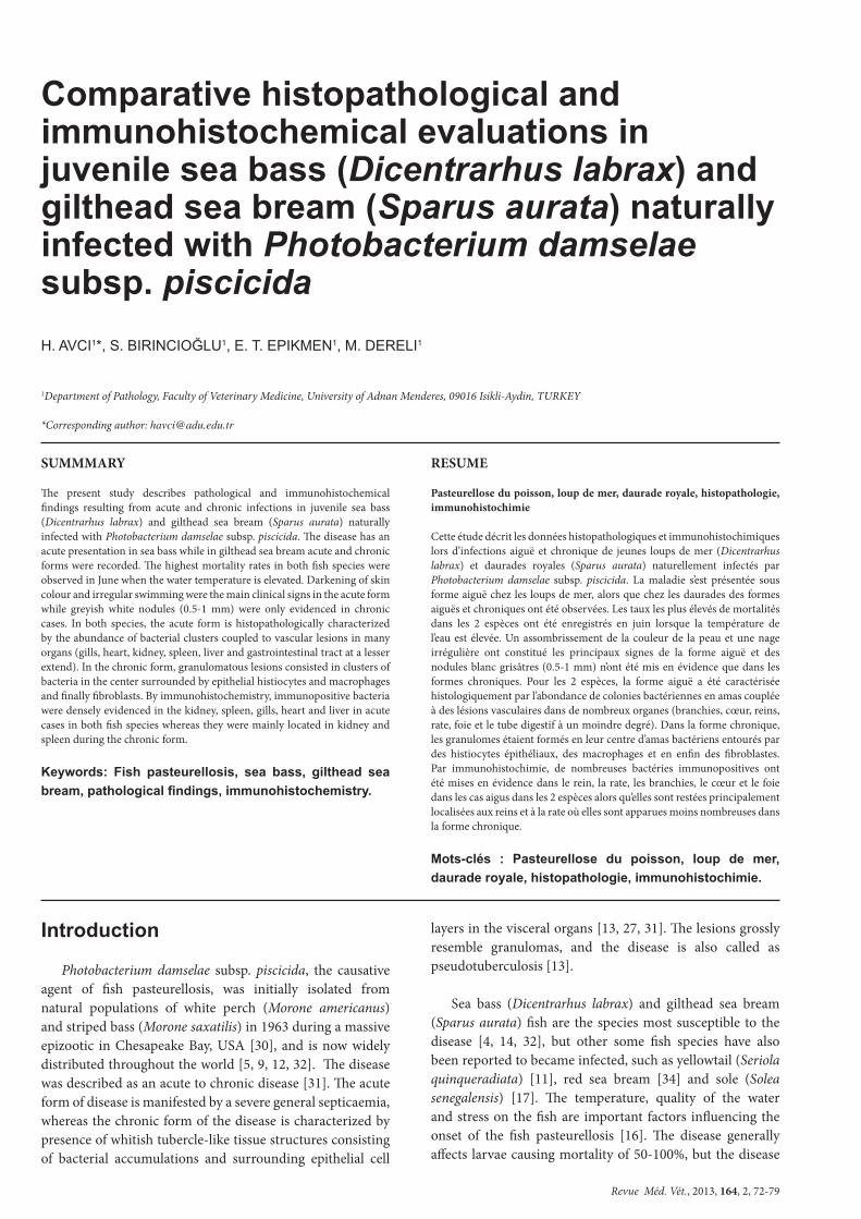
Photobacterium damselae subsp. piscicida was isolated and identified from the sea bass and gilthead sea bream samples. Clinical and macroscopic findings of the disease were summarised in Table I. Only acute form of the disease was observed in sea bass whereas chronic and acute forms were recorded in gilthead sea bream. In both fish species, the highest mortality rates were seen in June when the temperature of the water reached 24°C. During the investigation period, water salinity and quality were uniform, but it was noted that water temperatures ranged between 20-24°C during the epizootics. However, globally morbidity and mortality rates were higher in gilthead sea breams than in sea basses.
Sea bass Gilthead sea bream
Necropsied animalsLength of fish (cm)
Morbidity (%)Mortality (%)
Irregular swimmingColoration in the skin
Skin erosionExophthalmia
AdhesionGranuloma in kidney
Granuloma in the spleen
441-330
0.2 – 2.04435107000
522-484
3 – 4652391511854
Table I: Clinical and macroscopical findings (number of cases) in juvenile sea bass (Dicentrarhus labrax) and gilthead sea bream (Sparus aurata) naturally
infected with Photobacterium damselae subsp. piscicida.

Revue Méd. Vét., 2013, 164, 2, 72-79
AVCI (H.) AND COLLABORATORS74
In acute form, darkening of skin colour and swimming near to the water surface were among clinical findings. Fishes with these clinical findings usually died within 2-3 days. In chronic form (only seen in gilthead sea breams), the prominent clinical findings were decrease in food consumption, irregular swimming movements, lethargy and varying degrees of skin colour modifications (darkening or lightening). In addition, unilateral exophthalmia and erosive changes in the skin were also seen in acute and chronic forms.
Vascular lesions in many organs and tissues consisting in oedema, hyperaemia and haemorrhages were found during the acute form in both fish species. In the chronic form, adhesions were seen in the abdominal cavity of some gilthead sea breams but the prominent macroscopic finding was light greyish white nodules at the size of 0.5-1 mm, hardly visible with naked eye.
HISTOPATHOLOGICAL FINDINGS
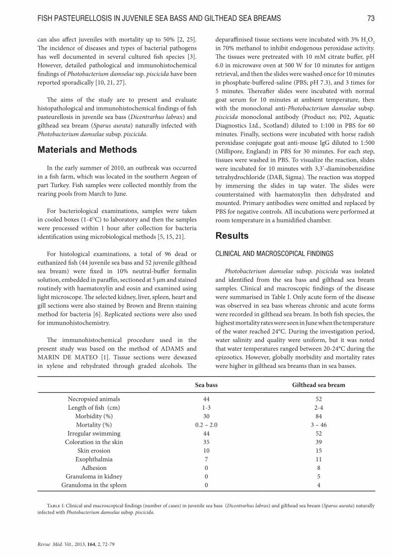
Histopathological findings of the disease were summarised in Table II. The most important histopathological finding seen in both sea basses and gilthead sea breams was the presence of dense bacterial clusters in the parenchyma and vessel lumens of kidney, spleen and liver and in vessel lumens of gills, heart, stomach, pyloric caecum, peritoneum, pancreas, muscles and meninges. In light microscopic examinations, it was determined that bacterial clusters seen in tissue sections were stained with Brown-Brenn stain and were gram negative.
In acute form, vessel lumens were enlarged in gills and they were filled with plasma, erythrocytes and clusters of bacteria. In secondary lamellae telangiectasias containing bacterial clusters were present in many cases of both fish
Sea bass Gilthead sea bream
Gill Cluster of bacteria Necrosis Telangiectasia Granuloma ImmunohistochemistryHeart Cluster of bacteria Pericardial haemorrhages Necrosis Endothelial macrophage activation ImmunohistochemistryLiver Cluster of bacteria Hyperaemia- Haemorrhages Degeneration Necrosis Fatty droplets in the hepatocytes ImmunohistochemistrySpleen Cluster of bacteria Hyperaemia- Haemorrhages Necrosis Granuloma ImmunohistochemistryKidney Cluster of bacteria Hyperaemia- Haemorrhages Necrosis (tubular epithelium) Necrosis (glomerulus) Necrosis (haematopoietic tissues) Granuloma Immunohistochemistry
251090
26
72455
24
141822133516
2122210
23
35351412130
31
2925232
34
1628139
31
222027174323
2929326
32
37431914157
39
Table II: Histopathological and immunohistochemical findings (number of cases) in juvenile sea bass (Dicentrarhus labrax) and gilthead sea bream (Sparus
aurata) naturally infected with Photobacterium damselae subsp. piscicida.

Revue Méd. Vét., 2013, 164, 2, 72-79
FISH PASTEURELLOSIS IN JUVENILE SEA BASS AND GILTHEAD SEA BREAMS 75
species (figure 1A). Necrosis was present in primary and secondary lamellar epithelium, but more commonly and severely in the secondary lamella.
In the heart, pericardial cavity was expanded due to haemorrhages containing large clusters of bacteria (figure 1B). In many cases, lumen of ventricle and atrium were filled with bacterial clusters and erythrocytes. In some cases, bacteria were attached to endocardium. In cases with high bacterial density, degenerations in muscle bundles in the myocardium and necrosis were observed. In myocardium, coupled to the activation of endothelial macrophages, few macrophages containing bacteria in their cytoplasm were detected.
In liver, vascular changes including hyperaemia and haemorrhages in addition to necrosis and bacterial clusters were marked findings. Haemorrhages were seen only around vena centralis. Adenoid structures were disordered, and hepatocytes exhibited enlarged cytoplasm with a granular appearance and cell borders were not differentiated in many of them. In cytoplasm of the remaining intact hepatocytes,
fat vacuoles with varying sizes were present. Necrosis, which is more commonly observed in gilthead sea breams, was usually in the form of single cell necrosis. In cases with less severe necrosis, hepatocyte cytoplasms had dark eosinophilic appearance and their nuclei were picnotic whilst in cases with severe necrosis, disappearing hepatocytes were replaced by irregular, dark eosinophilic, and homogenous necrotic material.
Spleen and kidney were the more affected organs both in sea bass and gilthead sea bream. In the acute form, spleen lesions including focal or irregularly located multifocal necrotic areas containing clusters of bacteria, oedema, severe hyperaemia and haemorrhages were related to septicaemia. Clusters of bacteria either completely filled the vessel lumens or were focally or diffusely distributed in parenchyma. In kidney, vessels were hyperaemic and in many cases, focal or diffuse haemorrhagic areas were present in interstitium.
In all cases, clusters of bacteria were distributed to the whole organ including vessel lumens and parenchyma (figure
Figure 1: A. Bacterial clusters (arrowheads) in the telangiectasias (arrows) of secondary lamellae of the gill. Gilthead sea bream. Haematoxylin-eosin, bar: 50 µm. B. Pericardial cavity (arrows) filled with erythrocytes and bacterial clusters (arrowheads). Sea bass. Haematoxylin-eosin, bar: 100 µm. C. Intensive bacterial clusters (arrows) in the haematopoietic tissue of the kidney. Sea bass. Haematoxylin-eosin, bar: 100 µm. D. Focal coagulation necrosis (N), and bacterial clusters in the necrotic area (arrowhead) and vessels lumens (arrow) in the kidney. Gilthead sea bream. Haematoxylin-eosin, bar: 50 µm.

Revue Méd. Vét., 2013, 164, 2, 72-79
AVCI (H.) AND COLLABORATORS76
1C). Many glomeruli were hardly recognizable due to clusters of bacteria and even completely disappeared due to necrotic changes. Tubular epithelia were generally swollen and in some epithelial cells, cytoplasm had granular eosinophilic appearance. In cases with a mild course, cytoplasms of tubular epithelial cells were homogenously dark pink while in severe cases, integrity of cell borders was disrupted and their nuclei were either picnotic or have disappeared. Similar necrotic changes were present in haematopoietic tissues of the kidney as well (figure 1D). Necrotic lesions were often focal or multifocal but sometimes they were in the form of single cell necrosis. Especially in gilthead sea breams, macrophages containing bacteria in their cytoplasm were seen in the renal haematopoietic tissue.
Oedema in peritoneum and serosa of swim-bladder, vascular lesions with hyperaemia and haemorrhages were histopathological findings observed more scarcely. In skin, necrosis in epidermis and hyperaemia and the eventual presence of bacterial clusters in the vessels in dermis near to necrotic areas were also found.
In the chronic form, marked lesions were noticed in spleen (figure 2A) and kidney (figure 2B) and at a lower degree in peritoneum and gills. The lesions were similar to those found in the acute form but they were generally less intense and were associated to the presence of granulomas with clusters of bacteria in the center surrounded by epithelial histiocytes and macrophages and finally fibroblasts at the outermost forming a thin connective tissue. In newly formed granulomas, macrophages with a dark eosinophilic cytoplasm containing bacteria and picnotic nucleus were often evidenced.
IMMUNOHISTOCHEMICAL FINDINGS
Bacteria found in many tissues and organs from sea basses and gilthead sea breams were stained positively by immunohistochemistry and bacterial morphology was
clearly evidenced. In necrotic areas, positive reactions were usually in granular appearance and less dense.
In the acute form, positive reactions were prominently encountered in kidney (figure 3A), spleen and gills (figure 3B) whereas immunolabelling was less intense in heart (figure 3C) and in liver. Immunopositive reactions were also detected in cytoplasm of few macrophages in the kidney, spleen, gills, heart and liver but also in the submucosa of pyloric caecum and intestines.
In the chronic form, immunopositive bacteria were essentially observed in the kidney and spleen. Positive staining was strongly marked in the vessel lumens (figure 3D) and in parenchyma while in the center of newly formed or old granulomas, immunopositive bacterial density was decreased or sometimes null. Additionally, strong immunopositive reactions were also detected in macrophage cytoplasms (figure 3D). Staining in the other organs and tissues was less dense than in the acute form.
Discussion
In fish pasteurellosis infection caused by Photobacterium damselae ssp. piscicida, the development of large economic costs has made this disease one of the most important bacterial diseases particularly of sea fish [3, 21, 23, 28]. The disease was first reported in USA in white perch and striped bass [30] and it has been described in many different regions of the world in many different species of fish [9, 15, 32]. In the present study, clinical, pathological and immunohisto-chemical findings of natural pasteurellosis occurring in juvenile sea basses and gilthead sea breams were investigated comparatively and findings were comprehensively evaluated.
It has been reported that in the appearance of pasteurellosis, crowded pools, size of fish and water temperature play important role and especially the water temperature over 20°C predisposes to disease [3, 5, 16]. In the fish included in the study, although optimum living conditions were present
Figure 2: Granulomatous lesions (arrows) consisted of clusters of bacteria (arrowheads), macrophages and histiocytes in a gilthead sea bream. A. Spleen, Haematoxylin-eosin, bar: 50µm. B. Kidney. Noted also the clusters of bacteria in the vessel lumen (*), Haematoxylin-eosin, bar: 30µm.

Revue Méd. Vét., 2013, 164, 2, 72-79
FISH PASTEURELLOSIS IN JUVENILE SEA BASS AND GILTHEAD SEA BREAMS 77
except for water temperature, the occurrence of disease at a water temperature similar to those reported in the literature indicates that water temperature is one of the most important stress factors in the emergence of the disease.
In the present study, clusters of bacteria were detected commonly in many organs and tissues, especially in gills, suggesting that, unlike the literatures [8, 19, 33], gills play primary role in the entrance of the disease to the body and that after the agent enters the body, it spreads throughout bloodstream. Absence of any pathological findings in the digestive system and in eyes and the incapacity here to detect bacteria or their clusters in the skin by light microscopy and immunoperoxidase staining enhance the probability that gills would be the entrance route of the disease. The presence of quite dense bacterial clusters in the tissue and organs was attributed to inadequate development of immune system in the offspring and to high pathogenic effect due to the fact that it appeared for the first time in the both fish populations.
In pasteurellosis cases reported by many investigators [2, 4, 22], kidney and spleen are among the organs where microscopic and macroscopic findings are commonly
seen. However, whereas clusters of bacteria were common in acute cases, sometimes clusters of bacteria were few or even absent in chronic cases. Additionally, immunopositive bacteria detected by immunohistochemistry were also found to be more abundant in acute cases than in chronic cases, suggesting that in juvenile fish, kidney and spleen might be not enough sufficient in isolation and identification of the pathogen agent, probably because of activation of cellular immunity, whilst in chronic cases, this situation should be taken into account [9, 19, 21, 22]. In addition, the presence of highly dense clusters of bacteria in the kidney in both species suggested that kidney should be microbiologically examined in a first attempt. In the present study, agent was isolated and identified in kidneys from many fishes, supporting the above idea.
In experimental and natural pasteurellosis cases, tubercle like granulomas appearing macroscopically as light brownish-white areas in spleen, kidney, liver and muscles and microscopically with necrosis and bacteria or clusters of bacteria in the center surrounded by fibroblasts have been defined as the most characteristic finding of the disease [4, 21, 24, 32]. In the present study, similar granulomatous
Figure 3: Immunolabelling of Photobacterium damselae subsp. piscicida (arrows) A. in interstitum and in vessel lumens of kidney. Sea bass, Immunohistoche-mistry, bar: 50 µm; B. in the vessel lumen of gill (arrowheads). Gilthead sea bream, Immunohistochemistry, bar: 20 µm; C. in the myocardium (arrowhead) and atrium (arrows). Gilthead sea bream, Immunohistochemistry, bar: 30 µm; D. in cytoplasm of macrophages (arrowheads) and in the vessel lumens (arrows) in the kidney. Gilthead sea bream (chronic form), Immunohistochemistry, bar: 20 µm.

Revue Méd. Vét., 2013, 164, 2, 72-79
AVCI (H.) AND COLLABORATORS78
structures which can be hardly seen macroscopically were encountered only in gilthead sea breams and in few fish, which increased the possibility that this finding can not be sufficient in the pathological diagnosis of pasteurellosis in juvenile fish. Granulomas described by some investigators in the liver [21] and muscles [15] were not seen in the present study. Granulomas found only in gilthead sea breams have quite weak cellular characteristics, suggesting inadequate maturation of immune system in juvenile fish. In the granulomas seen in the present study, giant cells described by some investigators [7, 9] were absent.
In pasteurellosis cases, vascular changes and coagulation necrosis in kidney have been reported by many investigators [2, 9, 15, 22]. In the present study, tubular necrosis seen in both sea basses and gilthead sea breams were consistent with the literature data. Although necrosis seen in the kidney haematopoetic tissue of both fish species were also reported only by FOYLE et al. [7] and as these authors could not succeed in evidencing the bacteria in these necrotic areas, it would be questioned whether necrosis was a finding of the disease. In the present study, both in light microscopic and immunohistochemical examinations, bacteria or bacterial clusters were seen in these necrotic areas, suggesting that this finding is strongly associated to the disease.
It has been reported that phospholipase and extracellular products (ECP) with cytotoxic and haemolytic effects are important in the pathogenesis of the disease [18, 23, 28]. It was thought that necrosis appearing in kidney, spleen and liver in the present study were associated with these compounds. In the juvenile both fish species, cytolytic effect was observed to be more severe as the immune system has not yet completely developed. Nevertheless, in tissues and organs with abundant bacterial clusters, erythrocyte breakdown and resulting haemosiderin accumulation were not seen, suggesting that haemolytic effects of would remain limited.
The intracellular presence of the bacteria in the cytoplasm of macrophages is considered as important in the occurrence of clinical and pathological signs of the disease [14, 26, 29]. In the present study, it was established that granulomas seen in few fish only among gilthead sea breams consisted of necrotic macrophages containing clusters of bacteria in the newly formed ones and in time these granulomas were transformed into mature granuloma according to the severity of the cellular response.
The immunoperoxidase method is used for the definitive diagnosis of fish pasteurellosis [7, 10, 20]. In the present study, when two forms of the disease were compared (acute vs. chronic forms), the distribution of immunopositive bacteria in tissues and organs varied: positive reactions were more common in the acute form than in the chronic one and even, in some fishes chronically affected no immunopositive bacteria were identified. This fact may be related to the destruction of the agent throughout the development of cellular response or to the decrease in antigenic characteristics which are
effective in the development of cellular response. In addition, the distribution of positive reactions was not different during the acute form between the 2 fish species, suggesting that this method can be used reliably in the definitive diagnosis of natural acute pasteurellosis occurring in young sea basses and gilthead sea breams.
Acknowledgments
The authors wish to thank Dr. Seza Eskiizmirliler (Bornova Veterinary Control and Research Institute) for kindly providing isolation and identification of the Photobacterium damselae subsp. piscicida in the present study.
References
1. ADAMS A., MARIN DE MATEO M.: Imnunohistochemical detection of fish pathogens. In: Techniques in fish imnunology, STOLEN J.S., FLETCHER T.C., ROWLEY A.F., ZELIKOFF J.T., KAATTARI S.L. and SMITH S.A. (eds.): SOS Publication, USA, 1994, pp.: 133-144.
2. BAKOPOULOS V., PERIC Z., RODGER H., ADAMS A., RICHARDS R.H.: First report of fish pasteurellosis from Malta. J. Aquat. Anim. Health, 1997, 9, 26-33.
3. BALEBONA M.C., ZORRILLA I., MORINIGO M.A., BORREGO J.J.: Survey of bacterial pathologies affecting farmed gilt-head sea bream (Sparus aurata L.) in southwestern Spain from 1990-1996. Aquaculture, 1998, 166, 19-35.
4. BAPTISTA T., ROMALDE J.L., TORANZO A.E.: First occurence of pasteurellosis in Portugal affecting cultured gilthead seabream (Sparus auratus). Bull. Eur. Assoc. Fish Pathol., 1996, 16, 92-95.
5. CANDAN A., KUCKER M.A., KARATAS S.: Pasteurellosis in cultured sea bass (Dicentrarchus labrax) in Turkey. Bull. Eur. Assoc. Fish Pathol., 1996, 16, 150-153.
6. CULLING A.F., ALLISON T.R., BARR T.W.: Cellular Pathology Technique, CULLING A.F., ALLISON T.R. and BARR T.W. (eds), 4th edition, Butterworth & Co.(Publ.). Ltd., London, 1985, pp.: 269-270.
7. FOYLE L., TURNBULL T., ELLIS A., BARNES A., ADAMS A., FERGUSON H.W.: Pasteurellosis in Atlantic salmon, Salmo salar L.: immunohistochemistry of the naturally-occuring disease. J. Fish Dis., 2003, 26, 373-376.
8. FUKUDA Y., KUSUDA R.: Efficacy of vaccination for pseudotuberculosis in cultured yellowtail by various routes of administration. B. Jpn. Soc. Sci. Fish., 1981, 47, 147-150.
9. JONES M.W., COX D.I.: Clinical Disease in seafarmed Atlantic salmon (Salmo salar) associated with a member of the family pasteurellaceae-a case history. Bull. Eur. Assoc. Fish Pathol., 1999, 19, 75-78.
10. JUNG T.S., THOMPSON K.D., MORRIS D.J., ADAMS A., SNEDDON K.: The production and characterization

Revue Méd. Vét., 2013, 164, 2, 72-79
FISH PASTEURELLOSIS IN JUVENILE SEA BASS AND GILTHEAD SEA BREAMS 79
of monoclonal antibodies against Photobacterium damselae ssp. piscicida and initial observations using immunohistochemistry. J. Fish Dis., 2001, 24, 67-77.
11. KAWAKAMI H., SAKAI M.: Comprasion of susceptibility of seven fishes to Photobacterium damsela subsp. piscicida. Bull. Eur. Assoc. Fish Pathol., 1999, 19, 153-155.
12. KIMURA M., KITAO T.: On the causative agent of tuberculosis of yellowtail. Fish Pathol., 1971, 6, 8-14.
13. KUBOTA S., KIMURA M., EGUSA S.: Studies of a bacterial tuberculoloidosis of the yellowtail. I. Symptomatology and histopathology. Fish Pathol., 1970, 4, 111-118.
14. KUSUDA R., KAWAI K.: Bacterial diseases of cultured marine fish in Japan. Fish Pathol., 1998, 33, 221-227.
15. LIU P.C., CHENG C.F., CHANG C.H., LIN S.L., WANG W.S., HUNG S.W., CHEN M.H., LIN C.C., TU C.Y., LIN Y.H.: Highly virulent Photobacterium damselae subsp. Piscicida isolated from Taiwan paradise fish, Macropodus opercularis (L.), in Taiwan. Afr. J. Microbiol. Res., 2011, 5, 2107-2113.
16. MAGARINOS B., COUSO N., NOYA M., MERINO P., TORANZO A.E., LAMAS J.: Effect of temparature on the development of pasteurellosis in carrier gilthead seabream (Sparus aurata). Aquaculture, 2001, 195, 17-21.
17. MAGARINOS B., ROMALDE J.L., LOPEZ-ROMALDE S., MORINIGO M.A., TORANZO A.E.: Pathobiological characterisation of Photobacterium damselae subsp. piscicida isolated from cultured sole (Solea senegalensis). Bull. Eur. Assoc. Fish Pathol., 2003, 23, 183-190.
18. MAGARINOS B., SANTOS Y., ROMALDE J.L., RIVAS C., BARJA J.L., TORANZO A.E.: Pathogenic activities of live cells and extracellular products of the fish pathogen Pasteurella piscicida. J. Gen. Microbiol., 1992, 138, 2491-2498.
19. MAGARINOS B., TORANZO A.E., ROMALDE J.L.: Different susceptibility of gilthead seabream and turbot to Pasteurella piscicida infection by the water route. Bull. Eur. Assoc. Fish Pathol., 1995, 15, 88-90.
20. MANIATIS K., MORRIS D.J., ADAMS A., PEARSON M.: Detection of Photobacterium damsela subspecies piscicida in fixed tissue sections using immnunohistochemistry and antigen retrieval immunohisto-chemistry. J. Fish Dis., 2000, 23, 343-347.
21. MLADINEO I., MILETIC I., BOCINA I.: Photobacterium damselae subsp. piscicida outbreak in cage-reared Atlantic bluefin tuna Thunnus thynnus. J. Aquat. Anim. Health, 2006, 18, 51-54.
22. NAGANO I., INOUE S., KAWAI K., OSHIMA S.I.: Repeatable immersion infection with Photobacterium damselae subsp. piscicida reproducing clinical signs and moderate mortality. Fish. Sci., 2009, 75, 707-714.
23. NAKAI T., FUJIIE N., Muroga K., Arimoto M., Mizuta Y., Matsuoka S.: Pasteurella piscicida infection in hatchery-reared juvenile striped jack. Fish Pathol., 1992, 27, 103-108.
24. NITZAN S., SHWARTSBURD B., VAIMAN R., HELLER E.D.: Some characteristics of Photobacterium damselae ssp. piscicida isolated in Israel during outbreaks of pasteurellosis in hybrid bass (Morone saxatilis x M. chrysops). Bull. Eur. Assoc. Fish Pathol., 2001, 21, 77-80.
25. NOYA M., MAGARINOS B., LAMAS J.: Interactions between peritonel exudate cells (PECs) of gilthead seabream (Sparus aurata) and Pasteurella piscicida. A morphological study. Aquaculture, 1995, 131, 11-21.
26. NOYA M., MAGARINOS B., TORANZO A.E., Lamas L.: Sequential pathology of experimental pasteurellosis in gilthead seabream, Sparus aurata. A light-and electron microcopic study. Dis. Aquat. Organ., 1995, 21, 177-186.
27. POULOS C., BAKOPOULOS V., ZOLOTA V., DIMITRIADIS G.J.: Histopathological findings after sea bass (Dicentrarhus labrax L.) exposure to extracellular products of Photobacterium damsela ssp. piscicida produced by invivo. Aquacult. Res., 2004, 35, 931-936.
28. ROMALDE J.L.: Photobacterium damselae subsp. piscicida: an integrated view of a bacterial fish pathogen. Int. Microbiol., 2002, 5, 3-9.
29. SKARMETA A.M., BANDIN I., SANTOS Y., TORANZO A.E.: In vitro killing of Pasteurella piscicida by fish macrophages. Dis Aquat. Organ., 1995, 23, 51-57.
30. SNIESZKO S.F., BULLOCK G.L., HOLLIS E., BOONE J.G.: Pasteurella sp. from an epizootic of white perch (Roccus americanus) in Chesapeake Bay tidewater areas. J. Bacteriol., 1964, 88, 1814-1815.
31. THUNE R.L. STANLEY L.A., COOPER R.K.: Pathogenesis of gram-negative bacterial infections in warmwater fish. Annu. Rev. Fish Dis., 1993, 3, 37-68.
32. TORANZO A.E. BARREIRO S., CASAL J.F., FIGUERAS A., MAGARINOS B., BARJA J.L.: Pasteurellosis in cultured gilthead seabream (Sparus aurata): first report in Spain. Aquaculture, 1991, 99, 1-15.
33. TUNG M.C., TSAI S.H., HO L.F., HUANG S.T., CHEN S.C.: An acute septisemic infection of pasteurella organism in pond-cultured Formasa snake-head fish (Channa maculata Lacepede) in Taiwan. Fish Pathol., 1985, 20, 143-148.
34. YASUNAGA N., HATAI K., TUKAHARA J.: Photobacterium piscicida from an epizootic of cultured red sea bream. Fish Pathol., 1983, 18, 107-110.
35.


![Instruments de Méd. Générael , O.R.L. & VET MEILLEURE · 01 [ 019 ] Instruments de Méd. Générael , O.R.L. & VET Éclairage à fibres optiques (F.O.) de haute qualité pour un](https://img.pdfslide.us/doc/110x75/5e1b9dc5606e447e755f1d67/instruments-de-md-gnrael-orl-vet-meilleure-01-019-instruments.jpg)