Embed Size (px)
Citation preview
ORIGINAL RESEARCHpublished: 29 August 2017
doi: 10.3389/fgene.2017.00111
Frontiers in Genetics | www.frontiersin.org 1 August 2017 | Volume 8 | Article 111
Edited by:
Eric Altermann,
AgResearch, New Zealand
Reviewed by:
Uri Gophna,
Tel Aviv University, Israel
Wayne Young,
AgResearch, New Zealand
*Correspondence:
Ines Thiele
Specialty section:
This article was submitted to
Evolutionary and Genomic
Microbiology,
a section of the journal
Frontiers in Genetics
Received: 06 March 2017
Accepted: 11 August 2017
Published: 29 August 2017
Citation:
Ravcheev DA and Thiele I (2017)
Comparative Genomic Analysis of the
Human Gut Microbiome Reveals a
Broad Distribution of Metabolic
Pathways for the Degradation of
Host-Synthetized Mucin Glycans and
Utilization of Mucin-Derived
Monosaccharides.
Front. Genet. 8:111.
doi: 10.3389/fgene.2017.00111
Comparative Genomic Analysis ofthe Human Gut Microbiome Revealsa Broad Distribution of MetabolicPathways for the Degradation ofHost-Synthetized Mucin Glycans andUtilization of Mucin-DerivedMonosaccharidesDmitry A. Ravcheev and Ines Thiele *
Luxembourg Centre for Systems Biomedicine, University of Luxembourg, Esch-sur-Alzette, Luxembourg
The colonic mucus layer is a dynamic and complex structure formed by secreted
and transmembrane mucins, which are high-molecular-weight and heavily glycosylated
proteins. Colonic mucus consists of a loose outer layer and a dense epithelium-attached
layer. The outer layer is inhabited by various representatives of the human gut microbiota
(HGM). Glycans of the colonic mucus can be used by the HGM as a source of carbon
and energy when dietary fibers are not sufficiently available. Both commensals and
pathogens can utilize mucin glycans. Commensals are mostly involved in the cleavage
of glycans, while pathogens mostly utilize monosaccharides released by commensals.
This HGM-derived degradation of the mucus layer increases pathogen susceptibility
and causes many other health disorders. Here, we analyzed 397 individual HGM
genomes to identify pathways for the cleavage of host-synthetized mucin glycans to
monosaccharides as well as for the catabolism of the derived monosaccharides. Our
key results are as follows: (i) Genes for the cleavage of mucin glycans were found in
86% of the analyzed genomes, which significantly higher than a previous estimation.
(ii) Genes for the catabolism of derived monosaccharides were found in 89% of the
analyzed genomes. (iii) Comparative genomic analysis identified four alternative forms of
themonosaccharide-catabolizing enzymes and four alternative forms of monosaccharide
transporters. (iv) Eighty-five percent of the analyzed genomesmay be involved in potential
feeding pathways for the monosaccharides derived from cleaved mucin glycans. (v) The
analyzed genomes demonstrated different abilities to degrade known mucin glycans.
Generally, the ability to degrade at least one type of mucin glycan was predicted for 81%
of the analyzed genomes. (vi) Eighty-two percent of the analyzed genomes can form
mutualistic pairs that are able to degrade mucin glycans and are not degradable by any
of the paired organisms alone. Taken together, these findings provide further insight into
the inter-microbial communications of the HGM as well as into host-HGM interactions.
Keywords: human gut microbiome, comparative genomics, mucin glycans, carbohydrate utilization, metabolism
reconstruction
Ravcheev and Thiele Degradation of Mucin Glycans by HGM
INTRODUCTION
The colonic mucus layer is a dynamic and complex structurethat is mainly composed of the glycoprotein mucin-2 (MUC2)(Johansson et al., 2008). MUC2 is characterized by abundantglycosylation. Highly variable glycan structures are O-linked toserine or threonine residues that are concentrated in so-calledPTS (proline, threonine, and serine) domains. The mass of theglycosylated MUC2 protein is approximately 2.5 MDa, and morethan 80% of this mass comes from carbohydrates (Lang et al.,2007). Glycosylated MUC2 can form a gel-like structure dueto its N- and C-terminal domains that form numerous cross-links between cysteine residues (Johansson et al., 2013). Due tothese cross-links, MUC2 forms a mucus structure. The colonicmucus consists of two layers, a loose outer layer and a denseepithelium-attached layer (Johansson et al., 2011). The innerlayer acts as a physical barrier preventing bacteria from accessingthe epithelium, whereas the outer layer is densely populated byvarious commensal microbes (Johansson et al., 2011, 2015; Liet al., 2015).
Interactions between the human gut microbiota (HGM) andcolonic mucus are not limited just to residing of the microbes inthe outer layer mucus. Thus, once in the outer mucus layer, gutmicrobes not only avoid washout by the contents flowing throughthe colon but also are able to access mucin glycans that can beused as sources of carbon and energy. Usually HGM microbesswitch to mucin glycans in a shortage of dietary fibers, becausemultiple intestinal microbes can switch between dietary and hostglycans (Mahowald et al., 2009). Both commensal and pathogenicmicrobes are able to degrade mucin glycans. The commensalmicrobes are usually able to cleave glycans with secretedglycosyl hydrolases (GHs) and lyases and further catabolize thederived monosaccharides, whereas most intestinal pathogensuse monosaccharides released by commensal-secreted enzymes(Martens et al., 2008; Koropatkin et al., 2012; Marcobal et al.,2013; Cameron and Sperandio, 2015; Pacheco and Sperandio,2015). Being both an environmental niche and a food source, themucus layer plays a key role in shaping the HGM composition(Koropatkin et al., 2012; Johansson et al., 2015). In turn, theHGM is able to modulate mucus chemical composition via thedegradation of glycans and peptides and the local release ofbioactive factors that can change the expression patterns of themucin-producing host cells (Deplancke and Gaskins, 2001). Thedegradation of mucin glycans is highly dependent on the hostdiet. Thus, decreased levels of fibers in the diet force the HGMto degrade more mucin glycans, resulting in the thinning anddepletion of the mucus layer, which subsequently enhances hostsusceptibility to pathogens (Marcobal et al., 2013; Tailford et al.,2015; Desai et al., 2016). Additionally, depletion of the colonicmucus is associated with such disorders as Crohn’s disease, celiacdisease, colonic ischemia, compound exocytosis, and ulcerativecolitis (Png et al., 2010; Joossens et al., 2011; Parmar et al., 2012;Johansson et al., 2013; Arike and Hansson, 2016; Cockburn andKoropatkin, 2016).
Because of the significant mucus-microbiota interaction,multiple HGM organisms have been analyzed for theirabilities to degrade mucin glycans. To date, more than
50 mucus-degrading bacterial strains, generally belongingto Akkermansia muciniphila, Bacteroides spp., Barnesiellaintestinihominis, Bifidobacterium spp., Eubacterium spp., andRuminococcus spp. (Derrien et al., 2004; Sonnenburg et al., 2005;Martens et al., 2008; Comstock, 2009; Png et al., 2010; Kiyoharaet al., 2012; Pudlo et al., 2015; Tailford et al., 2015; Desai et al.,2016), have been identified.
In this study, we analyzed the degradation of mucin glycans bythe HGM using comparative genomic analysis. The comparativegenomic analysis of sugar utilization by microbes is a recentbut actively developing research area. A comparative genomicapproach, combining a phylogenomics and genome-contextbased techniques (Osterman and Overbeek, 2003; Rodionov,2007), has been previously applied for the analysis of sugarutilization in various microbial taxa, including multiple HGMstrains (Leyn et al., 2012; Ravcheev et al., 2013; Zhang et al.,2015; Khoroshkin et al., 2016). Here, we used this comparativeapproach not to a set of related organisms, but tomicroorganismscohabiting a certain environment, being the human intestine.Unlike functional analysis of metagenomes, this analysis isbased on a reconstruction of metabolic pathways in individualgenomes of microbes found in the studied environmentalcommunity and further prediction of possible interactionsbetween different microorganisms. This approach has notpreviously been used for the detailed analysis of sugar utilizationbut it has been repeatedly used to reconstruct other metabolicpathways, including respiration (Ravcheev and Thiele, 2014),biosynthesis of B-vitamins (Magnúsdóttir et al., 2015), andquinones (Ravcheev and Thiele, 2016), as well central carbonmetabolism and biosynthesis of amino acids and nucleotides(Magnúsdóttir et al., 2017), in multiple HGM genomes.
Here, we used a comparative genomics approach to analyzethe degradation of mucin glycans as well utilization of thederived monosaccharides as carbon and energy sources in HGMgenomes. Additionally, we predicted potential feeding pathwaysfor mucin glycan-derived monosaccharides, the specificity ofvarious HGM strains for different types of mucin glycans, andmutualistic relationships for the cleavage of mucin glycans bydifferent HGM organisms.
MATERIALS AND METHODS
Analyzed GenomesThe analyzed genomes were selected using the following steps.(1) All genomes listed in the Human Microbiome Project (HMP,http://www.hmpdacc.org/HMRGD/) as of 17.09.2015 and 459genomes with the body site “Gastrointestinal tract” (i.e., isolatedfrom the intestine) were selected. (2) All the genomes absentfrom the PubSEED (Overbeek et al., 2005; Disz et al., 2010)and Integrated Microbial Genomes (IMG) databases (Markowitzet al., 2014) were excluded. Among the remaining 397 genomes,71 had a finished sequencing status whereas 326 others had adraft status (Table S1). These genomes represent 288 microbialspecies, 89 genera, 45 families, 19 orders, 14 classes, and 8phyla. All the selected genomes, except 2 Archaea, are bacterial.The phyletic distribution of the analyzed genomes is in goodagreement with that observed in various HGM (Eckburg et al.,
Frontiers in Genetics | www.frontiersin.org 2 August 2017 | Volume 8 | Article 111
Ravcheev and Thiele Degradation of Mucin Glycans by HGM
2005; Goodman et al., 2011; Walker et al., 2011; Graf et al.,2015). Thus, the most represented phyla are Actinobacteria(37 genomes, 9.3% of the analyzed genomes), Bacteroidetes(69 genomes, 17.3%), Firmicutes (197 genomes, 49.6%), andProteobacteria (71 genomes, 17.9%).
Approach, Tools, and DatabasesThe PubSEED platform was used to annotate the genesresponsible for the degradation of mucin glycans usingthe following comparative genomics approach. To avoidmisannotation, all of the proteins with the same function werechecked for orthology. Orthologs were defined as the bestbidirectional hits that have a similar genomic context. To searchfor the best bidirectional hits, a BLAST algorithm (Altschul et al.,1997) implemented in PubSEED and the IMG platform wasused with the following parameters, a score ≥150 bits, an e-value ≤ e−50, a protein identity and positives ≥25 and ≥40%,respectively, a query coverage at least 70%. To analyze genomiccontext, we used PubSEED and STRING v9.1 (Franceschiniet al., 2013) along with phylogenetic trees for protein domainsin MicrobesOnline (Dehal et al., 2010). To analyze proteindomain structure, we searched the Pfam (Finn et al., 2014)and CDD (Marchler-Bauer et al., 2013) databases and the“Domains & Families” option of the MicrobesOnline platformwith the threshold e−30. Additionally, functional annotations ofthe analyzed genes were performed using the UniProt (Magraneand Consortium, 2011), KEGG (Kanehisa et al., 2012), andMetaCyc (Caspi et al., 2014) databases.
After annotation of orthologs for the known analyzed genes,all the catabolic pathways (CPs) for monosaccharides utilizationwere checked for gaps. The CP was defined as sequence ofreactions from intracellular monosaccharide to the intermediateof the central carbohydrate metabolism (glycolytic pathwaysor TCA cycle). A gap was defined as an absence of enzyme-encoding genes responsible for one or more reactions. Lengthof the gap was defined as a number of successive reactionscorresponding to the absent genes. A CP was considered to bepresent in the organism if no more than two gaps were foundand length of each gap was maximally one reaction. A searchof the non-orthologous displacement was done for the gaps inthe present pathways as well as for transporters absent in thepresence of CP. If non-orthologous replacement was predicted,its orthologs were searched in all analyzed genomes. Afterwards,CPs were checked for gaps again while considering the identifiednon-orthologous replacements and the presence of CPs wasre-evaluated.
To search for the protein homologs, a BLAST algorithmimplemented in the PubSEED and the IMG platforms wasused, with the following parameters, an e-value ≤ e−20,a protein identity ≥20%. Multiple protein alignmentswere performed using the MUSCLE v. 3.8.31 tool (Edgar,2004a,b). Phylogenetic trees were constructed using themaximum-likelihood method with the default parametersimplemented in PhyML-3.0 (Guindon et al., 2010). Theobtained trees were visualized and midpoint-rootedusing the interactive viewer Dendroscope, version 3.2.10,build 19 (Huson et al., 2007). To clarify the taxonomic
affiliations of the analyzed genomes, the NCBI Taxonomydatabase (http://www.ncbi.nlm.nih.gov/taxonomy) wasused. Analysis of gene occurrence was performed using the“Phylogenetic Profiler for Single Genes” option of the IMGplatform.
The non-random distribution of the analyzed pathways, wastested using the Chi-squared test of observed frequencies ofcombinations with R (version 3.2.3).
All of the annotated genes are represented as a subsystemin PubSEED (http://pubseed.theseed.org/SubsysEditor.cgi;subsystem names are “Galactose utilization HGM,” “L-fucose utilization HGM,” “N-Acetylgalactosamine utilizationHGM,” “N-Acetylglucosamine utilization HGM,” and “N-Acetylneuraminic acid utilization HGM”) and in Tables S2–S7.The protein sequences for the annotated genes in FASTA formatare represented in the file Sequences S1 in the SupplementaryMaterials.
Genome-Context and PhylogenomicApproaches for Functional AnnotationThe functions of some genes cannot be correctly annotated basedon sequence similarity alone. Thus, a search of orthologs asdescribed in Approach, Tools, and Databases is often incorrectlyapplied to large protein families containing proteins withdifferent functions (Rodionov, 2007; Promponas et al., 2015).Additionally, the existence of non-orthologous displacements(Galperin and Koonin, 1998) require advanced approaches asgenome context-based methods. Below, we describe non-trivialmethods used in this work for functional annotation of theanalyzed genes.
Annotation of the fucK and fucA GenesThe main cause of misannotation for the fuc genes is theirsimilarity to the genes for rhamnose utilization. Both theFucK and RhaB proteins belong to the FGGY family ofcarbohydrate kinases (Pfam: PF02782), whereas the FucAand RhaD proteins belong to the Aldolase_II family (Pfam:PF00596). We used the following steps to distinguish fuc andrha genes. (1) The fucI gene was used as a signature genefor the Fuc utilization pathway because this gene has nohomologs among rhamnose-catabolizing enzymes. Orthologsfor the FucI were found as described in Approach, Tools,and Databases. FucI proteins marked as reviewed in UniProtdatabase were used as a query. Then, domain structures of allidentified orthologs were analyzed and all the FucI orthologsdemonstrated the presence of L-fucose isomerase domain (Pfam:PF07882) with e-value < e−110. The domain structure was usedas an additional confirmation of orthology. (2) Phylogeneticmaximum-likelihood trees were built for the FucK/RhaB-(Figure S1) and FucA/RhaD-like proteins (Figure S2) found inthe analyzed genomes. (3) Genes that chromosomally clusteredwith fucI were annotated as fucK and fucA. (4) Single-copygenes found in genomes having fucI but lacking any rhagenes were annotated as fucK and fucA. (5) The remainingnon-annotated genes were annotated by their positions onthe phylogenetic trees relative to the previously annotatedgenes.
Frontiers in Genetics | www.frontiersin.org 3 August 2017 | Volume 8 | Article 111
Ravcheev and Thiele Degradation of Mucin Glycans by HGM
Prediction of Non-orthologs Replacements for the Fcl
Fucose Catabolism PathwayNon-orthologous displacements for genes for the Fcl pathwayswere found by an analysis of the chromosomal clustersin genomes having fclBC gene clusters but lacking thefclA, fclD, and fclE genes. The six genomes satisfying thesecriteria, were analyzed, Bifidobacterium bifidum NCIMB41171, Bifidobacterium breve DSM 20213, Bifidobacteriumbreve HPH0326, Bifidobacterium longum ATCC 15697,Bifidobacterium pseudocatenulatum DSM 20438, andCorynebacterium ammoniagenes DSM 20306. Genes foundto be chromosomally clustered with the fclBC were proposed tobe non-orthologous replacements for the fclADE genes and theirpossible functions were predicted as described below.
The non-orthologs displacement for fclE, was predictedby similarity of the encoded protein to 4-hydroxy-tetrahydrodipicolinate synthase (DHDPS family, Pfam:PF00701). Because pyruvate is a product of FclE-catalyzedreaction (Yew et al., 2006) as well of reactions catalyzed byenzymes from DHDPS family (N-acetylneuraminate lyase andtrans-o-hydroxybenzylidenepyruvate hydratase-aldolase), weconcluded that the analyzed protein is an alternative form ofFclE and named it FclE2.
The non-orthologous displacement for FclA and FclDproteins was predicted by analysis of protein families andphylogenetic trees. Thus, the fclBC-clustered gene was foundto belong to the short-chain dehydrogenase family (Pfam:PF00106). Because this family includes experimentally analyzedFucA and FucD proteins from and Burkholderia multivorans(Hobbs et al., 2013), we constructed phylogenetic trees for FclAand FclD proteins as well short-chain dehydrogenase familyproteins encoded in the fclBC chromosomal clusters (FigureS3). Because all the fclBC-clustered proteins formed a branch,separated from both FclD and FclA branches, we proposed thatthey have two functions, thus being non-orthologs replacementsfor both FclD and FclA. These proteins were were designatedFclA2/FclD2.
Prediction of Novel Fucose-Specific ABC Transport
SystemPossible Fuc-specific ABC transport systems have been predictedby chromosomal clustering with the fuc genes in 25 analyzedgenomes, including Actinomyces spp., Clostridium spp.,Coprococcus spp., Lachnospiraceae bacterium, Ruminococcusspp., and Enterobacter spp. The closest experimentally analyzedhomolog of the substrate-binding subunit of this system is thesubstrate-binding protein AraF of the arabinose-specific ABCsystem from E. coli (Johnson and Schleif, 2000). Additionally,genes of this system co-cluster together with the fcl genes in thegenomes of Streptomyces sp. HGB0020, Bifidobacterium longumATCC 15697, and Bifidobacterium pseudocatenulatum DSM20438.
Annotation of Homologous Enzymes for GalNAc and
GlcNAc MetabolismPreviously it was found that at least some Firmicutes andProteobacteria can utilize only galactosamine (GalN) but
not GalNAc (Leyn et al., 2012; Zhang et al., 2015). Thecrucial feature of the GalNAc-utilizing microorganisms isthe presence of the agaA gene for N-acetylgalactosamine-6-phosphate deacetylase. For proper pathway reconstruction,AgaA should be distinguished from the N-acetylglucosamine-6-phosphate deacetylase NagA, which is involved in GlcNAcutilization. For this purpose, we used the following pipeline. (1)The agaS and gene was selected as signature gene for the GalNAcutilization pathway, and nagBwas used as a signature gene for theGlcNAc utilization pathway. These genes were selected becauseAgaS have no homologs in the GlcNAc utilization pathwayand NagB has no homologs in GalNAc utilization pathway.(2) Orthologs for AgaS were found as described in Approach,Tools, and Databases. For a query, proteins marked as reviewedin UniProt database as well AgaS proteins annotated at theRegPrecise database (Novichkov et al., 2013) were used. Thedomain structures of all found AgaS orthologs were analyzed andall them demonstrated the presence of SIS_AgaS_like domain(NCBI CDD: cd05010) with e-value < e−70. Additionally, thephylogenetic maximum-likelihood tree for the AgaS proteinswas constructed (Figure S4). The constructed tree was quitecompact, did not include long branches, and its structure wasin agreement with microbial taxonomy, which was used as anadditional corroboration for orthology of all the predicted AgaSproteins. (3) Orthologs for the NagB were found as described inApproach, Tools, and Databases. For a query, proteins markedas reviewed in UniProt database as well AgaS proteins annotatedat the RegPrecise database were used. All these orthologs belongto the GlcN6P_deaminase family (NCBI CDD: cd01399) with ane-value < e−100. (4) A phylogenetic maximum-likelihood treewas built for AgaA/NagA-like proteins (Figure S5) found in theanalyzed genomes. (5) Genes chromosomally clustered with agaSwere annotated as agaA, while genes chromosomally clusteredwith nagB were annotated as nagA. (6) Genes that co-occurredin the genomes with only agaS were annotated as agaA, andgenes that co-occurred in the genomes with only nagB wereannotated as nagA. (7) The remaining non-annotated genes wereannotated by their positions on the phylogenetic trees relative tothe previously annotated genes.
Annotation of the GalNAc-Specific TransportersThree PTSs associated with various aga genes were previouslyidentified, including a GalNAc-specific (AgaPTS), a GalN-specific (GamPTS), and a GnbPTS with multiple specificities(Leyn et al., 2012; Bidart et al., 2014; Zhang et al., 2015).The GnbPTS can transport and phosphorylate three differentcompounds: GalNAc, lacto-N-biose (Galβ-1,3-GlcNAc), andgalacto-N-biose (Galβ-1,3-GalNAc).
Only AgaPTS and GnbPTS but not GamPTS are involvedin GalNAc catabolism. We used the following steps todistinguish the various types of PTSs associated with aga genes.(1) Homologs were found for all the previously annotatedEIIC components of the AgaPTS, GamPTS, and GnbPTS asdescribed in Approach, Tools, and Databases. EIIC componentswere selected because these components are substrate-bindingsubunits. Query proteins were extracted from the RegPrecisedatabase. (2) A phylogenetic maximal-likelihood tree was
Frontiers in Genetics | www.frontiersin.org 4 August 2017 | Volume 8 | Article 111
Ravcheev and Thiele Degradation of Mucin Glycans by HGM
constructed for the EIIC components of the analyzed PTSsystems (Figure S6). (3) The EIIC components clustered with thegnbG gene were annotated as binding GalNAc as well lacto- andgalacto-N-biose. The gnbG gene encodes intracellular glycosyl-hydrolase, specific to lacto- and galacto-N-biose and clusteredwith the GnbPTS in all genomes previously analyzed (Bidartet al., 2014; Zhang et al., 2015). Additionally, the EIIC proteinsclustered with the GnbG are clearly separated on the phylogenetictree (Figure S6) and thus can be easily distinguished from theirhomologs. (4) The AgaS protein was used as a signature for theGalNAc and GalN utilization pathways, whereas the presence orabsence of AgaA protein was used as a signature for GalNAc orGalN utilization, respectively (see Annotation of HomologousEnzymes for GalNAc and GlcNAc Metabolism). Thus, proteinsco-clustered with the agaS and agaA genes were annotated asGalNAc specific and corresponding PTS systems were annotatedas AgaPTS. (5) The EIIC proteins that co-clustered with the agaSbut not the agaA genes were annotated as GalN-specific andcorresponding PTS systems were annotated as GamPTS.
Prediction of a Novel GlcNAc-Specific TransporterAn alternative ABC transporter for GlcNAc was predicted in thiswork and named NgcABCD. This transporter was predicted byanalysis of gene clusters. Thus, genes for putative ABC transportsystem were co-clustered together with the nagKAB genes in15 genomes of Bifidobacterium spp. The following differenceswere found between the NgcABCD and the previously describedABC-transporter NgcEFG (Xiao et al., 2002). First, the ngcEFGoperon encodes only the substrate-binding protein and twointermembrane proteins, whereas the ngcABCD operon encodesan additional protein, an ATP-binding protein. Second, thesubstrate-binding subunits of these systems belong to differentprotein families; NgcE is a member of the SBP_bac_8 family(Pfam: PF13416), whereas NgcA is a member of the SBP_bac_5family (Pfam: PF00496).
Prediction of a Non-orthologous Replacement for the
Uridylyltransferase GalTTo predict non-orthologous displacement of these genes, we usedan analysis of gene occurrence also known as “Phyletic patterns”(Osterman and Overbeek, 2003; Tatusov et al., 2003). For theprediction of the non-orthologous displacement of galT, twosets of genomes were selected. The first set included finishedgenomes having galK but not galT. The second set includedfinished genomes having both galK and galT (Table S8). Finishedgenomes were selected because draft genomes do not allow usto distinguish the actual absence of the gene in the genome orthe location of the gene in an unsequenced part of the genome.Candidate functional analogs of GalT were identified as genespresent in genomes having only galK but absent in genomeshaving galKT genes. For the analysis of gene co-occurrence, weused the “Phylogenetic Profiler for Single Genes” tool availableat the IMG JGI web-resource (https://img.jgi.doe.gov/). Forthe phylogenetic profiling the algorithm “By Present/AbsentHomologs” was applied with the following parameters, maximale-value = 1e-10, minimal identity = 30%, minimal percentage
of taxa with homologs = 50%, and minimal percentage of taxawithout homologs= 50%.
All the found 62 candidates (Table S8) were filtered to excludemembrane, regulatory, and secreted proteins, and then theirdomain structure and chromosomal environment were analyzed.The gene Amuc_0031 in the A. muciniphila genome (namedgalY), here was considered as the best candidate because ofthe following. (1) Encoded protein belongs to the nucleotidyltransferase (NTP_transferase, Pfam: PF00483) family, membersof that are able to transform phosphosugars onto nucleotidesugars (Jensen and Reeves, 1998). (2) Orthologs of this gene fromthe analyzed genomes form a single monophyletic branch ona phylogenetic maximum-likelihood tree (Figure S7). Analysisof the tree together with the gene occurrence patterns showedthat all homologs found in genomes lacking galT form amonophyletic branch. (3) Genes, encoding orthologs for thisprotein, were clustered with the galE gene in 15 analyzedgenomes as wall with the galMP operon in two analyzedgenomes.
Prediction of a Novel Galactose-Specific TransporterA new Gal-specific transporter was predicted based on co-clustering of its gene together with galK in the genomesof Propionibacterium spp. and with galKT in the genomesof Streptomyces spp. This transporter is member of the SSSfamily (sodium solute symporter, Pfam: PF00474). The predictedfunction was checked by an analysis of chromosomal context forthe orthologs of this gene.
RESULTS
Collecting the Data on Degradation ofMucin Glycans and Utilization of DerivedMonosaccharidesMucin glycans are complex polysaccharides that contain fivedifferent monosaccharides, L-fucose (Fuc), D-galactose (Gal),N-acetyl-D-galactosamine (GalNAc), N-acetyl-D-glucosamine(GlcNAc), and N-acetylneuraminic acid (Neu5Ac), and can formvarious glycosidic bonds (Podolsky, 1985; Tailford et al., 2015).Evidently, degradation of such complex structures requires alarge number of bacterial proteins interacting with the mucinglycans. Because of this complexity and the variability of mucinglycans, systematization of bacterial proteins for the degradationof mucin glycans became the first goal of this study.
All bacterial proteins involved in the degradation of mucinglycans were divided into two groups: (1) GHs, which splitglycans to oligo- and monosaccharides as well separatingglycans from mucin proteins, and (2) enzymes required for thecatabolism of the derived monosaccharides. This division wasmade based on the following aspects of mucin glycan degradationby the HGM. First, splitting of glycans and catabolismof the derived monosaccharides are spatially separated; thefirst process occurs outside of the microbial cell, while thesecond process occurs in the cell cytoplasm. Such spatialseparation is important for the metabolic modeling of HGM-host metabolism. Second, some HGM organisms have only
Frontiers in Genetics | www.frontiersin.org 5 August 2017 | Volume 8 | Article 111
Ravcheev and Thiele Degradation of Mucin Glycans by HGM
glycan-cleaving hydrolases or only monosaccharide-catabolizingenzymes. For example, Bacteroides thetaiotaomicron encodesonly sialidases that can release Neu5Ac from mucin glycansbut not genes for the catabolism of this compound (Marcobalet al., 2011). On the other hand, Clostridium difficile encodesonly genes for the catabolism of N-acetylneuraminic acidbut not sialidases (Sebaihia et al., 2006). Such differentiationof the enzymes provides for the cross-talk of the HGMorganisms, as was shown for B. thetaiotaomicron releasingNeu5Ac and Salmonella typhimurium consuming it (Ng et al.,2013). Thus, the distribution of the monosaccharide-releasingenzymes and monosaccharide catabolic pathways should beanalyzed separately.
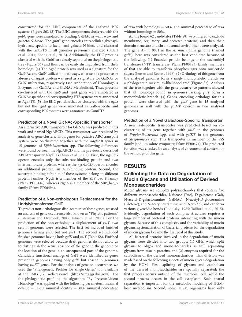
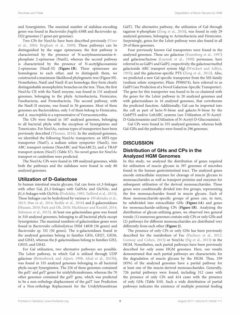
To identify all the GHs for mucin glycan degradation, we firstidentified all the glycosyl bonds previously detected in mucinglycans of the human intestine (Podolsky, 1985; Larsson et al.,2009; Tailford et al., 2015), which resulted in a collection of21 different glycosyl bonds (Figure 1A and Table S9). Then,we searched for all the enzymes able to hydrolase such bondsusing the KEGG (Kanehisa et al., 2012) and MetaCyc (Caspiet al., 2014) databases. Briefly, we searched the databases for bothof the monosaccharides that form the bond after the reactions
connected to this monosaccharide were filtered by EC numberto identify all the glycoside hydrolases (i.e., enzymes with EC3.2.-.-) for which this monosaccharide can be a substrate orproduct. After that, all the identified glycoside hydrolases weremanually checked for the corresponding analyzed bond. Finally,we collected 9 types of GHs (Table S9). For further informationon these enzymes, such as protein families and experimentallyanalyzed representatives, we carried out a search by EC numberin the CAZy database (Cantarel et al., 2012).
Pathways for catabolism of the derived monosaccharides wereidentified here as sets of reactions necessary to convert themonosaccharides into any intermediates of central metabolism.The pathway data were extracted from the KEGG (Kanehisaet al., 2012) and MetaCyc (Caspi et al., 2014) databases as wellas previous publications. For the two monosaccharides GalNAcand GlcNAc, only one pathway per monosaccharide has beendescribed (Figure 1B). GalNAc is catabolized through tagatose 6-phosphate to glyceraldehyde 3-phosphate and dihydroxyacetonephosphate (Leyn et al., 2012; Bidart et al., 2014), whereasGlcNAc is converted into fructose 6-phosphate (Afzal et al.,2015a; Plumbridge, 2015; Uhde et al., 2016). For Fuc, Gal,and Neu5Ac, two alternative pathways for the catabolism of
FIGURE 1 | Enzymes involved in the degradation of mucin glycans. (A) Splitting of a hypothetical mucin glycan by GHs. The following enzymes are shown: EC
3.2.1.18, Neuraminidase; EC 3.2.1.22, α-galactosidase; EC 3.2.1.23, Beta-galactosidase; EC 3.2.1.49, α-N-acetylgalactosaminidase; EC 3.2.1.50,
α-N-acetylglucosaminidase; EC 3.2.1.51, α-L-fucosidase; EC 3.2.1.52, β-N-hexosaminidase; EC 3.2.1.97, and Endo-α-N-acetylgalactosaminidase. (B) Known
pathways for utilization of the derived monosaccharides. Protein abbreviations are shown; for the corresponding functions see Table S2 in the Supplementary
Materials.
Frontiers in Genetics | www.frontiersin.org 6 August 2017 | Volume 8 | Article 111
Ravcheev and Thiele Degradation of Mucin Glycans by HGM
each monosaccharide have been described. Thus, Fuc may becatabolized through fuculose 1-phosphate to lactaldehyde anddihydroxyacetone phosphate or through fucolactone to lactateand pyruvate (Yew et al., 2006; Hobbs et al., 2013). For thegenes encoding enzymes of the latter pathway, no four-letterabbreviations have been designated. Thus, for these genes,we introduced the designation fclABCDE (from fucolactone;the last letter corresponds to the order of catalyzed reactionsin the pathway). Gal catabolism can also occur through twoalternative pathways: through galactose 1-phosphate and UDPgalactose (the Leloir pathway) (Bettenbrock and Alpert, 1998;Afzal et al., 2015b) or through galactose 6-phosphate and tagatose6-phosphate (Zeng et al., 2010). The last two steps of the secondpathway, phosphorylation and aldol splitting, are shared with thepathway for GalNAc catabolism. Neu5Ac is converted to fructose6-phosphate by two pathways, through GlcNAc or GlcNAc 6-phosphate (Vimr et al., 2004; Brigham et al., 2009). Therefore,these pathways overlap with GlcNAc by two or three reactions,respectively (Figure 1B).
Utilization of L-FucoseIntestinal mucin glycans contain Fuc moieties connected toGal by α1,2-linkage as well to GlcNAc by α1,2-, α1,3-, orα1,4- linkages (Podolsky, 1985; Tailford et al., 2015). Duringthe degradation of mucin glycans, these moieties can beremoved by α-L-fucosidases (Katayama et al., 2004; Nagaeet al., 2007; Ashida et al., 2009). α-L-fucosidases foundin the analyzed genomes belong to three families: GH29,GH42, and GH95. At least one α-L-fucosidase was foundin 131 genomes (Table S3). All of these genomes belongto only four phyla: Actinobacteria, Bacteroidetes, Firmicutes,and Verrucomicrobia. The largest number of genes encodingα-L-fucosidases were found in representatives of Firmicutes
(Lachnospiraceae bacterium 3_1_57FAA_CT1, 16 genes) andBacteroidetes (Bacteroides coprophilus DSM 18228, 14 genes).
Genes for both the alternative pathways for Fuc catabolismwere found in the analyzed genomes. Thus, genes for thepathway through fuculose 1-phosphate (fucIKA genes) werefound in the 125 genomes belonging to all studied bacterial phylaexcept Synergistetes and Tenericutes. The genes for the pathwaythrough fucolactone (fclABCDE) were found in only 8 genomes,all belonging to Actinobacteria.
In total, three different Fuc-specific transport systems werefound in the analyzed genomes, including two differentpermeases and one ABC transporter. The first permease, hereinreferred to as FucP1, was previously analyzed in Escherichia coli(Gunn et al., 1994) and predicted in the genomes of Bacteroidesspp. (Hooper et al., 1999; Ravcheev et al., 2013). This transporteris highly distributed in the analyzed genomes, co-clusteringwith the fuc genes in 88 genomes and with the fcl genes in 4genomes. An alternative Fuc permease named FucP2 belongs tothe Sugar_tr family (Pfam: PF00083). The gene encoding thistransporter is located inside the fuc gene cluster in the genomesof Pediococcus acidilactici and Lactobacillus rhamnosus. A thirdfucose transporter, the ABC-type one, was predicted in this study(see Prediction of Novel Fucose-Specific ABCTransport System).This transporter was found in 33 genomes belonging mostly toActinobacteria and Clostridia.
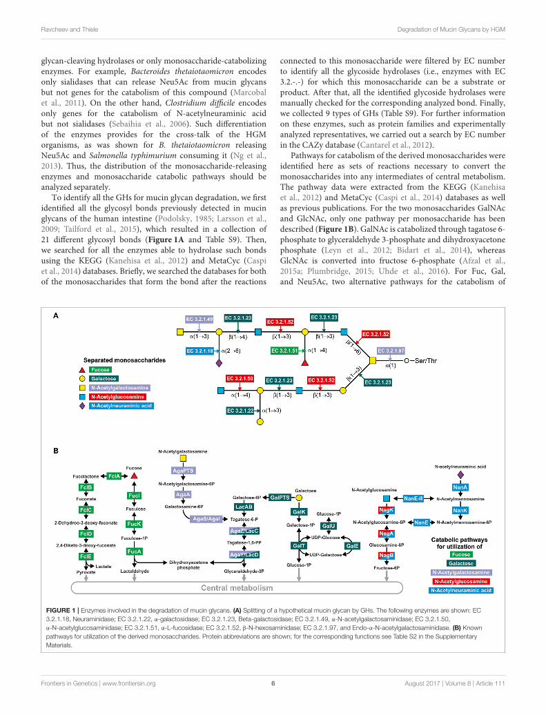
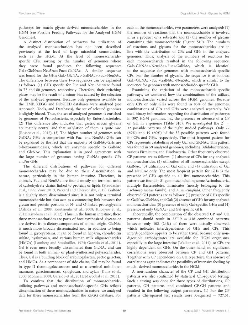
Generally, α-L-fucosidases were found in 131 analyzedgenomes, whereas Fuc CPs were found in 133 genomes. Both α-fucosidases and CPs were found together in 72 analyzed genomes(Figure 2, Table S3).
Utilization of N-Acetyl-D-GalactosamineGalNAc plays a crucial role in mucin glycans, forminglinks with side-chain oxygen atoms of Ser/Thr residues in
FIGURE 2 | Distribution of the CPs and GHs for mucin glycan-derived monosaccharides in the analyzed genomes. Pathways and GHs are grouped according to the
utilized monosaccharide. The bars “CP” correspond to the presence of catabolic pathways, the bars “GH” correspond to the presence of glycosyl hydrolases, the
bars “CP+GH” correspond to the presence of GHs and pathways for this monosaccharide in the same genomes.
Frontiers in Genetics | www.frontiersin.org 7 August 2017 | Volume 8 | Article 111
Ravcheev and Thiele Degradation of Mucin Glycans by HGM
mucin peptide chains. This linkage between the glycan andpeptide parts of mucin is mediated by various endo-α-N-acetylgalactosaminidases (Ashida et al., 2008; Koutsioulis et al.,2008; Kiyohara et al., 2012). Additionally, in intestinal mucinglycans, GalNAc moieties can be connected to Gal by α1,3- ,orβ1,4-linkage and to GalNAc by α1,6- or β1,3-linkage (Podolsky,1985; Tailford et al., 2015). Release of GalNAc from mucinglycans can be mediated by β-N-hexosaminidases (Cabezas,1989; Zwierz et al., 1999) and exo-α-N-acetylgalactosaminidases(Hoskins et al., 1997).
At least one GalNAc-releasing GH (GalNAc-GH) was foundin 218 genomes (Table S4). No GalNAc-GHs were found in thegenomes of Archaea or in the bacterial phyla Fusobacteria andSynergistetes. The maximal number of genes for the GalNAc-GHs was found in Bacteroides sp. 1_1_6 and Bacteroides sp.D22. Both of these organisms have 17 GalNAc-GH genes pergenome. All exo-α-N-acetylgalactosaminidases in the analyzedgenomes belong to the GH27 family. This type of GalNAc-GHswere found in only 13 genomes belonging to only four phyla:Actinobacteria, Bacteroidetes, Firmicutes, and Proteobacteria.Endo-α-N-acetylgalactosaminidases are also rarely representedin the analyzed genomes. These enzymes, belonging to theGH101 or GH129 families, were found in only 15 genomesfrom only two phyla: Actinobacteria and Firmicutes. In contrast,β-N-hexosaminidases were found in 217 genomes and in allbacterial phyla except Fusobacteria and Synergistetes. All the β-N-hexosaminidases found in the analyzed genomes belong to theGH3 or GH20 families.
GalNAc utilization pathway including agaS and agaA genes(see Annotation of Homologous Enzymes for GalNAc andGlcNAc Metabolism for Details on Annotation) was found in76 analyzed genomes. Three different GalNAc-specific transportsystems were found in the analyzed genomes, the PTS-typetransporters AgaPTS and GnbPTS (see Annotation of theGalNAc-Specific Transporters for Details on Annotation), as wellABC-type transporter LnbABC. The GnbPTS is a transporterwith multiple specificities and, in addition to GalNAc it can alsotransport oligosaccharides, lacto-N-biose (Galβ-1,3-GlcNAc),and galacto-N-biose (Galβ-1,3-GalNAc) (Bidart et al., 2014).Both lacto-N-biose and galacto-N-biose contain the glycosylbonds found in intestinal mucin glycans (Table S9). Thus,GnbPTS was included in the three analyzed pathways as GalNAc,GlcNAc, and Gal catabolism. Lacto- and galacto-N-biose are thenhydrolyzed by intracellular GH GnbG. Formally, GnbG is a GH,but because of its intracellular localization, it is considered to be apart of the GalNAc, GlcNAc and Gal CPs. The LnbABC transportsystem also transports lacto- and galacto-N-biose that afterwardshydrolyzed by intracellular phosphorylase LnbP (Nishimoto andKitaoka, 2007). As with GamPTS-GamB, the LnbABC-LnbPsystem was considered to be a part of the GalNAc, GlcNAc andGal CPs.
Overall, the GalNAc CP was found in the 76 analyzedgenomes belonging to the phyla Actinobacteria, Firmicutes, andProteobacteria (Figure 2, Table S4). Distribution of the GalNAc-releasing GHs is much broader; these enzymes were found in 218genomes. Both GHs and CPs were found in only 27 of analyzedgenomes.
Utilization of N-Acetyl-D-GlucosamineIntestinal mucin glycans contain GlcNAc moieties that formvarious glycosyl bonds, such as α1,4-, and β1,3-linkages withGal and β1,3-, and β1,6-linkages with GalNAc (Podolsky,1985; Tailford et al., 2015). The α-linkages are hydrolyzedby α-N-acetylglucosaminidases (Shimada et al., 2015), whereasthe β-linkages with Gal or GalNAc are hydrolyzed by β-N-hexosaminidases (see Utilization of N-Acetyl-D-Galactosamine).
At least one GlcNAc-releasing GH (GlcNAc-GH) was foundin 257 genomes (Table S5). The maximal numbers of thegenes encoding GlcNAc-GHs were found in Bacteroides sp.1_1_14 (23 genes) and B. thetaiotaomicron (22 genes). Inaddition to β-N-hexosaminidases (see Utilization of N-acetyl-D-Galactosamine), α-N-acetylglucosaminidases (GH89 family)were found in 60 genomes (Table S5). All genomes in which α-N-acetylglucosaminidase genes were found belong to the phylaActinobacteria, Bacteroidetes, Firmicutes, Proteobacteria, andVerrucomicrobia.
The GlcNAc CP (Yang et al., 2006; Plumbridge, 2015)was found in 218 of the analyzed genomes (Table S5). Thispathway is broadly distributed among analyzed taxa and isabsent only in Archaea and the bacterial phyla Synergistetes andVerrucomicrobia. Various GlcNAc-specific transport systemswere identified in the analyzed genomes. These are thePTSs NagE (Plumbridge et al., 1993; Plumbridge, 2015) andGnbPTS (see Utilization of N-Acetyl-D-Galactosamine), theABC transport systems NgcEFG (Xiao et al., 2002), NgcABCD(predicted in this work, see Prediction of a Novel GlcNAc-Specific Transporter) and LnbABC (Nishimoto and Kitaoka,2007), and the permease NagP (Ravcheev et al., 2013).
The GlcNAc moieties of intestinal mucin glycans can besulfated. This sulfation possibly protects these glycans fromdegradation by the HGM and is correlated with health anddisease status of the host organism (Tobisawa et al., 2010; Boltinet al., 2013). Only specific microbial species are able to removesulfate groups to make mucin glycans available for themselvesor to other microbes (Smalley et al., 1994; Robertson andWright, 1997; Jansen et al., 1999; Wright et al., 2000; Robinsonet al., 2012). Among the analyzed organisms, genes for mucin-desulfating sulfatase (GlcNAc-6-sulfatase) were found only in42 genomes from Bacteroidetes phyla and in the genome ofA. muciniphila (Table S5). Notably, more than half of these genesare chromosomally clustered with the nagKP operon or withgenes for β-N-hexosaminidases (Figure S8).
Overall, GlcNAc CP was found in 218 analyzed genomes,whereas GlcNAc-GHs were found in 266 genomes. Both thepathway and the GHs were found in 155 analyzed genomes(Figure 2, Table S5).
Utilization of N-Acetyl-D-Neuraminic AcidNeu5Ac is commonly found in the terminal location of intestinalmucin glycans (Johansson et al., 2011; Mcguckin et al., 2011),forming α2,3-linkages with Gal and α2,6-linkages with Gal orGalNAc (Tailford et al., 2015). These bonds can be hydrolyzed bysialidases (Juge et al., 2016). Sialidases, all belonging to the GH33family, were found in 112 analyzed genomes. Sialidases werefound in the genomes of all bacterial phyla except Fusobacteria
Frontiers in Genetics | www.frontiersin.org 8 August 2017 | Volume 8 | Article 111
Ravcheev and Thiele Degradation of Mucin Glycans by HGM
and Synergistetes. The maximal number of sialidase-encodinggenes was found in Bacteroides fragilis 638R and Bacteroides sp.D22 genomes (7 genes per genome).
Two CPs for Neu5Ac have been described previously (Vimret al., 2004; Brigham et al., 2009). These pathways can bedistinguished by the sugar epimerases; the first pathway ischaracterized by the presence of N-acetylmannosamine-6-phosphate 2-epimerase (NanE), whereas the second pathwayis characterized by the presence of N-acetylglucosamine2-epimerase (NanE-II) (Figure 1B). These epimerases arehomologous to each other, and to distinguish them, weconstructed amaximum-likelihood phylogenetic tree (Figure S9).Nonetheless, NanE and NanE-II are homologs; they form clearlydistinguishable monophyletic branches on the tree. Thus, the firstNeu5Ac CP, with the NanE enzyme, was found in 131 analyzedgenomes, belonging to the phyla Actinobacteria, Firmicutes,Fusobacteria, and Proteobacteria. The second pathway, withthe NanE-II enzyme, was found in 56 genomes. Most of thesegenomes are Bacteroidetes, only 3 of them belong to Firmicutes,and A. muciniphila is a representative of Verrucomicrobia.
The CPs were found in 187 analyzed genomes, belongingto all bacterial phyla with the exception of Synergistetes andTenericutes. For Neu5Ac, various types of transporters have beenpreviously described (Thomas, 2016). In the analyzed genomes,we identified the following Neu5Ac transporters: an MFS-typetransporter (NanT), a sodium solute symporter (NanX), twoABC transport systems (NanABC and NanABC2), and a TRAPtransport system (NeuT) (Table S7). No novel genes for Neu5Actransport or catabolism were predicted.
The Neu5Ac CPs were found in 189 analyzed genomes, whileboth the pathways and the sialidases were found in only 80analyzed genomes.
Utilization of D-GalactoseIn human intestinal mucin glycans, Gal can form α1,3-linkageswith other Gal, β1,3-linkages with GalNAc and GlcNAc, andβ1,4-linkages with GlcNAc (Podolsky, 1985; Tailford et al., 2015).These linkages can be hydrolyzed by various α- (Wakinaka et al.,2013; Han et al., 2014; Reddy et al., 2016) and β-galactosidases(Husain, 2010; Park and Oh, 2010; Michlmayr and Kneifel, 2014;Solomon et al., 2015). At least one galactosidase gene was foundin 310 analyzed genomes, belonging to all bacterial phyla exceptSynergistetes. The maximal numbers of galactosidase genes werefound in Bacteroides cellulosilyticus DSM 14838 (56 genes) andBacteroides sp. D2 (50 genes). The α-galactosidases found inthe analyzed genomes belong to families GH4, GH27, GH36,and GH43, whereas the β-galactosidases belong to families GH2,GH35, and GH42.
For Gal utilization, two alternative pathways are possible.The Leloir pathway, in which Gal is utilized through UDPgalactose (Bettenbrock and Alpert, 1998; Afzal et al., 2015b),was found in 335 analyzed genomes belonging to all bacterialphyla except Synergistetes. The 256 of these genomes containedthe galU and galT genes for uridylyltransferases, whereas the 70other genomes contained the galY gene, which was predictedto be a non-orthologs displacement of the galT (see Predictionof a Non-orthologs Replacement for the Uridylyltransferase
GalT). The alternative pathway, the utilization of Gal throughtagatose 6-phosphate (Zeng et al., 2010), was found in only 29analyzed genomes, belonging to Actinobacteria and Firmicutes.Surprisingly, genes for the Leloir pathway were also found in all29 of these genomes.
Four previously known Gal transporters were found in theanalyzed genomes. These are galactose (Essenberg et al., 1997)and galactose/lactose (Luesink et al., 1998) permeases, herereferred to as GalP1 and GalP2, respectively; the galactose/methylgalactoside ABC transport system Mgl (Weickert and Adhya,1993); and the galactose-specific PTS (Zeng et al., 2012). Also,we predicted a new Gal-specific transporter from the SSS family(sodium solute symporter, Pfam: PF00474), here referred to asGalP3 (see Prediction of a Novel Galactose-Specific Transporter).The gene for this transporter was found to be co-clustered withthe genes for the Leloir pathway in 20 analyzed genomes andwith galactosidases in 16 analyzed genomes, that corroboratethe predicted function. Additionally, Gal can be imported intothe cell as part of lacto-N-biose and galacto-N-biose by theGnbPTS and/or LnbABC systems (see Utilization of N-Acetyl-D-Galactosamine and Utilization of N-Acetyl-D-Glucosamine).
Gal CPs were found in 355 analyzed genomes, whereas bothGal-GHs and the pathways were found in 296 genomes.
DISCUSSION
Distribution of GHs and CPs in theAnalyzed HGM GenomesIn this study, we analyzed the distribution of genes requiredfor utilization of mucin glycans in 397 genomes of microbesfound in the human gastrointestinal tract. The analyzed genesencode extracellular enzymes for cleavage of mucin glycans tomonosaccharides as well as transport proteins and enzymes forsubsequent utilization of the derived monosaccharides. Thesegenes were conditionally divided into five groups, representingthe five monosaccharides found in mucin glycans. Each ofthese monosaccharide-specific groups of genes can, in turn,be subdivided into extracellular GHs (Figure 1A) and genesfor monosaccharide-utilizing CPs (Figure 1B). Analyzing thedistribution of glycan-utilizing genes, we observed two generaltrends: (1) numerous genomes contain only CPs or only GHs and(2) pathways for different monosaccharides are distributed verydifferently from each other (Figure 2).
The presence of only CPs or only GHs has been previouslydescribed for the metabolism of Fuc (Pacheco et al., 2012;Conway and Cohen, 2015) or Neu5Ac (Ng et al., 2013) in theHGM. Nonetheless, such partial pathways have been previouslydescribed for only some HGM genomes. Here, our resultsdemonstrated that such partial pathways are characteristic forthe degradation of mucin glycans by the HGM. Thus, 339(85%) of the analyzed genomes have a partial pathway forat least one of the mucin-derived monosaccharides. Generally,726 partial pathways were found, including 312 cases withthe presence of only CPs and 414 cases with the presenceof only GHs (Table S10). Such a wide distribution of partialpathways indicates the existence of multiple potential feeding
Frontiers in Genetics | www.frontiersin.org 9 August 2017 | Volume 8 | Article 111
Ravcheev and Thiele Degradation of Mucin Glycans by HGM
pathways for mucin glycan-derived monosaccharides in theHGM (see Possible Feeding Pathways for the Analyzed HGMGenomes).
A distinct distribution of pathways for utilization ofthe analyzed monosaccharides has not been describedpreviously at the level of large microbial communities,such as the HGM. At the level of monosaccharide-specific CPs, sorting by the number of genomes wherethey were found produces the following sequence:Gal>GlcNAc>Neu5Ac>Fuc>GalNAc. A similar sequencewas found for the GHs: Gal>GlcNAc>GalNAc>Fuc>Neu5Ac.The differences between these two sequences can be explainedas follows. (1) GHs specific for Fuc and Neu5Ac were foundin 72 and 80 genomes, respectively. Therefore, their switchingplaces may be the result of a minor bias caused by the selectionof the analyzed genomes. Because only genomes available inthe HMP, KEGG and PubSEED databases were analyzed (seeApproach, Tools, and Databases), the set of selected genomesis slightly biased. Thus, the set of analyzed genomes is enrichedby genomes of Proteobacteria, especially by Enterobacteriales.(2) This difference also indicates that gastric mucin glycansare mainly neutral and that sialylation of them is quite rare(Rossez et al., 2012). (3) The higher number of genomes withGalNAc-GHs in comparison with Fuc- and Neu5Ac-GHs canbe explained by the fact that the majority of GalNAc-GHs areβ-hexosaminidases, which are enzymes specific to GalNAcand GlcNAc. This explanation is in good agreement withthe large number of genomes having GlcNAc-specific CPsand/or GHs.
The different distributions of pathways for differentmonosaccharides may be due to their dissemination innature, particularly in the human intestine. Therefore, inanimals, Fuc and Neu5Ac are found mostly on terminal unitsof carbohydrate chains linked to proteins or lipids (Staudacheret al., 1999; Vimr, 2013; Pickard and Chervonsky, 2015). GalNAcis a slightly more disseminated since it is not only a terminalmonosaccharide but also acts as a connecting link between theglycan and protein portions of N- and O-linked proteoglycans(Ashida et al., 2008; Koutsioulis et al., 2008; Kilcoyne et al.,2012; Kiyohara et al., 2012). Thus, in the human intestine, thesethree monosaccharides are parts of host-synthesized glycans orare derived from dietary components of animal origin. GlcNAcis much more broadly disseminated and, in addition to beingfound in glycoproteins, it can be found in heparin, chondroitinsulfate, hyaluronan, and various human milk oligosaccharides(HMOs) (Lamberg and Stoolmiller, 1974; Garrido et al., 2011).Gal is even more broadly disseminated than GlcNAc and canbe found in both animal- or plant-synthesized polysaccharides.Thus, Gal is a building block of arabinogalactan, pectic galactan,and HMOs. As a component of side chains, Gal may be foundin type II rhamnogalacturonan and as a terminal unit in α-mannans, galactomannan, xyloglucan, and xylan (Kunz et al.,2000; Mohnen, 2008; Garrido et al., 2011; Marcobal et al., 2011).
To confirm that the distribution of monosaccharide-utilizing pathways and monosaccharide-specific GHs reflectsdissemination of these monosaccharides in nature, we analyzeddata for these monosaccharides from the KEGG database. For
each of the monosaccharides, two parameters were analyzed: (1)the number of reactions that the monosaccharide is involvedin as a product or a substrate and (2) the number of glycanscontaining this monosaccharide (Figure S10). The numbersof reactions and glycans for the monosaccharides are inline with the distribution of CPs and GHs in the analyzedgenomes. Thus, analysis of the numbers of reactions foreach monosaccharide resulted in the following sequence:Gal>GlcNAc>Neu5Ac>Fuc>GalNAc, which is identicalto the sequence for genomes with monosaccharide-specificCPs. For the number of glycans, the sequence is as follows:Gal>GlcNAc>Fuc>GalNAc>Neu5Ac, which is similar to thesequence for genomes with monosaccharide-specific GHs.
Examining the variation of the monosaccharide-specificpathways, we wondered how the combinations of the utilizedmonosaccharides varied across the HGM genomes. Becauseonly CPs or only GHs were found in 85% of the genomes,combinations of CPs and GHs were analyzed separately. Weused binary information regarding the distribution of pathwaysin 397 HGM genomes, i.e., the presence or absence of a CPor GH in a genome (Table S10). We investigated the 25 =
32 possible patterns of the eight studied pathways. Only 22(69%) and 19 (60%) of the 32 possible patterns were foundfor CPs and GHs, respectively. The most frequent pattern forCPs represents catabolism of only Gal and GlcNAc. This patternwas found in 59 analyzed genomes, including Bifidobacteriacea,various Firmicutes, and Fusobacteria. Other frequently observedCP patterns are as follows: (1) absence of CPs for any analyzedmonosaccharides, (2) utilization of all monosaccharides exceptGalNAc, (3) utilization of Gal only, and (4) utilization of Galand Neu5Ac only. The most frequent pattern for GHs is thepresence of GHs specific to all five monosaccharides. Thispattern was found in 81 genomes, including someActinobacteria,multiple Bacteroidetes, Firmicutes (mostly belonging to theLachnospiraceae family), and A. muciniphila. Other frequentlyobserved GH patterns are as follows: (1) presence of GHs specificto GalNAc, GlcNAc, and Gal; (2) absence of GHs for any analyzedmonosaccharides; (3) presence of only Gal-specific GHs; and (4)presence of only GlcNAc- and Gal-specific GHs.
Theoretically, the combination of the observed CP and GHpatterns should result in 22∗19 = 418 combined patterns;however, only 102 (24%) of them were actually observed,which indicates interdependence of GHs and CPs. Thisinterdependence appears to be rather trivial because only non-digestible carbohydrates are available for HGM organisms,especially in the large intestine (Walker et al., 2011), so CPs arehighly dependent on GHs. On the other hand, no significantcorrelations were observed between CP and GH patterns.Together with CP dependence on GH repertoire, this absence ofcorrelations again indicates the possibility of intensive feeding bymucin-derived monosaccharides in the HGM.
A non-random character of the CP and GH distributionpatterns was also confirmed by statistical Chi-squared testing.Such a testing was done for three types of distributions, CPpatterns, GH patterns, and combined CP-GH patterns andresulted in the following output parameters. (1) For the CPpatterns Chi-squared test results were X-squared = 727.51,
Frontiers in Genetics | www.frontiersin.org 10 August 2017 | Volume 8 | Article 111
Ravcheev and Thiele Degradation of Mucin Glycans by HGM
degree of freedom (DF) = 31, and p-value < 2.2e-16. (2) For theGH patterns the test results were X-squared = 1303, DF = 31,and p-value< 2.2e-16. (3) For the combined CP-GH patterns thetest results were X-squared = 10418, DF = 1023, and p-value <
2.2e-16. Thus, the tests showed that the distributions of all threepathways were non-random.
Surprisingly, the combined pattern corresponding to themost frequent GH and CP patterns was found in onlythree genomes: Clostridium nexile DSM 1787, Lachnospiraceaebacterium 2_1_46FAA, and Ruminococcus lactaris ATCC 29176.The combined pattern we observed most frequently was theabsence of all analyzed GHs and CPs. This pattern was found in28 genomes, belonging to Archaea, some Firmicutes, and Beta-and Epsilonproteobacteria. Other frequently observed combinedpatterns were as follows: (1) presence of GHs specific for GalNAc,GlcNAc, and Gal together with CPs for GlcNAc and Gal; (2)presence of GHs for all five monosaccharides together withutilization of all these monosaccharides except GalNAc; and(3) presence of GHs for all five monosaccharides together withutilization of Neu5Ac and Gal.
Taken together, an optimal strategy for glycan-utilizing HGMmicroorganisms includes (1) the presence of CPs specific toGlcNAc andGal as the components of multiple host- and dietary-derived carbohydrates and (2) the presence of GHs specific to aslarge as possible a number of glycan-building monosaccharides.
Possible Feeding Pathways for theAnalyzed HGM GenomesThe presence of only CPs for a certain monosaccharide inone HGM organism and only GHs for this monosaccharide inanother organism allows us to predict possible feeding pathways.Previously, such feeding pathways in the HGM have beenfound for Fuc (Pacheco et al., 2012; Conway and Cohen, 2015)and Neu5Ac (Ng et al., 2013). Here, we predicted multiplepotential feeding pathways for all five monosaccharides formingmucin glycans. The 339 (85%) analyzed genomes demonstratedthe presence of only CPs or only GHs for at least onemonosaccharide; therefore, the majority of HGM organisms areinvolved in these feeding pathways.
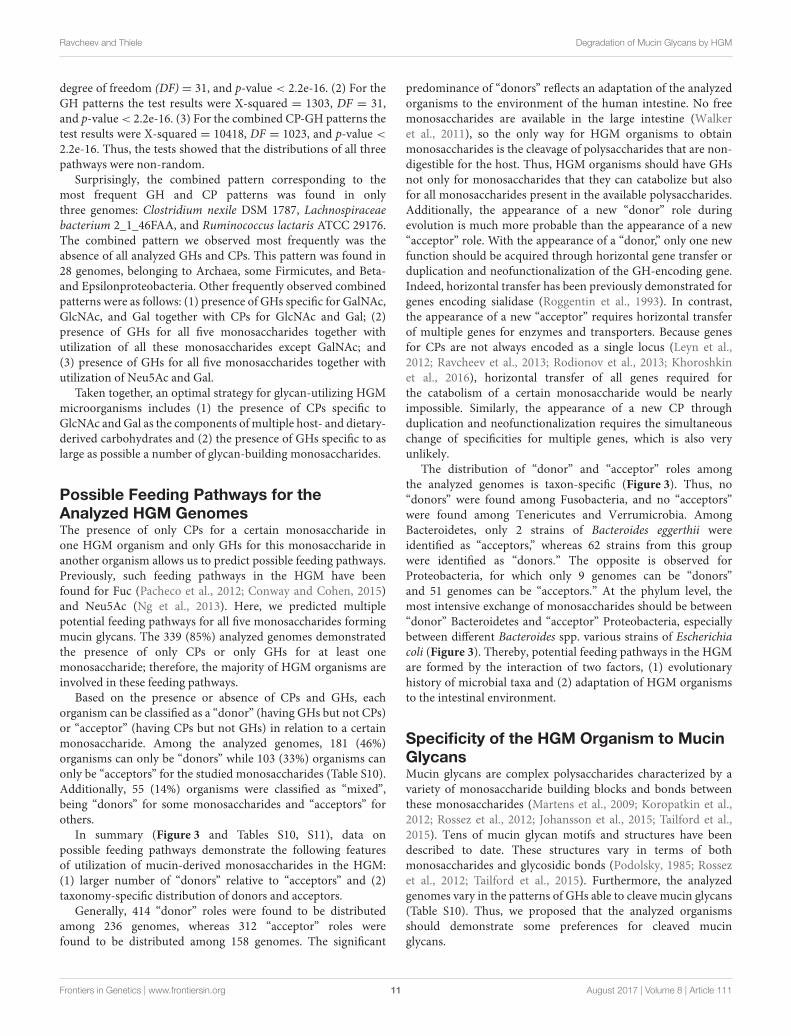
Based on the presence or absence of CPs and GHs, eachorganism can be classified as a “donor” (having GHs but not CPs)or “acceptor” (having CPs but not GHs) in relation to a certainmonosaccharide. Among the analyzed genomes, 181 (46%)organisms can only be “donors” while 103 (33%) organisms canonly be “acceptors” for the studied monosaccharides (Table S10).Additionally, 55 (14%) organisms were classified as “mixed”,being “donors” for some monosaccharides and “acceptors” forothers.
In summary (Figure 3 and Tables S10, S11), data onpossible feeding pathways demonstrate the following featuresof utilization of mucin-derived monosaccharides in the HGM:(1) larger number of “donors” relative to “acceptors” and (2)taxonomy-specific distribution of donors and acceptors.
Generally, 414 “donor” roles were found to be distributedamong 236 genomes, whereas 312 “acceptor” roles werefound to be distributed among 158 genomes. The significant
predominance of “donors” reflects an adaptation of the analyzedorganisms to the environment of the human intestine. No freemonosaccharides are available in the large intestine (Walkeret al., 2011), so the only way for HGM organisms to obtainmonosaccharides is the cleavage of polysaccharides that are non-digestible for the host. Thus, HGM organisms should have GHsnot only for monosaccharides that they can catabolize but alsofor all monosaccharides present in the available polysaccharides.Additionally, the appearance of a new “donor” role duringevolution is much more probable than the appearance of a new“acceptor” role. With the appearance of a “donor,” only one newfunction should be acquired through horizontal gene transfer orduplication and neofunctionalization of the GH-encoding gene.Indeed, horizontal transfer has been previously demonstrated forgenes encoding sialidase (Roggentin et al., 1993). In contrast,the appearance of a new “acceptor” requires horizontal transferof multiple genes for enzymes and transporters. Because genesfor CPs are not always encoded as a single locus (Leyn et al.,2012; Ravcheev et al., 2013; Rodionov et al., 2013; Khoroshkinet al., 2016), horizontal transfer of all genes required forthe catabolism of a certain monosaccharide would be nearlyimpossible. Similarly, the appearance of a new CP throughduplication and neofunctionalization requires the simultaneouschange of specificities for multiple genes, which is also veryunlikely.
The distribution of “donor” and “acceptor” roles amongthe analyzed genomes is taxon-specific (Figure 3). Thus, no“donors” were found among Fusobacteria, and no “acceptors”were found among Tenericutes and Verrumicrobia. AmongBacteroidetes, only 2 strains of Bacteroides eggerthii wereidentified as “acceptors,” whereas 62 strains from this groupwere identified as “donors.” The opposite is observed forProteobacteria, for which only 9 genomes can be “donors”and 51 genomes can be “acceptors.” At the phylum level, themost intensive exchange of monosaccharides should be between“donor” Bacteroidetes and “acceptor” Proteobacteria, especiallybetween different Bacteroides spp. various strains of Escherichiacoli (Figure 3). Thereby, potential feeding pathways in the HGMare formed by the interaction of two factors, (1) evolutionaryhistory of microbial taxa and (2) adaptation of HGM organismsto the intestinal environment.
Specificity of the HGM Organism to MucinGlycansMucin glycans are complex polysaccharides characterized by avariety of monosaccharide building blocks and bonds betweenthese monosaccharides (Martens et al., 2009; Koropatkin et al.,2012; Rossez et al., 2012; Johansson et al., 2015; Tailford et al.,2015). Tens of mucin glycan motifs and structures have beendescribed to date. These structures vary in terms of bothmonosaccharides and glycosidic bonds (Podolsky, 1985; Rossezet al., 2012; Tailford et al., 2015). Furthermore, the analyzedgenomes vary in the patterns of GHs able to cleave mucin glycans(Table S10). Thus, we proposed that the analyzed organismsshould demonstrate some preferences for cleaved mucinglycans.
Frontiers in Genetics | www.frontiersin.org 11 August 2017 | Volume 8 | Article 111
Ravcheev and Thiele Degradation of Mucin Glycans by HGM
FIGURE 3 | Distribution of monosaccharide donors and acceptors by taxa. Only genomes classified as “donors” (the horizontal axis) and “acceptors” (the vertical axis)
are shown. The numbers of monosaccharides are shown in agreement with the color scale. For details on each pair of organisms and possibly exchanged
monosaccharides, see Table S11.
Data on the known structures of mucin glycans found in thehuman intestine were collected from the literature (Podolsky,1985; Rossez et al., 2012), resulting in 56 different glycanstructures (Figure S11 and Table S12). For each analyzed genome,the ability to cleave each glycan structure was predicted. Glycanwas considered able to be cleaved by a certain organism if the GHsfor all glycoside bonds in the glycan were found in the genomeof the organism. Bonds between GalNAc and Ser/Thr residuesof the mucin peptide were excluded from the analysis becausethis bond is cleaved by endo-α-N-acetylgalactosaminidases foundin only 15 of the analyzed genomes (Table S4). On the basis ofthis prediction for each genome, the pattern of likely cleavedglycans was determined (Table S13). Among 397 analyzedgenomes, 321 (81%) were able to cleave at least one of theglycans; generally, 20 different glycan-cleavage patterns weredefined. It has previously been estimated that approximately40% of bacteria have glycan-degrading enzymes (Arike andHansson, 2016). Here, we demonstrated that, at least for
the HGM microbes, this figure is actually at least 2-foldhigher.
Based on the glycan-cleavage patterns, only 8 analyzedorganisms are able to cleave all 56 glycan structures belongingto the phyla Bacteroidetes (Bacteroides ovatus SD CMC3f, Bacteroides sp. 2_2_4, and Bacteroides sp. 3_1_23) andFirmicutes (Clostridium perfringens WAL-14572, 3 strains ofLachnospiraceae bacterium, and Ruminococcus torques ATCC27756). The three most frequently observed patterns were thefollowing. (1) Only Core 1 (Tailford et al., 2015) structurescan be cleaved, i.e., no GHs except β-galactosidases are present.This pattern was found in 77 genomes belonging mostlyto Lactobacillaceae, Ruminococcaceae and Enterobacteriales.(2) Glycans having only poly-lacto-N-biose but lacking anyspecific groups (Table S12) can be cleaved. These genomes haveonly GHs for the hydrolysis of β-Gal and β-GlcNAc bonds.This pattern was found in 56 genomes belonging mostly toActinobacteria, Firmicutes and Enterobacteriales. (3) All glycans
Frontiers in Genetics | www.frontiersin.org 12 August 2017 | Volume 8 | Article 111
Ravcheev and Thiele Degradation of Mucin Glycans by HGM
lacking α-GalNAc groups can be cleaved. These genomes haveall GHs but not α-N-acetylgalactosaminidases. This pattern wasfound in 41 genomes belonging mostly to Bacteroidaceae.
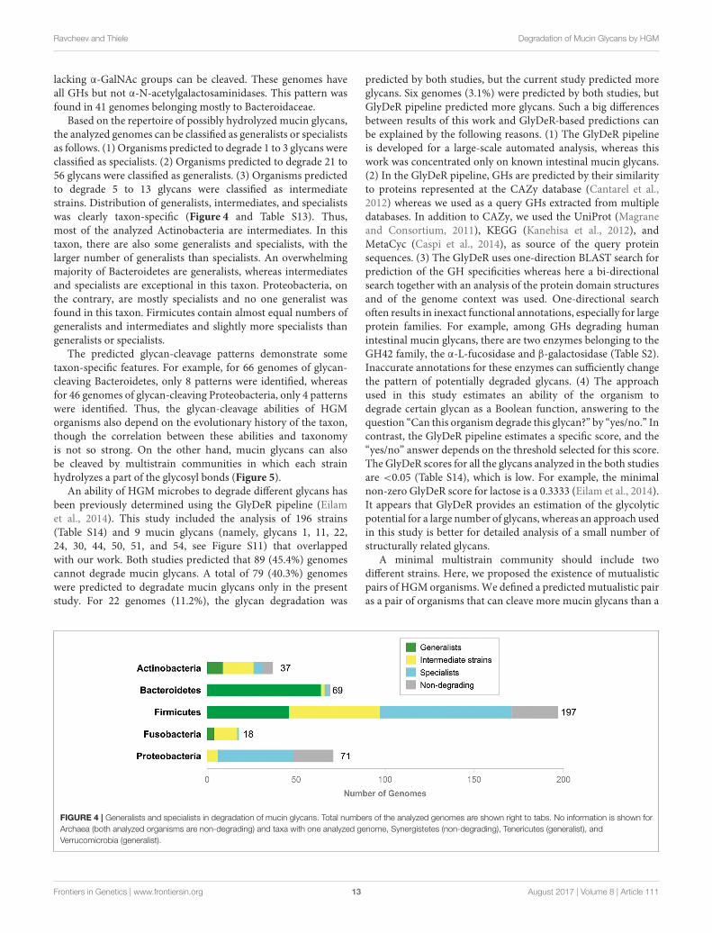
Based on the repertoire of possibly hydrolyzed mucin glycans,the analyzed genomes can be classified as generalists or specialistsas follows. (1) Organisms predicted to degrade 1 to 3 glycans wereclassified as specialists. (2) Organisms predicted to degrade 21 to56 glycans were classified as generalists. (3) Organisms predictedto degrade 5 to 13 glycans were classified as intermediatestrains. Distribution of generalists, intermediates, and specialistswas clearly taxon-specific (Figure 4 and Table S13). Thus,most of the analyzed Actinobacteria are intermediates. In thistaxon, there are also some generalists and specialists, with thelarger number of generalists than specialists. An overwhelmingmajority of Bacteroidetes are generalists, whereas intermediatesand specialists are exceptional in this taxon. Proteobacteria, onthe contrary, are mostly specialists and no one generalist wasfound in this taxon. Firmicutes contain almost equal numbers ofgeneralists and intermediates and slightly more specialists thangeneralists or specialists.
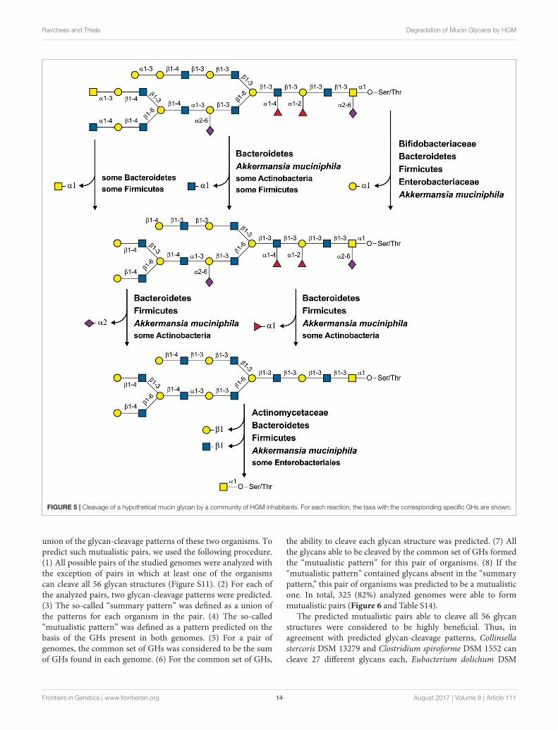
The predicted glycan-cleavage patterns demonstrate sometaxon-specific features. For example, for 66 genomes of glycan-cleaving Bacteroidetes, only 8 patterns were identified, whereasfor 46 genomes of glycan-cleaving Proteobacteria, only 4 patternswere identified. Thus, the glycan-cleavage abilities of HGMorganisms also depend on the evolutionary history of the taxon,though the correlation between these abilities and taxonomyis not so strong. On the other hand, mucin glycans can alsobe cleaved by multistrain communities in which each strainhydrolyzes a part of the glycosyl bonds (Figure 5).
An ability of HGM microbes to degrade different glycans hasbeen previously determined using the GlyDeR pipeline (Eilamet al., 2014). This study included the analysis of 196 strains(Table S14) and 9 mucin glycans (namely, glycans 1, 11, 22,24, 30, 44, 50, 51, and 54, see Figure S11) that overlappedwith our work. Both studies predicted that 89 (45.4%) genomescannot degrade mucin glycans. A total of 79 (40.3%) genomeswere predicted to degradate mucin glycans only in the presentstudy. For 22 genomes (11.2%), the glycan degradation was
predicted by both studies, but the current study predicted moreglycans. Six genomes (3.1%) were predicted by both studies, butGlyDeR pipeline predicted more glycans. Such a big differencesbetween results of this work and GlyDeR-based predictions canbe explained by the following reasons. (1) The GlyDeR pipelineis developed for a large-scale automated analysis, whereas thiswork was concentrated only on known intestinal mucin glycans.(2) In the GlyDeR pipeline, GHs are predicted by their similarityto proteins represented at the CAZy database (Cantarel et al.,2012) whereas we used as a query GHs extracted from multipledatabases. In addition to CAZy, we used the UniProt (Magraneand Consortium, 2011), KEGG (Kanehisa et al., 2012), andMetaCyc (Caspi et al., 2014), as source of the query proteinsequences. (3) The GlyDeR uses one-direction BLAST search forprediction of the GH specificities whereas here a bi-directionalsearch together with an analysis of the protein domain structuresand of the genome context was used. One-directional searchoften results in inexact functional annotations, especially for largeprotein families. For example, among GHs degrading humanintestinal mucin glycans, there are two enzymes belonging to theGH42 family, the α-L-fucosidase and β-galactosidase (Table S2).Inaccurate annotations for these enzymes can sufficiently changethe pattern of potentially degraded glycans. (4) The approachused in this study estimates an ability of the organism todegrade certain glycan as a Boolean function, answering to thequestion “Can this organism degrade this glycan?” by “yes/no.” Incontrast, the GlyDeR pipeline estimates a specific score, and the“yes/no” answer depends on the threshold selected for this score.The GlyDeR scores for all the glycans analyzed in the both studiesare <0.05 (Table S14), which is low. For example, the minimalnon-zero GlyDeR score for lactose is a 0.3333 (Eilam et al., 2014).It appears that GlyDeR provides an estimation of the glycolyticpotential for a large number of glycans, whereas an approach usedin this study is better for detailed analysis of a small number ofstructurally related glycans.
A minimal multistrain community should include twodifferent strains. Here, we proposed the existence of mutualisticpairs of HGMorganisms.We defined a predictedmutualistic pairas a pair of organisms that can cleave more mucin glycans than a
FIGURE 4 | Generalists and specialists in degradation of mucin glycans. Total numbers of the analyzed genomes are shown right to tabs. No information is shown for
Archaea (both analyzed organisms are non-degrading) and taxa with one analyzed genome, Synergistetes (non-degrading), Tenericutes (generalist), and
Verrucomicrobia (generalist).
Frontiers in Genetics | www.frontiersin.org 13 August 2017 | Volume 8 | Article 111
Ravcheev and Thiele Degradation of Mucin Glycans by HGM
FIGURE 5 | Cleavage of a hypothetical mucin glycan by a community of HGM inhabitants. For each reaction, the taxa with the corresponding specific GHs are shown.
union of the glycan-cleavage patterns of these two organisms. Topredict such mutualistic pairs, we used the following procedure.(1) All possible pairs of the studied genomes were analyzed withthe exception of pairs in which at least one of the organismscan cleave all 56 glycan structures (Figure S11). (2) For each ofthe analyzed pairs, two glycan-cleavage patterns were predicted.(3) The so-called “summary pattern” was defined as a union ofthe patterns for each organism in the pair. (4) The so-called“mutualistic pattern” was defined as a pattern predicted on thebasis of the GHs present in both genomes. (5) For a pair ofgenomes, the common set of GHs was considered to be the sumof GHs found in each genome. (6) For the common set of GHs,
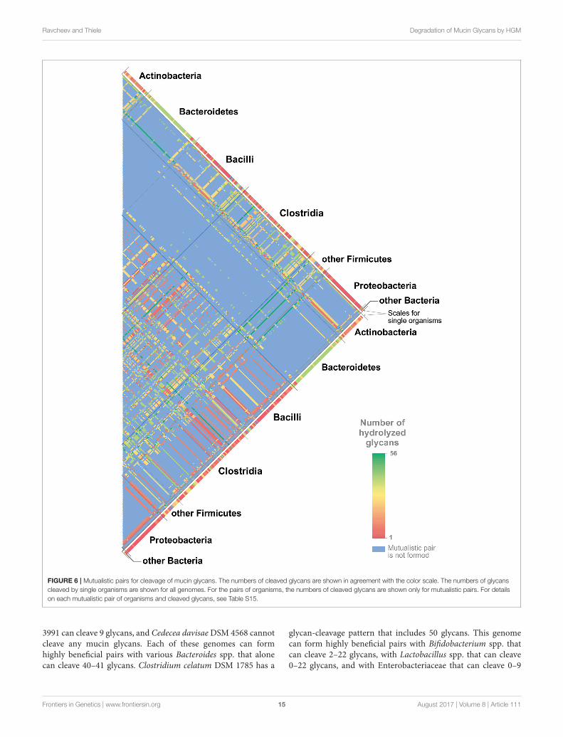
the ability to cleave each glycan structure was predicted. (7) Allthe glycans able to be cleaved by the common set of GHs formedthe “mutualistic pattern” for this pair of organisms. (8) If the“mutualistic pattern” contained glycans absent in the “summarypattern,” this pair of organisms was predicted to be a mutualisticone. In total, 325 (82%) analyzed genomes were able to formmutualistic pairs (Figure 6 and Table S14).
The predicted mutualistic pairs able to cleave all 56 glycanstructures were considered to be highly beneficial. Thus, inagreement with predicted glycan-cleavage patterns, Collinsellastercoris DSM 13279 and Clostridium spiroforme DSM 1552 cancleave 27 different glycans each, Eubacterium dolichum DSM
Frontiers in Genetics | www.frontiersin.org 14 August 2017 | Volume 8 | Article 111
Ravcheev and Thiele Degradation of Mucin Glycans by HGM
FIGURE 6 | Mutualistic pairs for cleavage of mucin glycans. The numbers of cleaved glycans are shown in agreement with the color scale. The numbers of glycans
cleaved by single organisms are shown for all genomes. For the pairs of organisms, the numbers of cleaved glycans are shown only for mutualistic pairs. For details
on each mutualistic pair of organisms and cleaved glycans, see Table S15.
3991 can cleave 9 glycans, and Cedecea davisaeDSM 4568 cannotcleave any mucin glycans. Each of these genomes can formhighly beneficial pairs with various Bacteroides spp. that alonecan cleave 40–41 glycans. Clostridium celatum DSM 1785 has a
glycan-cleavage pattern that includes 50 glycans. This genomecan form highly beneficial pairs with Bifidobacterium spp. thatcan cleave 2–22 glycans, with Lactobacillus spp. that can cleave0–22 glycans, and with Enterobacteriaceae that can cleave 0–9
Frontiers in Genetics | www.frontiersin.org 15 August 2017 | Volume 8 | Article 111
Ravcheev and Thiele Degradation of Mucin Glycans by HGM
glycans. All highly beneficial pairs described are organized ina similar manner. The five strains listed above are distantlyrelated to each other, and three of them, C. celatum, C. davisae,and C. spiroforme, are pathogens (Akinosoglou et al., 2012;Papatheodorou et al., 2012; Agergaard et al., 2016), whereas nodata about the pathogenicity of C. stercoris and E. dolichumwere found. Each of these five strains forms highly beneficialpairs with a large number of organisms closely related to eachother and highly represented in healthy HGM (Eckburg et al.,2005; Goodman et al., 2011; Walker et al., 2011; Graf et al.,2015). Based on these features of highly beneficial pairs, weproposed that these five organisms can be harmful to humanhealth not only due to pathogenicity itself but also because theycan greatly increase the ability of the HGM to forage the hostmucus layer.
The idea of mutualistic pairs is quite attractive but ratherspeculative at this stage, as it requires additional support, suchas ecological-statistical testing of its relevance. One would needto test how many of such pairs co-occur in actual human gutsamples compared to a random co-occurrence model. However,such analysis is associated with some challenges. For instance,the HGM taxonomical structure significantly varies dependingon host genetics, age, geography, lifestyle, and diet (Kurokawaet al., 2007; Clemente et al., 2012; Yatsunenko et al., 2012;Suzuki and Worobey, 2014; Allais et al., 2015). For example,only 75 microbial species were found in more than 50% ofindividuals (Qin et al., 2010). Second, HGM is characterizedby functional redundancy, namely the same functions can beconferred by multiple bacteria, both closely or distantly relatedto each other (Moya and Ferrer, 2016). The predicted mutualisticpairs illustrate this redundancy. For example, C. stercoris waspredicted to formmutualistic pairs, i.e., having an identical sets ofmucin glycans degraded by the pair, with such a distantly relatedorganisms as A. muciniphila, B. thetaiotaomicron, C. nexile, andStreptomyces sp. HGB0020 (Table S15). Finally, the results of thetesting may be biased because of the closely related organismsthat can be present in the HGM. For example, C. stercoris canform identical predicted mutualistic pairs with the 43 differentBacteroides spp. (Table S15). If different, but closely related toeach other, Bacteroides spp. will be present in samples fromdifferent individuals, this testing will not demonstrate a co-occurrence of C. stercoris with any of these strains in comparisonwith a random co-occurrence. Taken together, idea of themutualistic pairs in the HGM is a perspective area for furtherstudies.
Unresolved Problems and PossibleSolutionsThis study resulted in the prediction of a number of novel genesinvolved in utilization of human mucin glycans. Nonetheless,some problems related to monosaccharide utilization remainunresolved (Tables S3–S8). These problems are the absence ofone or two steps of certain CPs as well as the absence of knowntransporters in the presence of a corresponding CP. At leastone such problem was detected for 90 (23%) of the analyzedgenomes. The most frequently observed problems are as follows:
(1) the absence of known Gal transporters in 30 genomes, mostlyFirmicutes; (2) the absence of L-fuculose phosphate aldolase in23 genomes belonging to Bacteroidetes, Clostridia, and someActinobacteria; (3) the absence of L-fuculokinase in 19 genomesbelonging to Bacteroidetes, Clostridia, and some Actinobacteria;and (4) the absence of galactose kinase, which was observed in7 genomes of Firmicutes. Other problems have been observed in1–6 analyzed genomes.
These unresolved problems can be explained by threenon-exclusive hypotheses: (1) the incompleteness of genomesequences, (2) non-orthologous replacements for enzymes andtransporters, and (3) the existence of alternative reactions andpathways. A total of 326 (82%) of the analyzed genomes havedraft status, and some genes for the transport and utilization ofmonosaccharides may thus be absent from the current versionof the genome. Indeed, 77 genomes with absent genes havedraft status. Therefore, obtaining the finished genomes for thestudied organisms will help us to fill the gaps in the incompletepathways.
The problem of pathway incompleteness is only partiallyresolved by the finished versions of the analyzed genomes becauseincomplete pathways were also found in 13 finished genomes.For example, the finished genome of Clostridium difficile NAP07lacks genes for Gal- and GalNAc-specific transporters, as wellas for galactose kinase. These gaps may be filled by predictionor experimental identification of non-orthologous replacements,namely, genes that are not orthologs of the previously knowngenes but have the same functions. Such replacements havebeen previously described for the analyzed monosaccharide-utilization pathways. Thus, pairs of non-orthologs proteins wereknown for galactosamine-6-phosphate isomerase (AgaS andAgaI), glucosamine-6-phosphate deaminase (NagB1 andNagB2),and N-acetylglucosamine kinase (NagK1 and NagK2). Moreover,in this study, we predicted 4 non-orthologous replacements forenzymes (FclA2, FclD2, FclE2, and GalY) and 4 non-orthologousreplacements for transporters (FucABC, FucP2, GalP3, andNgcABCD). The idea of non-orthologous replacement is verypromising because these replacements can be found withcomputational methods alone.
Another possible way to resolve problems with incompletepathways is the prediction of alternative reactions or pathways.Unlike the case of non-orthologous displacements, here, weshould predict genes with previously unknown functions, butinvolved into metabolism of analyzed compounds. For example,catabolism of Fuc, Neu5Ac, and Gal is possible via twodifferent pathways for each of these compounds (Figure 1B).The prediction of novel reactions in pathways is usually moredifficult than the detection of non-orthologous displacements butis also possible using only computational methods. For example,an alternative pathway for the biosynthesis of menaquinone wasdiscovered using comparative genomics techniques (Hiratsukaet al., 2008), and gaps in this pathway have subsequentlybeen filled by computational analysis (Ravcheev and Thiele,2016).
Taken together, all the remaining problems may be resolvedusing comparative genomics-based analysis. The availability of anincreasing number of microbial genomes as well as completion of
Frontiers in Genetics | www.frontiersin.org 16 August 2017 | Volume 8 | Article 111
Ravcheev and Thiele Degradation of Mucin Glycans by HGM
existing genome sequences will provide significant opportunitiesfor the computational analyses of these microbes and theresolution of the described problems.
Concluding Remarks and Future PlansThis study included a comprehensive computational analysis ofthe degradation of mucin glycans by HGM microorganisms. Inaddition to novel functional annotations, which are standardfor comparative genomics studies, this analysis also predictedcommensal interactions between different microbes, specificityof the HGM strains to various types of glycans, and mutualisticinter-strain interactions at the level of mucin glycan degradation.These results demonstrate the efficiency of the selected approach:the analysis of individual genomes for members of the microbialcommunity.
Although this study significantly improved our understandingof the HGM and its interactions with the host, there is stillmuch to be discovered in this field. The areas lacking evidenceindicate the future directions for the analysis of the HGM asit relates to mucin glycan degradation. The first two futuredirections, which are defined by unresolved problems (seeUnresolved Problems and Possible Solutions), are to updatethe metabolic reconstructions with full versions of all analyzedgenomes and to fill in the remaining gaps in the studiedmetabolic pathways. The third future direction is to expandthe reconstructed metabolic, transport, and possible feedingpathways to novel genomes; this is aimed at the growingnumber of microbial genomes, including members of the HGMcommunity. Mucin glycans are not the only glycans that can bedegraded by HGM organisms. Thus, the fourth future directionis pathway reconstruction for the degradation of dietary-derivedglycans.
TheHGMhas been intensively studied in relation to its impacton human health, and more than 50 human diseases have beenshown to be associated with HGM alterations (Potgieter et al.,2015). Nonetheless, the use of HGM taxonomical compositionas a diagnostic tool is currently hampered by the high variabilityof the HGM depending on various factors (Kurokawa et al.,2007; Clemente et al., 2012; Yatsunenko et al., 2012; Suzukiand Worobey, 2014; Allais et al., 2015) and by functionalredundancy of the HGM. As it relates to the HGM, functionalredundancy is defined as the ability of multiple bacteria, bothclosely and distantly related to each other, to implement thesame metabolic functions (Moya and Ferrer, 2016). On theother hand, more than a dozen HGM-produced metabolitesare associated with various human diseases (Potgieter et al.,2015), indicating possible associations between human healthand the presence or absence of certain metabolic pathways inthe HGM. Therefore, functional redundancy may provide apossible approach for the use of the HGM to distinguish healthstates of an individual. Indeed, a comparison of metagenomicsdata from healthy or diseased subjects may highlight HGMgenes that are associated with disease. Because accuratefunctional annotation of all the genes in each metagenomeis extremely time-consuming and costly, it makes sense tocompare different microbiomes for the presence or absence of
particular metabolic pathways that are already annotated andpossibly associated with a health state. Because the state ofthe intestinal mucus layer is closely associated with humanhealth (Png et al., 2010; Johansson et al., 2013; Cockburn andKoropatkin, 2016; Desai et al., 2016), HGM genes involvedin the degradation of mucin glycans are perfect candidatesto be tested for associations with health and disease. Thus,the fifth future direction is an analysis of the presence orabsence of the analyzed genes in health and disease HGMmetagenomes.
Computational modeling of metabolism (Palsson, 2006; Orthet al., 2010) may be used for elucidation of mucin glycandegradation by HGM organisms. Previously, genome-basedmodels have been published for single representatives of theHGM (Thiele et al., 2005, 2011, 2012; Orth et al., 2011; Heinkenet al., 2014) as well as for whole HGM communities (Levyand Borenstein, 2013; Bauer et al., 2015; Heinken and Thiele,2015; Noecker et al., 2016; Shashkova et al., 2016; Magnúsdóttiret al., 2017). Additionally, computational models for humanmetabolism are also available (Thiele et al., 2013b; Mardinogluet al., 2014), and host-microbial metabolic interactions havebeen modeled (Heinken et al., 2013, 2016; Thiele et al., 2013a;Shoaie and Nielsen, 2014; Levy et al., 2015). Thus, the finalfuture direction is to update the existing HGM and host-HGMmetabolic models of reactions for all the pathways reconstructedin this study. Such an update would improve the existing modelsand would improve our understanding of the interaction betweenhumans and their microbiome.
AUTHOR CONTRIBUTIONS
DR and IT conceived of and designed the research project andwrote the manuscript. DR performed the genomic analysis of thepathways for utilization of mucin glycans. All authors read andapproved the final manuscript.
FUNDING
This study was funded by the National Research Fund (FNR,#6847110), Luxembourg, and co-funded under the Marie CurieActions of the European Commission (FP7-COFUND), and aFNR ATTRACT programme grant (FNR/A12/01).
ACKNOWLEDGMENTS
We are grateful to Katherine Ogurtsova, (Institute for HealthServices Research and Health Economics, German DiabetesCentre, Düsseldorf, Germany) for help with statistical analysis ofthe pathways distribution.
SUPPLEMENTARY MATERIAL
The Supplementary Material for this article can be foundonline at: http://journal.frontiersin.org/article/10.3389/fgene.2017.00111/full#supplementary-material
Frontiers in Genetics | www.frontiersin.org 17 August 2017 | Volume 8 | Article 111
Ravcheev and Thiele Degradation of Mucin Glycans by HGM
REFERENCES
Afzal, M., Shafeeq, S., Ahmed, H., and Kuipers, O. P. (2015a). Sialic acid-mediated
gene expression in Streptococcus pneumoniae and the role of NanR as a
transcriptional activator of the nan gene cluster. Appl. Environ. Microbiol. 81,
3121–3131. doi: 10.1128/AEM.00499-15
Afzal, M., Shafeeq, S., Manzoor, I., and Kuipers, O. P. (2015b). GalR acts as a
transcriptional activator of galKT in the presence of galactose in Streptococcus
pneumoniae. J. Mol. Microbiol. Biotechnol. 25, 363–371. doi: 10.1159/0004
39429
Agergaard, C. N., Hoegh, S. V., Holt, H. M., and Justesen, U. S. (2016). Two serious
cases of infection with Clostridium celatum after 40 years in hiding? J. Clin.
Microbiol. 54, 236–238. doi: 10.1128/JCM.01938-15
Akinosoglou, K., Perperis, A., Siagris, D., Goutou, P., Spiliopoulou, I., Gogos,
C. A., et al. (2012). Bacteraemia due to Cedecea davisae in a patient with
sigmoid colon cancer: a case report and brief review of the literature.
Diagn. Microbiol. Infect. Dis. 74, 303–306. doi: 10.1016/j.diagmicrobio.2012.
06.019
Allais, L., Kerckhof, F. M., Verschuere, S., Bracke, K. R., De Smet, R., Laukens,
D., et al. (2015). Chronic cigarette smoke exposure induces microbial and
inflammatory shifts and mucin changes in the murine gut. Environ Microbiol.
18, 1352–1363. doi: 10.1111/1462-2920.12934
Altschul, S. F., Madden, T. L., Schäffer, A. A., Zhang, J., Zhang, Z., Miller, W., et al.
(1997). Gapped BLAST and PSI-BLAST: a new generation of protein database
search programs.Nucleic Acids Res. 25, 3389–3402. doi: 10.1093/nar/25.17.3389
Arike, L., and Hansson, G. C. (2016). The densely o-glycosylated MUC2 mucin
protects the intestine and provides food for the commensal bacteria. J. Mol.
Biol. 428, 3221–3229. doi: 10.1016/j.jmb.2016.02.010
Ashida, H., Maki, R., Ozawa, H., Tani, Y., Kiyohara, M., Fujita, M., et al.
(2008). Characterization of two different endo-α-N-acetylgalactosaminidases
from probiotic and pathogenic enterobacteria, Bifidobacterium longum and
Clostridium perfringens. Glycobiology 18, 727–734. doi: 10.1093/glycob/cwn053
Ashida, H., Miyake, A., Kiyohara, M., Wada, J., Yoshida, E., Kumagai, H.,
et al. (2009). Two distinct alpha-L-fucosidases from Bifidobacterium bifidum
are essential for the utilization of fucosylated milk oligosaccharides and
glycoconjugates. Glycobiology 19, 1010–1017. doi: 10.1093/glycob/cwp082
Bauer, E., Laczny, C. C., Magnusdottir, S., Wilmes, P., and Thiele, I.
(2015). Phenotypic differentiation of gastrointestinal microbes is
reflected in their encoded metabolic repertoires. Microbiome 3:55.
doi: 10.1186/s40168-015-0121-6
Bettenbrock, K., and Alpert, C. A. (1998). The gal genes for the Leloir pathway of
Lactobacillus casei 64H. Appl. Environ. Microbiol. 64, 2013–2019.
Bidart, G. N., Rodríguez-Díaz, J., Monedero, V., and Yebra, M. J. (2014). A
unique gene cluster for the utilization of the mucosal and human milk-
associated glycans galacto-N-biose and lacto-N-biose in Lactobacillus casei.
Mol. Microbiol. 93, 521–538. doi: 10.1111/mmi.12678
Boltin, D., Perets, T. T., Vilkin, A., and Niv, Y. (2013). Mucin function in
inflammatory bowel disease: an update. J. Clin. Gastroenterol. 47, 106–111.
doi: 10.1097/MCG.0b013e3182688e73
Brigham, C., Caughlan, R., Gallegos, R., Dallas, M. B., Godoy, V. G., and Malamy,
M. H. (2009). Sialic acid (N-acetyl neuraminic acid) utilization by Bacteroides
fragilis requires a novel N-acetyl mannosamine epimerase. J. Bacteriol. 191,
3629–3638. doi: 10.1128/JB.00811-08
Cabezas, J. A. (1989). Some comments on the type references of the
official nomenclature (IUB) for beta-N-acetylglucosaminidase, beta-N-
acetylhexosaminidase and beta-N-acetylgalactosaminidase. Biochem. J. 261,
1059–1060. doi: 10.1042/bj2611059b
Cameron, E. A., and Sperandio, V. (2015). Frenemies: signaling and nutritional
integration in pathogen-microbiota-host interactions. Cell Host Microbe 18,
275–284. doi: 10.1016/j.chom.2015.08.007
Cantarel, B. L., Lombard, V., and Henrissat, B. (2012). Complex carbohydrate
utilization by the healthy human microbiome. PLoS ONE 7:e28742.
doi: 10.1371/journal.pone.0028742
Caspi, R., Altman, T., Billington, R., Dreher, K., Foerster, H., Fulcher, C. A.,
et al. (2014). The MetaCyc database of metabolic pathways and enzymes and
the BioCyc collection of pathway/genome databases. Nucleic Acids Res. 42,
D459–D471. doi: 10.1093/nar/gkt1103
Clemente, J. C., Ursell, L. K., Parfrey, L. W., and Knight, R. (2012). The impact of
the gut microbiota on human health: an integrative view. Cell 148, 1258–1270.
doi: 10.1016/j.cell.2012.01.035
Cockburn, D. W., and Koropatkin, N. M. (2016). Polysaccharide degradation by
the intestinal microbiota and its influence on human health and disease. J. Mol.
Biol. 428, 3230–3252. doi: 10.1016/j.jmb.2016.06.021
Comstock, L. E. (2009). Importance of glycans to the host-bacteroides
mutualism in the mammalian intestine. Cell Host Microbe 5, 522–526.
doi: 10.1016/j.chom.2009.05.010
Conway, T., and Cohen, P. (2015). Commensal and pathogenic Escherichia coli
metabolism in the gut. Microbiol. Spectr. 3:0006. doi: 10.1128/microbiolspec.
MBP-0006-2014
Dehal, P. S., Joachimiak, M. P., Price, M. N., Bates, J. T., Baumohl, J.
K., Chivian, D., et al. (2010). MicrobesOnline: an integrated portal for
comparative and functional genomics. Nucleic Acids Res. 38, D396–D400.
doi: 10.1093/nar/gkp919
Deplancke, B., and Gaskins, H. R. (2001). Microbial modulation of innate defense:
goblet cells and the intestinal mucus layer. Am. J. Clin. Nutr. 73, 1131S–1141S.
Available online at: http://ajcn.nutrition.org/content/73/6/1131S
Derrien, M., Vaughan, E. E., Plugge, C. M., and De Vos, W. M. (2004).
Akkermansia muciniphila gen. nov., sp. nov., a human intestinal
mucin-degrading bacterium. Int. J. Syst. Evol. Microbiol. 54, 1469–1476.
doi: 10.1099/ijs.0.02873-0
Desai, M., Seekatz, A. M., Koropatkin, N. M., Kamada, N., Hickey, C. A.,
Wolter, M., et al. (2016). A dietary fiber-deprived gut microbiota degrades
the colonic mucus barrier and enhances pathogen susceptibility. Cell 167,
1339–1353.e1321. doi: 10.1016/j.cell.2016.10.043
Disz, T., Akhter, S., Cuevas, D., Olson, R., Overbeek, R., Vonstein, V., et al.
(2010). Accessing the SEED genome databases via Web services API: tools for
programmers. BMC Bioinformatics 11:319. doi: 10.1186/1471-2105-11-319
Eckburg, P. B., Bik, E. M., Bernstein, C. N., Purdom, E., Dethlefsen, L., Sargent,
M., et al. (2005). Diversity of the human intestinal microbial flora. Science 308,
1635–1638. doi: 10.1126/science.1110591
Edgar, R. C. (2004a). MUSCLE: a multiple sequence alignment method
with reduced time and space complexity. BMC Bioinformatics 5:113.
doi: 10.1186/1471-2105-5-113
Edgar, R. C. (2004b). MUSCLE: multiple sequence alignment with high
accuracy and high throughput. Nucleic Acids Res. 32, 1792–1797.
doi: 10.1093/nar/gkh340
Eilam, O., Zarecki, R., Oberhardt, M., Ursell, L. K., Kupiec, M., Knight, R.,
et al. (2014). Glycan degradation (GlyDeR) analysis predicts mammalian gut
microbiota abundance and host diet-specific adaptations. MBio. 5:e01526-14.
doi: 10.1128/mBio.01526-14
Essenberg, R. C., Candler, C., and Nida, S. K. (1997). Brucella abortus strain 2308
putative glucose and galactose transporter gene: cloning and characterization.
Microbiology 143(Pt 5), 1549–1555. doi: 10.1099/00221287-143-5-1549
Finn, R. D., Bateman, A., Clements, J., Coggill, P., Eberhardt, R. Y., Eddy, S. R., et al.
(2014). Pfam: the protein families database. Nucleic Acids Res. 42, D222–D230.
doi: 10.1093/nar/gkt1223
Franceschini, A., Szklarczyk, D., Frankild, S., Kuhn, M., Simonovic, M., Roth,
A., et al. (2013). STRING v9.1: protein-protein interaction networks, with
increased coverage and integration. Nucleic Acids Res. 41, D808–D815.
doi: 10.1093/nar/gks1094
Galperin, M. Y., and Koonin, E. V. (1998). Sources of systematic error in
functional annotation of genomes: domain rearrangement, non-orthologous
gene displacement and operon disruption. In Silico Biol. 1, 55–67.
Garrido, D., Kim, J. H., German, J. B., Raybould, H. E., and Mills, D.
A. (2011). Oligosaccharide binding proteins from Bifidobacterium longum
subsp. infantis reveal a preference for host glycans. PLoS ONE 6:e17315.
doi: 10.1371/journal.pone.0017315
Goodman, A. L., Kallstrom, G., Faith, J. J., Reyes, A., Moore, A., Dantas, G.,
et al. (2011). Extensive personal human gut microbiota culture collections
characterized andmanipulated in gnotobiotic mice. Proc. Natl. Acad. Sci. U.S.A.
108, 6252–6257. doi: 10.1073/pnas.1102938108
Graf, D., Di Cagno, R., Fåk, F., Flint, H. J., Nyman, M., Saarela, M., et al. (2015).
Contribution of diet to the composition of the human gut microbiota. Microb.
Ecol. Health Dis. 26:26164. doi: 10.3402/mehd.v26.26164
Frontiers in Genetics | www.frontiersin.org 18 August 2017 | Volume 8 | Article 111
Ravcheev and Thiele Degradation of Mucin Glycans by HGM
Guindon, S., Dufayard, J. F., Lefort, V., Anisimova, M., Hordijk, W., and Gascuel,
O. (2010). New algorithms and methods to estimate maximum-likelihood
phylogenies: assessing the performance of PhyML 3.0. Syst. Biol. 59, 307–321.
doi: 10.1093/sysbio/syq010
Gunn, F. J., Tate, C. G., and Henderson, P. J. (1994). Identification of a
novel sugar-H+ symport protein, FucP, for transport of L-fucose into
Escherichia coli. Mol. Microbiol. 12, 799–809. doi: 10.1111/j.1365-2958.1994.
tb01066.x
Han, Y. R., Youn, S. Y., Ji, G. E., and Park, M. S. (2014). Production
of alpha- and beta-galactosidases from Bifidobacterium longum subsp.
longum RD47. J. Microbiol. Biotechnol. 24, 675–682. doi: 10.4014/jmb.1402.
02037
Heinken, A., Khan, M. T., Paglia, G., Rodionov, D. A., Harmsen, H. J., and
Thiele, I. (2014). Functional metabolic map of Faecalibacterium prausnitzii, a
beneficial human gut microbe. J. Bacteriol. 196, 3289–3302. doi: 10.1128/JB.
01780-14
Heinken, A., Ravcheev, D. A., and Thiele, I. (2016). “Systems biology of bacteria-
host interactions,” in The HumanMicrobiota and Chronic Disease, eds L. Nibali
and B. Henderson (Hoboken, NJ: John Wiley & Sons, Inc.), 113–137.
Heinken, A., Sahoo, S., Fleming, R. M., and Thiele, I. (2013). Systems-level
characterization of a host-microbe metabolic symbiosis in the mammalian gut.
Gut Microbes 4, 28–40. doi: 10.4161/gmic.22370
Heinken, A., and Thiele, I. (2015). Anoxic conditions promote species-specific
mutualism between gut microbes in silico. Appl. Environ. Microbiol. 81,
4049–4061. doi: 10.1128/AEM.00101-15
Hiratsuka, T., Furihata, K., Ishikawa, J., Yamashita, H., Itoh, N., Seto,
H., et al. (2008). An alternative menaquinone biosynthetic pathway
operating in microorganisms. Science 321, 1670–1673. doi: 10.1126/science.11
60446
Hobbs, M. E., Vetting, M., Williams, H. J., Narindoshvili, T., Kebodeaux,
D. M., Hillerich, B., et al. (2013). Discovery of an L-fucono-1,5-lactonase
from cog3618 of the amidohydrolase superfamily. Biochemistry 52, 239–253.
doi: 10.1021/bi3015554
Hooper, L. V., Xu, J., Falk, P. G., Midtvedt, T., and Gordon, J. I. (1999). A
molecular sensor that allows a gut commensal to control its nutrient foundation
in a competitive ecosystem. Proc. Natl. Acad. Sci. U.S.A. 96, 9833–9838.
doi: 10.1073/pnas.96.17.9833
Hoskins, L. C., Boulding, E. T., and Larson, G. (1997). Purification
and characterization of blood group A-degrading isoforms of α-N-
acetylgalactosaminidase from Ruminococcus torques strain IX-70. J. Biol.
Chem. 272, 7932–7939. doi: 10.1074/jbc.272.12.7932
Husain, Q. (2010). Beta galactosidases and their potential applications:
a review. Crit. Rev. Biotechnol. 30, 41–62. doi: 10.3109/07388550903
330497
Huson, D. H., Richter, D. C., Rausch, C., Dezulian, T., Franz, M., and Rupp, R.
(2007). Dendroscope: An interactive viewer for large phylogenetic trees. BMC
Bioinformatics 8:460. doi: 10.1186/1471-2105-8-460
Jansen, H. J., Hart, C. A., Rhodes, J. M., Saunders, J. R., and Smalley,
J. W. (1999). A novel mucin-sulphatase activity found in Burkholderia
cepacia and Pseudomonas aeruginosa. J. Med. Microbiol. 48, 551–557.
doi: 10.1099/00222615-48-6-551
Jensen, S. O., and Reeves, P. R. (1998). Domain organisation in phosphomannose
isomerases (types I and II). Biochim. Biophys. Acta 1382, 5–7.
doi: 10.1016/S0167-4838(97)00122-2
Johansson, M. E., Jakobsson, H. E., Holmén-Larsson, J., Schutte, A., Ermund,
A., Rodriguez-Pineiro, A. M., et al. (2015). Normalization of host intestinal
mucus layers requires long-term microbial colonization. Cell Host Microbe 18,
582–592. doi: 10.1016/j.chom.2015.10.007
Johansson, M. E., Larsson, J. M., and Hansson, G. C. (2011). The two
mucus layers of colon are organized by the MUC2 mucin, whereas
the outer layer is a legislator of host-microbial interactions. Proc. Natl.
Acad. Sci. U.S.A. 108(Suppl. 1), 4659–4665. doi: 10.1073/pnas.1006
451107
Johansson, M. E., Phillipson, M., Petersson, J., Velcich, A., Holm, L., and Hansson,
G. C. (2008). The inner of the two Muc2 mucin-dependent mucus layers in
colon is devoid of bacteria. Proc. Natl. Acad. Sci. U.S.A. 105, 15064–15069.
doi: 10.1073/pnas.0803124105
Johansson, M. E., Sjövall, H., and Hansson, G. C. (2013). The gastrointestinal
mucus system in health and disease. Nat. Rev. Gastroenterol. Hepatol. 10,
352–361. doi: 10.1038/nrgastro.2013.35
Johnson, C. M., and Schleif, R. F. (2000). Cooperative action of the catabolite
activator protein and AraC in vitro at the araFGH promoter. J. Bacteriol. 182,
1995–2000. doi: 10.1128/JB.182.7.1995-2000.2000
Joossens, M., Huys, G., Cnockaert, M., De Preter, V., Verbeke, K., Rutgeerts,
P., et al. (2011). Dysbiosis of the faecal microbiota in patients with Crohn’s
disease and their unaffected relatives. Gut 60, 631–637. doi: 10.1136/gut.2010.
223263
Juge, N., Tailford, L., and Owen, C. D. (2016). Sialidases from gut bacteria:
a mini-review. Biochem. Soc. Trans. 44, 166–175. doi: 10.1042/BST201
50226
Kanehisa, M., Goto, S., Sato, Y., Furumichi, M., and Tanabe, M. (2012). KEGG for
integration and interpretation of large-scale molecular data sets. Nucleic Acids
Res. 40, D109–D114. doi: 10.1093/nar/gkr988
Katayama, T., Sakuma, A., Kimura, T., Makimura, Y., Hiratake, J., Sakata,
K., et al. (2004). Molecular cloning and characterization of Bifidobacterium
bifidum 1,2-α-L-fucosidase (AfcA), a novel inverting glycosidase (glycoside
hydrolase family 95). J. Bacteriol. 186, 4885–4893. doi: 10.1128/JB.186.15.4885-
4893.2004
Khoroshkin, M. S., Leyn, S. A., Van Sinderen, D., and Rodionov, D. A.
(2016). Transcriptional regulation of carbohydrate utilization pathways in
the Bifidobacterium genus. Front. Microbiol. 7:120. doi: 10.3389/fmicb.2016.
00120
Kilcoyne, M., Gerlach, J. Q., Gough, R., Gallagher, M. E., Kane, M., Carrington,
S. D., et al. (2012). Construction of a natural mucin microarray and
interrogation for biologically relevant glyco-epitopes. Anal. Chem. 84,
3330–3338. doi: 10.1021/ac203404n
Kiyohara, M., Nakatomi, T., Kurihara, S., Fushinobu, S., Suzuki, H., Tanaka, T.,
et al. (2012). α-N-acetylgalactosaminidase from infant-associated bifidobacteria
belonging to novel glycoside hydrolase family 129 is implicated in
alternative mucin degradation pathway. J. Biol. Chem. 287, 693–700.
doi: 10.1074/jbc.M111.277384
Koropatkin, N. M., Cameron, E. A., and Martens, E. C. (2012). How glycan
metabolism shapes the human gutmicrobiota.Nat. Rev. Microbiol. 10, 323–335.
doi: 10.1038/nrmicro2746
Koutsioulis, D., Landry, D., and Guthrie, E. P. (2008). Novel endo-alpha-N-
acetylgalactosaminidases with broader substrate specificity. Glycobiology 18,
799–805. doi: 10.1093/glycob/cwn069
Kunz, C., Rudloff, S., Baier, W., Klein, N., and Strobel, S. (2000). Oligosaccharides
in human milk: structural, functional, and metabolic aspects. Annu. Rev. Nutr.
20, 699–722. doi: 10.1146/annurev.nutr.20.1.699
Kurokawa, K., Itoh, T., Kuwahara, T., Oshima, K., Toh, H., Toyoda, A., et al.
(2007). Comparative metagenomics revealed commonly enriched gene sets in
human gut microbiomes. DNA Res. 14, 169–181. doi: 10.1093/dnares/dsm018
Lamberg, S. I., and Stoolmiller, A. C. (1974). Glycosaminoglycans. A
biochemical and clinical review. J. Invest. Dermatol. 63, 433–449.
doi: 10.1111/1523-1747.ep12680346
Lang, T., Hansson, G. C., and Samuelsson, T. (2007). Gel-forming mucins
appeared early in metazoan evolution. Proc. Natl. Acad. Sci. U.S.A. 104,
16209–16214. doi: 10.1073/pnas.0705984104
Larsson, J. M., Karlsson, H., Sjövall, H., and Hansson, G. C. (2009). A
complex, but uniform O-glycosylation of the human MUC2 mucin from
colonic biopsies analyzed by nanoLC/MSn. Glycobiology 19, 756–766.
doi: 10.1093/glycob/cwp048
Levy, R., and Borenstein, E. (2013). Metabolic modeling of species interaction in
the human microbiome elucidates community-level assembly rules. Proc. Natl.
Acad. Sci. U.S.A. 110, 12804–12809. doi: 10.1073/pnas.1300926110
Levy, R., Carr, R., Kreimer, A., Freilich, S., and Borenstein, E. (2015).
NetCooperate: a network-based tool for inferring host-microbe
and microbe-microbe cooperation. BMC Bioinformatics 16:164.
doi: 10.1186/s12859-015-0588-y
Leyn, S. A., Gao, F., Yang, C., and Rodionov, D. A. (2012). N-Acetylgalactosamine
utilization pathway and regulon in proteobacteria: genomic reconstruction and
experimental characterization in Shewanella. J. Biol. Chem. 287, 28047–28056.
doi: 10.1074/jbc.M112.382333
Frontiers in Genetics | www.frontiersin.org 19 August 2017 | Volume 8 | Article 111
Ravcheev and Thiele Degradation of Mucin Glycans by HGM
Li, H., Limenitakis, J. P., Fuhrer, T., Geuking, M. B., Lawson, M. A., Wyss, M., et al.
(2015). The outer mucus layer hosts a distinct intestinal microbial niche. Nat.
Commun. 6:8292. doi: 10.1038/ncomms9292
Luesink, E. J., Van Herpen, R. E., Grossiord, B. P., Kuipers, O. P., and
De Vos, W. M. (1998). Transcriptional activation of the glycolytic las
operon and catabolite repression of the gal operon in Lactococcus lactis are
mediated by the catabolite control protein CcpA. Mol. Microbiol. 30, 789–798.
doi: 10.1046/j.1365-2958.1998.01111.x
Magnúsdóttir, S., Heinken, A., Kutt, L., Ravcheev, D. A., Bauer, E., Noronha,
A., et al. (2017). Generation of genome-scale metabolic reconstructions for
773 members of the human gut microbiota. Nat. Biotechnol. 35, 81–89.
doi: 10.1038/nbt.3703
Magnúsdóttir, S., Ravcheev, D. A., De Crécy-Lagard, V., and Thiele, I. (2015).
Systematic genome assessment of B-vitamin biosynthesis suggests co-operation
among gut microbes. Front. Genet. 6:148. doi: 10.3389/fgene.2015.00148
Magrane, M., and Consortium, U. (2011). UniProt Knowledgebase:
a hub of integrated protein data. Database (Oxford) 2011:bar009.
doi: 10.1093/database/bar009
Mahowald, M. A., Rey, F. E., Seedorf, H., Turnbaugh, P. J., Fulton, R. S., Wollam,
A., et al. (2009). Characterizing a model human gut microbiota composed of
members of its two dominant bacterial phyla. Proc. Natl. Acad. Sci. U.S.A. 106,
5859–5864. doi: 10.1073/pnas.0901529106
Marchler-Bauer, A., Zheng, C., Chitsaz, F., Derbyshire, M. K., Geer, L. Y., Geer,
R. C., et al. (2013). CDD: conserved domains and protein three-dimensional
structure. Nucleic Acids Res. 41, D348–D352. doi: 10.1093/nar/gks1243
Marcobal, A., Barboza, M., Sonnenburg, E. D., Pudlo, N., Martens, E. C.,
Desai, P., et al. (2011). Bacteroides in the infant gut consume milk
oligosaccharides via mucus-utilization pathways. Cell Host Microbe 10,
507–514. doi: 10.1016/j.chom.2011.10.007
Marcobal, A., Southwick, A. M., Earle, K. A., and Sonnenburg, J. L. (2013). A
refined palate: bacterial consumption of host glycans in the gut. Glycobiology
23, 1038–1046. doi: 10.1093/glycob/cwt040
Mardinoglu, A., Agren, R., Kampf, C., Asplund, A., Uhlen, M., and Nielsen,
J. (2014). Genome-scale metabolic modelling of hepatocytes reveals serine
deficiency in patients with non-alcoholic fatty liver disease. Nat. Commun.
5:3083. doi: 10.1038/ncomms4083
Markowitz, V. M., Chen, I. M., Palaniappan, K., Chu, K., Szeto, E., Pillay, M.,
et al. (2014). IMG 4 version of the integrated microbial genomes comparative
analysis system. Nucleic Acids Res. 42, D560–D567. doi: 10.1093/nar/gkt963
Martens, E. C., Chiang, H. C., and Gordon, J. I. (2008). Mucosal glycan foraging
enhances fitness and transmission of a saccharolytic human gut bacterial
symbiont. Cell Host Microbe 4, 447–457. doi: 10.1016/j.chom.2008.09.007
Martens, E. C., Koropatkin, N. M., Smith, T. J., and Gordon, J. I. (2009). Complex
glycan catabolism by the human gut microbiota: the Bacteroidetes Sus-like
paradigm. J. Biol. Chem. 284, 24673–24677. doi: 10.1074/jbc.R109.022848
Mcguckin, M. A., Lindén, S. K., Sutton, P., and Florin, T. H. (2011).
Mucin dynamics and enteric pathogens. Nat. Rev. Microbiol. 9, 265–278.
doi: 10.1038/nrmicro2538
Michlmayr, H., and Kneifel, W. (2014). β-Glucosidase activities of lactic acid
bacteria: mechanisms, impact on fermented food and human health. FEMS
Microbiol. Lett. 352, 1–10. doi: 10.1111/1574-6968.12348
Mohnen, D. (2008). Pectin structure and biosynthesis. Curr. Opin. Plant Biol. 11,
266–277. doi: 10.1016/j.pbi.2008.03.006
Moya, A., and Ferrer, M. (2016). Functional redundancy-induced stability of
gut microbiota subjected to disturbance. Trends Microbiol. 24, 402–413.
doi: 10.1016/j.tim.2016.02.002
Nagae, M., Tsuchiya, A., Katayama, T., Yamamoto, K., Wakatsuki, S., and Kato,
R. (2007). Structural basis of the catalytic reaction mechanism of novel 1,2-α-
L-fucosidase from Bifidobacterium bifidum. J. Biol. Chem. 282, 18497–18509.
doi: 10.1074/jbc.M702246200
Ng, K. M., Ferreyra, J. A., Higginbottom, S. K., Lynch, J. B., Kashyap, P.
C., Gopinath, S., et al. (2013). Microbiota-liberated host sugars facilitate
post-antibiotic expansion of enteric pathogens. Nature 502, 96–99.
doi: 10.1038/nature12503
Nishimoto, M., and Kitaoka, M. (2007). Identification of N-acetylhexosamine
1-kinase in the complete lacto-N-biose I/galacto-N-biose metabolic pathway
in Bifidobacterium longum. Appl. Environ. Microbiol. 73, 6444–6449.
doi: 10.1128/AEM.01425-07
Noecker, C., Eng, A., Srinivasan, S., Theriot, C. M., Young, V. B., Jansson, J. K.,
et al. (2016). Metabolic model-based integration of microbiome taxonomic
and metabolomic profiles elucidates mechanistic links between ecological
and metabolic variation. mSystems 1:e00013–15. doi: 10.1128/mSystems.
00013-15
Novichkov, P. S., Kazakov, A. E., Ravcheev, D. A., Leyn, S. A., Kovaleva, G. Y.,
Sutormin, R. A., et al. (2013). RegPrecise 3.0–A resource for genome-scale
exploration of transcriptional regulation in bacteria. BMC Genomics 14:745.
doi: 10.1186/1471-2164-14-745
Orth, J. D., Conrad, T. M., Na, J., Lerman, J. A., Nam, H., Feist, A. M., et al. (2011).
A comprehensive genome-scale reconstruction of Escherichia colimetabolism–
2011.Mol. Syst. Biol. 7:535. doi: 10.1038/msb.2011.65
Orth, J. D., Thiele, I., and Palsson, B. Ø. (2010). What is flux balance analysis? Nat.
Biotechnol. 28, 245–248. doi: 10.1038/nbt.1614
Osterman, A., and Overbeek, R. (2003). Missing genes in metabolic pathways:
a comparative genomics approach. Curr. Opin. Chem. Biol. 7, 238–251.
doi: 10.1016/S1367-5931(03)00027-9
Overbeek, R., Begley, T., Butler, R. M., Choudhuri, J. V., Chuang, H. Y., Cohoon,
M., et al. (2005). The subsystems approach to genome annotation and its use
in the project to annotate 1000 genomes. Nucleic Acids Res. 33, 5691–5702.
doi: 10.1093/nar/gki866
Pacheco, A. R., Curtis, M. M., Ritchie, J. M., Munera, D., Waldor, M. K., Moreira,
C. G., et al. (2012). Fucose sensing regulates bacterial intestinal colonization.
Nature 492, 113–117. doi: 10.1038/nature11623
Pacheco, A. R., and Sperandio, V. (2015). Enteric pathogens exploit the
microbiota-generated nutritional environment of the gut. Microbiol. Spectr.
3:0001. doi: 10.1128/microbiolspec.MBP-0001-2014
Palsson, B. (2006). Systems Biology: Properties of Reconstructed Networks.
Cambridge: Cambridge University Press.
Papatheodorou, P., Wilczek, C., Nölke, T., Guttenberg, G., Hornuss, D., Schwan,
C., et al. (2012). Identification of the cellular receptor of Clostridium spiroforme
toxin. Infect. Immun. 80, 1418–1423. doi: 10.1128/IAI.06378-11
Park, A. R., and Oh, D. K. (2010). Galacto-oligosaccharide production using
microbial beta-galactosidase: current state and perspectives. Appl. Microbiol.
Biotechnol. 85, 1279–1286. doi: 10.1007/s00253-009-2356-2
Parmar, A. S., Alakulppi, N., Paavola-Sakki, P., Kurppa, K., Halme, L., Färkkilä,
M., et al. (2012). Association study of FUT2 (rs601338) with celiac disease
and inflammatory bowel disease in the Finnish population. Tissue Antigens 80,
488–493. doi: 10.1111/tan.12016
Pickard, J. M., and Chervonsky, A. V. (2015). Intestinal fucose as a
mediator of host-microbe symbiosis. J. Immunol. 194, 5588–5593.
doi: 10.4049/jimmunol.1500395
Plumbridge, J. (2015). Regulation of the utilization of amino sugars by Escherichia
coli and Bacillus subtilis: same genes, different control. J. Mol. Microbiol.
Biotechnol. 25, 154–167. doi: 10.1159/000369583
Plumbridge, J. A., Cochet, O., Souza, J. M., Altamirano, M. M., Calcagno, M.
L., and Badet, B. (1993). Coordinated regulation of amino sugar-synthesizing
and -degrading enzymes in Escherichia coli K-12. J. Bacteriol. 175, 4951–4956.
doi: 10.1128/jb.175.16.4951-4956.1993
Png, C. W., Lindén, S. K., Gilshenan, K. S., Zoetendal, E. G., Mcsweeney, C. S., Sly,
L. I., et al. (2010). Mucolytic bacteria with increased prevalence in IBD mucosa
augment in vitro utilization of mucin by other bacteria. Am. J. Gastroenterol.
105, 2420–2428. doi: 10.1038/ajg.2010.281
Podolsky, D. K. (1985). Oligosaccharide structures of human colonicmucin. J. Biol.
Chem. 260, 8262–8271.
Potgieter, M., Bester, J., Kell, D. B., and Pretorius, E. (2015). The dormant
blood microbiome in chronic, inflammatory diseases. FEMSMicrobiol. Rev. 39,
567–591. doi: 10.1093/femsre/fuv013
Promponas, V. J., Iliopoulos, I., and Ouzounis, C. A. (2015). Annotation
inconsistencies beyond sequence similarity-based function prediction-
phylogeny and genome structure. Stand. Genomic Sci. 10:108.
doi: 10.1186/s40793-015-0101-2
Pudlo, N. A., Urs, K., Kumar, S. S., German, J. B., Mills, D. A., and Martens, E.
C. (2015). Symbiotic human gut bacteria with variable metabolic priorities for
host mucosal glycans.MBio 6, e01282–e01215. doi: 10.1128/mBio.01282-15
Qin, J., Li, R., Raes, J., Arumugam, M., Burgdorf, K. S., Manichanh, C., et al.
(2010). A human gut microbial gene catalogue established by metagenomic
sequencing. Nature 464, 59–65. doi: 10.1038/nature08821
Frontiers in Genetics | www.frontiersin.org 20 August 2017 | Volume 8 | Article 111
Ravcheev and Thiele Degradation of Mucin Glycans by HGM
Ravcheev, D. A., Godzik, A., Osterman, A. L., and Rodionov, D. A.
(2013). Polysaccharides utilization in human gut bacterium Bacteroides
thetaiotaomicron: comparative genomics reconstruction of metabolic and
regulatory networks. BMC Genomics 14:873. doi: 10.1186/1471-2164-14-873
Ravcheev, D. A., and Thiele, I. (2014). Systematic genomic analysis reveals the
complementary aerobic and anaerobic respiration capacities of the human gut
microbiota. Front. Microbiol. 5:674. doi: 10.3389/fmicb.2014.00674
Ravcheev, D. A., and Thiele, I. (2016). Genomic analysis of the human gut
microbiome suggests novel enzymes involved in quinone biosynthesis. Front.
Microbiol. 7:128. doi: 10.3389/fmicb.2016.00128
Reddy, S. K., Bagenholm, V., Pudlo, N. A., Bouraoui, H., Koropatkin, N. M.,
Martens, E. C., et al. (2016). A β-mannan utilization locus in Bacteroides ovatus
involves a GH36 α-galactosidase active on galactomannans. FEBS Lett. 590,
2106–2118. doi: 10.1002/1873-3468.12250
Robertson, A. M., and Wright, D. P. (1997). Bacterial glycosulphatases
and sulphomucin degradation. Can. J. Gastroenterol. 11, 361–366.
doi: 10.1155/1997/642360
Robinson, C. V., Elkins, M. R., Bialkowski, K. M., Thornton, D. J., and Kertesz, M.
A. (2012). Desulfurization of mucin by Pseudomonas aeruginosa: influence of
sulfate in the lungs of cystic fibrosis patients. J. Med. Microbiol. 61, 1644–1653.
doi: 10.1099/jmm.0.047167-0
Rodionov, D. A. (2007). Comparative genomic reconstruction of
transcriptional regulatory networks in bacteria. Chem. Rev. 107, 3467–3497.
doi: 10.1021/cr068309+
Rodionov, D. A., Rodionova, I. A., Li, X., Ravcheev, D. A., Tarasova, Y.,
Portnoy, V. A., et al. (2013). Transcriptional regulation of the carbohydrate
utilization network in Thermotoga maritima. Front. Microbiol. 4:244.
doi: 10.3389/fmicb.2013.00244
Roggentin, P., Schauer, R., Hoyer, L. L., and Vimr, E. R. (1993). The sialidase
superfamily and its spread by horizontal gene transfer. Mol. Microbiol. 9,
915–921. doi: 10.1111/j.1365-2958.1993.tb01221.x
Rossez, Y., Maes, E., Lefebvre Darroman, T., Gosset, P., Ecobichon, C., Joncquel
Chevalier Curt, M., et al. (2012). Almost all human gastric mucin O-
glycans harbor blood group A, B or H antigens and are potential binding
sites for Helicobacter pylori. Glycobiology 22, 1193–1206. doi: 10.1093/glycob/
cws072
Sebaihia, M., Wren, B. W., Mullany, P., Fairweather, N. F., Minton, N., Stabler, R.,
et al. (2006). The multidrug-resistant human pathogen Clostridium difficile has
a highly mobile, mosaic genome. Nat. Genet. 38, 779–786. doi: 10.1038/ng1830
Shashkova, T., Popenko, A., Tyakht, A., Peskov, K., Kosinsky, Y., Bogolubsky, L.,
et al. (2016). Agent based modeling of human gut microbiome interactions and
perturbations. PLoS ONE 11:e0148386. doi: 10.1371/journal.pone.0148386
Shimada, Y., Watanabe, Y., Wakinaka, T., Funeno, Y., Kubota, M., Chaiwangsri,
T., et al. (2015). α-N-Acetylglucosaminidase from Bifidobacterium bifidum
specifically hydrolyzes alpha-linked N-acetylglucosamine at nonreducing
terminus of O-glycan on gastric mucin. Appl. Microbiol. Biotechnol. 99,
3941–3948. doi: 10.1007/s00253-014-6201-x
Shoaie, S., and Nielsen, J. (2014). Elucidating the interactions between the human
gut microbiota and its host through metabolic modeling. Front. Genet. 5:86.
doi: 10.3389/fgene.2014.00086
Smalley, J. W., Dwarakanath, D., Rhodes, J. M., and Hart, C. A. (1994).
Mucin-sulphatase activity of some oral streptococci. Caries Res. 28, 416–420.
doi: 10.1159/000262014
Solomon, H. V., Tabachnikov, O., Lansky, S., Salama, R., Feinberg, H.,
Shoham, Y., et al. (2015). Structure-function relationships in Gan42B, an
intracellular GH42 β-galactosidase from Geobacillus stearothermophilus. Acta
Crystallogr. D Biol. Crystallogr. 71, 2433–2448. doi: 10.1107/S13990047150
18672
Sonnenburg, J. L., Xu, J., Leip, D. D., Chen, C. H., Westover, B. P., Weatherford, J.,
et al. (2005). Glycan foraging in vivo by an intestine-adapted bacterial symbiont.
Science 307, 1955–1959. doi: 10.1126/science.1109051
Staudacher, E., Altmann, F., Wilson, I. B., and März, L. (1999). Fucose in
N-glycans: from plant to man. Biochim. Biophys. Acta 1473, 216–236.
doi: 10.1016/S0304-4165(99)00181-6
Suzuki, T. A., and Worobey, M. (2014). Geographical variation of human
gut microbial composition. Biol. Lett. 10:20131037. doi: 10.1098/rsbl.
2013.1037
Tailford, L., Crost, E., Kavanaugh, D., and Juge, N. (2015).Mucin glycan foraging in
the human gut microbiome. Front. Genet. 6:81. doi: 10.3389/fgene.2015.00081
Tatusov, R. L., Fedorova, N. D., Jackson, J. D., Jacobs, A. R., Kiryutin, B., Koonin,
E. V., et al. (2003). The COG database: an updated version includes eukaryotes.
BMC Bioinformatics 4:41. doi: 10.1186/1471-2105-4-41
Thiele, I., Fleming, R. M., Que, R., Bordbar, A., Diep, D., and Palsson, B. O.
(2012). Multiscale modeling of metabolism and macromolecular synthesis in
E. coli and its application to the evolution of codon usage. PLoS ONE 7:e45635.
doi: 10.1371/journal.pone.0045635
Thiele, I., Heinken, A., and Fleming, R. M. (2013a). A systems biology approach
to studying the role of microbes in human health. Curr. Opin. Biotechnol. 24,
4–12. doi: 10.1016/j.copbio.2012.10.001
Thiele, I., Hyduke, D. R., Steeb, B., Fankam, G., Allen, D. K., Bazzani, S., et al.
(2011). A community effort towards a knowledge-base and mathematical
model of the human pathogen Salmonella Typhimurium LT2. BMC Syst. Biol.
5:8. doi: 10.1186/1752-0509-5-8
Thiele, I., Swainston, N., Fleming, R. M., Hoppe, A., Sahoo, S., Aurich, M. K., et al.
(2013b). A community-driven global reconstruction of human metabolism.
Nat. Biotechnol. 31, 419–425. doi: 10.1038/nbt.2488
Thiele, I., Vo, T. D., Price, N. D., and Palsson, B. Ø. (2005). Expanded metabolic
reconstruction of Helicobacter pylori (iIT341 GSM/GPR): an in silico genome-
scale characterization of single- and double-deletion mutants. J. Bacteriol. 187,
5818–5830. doi: 10.1128/JB.187.16.5818-5830.2005
Thomas, G. H. (2016). Sialic acid acquisition in bacteria-one substrate, many
transporters. Biochem. Soc. Trans. 44, 760–765. doi: 10.1042/BST20160056
Tobisawa, Y., Imai, Y., Fukuda, M., and Kawashima, H. (2010). Sulfation of colonic
mucins by N-acetylglucosamine 6-O-sulfotransferase-2 and its protective
function in experimental colitis in mice. J. Biol. Chem. 285, 6750–6760.
doi: 10.1074/jbc.M109.067082
Uhde, A., Brühl, N., Goldbeck, O., Matano, C., Gurow, O., Rückert, C., et al. (2016).
Transcription of sialic acid catabolism genes in Corynebacterium glutamicum
is subject to catabolite repression and control by the transcriptional repressor
NanR. J. Bacteriol. 198, 2204–2218. doi: 10.1128/JB.00820-15
Vimr, E. R. (2013). Unified theory of bacterial sialometabolism: how and
why bacteria metabolize host sialic acids. ISRN Microbiol. 2013:816713.
doi: 10.1155/2013/816713
Vimr, E. R., Kalivoda, K. A., Deszo, E. L., and Steenbergen, S. M. (2004). Diversity
of microbial sialic acid metabolism. Microbiol. Mol. Biol. Rev. 68, 132–153.
doi: 10.1128/MMBR.68.1.132-153.2004
Wakinaka, T., Kiyohara, M., Kurihara, S., Hirata, A., Chaiwangsri, T., Ohnuma, T.,
et al. (2013). Bifidobacterial α-galactosidase with unique carbohydrate-binding
module specifically acts on blood group B antigen. Glycobiology 23, 232–240.
doi: 10.1093/glycob/cws142
Walker, A. W., Ince, J., Duncan, S. H., Webster, L. M., Holtrop, G., Ze, X., et al.
(2011). Dominant and diet-responsive groups of bacteria within the human
colonic microbiota. ISME J. 5, 220–230. doi: 10.1038/ismej.2010.118
Weickert, M. J., and Adhya, S. (1993). The galactose regulon of Escherichia coli.
Mol. Microbiol. 10, 245–251. doi: 10.1111/j.1365-2958.1993.tb01950.x
Wright, D. P., Knight, C. G., Parkar, S. G., Christie, D. L., and Roberton, A. M.
(2000). Cloning of a mucin-desulfating sulfatase gene from Prevotella strain
RS2 and its expression using a Bacteroides recombinant system. J. Bacteriol.
182, 3002–3007. doi: 10.1128/JB.182.11.3002-3007.2000
Xiao, X., Wang, F., Saito, A., Majka, J., Schlösser, A., and Schrempf, H. (2002).
The novel Streptomyces olivaceoviridisABC transporter Ngcmediates uptake of
N-acetylglucosamine and N,N’-diacetylchitobiose. Mol. Genet. Genomics 267,
429–439. doi: 10.1007/s00438-002-0640-2
Yang, C., Rodionov, D. A., Li, X., Laikova, O. N., Gelfand, M. S., Zagnitko, O. P.,
et al. (2006). Comparative genomics and experimental characterization of N-
acetylglucosamine utilization pathway of Shewanella oneidensis. J. Biol. Chem.
281, 29872–29885. doi: 10.1074/jbc.M605052200
Yatsunenko, T., Rey, F. E., Manary, M. J., Trehan, I., Dominguez-Bello, M. G.,
Contreras, M., et al. (2012). Human gut microbiome viewed across age and
geography. Nature 486, 222–227. doi: 10.1038/nature11053
Yew, W. S., Fedorov, A. A., Fedorov, E. V., Rakus, J. F., Pierce, R. W., Almo, S.
C., et al. (2006). Evolution of enzymatic activities in the enolase superfamily:
L-fuconate dehydratase from Xanthomonas campestris. Biochemistry 45,
14582–14597. doi: 10.1021/bi061687o
Frontiers in Genetics | www.frontiersin.org 21 August 2017 | Volume 8 | Article 111
Ravcheev and Thiele Degradation of Mucin Glycans by HGM
Zeng, L., Das, S., and Burne, R. A. (2010). Utilization of lactose and galactose
by Streptococcus mutans: transport, toxicity, and carbon catabolite repression.
J. Bacteriol. 192, 2434–2444. doi: 10.1128/JB.01624-09
Zeng, L., Martino, N. C., and Burne, R. A. (2012). Two gene clusters coordinate
galactose and lactose metabolism in Streptococcus gordonii. Appl. Environ.
Microbiol. 78, 5597–5605. doi: 10.1128/AEM.01393-12
Zhang, H., Ravcheev, D. A., Hu, D., Zhang, F., Gong, X., Hao, L., et al.
(2015). Two novel regulators of N-acetyl-galactosamine utilization pathway
and distinct roles in bacterial infections. Microbiologyopen 4, 983–1000.
doi: 10.1002/mbo3.307
Zwierz, K., Zalewska, A., and Zoch-Zwierz, A. (1999). Isoenzymes of N-acetyl-
beta-hexosaminidase. Acta Biochim. Pol. 46, 739–751.
Conflict of Interest Statement: The authors declare that the research was
conducted in the absence of any commercial or financial relationships that could
be construed as a potential conflict of interest.
The reviewer WY and handling Editor declared their shared affiliation.
Copyright © 2017 Ravcheev and Thiele. This is an open-access article distributed
under the terms of the Creative Commons Attribution License (CC BY). The use,
distribution or reproduction in other forums is permitted, provided the original
author(s) or licensor are credited and that the original publication in this journal
is cited, in accordance with accepted academic practice. No use, distribution or
reproduction is permitted which does not comply with these terms.
Frontiers in Genetics | www.frontiersin.org 22 August 2017 | Volume 8 | Article 111