Embed Size (px)
Citation preview
COMPARATIVE EVALUATION OF PUSHOUT BOND STRENGTH AND
APICAL SEALING ABILITY OF THREE DIFFERENT ROOT CANAL
SEALERS: AN INVITRO STUDY
Dissertation submitted to
THE TAMIL NADU DR. M.G.R. MEDICAL UNIVERSITY
In partial fulfillment for the degree of
MASTER OF DENTAL SURGERY
BRANCH – IV
CONSERVATIVE DENTISTRY AND ENDODONTICS
APRIL 2014- 2017
ENDORSEMENT BY THE H.O.D. PRINCIPAL / THE HEAD OF THE INSTITUTION
This is to certify that Dr. SREEDEV C P, Post Graduate student (2014–2017) in the Department of
Conservative Dentistry and Endodontics, K.S.R. Institute of Dental Science and Research, has done
this dissertation titled “COMPARATIVE EVALUATION OF PUSHOUT BOND
STRENGTH AND APICAL SEALING ABILITY OF THREE DIFFERENT
ROOT CANAL SEALERS: AN INVITRO STUDY” under our guidance and supervision
in partial fulfillment of the regulations laid down by The Tamil Nadu Dr. M.G.R. Medical University,
Chennai – 600 032 for M.D.S., (Branch – IV) CONSERVATIVE DENTISTRY AND
ENDODONTICS degree examination.
Seal & Signature of H.O.D.
Dr. SEBEENA MATHEW
PROFESSOR
Seal & Signature of Principal
Dr. G.S. KUMAR.,M.D.S
PRINCIPAL
CERTIFICATE BY THE GUIDE
This is to certify that the dissertation titled “COMPARATIVE EVALUATION OF
PUSHOUT BOND STRENGTH AND APICAL SEALING ABILITY OF THREE
DIFFERENT ROOT CANAL SEALERS: AN INVITRO STUDY ” Is a bonafide
research work done by Dr. SREEDEV C P in partial fulfillment of the requirements for the degree of
MASTER OF DENTAL SURGERY in the speciality of CONSERVATIVE DENTISTRY AND
ENDODONTICS
Signature of the Guide
DR. HARIKARAN. J , M.D.S
PROFESSOR
K.S.R. INSTITUTE OF DENTAL SCIENCE AND RESEARCH
TIRUCHENGODE
Date:
Place:
DECLARATION BY THE CANDIDATE
TITLE OF DISSERTATION
COMPARATIVE EVALUATION OF
PUSHOUT BOND STRENGTH AND APICAL
SEALING ABILITY OF THREE DIFFERENT
ROOT CANAL SEALERS: AN INVITRO
STUDY
PLACE OF STUDY K.S.R. Institute of Dental Science and Research
DURATION OF THE COURSE 3 Years
NAME OF THE GUIDE Dr. Harikaran. J
HEAD OF THE DEPARTMENT Dr. Sebeena Mathew
I here by declare that no part of the dissertation will be utilized for gaining financial assistance for
research or other promotions without obtaining prior permission of the Principal, K.S.R. Institute of
Dental Science and Research, Tiruchengode. In addition, I declare that no part of this work will be
published either in print or in electronic media without the guide who has been actively involved in
dissertation. The author has the right to publish this study solely with the prior permission of the
Principal, K.S.R. Institute of Dental Science and Research, Tiruchengode.
Head of the Department Signature of the candidate
Acknowledgement
I express my sincere thanks to Chairman Thiru. Lion. Dr. K.S.
Rangasamy,MJF., Principal Dr. G.S. Kumar, M.D.S . , the former Head of the
Department of Conservative Dentistry and Endod ontics, Dr. Sivakumar Kailasam,
M.D.S., KSR Institute of Dental Science and Research, Thiruchengode, for permitting
me to pursue this course and avail the facilities of this college.
With overwhelming gratitude I thank my Professor Dr.Harikaran. J M.D.S.,
my guide and mentor, for his immense support and valuable guidance through the
journey of my course and main dissertation.
My sincere thanks to , Dr.Sebeena Mathew, M.D.S., Professor and Head of the
Department of Conservative Dentistry and Endodontics , who has taken extreme pain
and patience in helping me, prepare my main dissertation within the stipulated period.
My sincere thanks to Dr. K. Karthick, M.D.S., Professor , Department of
Conservative Dentistry and Endodontics , for his constant guidance and immense
support during my M.D.S. course and main dissertation.
I would also like to engrave here, my heartfelt respects and thanks to all my
teachers in the department, Dr. Boopathi.T, M.D.S., Dr. Deepa, M.D.S., and other
staff members for their constan t encouragement in all aspects of my career as an MDS
student.
I would also l ike to thank my father Pushpahasan C.V, my mother
Sheela P.V my sister Anjaly C.P and brother in law Mr.Kiran.A whose expeditious
encouragement and prayers have always been a pillar of support for me.
I would also love to thank my best friends Dr.Allwyn, Dr.Sibi, Dr.Loganathan, Dr.Poojitha,
Dr.Kumar, Dr.Abitha, Dr.Jhony, Dr.Rijil and my juniors for their timely help and constant
support.
I extend my heartfelt thanks to my co PG’s Dr.Nishan.A and Dr.Iswarya.Raju for
their help, advice and support.
I thank the librarian Mr. R. Madeshwaran, M.A., B.Ed., M.Phil. , for his
valuable work in helping me to access and collect the articles and text books for this
dissertation.
I l ike to thank Dr.Rajasekar,SITRA institute,coimbatore for his ideas and
guidance during Scanning electron microscope imaging.
I extend my thanks to Spy Printers , Erode, for their help in compiling the
dissertation, printing and binding.
Last but not the least; I thank God, the Almighty , for blessing me abundantly
and for giving me the time and inclination to write this main Dissertation.
CONTENTS
S.NO
TITLE
PAGE NO.
1.
INTRODUCTION
1
2.
AIM AND OBJECTIVES
4
3.
REVIEW OF LITERATURE
5
4.
METHODOLOGY
18
5.
RESULTS
33
6.
STATISTICAL ANALYSIS
43
7.
DISCUSSION
52
8.
SUMMARY
57
9.
CONCLUSION
59
10.
REFERENCES
60
11.
APPENDIX
69
LIST OF FIGURES
SL NO. TITLE PAGE
NO.
1.
X Smart Plus and Protaper files.
26
2.
Armamentarium used
26
3.
Teeth samples used
27
4.
Horizontal sections of teeth
28
5.
Horizontal sections of teeth at Coronal, Middle and
Apical region
28
6.
Push out bond strength examination using universal
testing machine
29
7.
Extruded root canal filling materials after push out
bond strength test
30
8.
Specimens coated with wax except apical foramen
before dye immersion
30
SL
NO.
TITLE PAGE NO.
9.
Longitudinal sectioning done after dye immersion
31
10
Specimens before stereo microscopic evaluation
31
11
Stereo Microscope
32
12
Scanning Electron Microscope Zeiss Sigma V used for
Analysis
32
13
Adhesive failure
33
14
Mixed failure
33
15
SEM image taken in root canal dentine surface after
cleaning and shaping
33
16
SEM image of epoxy resin (AH plus) based sealer
34
17
SEM image of MTA sealer dentine interface
34
LIST OF TABLES
SL.NO. TITLE PAGE
NO.
1
Shows push out bond strength in N/mm2 of various sealers
37
2
Shows push out bond strength in N/mm2 of AH plus sealer
38
3
Shows push out bond strength in N/mm2 of Zinc oxide Eugenol
sealer
39
4
Shows push out bond strength in N/mm2 of MTA plus sealer
40
SL
NO.
TITLE PAGE NO.
18.
MTA plus sealer group
35
19
Zinc Oxide Eugenol sealer group
35
20
AH plus sealer group
36
5
Apical microleakage of different sealers in micrometer
41
6
Mean bond strength of 3 sealers
43
7
Mean bond strength of AH plus sealer sealers
44
8
Mean Bond Strength of Zinc Oxide Eugenol Sealer
.
44
9
Mean bond strength of MTA plus sealer
45
10
Mann whitney and Wilcoxon test comparing AH plus and Zinc
oxide Eugenol group
46
11 Mann whitney and Wilcoxon test comparing AH plus and MTA
plus group
47
12
Mann whitney and Wilcoxon test comparing zinc oxide Eugenol
and MTA plus group
48
13
Mean microleakage of three sealers
.
50
14
Mean microleakage of each sealer
50
LIST OF GRAPHS
SL NO. TITLE PAGE
NO.
1
Shows push out bond strength in N/mm2 of AH plus sealer
38
2
Shows push out bond strength in N/mm2 of Zinc Oxide Eugenol
Sealer
39
3
Shows push out bond strength in N/mm2 of MTA plus sealer
40
4
Apical microleakage of different sealers in micrometer
41
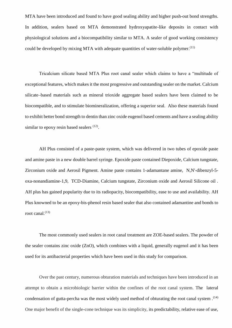
INTRODUCTION
Successful root canal treatment depends on the thorough debridement of the root canal system
by elimination of pathogenic organisms and to seal the root canal space completely to prevent ingress
of bacteria from the oral environment (1). The physical properties necessary for this function include
adaptation and adhesion of the filling material to the root canal wall, because gutta-percha does not
directly bond to the dentine surface (2). The sealer should be capable of producing a bond between core
material and dentine wall.
The ultimate goal of root canal therapy is total obturation of the root canal system. Most of the
obturation techniques utilize gutta-percha in conjunction with a cementing medium. However, sixty
percent of failures in endodontic therapy may be due to inadequate sealing of the root canal system. (3,4,5)
Other studies have revealed that inadequate flow of gutta percha and its inability to adhere to dentinal
walls leads to an insufficient seal. Thus, over the years many different obturation techniques have been
introduced in a hope of increasing the quality of the apical seal. (6,7)
Numerous studies have clearly demonstrated that when sealer is used with any obturation
technique the apical seal is improved significantly (8,9). Currently, none of the commercially available
sealers possess all of the properties required to be considered an ideal root canal sealer. Therefore,
dentistry must continue with research and development of new sealers until the perfect sealer is found.
(10)
Lee et al. introduced mineral trioxide aggregate, a tricalcium silicate cement as a perforation
repair material. Being bioactive it and biocompatible gained popularity for pulp capping, apexification,
pulpotomy, and as root end filling material. Recently, it was developed as a root canal sealer and as an
obturation material. When used as an endodontic sealer, MTA has been shown to have the ability to
regenerate periodontal ligament and cementum in the periapical region. More recently, sealers based on
MTA have been introduced and found to have good sealing ability and higher push-out bond strengths.
In addition, sealers based on MTA demonstrated hydroxyapatite-like deposits in contact with
physiological solutions and a biocompatibility similar to MTA. A sealer of good working consistency
could be developed by mixing MTA with adequate quantities of water-soluble polymer.(11)
Tricalcium silicate based MTA Plus root canal sealer which claims to have a “multitude of
exceptional features, which makes it the most progressive and outstanding sealer on the market. Calcium
silicate–based materials such as mineral trioxide aggregate based sealers have been claimed to be
biocompatible, and to stimulate biomineralization, offering a superior seal. Also these materials found
to exhibit better bond strength to dentin than zinc oxide eugenol based cements and have a sealing ability
similar to epoxy resin based sealers (12).
AH Plus consisted of a paste-paste system, which was delivered in two tubes of epoxide paste
and amine paste in a new double barrel syringe. Epoxide paste contained Diepoxide, Calcium tungstate,
Zirconium oxide and Aerosil Pigment. Amine paste contains 1-adamantane amine, N,N'-dibenzyl-5-
oxa-nonandiamine-1,9, TCD-Diamine, Calcium tungstate, Zirconium oxide and Aerosil Silicone oil .
AH plus has gained popularity due to its radiopacity, biocompatibility, ease to use and availability. AH
Plus knowned to be an epoxy-bis-phenol resin based sealer that also contained adamantine and bonds to
root canal.(13)
The most commonly used sealers in root canal treatment are ZOE-based sealers. The powder of
the sealer contains zinc oxide (ZnO), which combines with a liquid, generally eugenol and it has been
used for its antibacterial properties which have been used in this study for comparison.
Over the past century, numerous obturation materials and techniques have been introduced in an
attempt to obtain a microbiologic barrier within the confines of the root canal system. The lateral
condensation of gutta-percha was the most widely used method of obturating the root canal system .(14)
One major benefit of the single-cone technique was its simplicity, its predictability, relative ease of use,
conservative preparation, and controlled placement of materials. Studies have showned that the single-
cone technique with a suitable resin-based sealer could achieve the same rates of success than other
types of obturation methods. The apical sealing ability of matched-taper single-cone obturation was
comparable with that of lateral condensation.(15,16,17 ) The primary disadvantage of thermoplasticized
guttapercha technique was that heated gutta-percha tends to contract as it cools.
Apical leakage was considered to be the common cause for endodontic failure and were
influenced by many variables such as different filling techniques, chemical and physical properties of
root canal filling materials and presence and absence of smear layer. Dye penetration, fluid filtration,
dye extraction or dissolution method, bacteria and toxin infiltration method, air pressure method,
electrochemical method etc were some of the various methods used for detection of microleakage.
Micro push-out bond strength and dye penetration methods were used in the current study for
detection of microleakage.(18,19)
The null hypothesis tested was that apical sealing ability and push out bond strength of three
sealers are same and no differences in microleakage and pushout bond strength after single cone
technique obturation.

AIMS
To evaluate and compare the Push out bond strength and Apical sealing ability of AH plus, Zinc Oxide
Eugenol and MTA plus root canal sealers after obturation using single cone technique.
OBJECTIVES
1. To evaluate and compare the push out bond strength of gutta-percha after obturation in Coronal,
Middle and Apical region of root canal using three different root canal sealers.
2. To evaluate and compare Apical microleakage after obturation using three different root canal
sealers.
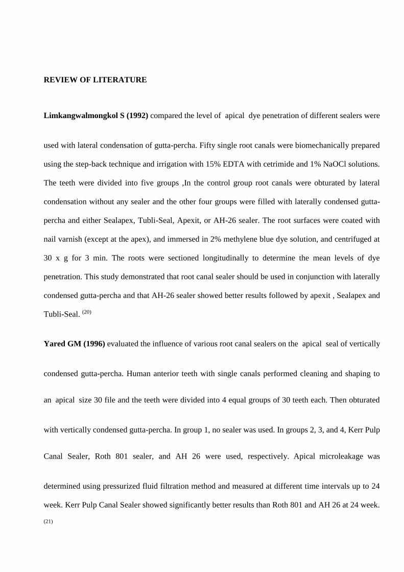
REVIEW OF LITERATURE
Limkangwalmongkol S (1992) compared the level of apical dye penetration of different sealers were
used with lateral condensation of gutta-percha. Fifty single root canals were biomechanically prepared
using the step-back technique and irrigation with 15% EDTA with cetrimide and 1% NaOCl solutions.
The teeth were divided into five groups ,In the control group root canals were obturated by lateral
condensation without any sealer and the other four groups were filled with laterally condensed gutta-
percha and either Sealapex, Tubli-Seal, Apexit, or AH-26 sealer. The root surfaces were coated with
nail varnish (except at the apex), and immersed in 2% methylene blue dye solution, and centrifuged at
30 x g for 3 min. The roots were sectioned longitudinally to determine the mean levels of dye
penetration. This study demonstrated that root canal sealer should be used in conjunction with laterally
condensed gutta-percha and that AH-26 sealer showed better results followed by apexit , Sealapex and
Tubli-Seal. (20)
Yared GM (1996) evaluated the influence of various root canal sealers on the apical seal of vertically
condensed gutta-percha. Human anterior teeth with single canals performed cleaning and shaping to
an apical size 30 file and the teeth were divided into 4 equal groups of 30 teeth each. Then obturated
with vertically condensed gutta-percha. In group 1, no sealer was used. In groups 2, 3, and 4, Kerr Pulp
Canal Sealer, Roth 801 sealer, and AH 26 were used, respectively. Apical microleakage was
determined using pressurized fluid filtration method and measured at different time intervals up to 24
week. Kerr Pulp Canal Sealer showed significantly better results than Roth 801 and AH 26 at 24 week.
(21)
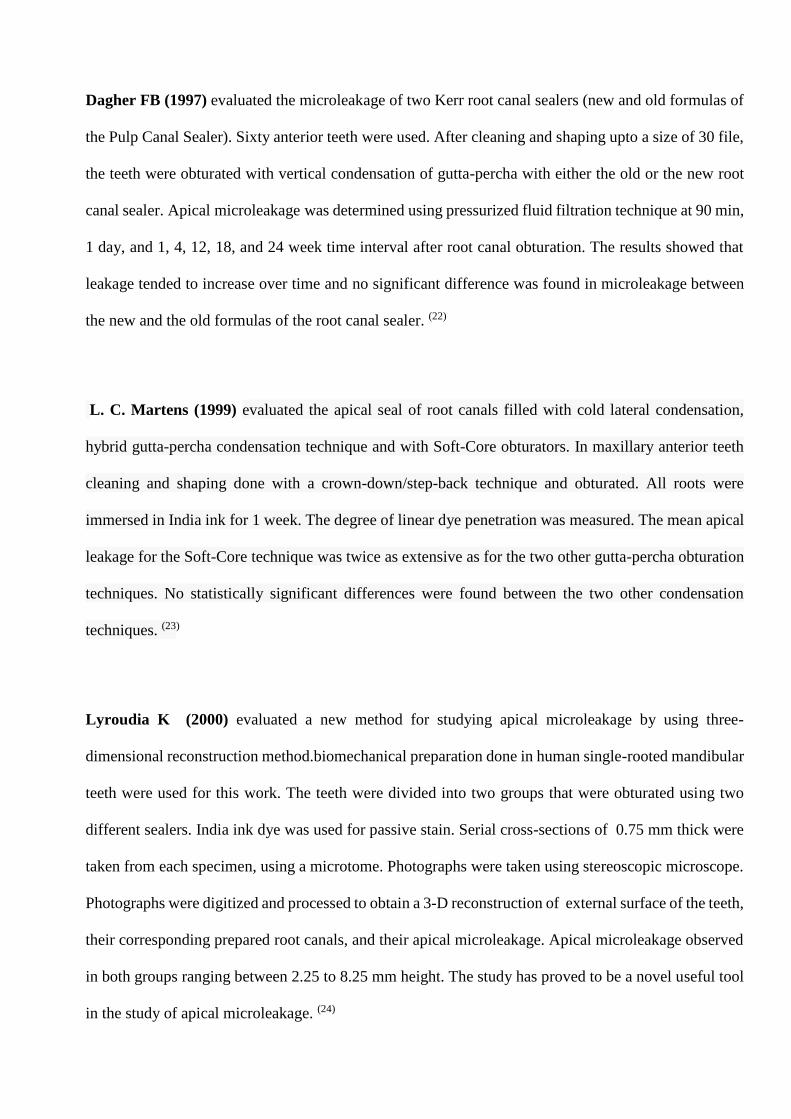
Dagher FB (1997) evaluated the microleakage of two Kerr root canal sealers (new and old formulas of
the Pulp Canal Sealer). Sixty anterior teeth were used. After cleaning and shaping upto a size of 30 file,
the teeth were obturated with vertical condensation of gutta-percha with either the old or the new root
canal sealer. Apical microleakage was determined using pressurized fluid filtration technique at 90 min,
1 day, and 1, 4, 12, 18, and 24 week time interval after root canal obturation. The results showed that
leakage tended to increase over time and no significant difference was found in microleakage between
the new and the old formulas of the root canal sealer. (22)
L. C. Martens (1999) evaluated the apical seal of root canals filled with cold lateral condensation,
hybrid gutta-percha condensation technique and with Soft-Core obturators. In maxillary anterior teeth
cleaning and shaping done with a crown-down/step-back technique and obturated. All roots were
immersed in India ink for 1 week. The degree of linear dye penetration was measured. The mean apical
leakage for the Soft-Core technique was twice as extensive as for the two other gutta-percha obturation
techniques. No statistically significant differences were found between the two other condensation
techniques. (23)
Lyroudia K (2000) evaluated a new method for studying apical microleakage by using three-
dimensional reconstruction method.biomechanical preparation done in human single-rooted mandibular
teeth were used for this work. The teeth were divided into two groups that were obturated using two
different sealers. India ink dye was used for passive stain. Serial cross-sections of 0.75 mm thick were
taken from each specimen, using a microtome. Photographs were taken using stereoscopic microscope.
Photographs were digitized and processed to obtain a 3-D reconstruction of external surface of the teeth,
their corresponding prepared root canals, and their apical microleakage. Apical microleakage observed
in both groups ranging between 2.25 to 8.25 mm height. The study has proved to be a novel useful tool
in the study of apical microleakage. (24)
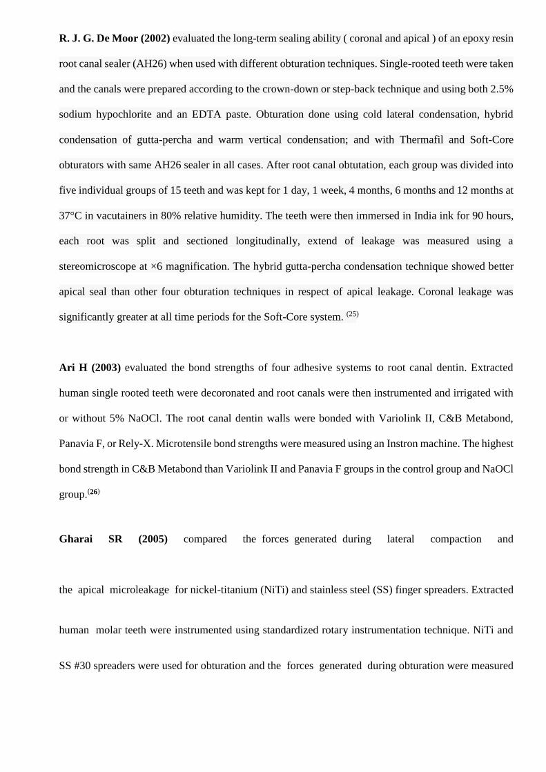
R. J. G. De Moor (2002) evaluated the long-term sealing ability ( coronal and apical ) of an epoxy resin
root canal sealer (AH26) when used with different obturation techniques. Single-rooted teeth were taken
and the canals were prepared according to the crown-down or step-back technique and using both 2.5%
sodium hypochlorite and an EDTA paste. Obturation done using cold lateral condensation, hybrid
condensation of gutta-percha and warm vertical condensation; and with Thermafil and Soft-Core
obturators with same AH26 sealer in all cases. After root canal obtutation, each group was divided into
five individual groups of 15 teeth and was kept for 1 day, 1 week, 4 months, 6 months and 12 months at
37°C in vacutainers in 80% relative humidity. The teeth were then immersed in India ink for 90 hours,
each root was split and sectioned longitudinally, extend of leakage was measured using a
stereomicroscope at ×6 magnification. The hybrid gutta-percha condensation technique showed better
apical seal than other four obturation techniques in respect of apical leakage. Coronal leakage was
significantly greater at all time periods for the Soft-Core system. (25)
Ari H (2003) evaluated the bond strengths of four adhesive systems to root canal dentin. Extracted
human single rooted teeth were decoronated and root canals were then instrumented and irrigated with
or without 5% NaOCl. The root canal dentin walls were bonded with Variolink II, C&B Metabond,
Panavia F, or Rely-X. Microtensile bond strengths were measured using an Instron machine. The highest
bond strength in C&B Metabond than Variolink II and Panavia F groups in the control group and NaOCl
group.(26)
Gharai SR (2005) compared the forces generated during lateral compaction and
the apical microleakage for nickel-titanium (NiTi) and stainless steel (SS) finger spreaders. Extracted
human molar teeth were instrumented using standardized rotary instrumentation technique. NiTi and
SS #30 spreaders were used for obturation and the forces generated during obturation were measured
on a Universal testing machine. Apical microleakage was determined using fluid filtration method.
There was no significant difference in microleakage between two type of spreaders. (27)
Vasiliadis L (2010) evaluated the ex vivo short term and long term microleakage along root canals
filled with Gutta-flow and AH-Plus using the cold lateral compaction technique.
Single-rooted human teeth were sectioned below their cemento–enamel junctions to adjust a length of
the roots to approximately 15 mm. The root canals were instrumented by step-back technique and filled
using cold lateral condensation. The sealer used was either Gutta-flow (Group A) or AH-Plus (Group
B). Microleakage evaluation was done by using a fluid transport model after periods of 1 week and
3 months. Study showed no significant difference between AH-plus and Gutta-flow in terms of sealing
ability. (28)
Shokouhinejad (2012) compared push-out bond strength of a bioceramic endodontic sealer,
EndoSequence BC sealer used with gutta-percha in the presence or absence of phosphate-buffered
saline solution (PBS) within the root canals. Single-rooted human teeth were prepared and obturated
with gutta-percha/EndoSequence BC sealer. The specimens were stored in PBS and underwent Push-
out bond strength evaluation. The presence of PBS in the root canals increased the bond strength values
of EndoSequence BC sealer/gutta-percha after 1 week. Whereas no difference was found between the
bond strength of EndoSequence BC sealer/guttapercha in the presence or absence of PBS in
the root canals after 2 month.(29)
Souza Bier (2012) studied about adhesiveness of six root canal sealers: Endo CPM, Epiphany, White
MTA, Acroseal, Sealapex and Sealer 26 to dentin, in a push-out test design. Teeth were sectioned
horizontally of 2 mm were prepared, rinsed with 5.25% NaOCl followed with 17% EDTA, and filled
with one of the sealers and underwent bond strength evaluation after setting. The bond strength between
endodontic sealers and root dentin was maximal bond strength was when Acroseal, Sealer 26 and
Epiphany; where as Sealapex , Endo CPM, had the lowest bond strength.(30)
Bhardwaj (2013) Compared the sealing efficacy of the AH 26 with gutta percha, AH Plus with gutta
percha and Resilon sealer with its obturator. The apical leakage study done by using the dye penetration
methodology wiyh the help of a stereomicroscope. The highest microleakage in AH 26 , followed by
AH Plus and Resilon sealer with least microleakage (31)
Carvalho (2013) evaluated the influence of calcium hydroxide paste used as intracanal medication to
the bond strength of AH Plus and Epiphany sealers to root dentin. Maxillary first molars
palatal canals were taken in human, using a rotary system. Half of the specimens were placed with
calcium hydroxide for fourteen days and other half without. The calcium hydroxide were removed and
filled with either AH or EP and underwent push out bond strength analysis. Regardless of the intracanal
medicament used, AH plus showed higher bond strength values compared with Epiphany sealers.(32)
Evren (2013) compared the effect of photoactivated disinfection (PAD) on the bond strength between
root canal sealers and human root canal dentin using a push-out test. Extracted and decoronated
human mandibular premolar teeth were used and prepared with the ProTaper rotary system upto the
size of the F3 file. The smear layer of the roots were removed by using 17% EDTA followed by 5.25%
sodium hypochlorite and distillate water. The roots were grouped according to the final irrigation
regimen. In group 1, PAD was applied to the root canals and a light curing for 20 seconds. In Group 2
finally irrigated with a 2% solution of chlorhexidine , whereas group 3 served as a control group (NaOCl
+ EDTA). After obturation with the lateral condensation technique using gutta-percha and AH
Plus sealer sealer. One-millimeter-thickened horizontal sections from the coronal and middle thirds of
each root were taken for the push-out bond strength measurement. Study showed that PAD do not
adversely affect the bond strength between the AH Plus sealer and root canal dentin and that it can be
used for final disinfection of root canals.(33)
Ozcan (2013) evaluated the sealing abilities of two different root canal sealers (epoxy resin-based AH
Plus and polydimethylsiloxane-based GuttaFlow) and of five obturation techniques (lateral
condensation, Thermafil, matched taper single gutta-percha point, laterally condensed-matched taper
gutta-percha point and continuous wave of condensation), through a bacterial leakage model. Single-
rooted human teeth were taken and were obturated with the test material, using the different root filling
techniques and were mounted to a two-chamber bacterial leakage model and Enterococcus faecalis were
added to the upper chambers. The lower chambers of all of the specimens were checked every day during
the test period (100 days). The day of turbidity were noted for each sample. The continuous wave of
condensation technique was found to be better than the other techniques. Both AH Plus and
GuttaFlow sealers showed a similar levels of sealing ability. (34)
Shokouhinejad (2013) compared the bond strength of a bioceramic sealer (EndoSequence BC Sealer)
and AH Plus based on the smear layer presence or absence. Extracted single-rooted human teeth were
instrumented each underwent irrigation by 5.25% NaOCl and smear layer was not removed ; or
the root canals were finally irrigated with 17% EDTA and 5.25% NaOCl to remove the smear layer.
Then root canals were obturated using gutta-percha/AH Plus; or obturation was performed using gutta-
percha/EndoSequence BC Sealer and underwent Push-out bond strength evaluation. In conclusion, the
bond strength of the new..bond strength was simar for bioceramic sealer and AH Plus with or without
the smear layer. (35)
Topçuoglu (2014) evaluated the effects of various gutta-percha solvents on the push-out bond strength
between several root canal sealers and root dentine. Single-rooted human teeth were prepared using
the ProTaper System (Dentsply Maillefer, Ballaigues, Switzerland) up to file size of F4. Different
solvent type (Eucalyptol, chloroform and orange oil), at time (2 and 5 min), sealer type ( MTA Fillapex,
AH Plus and Sealapex) and at different root areas (coronal, middle and apical). After obturation, three
1-mm-thick slices were taken and underwent push-out bond strength test. The use of chloroform solvent
for 5 min in the root canal caused a decrease of bond strength of all sealers but Eucalyptol and orange
oil were no adversely affected the bond strength of the sealers. The push-out bond strength was highest
for AH Plus group and lowest for MTA Fillapex group and decreses from coronal to apical. (36)
ULUSOY (2014) compared the effects of EDTA and maleic acid on the sealing ability of
different root canal sealers. Root canals were instrumented and irrigated with EDTA or MA. They
were divided into groups and obturated as follows: Group 1: MA + Hybrid Root SEAL/gutta-percha.
Group 2: EDTA + Hybrid Root SEAL/gutta-percha. Group 3: MA + iRoot SP/gutta-percha. Group 4:
EDTA + iRoot SP/gutta-percha. Group 5: MA + EndoREZ/EndoREZ points. Group 6: EDTA +
EndoREZ/EndoREZ points. Group 7: MA + AH Plus/gutta-percha. Group 8: EDTA + AH Plus/gutta-
percha. The microleakage was measured after 2 min and 8 min using the fluid filtration method. The
microleakage were minimum for the teeth obturated with EndoREZ selaers and AH Plus whereas
maximum leakage were in Hybrid Root SEAL. Use of EDTA resulted in lower microleakage values
compared with those using MA. (37)
Al-Dwairi (2015) compared the effect of resin-based endodontic sealers and eugenol-based on the push-
out bond strengths of prefabricated fiber posts luting with different resin cements. Luting of
prefabricated fiber posts were done into extracted single rooted teeth with either of three resin cements
(ParaCore, Variolink II, or Rely X Unicem). Each group was again subdivided into three groups with
10 teeth each. Obturation done with gutta percha and each of endodontic sealers Endofil , TubliSeal and
AH26 as three groups respectively. After push-out tests, highest mean bond strength value was recorded
for the AH26 sealer group luted with Rely X Unicem resin cement , while the lowest mean bond strength
value was observed with posts luted with Variolink II resin cement in the canal with endofil sealer and
gutta-percha obturation done. (38)
Berkan Celikten (2015) used micro-CT to compare three different obturation techniques with respect
to the voids occurrence in canal filled with bioceramic sealer. Extracted mandibular premolars were
prepared with ProTaper Universal system. Obturation done using three techniques single-cone, lateral
compaction, or Thermafil filling technique with gutta-percha and bioceramic root canal sealer.
Assessment of voids in apical thirds was done with images taken from micro-CT. Highest Void volumes
were for the single-cone technique and lowest for Thermafil, in all regions. (39)
Epita S (2015) evaluated tensile and shear bond strengths of one epoxy (AH plus) and two methacrylate
resin-based sealers (EZ and RS) of varying thin and thick layers bonded to root dentine. Bond strength
tests were conducted using an alignment device. Mode of failures in representative surfaces were
evaluated. The study showed thick layer of sealer produced higher bond strength, except for EZ.
Differences between thin and thick layers were found only in tensile bond strengths of RS and AH.
Mixed type of failure was found with all sealers. Bond strengths of thin layers of resin-based sealers to
root dentine tended to be lower than with thick layers. (40)
Elbatouty ( 2015) evaluated the push-out bond strength of the
bioceramic root canal sealer (EndoSequence BC) in comparison to zinc oxide-eugenol-based (Kerr
EWT) sealer and resin-based (AH Plus) sealer. Extracted roots were divided into three groups with
sealers EndoSequence BC; AH Plus; and Kerr EWT sealers. Two millimetre thickness horizontal
sections at the coronal, middle, and apical thirds the root were sliced for the push-out bond strength
measurement using universal testing machine after 7, 14 and 30 days. Scanning electron microscope
used for mode of failure analysis. The study showed the highest mean push-out bond strength values
after 1 and 4 weeks for EndoSequence BC, followed by AH Plus and Kerr EWT. After 2 weeks, the AH
Plus had the highest mean push-out bond strength followed by EndoSequence BC. (41)
Ehsen Abdelmoumen (2015) compared the sealing ability of Zinc oxide eugenol sealer and resin-based
sealer (Endo REZ) with two different root canal obturation techniques (cold lateral condensation and
single cone technique). Human incisors, canines and premolars were selected and shaping done using
protaper rotary files. Nail polish were applied to the tooth surface, after obturation except for the apical
2 mm. Evaluation of the sealing ability was performed by measuring the dye penetration along the canal
walls for the different techniques. The apical sealing ability of EndoREZ is less effective as that of Zinc-
oxide when used with other two different clinical obturation methods. (42)
Fernanda Leal (2015) evaluated the effect of different final irrigation techniques on push-out bond
strength of epoxy resin root canal sealer to dentin. Single-rooted anterior teeth root canals were prepared
using a rotary system, the final diameter was kept as #5 Gates-Glidden. During chemomechanical
preparation, 2% CHX gel or 5.25% NaOCl was used. For smear layer removal, 17% EDTA or QMix 2
in 1 applied for 3 min. 1 mL of NaOCl, CHX solution or distilled water was used as final irrigant,. On
conclusion of preparation, canals were filled with gutta-percha/AH Plus sealer. After obturation bond
strength was measured by the push-out test. The group NaOCl/EDTA/NaOCl showed higher bond
strength values than other groups. The final irrigant affect the push-out bond strength of AH Plus to
dentin.(43)
Neelakantan (2015) evaluated the influence of various irrigation between root canal sealers and dentin
and was analyzed by using Fourier transform infrared spectroscopy (FTIRS) . Single-rooted teeth were
instrumented with 3% NaOCl as the irrigant divided into 4 groups based on irrigation protocol with 3%
NaOCl, 17% EDTA as first group, ; 17% EDTA, 3% NaOCl, as second group ; 3% NaOCl, QMix, as
group three and 3% NaOCl, water as group four. Again each group was divided into 3 subgroups on
the basis of the root canal sealer: epoxy resin (AH Plus); silicone (RoekoSeal) and calcium hydroxide
(Sealapex). The dislocation resistance was calculated by using push-out bond strength test. AH Plus
showed the highest bond strength . Chemical bonding between AH Plus and dentinal collagen were
revealed by FTIRS. The epoxy resin sealer (AH Plus) chemically bonds to dentinal collagen and
irrigation protocol plays a major role in it.(44)
Pradeep (2015) used traditional gutta-percha, epoxy resin and DBA to make the root canal leak proof.
Extracted maxillary incisors teeth were decoronated at cemento-enamel junction. Instrumentation done
to size 40 and were randomly assigned to four groups according to the dentin adhesive system and
obturation done with AH Plus sealer and gutta-percha points. The formation of hybrid layer was
observed using scanning electron microscope and the dye penetration extension was measured using a
stereomicroscope. Based on the hybrid layer divided into Control group without hybrid layer formation,
Uniform thin hybrid layer, with short multiple resin tags and lateral branchings , Uniform hybrid layer
with short and thick resin tag formations and Hybrid layer with numerous long discontinuous resin tags
respectively as four groups. Group without adhesive showed the highest apical microleakage implying
the need for dental adhesive.(45)
Levent Demiriz (2016) evaluated the adaptation ability of MTA Fillapex root canal sealer to dentinal
wall using stereo electron microscope . Single-rooted, human maxillary incisor teeth root canals were
prepared with a rotary nickel–titanium instrument to a size F3 file. Divided into two equal groups and
one group was filled with AH Plus, and the other group was filled with MTA Fillapex using Gutta-
percha single cone as a core material. The roots were performed for SEM evaluation, and scanning
electron images were taken at ×50, ×100, ×500, and ×1000 magnifications. The gaps between the root
canal sealer and canal walls were measured in coronal, middle, and apical thirds. The highest value
among the detected gap formations was recorded for each section. MTA Fillapex has similar dentinal
wall adaptation ability as AH Plus does.(46).
Patni PM (2016) investigated the effect of the apical seal obtained by different sealers used in with cold
lateral condensation technique of obturation done gutta-percha using stereomicroscope. One hundred
single-rooted extracted human permanent teeth with a single root canal were used in this in-vitro study.
The sealers tested were conventional Zinc oxide eugenol, Roekoseal Automix (RSA) and Apexit, AH-
Plus. The polydimethylsiloxane root canal sealer RSA provided a better apical seal followed by AH plus
and Apexit, but conventional zinc oxide eugenol showed lowest sealing ability. The shrinkage related
to setting and potential dissolution might be the reason for proper seal of the root canal leading to
treatment failure. (47)
Ahuja L (2016) evaluated and compared the apical microleakage of a resin based sealer; Adseal with
Mineral Trioxide Aggregate (MTA) based sealers; MTA Fillapex and Pro root MTA. Single rooted
teeth were decoronated at cemento-enamel junction and biomechanical preparation done using
endodontic rotary system. The teeth were randomly divided into five groups with n=15; Group I - Gutta-
percha and Adseal ; Group II - Gutta-percha and MTA Fillapex; Group III- Gutta-percha and Proroot
MTA; Group IV- Gutta-percha without sealer (positive control group) and Group V- Root canal
remained empty (negative control). Root surfaces were covered with nail varnish except the apical 2mm
and then immersed in 2% methylene blue dye for 72 hours. Roots were longitudinally splitted and
linear apical dye penetration was measured under Stereomicroscope at 40X magnification. MTA
Fillapex group showed maxmium apical microleakage followed by Pro root MTA and Adseal sealer.(48)
Widcha Asawaworarit (2016) evaluated the apical sealing ability of tricalcium silicate-based MTA
Fillapex and resin-based AH Plus sealers at time periods 24 hrs, 7 days and 4 weeks. Decoronated
human upper anterior teeth were instrumented using a set of ProTaper rotary instruments. Obturated
with MTA Fillapex and gutta-percha and AH Plus and gutta-percha using a warm vertical compaction
technique. The apical sealing ability was measured using the fluid-filtration method with 200 mm
mercury (26.67 KPa) above atmospheric pressure at 24 h, 7 days and 4 weeks. MTA Fillapex had

significantly more micro leakage than AH Plus 1 week after filling. After 4 weeks, AH Plus showed less
result than MTA Fillapex . (49)
MATERIALS AND METHODS
Armamentarium used in the study:
Extracted teeth
Endodontic files size 8,10,15 size ( Denstply Maillefer )
X smart plus ( Dentsply maillefer )
Protaper Rotary files ( Dentsply )
Sodium hypochlorite ( VIP Vensons, India )
Normal saline ( Nirlife NIRMA LIMITED )
EDTA ( Prevest Denpro )
Guttapercha cones ( Dentsply )
Diamond disc and mandrel
Chisel
Scanning electron microscope ( Zeiss sigma V )
Olympus Zoom Stereomicroscope, Sz 40-45, Japan
Methylene blue dye
Kalpak universal testing machine
Modelling wax
Disposable syringe ( Dispovan )
Zinc oxide Eugenol sealer (Deepak Enterprise)
AH plus sealer ( Dentsply )
MTA plus sealer (Prevest Denpro Limited)
SOURCE OF DATA
Extracted mandibular premolar teeth has been collected from department of Oral Maxillofacial surgery
in KSR Institute of Dental science and Research, study has been conducted in the department of
Conservative Dentistry and Endodontics in KSR Institute of dental science and research. Push out bond
strength evaluation was done in LMPRD Laboratory, Erode
MATERIALS AND METHODS
This experimental study included 120 extracted single rooted teeth (Mandibular premolars) collected
from department of Oral Maxillofacial surgery in KSR Institute of Dental science and Research.
Extracted due to Orthodontic and Periodontic reasons
INCLUSION CRITERIA:
Mandibular first premolars.
Complete root formation without signs of internal or external resorption, no fracture or crack in
the root.
No coronal restoration or decay below the cemento enamel junction (CEJ)
Straight cone-shaped root without curvature in the apical third.
No calcification in the root canal.
EXCLUSION CRITERIA:
K files #10 and #15 not passing beyond 11 mm from CEJ into the root canal.
PREPARATION OF THE TEETH
The collected teeth were cleaned immediately after extraction by removing all attached hard and
soft tissues and immersing in 1000 ml of 5.25% sodium hypochlorite for 24 hours. Then the teeth were
stored in the container with lid containing 0.9% sterile saline at room temperature until further
processing. The crowns of the teeth were decapitated at the cemento-enamel junction with a diamond
disk under water coolant. All prepared teeth were again held in 0.9% sterile saline at room temperature
until the test time.
For root canal preparation, Xmart plus (DENTSPLY Maillefer) and Protaper (DENTSPLY
Maillefer) with single length technique were used. The preparation of root canal were done upto F3
Protaper file. The system settings for all teeth were as follows: speed = 250 rpm, torque = 3Ncm. 2.5%
NaOCl was used as an irrigant between each file. The final rinse was performed using 3 mL of 2.5%
NaOCl and 17% EDTA, followed by 3 mL of normal saline. The root canals were dried with #30 paper
points. To confirm drying of the canal, five consecutive #30 paper points were placed in the canal for
five seconds and had to remain dry. The teeth were randomly divided into three groups, each containing
20 specimens. Obturation was done using single cone of 30 6% gutta-percha using Zinc oxide Eugenol,
AH plus and MTA plus sealers.
GROUPING
Group I - Gutta-percha and Zinc oxide Eugenol sealer
Group II - Gutta-percha and AH Plus sealer
Group III- Gutta-percha and MTA Plus sealer
PUSH OUT BOND STRENGTH TEST
Out of obturated teeth, 60 teeth were taken for push out bond strength evaluation. 20 teeth from
each group were horizontal sectioned at coronal, middle and apical thirds of root using a diamond disc
with the help of a coolant. The sections were made uniformly of 2 mm thickness. At this time the apical
side of the disk was marked to ensure the plunger of the push-out test pushed from the apical to coronal
direction, to avoid any interference owing to root canal taper. Plunger tip end had a thickness of 0.5mm
diameter.
A vertical load was applied to the obturation material in an apical to coronal direction at the rate
of 0.5mm/min. A load versus time curve was plotted in real time by a software program connected to
the universal testing machine. Failure of bond was determined when a sharp decline was observed on
the graph. If any evidence of the plunger scrapping dentin was observed, the specimen was discarded
and the data collected was not included in further analysis. The bond strength, expressed in MPa, was
calculated by dividing the maximum load in Newtons by the area of the bonded interface. The area of
the bonded interface was calculated using the formula, area = 2𝜋r X h, where 𝜋 is the constant 3.14 and
r and h were the measured radius and height in millimeters of the filling material.
After the pushout bond strength test was performed, each of the root slices were examined
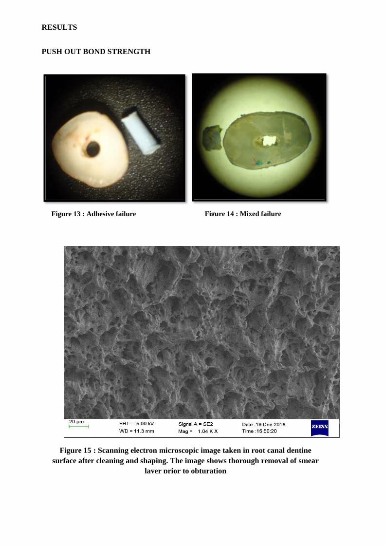
under a stereo microscope at 40 X magnification to determine the failure mode.
Modes of bond failure were considered as follows:
(1) Adhesive Failure ( Sealer / Dentine interface or Mastercone / Sealer interface)
(2) Cohesive Failure ( Within the sealer)
(3) Mixed failure (Both adhesive and cohesive failure)
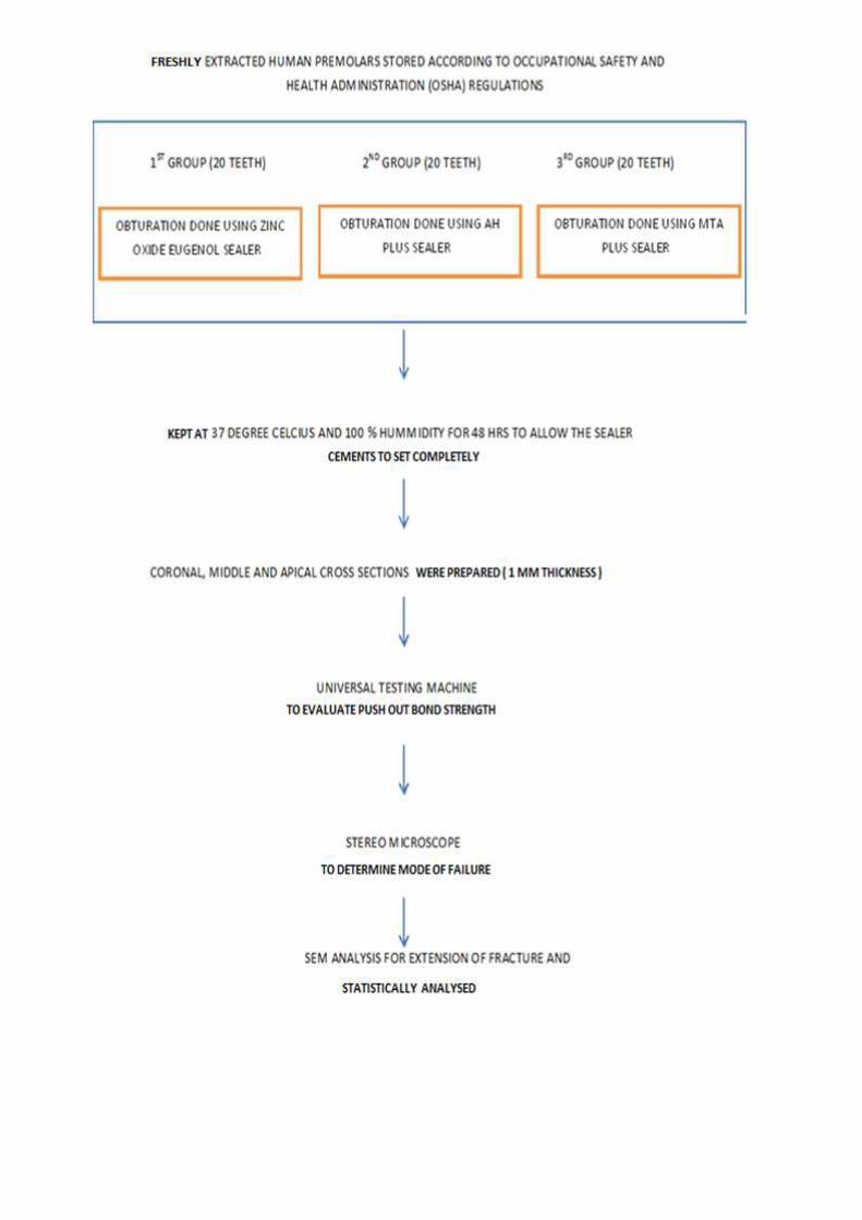
In all groups, the debonded surfaces of specimens were dried, mounted on aluminum stubs,sputter-
coated with gold/palladium analyzed with a scanning electron microscope (Zeiss sigma V) to
determine the extent of failure mode.
APICAL SEAL EVALUATION
The obturated teeth were isolated with modelling wax, leaving the apical foramen. The leakage
was shown using dye penetration test with methylene blue. The external surfaces of all roots were made
attached or bonded to a rod in such a way that apical 1 mm of each teeth touching the dye solution in a
bowl. The roots were held vertically with the help of meshwork in a plastic box containing 2% methylene
blue, so that only the apical 1 mm of each root were immersed in the dye. The roots were left in the dye
for two days at 37 degree celsius, after which they were removed, washed and the wax covering was
removed with scalpel. Teeth roots were sectioned longitudinally along the canal centers in buccolingual
direction.
The binocular stereomicroscope (Olympus Zoom Stereomicroscope, Sz 40-45, Japan) were used
to measure extent of dye- penetration up to the most coronal mark in micrometers (at magnification of
X 40). The measurements of the dye penetration were done with the help of CMEIAS software. Micro
scale tool in the CMEIAS software were used for measuring the extend of dye penetration. The data
were collected and subjected to statistical analysis.
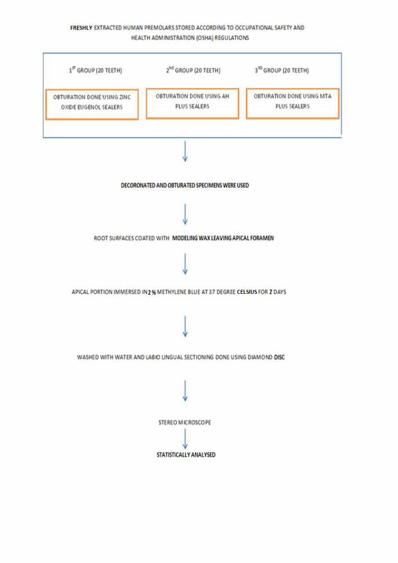
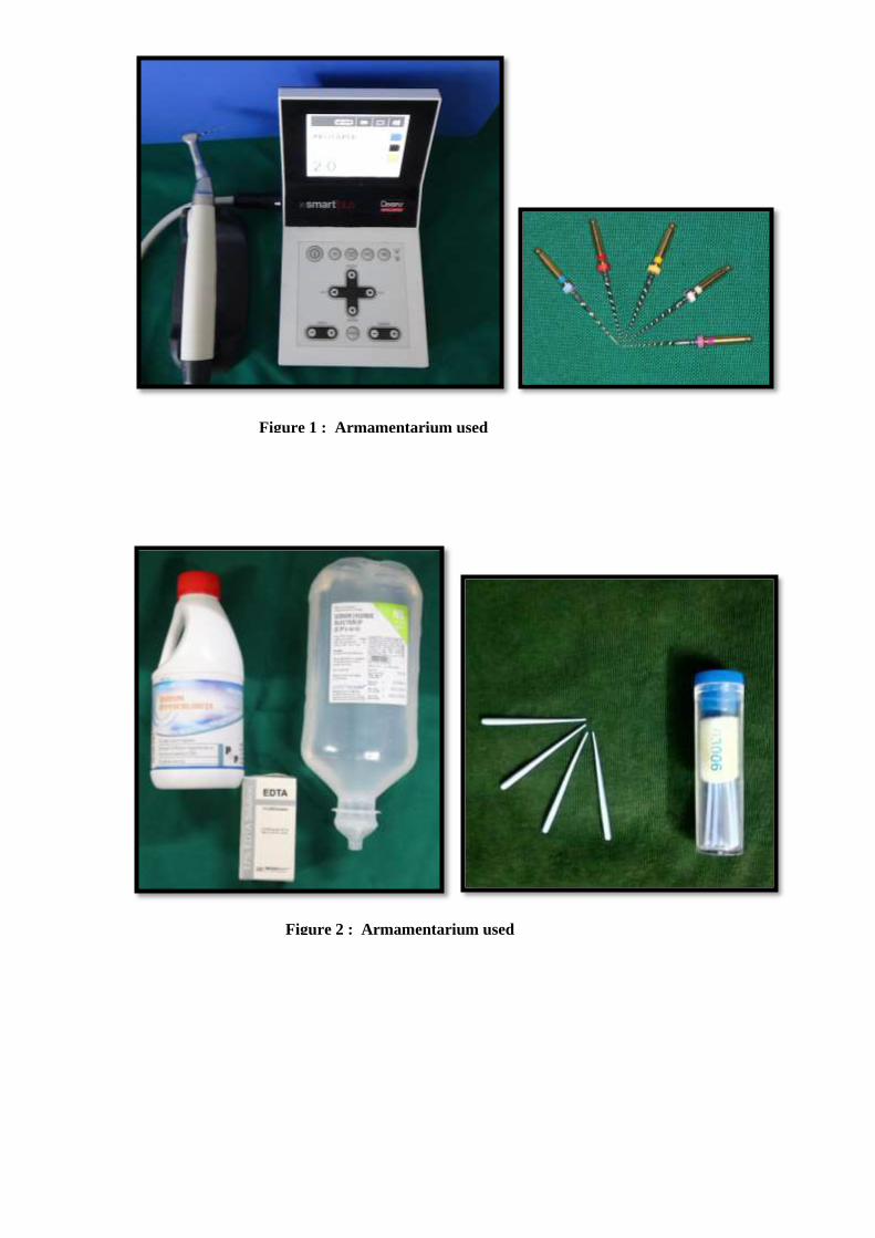
Figure 1 : Armamentarium used
Figure 2 : Armamentarium used
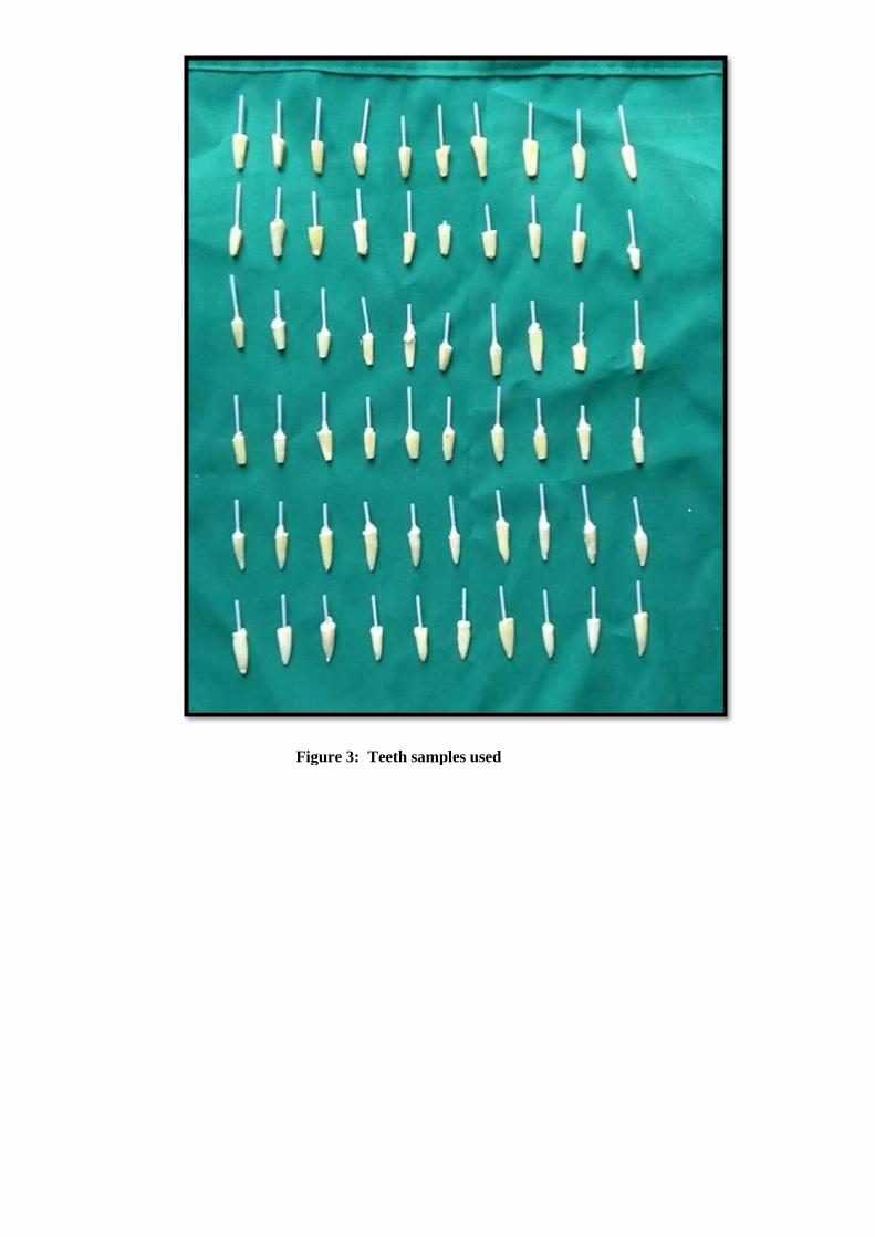
Figure 3: Teeth samples used
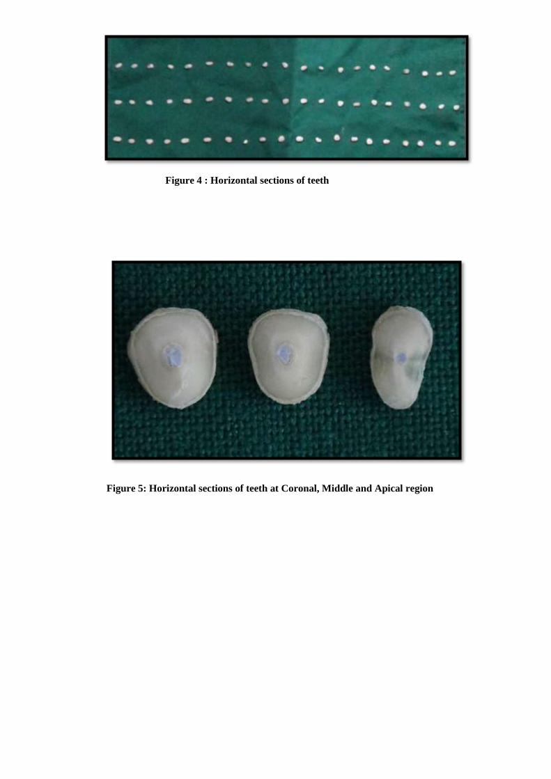
Figure 4 : Horizontal sections of teeth
Figure 5: Horizontal sections of teeth at Coronal, Middle and Apical region
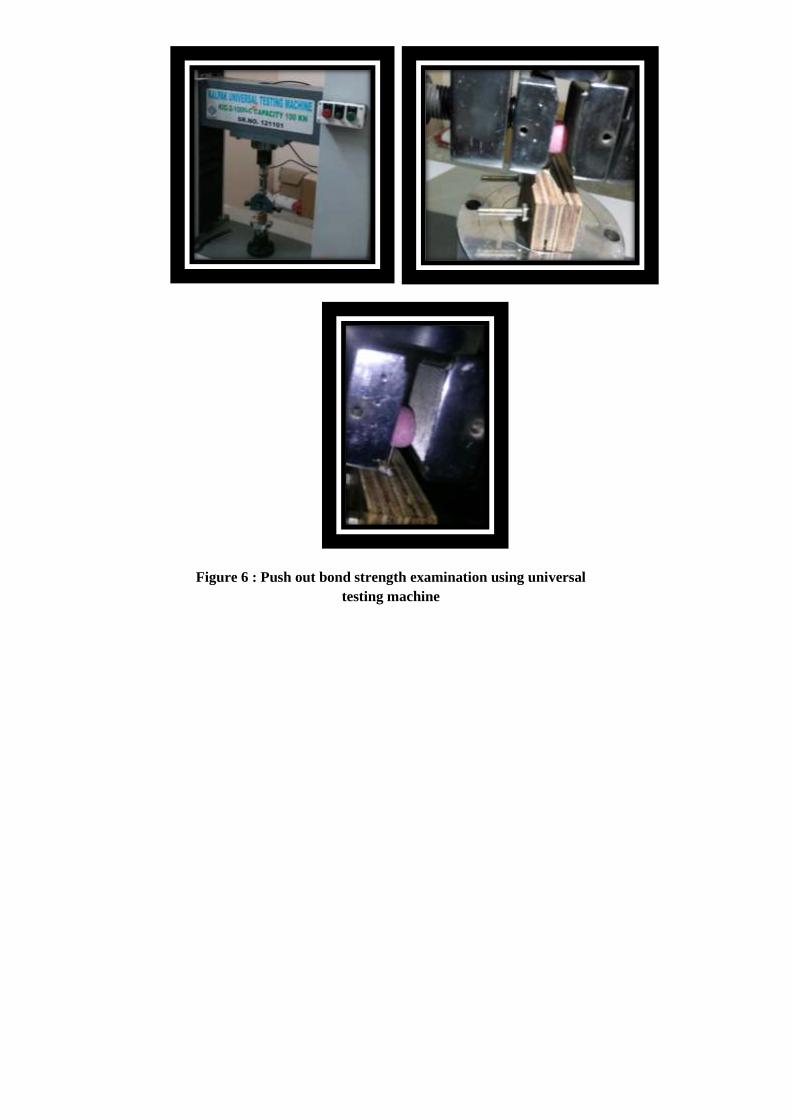
Figure 6 : Push out bond strength examination using universal
testing machine
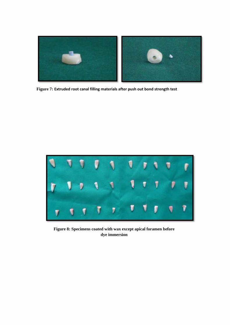
Figure 8: Specimens coated with wax except apical foramen before
dye immersion
Figure 7: Extruded root canal filling materials after push out bond strength test
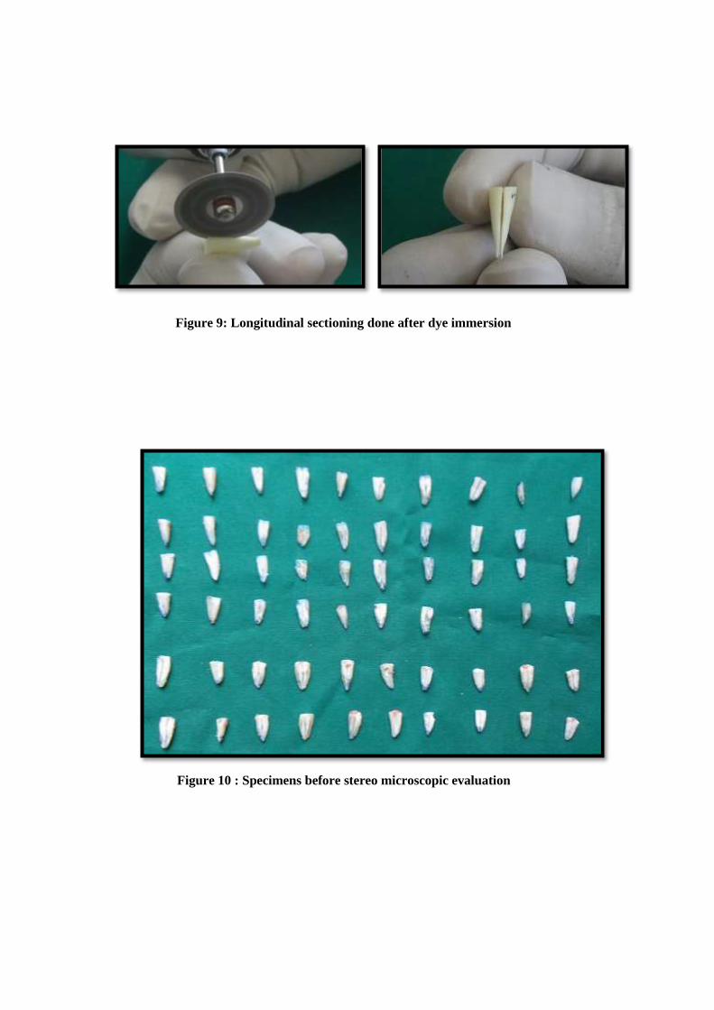
Figure 9: Longitudinal sectioning done after dye immersion
Figure 10 : Specimens before stereo microscopic evaluation
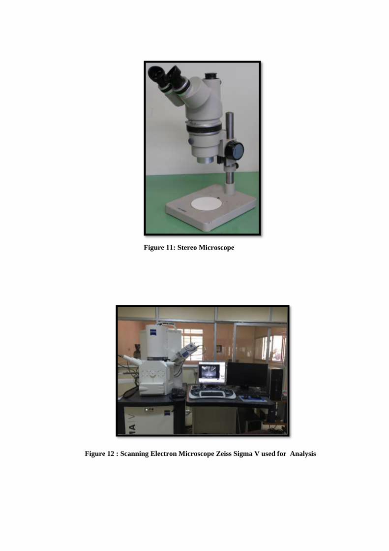
Figure 11: Stereo Microscope
Figure 12 : Scanning Electron Microscope Zeiss Sigma V used for Analysis
RESULTS
PUSH OUT BOND STRENGTH
Armamentarium used
Figure 13 : Adhesive failure Figure 14 : Mixed failure
Figure 15 : Scanning electron microscopic image taken in root canal dentine
surface after cleaning and shaping. The image shows thorough removal of smear
layer prior to obturation
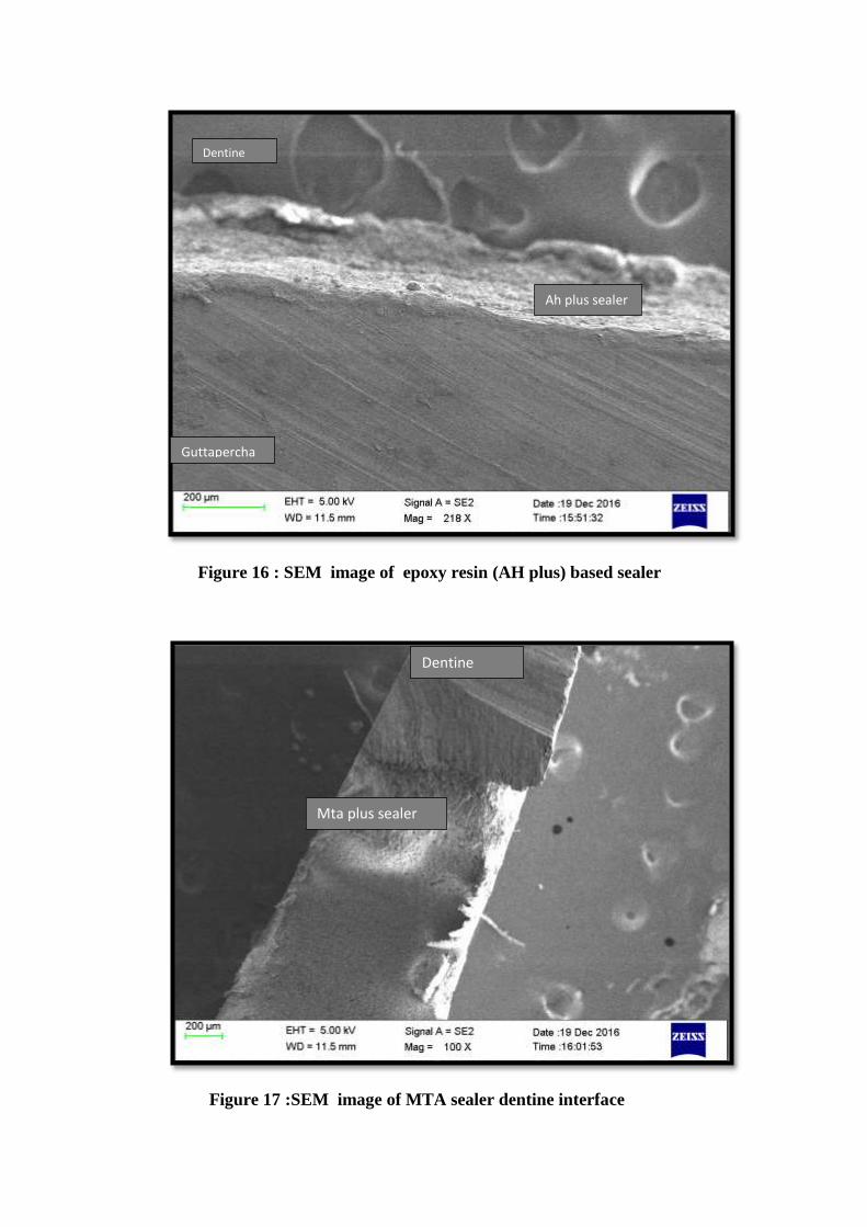
Guttapercha
Ah plus sealer
Dentine
Figure 16 : SEM image of epoxy resin (AH plus) based sealer
dentine interface
Figure 17 :SEM image of MTA sealer dentine interface
Mta plus sealer
Dentine
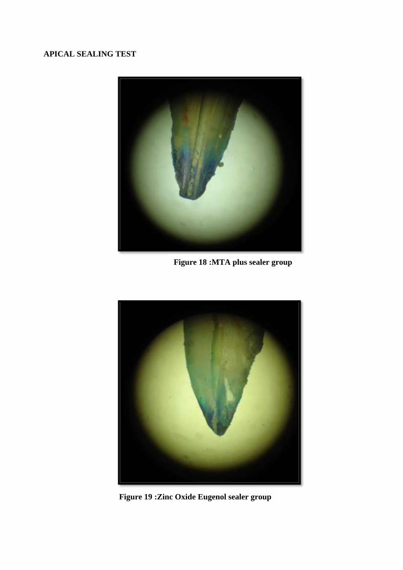
APICAL SEALING TEST
Figure 18 :MTA plus sealer group
Figure 19 :Zinc Oxide Eugenol sealer group
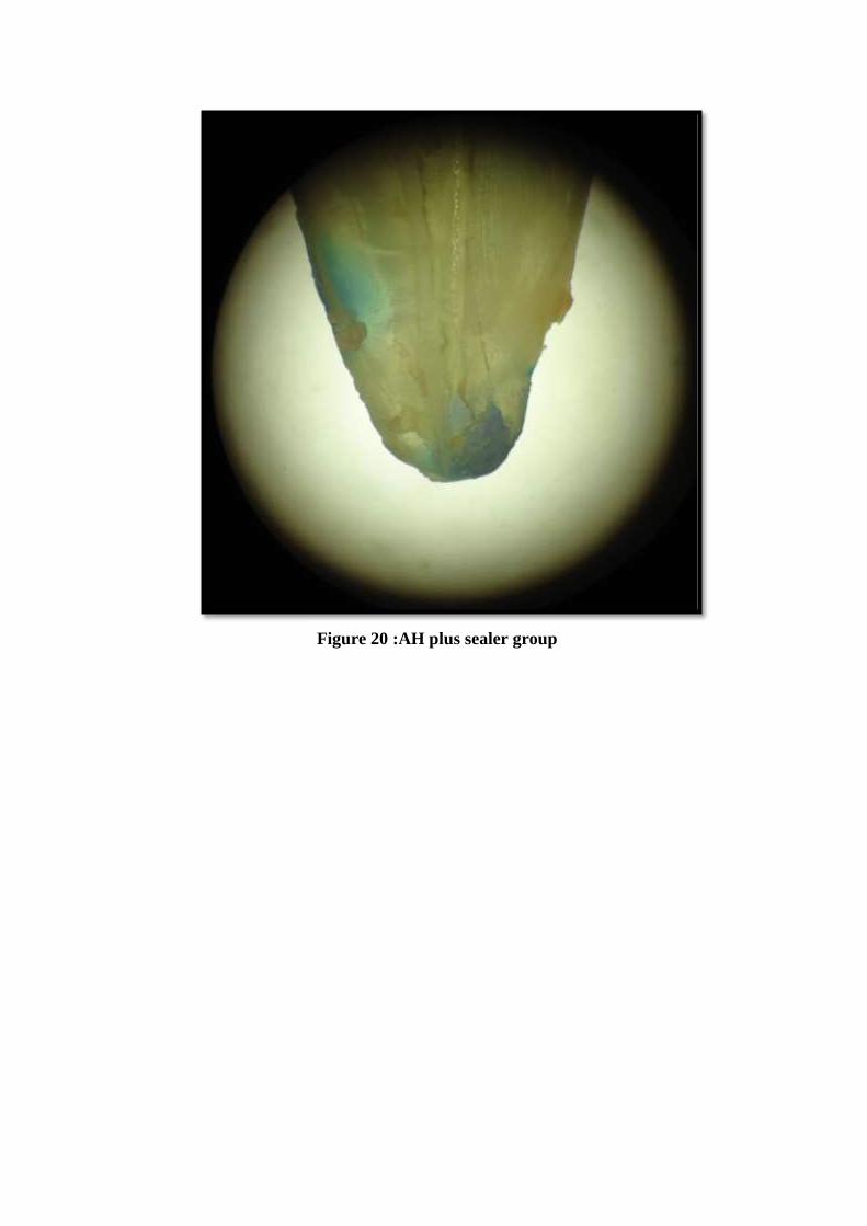
Figure 20 :AH plus sealer group
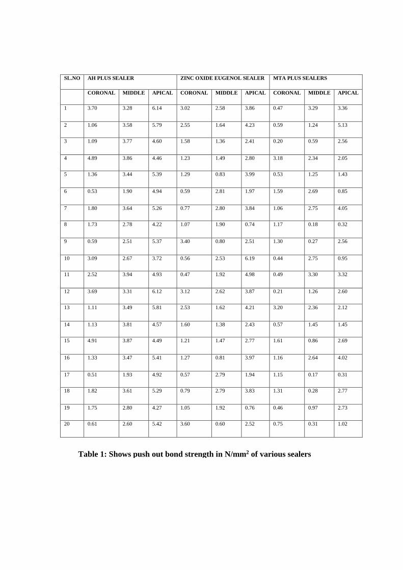
SL.NO AH PLUS SEALER ZINC OXIDE EUGENOL SEALER MTA PLUS SEALERS
CORONAL MIDDLE APICAL CORONAL MIDDLE APICAL CORONAL MIDDLE APICAL
1 3.70 3.28 6.14 3.02 2.58 3.86 0.47 3.29 3.36
2 1.06 3.58 5.79 2.55 1.64 4.23 0.59 1.24 5.13
3 1.09 3.77 4.60 1.58 1.36 2.41 0.20 0.59 2.56
4 4.89 3.86 4.46 1.23 1.49 2.80 3.18 2.34 2.05
5 1.36 3.44 5.39 1.29 0.83 3.99 0.53 1.25 1.43
6 0.53 1.90 4.94 0.59 2.81 1.97 1.59 2.69 0.85
7 1.80 3.64 5.26 0.77 2.80 3.84 1.06 2.75 4.05
8 1.73 2.78 4.22 1.07 1.90 0.74 1.17 0.18 0.32
9 0.59 2.51 5.37 3.40 0.80 2.51 1.30 0.27 2.56
10 3.09 2.67 3.72 0.56 2.53 6.19 0.44 2.75 0.95
11 2.52 3.94 4.93 0.47 1.92 4.98 0.49 3.30 3.32
12 3.69 3.31 6.12 3.12 2.62 3.87 0.21 1.26 2.60
13 1.11 3.49 5.81 2.53 1.62 4.21 3.20 2.36 2.12
14 1.13 3.81 4.57 1.60 1.38 2.43 0.57 1.45 1.45
15 4.91 3.87 4.49 1.21 1.47 2.77 1.61 0.86 2.69
16 1.33 3.47 5.41 1.27 0.81 3.97 1.16 2.64 4.02
17 0.51 1.93 4.92 0.57 2.79 1.94 1.15 0.17 0.31
18 1.82 3.61 5.29 0.79 2.79 3.83 1.31 0.28 2.77
19 1.75 2.80 4.27 1.05 1.92 0.76 0.46 0.97 2.73
20 0.61 2.60 5.42 3.60 0.60 2.52 0.75 0.31 1.02
Table 1: Shows push out bond strength in N/mm2 of various sealers
SL.NO
AH PLUS SEALER
CORONAL MIDDLE APICAL
1 3.70 3.28 6.14
2 1.06 3.58 5.79
3 1.09 3.77 4.60
4 4.89 3.86 4.46
5 1.36 3.44 5.39
6 0.53 1.90 4.94
7 1.80 3.64 5.26
8 1.73 2.78 4.22
9 0.59 2.51 5.37
10 3.09 2.67 3.72
11 2.52 3.94 4.93
12 3.69 3.31 6.12
13 1.11 3.49 5.81
14 1.13 3.81 4.57
15 4.91 3.87 4.49
16 1.33 3.47 5.41
17 0.51 1.93 4.92
18 1.82 3.61 5.29
19 1.75 2.80 4.27
20 0.61 2.60 5.42
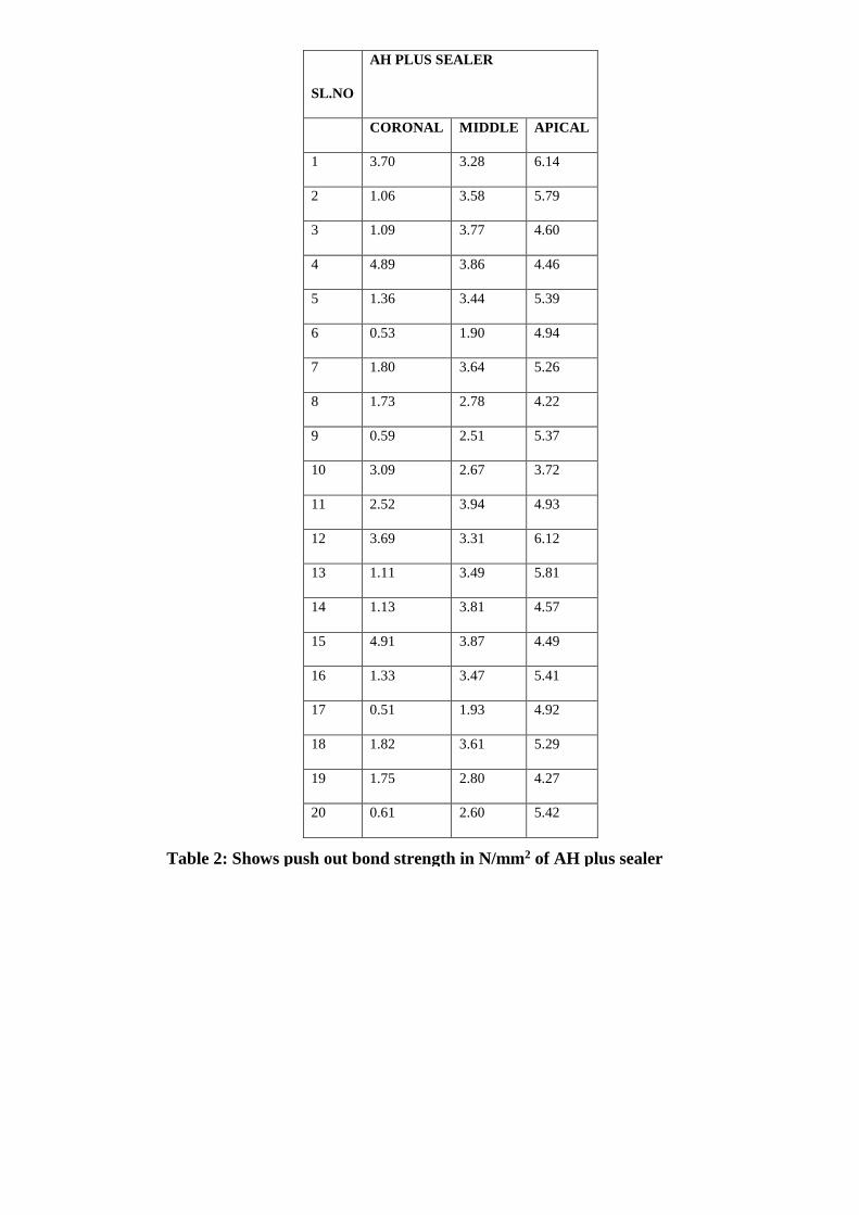
Table 2: Shows push out bond strength in N/mm2 of AH plus sealer
Sl.no ZINC OXIDE EUGENOL
SEALER
CORONAL MIDDLE APICAL
1 3.02 2.58 3.86
2 2.55 1.64 4.23
3 1.58 1.36 2.41
4 1.23 1.49 2.80
5 1.29 0.83 3.99
6 0.59 2.81 1.97
7 0.77 2.80 3.84
8 1.07 1.90 0.74
9 3.40 0.80 2.51
10 0.56 2.53 6.19
11 0.47 1.92 4.98
12 3.12 2.62 3.87
13 2.53 1.62 4.21
14 1.60 1.38 2.43
0
1
2
3
4
5
6
7
1 2 3 4 5 6 7 8 9 10 11 12 13 14 15 16 17 18 19 20
Chart Title
AH PLUS SEALER CORONAL AH PLUS SEALER MIDDLE AH PLUS SEALER APICAL
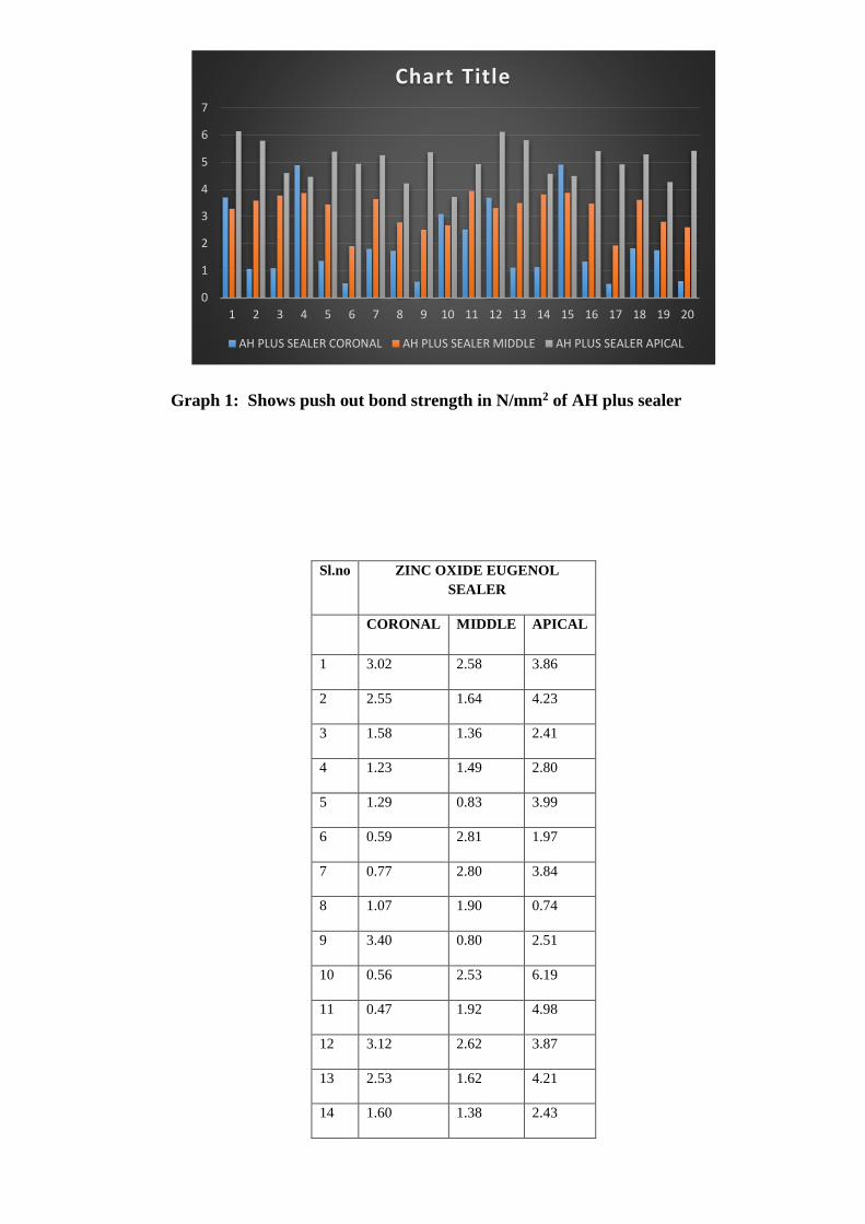
Graph 1: Shows push out bond strength in N/mm2 of AH plus sealer
15 1.21 1.47 2.77
16 1.27 0.81 3.97
17 0.57 2.79 1.94
18 0.79 2.79 3.83
19 1.05 1.92 0.76
20 3.60 0.60 2.52
0
1
2
3
4
5
6
7
1 2 3 4 5 6 7 8 9 10 11 12 13 14 15 16 17 18 19 20
Chart Title
ZINC OXIDE EUGENOL SEALER CORONAL ZINC OXIDE EUGENOL SEALER MIDDLE
ZINC OXIDE EUGENOL SEALER APICAL
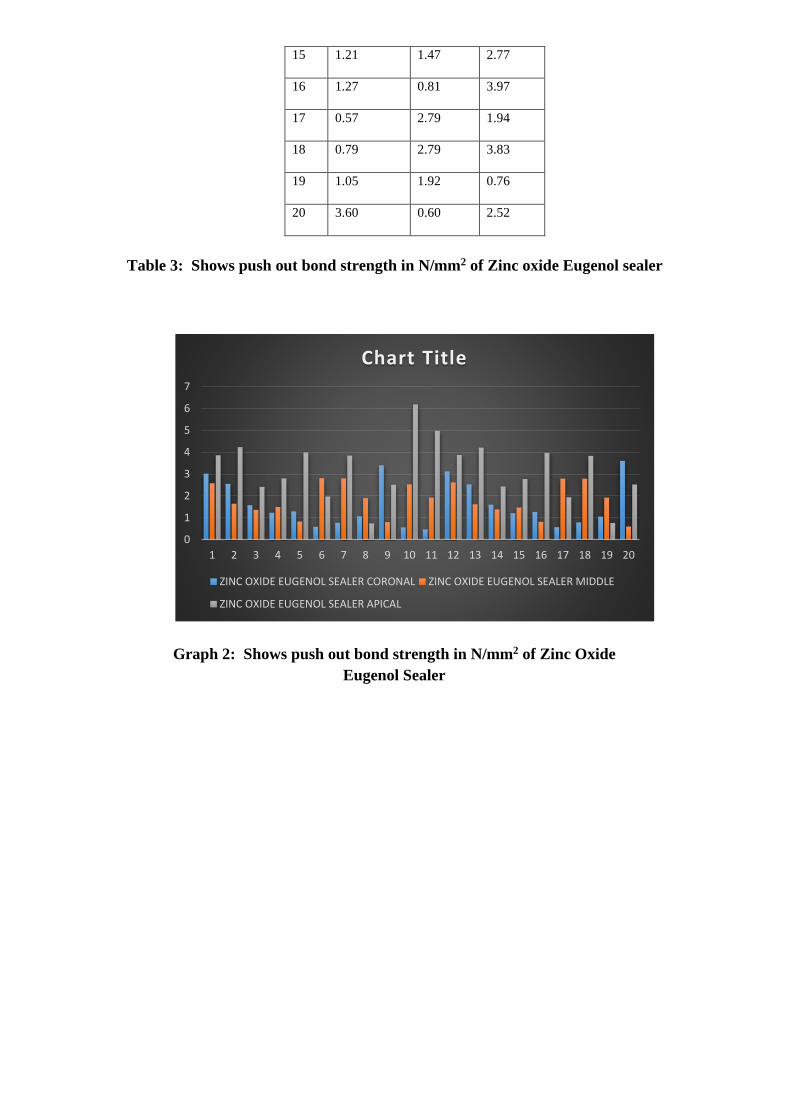
Table 3: Shows push out bond strength in N/mm2 of Zinc oxide Eugenol sealer
Graph 2: Shows push out bond strength in N/mm2 of Zinc Oxide
Eugenol Sealer

Sl.no MTA PLUS SEALERS
CORONAL MIDDLE APICAL
1 0.47 3.29 3.36
2 0.59 1.24 5.13
3 0.20 0.59 2.56
4 3.18 2.34 2.05
5 0.53 1.25 1.43
6 1.59 2.69 0.85
7 1.06 2.75 4.05
8 1.17 0.18 0.32
9 1.30 0.27 2.56
10 0.44 2.75 0.95
11 0.49 3.30 3.32
12 0.21 1.26 2.60
13 3.20 2.36 2.12
14 0.57 1.45 1.45
15 1.61 0.86 2.69
16 1.16 2.64 4.02
17 1.15 0.17 0.31
18 1.31 0.28 2.77
19 0.46 0.97 2.73
20 0.75 0.31 1.02
Table 4: Shows push out bond strength in N/mm2 of MTA plus sealer
0
1
2
3
4
5
6
1 2 3 4 5 6 7 8 9 10 11 12 13 14 15 16 17 18 19 20
Chart Title
MTA PLUS SEALER S CORONAL MTA PLUS SEALER S MIDDLE
MTA PLUS SEALER S APICAL
Sl.no. MICROLEAKAGE VALUES IN
MICROMETER
AH Plus Zinc Oxide
Eugenol
MTA plus
1 76.92 348.75 563.78
2 189.02 405.01 615.82
3 162.36 263.9 339.48
4 114.07 474.4 479.45
5 56.57 712.64 263.35
6 64.76 254.39 205.08
7 149.35 531.87 211.37
8 98.27 483.35 366.59
9 114.54 261 248.29
10 75.18 173.12 168.96
11 174.93 362.49 202.81
12 138.46 342.12 349.05
13 113.14 267.59 576.68
14 116.48 305.76 279.58
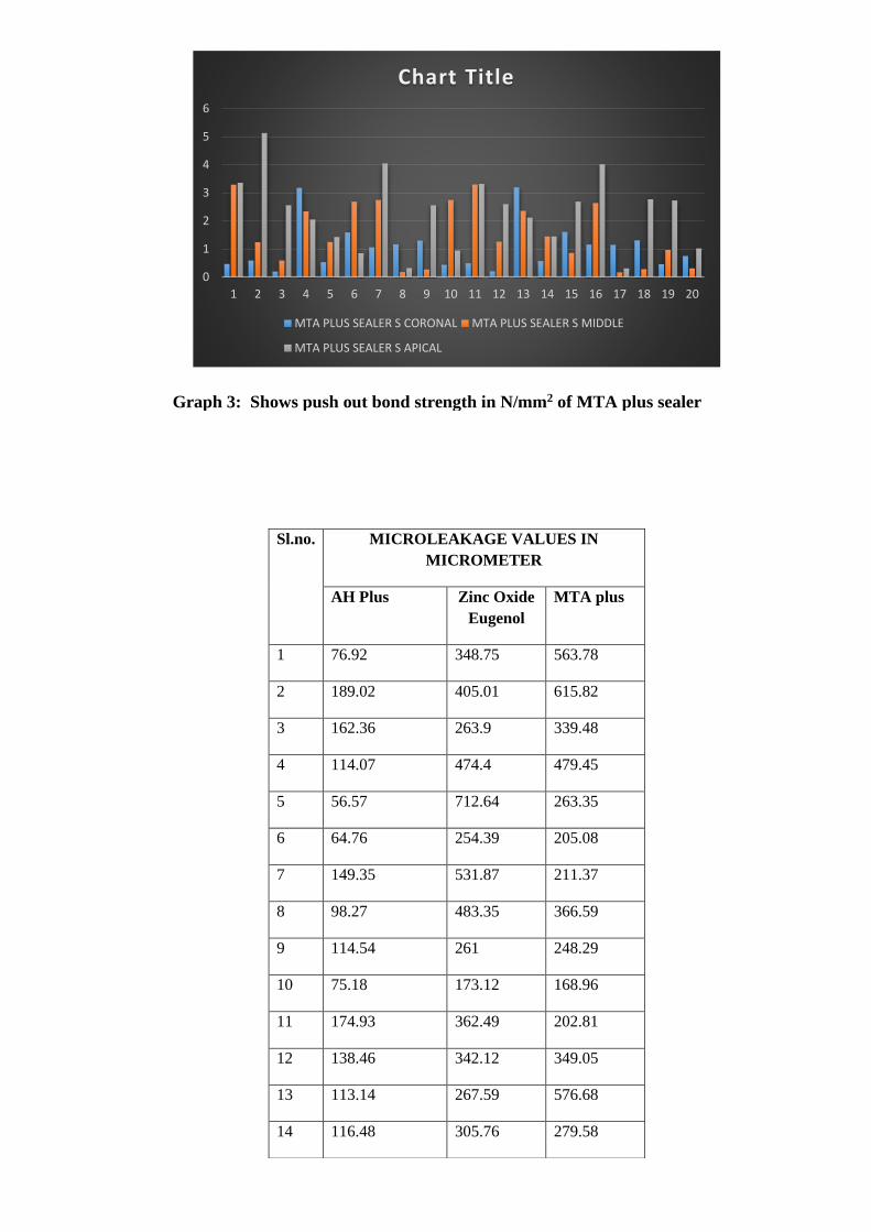
Graph 3: Shows push out bond strength in N/mm2 of MTA plus sealer
0
50
100
150
200
250
300
350
400
Category 1
Chart Title
AH plus Zinc Oxide Egenol MTA plus
15 152.05 393.24 184.2
16 149.58 475.6 182.48
17 122.08 262.21 246.6
18 135.69 324.9 554.35
19 160.37 317.32 212.76
20 184.34 385.4 324.9
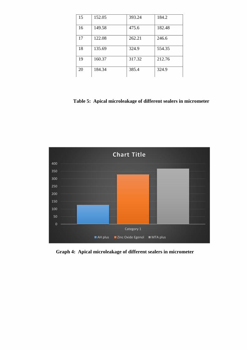
Table 5: Apical microleakage of different sealers in micrometer
Graph 4: Apical microleakage of different sealers in micrometer
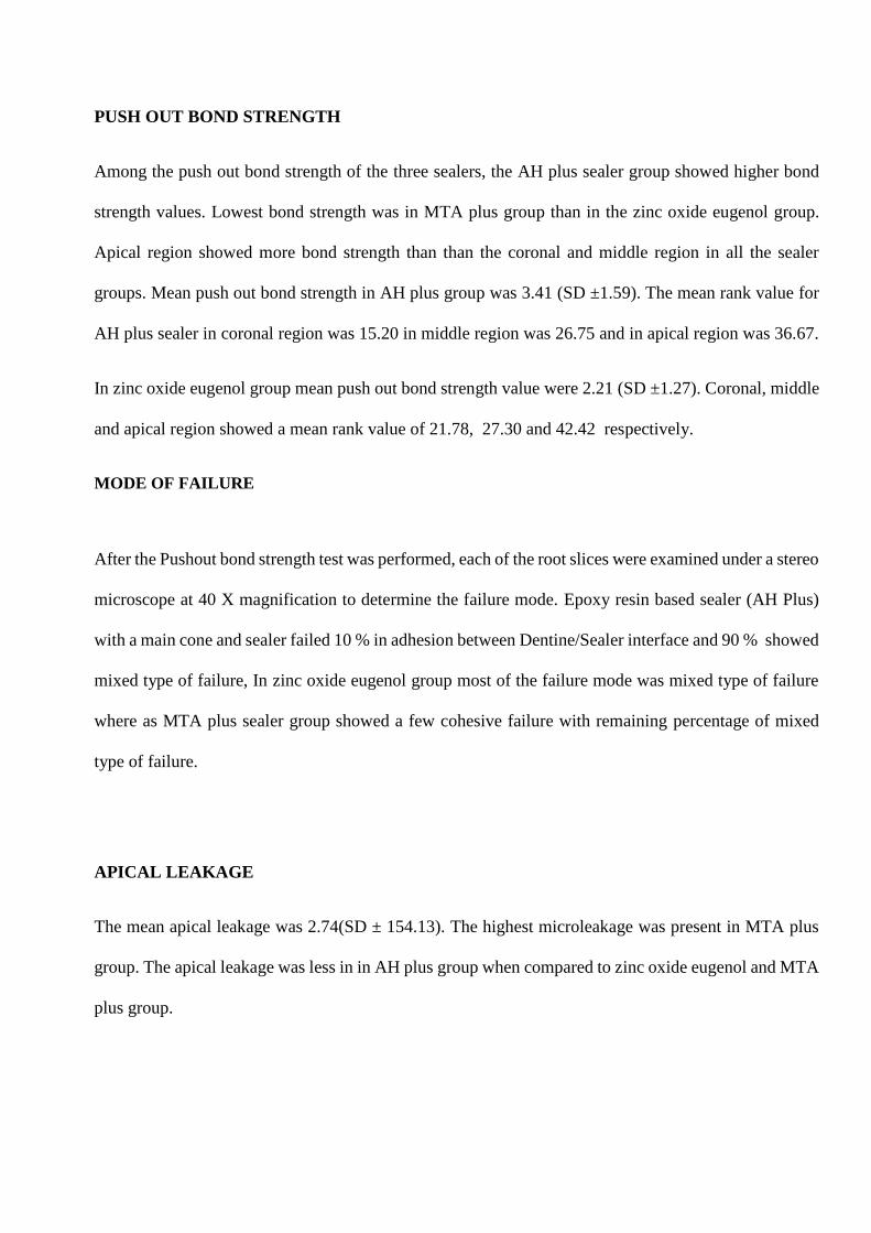
PUSH OUT BOND STRENGTH
Among the push out bond strength of the three sealers, the AH plus sealer group showed higher bond
strength values. Lowest bond strength was in MTA plus group than in the zinc oxide eugenol group.
Apical region showed more bond strength than than the coronal and middle region in all the sealer
groups. Mean push out bond strength in AH plus group was 3.41 (SD ±1.59). The mean rank value for
AH plus sealer in coronal region was 15.20 in middle region was 26.75 and in apical region was 36.67.
In zinc oxide eugenol group mean push out bond strength value were 2.21 (SD ±1.27). Coronal, middle
and apical region showed a mean rank value of 21.78, 27.30 and 42.42 respectively.
MODE OF FAILURE
After the Pushout bond strength test was performed, each of the root slices were examined under a stereo
microscope at 40 X magnification to determine the failure mode. Epoxy resin based sealer (AH Plus)
with a main cone and sealer failed 10 % in adhesion between Dentine/Sealer interface and 90 % showed
mixed type of failure, In zinc oxide eugenol group most of the failure mode was mixed type of failure
where as MTA plus sealer group showed a few cohesive failure with remaining percentage of mixed
type of failure.
APICAL LEAKAGE
The mean apical leakage was 2.74(SD ± 154.13). The highest microleakage was present in MTA plus
group. The apical leakage was less in in AH plus group when compared to zinc oxide eugenol and MTA
plus group.
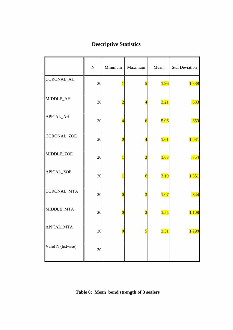
N Minimum Maximum Mean Std. Deviation
CORONAL_AH 20 1 5 1.96 1.388
MIDDLE_AH 20 2 4 3.21 .633
APICAL_AH 20 4 6 5.06 .659
CORONAL_ZOE 20 0 4 1.61 1.031
MIDDLE_ZOE 20 1 3 1.83 .754
APICAL_ZOE 20 1 6 3.19 1.351
CORONAL_MTA 20 0 3 1.07 .844
MIDDLE_MTA 20 0 3 1.55 1.108
APICAL_MTA 20 0 5 2.31 1.298
Valid N (listwise) 20
Table 6: Mean bond strength of 3 sealers
Descriptive Statistics
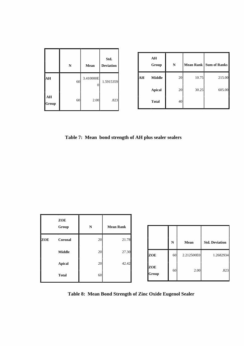
N Mean
Std.
Deviation
AH 60
3.410000E
0 1.5915359
AH
Group 60 2.00 .823
AH
Group N Mean Rank Sum of Ranks
AH Middle 20 10.75 215.00
Apical 20 30.25 605.00
Total 40
ZOE
Group N Mean Rank
ZOE Coronal 20 21.78
Middle 20 27.30
Apical 20 42.42
Total 60
N Mean Std. Deviation
ZOE 60 2.212500E0 1.2682934
ZOE
Group 60 2.00 .823
Table 7: Mean bond strength of AH plus sealer sealers
Table 8: Mean Bond Strength of Zinc Oxide Eugenol Sealer
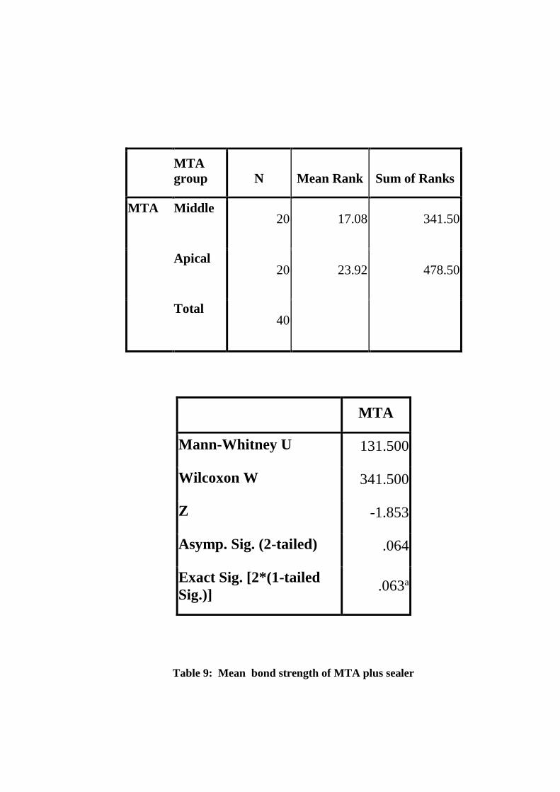
MTA
group N Mean Rank Sum of Ranks
MTA Middle 20 17.08 341.50
Apical 20 23.92 478.50
Total 40
MTA
Mann-Whitney U 131.500
Wilcoxon W 341.500
Z -1.853
Asymp. Sig. (2-tailed) .064
Exact Sig. [2*(1-tailed
Sig.)] .063a
Table 9: Mean bond strength of MTA plus sealer
sealers
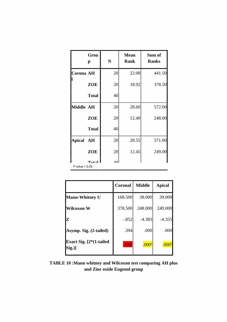
Grou
p N
Mean
Rank
Sum of
Ranks
Corona
l
AH 20 22.08 441.50
ZOE 20 18.92 378.50
Total 40
Middle AH 20 28.60 572.00
ZOE 20 12.40 248.00
Total 40
Apical AH 20 28.55 571.00
ZOE 20 12.45 249.00
Total 40
Coronal Middle Apical
Mann-Whitney U 168.500 38.000 39.000
Wilcoxon W 378.500 248.000 249.000
Z -.852 -4.383 -4.355
Asymp. Sig. (2-tailed) .394 .000 .000
Exact Sig. [2*(1-tailed
Sig.)] .398a .000a .000a
P value < 0.05
TABLE 10 :Mann whitney and Wilcoxon test comparing AH plus
and Zinc oxide Eugenol group
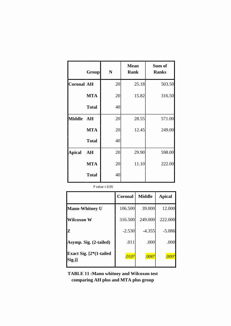
Coronal Middle Apical
Mann-Whitney U 106.500 39.000 12.000
Wilcoxon W 316.500 249.000 222.000
Z -2.530 -4.355 -5.086
Asymp. Sig. (2-tailed) .011 .000 .000
Exact Sig. [2*(1-tailed
Sig.)] .010a .000a .000a
Group N
Mean
Rank
Sum of
Ranks
Coronal AH 20 25.18 503.50
MTA 20 15.82 316.50
Total 40
Middle AH 20 28.55 571.00
MTA 20 12.45 249.00
Total 40
Apical AH 20 29.90 598.00
MTA 20 11.10 222.00
Total 40
TABLE 11 :Mann whitney and Wilcoxon test
comparing AH plus and MTA plus group
P value < 0.05
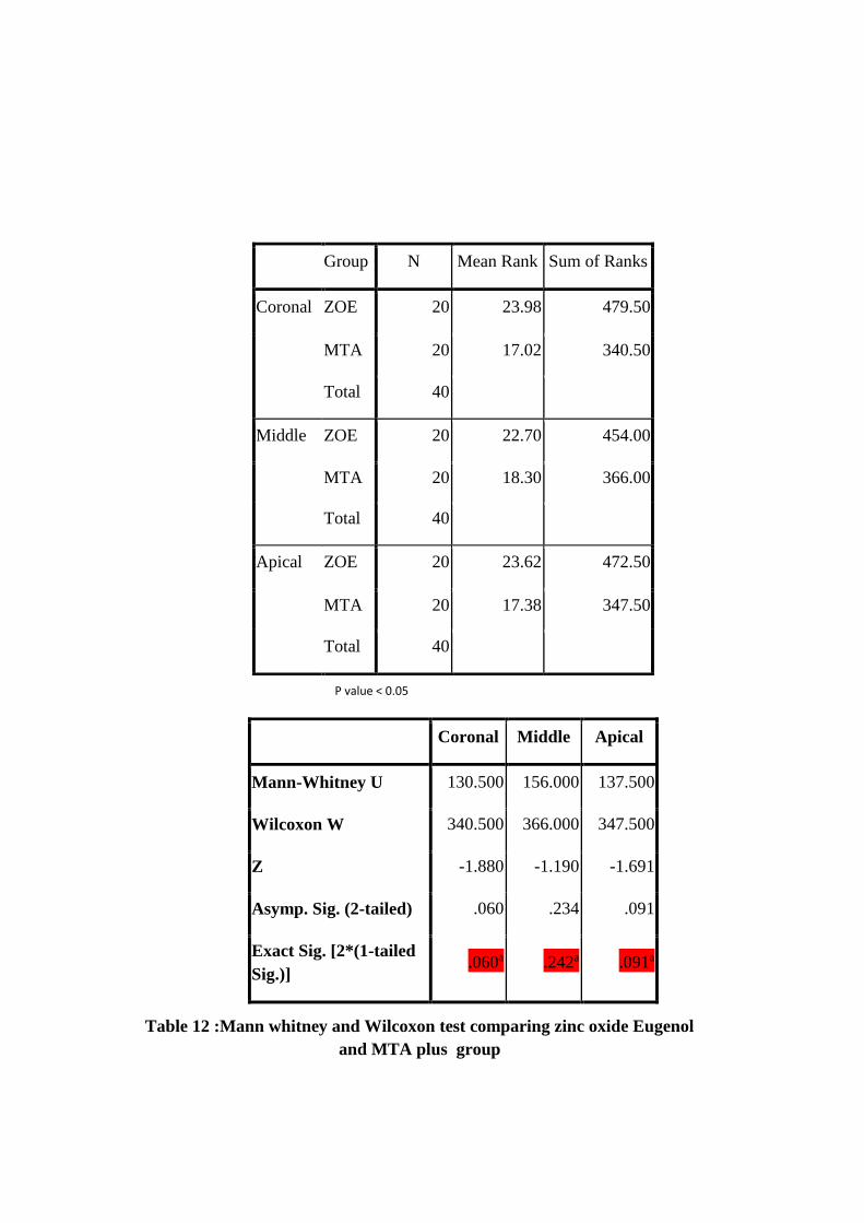
Coronal Middle Apical
Mann-Whitney U 130.500 156.000 137.500
Wilcoxon W 340.500 366.000 347.500
Z -1.880 -1.190 -1.691
Asymp. Sig. (2-tailed) .060 .234 .091
Exact Sig. [2*(1-tailed
Sig.)] .060a .242a .091a
Group N Mean Rank Sum of Ranks
Coronal ZOE 20 23.98 479.50
MTA 20 17.02 340.50
Total 40
Middle ZOE 20 22.70 454.00
MTA 20 18.30 366.00
Total 40
Apical ZOE 20 23.62 472.50
MTA 20 17.38 347.50
Total 40
Table 12 :Mann whitney and Wilcoxon test comparing zinc oxide Eugenol
and MTA plus group
P value < 0.05
Kruskal Wallis test descriptive statistics in AH plus group showed high significance among
Coronal , Middle and Apical region. Mann Whitney test showed there was a significant difference
between Coronal region and Middle region, Coronal region and Apical region, and the Middle region
and Apical region in AH plus group. In MTA plus group there was no statistically significant difference
between Coronal region and Middle region, and Middle region and Apical region whereas Coronal and
Apical region showed statistical significance difference.
In Zinc Oxide Eugenol there was no statistically significance difference between Coronal and
Middle region. Whereas Coronal and Apical region , Middle and Apical region showed statistical
significance.
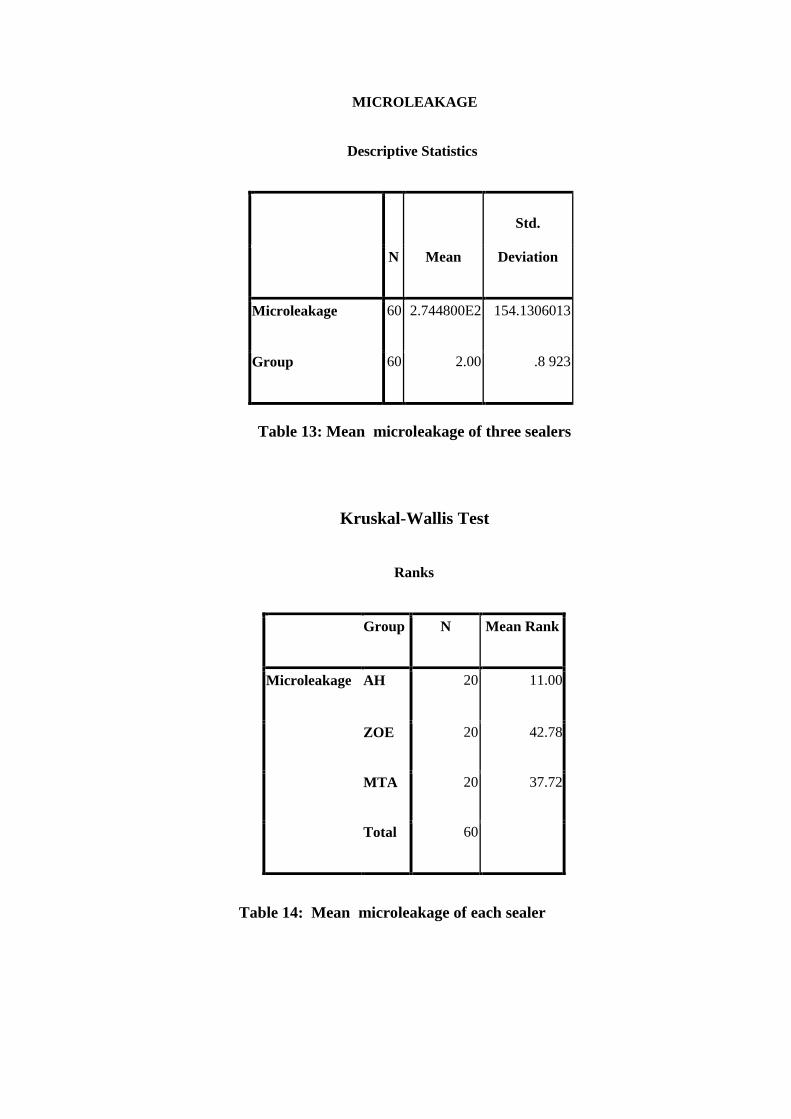
MICROLEAKAGE
Descriptive Statistics
N Mean
Std.
Deviation
Microleakage 60 2.744800E2 154.1306013
Group 60 2.00 .8 923
Kruskal-Wallis Test
Ranks
Group N Mean Rank
Microleakage AH 20 11.00
ZOE 20 42.78
MTA 20 37.72
Total 60
Table 13: Mean microleakage of three sealers
Table 14: Mean microleakage of each sealer
Descriptive statistical analysis of microleakage among the three groups were done using
kruskal- wallis test. Inter group comparisons of microleakage was done using mann-whitney test.
There was no significance difference in microleakage between MTA plus group and Zinc oxide
Eugenol group. AH plus group and MTA plus group showed highly statistical significance. AH plus
group and Zinc oxide Eugenol showed statistical significance.
DISCUSSION
The ideal requirement for root canal filling was that the entire root canal space should have no
gaps or voids. Sealer and core materials should form a uniform, chemically bonded mass that should be
bonded to dentine to minimize leakage.(1) In the current study three root canal sealers were compared
by evaluating push out bond strength and apical sealing ability . With the development of resin-based
sealers, the strength of the bond have received greater attention and the possibility of creating a
‘monoblock’ between the sealer and core material which bonds to the canal walls has introduced the
prospect of strengthening the root-filled tooth against fracture .(50,51) The sealers included a tricalcium
silicate‐containing sealer(MTA Plus) , an epoxy resin‐based sealer (AH Plus) and a zinc oxide eugenol
sealer.
The result of study showed higher push out bond strength for Epoxy resin sealer.The sealer group
interacted with dentine mechanically by penetrating into open dentinal tubules and moreover the
penetrating ability was enhanced by smear layer removal . Pretreatment of the dentine surface with
EDTA caused a significant increase in bond strength for epoxy resin based sealer and zinc oxide eugenol
based sealer .(52) . AH plus had greater adhesion to root dentin , it could be likely due to the fact that, as
an epoxy resin-based sealer, AH Plus had better penetration into the micro-irregularities because of its
creep capacity and long setting time, which increases the mechanical interlocking between sealer and
root dentin.(53)
Epoxy resin-based sealers have the possibility of adhesion to dentin with lower rates of water
solubility, are well tolerated by tissues, have low water sorption, and have a potential of forming
monoblock, thus reinforcing root canal.(54) This fact, allied to the cohesion among sealer molecules,
increased the resistance to removal and/or displacement from dentin, which could be translated as greater
adhesion.
The particle size of the filler had a decisive effect on the film thickness of the mixed material.
AH Plus had a film thickness of 16 µm, which showed clearly below the value of less than 50 µm
required by the ISO standard for root canal sealing materials whereas film thickness for MTA plus sealer
showed as 47 µm. The quality of the root canal filling directly depends on the shrinkage upon setting
and the solubility of the material used, as these properties were decisive for the impermeability of the
treated root canal. AH plus ( 0.1 %) sealer showed much less solubility than MTA sealer (1.9 %) (55)
Push-out bond strengths of resin sealers were much lower when the sealer was present as a thin
layer.(1) The MTA Plus kit includes 2 mixing liquids: a proprietary salt-free polymer gel and water.
MTA Plus has been indicated as a root canal sealer as well as a root-end filling material and a pulp
capping cement. By using the gel and varying the powder to gel ratio, different setting times and
physical-rheologic properties can be obtained. The gel has been formulated to confer washout resistance,
whereas its fine powder particle size improves handling and placement. Recent studies showed the
possibility to perform retreatment of teeth filled with MTA cements (56,57) although it has been suggested
to avoid filling procedures using MTA-like cements to completely obturate the root canal because the
collagen and flexural strength of the dentin can be negatively affected(58)
No previous studies or articles were currently available for supporting the poor micro push out
bond strength and apical sealing ability in MTA plus sealer. The poor sealing ability of MTA plus might
be due to the presence of voids in the two interfaces of sealer. One study reported that the use of MTA
as an orthograde filling material produced significantly higher percentage of voids than did GuttaPercha.
Moreover, with MTA, several voids were observed in the apical region. MTA exhibited significantly
poorer sealing quality in root canals with complex canal types; however, this difference was not
observed in the root canals taken for this study.(59)
The reason for the low adhesion strength of MTA plus was reported to be the low adhesion
property of its tag-like structures to root canal dentin.(60) MTA Plus in direct contact with fluids exhibited
partial decalcification of calcium silicate hydrate in contact with the solution, microcracking and
leaching of calcium hydroxide. Interaction with a physiological solution resulted in inhibition of
hydration.(61)
The apical sealing ability of matched-taper single-cone obturation was comparable with that of
lateral condensation and Thermafil techniques.(62) It has been shown that gutta-percha frequently
separates from the sealer resulting in gap formation (63), which could be the “weak link” in endodontic
therapy. Although some sealers might have adhesive properties to dentine (e.g. AH 26 and AH Plus,
both epoxy resin based) (Dentsply Caulk, Milford, DE, USA) Bacteria could penetrate through the entire
length of the canal as early as 19 days after obturation; the entire sample was contaminated in 90 days.
It was obvious that the obturation material could be improved to prevent the passage of oral bacteria, or
to entomb any remaining bacteria in the root canal, thus limiting their ability to cause disease in the
periapical tissues.(64)
Wennberg et al found failure of adhesion to dentine in zinc oxide based sealers and failure of
adhesion to gutta-percha for epoxy resin based sealer. The commonly used evaluation methods for apical
microleakage methodology are fluid filtration method and dye extraction method. Other dyes like
rhodamine, indian ink blue, malachite green, eosin etc were the other dyes that could replace the
methylene blue which was used in the current study. Methylene blue dye was used in this study as its
molecular size is similar to bacterial by-products such as butyric acid which can leak out of infected root
canals to irritate periapical tissues, also it was easy to use, pH manipulation and availability add to it
advantages.(65,66)
The setting reaction of zinc and eugenol was basically an ionic reaction, where eugenol acts as
a proton donator (H+). The setting mechanism of the zinc-oxide-eugenol-based cements was the result
of equimolar mixtures of zinc oxide and eugenol, consisting of zinc oxide involved in a long crystal
matrix of zinc eugenolate chelate. Any excess of eugenol get absorbed either by eugenolate and zinc
oxide . Based on the research of Fragola, Brauer and Grossman et al, the influence of pH on the setting
reaction could be explained as follows: zinc oxide (ZnO) reacts with water, producing zinc hydroxide
(Zn(OH)2). By reacting with hydrogen (2H+), ionic zinc (Zn2+) and water (2H2O) are produced. Phenolic
hydrogen from eugenol get substituted by zinc ions in order to form a zinc-oxide-eugenol chelate, the
solidification of the cement then occurring. (67) Sealing properties of ZnOE sealers were inferior in
comparison to other sealers due to the relatively high solubility of the ZOE sealer; so, adhesion between
guttapercha and zinc oxide eugenol was weak. (68)
The results of this study were similar with the results of many other studies comparing the bond
strength of epoxy resin based sealer and tricalcium silicate based sealer to dentin. (69,70,71,72 ) Generally
these studies have been showned gutta-percha and AH Plus to have higher bond strengths than MTA
based sealers and zinc oxide based sealers obturation systems.
The presence of phosphates in the canal has been reported to enhance biomineralization and also
increased the push‐out bond strength of calcium silicate‐based materials. When EDTA was used as final
irrigant chelating agent opens dentinal tubules and exposes dentine collagen fibrils enhancing the sealers
entrapping in dentine structure . Also the residual EDTA chelates calcium from the materials and the
dentine, resulted in more free calcium helped to provide bonding(73,74).
Push-out test has been described to measure the bond between sealer, canal wall and the core
material. The test was intended to assess the extent to which the sealer and core material were bonded
into a solid mass as well as the strength of the bond to the canal wall.(75)
The push-out bond strength test was one method to evaluate the effectiveness of root canal
obturation material or technique. The other methods of testing include bacterial leakage, fluid filtration
and dye penetration testing (76). While every method of in vitro testing supposed to replicate the clinical
environment, and the correlation between leakage studies and clinical success has been questioned.(77)
The push-out models has been used widely to evaluate dentin obturation interface, but its relevance has
also been called into question.(78) There was also no evidence that any of these methods was the best for
measuring clinical effectiveness of root canal obturation material or techniques.
The current study demonstrated that highest mean bond strengths were found at the apical
segment of each group. This could be due to the pooling of sealer at the apical segment as an evident
during obturation. Each tooth was prepared so that tug-back was felt when placing the master cone. This
demonstrates a tight fit that could cause an influence in push-out bond strength. The apical segments
exhibited highest standard deviation. This might be due to the small diameter of obturation material
found near the apex. Attempts were made to match the diameter of the plunger which used in the push
out testing compared to the diameter of the filling material to prevent the plunger touching the wall of
the canal. All the slices were examined under 40X magnification after push-out. If evidence was found
that the canal wall touched the sample was discarded. The middle and coronal segments were more
easily aligned using the push-out plungers by minimizing the need to discard slices than the apical
segments.
SUMMARY
The present study was done in the Department of Conservative and Endodontics ,KSR Institute
of Dental Science and Research. After taking approval from institutional review board,a randomised
control trial was planned. . The ultimate goal of root canal therapy is total obturation of the root canal
system. The physical properties necessary for this function included adaptation and adhesion of the
filling material to the root canal wall, because gutta-percha does not directly bond to the dentine surface.
Ideally, the sealer should be capable of producing a bond between core material and dentine wall. So
the present study aimed to evaluate and compare the push out bond strength and apical sealing ability
of AH plus, Zinc Oxide Eugenol and MTA plus root canal sealers after obturation using single cone
technique. Total of 120 mandibular premolar teeth were used and decoronated. All teeth were prepared
using protaper rotary file system upto F3 file size using Xmart plus (DENTSPLY Maillefer) and
obturated with guttapercha size of final instrumentation using single cone obturation technique. The
irrigation solution used after each instrumentation were 2.5% NaOCl, 17% EDTA and normal saline.
Samples were grouped into three based on the root canal sealer used of each group containing 40 number
of teeth. In Group I, specimens, obturation done using Gutta-percha and zinc oxide Eugenol sealer;
Group II - Gutta-percha and AH Plus sealer and Group III- Gutta-percha and MTA Plus sealer. Half of
the specimens in each group were used for push out bond strength analysis. Horizontal sections of 2 mm
thickness were made from coronal, middle and apical region of samples and underwent push out bond
strength test with the help of universal testing machine at a cross head speed of 0.5 mm/ min. Specimens
were analysed using stereo microscope for the mode of failure and extent of fracture using scanning
electron microscope. Remaining half of the specimens were coated with modelling wax except apical
foramen and immersed in methylene blue for 2 days. Stereo microscopic photographs of each samples
were taken and linear measurement was calculated using CMEIAS software.
The findings of the present study can be summerized as follows:
1. AH plus sealer group showed the highest push out bond strength than Zinc oxide Eugenol and
MTA plus root canal sealer.
2. Apical segment of each test group demonstrated the highest mean bond strengths than the
coronal and middle segments.
3. Apical microleakage value in AH plus sealer group was less on comparing Zinc Oxide Eugenol
and MTA plus root canal sealer.
CONCLUSION
Within the limitations of the present study, push out bond strength values of each sealer were
different and varied significantly in each area of root canal Coronal, Middle and Apical. AH plus sealer
showed higher push out bond strength than Zinc oxide Eugenol and MTA plus root canal sealer. Apical
segment of each test group demonstrated the highest mean bond strengths than the coronal and middle
segments Apical microleakage value in AH plus was less on comparing zinc oxide Eugenol and MTA
plus root canal sealer.
REFERENCES
1. A. Jainaen, J. E. A. Palamara& H. H. Messer et al. Push-out bond strengths of the dentine–sealer
interface with and without a main cone, International Endodontic Journal, 2007; 40(2): 882–890.
2. ThompsonJI, Gregson PJ, Revell PA et al, Analysis of pushout test data based on interfacial
fracture energy. Journal of Materials Science: Materials in Medicine 1999; 10(5): 863–8.
3. Dow, P.R., Ingle, J.I. et al, Isotope Determination of Root Canal Failure. Oral Surgery 1955;8
(5) :1100-4,.
4. Ingle, J.I,Taintor, et al, J.F. Endodontics. 3rd ed. Philadelphia: Lea &Febiger, 1985; 36.
5. Weine et al, F.S. Endodontic Theory. 4th ed. St. Louis: CV Mosby, 1989;13.
6. Caicedo,R, Von Fraunhofer, J. A et al, The Properties of Endodontic Sealer Cements. Journal
of Endodontics. 1988; Nov: 527-534.
7. Kapsilmalis, P, Evans, R et al, Sealing properties of endodontic filling materials
usingradioactive polar and nonpolar isotopes. Oral Surgery. 1966; 22 (2) :386,.
8. Lares, C, El Deeb M.E et al, The sealing ability of the Thermafilobturation technique. Journal
of Endodontics. 1999; 16 (2) :474,-9
9. Marshall, F.J.,Massler, M. et al, Sealing of pulpless teeth evaluated with radioisotopes Journal
of Endodontics 1987; 13 (7) :315-7,
10. Skidmore LJ, Berzins DW, Bahcall JK et al,. An in vitro comparison of the intraradicular dentin
bond strength of resilon and gutta-percha. Journal of Endodontics 2006; 32 (3) :963-6.
11. Sophia Thakur, Jonathan Emil et al, Evaluation of mineral trioxide aggregate as root canal
sealer: A clinical study Journal of Conservative Dentistry. 2013; 16(6): 494–498.
12. PrasannaNeelakantan et al, Retreatability of 2 Mineral Trioxide Aggregate–based Root Canal
Sealers: A Cone-beam Computed Tomography Analysis Journal of Endodontics, 2013;12(7):1–
4.
13. A. D. B. Garrido, R. C. C. Lia, S. C. França, J. F. da Silva, S. Astolfi-Filho, et al, “Laboratory
evaluation of the physicochemical properties of a new root canal sealer based on
Copaiferamultijuga oil-resin,” International Endodontic Journal, 2010; 43 (4) : 283–291.
14. Kapsilmalis, P, Evans R et al Sealing properties of endodontic filling materials usingradioactive
polar and nonpolar isotopes. Oral Surgery. 1966; 22:386.
15. Yilmaz Z, Deniz D, Ozcelik B, et al, Sealing efficiency of BeeFill 2in1 and System B/Obtura II
versus single-cone and cold lateral compaction techniques. Oral Surgery Oral Medicine Oral
Pathology Oral Radiology and Endodontology. 2009;108(6):e51-e55.
16. Antonopoulos et al Evaluation of the apical seal of root canal fillings with different methods
journal of Endodontics1998; 24 (11):655-8.
17. Wu et al. The capability of two hand instrumentation techniques to remove the inner layer of
dentine in oval canals International Endodontic Journal, 2003; 36 (2) :218-224,
18. Kardonet al An In Vitro Evaluation of the Sealing Ability of a New Root-canal–obturation
System. Journal of endodontics 2003 ; 29 (10) : 201
19. Sabir Muliyar, K Abdul Shameem, Rekha P Thankachan et al, Microleakage in Endodontics,
Journal of International Oral Health. 2014; 6(6): 99–104
20. Limkangwalmongkol Set al, Apical dye penetration with four root canal sealers and guttapercha
using longitudinal sectioning. Journal of Endodontics. 1992; 18(11):535-9
21. Yared GM et al, Sealing ability of the vertical condensation with different root canal sealers.
Journal of Endodontics. 1996 ;22(1):6-8
22. Dagher FB et al, Microleakage of a new and an old Kerr root canal sealers. Journal of
Endodontics 1997; 23(7):442-3
23. L. C. Martens et al, Apical microleakage after lateral condensation, hybrid gutta-percha
condensation and Soft-Core obturation: an in vitro evaluation International Endodontic Journal
1999; 15 (5) : 239–243
24. 24.Lyroudia K et al, Three-dimensional reconstruction: a new method for the evaluation of
apical microleakage.Journal of Endodontics. 2000 ;26(1):36-8
25. R. J. G. De Moor et al, The long-term sealing ability of an epoxy resin root canal sealer used
with five gutta-percha obturation techniques, International Endodontic Journal2002 ; 35 (3) :
275–282.
26. Ari H et al, Effects of NaOCl on bond strengths of resin cements to root canal dentin. Journal of
Endodontics. 2003 ;29(4):248-51
27. Gharai SR et al, SS spreaders produced significantly high force than NiTi spreaders in all
specimens Journal of Endodontics. 2005 ;31(3):198-200
28. Vasiliadis L et al, Short- and long-term sealing ability of Gutta-flow and AH-Plus using an ex
vivo fluid transport model, International Endodontic Journal, 2010; 43 (3): 377–381,
29. Shokouhinejad et al,Effect of phosphate-buffered saline on push-out bond strength of a new
bioceramic sealer to root canal dentin.Dental Research Journal. 2012 ; 9 (5): 595-599
30. Souza Bier et al, Push-out bond strength of Calcium hydroxide and Mineral Trioxide Aggregate
based sealers to root canal dentin. RevistaOdontoCiencia. 2012; 27 (4) :320-324
31. Bhardwaj et al, comparative evaluation of apical leakage of root canal sealers in
vitro.International Journal of Clinical Dentistry. 2013 ; 6 (1):17-24
32. Carvalho et al, Influence of calcium hydroxide intracanal medication on bond strength of two
endodontic resin-based sealersassessed by micropush-out test.Dental Traumatology. 2013; 29
(1) :73-76
33. Evren et al, Effect of Photoactivated Disinfection on Bond Strength of Root Canal Filling.Journal
of Endodontics. 2013 ; 39 (11):1428-1430
34. Ozcan et al, Assessment of the sealing abilities of several root canal sealers and filling
methods.ActaOdontologicaScandinavica. 2013; 71 ( 6) :1362-1369
35. Shokouhinejad et al, Push-out bond strength of gutta-percha with a new bioceramic sealer in
the presence or absence of smear layer.Australian Endodontic Journal. 2013; 39 (3) :102-106
36. Topçuoglu et al, The bond strength of endodontic sealers to root dentine exposed to different
gutta-percha solvents. International Endodontic Journal. 2014; 47 (12) :1100-1106
37. Ulusoy et al, Apicalmicroleakage of different root canal sealers after use of maleic acid and
EDTA as final irrigants Brazilian Oral Research. 2014; 28 (5) :337-342
38. Al-Dwairi et al, Effect of endodontic sealers on push-out bond strength of cemented fiber posts
Quintessence International. 2015; 46 (4) :299-307
39. BerkanCelikten et al, Micro-CT assessment of the sealing ability of three root canal filling
techniques, Journal of Oral Science, 2015 ; 57( 4) : 361-366,
40. Epita S et al, Thin and thick layers of resin-based sealer cement bonded to root dentine
compared: Adhesive behaviour Australian Endodontic Journal 2015; 41: 128–134
41. Elbatoutyet al, In vitro bond strength of bioceramic root canal sealer in comparison to resin-
based and eugenol-based rootcanal sealersEndodontic Practice Today. 2015 ; 9 ( 1) : 59-63
42. EhsenAbdelmoumen et al, In-Vitro Evaluation of Apical Sealing Ability of Two Root Canal
Sealers Used With two Root Canal Filling-Techniques. International Dental Journal. 2015 ; 65
(9) : 163-171.
43. Fernanda Leal et al, Effect of final irrigation protocols on push-out bond strength of an epoxy
resin root canal sealer to dentin. Australian Endodontic Journal, 2015; 41: 135–139
44. Neelakantan et al, Influence of Irrigation Sequence on the Adhesion of Root Canal Sealers to
Dentin: A Fourier Transform Infrared Spectroscopy and Push-out Bond Strength
Analysis.Journal of Endodontics. 2015 ; 41 (7):1108-1111
45. Pradeep et al, Evaluation of Different Dentin Adhesive Systems and its Effect on Apical
Microleakage: An In Vitro Study.Journal of International Oral Health.2015; 7 (5): 44-48
46. LeventDemiriz et al, Evaluation of the dentinal wall adaptation ability of MTA Fillapex using
stereo electron microscope Journal of Conservative Dentistry 2016;19:220-4
47. Patni PM et al, Stereomicroscopic Evaluation of Sealing Ability of Four Different Root Canal
Sealers- An invitro Study Journal of Clinical and Diagnostic Research. 2016 ; 10 (8) :37-9
48. Ahuja L et al, A Comparative Evaluation of Sealing Ability of New MTA Based Sealers with
Conventional Resin Based Sealer: An In-vitro Study, Journal of Clinical and Diagnostic
Research 2016; 10(7): ZC76–ZC79
49. Widcha Asawaworarit et al, Comparison of the Apical Sealing Ability of Calcium Silicate-
Based Sealer and Resin-Based Sealer Using the Fluid-Filtration Technique. Medical Principles
and Practice 2016;25:561–565
50. Teixeira FB, Teixeira EC, Thompson JY et al, Fracture resistance of roots endodontically treated
with a new resin filling material. Journal of the American Dental Association 2014; 135, 646–
52
51. Schafer E, Zandbiglari T, et al, Influence of resinbased adhesive root canal fillings on the
resistance to fracture of endodontically treated roots: an in vitro preliminary study. Oral Surgery,
Oral Medicine, Oral Pathology, Oral Radiology, and Endodontology2005;103:274–9.
52. Figdor D et al, Apical periodontitis: A very prevalent problem. Oral Surgery, Oral Medicine,
Oral Pathology, Oral Radiology, and Endodontology. 2002;94:651–2
53. Ruddle CJ et al, Advanced Endodontics. Gauging the Terminus: A Novel Method.. Available
from: http://www.endoruddle.com/blogs/show/21. 2012: May, 25.
54. Hargreaves KM, Cohen S, Berman LH et al, Cohen′s pathways of the pulp. Mosby Elsevier;
2011
55. Kenneth, Louis Berman et al, Pathways of Pulp, first South Asia edition page 296
56. Neelakantan P, Grotra D, Sharma S et al ,Retreatability of 2 mineral trioxide aggregatebased
root canal sealers: a cone-beam computed tomography analysis. Journal of Endodontics 2013;39
(11) :893–6.
57. Pirani C, Iacono F, Chersoni S, et al. The effect of ultrasonic removal of various rootend filling
materials. International Endodontic Journal,2009;42 (12) :1015–25.
58. Leiendecker AP, Qi YP, Sawyer AN, et al. Effects of calcium silicate-based materials on
collagen matrix integrity of mineralized dentin. Journal of Endodontics,2012;38 (24) 829–33
59. Wonkyung JHO et al Comparison of root canal filling quality by Mineral Trioxide Aggregate
and Guttapercha cones/AH plus sealer ,Dental Materials Journal 2016; 35(4): 644–650
60. HakanGokturk et al, Effect of double Antibiotic and Calcium Hydroxide Pastes on dislodgement
resistance of an epoxy resin-based and two calcium silicate-based root canal sealers, Clinical
Oral Investigations2016; 10 (6) : 112- 119
61. Camilleriet al, The setting characteristics of MTA plus in different environmental conditions
International Endodontic Journal. 2013 ;46(9):831-40
62. U Inan et al, In Vitro Evaluation of Matched-Taper Single-Cone Obturation with a Fluid
Filtration Method March Journal of the Canadian Dental Association 2009 ;75 (2) : 215- 222
63. G Shipper et al, In vitro microbial leakage of endodontically treated teeth using new and standard
obturation techniques Journal of Endodontics, 2004;30(3):154-8
64. Torabinejad M, Ung B, Kettering JD et al, In vitro bacterial penetration of coronally unsealed
endodontically treated teeth. Journal of Endodontics2016; 10 (12): 566-9
65. Tanomaru-Filho M, Pinto RV, Bosso R et al, Evaluation of the thermoplasticity of gutta-percha
and Resilon using the Obtura II system at different temperature settings. International Endodontic
Journal. 2011;44(5) :764–8.
66. Figdor D et al, Apical periodontitis: A very prevalent problem. Oral Surgery, Oral Medicine,
Oral Pathology, Oral Radiology, and Endodontology. 2002;94(2):651–2
67. Fragola A, Pascal S, Rosengarten M et al, The effect of varying particle size of the components
of Grossman's cement. Journal of Endodontics 1979; 5(4): 336-339
68. Upadhyay V, Upadhyay M, Panday RK et al, A SEM evaluation of dentinal adaptation of root
canal obturation with GuttaFlow and conventional obturating material. Indian Journal of Dental
Research 2011;22 (7):1-6
69. Topcuoglu et al, The bond strength of endodontic sealers to root dentine exposed to different
gutta-percha solvents, International Endodontic Journal 2014; 47(5): 1100–1106,
70. Widcha Aawaworarit et al, Comparison of the Apical Sealing Ability of Calcium Silicate-Based
Sealer and Resin-Based Sealer Using the Fluid-Filtration TechniqueMedical Principles and
Practice2016;25 (6) : 561–565.
71. Patni PMet al, Stereomicroscopic Evaluation of Sealing Ability of Four Different Root Canal
Sealers- An invitro Study Journal of clinical and diagnostic research . 2016 ;10(8):ZC37-9,
72. Ahuja L A et al, Comparative Evaluation of Sealing Ability of New MTA Based Sealers with
Conventional Resin Based Sealer: An In-vitro Study Journal of clinical and diagnostic
research 2016;10(7):Z76-9
73. A Wennberg et al, Adhesion of root canal sealers to bovine dentine and gutta-percha,
International Endodontic Journal 1990 ;23(1):13-9
74. Reyes‐Carmona JF, Felippe MS et al, The biomineralization ability of mineral trioxide aggregate
and Portland cement on dentin enhances the push‐out strength. Journal of Endodontics
2010;36(2):286‐91.
75. Thompson JI, Gregson PJ et al, Analysis of pushout test data based on interfacial fracture energy.
Journal of Materials Science: Materials in Medicine 2010;10: 863–8.
76. Wu MK, Wesselink PR et al, Endodontic leakage studies reconsidered. Part 1. Methodology ,
application and relevance. International Endodontic Journal 1993; 26 (4) :37-43
77. Oliver CM, Abbott PV et al, Correlation between clinical success and apical dye penetration.
International Endodontic Journal 2001; 34 (8) :637-44
78. Sudsangiam S, Van Noort R et al, Do dentin bond strength tests serve a useful purpose? Jornal
of Adhesive Dentistry, 1999; 1 (11):57-67.
APPENDIX – I
ABBREVATIONS
SEM – Scanning Electron Microscope
EDTA – Ethylene diamine tetra acetic acid.
MTA – Mineral trioxide aggregate
ZOE – Zinc oxide Eugenol
STD – Standard deviation
GRP – Group
Asymp. Sig – Asymptomatic significance
Exact Sig – Exact significance
CEJ – Cemento Enamel Junction