Embed Size (px)
Citation preview
261
Journal of International Oral Health 2016; 8(2):261-266Evaluation of sealing ability among restorative materials … Velagapudi NJ et al
Original ResearchReceived: 15th September 2015 Accepted: 20th December 2015 Conflict of Interest: None
Source of Support: Nil
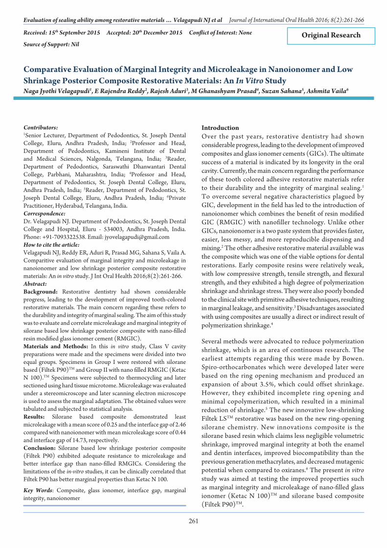
Comparative Evaluation of Marginal Integrity and Microleakage in Nanoionomer and Low Shrinkage Posterior Composite Restorative Materials: An In Vitro StudyNaga Jyothi Velagapudi1, E Rajendra Reddy2, Rajesh Aduri3, M Ghanashyam Prasad4, Suzan Sahana5, Ashmita Vaila6
Contributors:1Senior Lecturer, Department of Pedodontics, St. Joseph Dental College, Eluru, Andhra Pradesh, India; 2Professor and Head, Department of Pedodontics, Kamineni Institute of Dental and Medical Sciences, Nalgonda, Telangana, India; 3Reader, Department of Pedodontics, Saraswathi Dhanwantari Dental College, Parbhani, Maharashtra, India; 4Professor and Head, Department of Pedodontics, St. Joseph Dental College, Eluru, Andhra Pradesh, India; 5Reader, Department of Pedodontics, St. Joseph Dental College, Eluru, Andhra Pradesh, India; 6Private Practitioner, Hyderabad, Telangana, India.Correspondence:Dr. Velagapudi NJ. Department of Pedodontics, St. Joseph Dental College and Hospital, Eluru - 534003, Andhra Pradesh, India. Phone: +91-7093322538. Email: [email protected] to cite the article:Velagapudi NJ, Reddy ER, Aduri R, Prasad MG, Sahana S, Vaila A. Comparitive evaluation of marginal integrity and microleakage in nanoionomer and low shrinkage posterior composite restorative materials: An in vitro study. J Int Oral Health 2016;8(2):261-266.Abstract:Background: Restorative dentistry had shown considerable progress, leading to the development of improved tooth-colored restorative materials. The main concern regarding these refers to the durability and integrity of marginal sealing. The aim of this study was to evaluate and correlate microleakage and marginal integrity of silorane based low shrinkage posterior composite with nano-filled resin modified glass ionomer cement (RMGIC).Materials and Methods: In this in vitro study, Class V cavity preparations were made and the specimens were divided into two equal groups. Specimens in Group I were restored with silorane based (Filtek P90)TM and Group II with nano filled RMGIC (Ketac N 100).TM Specimens were subjected to thermocycling and later sectioned using hard tissue microtome. Microleakage was evaluated under a stereomicroscope and later scanning electron microscope is used to assess the marginal adaptation. The obtained values were tabulated and subjected to statistical analysis.Results: Silorane based composite demonstrated least microleakage with a mean score of 0.25 and the interface gap of 2.46 compared with nanoionomer with mean microleakage score of 0.44 and interface gap of 14.73, respectively.Conclusion: Silorane based low shrinkage posterior composite (Filtek P90) exhibited adequate resistance to microleakage and better interface gap than nano-filled RMGICs. Considering the limitations of the in-vitro studies, it can be clinically correlated that Filtek P90 has better marginal properties than Ketac N 100.
Key Words: Composite, glass ionomer, interface gap, marginal integrity, nanoionomer
IntroductionOver the past years, restorative dentistry had shown considerable progress, leading to the development of improved composites and glass ionomer cements (GICs). The ultimate success of a material is indicated by its longevity in the oral cavity. Currently, the main concern regarding the performance of these tooth colored adhesive restorative materials refer to their durability and the integrity of marginal sealing.1 To overcome several negative characteristics plagued by GIC, development in the field has led to the introduction of nanoionomer which combines the benefit of resin modified GIC (RMGIC) with nanofiller technology. Unlike other GICs, nanoionomer is a two paste system that provides faster, easier, less messy, and more reproducible dispensing and mixing.2 The other adhesive restorative material available was the composite which was one of the viable options for dental restorations. Early composite resins were relatively weak, with low compressive strength, tensile strength, and flexural strength, and they exhibited a high degree of polymerization shrinkage and shrinkage stress. They were also poorly bonded to the clinical site with primitive adhesive techniques, resulting in marginal leakage, and sensitivity.3 Disadvantages associated with using composites are usually a direct or indirect result of polymerization shrinkage.4
Several methods were advocated to reduce polymerization shrinkage, which is an area of continuous research. The earliest attempts regarding this were made by Bowen. Spiro-orthocarbonates which were developed later were based on the ring opening mechanism and produced an expansion of about 3.5%, which could offset shrinkage. However, they exhibited incomplete ring opening and minimal copolymerization, which resulted in a minimal reduction of shrinkage.5 The new innovative low-shrinking Filtek LSTM restorative was based on the new ring-opening silorane chemistry. New innovations composite is the silorane based resin which claims less negligible volumetric shrinkage, improved marginal integrity at both the enamel and dentin interfaces, improved biocompatibility than the previous generation methacrylates, and decreased mutagenic potential when compared to oxiranes.6 The present in vitro study was aimed at testing the improved properties such as marginal integrity and microleakage of nano-filled glass ionomer (Ketac N 100)TM and silorane based composite (Filtek P90)TM.
262
Evaluation of sealing ability among restorative materials … Velagapudi NJ et al Journal of International Oral Health 2016; 8(2):261-266
Materials and MethodsSample collectionA total of 90 non-carious premolars, extracted atraumatically for orthodontic purpose were collected. These teeth were stored in distilled water until they were subjected to the inclusion and exclusion criteria which were as follows:
Inclusion criteria• Non-carious premolars indicated for extraction for
orthodontic purpose.Exclusion criteria• Teeth with craze lines and cracks• Cervical abrasions• Affected by abfraction• Endodontically treated teeth• Dental caries.
The craze lines if present on the collected teeth were detected by tactile examination and transillumination.
Specimen preparation/preparation of samplesTotal of 40 teeth out of collected sample which satisfied the criteria of the study were surface debrided with ultrasonic scaling unit and preserved in deionized water. Standardized Class V cavity of size 4 mm × 3 mm × 2 mm mesio-distal width, occluso gingival length, depth, respectively were made on the facial surface of each tooth, 1 mm above the cementoenamel junction. William’s graduated periodontal probe and a steel scale were used to gauge the dimensions
Grouping of specimensAll the prepared teeth were randomly divided into two groups. The groupings are as follows.
Group 1: Teeth were restored with low shrinkage posterior composite (Filtek P 90).
Group 2: Teeth were restored with nanoionomer (Ketac N 100).
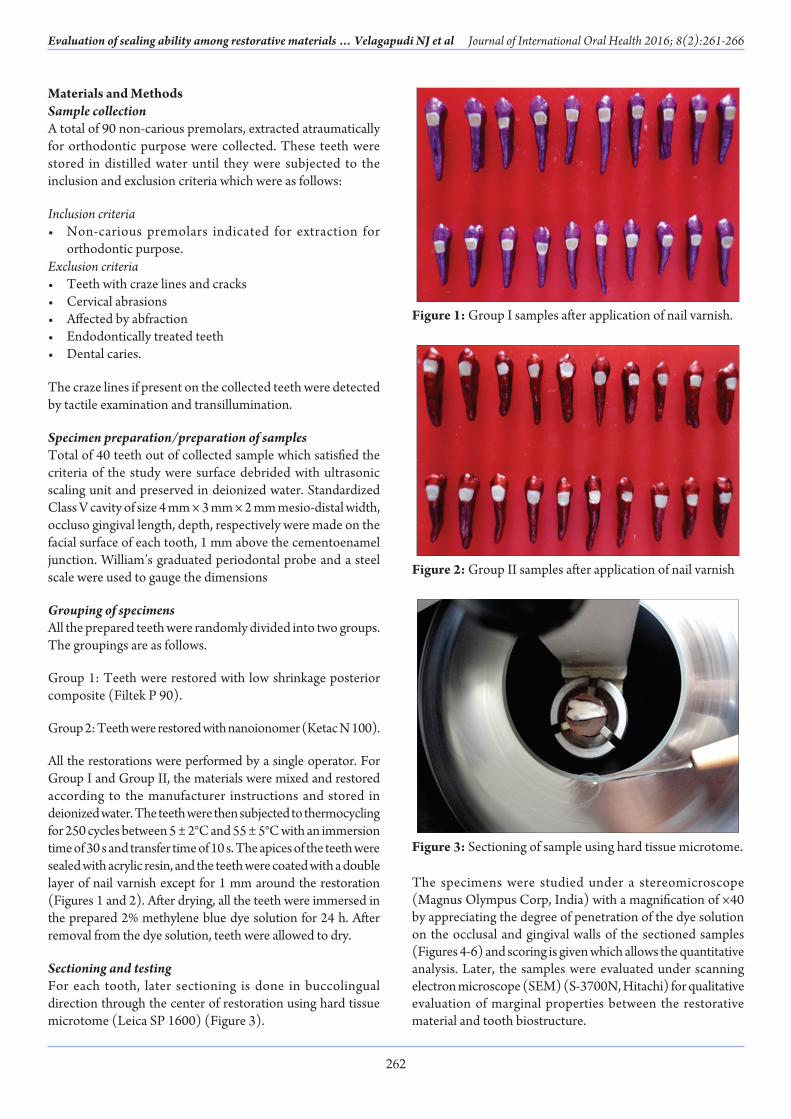
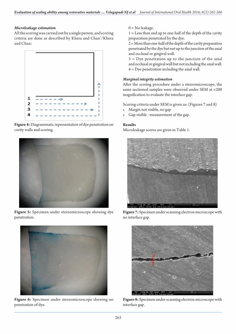
All the restorations were performed by a single operator. For Group I and Group II, the materials were mixed and restored according to the manufacturer instructions and stored in deionized water. The teeth were then subjected to thermocycling for 250 cycles between 5 ± 2°C and 55 ± 5°C with an immersion time of 30 s and transfer time of 10 s. The apices of the teeth were sealed with acrylic resin, and the teeth were coated with a double layer of nail varnish except for 1 mm around the restoration (Figures 1 and 2). After drying, all the teeth were immersed in the prepared 2% methylene blue dye solution for 24 h. After removal from the dye solution, teeth were allowed to dry.
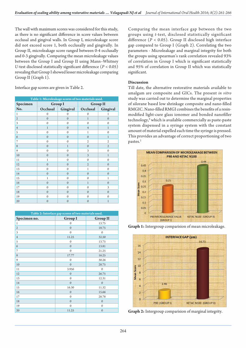
Sectioning and testingFor each tooth, later sectioning is done in buccolingual direction through the center of restoration using hard tissue microtome (Leica SP 1600) (Figure 3).
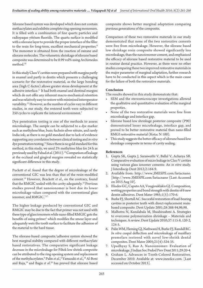
The specimens were studied under a stereomicroscope (Magnus Olympus Corp, India) with a magnification of ×40 by appreciating the degree of penetration of the dye solution on the occlusal and gingival walls of the sectioned samples (Figures 4-6) and scoring is given which allows the quantitative analysis. Later, the samples were evaluated under scanning electron microscope (SEM) (S-3700N, Hitachi) for qualitative evaluation of marginal properties between the restorative material and tooth biostructure.
Figure 1: Group I samples after application of nail varnish.
Figure 2: Group II samples after application of nail varnish
Figure 3: Sectioning of sample using hard tissue microtome.
263
Journal of International Oral Health 2016; 8(2):261-266Evaluation of sealing ability among restorative materials … Velagapudi NJ et al
Microleakage estimationAll the scoring was carried out by a single person, and scoring criteria are done as described by Khera and Chan7/Khera and Chan:
0 = No leakage. 1 = Less than and up to one-half of the depth of the cavity
preparation penetrated by the dye. 2 = More than one-half of the depth of the cavity preparation
penetrated by the dye but not up to the junction of the axial and occlusal or gingival wall.
3 = Dye penetration up to the junction of the axial and occlusal or gingival wall but not including the axial wall.
4 = Dye penetration including the axial wall.
Marginal integrity estimationAfter the scoring procedure under a stereomicroscope, the same sectioned samples were observed under SEM at ×200 magnification to evaluate the interface gap.
Scoring criteria under SEM is given as: (Figures 7 and 8)• Margin not visible, no gap• Gap visible - measurement of the gap.
ResultsMicroleakage scores are given in Table 1.
Figure 4: Diagrammatic representation of dye penetration on cavity walls and scoring.
Figure 5: Specimen under stereomicroscope showing dye penetration.
Figure 6: Specimen under stereomicroscope showing no penetration of dye.
Figure 7: Specimen under scanning electron microscope with no interface gap.
Figure 8: Specimen under scanning electron microscope with interface gap.
264
Evaluation of sealing ability among restorative materials … Velagapudi NJ et al Journal of International Oral Health 2016; 8(2):261-266
The wall with maximum scores was considered for this study, as there is no significant difference in score values between occlusal and gingival walls. In Group I, microleakage score did not exceed score 1, both occlusally and gingivally. In Group II, microleakage score ranged between 0-4 occlusally and 0-3 gingivally. Comparing the mean microleakage values between the Group I and Group II using Mann–Whitney U-test disclosed statistically significant difference (P < 0.05) revealing that Group I showed lesser microleakage comparing Group II (Graph 1).
Interface gap scores are given in Table 2.
Comparing the mean interface gap between the two groups using t-test, disclosed statistically significant difference (P < 0.05). Group II disclosed high interface gap compared to Group I (Graph 2). Correlating the two parameters - Microleakage and marginal integrity for both the groups using spearman’s rank correlation revealed 93% of correlation in Group I which is significant statistically and 95% of correlation in Group II which was statistically significant.
DiscussionTill date, the alternative restorative materials available to amalgam are composite and GICs. The present in vitro study was carried out to determine the marginal properties of silorane based low shrinkage composite and nano-filled RMGIC. Nano-filled RMGI combines the benefits of a resin-modified light-cure glass ionomer and bonded nanofiller technology,8 which is available commercially as paste-paste system dispensed in a syringe system with the constant amount of material expelled each time the syringe is pressed. This provides an advantage of correct proportioning of two pastes.2
Table 2: Interface gap scores of two materials used.Specimen no. Group I Group II1 0 13.752 0 18.753 0 04 11.25 32.505 0 13.756 0 13.817 0 21.258 17.77 16.259 0 30.2610 0 28.7511 3.956 012 0 26.7513 0 12.3114 0 015 16.30 11.3216 0 15.6617 0 28.7818 0 019 0 020 11.25 0
Table 1: Microleakage scores of two materials used.Specimen No.
Group I Group IIOcclusal Gingival Occlusal Gingival
1 0 0 0 12 0 0 1 03 0 0 0 04 1 0 4 15 0 0 1 06 0 0 0 17 0 0 2 28 0 1 0 29 0 0 3 010 0 0 3 111 1 0 0 012 0 0 2 013 0 0 1 014 0 0 0 015 1 0 0 116 0 0 1 017 0 0 0 318 0 0 0 019 0 0 0 020 0 0 0 1
Graph 1: Intergroup comparision of mean microleakage.
Graph 2: Intergroup comparision of marginal integrity.
265
Journal of International Oral Health 2016; 8(2):261-266Evaluation of sealing ability among restorative materials … Velagapudi NJ et al
Silorane based system was developed which does not contain methacrylates and exhibits complete ring opening monomers. It is filled with a combination of fine quartz particles and radiopaque yttrium fluoride. The quartz surface is modified with a silorane layer to provide the proper interface of the filler to the resin for long-term, excellent mechanical properties.9 The monomer is obtained from the reaction of oxirane and silorane molecules. The volumetric shrinkage of silorane based composite was determined to be 0.99 vol% using Archimedes method.10
In this study Class V cavities were prepared with margins partly in enamel and partly in dentin which presents a challenging scenario for the restorative material, as the large bonding area (high C-factor) allows greater stress development at the adhesive interface.11 It had both enamel and dentinal margins which do not offer any inherent macro-mechanical retention and was relatively easy to restore with minimized interoperator variability.12 However, as the number of cycles vary in different studies, in our study, the restored teeth were subjected to 250 cycles to replicate the intraoral environment.7
Dye penetration testing is one of the methods to assess microleakage. The sample can be subjected to a dye marker such as methylene blue, basic fuchsin silver nitrate, and rarely India ink; as there is no gold standard due to lack of evidence supporting any correlation between clinical testing and in vitro dye penetration testing.13 Since there is no gold standard for this method, in this study, we used 2% methylene blue for 24 h as previously used by Fahad et al. (2011).14 Comparison of leakage at the occlusal and gingival margins revealed no statistically significant difference in this study.
Puckett et al. found that the degree of microleakage of the conventional GIC was less than that of the resin-modified cement.15 However, Brackett et al., on the contrary, found that the RMGIC sealed with the cavity adequately.16 Previous studies proved that nanoionomer is best due its lower microleakage values compared with the conventional glass ionomer, and RMGIC.7,17
The higher leakage produced by conventional GIC and RMGIC may be due to the fact that primer was not used with these type of glass ionomers while nano-filled RMGIC gets the benefits of using primer1 which modifies the smear layer and adequately wets the tooth surface to facilitate the adhesion of the material to the hard tissue.
The silorane-based composite/adhesive system showed the best marginal stability compared with different methacrylate based restoratives. The comparative significant leakage decrease in the microleakage for Filtek low shrink composite can be attributed to the ring opening system and replacement of the methylacrylates.9 Palin et al.,6 Yamazaki et al.,18 Al-Boni and Raja,19 and Bagis et al.20 has proved that silorane based
composite shows better marginal adaptation comparing previous generations of the composite.
Comparison of these two restorative materials in our study demonstrated that none of the two restorative cements were free from microleakage. However, the silorane based low shrinkage resin composite showed significantly less microleakage, than the nanoionomer cement, and emphasized the efficacy of silorane based restorative material to be used in routine dental practice. However, as there were no other studies comparing these two improved restorative materials in the major parameter of marginal adaptation, further research have to be conducted in this aspect which is the main cause for the failure of both the restorative materials.
ConclusionThe results showed in this study demonstrate that:• SEM and the stereomicroscope investigations allowed
the qualitative and quantitative evaluation of the marginal properties.
• None of the two restorative materials were free from microleakage and interface gap.
• Silorane based low shrinkage posterior composite (P90) demonstrated lesser microleakage, interface gap, and proved to be better restorative material than nano-filled RMGI restorative material (Ketac N 100).
• This study suggests the better efficacy of silorane based low shrinkage composite in terms of cavity sealing.
References1. Gupta SK, Gupta J, Saraswathi V, Ballal V, Acharya SR.
Comparative evaluation of micro leakage in Class V cavities using various glass ionomer cements: An in vitro study. J Interdiscip Dent 2012;2:164-9.
2. Available from: http://www.3MESPE.com/ketacnano. [http://www.3MESPE.com/ketacnano (Last Accessed on 2015 Aug 10].
3. Eliades GC, Caputo AA, Vougiouklakis GJ. Composition, wetting properties and bond strength with dentin of 6 new dentin adhesives. Dent Mater 1985;1(5):170-6.
4. Burke FJ, Shortall AC. Succesful restoration of load-bearing cavities in posterior teeth with direct-replacement resin-based composite. Dent Update 2001;28:388-94,96,98.
5. Malhotra N, Kundabala M, Shashirashmi A. Strategies to overcome polymerization shrinkage – Materials and techniques. A review. Dent Update 2010;37:115-8, 120-2, 124-5.
6. Palin WM, Fleming GJ, Nathwani H, Burke FJ, Randall RC. In vitro cuspal deflection and microleakage of maxillary premolars restored with novel low-shrink dental composites. Dent Mater 2005;21(4):324-35.
7. Upadhyay S, Rao A. Nanoionomer: Evaluation of microleakage. J Indian Soc Pedod Prev Dent 2011;29:20-4.
8. Graham L. Advances in Tooth-Colored Restoratives. December 2010. Available at: www.ineedce.com. [Last accessed on October 2013].
266
Evaluation of sealing ability among restorative materials … Velagapudi NJ et al Journal of International Oral Health 2016; 8(2):261-266
9. Available from: http://solutions.3mindia.co.in/filtek-p90 [Last accessed on 2015 July 14].
10. Weinmann W, Thalacker C, Guggenberger R. Siloranes in dental composites. Dent Mater 2005;21(1):68-74.
11. Smith DC. Polyacrylic acid-based cements: Adhesion to enamel and dentin. Oper Dent 1992;Suppl 5:177-83.
12. Ritter AV, Swift EJ Jr, Heymann HO, Sturdevant JR, Wilder AD Jr. An eight-year clinical evaluation of filled and unfilled one-bottle dental adhesives. J Am Dent Assoc 2009;140(1):28-37.
13. Heintze SD. Systematic reviews: I. The correlation between laboratory tests on marginal quality and bond strength. II. The correlation between marginal quality and clinical outcome. J Adhes Dent 2007;9 Suppl 1:77-106.
14. Umer F, Naz F, Khan FR. An in vitro evaluation of microleakage in class V preparations restored with Hybrid versus Silorane composites. J Conserv Dent 2011;14:103-7.
15. Puckett AD, Fitchie JG, Bennett B, Hembree JH. Microleakage and thermal properties of hybrid ionomer
restoratives. Quintessence Int 1995;26:577-81.16. Brackett WW, Gunnin TD, Johnson WW, Conkin JE.
Microleakage of light-cured glass-ionomer restorative materials. Quintessence Int 1995;26(8):583-5.
17. Goresta K,Glavina D, Skrinjaric I. Microleakage of newly developed nano-ionomer and glass ionomer cement restoration. Available from:http://www.iadr.confex.com/iadr/ced09/webprogram/paper123622.html. [Last accessed on 2014 July 16].
18. Yamazaki PC, Bedran-Russo AK, Pereira PN, Wsift EJ Jr. Microleakage evaluation of a new low-shrinkage composite restorative material. Oper Dent 2006;31:670-6.
19. Al-Boni R, Raja OM. Microleakage evaluation of silorane based composite versus methacrylate based composite. J Conserv Dent 2010;13(3):152-5.
20. Bagis YH, Baltacioglu IH, Kahyaogullari S. Comparing microleakage and the layering methods of silorane-based resin composite in wide Class II MOD cavities. Oper Dent 2009;34(5):578-85.