Embed Size (px)
Citation preview

Copyright © 2020 the authorsThis is an open-access article distributed under the terms of the Creative Commons Attribution4.0 International license, which permits unrestricted use, distribution and reproduction in anymedium provided that the original work is properly attributed.
Research Articles: Behavioral/Cognitive
COMPARATIVE BRAIN IMAGING REVEALSANALOGOUS AND DIVERGENT PATTERNS OFSPECIES- AND FACE-SENSITIVITY IN HUMANSAND DOGS
https://doi.org/10.1523/JNEUROSCI.2800-19.2020
Cite as: J. Neurosci 2020; 10.1523/JNEUROSCI.2800-19.2020
Received: 18 December 2019Revised: 26 May 2020Accepted: 20 June 2020
This Early Release article has been peer-reviewed and accepted, but has not been throughthe composition and copyediting processes. The final version may differ slightly in style orformatting and will contain links to any extended data.
Alerts: Sign up at www.jneurosci.org/alerts to receive customized email alerts when the fullyformatted version of this article is published.

1
Running head: SPECIES- AND FACE-SENSITIVITY IN HUMANS AND DOGS 1 2 3 4 5 6 7
COMPARATIVE BRAIN IMAGING REVEALS ANALOGOUS AND DIVERGENT 8 PATTERNS OF SPECIES- AND FACE-SENSITIVITY IN HUMANS AND DOGS 9
10 11
Nóra Bunford1,2,§, Raúl Hernández-Pérez1,3,4§, Eszter Borbála Farkas1,3, Laura V. Cuaya1,3,4, 12 Dóra Szabó1, Ádám György Szabó5, Márta Gácsi1,6, Ádám Miklósi1,6, and Attila Andics1,3,6 13
14 § These Authors contributed equally to this work. 15 1 Department of Ethology, Institute of Biology, Eötvös Loránd University, 1117 Budapest, Pázmány Péter 16
sétány 1/C, Hungary 17 2 ‘Lendület’ Developmental and Translational Neuroscience Research Group, Institute of Cognitive 18
Neuroscience and Psychology, Research Centre for Natural Sciences, 1117 Budapest, Magyar Tudósok 19 körútja 2, Hungary 20
3 MTA-ELTE ‘Lendület’ Neuroethology of Communication Research Group, Eötvös Loránd University, 1117 21 Budapest, Pázmány Péter sétány 1/C, Hungary 22
4 Instituto de Neurobiología, Universidad Nacional Autónoma de México, 76230 Querétaro, Boulevard 23 Juriquilla 3001, Santiago de Querétaro, Mexico 24
5 Department of Neuroradiology, Medical Imaging Centre, Semmelweis University, 1083 Budapest, Balassa u. 25 6, Hungary 26
6 MTA-ELTE Comparative Ethology Research Group, 1117 Budapest, Pázmány Péter sétány 1/C, Hungary 27 28 29 Correspondence: Attila Andics: [email protected] & Nóra Bunford: [email protected] 30 31 Number of pages: 44 32 Number of figures and tables (separately): 4 and 1 (not inlcuding extended data figures and tables) 33 Number of words for abstract, introduction, and discussion (separately): 196, 723, 2536 34 35 Acknowledgments: NB was funded by the New National Excellence Program 2017/2018 (ÚNKP-17-4-36 ELTE/12423/11) and the MTA Lendület (“Momentum”) Programme (LP2018-3/2018). RH, EF, LC, and AA 37 were funded by the MTA Lendület (“Momentum”) Programme (LP2017-13/2017) and by the Eötvös Loránd 38 University. RH and LC were also funded by the Mexican Council of Science and Technology (CONACYT; 39 409258 and 407590, respectively). DSz was supported by a European Research Council (ERC; 680040) grant of 40 the European Union’s Horizon 2020 Research and Innovation Programme; NB and MG by the National 41 Research, Development and Innovation Office (115862K); and NB, MG, and ÁM by the Hungarian Academy 42 of Sciences (F01/031). ÁM also received support from the National Brain Research Program (2017-1.2.1-NKP-43 2017-00002). The funders had no role in study design, data collection and analysis, decision to publish, or 44 preparation of the manuscript. 45
We thank the Medical Imaging Centre at the Semmelweis University as well as the Programa de 46 Doctorado en Ciencias Biomédicas, Universidad Nacional Autónoma de México (UNAM) and the National 47 Laboratory for Magnetic Resonance Imaging which was funded by CONACYT (23676, 251216 and 280283). 48 We thank all human and dog participants and dogs’ owners for their participation and contribution to these data. 49 We thank Boróka Babarczi-Bereczky, Luis Concha, Kálmán Czeibert, Juan Ortiz, and Erick Pasaye for their 50 assistance. We thank István Ulbert for his valuable comments. 51

SPECIES- AND FACE-SENSITIVITY IN HUMANS AND DOGS 2
Abstract 52
Conspecific-preference in social perception is evident for multiple sensory modalities and in 53
many species. There is also a dedicated neural network for face processing in primates. Yet, 54
the evolutionary origin and the relative role of neural species-sensitivity and face-sensitivity 55
in visuo-social processing are largely unknown. In this comparative study, species- and face-56
sensitivity to identical visual stimuli (videos of human and dog faces and occiputs) were 57
examined using functional magnetic resonance imaging in dogs (n=20; 45% female) and 58
humans (n=30; 50% female). In dogs, the bilateral mid suprasylvian gyrus showed 59
conspecific-preference, no regions exhibited face-preference, and the majority of the visually-60
responsive cortex showed greater conspecific- than face-preference. In humans, conspecific-61
preferring regions (the right amygdala/hippocampus and the posterior superior temporal 62
sulcus) also showed face-preference, and much of the visually-responsive cortex showed 63
greater face- than conspecific-preference. Multivariate pattern analyses identified species-64
sensitive regions in both species, but face-sensitive regions only in humans. Across-species 65
representational similarity analyses revealed stronger correspondence between dog and 66
human response patterns for distinguishing con- from heterospecific faces than other 67
contrasts. Results unveil functional analogies in dog and human visuo-social processing of 68
conspecificity but suggest that cortical specialization for face perception may not be 69
ubiquitous across mammals. 70
Keywords: comparative neuroscience, face-sensitivity, conspecific-preference, 71 functional magnetic resonance imaging (fMRI), visuo-social processing, across-species 72 representational similarity analysis (RSA) 73

SPECIES- AND FACE-SENSITIVITY IN HUMANS AND DOGS 3
Significance statement 74
To explore the evolutionary origins of human face-preference and its relationship to 75
conspecific-preference, we conducted the first comparative and noninvasive visual 76
neuroimaging study of a non-primate and a primate species, dogs and humans. Conspecific-77
preferring brain regions were observed in both species, but face-preferring brain regions were 78
observed only in humans. In dogs, an overwhelming majority of visually-responsive cortex 79
exhibited greater conspecific- than face-preference whereas in humans, much of the visually-80
responsive cortex showed greater face- than conspecific-preference. Together, these findings 81
unveil functional analogies and differences in the organizing principles of visuo-social 82
processing across two phylogenetically distant mammal species. 83

SPECIES- AND FACE-SENSITIVITY IN HUMANS AND DOGS 4
4
Tuning to relevant classes of social stimuli is evidenced by both behavioral and neural 84
processing preferences, but whether such preferences are due to comparable neural 85
mechanisms across mammals remains equivocal. Conspecific-preference is reported in many 86
species and across sensory modalities. Con- relative to heterospecific smells (Boulet, 87
Charpentier, & Drea, 2009; Guo et al., 2018) and vocalizations (Andics, Gácsi, Faragó, Kis, 88
& Miklósi, 2014; Belin, Zatorre, Lafaille, Ahad, & Pike, 2000; Dooling, Brown, Klump, & 89
Okanoya, 1992; Petkov et al., 2008) elicit stronger behavioral and neural responses in 90
multiple species. Visual conspecific-preference is also well-documented across mammals 91
behaviorally (Da Costa, Leigh, Man, & Kendrick, 2004; Dufour, Pascalis, & Petit, 2006; 92
Pascalis & Bachevalier, 1998) but only in primates neurally (Anzellotti & Caramazza, 2014; 93
Blonder et al., 2004; Kriegeskorte, Mur, Ruff, et al., 2008; Minxha et al., 2017). 94
A visual processing preference that has received considerable empirical attention is 95
face-sensitivity. In primates, behavioral data implicate highly developed and specialized 96
visual skills in facial information processing (Cassia, Turati, & Simion, 2004; Dufour et al., 97
2006; Kanwisher, McDermott, & Chun, 1997; Morton & Johnson, 1991; Valenza, Simion, 98
Cassia, & Umiltà, 1996). Imaging findings suggest that face processing in nonhuman 99
primates (Tsao, Freiwald, Knutsen, Mandeville, & Tootell, 2003) and in humans (Duchaine 100
& Yovel, 2015; Kanwisher et al., 1997) is supported by dedicated cortical patches/regions. 101
The presence of non-conspecific-preferring face-sensitive regions in humans (Blonder et al., 102
2004; Tong, Nakayama, Moscovitch, Weinrib, & Kanwisher, 2000) and non-conspecific-103
preferring face-sensitive neurons in macaques (Minxha et al., 2017) is further support of the 104
potential centrality of faceness – in addition to or even beyond conspecificity – as an 105
organizing principle for primate visual social perception. 106
In non-primate mammals, the role of faces in visuo-social perception is largely 107
unknown. For navigating the environment, relative to primates, many non-primates rely less 108

SPECIES- AND FACE-SENSITIVITY IN HUMANS AND DOGS 5
5
on vision, or rely more on non-facial visual cues (Leopold & Rhodes, 2010). Although to 109
various non-primates faces are attractive stimuli, direct behavioral evidence for strictly-110
defined face sensitivity is scarce (Leopold & Rhodes, 2010). Up until most recently, neural 111
face sensitivity has only been reported in sheep (Kendrick & Baldwin, 1987; Peirce, Leigh, 112
Dacosta, & Kendrick, 2001). 113
Domestic dogs are an ideal test case for comparative investigations of non-primate 114
face processing. Due to social proximity to humans, dogs have been a species of choice in 115
comparative studies of social perception and, with recent advances in awake dog fMRI 116
(Bunford, Andics, Kis, Miklósi, & Gácsi, 2017), neural mechanisms thereof. Similarly to 117
humans, dogs can differentiate conspecific from heterospecific visual stimuli (Racca et al., 118
2010). Furthermore, evidence indicates that dogs also rely on faces as an important source of 119
information that is socially relevant (Gácsi, Miklósi, Varga, Topál, & Csányi, 2004) and that 120
dogs are attracted to human faces and can differentiate familiar from novel human faces 121
(Huber, Racca, Scaf, Virányi, & Range, 2013). Although prior data suggest that specific 122
canine temporal regions respond more strongly to (human) faces than objects (Cuaya, 123
Hernández-Pérez, & Concha, 2016; Dilks et al., 2015), the designs of these small-sample 124
fMRI studies do not allow for inferences about whether the observed sensitivity to (human) 125
faces is driven by sensitivity to animacy or bodily stimuli in general, or to faceness in 126
particular. Recent data show that some canine temporal regions respond more strongly to dog 127
than human faces (Thompkins et al., 2018), but whether this conspecific-preference is face-128
specific, remains untested. Others did not find any dog brain regions to respond more 129
strongly to faces than scrambled images (Dilks et al., 2015; Szabó et al., 2020). 130
To comparatively assess the role of conspecificity and faceness in visuo-social 131
perception beyond the primate order, here, we performed the same fMRI experiment in 132
humans and dogs, using identical stimuli for both species: videos of human and dog faces and 133

SPECIES- AND FACE-SENSITIVITY IN HUMANS AND DOGS 6
6
occiputs (i.e., back of the head – stringent comparison stimuli that are similar to faces in 134
terms of animacy, familiarity [of the species to the viewer], intactness, and shape). We 135
hypothesized that (1) as in the auditory modality (Andics et al., 2014), conspecific-preference 136
is a relevant organizing principle of visuo-social perception in both dog and human brains, 137
and (2) face-preference is less central, relative to conspecific-preference, in dogs than in 138
humans. To test these hypotheses, we conducted whole brain univariate and multivariate 139
analyses, directly contrasted processing preferences in visually-responsive cortices of both 140
species, and performed across-species representational similarity analyses. 141
Materials and Methods 142
Participants 143
Data were collected in the context of a two-site (Hungary: Eötvös Loránd University and 144
Mexico: Universidad Nacional Autónoma de México) project. Participants were 20 family 145
dogs (Mage=5.37 years, SD=2.91, range=2.5-11 years; 5 intact males, 4 intact females, 6 146
neutered males, 5 spayed females) and 30 humans (Mage=32.3 years, SD=7.5, range=21-50 147
years; 50% female). Fourteen dogs were recruited from the Hungarian site and 6 were 148
recruited from the Mexican site and all 30 humans were recruited from the Hungarian site. In 149
Hungary, dog owners and humans were recruited through the Department of Ethology 150
participant pool and website, popular social networking sites, and via snowball sampling and 151
in Mexico, dog owners were recruited by research staff in dog parks and via snowball 152
sampling. All procedures involving dogs met national and international guidelines for animal 153
care and were approved by the appropriate ethics committees (the Food Chain Safety and 154
Animal Health Directorate Government Office [Hungary] and the Bioethics Committee of the 155
Institute of Neurobiology, Universidad Nacional Autónoma de México [Mexico]). All 156
procedures involving humans were approved by the appropriate ethics committee (Committee 157
of Scientific and Research Ethics [ETT-TUKEB], Budapest, Hungary) and were in 158

SPECIES- AND FACE-SENSITIVITY IN HUMANS AND DOGS 7
7
accordance with the 1964 Helsinki declaration and its later amendments. All humans 159
participated voluntarily and provided written informed consent. 160
Dogs had an average age of 5.37 years (SD=2.91, range=2.5 to 11 years) and were all 161
family dogs. Independent samples t-tests indicated no cross-site differences in dogs’ age or 162
average number of scanning sessions needed (ps>.211). Humans had an average age of 32.3 163
years (SD=7.5, range=21-50 years). Most completed a master’s degree or equivalent (47%), 164
followed by bachelor’s degree (37%), and high school degree (16%). Seven women and four 165
men currently owned a dog and 12 women and 14 men had ever owned a dog. All 166
participants had intact or corrected-to-intact vision and were free of major medical or 167
neurologic illness as indicated by self-report. Exclusion criteria were contraindications to 168
magnetic resonance imaging (e.g., claustrophobia, pregnancy, non-removable ferrous 169
objects). No participants reported having experienced a traumatic experience with dogs. 170
Experimental Design and Procedure 171
Experimental and stimulus design were identical for dogs and humans. The experiment 172
comprised six runs, each run containing 12 blocks, each block comprised of 4, 2-s long 173
stimuli representing one of four conditions: dog face (DF), dog occiput (DO), human face 174
(HF), and human occiput (HO). Each block was preceded by a 10 s window during which a 175
fixation cross was presented and, during the pre-block windows, participants were presented 176
with a brief alerting sound via headphones. Stimulus order within blocks and block order 177
within runs was pseudo-randomized so that within blocks 1-4, 5-8, or 9-12, there was not 178
more than one block of the same condition, and so that across the 12 blocks, blocks of the 179
same condition did not immediately follow one another. Participants received one of six 180
randomizations. The total length of a run was 226 s. Stimuli were presented ~155cm in front 181
of participants’ eyes and controlled using Matlab (version R2016a) Psychophysics Toolbox 182
Version 3. Dogs were trained to lay motionless during scanning (Andics et al., 2016, 2014) 183

SPECIES- AND FACE-SENSITIVITY IN HUMANS AND DOGS 8
8
and to look at the stimuli. Dogs viewed the presentation screen directly (on an MR 184
compatible LCD Monitor NordicNeuroLab AS in Hungary, and via back-projected onto a 185
white screen using an Epson x14+ projector in Mexico), while maintaining a sphinx position 186
with their heads supported by a chinrest (Berns, Brooks, & Spivak, 2013; Cuaya et al., 2016) 187
and humans viewed the screen through a mirror attached to the head coil. 188
Dogs were tested in one run per session, with no more than four sessions per day and 189
humans were tested in a single session. Sessions with dogs were continued until six 190
functional runs (average number of days needed was 3.15, range 2-6) were obtained. 191
Sessions were continuously monitored (for dogs closing their eyes, or not being 192
fixated at the stimuli for longer than 4 secs) on a monitor by experimenters responsible for 193
scanning participants (the first four authors). No scans had to be discarded for these reasons. 194
Humans were instructed to passively view the stimuli. 195
fMRI Stimuli 196
Stimuli consisted of color videos (with an approximate size of faces/occiputs from lowest 197
point of the chin to highest point on top of the head=28 cm) of unknown human and dog 198
faces and human and dog occiputs (36 images of each), depicted in front of a uniform blue 199
background (see Movie 1). 200
Movement in the videos involved minor facial movements, such as eyeblinks or subtle 201
change in the exact direction or location of eye gaze, or minor head movements, such as 202
vertical movement due to inhalation/exhalation. In case of human faces, individuals posed 203
without glasses, jewelry, or intense make-up and with as neutral expression as possible. To 204
avoid creating stimuli that are potentially threatening for the dogs, human individuals were 205
instructed not to look directly into the camera and similar dog footage was selected (i.e., with 206
gaze slightly averted). In selecting our stimuli, we aimed to balance ecological validity, 207
experimental control, and feasibility. Specifically, to enhance ecological validity and 208

SPECIES- AND FACE-SENSITIVITY IN HUMANS AND DOGS 9
9
feasibility, we chose natural color images as it was essential that images are engaging and 209
easily perceivable for dogs to ensure that they look at them during scanning. To ensure 210
experimental rigor, recording of images was done under identical settings and circumstances 211
(e.g., with regard to lighting and time of day) and differences in visual properties (brightness, 212
contrast, hue, saturation, motion) were considered in analyses (see below). 213
fMRI Data Acquisition and Preprocessing 214
At both test sites, scanning was performed on a 3T MRI scanner (Ingenia 3T, Philips Medical 215
System) using, for both dogs and humans, a BOLD-sensitive T2*-weighted echo-planar 216
imaging sequence (both dogs and humans: TR=3200 ms, TE=29 ms, flip angle=90º, 2.5 mm-217
thick slices with .5 mm gap; dogs: field of view: 300*198*110 mm, acquisition matrix 218
120×79; 37 axial slices; humans: field of view: 300*198*132 mm, acquisition matrix 219
120×79; 44 axial slices). Each of the six runs included 75 volumes. A high-resolution 220
anatomical scan was also acquired at a separate session for dogs and at the end of the 221
functional imaging session for humans, using a T1-weighted 3D TFE sequence, with 1×1×1 222
mm resolution with 180 slices, covering the whole brain, for anatomical localization. 223
For dogs at both sites, Philips SENSE coils and for humans a Philips dStream Head 224
32ch coil was used. The former at the Hungarian site consisted of two, 14x17 cm elliptical 225
elements (Flex-M) and at the Mexican site of two 11 cm-diameter circular elements (Flex-S), 226
with one placed under the dog’s head and the other on top of the dog’s head, fixed with 227
plastic strips, as in previous studies (Andics et al., 2014, 2016). 228
Image preprocessing and statistical analysis were performed using SPM12 229
(www.fil.ion.ucl.ac.uk/spm) and followed conventional preprocessing steps (realignment, 230
normalization to a pre-selected, individually labelled canine brain of an average-sized dog as 231
template for dogs (Czeibert et al., 2019) and a Montreal Neurological Institute (MNI) 232
template for humans, resampling to 2x2x2mm3 voxels, and smoothing with an isotropic 233

SPECIES- AND FACE-SENSITIVITY IN HUMANS AND DOGS 10
10
Gaussian kernel (FWHM 4 mm for dogs, 8 mm for humans). For dogs, the longitudinal axis 234
of the brain was established through the rostral and caudal commissures, and the origin for 235
obtaining coordinates was set to the mid of the rostral commissure. Negative to positive x, y, 236
and z coordinates are in mm and, as in MNI space for humans, denote left to right, posterior 237
to anterior, and inferior to superior directions, respectively. For dogs and humans, if 238
translation exceeded 3 mm, the scan was excluded (3 dog runs and no human runs were 239
affected by these criteria). 240
The average of maximal movements for each translation direction was below 2.059 241
mm for dogs (1.523 in the x, .947 in the y, and 2.059 in the z direction) and .605 mm for 242
humans (.183 in the x, .434 in the y, and .605 in the z direction) and, for each rotation axis, 243
was below 1.196° (.698 for pitch, 1.196 for roll, and .773 for yaw) and .571° (.571 for pitch, 244
.199 for roll, and .231 for yaw), respectively. The average of the maximum scan-to-scan 245
movement per dog and per translation direction was .853 mm (.730 in the x, .618 in the y, 246
and 1.212 in the z direction) and per human and per direction was .212 mm (.068 in the x, 247
.277 in the y, and .289 in the z direction). The average of the maximum scan-to-scan 248
movement per dog and per rotation axis was .416° (.475 for pitch, .469 for roll, and .305 for 249
yaw) and per human and per axis was .151° (.281 for pitch, .077 for roll, and .095 for yaw). 250
fMRI Data and Statistical Analysis 251
All statistical tests were two-tailed unless otherwise noted. 252
GLM. A general linear model was applied to the time series, convolved with the 253
canonical hemodynamic response function and with a 128 s high-pass filter. Condition 254
regressors were constructed for each condition, resulting in four regressors: DF, DO, HF, and 255
HO, the effects of which were estimated for each voxel for each participant, with first level 256
individual models also including movement correction parameters as nuisance regressors, and 257
taken to the second level for whole-volume random effects analysis on the group level. 258

SPECIES- AND FACE-SENSITIVITY IN HUMANS AND DOGS 11
11
Threshold for reporting for contrasts were p<.001 uncorrected and p<.05 cluster-corrected for 259
FWE for dogs and p<.000001 uncorrected and p<.001 cluster-corrected for FWE for humans. 260
To establish that findings are not a result of shortcomings of experimental design or stimuli, 261
the overall level of visual responsiveness within the dog and human brain was examined in 262
GLM analyses comparing all conditions to baseline (i.e., fixation cross) (p<.001 uncorrected 263
and p<.05 cluster-corrected for FWE). 264
To create a set of all face-sensitive and conspecific-sensitive regions for further 265
characterization, first, we selected peaks from the F>O and conspecific (C)>heterospecific 266
(He) (i.e., H>D for humans, D>H for dogs) main contrasts, starting with the strongest peaks. 267
Peaks closer than 16 mm to those already selected were skipped. Next, in case of unilateral 268
response, specific contrasts were examined to determine if a contralateral region can be 269
identified. Two dog regions and eight human regions were thus identified and included in 270
further analyses: for dogs, bilateral mSSG based on D>H and for humans, bilateral FuG and 271
IOG, right pMTG, right aMTG and right AMY, based on F>O, and left pMTG based on 272
HF>HO. (For data on all specific contrasts, see Table 1-1). 273
To further characterize these regions, 2(F, O)x2(H, D)x2(left, right) ANOVAs (2x2 in 274
case of unilateral activity) were conducted (interpreting only side main effects and 275
interactions but not interpreting face and species main effects, to avoid double-dipping). 276
Control tests for low-level visual property effects. To assess whether observed 277
differences in brain response were due to differences in visual properties or motion of the 278
four stimulus categories, the parametric effects of the four visual properties (brightness, 279
contrast, hue, and saturation) or motion of the experimental stimuli were tested in random 280
effects parametric modulation analyses. First, to quantify each video’s brightness, contrast, 281
hue, and saturation, the value of each property on each frame was calculated and then 282
averaged. The brightness, hue, and saturation of each pixel was calculated by converting it to 283

SPECIES- AND FACE-SENSITIVITY IN HUMANS AND DOGS 12
12
its’ representation in the HSL color representation, in which the appearance of a pixel is 284
determined by a number value of these three components. The contrast of each image was 285
defined as the standard deviation of the pixel intensities (root mean square contrast). The 286
level of motion across consecutive frames was evaluated using the motion estimation 287
functions of MATLAB’s Computer Vision System Toolbox, and then averaged over the 288
whole clip. 289
To this end, we first checked for differences across conditions in 2(F, O)x2(H, D) 290
ANOVAs. Then, low-level visual properties that emerged as significantly different in 291
faceness contrasts were modeled as parametric modulators in face-sensitive regions and low-292
level visual properties that emerged as significantly different in conspecificity contrasts were 293
modeled as parametric modulators in conspecific-preferring regions. Obtained mean beta 294
values were compared to zero, in a total of 23 Benjamini-Hochberg-corrected one-sample t-295
tests, considering each of eight GLM-derived regions, the contrast based on which the region 296
was identified, and whether or not there was a difference in any visual property for the 297
pertinent comparison. For example, the bilateral IOG was selected based on F>O and, 298
because faces and occiputs differed only in brightness, left and right IOG response to 299
brightness was compared to zero. Accordingly, we tested R/L mSSG, R AMY and R/L 300
pMTG response to contrast, hue, and saturation, and R/L FuG, R/L IOG, R/L pMTG, R 301
aMTG and R AMY response to brightness. 302
As another test of the degree to which variations in visual properties modulated neural 303
response, GLM analyses and then ANOVAs were repeated controlling for variations in visual 304
properties, i.e., following removal of a single, visually most deviant block per condition, per 305
run. To identify the visually most deviant block, we ranked all blocks within each condition 306
and each run, giving the highest rank to the block which contributed the most to the visual 307
difference across conditions. This ranking was done for all four visual properties across runs, 308

SPECIES- AND FACE-SENSITIVITY IN HUMANS AND DOGS 13
13
and ranks were summed. For each condition and each run, the block with the highest rank 309
was identified as the visually most deviant one. After removal of these deviant blocks, visual 310
properties did not differ for the remaining trials, ps>.05. 311
Comparing conspecific- and face-preference. To examine the extent to which 312
visually-responsive voxels respond stronger to the conspecificity or to the faceness of stimuli, 313
first, the proportion of voxels with greater sensitivity to conspecificity than to faceness and 314
the proportion with greater sensitivity to faceness than to conspecificity was assessed, by 315
calculating: (a) the number of voxels with larger positive beta values in the C>He contrast at 316
the group level than in the F>O contrast and (b) the number of voxels with larger positive 317
beta values in the F>O contrast at the group level than in the C>He contrast, respectively. 318
Second, the proportion of these two sets of voxels was determined (a/(a+b)). 319
To assess the likelihood of obtaining the observed proportions by chance, we first 320
modelled the proportion with greater sensitivity to conspecificity than to faceness and the 321
proportion with greater sensitivity to faceness than to conspecificity under a “no signal” 322
condition, by randomly re-labelling each stimulus block. Second, we determined the number 323
of conspecific-preferring and face-preferring voxels and third, we employed permutation 324
testing with 10000 resamples. 325
To determine whether, across participants, there are sets of voxels exhibiting 326
consistently greater conspecific-preference than face-preference (or vice versa), within the 327
visually-responsive regions of each participant, a “response preference map” was created. A 328
value of 1 was assigned to each voxel whose beta value of the C>He contrast was positive 329
and greater than the beta value of the F>O contrast. A value of -1 was assigned to each voxel 330
whose beta value of the F>O contrast was positive and greater than the beta value of the 331
C>He contrast and a value of 0 was assigned to all other voxels. Then, the response 332
preference map was compared to a mean of random permutations in one-sample t-tests (one-333

SPECIES- AND FACE-SENSITIVITY IN HUMANS AND DOGS 14
14
tailed) at the group level, using SnPM. Thresholds for reporting for contrasts were p<.005 334
uncorrected and p<.05 cluster-corrected for FWE for dogs and p<.0001 uncorrected and 335
p<.001 cluster-corrected for FWE for humans. 336
Multivariate pattern analysis. To assess which regions can accurately discriminate 337
faces from occiputs (face-sensitivity, F vs. O) and conspecific from heterospecific stimuli 338
(species-sensitivity, C vs. He) in each species, we performed multivariate pattern analyses 339
(MVPA) on stimulus blocks using PyMVPA software package (Hanke, 2009) and the 340
LibSVM’s implementation of the linear support vector machine classifier (LSVM 341
www.csie.ntu.edu.tw/~cjlin/libsvm). Final processing was done using custom-made 342
MATLAB scripts. The events in the time series of each acquisition were convolved to the 343
hemodynamic response function, then each acquisition was linearly detrended and z-scored. 344
A two-way classification was performed, wherein a LSVM classifier was trained with the 345
time series values corresponding to the two stimulus categories for each analysis (either F vs. 346
O or C vs. He). Classifier performance in each participant was evaluated using a leave-one-347
out cross-validation scheme, that is, all but one acquisitions were used to train the classifier 348
(train), and the classifier predicted the stimulus category in the remaining acquisition (test). 349
This process was repeated so that each acquisition was ‘test’ once. Classifier performance 350
was then calculated as the average number of correct classifications across participants and 351
acquisitions. 352
We searched within the visually-responsive cortex using a searchlight approach 353
(Kriegeskorte, Goebel, & Bandettini, 2006) and a spherical kernel: In each voxel within the 354
visually responsive regions of each participant we created a sphere (radius=4 mm for dogs 355
and 8 mm for humans), all the voxels contained within the sphere were used to train and test 356
a LSVM classifier using a training and testing scheme identical to the one mentioned above. 357

SPECIES- AND FACE-SENSITIVITY IN HUMANS AND DOGS 15
15
The resulting classification accuracy was projected back to the center of the sphere. We 358
repeated this process for every voxel, thus creating an accuracy map for each participant. 359
To determine if classifier performance was better than chance, random permutation 360
testing (Stelzer, Chen, & Turner, 2013) was used. We calculated classifier performance that 361
would be expected by chance for each voxel, by randomly re-labelling each stimulus block 362
and repeating this process 10,000 times (to create a distribution of the possible values each 363
voxel can have by chance) for dogs, and 1,000,000 times for humans. The probability of a 364
given group mean classifier performance was then estimated, by comparing such 365
performance to the performance that would be expected by chance. To test whether a region 366
encoded information about a stimulus at the group level, we averaged the classification 367
accuracy of each voxel across all participants. The resulting group map was then thresholded 368
using permutation testing as described above (p<.001 for dogs and p<.000001 for humans). 369
To estimate the probability of obtaining a cluster with a certain size, we used random 370
permutation testing by repeating the same procedure. We then thresholded the obtained maps 371
and calculated the number and size of clusters under chance conditions, and then used this 372
distribution of cluster sizes to estimate the cluster size that would be expected by chance. 373
Only clusters with sizes above threshold were retained (p<.05 for dogs and p<.001 for 374
humans). 375
Representational similarity analysis. To assess if stimuli are represented similarly in 376
GLM-derived human brain regions and the dog brain, across-species representational 377
similarity analyses (RSA, see Kriegeskorte, Mur, & Bandettini, 2008, for a similar across-378
species comparison) were performed, in multiple steps. 379
First, we calculated a representational dissimilarity matrix (RDM) for all stimulus 380
categories across all runs of each participant. RDMs represent how different the patterns of 381
activity are, related to a pair of stimuli, in a given set of voxels. For humans, we obtained 382

SPECIES- AND FACE-SENSITIVITY IN HUMANS AND DOGS 16
16
RDMs for GLM-derived selected human peaks, creating a sphere (radius=8 mm) around each 383
peak. For dogs, we obtained RDMs using a searchlight approach (Connolly et al., 2012) by 384
creating a sphere (radius=4 mm) around each voxel in the visually-responsive cortex. (For 385
completeness, we also report across-species representational similarities between the same 386
human peaks and the whole dog brain in Figure 4-1) RDMs were calculated as the correlation 387
distance (1 - Pearson correlation) of each stimulus type-run pair of the activity pattern of the 388
set of voxels within the sphere. To reduce differences between low- and high-noise voxels 389
with regard to their impact, a transformation equivalent to univariate noise normalization 390
suggested by Walther et al. (2016) was implemented. Specifically, prior to calculation of 391
RDMs, the data of each voxel were rescaled, using the SD of changes in its “activation” 392
during baseline periods. 393
Second, we compared human RDMs to dog RDMs. Two ways of across-species 394
matching of conditions were tested. (1) Direct matching: human representations of human 395
stimuli were compared to dog representations of human stimuli, and human representations 396
of dog stimuli were compared to dog representations of dog stimuli. (2) Functional matching: 397
human representations of human stimuli were compared to dog representations of dog 398
stimuli, and human representations of dog stimuli were compared to dog representations of 399
human stimuli. Direct matching therefore referenced stimulus identity, while functional 400
matching referenced con/heterospecificity. We calculated Pearson correlation coefficients 401
between RDMs, repeated this procedure for each voxel, and projected back the result of the 402
correlation to the center of the sphere, obtaining a similarity map. We repeated this procedure 403
for all dog-human pairs and averaged the maps of each human. A one-tailed one sample t-test 404
was run on each voxel at the group level (p<.001) to test whether the values of the voxel 405
differed from chance (calculated by taking random coordinates and performing the same 406
procedure, n=1000). 407

SPECIES- AND FACE-SENSITIVITY IN HUMANS AND DOGS 17
17
Third, in cases where suprathreshold representational similarity across species was 408
observed (this happened only for Functional matching), to determine what is driving that 409
similarity, follow-up pairwise comparisons were calculated in one sample t-tests, comparing 410
observed mean ρ values to expected (by chance) mean ρ values. (To obtain mean ρ values, 411
correlation coefficients were calculated for every stimulus pair for each human*dog pair and 412
then the means of these correlation coefficients for every stimulus pair were calculated for 413
each participant. To obtain expected mean ρ values, the same procedure as for observed mean 414
ρs was followed, except we randomly swapped condition labels, thereby obtained a chance 415
mean ρ, repeated this 10,000 times and calculated their mean). Comparisons of stimulus pairs 416
CF vs. CO (indicative of face-sensitivity for conspecifics), HeF vs. HeO (face-sensitivity for 417
heterospecifics), HeF vs. CF (species-sensitivity for faces), HeO vs. CO (species-sensitivity 418
for occiputs) were performed. To determine the magnitude of the obtained differences, 419
Cohen’s D values as indices of effect size were calculated for each pair compared. 420
Data Availability 421
The datasets generated and/or analyzed during the current study are available from the 422
corresponding author on reasonable request. 423
Results 424
GLM. For GLM results for each main contrast (F>O, O>F, H>D, D>H) and 425
interactions in dogs and humans, see Table 1 and Figure 1. For visual responsiveness results 426
in dogs and humans, see Figure 2–1 and Figure 2–2. 427
In dogs, we found significant main effects only for the D>H contrast. Specifically, the 428
bilateral mSSG responded more strongly to dog relative to human stimuli. Even with a more 429
liberal, p<.005 uncorrected voxel threshold, we obtained no face-preferring >3-voxel clusters, 430
ps(cluster-corrected for FWE)>.991 for 1-3-voxel clusters. In dogs, we found no interaction 431
effects. 432

SPECIES- AND FACE-SENSITIVITY IN HUMANS AND DOGS 18
18
In humans, we found significant main effects for all four contrasts, with H>D regions 433
essentially being a subset of F>O regions. Specifically, the bilateral fusiform gyrus (FuG) and 434
inferior occipital gyrus (IOG), right posterior middle temporal gyrus (pMTG), right anterior 435
middle temporal gyrus (aMTG) and right amygdala/hippocampus (AMY) responded more 436
strongly to faces relative to occiputs. Both the right pMTG and the right AMY responded 437
more strongly to human than to dog stimuli. In the left hemisphere, the middle occipital gyrus 438
(MOG), precuneus (PCUN), and inferior parietal lobule (IPL) and in the right hemisphere a 439
medial FuG region (mFuG) and the superior frontal gyrus (SFG) responded more strongly to 440
occiputs than to faces; and the left superior occipital region spanning to the cuneus (SOG) 441
and bilateral lateral occipitotemporal cortex (LOTC) showed stronger response to dog than to 442
human stimuli. In humans, we also found interaction effects: in the right pMTG and aMTG, 443
there was stronger face-preference for conspecifics than heterospecifics. Follow-up 444
comparisons indicated that response was greatest to human faces relative to all other stimuli 445
(pMTG ps<.007, aMTG ps<.001), with no response difference among the other three 446
conditions (pMTG ps>.877, aMTG ps>.993). This reveals conspecific face-sensitivity in the 447
right pMTG and aMTG. In the bilateral FuG/MOG, response was weaker to human faces 448
than to either dog faces (L p=.012, R p=.071) or human occiputs (L p=.033, R p=.094), with 449
no difference among other conditions (L ps>.129, R ps>.500). 450
Activity response profiles for selected GLM-derived regions in dogs and humans are 451
shown in Figures 1A-B. 452
Further characterizing these regions, in dogs, for mSSG, neither the side main effect, 453
nor any of the two- or three-way interactions were significant (all ps>.164). In humans, for 454
IOG, the main effect of side was significant, F(1, 239)=20.286, p<.001 (left>right), and so 455
was the interaction effect between face and species on IOG response, F(1, 239)=8.530, 456
p=.004, with greatest IOG response to dog faces. For FuG, neither the main effect of side, nor 457

SPECIES- AND FACE-SENSITIVITY IN HUMANS AND DOGS 19
19
any of the two- or three-way interactions were significant (all ps>.092). For pMTG, the main 458
effect of side was significant, F(1, 239)=66.947, p<.001 (right>left). Interactions between 459
face and species, F(1, 239)=6.396, p=.012 and face and side, F(1, 239)=4.073, p=.045 were 460
also significant. In case of the face by species interaction, greatest pMTG response was to 461
human faces. In case of the face by side interaction, greatest pMTG response was to faces in 462
the right hemisphere. For right AMY and right aMTG, the face by species interactions were 463
not significant (p=.079 and .053, respectively). 464
Control tests for low-level visual property effects. 2(F, O)x2(H, D) ANOVAs 465
indicated a visual difference for four properties: for F>O, there was a difference in brightness 466
F(1, 144)=6.187, p=.014; but not hue, contrast, or saturation (all ps>.404). For H>D, there 467
was a difference in contrast, F(1, 144)=8.334, p=.004; hue, F(1, 144)=4.007, p=.047; and 468
saturation, F(1, 144)=7.252, p=.008. There was no difference in motion (both ps>.353). 469
One-sample t-tests indicated three cases with visual effects, all for humans: brightness 470
contributed with a negative parametric modulatory effect to the right IOG response, t(29)=-471
3.588, p=.001 (faces had greater brightness than occiputs), contrast contributed with a 472
positive parametric modulatory effect to the right pMTG response, t(29)=3.453, p=.001 473
(human stimuli had greater contrast than dog stimuli), and brightness contributed with a 474
positive parametric modulatory effect to the right pMTG response, t(29)=3.301, p=.002 (face 475
stimuli had greater brightness than occiput stimuli) (see Table 1–2). 476
When GLM analyses and then ANOVAs were repeated following removal of a single, 477
visually most deviant block per condition, there were no changes in face or species main 478
effects in any of the selected regions: all previously significant effects remained significant 479
and no nonsignificant face or species main effect emerged as significant (see Table 1–3). 480
Comparing conspecific- and face-preference. Analyses of the extent to which 481
visually-responsive voxels respond stronger to the conspecificity or to the faceness of stimuli 482

SPECIES- AND FACE-SENSITIVITY IN HUMANS AND DOGS 20
20
indicated that in dogs, 94.6% of the visually-responsive cortex showed greater preference for 483
conspecificity than for faces (likelihood of obtaining the observed proportions by chance, 484
using permutation testing: p<.01). In humans, 10.8% of the visually-responsive cortex 485
showed this pattern (p<.05). Consequently, 5.4% of dog and 89.2% of human visually-486
responsive cortex showed greater preference for faces than for conspecificity (see Figure 2). 487
Non-parametric group analyses of the subject-level binary response preference maps 488
(see Figure 2–2) showed that, in dogs, the bilateral mSSG and a splenial gyrus (SpG) cluster 489
exhibited greater conspecific- than face-preference, and these clusters were overlapping with 490
those responding stronger to dog relative to human stimuli. In humans, the opposite pattern 491
emerged: a bilateral IOG cluster and a right inferior temporal gyrus (ITG) cluster exhibited 492
greater face- than conspecific-preference, and these clusters were overlapping with those 493
responding stronger to face than to occiput stimuli. 494
Multivariate pattern analysis. We found two clusters in dogs for the C vs. He 495
comparison, one in the left mSSG, with group mean classifier accuracy M=.642, SD=.124 496
and one in the right caudal suprasylvian gyrus (cSSG), M=.629, SD=.136. No clusters were 497
revealed in dogs for the F vs. O comparison. In humans, a cluster was revealed for the C vs. 498
He comparison, in the right pMTG, M=.675, SD=.163. Four clusters were revealed for the F 499
vs. O comparison: a large cluster including parts of the right FuG, IOG, MOG and MTG, 500
M=.761, SD=.180, a large cluster including parts of the left FuG, IOG, MOG and MTG, 501
M=.797, SD=.148, the right inferior frontal gyrus (IFG), M=.672, SD=.152, and a left MOG 502
cluster, M=.667, SD=.112. All results were cluster corrected for FWE p<.05 for dogs and 503
p<.001 for humans (Figure 3; for the full list of peaks and subpeaks see Figure 3–1). 504
Representational similarity analysis. Across-species RSA using the direct matching 505
model indicated no visually-responsive dog regions that represented stimuli similarly to the 506
GLM-derived human regions. Across-species RSA using the functional matching model 507

SPECIES- AND FACE-SENSITIVITY IN HUMANS AND DOGS 21
21
showed that the canine left mid ectosylvian gyrus (mESG), t(29)=4.994, right ectomarginal 508
gyrus (EMG), t(29)=4.882, left cSSG, t(29)=4.732 and right and left mSSG, t(29)=[6.378 and 509
4.997] represented stimuli similarly to the human right AMY (ps<.001), and the canine left 510
rESG, t(29)=4.383, right MG, t(29)=4.741 and right mSSG, t(29)=4.632 represented stimuli 511
similarly to the human right FuG (ps<.001) (see Figure 4). Follow-up pairwise comparisons 512
indicated that a medium species effect for faces (i.e., HeF-CF) drove the representational 513
similarity effect between the dog left (D=.657) and right mSSG (D=.581), left mESG 514
(D=.640), and right EMG (D=.641) and the human right AMY; a medium species effect for 515
faces in case of the representational similarity between the dog right MG (D=.656) and the 516
human right FuG; and a medium faceness effect for heterospecifics (i.e., HeF-HeO) in case of 517
the representational similarity between the dog right mSSG (D=.580) and the human right 518
FuG. All across-species RSA results are summarized in Figure 4–1, Figure 4–2, and Figure 519
4–3. 520
Individual difference-focused analyses. To determine if lack of support for face-521
sensitivity in dogs generalizes across the 20 dogs tested, we assessed for face-preference in 522
each dog, by testing whether there is suprathreshold F>O or DF>DO sensitivity in any 523
individual. Using a p<.001 uncorrected voxel threshold on individual contrast maps, we 524
found that no dogs had a meaningful number of suprathreshold face-preferring voxels (3 dogs 525
had such F>O voxels, Mnr of voxels=1.33, range 1-2; 2 dogs had such DF>DO voxels, Mnr of 526
voxels=2.5, range 2-3). In comparison, similarly thresholded individual D>H contrast maps 527
yielded sizeable clusters in many dogs (10 dogs had such voxels, Mnr of voxels=61, range 1-528
227). 529
To assess for any effects that relevant dog individual difference variables may have 530
had on our results, experience and breeding variables (see Table 1–4 for details) were entered 531
into GLM analyses as covariates to assess their effects on HF-preference (quantified in the 532

SPECIES- AND FACE-SENSITIVITY IN HUMANS AND DOGS 22
22
HF>HO and HF>DF contrasts) in the visually-responsive cortex of dogs. To index 533
“experience”, the type of training each dog received was considered, quantifying the degree 534
to which such training was face-oriented (involved/ necessitated attending to human faces) on 535
a four-point scale. To index “breeding”, a brain-based cephalic index was calculated for each 536
dog. Not only is a brain-based cephalic index appropriate to quantify the effects of breeding 537
on the architecture of the dog brain (Hecht et al., 2019), it is also relevant with regard to 538
attraction to human faces in dogs (Bognár, Iotchev, & Kubinyi, 2018). Findings indicated 539
neither individual difference variable covaried with HF-preference, neither at a more standard 540
(p<.001), nor at a more liberal voxel threshold (p<.01), p<.05 cluster-corrected for FWE. 541
To assess for any effects that relevant human individual difference variables may have 542
had on our results, self-reported dog ownership (as a proxy for expertise), was entered into 543
GLM analyses as a covariate. We assessed the covariate effect on D>H, DF>HF and DF>DO 544
responses – interest was in whether individuals who owned a dog would show greater 545
responses to dog stimuli overall, or to dog face stimuli specifically, compared to those who 546
did not own a dog – in the visually responsive cortex of humans. Results indicated that 547
expertise covaried with D>H response in the right lingual gyrus (LiG) (an 11-voxel-large 548
cluster, peak at 8,-80,-8) (thresholded at p<.000001 uncorrected and p<.001 cluster-corrected 549
for FWE). This pattern was driven by a difference in dog owners (n=11), who showed greater 550
right LiG response to dog (M=3.212, SD=1.628) than human stimuli (M=3.212, SD=1.628), 551
t(10)=6.934, p<.001. In non-owners (n=19), R LiG response was not affected by species, 552
t(18)=1.459, p=.162. Expertise did not covary with DF>HF or DF>DO response. 553
Discussion 554
Univariate and multivariate pattern analyses identified species-sensitive visual regions in both 555
human and dog brains, but face-sensitive regions in humans only. Our findings also 556
demonstrate that the relative roles of conspecific- and face-preference in visuo-social 557

SPECIES- AND FACE-SENSITIVITY IN HUMANS AND DOGS 23
23
perception differ between humans and dogs. In humans, all conspecific-preferring regions 558
were face-preferring whereas in dogs none of the conspecific-preferring regions exhibited 559
face-preference. Direct comparisons of conspecific- and face-preference in the visually-560
responsive cortex confirmed this difference in the relative roles of processing preferences 561
across species. In humans, only regions exhibiting greater face- than conspecific-preference 562
were identified. In contrast, in dogs, only regions exhibiting greater conspecific- than face-563
preference were identified. These results imply that, unlike in humans, face-preference is not 564
primary to conspecific-preference in the dog visually-responsive cortex. 565
Face-preference 566
Regarding face-preference, in humans, the cortical regions that showed stronger response to 567
faces relative to occiputs corresponded to key structures of the face network (Duchaine & 568
Yovel, 2015). In contrast, in dogs, no cortical regions preferred faces to occiputs. 569
Accordingly, although neural face-sensitivity appears general across primates, it may not be a 570
general organizing principle of visuo-social perception across mammals. Neural face-571
sensitivity does not appear to be such an organizing principle in dogs, who, e.g., for 572
assessment of attentional or motivational state, rely less on information in faces and more on 573
information in larger bodily units (Emery, 2000). Related, in dogs, there is no evidence that 574
for kin recognition or mate selection facial cues would be more important than non-facial 575
bodily cues, acoustic or chemical signals (Leopold & Rhodes, 2010). However, behaviorally, 576
dogs are attracted to faces (Adachi, Kuwahata, & Fujita, 2007; Gácsi, Miklósi, Varga, Topál, 577
& Csányi, 2004) and can differentiate dog from human faces (Racca et al., 2010), though this 578
ability is limited: even after training, only a minority (20%) can discriminate their owner’s 579
and a stranger’s face in the absence of head-contour (but with eyes, mouth, and nose clearly 580
visible) (Huber et al., 2013). All current and prior data considered, we propose that our 581
results are reconcilable with earlier neuroimaging findings that indicated face-preferring dog 582

SPECIES- AND FACE-SENSITIVITY IN HUMANS AND DOGS 24
24
brain regions based on faces vs. objects (Cuaya et al., 2016; Dilks et al., 2015) and human 583
faces vs. dog faces (Dilks et al., 2015; Thompkins et al., 2018) comparisons. As further 584
support for reconcilability of current and these past findings, none of the earlier studies 585
involved examination of face-preference, controlling for animate-inanimate and conspecific-586
heterospecific confounds. Of note, consistent with the current results, no face-preference was 587
observed in earlier studies to faces vs. scrambled faces comparisons (Dilks et al., 2015; Szabó 588
et al., 2020). In these prior studies, however, pertinent comparisons were not of dog faces vs. 589
scrambled dog faces (Dilks et al., 2015 report data for dog and human faces pooled together; 590
and Szabó et al., 2020 for human faces only). Accordingly, although the corresponding 591
findings may be indicative of lack of face-preference in dogs, those may also reflect 592
limitations of chosen experimental stimuli. Contrasts involving conspecific stimuli, rather 593
than human stimuli, may be more sensitive to probe face-sensitivity in dogs. Nevertheless, in 594
further support of our conclusion, we observe neither any clusters with greater response to 595
DF>DO (see Table 1–1), nor a meaningful number of suprathreshold face-preferring (F>O or 596
DF>DO) voxels in any individual dog. 597
It is important to note that our negative findings are not conclusive evidence against 598
dog face areas. It is possible that our measurement settings may have not been sufficiently 599
sensitive. However, the (1) relatively high number of dogs tested (compared to prior 600
neuroimaging studies), (2) consistency between the herein and earlier identified (Dilks et al., 601
2015) dog visually-responsive areas, (3) clear positive effects for the D vs. H contrast in 602
dogs, (4) clear F vs. O effects for the same stimuli in humans, and (5) consistency of our 603
univariate (macromap-level) and MVPA (micromap-level) (Dehaene & Cohen, 2007) 604
findings – in combination – make the measurement insensitivity explanation unlikely. 605
Instead, across-study differences in findings of face-preference may reflect differences in 606
control conditions, underscoring the importance of re-assessing earlier claims of dog face 607

SPECIES- AND FACE-SENSITIVITY IN HUMANS AND DOGS 25
25
areas using stricter controls. It is further possible that the lack of observed face-preferring 608
regions in dogs can be partly explained by power issues, i.e., it may have been a result of our 609
“strict” threshold that we did not detect a weak face-preference effect in our (lower-than-610
human quality) dog data. However, that we found strong conspecific effects in dogs suggests 611
otherwise. Also, that at the group level, even a lower threshold did not indicate a face-612
preference effect, and at the individual level, no dogs had a meaningful number of face-613
preferring voxels make this improbable. 614
Conspecific-preference 615
Findings of conspecific-preferring regions in the visually-responsive cortex of humans and 616
dogs support the hypothesis that, similarly to the auditory modality (Andics et al., 2014; 617
Petkov et al., 2008), neural conspecific-preference is present in phylogenetically distant 618
mammal species in the visual modality. In dogs, we identified a robust conspecific-preferring 619
cluster in the bilateral mSSG; a visual association area at the parieto-temporo-occipital 620
junction (Kowalska, 2000). The involvement of the mSSG in visuo-social perception is 621
consistent with corresponding regions having been broadly implicated in visual processing in 622
cats (e.g., Dow & Dubner, 1971; Yin & Greenwood, 1992) and marmosets (Hupfeld, Distler, 623
& Hoffmann, 2007), with homologies across the cat suprasylvian sulcus and the macaque V5 624
(involved in early visual processing) (Payne, 1993) and the cat mSSG and monkey inferior 625
parietal lobe (IPL; involved in directing visual attention) (Krüger, Kiefer, Groh, Dinse, & von 626
Seelen, 1993). In humans, only face-preferring regions (specifically, the pMTG, the aMTG 627
(for faces) and the AMY) showed conspecific-preference. This corroborates previous 628
findings of the AMY being conspecific-preferring (Blonder et al., 2004). Within the face 629
network, both AMY and pMTG are thought to be involved in emotional cue processing 630
(Duchaine & Yovel, 2015) – our findings may thus reflect a greater relevance of 631
conspecificity in emotional than in structural information processing for faces in humans. 632

SPECIES- AND FACE-SENSITIVITY IN HUMANS AND DOGS 26
26
Regarding the right aMTG, our findings are consistent with earlier results indicating this 633
region is involved in dynamic human face processing (Duchaine & Yovel, 2015) and suggest 634
that, similarly to ventral subregions of the face-sensitive anterior temporal lobe (Collins & 635
Olson, 2014), this dorsal face area prefers conspecific face stimuli. 636
Conspecific-preference, as observed here in the dog parieto-temporo-occipital 637
junction, a region purportedly involved in structural processing, may be of a different nature 638
than face-preference, as observed in the human occipito-temporal cortex. The hypothesized 639
underlying neural mechanism behind face-preference in the human visual cortex is category 640
selectivity (Kanwisher, 2017; Op de Beeck, Pillet, & Ritchie, 2019). Conspecific-preference, 641
however, may also be explainable by sensitivity to motivational relevance, a mechanism that 642
in humans modulates visual cognition through attention (Summerfield & Egner, 2009), and 643
not category-selectivity. In support, in humans, we observed conspecific-preference only in 644
(face-preferring) regions involved in emotional cue processing (Duchaine & Yovel, 2015) but 645
not in (face-preferring) regions involved in structural processing. Additionally, fine-grained, 646
feature-based category-selectivity in visual processing may be better developed in species 647
with greater visual acuity, such as primates (Leopold & Rhodes, 2010), but less so in species 648
with poorer visual acuity, such as dogs (Odom, Bromberg, & Dawson, 1983; Pongrácz, 649
Ujvári, Faragó, Miklósi, & Péter, 2017). In the absence of empirical data, it remains an open 650
question whether conspecific-preference is driven by category-selectivity or motivational 651
relevance in the dog visual cortex. 652
Neural mechanisms controlling processing preferences 653
Processing preferences for natural stimulus classes may not necessarily reflect functional 654
distinctions. Rather, such differences may be explained by sensitivity to visual similarity 655
(Kriegeskorte, Mur, & Bandettini, 2008). In our findings, differences in processing 656
preferences being driven by functional distinctions are supported by results of two analyses. 657

SPECIES- AND FACE-SENSITIVITY IN HUMANS AND DOGS 27
27
First, all species and face main effects were unchanged when controlling for differences in 658
low-level visual properties across conditions. Second, it was only in the functional matching 659
RSA model (i.e., when representation of dog stimuli in dogs was matched with representation 660
of human stimuli in humans and vice versa), but not in the direct matching RSA model (i.e., 661
when representation of dog stimuli in dogs was matched with representation of dog stimuli in 662
humans and vice versa) that we identified dog regions with a response pattern comparable to 663
any human face- or conspecific-preferring region’s response pattern. Specifically, visually-664
responsive dog regions, involving the mSSG, showed representational similarity to the 665
human FuG and AMY in the functional matching model. Arguably, this functional matching 666
model advantage indicates that response pattern similarities reference a relative, 667
motivationally relevant distinction between conspecific and heterospecific stimuli to the 668
perceiver, rather than absolute visual differences between dog and human stimuli. Of note, 669
representational similarities across species were primarily driven by species distinctions for 670
faces. Accordingly, visual conspecific-preference for faces may involve functionally 671
analogue neural response patterns in dogs and humans. 672
Effects of individual differences in dogs and humans 673
In dogs, we found no evidence to indicate that individual differences in experience 674
with human faces or breeding-related structural properties systematically affect brain 675
response to human faces. Of note, our sample was relatively homogeneous in these aspects; 676
all 20 dogs were highly trained (similar to in [Dilks et al., 2015]) family dogs, regularly 677
exposed to human faces (as such, any experience-related bias in this sample would have been 678
in the direction of increased likelihood of human face-sensitivity). Further, most dogs 679
represented modern, cooperative breed types. Thus, although generalizing our findings across 680
all domestic dogs in absence of a more heterogeneous sample may be inappropriate, there is 681
no reason to assume that dogs with less experience or dogs representing basal or non-682

SPECIES- AND FACE-SENSITIVITY IN HUMANS AND DOGS 28
28
cooperative breed types would show greater neural human face-sensitivity. Finally, even 683
though brain shape varied across the sample, all dogs were mesocephalic (medium-headed). 684
Given a potential association between differences in cephalic index (Hecht et al., 2019) and 685
readiness to attend to faces (Bognár et al., 2018), additional research with brachycephalic 686
(short-headed) dogs may be informative. 687
In humans, regarding individual differences in experience findings are both consistent 688
with and extend prior findings, in indicating that participants who owned a dog, unlike those 689
who did not, exhibited greater right LiG response to dog than to human stimuli. It has been 690
argued that real-world expertise shapes human behavior and neural processing (Harel, 691
Kravitz, & Baker, 2013). Neural evidence suggests that experts exhibit greater brain response 692
to objects of expertise than to other objects throughout (and outside of) the visual cortex 693
(Harel et al., 2013), including the FFA (Gauthier, Skudlarski, Gore, & Anderson, 2000; Xu, 694
2005), collateral sulcus/LiG, precuneus, and STS (Harel, Gilaie-Dotan, Malach, & Bentin, 695
2010; McGugin, Gatenby, Gore, & Gauthier, 2012). Dog ownership can be conceptualized as 696
real-world expertise. Relevant behavioral evidence indicates that dog experts (i.e., dog show 697
judges) have enhanced recognition of individual dogs (only) of the specific breeds with 698
which they are familiar (Diamond & Carey, 1986; Robbins & McKone, 2007). We suggest 699
that the activity pattern we found in the right LiG is thus consistent with an account of 700
expertise-based individual differences in human visual processing. Notably, we found no 701
such expertise effects in any other brain regions. 702
Potential mechanisms for greater response to heterospecific and occiput stimuli in 703
humans 704
In humans, greater response to heterospecific than conspecific stimuli was observed in the 705
(also face-preferring) IOG/LOC; a left superior occipital region (SOG); and in bilateral lateral 706
occipitotemporal cortex (LOTC). Finally, in a large bilateral cluster including parts of FuG, 707

SPECIES- AND FACE-SENSITIVITY IN HUMANS AND DOGS 29
29
IOG, MOG and SOG, response was weaker to human than to dog faces (or human occiputs). 708
Greater response to occiput than face stimuli was also observed mainly in regions associated 709
with visual functions, i.e., the left MOG, the PCUN, the left IPL and the right mFuG; and 710
also in the right SFG. There are a handful of accounts – albeit related, presuming different 711
mechanisms – that may explain observed greater response to heterospecific and occiput 712
stimuli. Which, if any of these accounts best explains these results, cannot be determined in 713
the absence of further control conditions and the current study was not designed to do so. 714
First, increased processing demands (e.g., due to addition of phase noise to face 715
stimuli) are associated with greater bilateral LOC (Bankó, Gál, Körtvélyes, Kovács, & 716
Vidnyánszky, 2011) and bilateral MOG (Hermann, Bankó, Gál, & Vidnyánszky, 2015) 717
response and processing heterospecific and occiput stimuli may be more effortful. Second, 718
norm-based processing involves evaluation of degree to which a stimulus differs from a 719
prototype (Rhodes et al., 2005). Face stimuli further from the prototype generate stronger 720
neural responses in face-sensitive brain regions in humans (Loffler, Yourganov, Wilkinson, 721
& Wilson, 2005; Tsao & Livingstone, 2008) and monkeys (Leopold, Bondar, Giese, & 722
Logothetis, 2006). Conspecific (face) stimuli may better match a potentially referenced 723
(face) prototype. Third, findings may be explainable by a novelty effect; others found 724
greater response to novel relative to familiar stimuli in the IOG (Geiger et al., 2018; Kiehl, 725
Laurens, Duty, Forster, & Liddle, 2001; Manahova, Mostert, Kok, Schoffelen, & de Lange, 726
2018; Ousdal, Andreassen, Server, & Jensen, 2014) and heterospecific and occiput stimuli 727
are arguably less familiar than conspecific and face stimuli. Fourth, others observed greater 728
response in the SOG to dog barking/ monkey lipsmacking than human lipreading (Buccino 729
et al., 2004) and the LOTC to human bodies/ body parts than human faces (Lingnau & 730
Downing, 2015). Representations of the human body may extend to animals (Konkle & 731
Caramazza, 2013), although such animal/body category-sensitive regions are small. 732

SPECIES- AND FACE-SENSITIVITY IN HUMANS AND DOGS 30
30
Lateralization 733
Regarding lateralization, human temporal and limbic structures implicated here showed 734
greater involvement of the right hemisphere. In both the pMTG and the AMY, both 735
conspecific-preference and face-preference were observed only on the right side. In the 736
pMTG, direct hemispheric comparisons confirmed a right bias in face-preference. In the 737
aMTG, face-preference was observed only in the right hemisphere. These findings of right 738
hemispheric dominance are consistent with prior behavioral and neural studies on face 739
perception (Duchaine & Yovel, 2015). Of note, the human ventral face-selective areas 740
exhibited no clear right-hemisphere dominance of face-preference in the present study. This 741
may be explained by our use of occiputs as comparison stimuli. Although traditionally 742
reported core- and extended face network regions were identified by our face vs. occiput 743
contrast, a different response pattern from that for e.g., faces vs. objects (as was done in 744
studies indicating lateralization in the human FFA) (Kanwisher, McDermott, & Chun, 1997; 745
Kanwisher & Yovel, 2006) may have been elicited by it. This finding may also be explained 746
by our relatively more coarse and macro-level design, experimental manipulations, and peak 747
selection (Rossion, 2014). Finally, visual association areas revealed by our contrasts in dogs 748
exhibited no lateralization in conspecific-preference. This is consistent with earlier findings 749
on human and dog auditory conspecificity processing in auditory association areas (Andics et 750
al., 2014). 751
Summary 752
The research presented here constitutes the first directly comparative, noninvasive visual 753
neuroimaging study of a non-primate and a primate species. We presented neuroimaging 754
evidence for visual species-sensitivity in both dogs and humans and showed that in 755
dogs, conspecific-preference is primary over face-preference whereas in humans, face-756
preference is primary over conspecific-preference. Further, we identified dog and human 757

SPECIES- AND FACE-SENSITIVITY IN HUMANS AND DOGS 31
31
brain regions with a similar representational pattern for processing visuo-social stimuli, and 758
this similarity effect was mainly driven by species distinctions based on faces. Together, 759
these results indicate functional analogies in dog and human visuo-social processing of 760
conspecificity, but suggest that cortical specialization for face perception may not be 761
ubiquitous across mammals. 762

SPECIES- AND FACE-SENSITIVITY IN HUMANS AND DOGS 32
32
References 763
Adachi, I., Kuwahata, H., & Fujita, K. (2007). Dogs recall their owner’s face upon hearing 764
the owner’s voice. Animal Cognition. https://doi.org/10.1007/s10071-006-0025-8 765
Andics, A., Gabor, A., Gacsi, M., Farago, T., Szabo, D., Miklosi, A., … Miklósi, Á. (2016). 766
Neural mechanisms for lexical processing in dogs. Science, 353(6303), 1030–1032. 767
https://doi.org/10.1126/science.aaf3777 768
Andics, A., Gácsi, M., Faragó, T., Kis, A., & Miklósi, Á. (2014). Voice-sensitive regions in 769
the dog and human brain are revealed by comparative fMRI. Current Biology, 24(5), 770
574–578. 771
Anzellotti, S., & Caramazza, A. (2014). Individuating the neural bases for the recognition of 772
conspecifics with MVPA. NeuroImage. 773
https://doi.org/10.1016/j.neuroimage.2013.12.005 774
Bankó, É. M., Gál, V., Körtvélyes, J., Kovács, G., & Vidnyánszky, Z. (2011). Dissociating 775
the effect of noise on sensory processing and overall decision difficulty. Journal of 776
Neuroscience. https://doi.org/10.1523/JNEUROSCI.2725-10.2011 777
Belin, P., Zatorre, R. J., Lafaille, P., Ahad, P., & Pike, B. (2000). Voice-selective areas in 778
human auditory cortex. Nature. https://doi.org/10.1038/35002078 779
Berns, G. S., Brooks, A., & Spivak, M. (2013). Replicability and heterogeneity of awake 780
unrestrained canine fMRI responses. PLoS ONE, 8(12), e81698. 781
https://doi.org/10.1371/journal.pone.0081698 782
Blonder, L. X., Smith, C. D., Davis, C. E., Kesler West, M. L., Garrity, T. F., Avison, M. J., 783
& Andersen, A. H. (2004). Regional brain response to faces of humans and dogs. 784
Cognitive Brain Research. https://doi.org/10.1016/j.cogbrainres.2004.03.020 785
Bognár, Z., Iotchev, I. B., & Kubinyi, E. (2018). Sex, skull length, breed, and age predict 786
how dogs look at faces of humans and conspecifics. Animal Cognition. 787
SPECIES- AND FACE-SENSITIVITY IN HUMANS AND DOGS 33
33
https://doi.org/10.1007/s10071-018-1180-4 788
Boulet, M., Charpentier, M. J., & Drea, C. M. (2009). Decoding an olfactory mechanism of 789
kin recognition and inbreeding avoidance in a primate. BMC Evolutionary Biology. 790
https://doi.org/10.1186/1471-2148-9-281 791
Buccino, G., Lui, F., Canessa, N., Patteri, I., Lagravinese, G., Benuzzi, F., … Rizzolatti, G. 792
(2004). Neural Circuits Involved in the Recognition of Actions Performed by 793
Nonconspecifics: An fMRI Study. Journal of Cognitive Neuroscience. 794
https://doi.org/10.1162/089892904322755601 795
Bunford, N., Andics, A., Kis, A., Miklósi, Á., & Gácsi, M. (2017). Canis familiaris as model 796
for non-invasive comparative neuroscience. Trends in Neurosciences, 40, 438–452. 797
https://doi.org/10.1016/j.tins.2017.05.003 798
Cassia, V. M., Turati, C., & Simion, F. (2004). Can a nonspecific bias toward top-heavy 799
patterns explain newborns’ Face preference? Psychological Science. 800
https://doi.org/10.1111/j.0956-7976.2004.00688.x 801
Collins, J. A., & Olson, I. R. (2014). Beyond the FFA: The role of the ventral anterior 802
temporal lobes in face processing. Neuropsychologia. 803
https://doi.org/10.1016/j.neuropsychologia.2014.06.005 804
Cuaya, L. V, Hernández-Pérez, R., & Concha, L. (2016). Our faces in the dog’s brain: 805
Functional imaging reveals temporal cortex activation during perception of human faces. 806
PLoS ONE, 11(3), 1–13. https://doi.org/10.1371/journal.pone.0149431 807
Czeibert, K., Baksa, G., Grimm, A., Nagy, S. A., Kubinyi, E., & Petneházy, Ö. (2019). MRI, 808
CT and high resolution macro-anatomical images with cryosectioning of a Beagle brain: 809
Creating the base of a multimodal imaging atlas. PLoS ONE. 810
https://doi.org/10.1371/journal.pone.0213458 811
Da Costa, A. P., Leigh, A. E., Man, M. S., & Kendrick, K. M. (2004). Face pictures reduce 812

SPECIES- AND FACE-SENSITIVITY IN HUMANS AND DOGS 34
34
behavioural, autonomic, endocrine and neural indices of stress and fear in sheep. 813
Proceedings of the Royal Society B: Biological Sciences. 814
https://doi.org/10.1098/rspb.2004.2831 815
Dehaene, S., & Cohen, L. (2007). Cultural recycling of cortical maps. Neuron. 816
https://doi.org/10.1016/j.neuron.2007.10.004 817
Diamond, R., & Carey, S. (1986). Why Faces Are and Are Not Special. An Effect of 818
Expertise. Journal of Experimental Psychology: General. https://doi.org/10.1037/0096-819
3445.115.2.107 820
Dilks, D. D., Cook, P., Weiller, S. K., Berns, H. P., Spivak, M., & Berns, G. S. (2015). 821
Awake fMRI reveals a specialized region in dog temporal cortex for face processing. 822
PeerJ, 3, e1115. https://doi.org/10.7717/peerj.1115 823
Dooling, R. J., Brown, S. D., Klump, G. M., & Okanoya, K. (1992). Auditory perception of 824
conspecific and heterospecific vocalizations in birds: evidence for special processes. 825
Journal of Comparative Psychology. https://doi.org/10.1037/0735-7036.106.1.20 826
Dow, B. M., & Dubner, R. (1971). Single-unit responses to moving visual stimuli in middle 827
suprasylvian gyrus of the cat. Journal of Neurophysiology, 34, 47–55. 828
https://doi.org/10.1152/jn.1971.34.1.47 829
Duchaine, B., & Yovel, G. (2015). A Revised Neural Framework for Face Processing. 830
Annual Review of Vision Science, 1(1), 393–416. https://doi.org/10.1146/annurev-vision-831
082114-035518 832
Dufour, V., Pascalis, O., & Petit, O. (2006). Face processing limitation to own species in 833
primates: A comparative study in brown capuchins, Tonkean macaques and humans. 834
Behavioural Processes, 73(1), 107–113. https://doi.org/10.1016/j.beproc.2006.04.006 835
Emery, N. J. (2000). The eyes have it: The neuroethology, function and evolution of social 836
gaze. Neuroscience and Biobehavioral Reviews. https://doi.org/10.1016/S0149-837

SPECIES- AND FACE-SENSITIVITY IN HUMANS AND DOGS 35
35
7634(00)00025-7 838
Gácsi, M., Miklósi, Á., Varga, O., Topál, J., & Csányi, V. (2004). Are readers of our face 839
readers of our minds? Dogs (Canis familiaris) show situation-dependent recognition of 840
human’s attention. Animal Cognition, 7(3), 144–153. 841
Gauthier, I., Skudlarski, P., Gore, J. C., & Anderson, A. W. (2000). Expertise for cars and 842
birds recruits brain areas involved in face recognition. Nature Neuroscience. 843
https://doi.org/10.1038/72140 844
Geiger, L. S., Moessnang, C., Schäfer, A., Zang, Z., Zangl, M., Cao, H., … Tost, H. (2018). 845
Novelty modulates human striatal activation and prefrontal–striatal effective 846
connectivity during working memory encoding. Brain Structure and Function. 847
https://doi.org/10.1007/s00429-018-1679-0 848
Guo, X., Ma, Z., Du, B., Li, T., Li, W., Xu, L., … Kang, L. (2018). Dop1 enhances 849
conspecific olfactory attraction by inhibiting MIR-9a maturation in locusts. Nature 850
Communications. https://doi.org/10.1038/s41467-018-03437-z 851
Harel, A., Gilaie-Dotan, S., Malach, R., & Bentin, S. (2010). Top-Down engagement 852
modulates the neural expressions of visual expertise. Cerebral Cortex. 853
https://doi.org/10.1093/cercor/bhp316 854
Harel, A., Kravitz, D., & Baker, C. I. (2013). Beyond perceptual expertise: Revisiting the 855
neural substrates of expert object recognition. Frontiers in Human Neuroscience. 856
https://doi.org/10.3389/fnhum.2013.00885 857
Hecht, E. E., Smaers, J. B., Dunn, W. D., Kent, M., Preuss, T. M., & Gutman, D. A. (2019). 858
Significant Neuroanatomical Variation Among Domestic Dog Breeds. The Journal of 859
Neuroscience : The Official Journal of the Society for Neuroscience. 860
https://doi.org/10.1523/JNEUROSCI.0303-19.2019 861
Hermann, P., Bankó, É. M., Gál, V., & Vidnyánszky, Z. (2015). Neural basis of identity 862
SPECIES- AND FACE-SENSITIVITY IN HUMANS AND DOGS 36
36
information extraction from noisy face images. Journal of Neuroscience. 863
https://doi.org/10.1523/JNEUROSCI.3572-14.2015 864
Huber, L., Racca, A., Scaf, B., Virányi, Z., & Range, F. (2013). Discrimination of familiar 865
human faces in dogs (Canis familiaris). Learning and Motivation. 866
https://doi.org/10.1016/j.lmot.2013.04.005 867
Hupfeld, D., Distler, C., & Hoffmann, K. P. (2007). Deficits of visual motion perception and 868
optokinetic nystagmus after posterior suprasylvian lesions in the ferret (Mustela putorius 869
furo). Experimental Brain Research. https://doi.org/10.1007/s00221-007-1009-x 870
Kanwisher, N. (2017). The Quest for the FFA and Where It Led. The Journal of 871
Neuroscience. https://doi.org/10.1523/JNEUROSCI.1706-16.2016 872
Kanwisher, N., McDermott, J., & Chun, M. M. (1997). The fusiform face area: a module in 873
human extrastriate cortex specialized for face perception. The Journal of Neuroscience : 874
The Official Journal of the Society for Neuroscience, 17(11), 4302–4311. 875
https://doi.org/10.1098/Rstb.2006.1934 876
Kendrick, K. M., Atkins, K., Hinton, M. R., Broad, K. D., Fabre-Nys, C., & Keverne, B. 877
(1995). Facial and vocal discrimination in sheep. Animal Behaviour. 878
https://doi.org/10.1016/0003-3472(95)90088-8 879
Kendrick, K. M., & Baldwin, B. A. (1987). Cells in temporal cortex of conscious sheep can 880
respond preferentially to the sight of faces. Science. 881
https://doi.org/10.1126/science.3563521 882
Kendrick, K. M., Da Costa, A. P., Leigh, A. E., Hinton, M. R., & Peirce, J. W. (2001). Sheep 883
don’t forget a face. Nature. https://doi.org/10.1038/35102669 884
Kiehl, K. A., Laurens, K. R., Duty, T. L., Forster, B. B., & Liddle, P. F. (2001). Neural 885
sources involved in auditory target detection and novelty processing: An event-related 886
fMRI study. Psychophysiology. https://doi.org/10.1017/S0048577201981867 887

SPECIES- AND FACE-SENSITIVITY IN HUMANS AND DOGS 37
37
Konkle, T., & Caramazza, A. (2013). Tripartite organization of the ventral stream by animacy 888
and object size. Journal of Neuroscience. https://doi.org/10.1523/JNEUROSCI.0983-889
13.2013 890
Kowalska, D. M. (2000). Cognitive functions of the temporal lobe in the dog: A review. 891
Progress in Neuro-Psychopharmacology & Biological Psychiatry, 24, 855–880. 892
Kriegeskorte, N., Goebel, R., & Bandettini, P. (2006). Information-based functional brain 893
mapping. Proceedings of the National Academy of Sciences of the United States of 894
America, 103(10), 3863–3868. https://doi.org/10.1073/pnas.0600244103 895
Kriegeskorte, N., Mur, M., & Bandettini, P. (2008). Representational similarity analysis – 896
connecting the branches of systems neuroscience. Frontiers in Systems Neuroscience, 2. 897
https://doi.org/10.3389/neuro.06.004.2008 898
Kriegeskorte, N., Mur, M., Ruff, D. A., Kiani, R., Bodurka, J., Esteky, H., … Bandettini, P. 899
A. (2008). Matching Categorical Object Representations in Inferior Temporal Cortex of 900
Man and Monkey. Neuron. https://doi.org/10.1016/j.neuron.2008.10.043 901
Krüger, K., Kiefer, W., Groh, A., Dinse, H. R., & von Seelen, W. (1993). The role of the 902
lateral suprasylvian visual cortex of the cat in object-background interactions: 903
Permanent deficits following lesions. Experimental Brain Research. 904
https://doi.org/10.1007/BF00228816 905
Leopold, D. A., & Rhodes, G. (2010). A Comparative view of face perception. Journal of 906
Comparative Psychology. https://doi.org/10.1037/a0019460 907
Lingnau, A., & Downing, P. E. (2015). The lateral occipitotemporal cortex in action. Trends 908
in Cognitive Sciences. https://doi.org/10.1016/j.tics.2015.03.006 909
Loffler, G., Yourganov, G., Wilkinson, F., & Wilson, H. R. (2005). fMRI evidence for the 910
neural representation of faces. Nature Neuroscience. https://doi.org/10.1038/nn1538 911
Manahova, M. E., Mostert, P., Kok, P., Schoffelen, J. M., & de Lange, F. P. (2018). Stimulus 912
SPECIES- AND FACE-SENSITIVITY IN HUMANS AND DOGS 38
38
familiarity and expectation jointly modulate neural activity in the visual ventral stream. 913
Journal of Cognitive Neuroscience. https://doi.org/10.1162/jocn_a_01281 914
McGugin, R. W., Gatenby, J. C., Gore, J. C., & Gauthier, I. (2012). High-resolution imaging 915
of expertise reveals reliable object selectivity in the fusiform face area related to 916
perceptual performance. Proceedings of the National Academy of Sciences of the United 917
States of America. https://doi.org/10.1073/pnas.1116333109 918
Minxha, J., Mosher, C., Morrow, J. K., Mamelak, A. N., Adolphs, R., Gothard, K. M., & 919
Rutishauser, U. (2017). Fixations Gate Species-Specific Responses to Free Viewing of 920
Faces in the Human and Macaque Amygdala. Cell Reports. 921
https://doi.org/10.1016/j.celrep.2016.12.083 922
Morton, J., & Johnson, M. H. (1991). CONSPEC and CONLERN: A two-process theory of 923
infant face recognition. Psychological Review. https://doi.org/10.1037/0033-924
295X.98.2.164 925
Odom, J. V., Bromberg, N. M., & Dawson, W. W. (1983). Canine visual acuity: Retinal and 926
cortical field potentials evoked by pattern stimulation. American Journal of Physiology - 927
Regulatory Integrative and Comparative Physiology. 928
https://doi.org/10.1152/ajpregu.1983.245.5.r637 929
Op de Beeck, H. P., Pillet, I., & Ritchie, J. B. (2019). Factors Determining Where Category-930
Selective Areas Emerge in Visual Cortex. Trends in Cognitive Sciences. 931
https://doi.org/10.1016/j.tics.2019.06.006 932
Ousdal, O. T., Andreassen, O. A., Server, A., & Jensen, J. (2014). Increased amygdala and 933
visual cortex activity and functional connectivity towards stimulus novelty is associated 934
with state anxiety. PLoS ONE. https://doi.org/10.1371/journal.pone.0096146 935
Pascalis, O., & Bachevalier, J. (1998). Face recognition in primates: A cross-species study. 936
Behavioural Processes, 43(1), 87–96. https://doi.org/10.1016/S0376-6357(97)00090-9 937

SPECIES- AND FACE-SENSITIVITY IN HUMANS AND DOGS 39
39
Payne, B. R. (1993). Evidence for visual cortical area homologs in cat and macaque monkey. 938
Cerebral Cortex. https://doi.org/10.1093/cercor/3.1.1 939
Peirce, J. W., Leigh, A. E., Dacosta, A. P. C., & Kendrick, K. M. (2001). Human face 940
recognition in sheep: Lack of configurational coding and right hemisphere advantage. 941
Behavioural Processes. https://doi.org/10.1016/S0376-6357(01)00158-9 942
Petkov, C. I., Kayser, C., Steudel, T., Whittingstall, K., Augath, M., & Logothetis, N. K. 943
(2008). A voice region in the monkey brain. Nature Neuroscience. 944
https://doi.org/10.1038/nn2043 945
Pongrácz, P., Ujvári, V., Faragó, T., Miklósi, Á., & Péter, A. (2017). Do you see what I see? 946
The difference between dog and human visual perception may affect the outcome of 947
experiments. Behavioural Processes. https://doi.org/10.1016/j.beproc.2017.04.002 948
Racca, A., Amadei, E., Ligout, S., Guo, K., Meints, K., & Mills, D. (2010). Discrimination of 949
human and dog faces and inversion responses in domestic dogs (Canis familiaris). 950
Animal Cognition. https://doi.org/10.1007/s10071-009-0303-3 951
Rhodes, G., Robbins, R., Jaquet, E., McKone, E., Jeffery, L., & Clifford, C. W. G. (2005). 952
Adaptation and Face Perception: How Aftereffects Implicate Norm-Based Coding of 953
Faces. In Fitting the Mind to the World: Adaptation and After-Effects in High-Level 954
Vision. https://doi.org/10.1093/acprof:oso/9780198529699.003.0009 955
Robbins, R., & McKone, E. (2007). No face-like processing for objects-of-expertise in three 956
behavioural tasks. Cognition. https://doi.org/10.1016/j.cognition.2006.02.008 957
Rossion, B. (2014). Understanding face perception by means of prosopagnosia and 958
neuroimaging. Frontiers in Bioscience - Elite. https://doi.org/10.2741/e706 959
Stelzer, J., Chen, Y., & Turner, R. (2013). Statistical inference and multiple testing correction 960
in classification-based multi-voxel pattern analysis (MVPA): Random permutations and 961
cluster size control. NeuroImage. https://doi.org/10.1016/j.neuroimage.2012.09.063 962

SPECIES- AND FACE-SENSITIVITY IN HUMANS AND DOGS 40
40
Summerfield, C., & Egner, T. (2009). Expectation (and attention) in visual cognition. Trends 963
in Cognitive Sciences. https://doi.org/10.1016/j.tics.2009.06.003 964
Szabó, D., Gábor, A., Gácsi, M., Faragó, T., Kubinyi, E., Miklósi, Á., & Andics, A. (2020). 965
On the face of it: no differential sensitivity to internal facial features in the dog brain. 966
Frontiers in Behavioral Neuroscience. 967
Thompkins, A. M., Ramaiahgari, B., Zhao, S., Gotoor, S. S. R., Waggoner, P., Denney, T. S., 968
… Katz, J. (2018). Separate brain areas for processing human and dog faces as revealed 969
by awake fMRI in dogs (Canis familiaris). Learning & Behavior. 970
https://doi.org/0.3758/s13420-018-0352-z 971
Tong, F., Nakayama, K., Moscovitch, M., Weinrib, O., & Kanwisher, N. (2000). Response 972
properties of the human fusiform face area. Cognitive Neuropsychology. 973
https://doi.org/10.1080/026432900380607 974
Tsao, D. Y., Freiwald, W. A., Knutsen, T. A., Mandeville, J. B., & Tootell, R. B. H. (2003). 975
Faces and objects in macaque cerebral cortex. Nature Neuroscience. 976
https://doi.org/10.1038/nn1111 977
Tsao, D. Y., & Livingstone, M. S. (2008). Mechanisms of Face Perception. Annual Review of 978
Neuroscience, 31(1), 411–437. https://doi.org/10.1146/annurev.neuro.30.051606.094238 979
Valenza, E., Simion, F., Cassia, V. M., & Umiltà, C. (1996). Face Preference at Birth. 980
Journal of Experimental Psychology: Human Perception and Performance. 981
https://doi.org/10.1037/0096-1523.22.4.892 982
Watson, R., Latinus, M., Charest, I., Crabbe, F., & Belin, P. (2014). People-selectivity, 983
audiovisual integration and heteromodality in the superior temporal sulcus. Cortex. 984
https://doi.org/10.1016/j.cortex.2013.07.011 985
Xu, Y. (2005). Revisiting the role of the fusiform face area in visual expertise. Cerebral 986
Cortex. https://doi.org/10.1093/cercor/bhi006 987

SPECIES- AND FACE-SENSITIVITY IN HUMANS AND DOGS 41
41
Yin, T. C. T., & Greenwood, M. (1992). Visuomotor interactions in responses of neurons in 988
the middle and lateral suprasylvian cortices of the behaving cat. Experimental Brain 989
Research. https://doi.org/10.1007/BF02259125 990

SPECIES- AND FACE-SENSITIVITY IN HUMANS AND DOGS 42
42
Table 1 Main GLM results for dogs and humans. Brain region cluster size
(voxels) peak
T coordinates
(x, y, z) Dogs
D>H R mSSGa 347 6.600 14, -32, 22
L mSSGa 4.964 -16, -22, 20
Humans – Main Effects F>O R aMTG 180 10.318 48, -12, -14 R IOG 398 10.262 28, -92, -2 L IOG 410 9.932 -38, -82, -10 R pMTG 307 9.407 52, -50, 10 L FuG 230 8.984 -42, -54, -22 R FuG 235 8.952 42, -46, -20 R AMY 56 8.260 22, -6, -12 L pMTGb 51 7.520 -50, -46, 12 O>F L IPL 122 8.279 -54, -30, 42 L MOG 65 7.942 -28, -78, 42 R SFG 44 7.595 22, 10, 58 R mFuG 83 6.914 30, -52, -2 L PCUN 81 6.824 -10, -68, 56 H>D R pMTG 197 8.110 50, -40, 6 R AMY 77 7.745 18, -12, -16 D>H L LOTC 251 8.537 -52, -68, -4 R LOTC 204 7.817 44, -62, 2 L SOG 47 7.755 -8, -92, 24
Humans – Interaction Effects HF-DF>HO-DO R pMTG 210 8.508 52, -44, 16 R aMTG 33 7.691 56, -8, -14 DF-HF>DO-HO L FuG/MOG 2562 12.093 -32, -86, 14 R FuG/MOG 2045 9.741 24, -70, -16 Note. Threshold for reporting for all higher-level contrasts was p<.000001 and cluster p<.001 for FWE for humans and p<.001 and cluster p<.05 for FWE for dogs. All peaks ≥16 mm apart are reported. a At p<.001 these two peaks result from D>H as a single cluster’s two main peaks. When checked with a stricter p<.0005 threshold, a left and a right, cluster-corrected significant cluster are obtained, with the same peaks. Thus, in dogs, the main and the subpeak are reported but in humans, in the absence of single bilateral clusters, subpeaks are not reported. b Region identified based on HF>HO. L=left; R=right; mSSG=mid suprasylvian gyrus; aMTG=anterior middle temporal gyrus; IOG=inferior occipital gyrus; pMTG=posterior middle temporal gyrus; FuG=fusiform gyrus; AMY=amygdala/hippocampus; IPL=inferior parietal lobule; MOG=middle occipital gyrus; SFG=superior frontal gyrus; medial fusiform gyrus=mFuG; PCUN=precuneus; LOTC=lateral occipitotemporal cortex; SOG=superior occipital gyrus/cuneus; FuG/MOG=a cluster including parts of FuG, IOG, MOG and SOG. Selected conspecific-preferring and face-sensitive regions are in bold.

SPECIES- AND FACE-SENSITIVITY IN HUMANS AND DOGS 43
43
See also Table1–1, Table 1–2, Table 1–3, and Table 1–4.
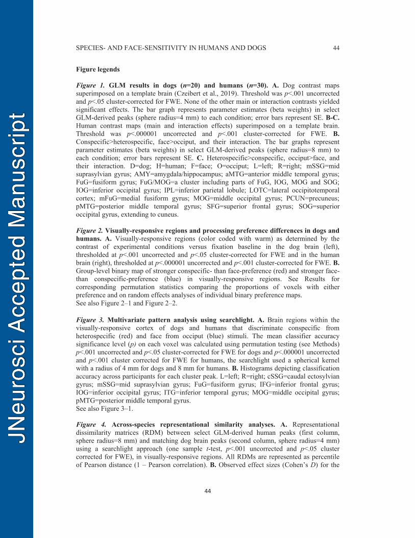
SPECIES- AND FACE-SENSITIVITY IN HUMANS AND DOGS 44
44
Figure legends Figure 1. GLM results in dogs (n=20) and humans (n=30). A. Dog contrast maps superimposed on a template brain (Czeibert et al., 2019). Threshold was p<.001 uncorrected and p<.05 cluster-corrected for FWE. None of the other main or interaction contrasts yielded significant effects. The bar graph represents parameter estimates (beta weights) in select GLM-derived peaks (sphere radius=4 mm) to each condition; error bars represent SE. B-C. Human contrast maps (main and interaction effects) superimposed on a template brain. Threshold was p<.000001 uncorrected and p<.001 cluster-corrected for FWE. B. Conspecific>heterospecific, face>occiput, and their interaction. The bar graphs represent parameter estimates (beta weights) in select GLM-derived peaks (sphere radius=8 mm) to each condition; error bars represent SE. C. Heterospecific>conspecific, occiput>face, and their interaction. D=dog; H=human; F=face; O=occiput; L=left; R=right; mSSG=mid suprasylvian gyrus; AMY=amygdala/hippocampus; aMTG=anterior middle temporal gyrus; FuG=fusiform gyrus; FuG/MOG=a cluster including parts of FuG, IOG, MOG and SOG; IOG=inferior occipital gyrus; IPL=inferior parietal lobule; LOTC=lateral occipitotemporal cortex; mFuG=medial fusiform gyrus; MOG=middle occipital gyrus; PCUN=precuneus; pMTG=posterior middle temporal gyrus; SFG=superior frontal gyrus; SOG=superior occipital gyrus, extending to cuneus. Figure 2. Visually-responsive regions and processing preference differences in dogs and humans. A. Visually-responsive regions (color coded with warm) as determined by the contrast of experimental conditions versus fixation baseline in the dog brain (left), thresholded at p<.001 uncorrected and p<.05 cluster-corrected for FWE and in the human brain (right), thresholded at p<.000001 uncorrected and p<.001 cluster-corrected for FWE. B. Group-level binary map of stronger conspecific- than face-preference (red) and stronger face- than conspecific-preference (blue) in visually-responsive regions. See Results for corresponding permutation statistics comparing the proportions of voxels with either preference and on random effects analyses of individual binary preference maps. See also Figure 2–1 and Figure 2–2. Figure 3. Multivariate pattern analysis using searchlight. A. Brain regions within the visually-responsive cortex of dogs and humans that discriminate conspecific from heterospecific (red) and face from occiput (blue) stimuli. The mean classifier accuracy significance level (p) on each voxel was calculated using permutation testing (see Methods) p<.001 uncorrected and p<.05 cluster-corrected for FWE for dogs and p<.000001 uncorrected and p<.001 cluster corrected for FWE for humans, the searchlight used a spherical kernel with a radius of 4 mm for dogs and 8 mm for humans. B. Histograms depicting classification accuracy across participants for each cluster peak. L=left; R=right; cSSG=caudal ectosylvian gyrus; mSSG=mid suprasylvian gyrus; FuG=fusiform gyrus; IFG=inferior frontal gyrus; IOG=inferior occipital gyrus; ITG=inferior temporal gyrus; MOG=middle occipital gyrus; pMTG=posterior middle temporal gyrus. See also Figure 3–1. Figure 4. Across-species representational similarity analyses. A. Representational dissimilarity matrices (RDM) between select GLM-derived human peaks (first column, sphere radius=8 mm) and matching dog brain peaks (second column, sphere radius=4 mm) using a searchlight approach (one sample t-test, p<.001 uncorrected and p<.05 cluster corrected for FWE), in visually-responsive regions. All RDMs are represented as percentile of Pearson distance (1 – Pearson correlation). B. Observed effect sizes (Cohen’s D) for the

SPECIES- AND FACE-SENSITIVITY IN HUMANS AND DOGS 45
45
across-species matching of RDMs for each peak-pair (first column), and modelled effect size patterns reflecting potential driving forces underlying across-species matching (second column), see also Figure 4–3. C=conspecific; He=heterospecific; F=face; O=occiput; L=left; R=right; AMY=amygdala/hippocampus; FuG=fusiform gyrus; cSSG=caudal suprasylvian gyrus; EMG=ectomarginal gyrus; mESG=mid ectosylvian gyrus; MG=marginal gyrus; mSSG=mid suprasylvian gyrus; rESG=rostral ectosylvian gyrus. See also Figure 4–1, Figure 4–2, Figure 4–3. Video legends Movie 1. fMRI stimuli. Video shows sample dynamic images, presented in color and dynamically for 2 s, representing each of four conditions human face, human occiput, dog face, and dog occiput. Note. Stimulus design was identical for dogs and humans.


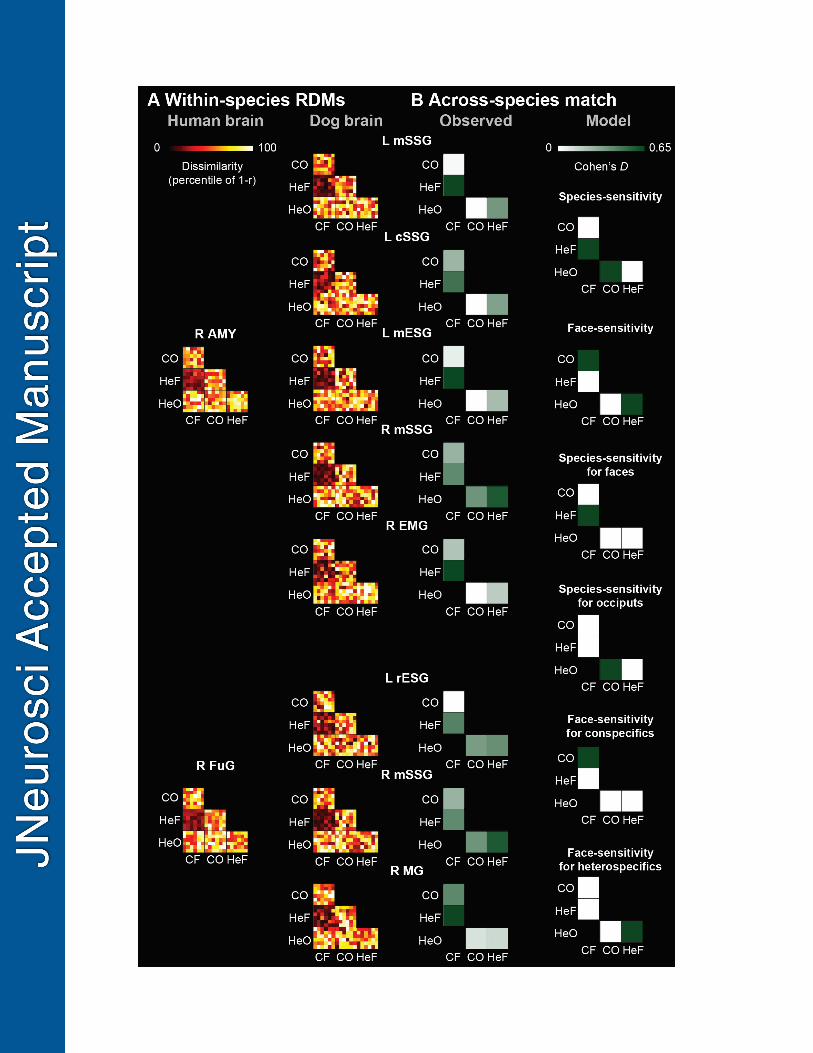


![Comparative Analysis of Arabidopsis Ecotypes Reveals a · Comparative Analysis of Arabidopsis Ecotypes Reveals a Role for Brassinosteroids in Root Hydrotropism1[OPEN] Rui Miao,a,2](https://img.pdfslide.us/doc/110x75/5fd77669ed73d6608c569009/comparative-analysis-of-arabidopsis-ecotypes-reveals-a-comparative-analysis-of-arabidopsis.jpg)