Embed Size (px)
Citation preview
University of Tennessee, Knoxville University of Tennessee, Knoxville
TRACE: Tennessee Research and Creative TRACE: Tennessee Research and Creative
Exchange Exchange
Doctoral Dissertations Graduate School
12-2001
Comparative Anatomy of the Lower Respiratory Tract of the Gray Comparative Anatomy of the Lower Respiratory Tract of the Gray
Short-tailed Opossum (Monodelphis domestica) and North Short-tailed Opossum (Monodelphis domestica) and North
American Opossum (Didelphis virginiana) American Opossum (Didelphis virginiana)
Lee Anne Cope University of Tennessee - Knoxville
Follow this and additional works at: https://trace.tennessee.edu/utk_graddiss
Part of the Animal Sciences Commons
Recommended Citation Recommended Citation Cope, Lee Anne, "Comparative Anatomy of the Lower Respiratory Tract of the Gray Short-tailed Opossum (Monodelphis domestica) and North American Opossum (Didelphis virginiana). " PhD diss., University of Tennessee, 2001. https://trace.tennessee.edu/utk_graddiss/2046
This Dissertation is brought to you for free and open access by the Graduate School at TRACE: Tennessee Research and Creative Exchange. It has been accepted for inclusion in Doctoral Dissertations by an authorized administrator of TRACE: Tennessee Research and Creative Exchange. For more information, please contact [email protected].
To the Graduate Council:
I am submitting herewith a dissertation written by Lee Anne Cope entitled "Comparative
Anatomy of the Lower Respiratory Tract of the Gray Short-tailed Opossum (Monodelphis
domestica) and North American Opossum (Didelphis virginiana)." I have examined the final
electronic copy of this dissertation for form and content and recommend that it be accepted in
partial fulfillment of the requirements for the degree of Doctor of Philosophy, with a major in
Animal Science.
Robert W. Henry, Major Professor
We have read this dissertation and recommend its acceptance:
Dr. R.B. Reed, Dr. C. Mendis-Handagama, Dr. J. Schumacher, Dr. S.E. Orosz
Accepted for the Council:
Carolyn R. Hodges
Vice Provost and Dean of the Graduate School
(Original signatures are on file with official student records.)
To the Graduate Council:
I am submitting herewith a dissertation written by Lee Anne Cope entitled “Comparative
Anatomy of the Lower Respiratory Tract of the Gray Short-tailed Opossum
(Monodelphis domestica) and North American Opossum (Didelphis virginiana).” I have
examined the final electronic copy of this dissertation for form and content and
recommend that it be accepted in partial fulfillment of the requirements for the degree of
Doctor of Philosophy, with a major in Animal Science.
Robert W. Henry, Major Professor We have read this dissertation and recommend its acceptance: Dr. R.B. Reed Dr. C. Mendis-Handagama Dr. J. Schumacher Dr. S.E. Orosz Accepted for the Council: Dr. Anne Mayhew Vice Provost and Dean of
Graduate Studies
(Original signatures are on file in the Graduate Student Services Office.)
COMPARATIVE ANATOMY OF THE LOWER RESPIRATORY TRACT OF
THE GRAY SHORT-TAILED OPOSSUM (Monodelphis domestica) AND NORTH
AMERICAN OPOSSUM (Didelphis virginiana)
A Dissertation
Presented for the
Doctor of Philosophy
Degree
The University of Tennessee, Knoxville
Lee Anne Cope
December 2001

ii
Copyright © Lee Anne Cope, 2001
All rights reserved
iii
DEDICATION
This dissertation is dedicated to my family
Mr. and Mrs. Charles Lee Cope
and
Mr. and Mrs. Jeffrey Morrow Cope and their children
Jeffrey Scott Cope and Ashley Channing Cope.
iv
ABSTRACT The present study describes the lower respiratory tract anatomy of the gray short-
tailed opossum (Monodelphis domestica) and North American opossum (Didelphis
virginiana). The trachea of the gray short-tailed opossum consists of 25 c-shaped
tracheal cartilages. The trachea of the North American opossum consists of 28 c-shaped
cartilages. The right lung of both species is separated into cranial, middle, caudal and
accessory lobes by interlobar fissures. The left lung consists of unseparated cranial and
caudal lobes. The right and left pulmonary arteries of the gray short-tailed and North
American opossums divide into pulmonary lobar arteries. The pulmonary lobar veins
join to form pulmonary veins. In the gray short-tailed opossum, the pulmonary lobar
veins join to form a right and left pulmonary vein which join to form a common
pulmonary venous trunk. In the North American opossum, a similar pattern occurs
however the common pulmonary venous trunk is formed from three pulmonary veins
(right, left and middle). Vascularization of the lung parenchyma is via the bronchial
artery, a branch of the bronchoesophageal artery. Right and left bronchial branches
course along the dorsal surface of the principal bronchi toward the hilus of the lung. In
both species, the left bronchial branch anastomoses with a mediastinal artery originating
from the aorta. Cranial deep cervical, cranial mediastinal and tracheobronchial lymph
nodes drain the lower respiratory tract of both species. Sympathetic innervation to the
lungs of the opossums comes from the sympathetic trunks as thoracic splanchnic nerves.
Parasympathetic innervation to the lungs is via branches from the vagus nerves.
v
The trachea and principal bronchi of the gray short-tailed opossum are lined by
pseudostratified ciliated columnar epithelium. Bronchial cartilages are irregular, shaped
plates and localized to the extrpulmonary portion of the principal bronchus. The
secondary and tertiary bronchi and primary and secondary bronchioles are lined by
simple ciliated columnar epithelium. The terminal bronchioles and proximal portion of
the respiratory bronchioles are lined by simple ciliated cuboidal epithelium. The distal
portion of the respiratory bronchioles and the alveolar ducts are lined by simple
squamous epithelium. The alveoli are lined by type I and II pneumocytes.
vi
TABLE OF CONTENTS
CHAPTER PAGE
I. INTRODUCTION…………………………………………………………... 1
II. REVIEW OF THE LITERATURE…………………………………………. 4
Trachea…………………………………………………………………..….. 4
Bronchi………………………………………………………………….…... 7
Alveoli and their cells……………………………………………………… 10
Lungs……………………………………………………………………….. 11
Pleura……………………………………………………………………..… 12
Pulmonary vessels…………………………………………………………... 13
Bronchial artery…………………………………………………………….. 14
Lymph nodes of the lower respiratory tract…… …………………………. 14
III. MATERIALS AND METHODS…………………………………………... 18
Anesthesia, catheterization and exsanguination…………………………… 18
Group I (Macroscopic Anatomy)…………………………………………… 19
Group II (Microscopic Anatomy)…………………...……………………… 23
IV. MACROSCOPIC ANATOMY OF THE LOWER RESPIRATORY
TRACT OF THE GRAY SHORT-TAILED OPOSSUM
(Monodelphis domestica)…………………………………………………... 26
vii
TABLE OF CONTENTS
CHAPTER PAGE
Abstract……………………………………………………………………. 26
Introduction………………………………………………………………… 26
Materials and Methods…………………………………………………….. 27
Results……………………………………………………………………... 28
Discussion…………………………………………………………………. 40
V. MACROSCOPIC ANATOMY OF THE LOWER
RESPIRATORY TRACT OF THE NORTH AMERICAN
OPOSSUM (Didelphis virginiana)………………………………………. 44
Abstract……………………………………………………………………. 44
Introduction………………………………………………………………... 44
Materials and Methods…………………………………………………….. 45
Results……………………………………………………………………… 46
Discussion………………………………………………………………….. 58
VI. ANATOMY OF STRUCTURES ASSOCIATED WITH THE
LOWER RESPIRATORY TRACT OF THE GRAY
SHORT-TAILED OPOSSUM (Monodelphis domestica)…………………. 62
Abstract……………………………………………………………………. 62
Introduction………………………………………………………………… 62
Materials and Methods……………………………………………………... 63
viii
TABLE OF CONTENTS
CHAPTER PAGE
Results……………………………………………………………………… 64
Discussion………………………………………………………………….. 79
VII. ANATOMY OF STRUCTURES ASSOCIATED WITH THE
LOWER RESPIRATORY TRACT OF THE NORTH AMERICAN
OPOSSUM (Didelphis virginiana)……………………………………….. 82
Abstract……………………………………………………………………. 82
Introduction………………………………………………………………… 83
Materials and Methods…………………………………………………….. 83
Results……………………………………………………………………... 84
Discussion…………………………………………………………………. 97
VIII. MICROSCOPIC ANATOMY OF THE LOWER RESPIRATORY
TRACT OF THE GRAY SHORT-TAILED
OPOSSUM (Monodelphis domestica)……………………………………. 100
Abstract…………………………………………………………………… 100
Introduction……………………………………………………………….. 101
Materials and Methods…………………………………………………… 102
Results……………………………………………………………………. 105
Discussion………………………………………………………………... 128
ix
TABLE OF CONTENTS
CHAPTER PAGE
IX. CONCLUSION………………………………………………………….. 135
REFERENCES………………………………………………………………………. 137
VITA………………………………………………………………………………….. 149
x
LIST OF FIGURES
FIGURE PAGE
4 – 1. Ventral view of lower respiratory tract in situ………………………………... 29
4 – 2. Cross section of trachea………………………………………………………. 30
4 – 3. Lateral view of right lung……………………………………………………... 33
4 – 4. Ventral view of thoracic viscera………………………………………………. 35
4 – 5. Lateral view of left lung………………………………………………………. 37
4 – 6. Ventral view of tracheobronchial cast………………………………….……... 38
5 – 1. Ventral view of thoracic respiratory tract (heart and great
vessels removed)……………………………………………………….. …... 47
5 – 2. Caudal view of lungs……………………………………………………. …… 49
5 – 3. Dorsal view of the ventral thoracic cavity and sternum………………….…… 50
5 – 4. Lateral view of right lung……………………………………………………. . 52
5 – 5. Lateral view of left lung………………………………………………………. 55
5 – 6. Ventral view of lower respiratory system cast…………………………. ……. 56
5 – 7. Ventral view of bronchial cast…………………………………………………. 57
6 – 1. Tracheobronchial vascular cast – ventral view…………………………. …….. 65
6 – 2. Ventral view of cranial portion of lungs……………………………………….. 67
6 – 3. Right view of thoracic cavity (right lung (L) reflected
ventrally)………………………………………………………………. ……. 68
xi
LIST OF FIGURES
FIGURE PAGE
6 – 4. Right view of thoracic cavity (right lung (L) reflected)………………... ……... 69
6 – 5. Left view of thoracic cavity (left lung (L) reflected
ventrally)……………………………………………………………………… 71
6 – 6. Left ventral view of cervical region……………………………………. …….. 73
6 – 7. Ventral view of tracheobronchial lymph nodes……………………………….. 74
6 – 8. Ventral view of cervical and thoracic region………………………….……….. 78
7 – 1. Ventral view of tracheobronchial vascular cast……………………….………... 86
7 – 2. Right lateral view of dorsal mediastinal structures (ribs
reflected dorsally)……………………………………………………………… 87
7 – 3. Dorsal mediastinal structures (lateral view)……………………………………. 89
a. Left lateral view of dorsal mediastinal structures (dorsal
aspect of left lung pulled ventrally)………………………………………… 89
b. Close up of lateral view of dorsal mediastinal structures
(dorsal aspect of left lung retracted ventrally)……………………………... 90
7 – 4. Left ventrolateral view of cranial cervical region……………………………… 92
7 – 5. Ventral view of cranial mediastinal lymph nodes…………………………….. 93
7 – 6. Tracheobronchial lymph nodes………………………………………………. 94
8 – 1. Cross section through the tunica mucosa of the trachea………………………. 106 8 – 2. Cross section of the trachea…………………………………………………… 108
xii
LIST OF FIGURES
FIGURE PAGE
8 – 3. Tracheal gland in the tela submucosa of the trachea. ………………………… 108 8 – 4. Tracheal gland duct (D)………………………………………………………. 109 8– 5. Tracheal gland. ………………………………………………………….……. 109 8 – 6. Trachealis muscle (T) attaching to the luminal side of
the tracheal cartilage…………………………………………………….……. 111
8 – 7. Membranous carina of the tracheal bifurcation………………………….……. 113
8 – 8. Autonomic ganglion…………………………………………………………... 113 8 – 9. Cross section of tunica mucosa of principal bronchus………………………… 115 8 – 10. Extrapulmonary part of principal bronchus………………………………….. 116 8 – 11. Layers of intrapulmonary portion of principal bronchus……………….…. .. 118 8 – 12. Longitudinal section of secondary bronchus………………………………… 120 8 – 13. Longitudinal section of tertiary bronchus……………………………….…… 120 8– 14. Cross section of primary bronchiole………………………………………….. 121 8– 15. Tunica mucosa of primary bronchiole………………………………………… 121 8– 16. Longitudinal section of terminal airways…………………………………….. 123 8– 17. Cross section of respiratory bronchiole………………………………………. 123 8– 18. Tunica mucosa of the respiratory bronchiole………………………………….. 124 8 – 19. Lung parenchyma……………………………………………………………. 126 8 – 20. Alveolar cells………………………………………………………………… 126
xiii
LIST OF FIGURES
FIGURE PAGE
8 – 21. Alveolar macrophage passing through alveolar pore………………………… 127 8 – 22. Visceral pleura of the lung…………………………………………………… 127
1
CHAPTER I
INTRODUCTION:
Marsupials have been considered an ideal animal for biomedical research because of
their short gestation period and a semiembryonic state of development of the offspring at
birth (VandeBerg, 1983). However, the only indigenous species of marsupial in the
United States is the North American opossum (Didelphis virginiana) and it has not
proven to be an ideal laboratory animal due to its large size. The North American
opossum also shows decreased reproductive capability, exhibits cannibalism of their
offspring and has an aggressive behavior toward other animals in captivity (VandeBerg,
1983). Also, the only commercial source for the North American opossum is wild caught
animals. Other species of marsupials from Australia and New Guinea have been used but
they also exhibit the same limitations as a lab animal as the North American opossum
(VandeBerg, 1983).
Until 1979, no marsupial species was considered to be ideal for biomedical research
or easily adaptable to laboratory conditions (VandeBerg, 1983). However, the gray
short-tailed opossum (Monodelphis domestica) has most of the desirable characteristics
of a lab animal without the problems encountered in other marsupials. The adult, gray
short-tailed opossum weighs only 80 to 150 grams allowing it to be housed in pairs in
standard plastic rat cages with little aggression (Fadem et al., 1982; VandeBerg, 1983).
The gestation period of the female opossum is two weeks and the size of the litter varies
2
from 8 to 14 pups which reach sexual maturity at 4 to 5 months of age. Gray short-
tailed opossums are also docile making them easier to handle. These opossums, when
raised under laboratory conditions, are free of diseases and parasites found in wild -
caught marsupials. (Fadem et al., 1982; VandeBerg, 1983).
In 1978, nine gray short-tailed opossums were captured in Brazil by the National
Zoological Park. The following year, twenty of the offspring from the first and second
generations were donated to the Southwest Foundation for Biomedical Research
(VandeBerg, 1983). Since then, this foundation has established and maintained a colony
of approximately 1500 animals (VandeBerg, 1983; Oorschot et al., 1992). These
opossums have been used for in house studies on genetics and reproduction (VandeBerg,
1983) while some of the opossums have been made available to outside investigators for
studies in various areas such as the development of the central nervous system (Kuehl-
Kovarik et al., 1995), embryogenesis (Klima, 1987; Baggott and Moore, 1990; Renfree,
1990; Selwood and VandeBerg, 1992; Szalay, 1994; Kuehl-Kovarik et al., 1995) and
embryonic morphogenesis and differentation (Filan, 1991; Hubbard et al., 1991; Krause,
1992; Morohunfola et al., 1992).
Since the gray short-tailed opossum has been used as a model in these various areas
of research, we realized a need for documentation of the normal macroscopic and
microscopic anatomy of the various organ systems. This study documents the normal
macroscopic and microscopic anatomy of the lower respiratory tract of the gray short-
tailed opossum including associated systems and structures. In addition, since the North

3
American opossum is native to much of the United States, comparisons are made to the
respective areas of the lower respiratory tract studied in the gray short-tailed opossum.
4
CHAPTER II
REVIEW OF LITERATURE:
The literature on the lower respiratory tract of marsupials is incomplete and scattered
over 144 years. This literature focused only on select regions such as the trachea
(Sonntag 1921a; Krause and Leeson 1973, 1975; Tucker, 1974), principal bronchi
(Krause and Leeson 1973, 1975), bronchial glands (Sorokin, 1965), type II pneumocytes
(Sorokin, 1967; Krause et al., 1976), lung lobation (Owen, 1852; Owen, 1868; Forbes,
1881; Parsons, 1903; Osgood, 1921; Sonntag 1921a, 1921b; Jones, 1949), pulmonary
vessels (McClure, 1903; Wade and Neely, 1949; Hill, 1955; Dowd, 1969), bronchial
circulation (Bernard et al., 1996) and associated lymph nodes (Zimmerman, 1940; Azzali
and Didio, 1965; Kampmeirer, 1969) of various marsupials.
TRACHEA:
The available literature on the trachea of marsupials such as the common shrew
opossum (Caenolestes obscurus) (Osgood, 1921), Perameles (Owen, 1868), native cat
(quoll) (Dasyurus viverrinus) (Hill, 1955) and mulgara (Dasycercus cristicauda) (Jones,
1949) concentrate on the shape of the tracheal cartilages. These articles indicate the
tracheal rings are incomplete dorsally similar to those of domestic mammals (Getty,
1975; Nickel et al., 1979; Banks, 1993; Evans, 1993). In addition, Owen (1868)
observed that the first twenty-four of the twenty-nine tracheal rings of the common
brush-tailed opossum (Trichosurus vulpecula) were complete. However, based on his
description it is not clear if the tracheal rings were truly complete or if the dorsal ends
5
overlapped similar to that of large domestic mammals such as the horse, cow, pig and
goat (Getty, 1975; Nickel et al., 1979). Along with the variation in the shape of the
tracheal rings, their number also varies among the marsupial species (Sonntag, 1921b) as
it does in domestic mammals (Getty, 1975; Nickel et al., 1979).
The literature found on the microscopic anatomy of the lower respiratory tract of
marsupials is limited to one report on the tracheal epithelium of the North American
opossum (Krause and Leeson, 1975). It reports the epithelium is simple ciliated
columnar in areas overlying cartilage rings and nonciliated simple columnar between the
tracheal cartilages. This differs from the pseudostratified ciliated columnar epithelium
lining the trachea of domestic mammals (Getty, 1975; Banks, 1993; Dellmann and Eurell,
1998) and several species of small rodents (Hansell, 1968; Jeffery, 1975; Becci, 1978;
Pack et. al, 1981).
In the North American opossum (Krause and Leeson, 1975), domestic mammals
(Banks, 1993; International Committee on Veterinary Gross Anatomical Nomenclature,
1994; Dellmann and Eurell, 1998) and rodents such as the rat (Jeffery, 1975), mouse
(Hansell, 1968; Pack et. al, 1981) and hamster (Becci, 1978), the predominant cell type
of the tracheal epithelium is the columnar cell. This cell has a basally placed nucleus.
Also depending on the species and location in the trachea, the apical (luminal) surface of
the columnar cell may be covered with cilia (Breeze and Wheeldon, 1977; Dellmann and
Eurell, 1998). Among these columnar cells are goblet cells and basal cells. In the North
American opossum, rat, mouse and hamster only a few goblet cells are seen in the entire
trachea in comparison to domestic mammals where they are found throughout the length
6
of the trachea. In all of these species, goblet cells are mucous secreting cells which have
a mass of mucous granules within the apical cytoplasm. The nucleus of the goblet cell is
basally located and surrounded by cytoplasmic organelles involved in the production of
mucus (Breeze and Turk, 1984; Burkitt et al., 1993; Dellmann and Eurell, 1998).
Basal cells are small, round to ovoid cells with a centrally located nucleus. These
cells function as germinal cells that differentiate and replace other epithelial cells
including the columnar or goblet cells (Breeze and Wheeldon, 1977; Dellmann and
Eurell, 1998). Basal cells are wedged between the other cells giving the epithelium a
pseudostratified appearance in domestic mammals (Breeze and Wheeldon, 1977;
Dellmann and Eurell, 1998), rat (Jeffery, 1975), mouse (Hansell, 1968; Pack et. al, 1981)
and hamster (Becci, 1978). These cells are not discussed in the North American
opossum.
Beneath the tracheal epithelium, a prominent vascular layer has been described in the
koala and phalanger (Tucker, 1974). This vascular layer is composed of two or three
rows of capillaries that may be close together (phalanger) or loosely organized (koala).
Based on Tucker’s (1974) description, this vascular layer must be part of the lamina
propria mucosae as it is in domestic mammals (Dellmann and Eurell, 1998).
The tracheal glands of the North American opossum are found within the tela
submucosa similar to that of domestic mammals (Getty, 1975; Burkitt et al., 1993;
Dellman and Eurell, 1998). These glands are abundant throughout the length of the
trachea in contrast to domestic carnivores where they are numerous in the ventral and
lateral walls of the proximal trachea and decrease in number distally (Getty, 1975; Burkitt
7
et al, 1993; Dellman and Eurell, 1998). The tracheal glands of the North American
opossum are composed of mucous and serous cells which form acini (adenomeres) that
are surrounded by myoepithelial cells (Krause and Leeson, 1973). Their description of
these mucous cells suggest similarities to domestic mammals due to their pear shape, the
mass of mucus granules within the apical cytoplasm and the flattened basally positioned
nucleus. However, Krause and Leeson (1973) do not describe the histological features of
the serous cells in the opossum tracheal glands. They assume these cells have similar
features to the serous cells described in a detailed study of the bronchial glands of the
North American opossum by Sorokin (1965b). In this article, Sorokin (1965b) states the
serous cells of these glands are more specialized than those of domestic carnivores
because of their ultrastructural and functional characteristics which are similar to cells
specialized for ion and water secretion such renal tubular cells. In the North American
opossum (Krause and Leeson, 1973) and domestic mammals (Getty, 1975; Schaller,
1992; Banks, 1993; Dellmann and Eurell, 1998), the tracheal glands open into the
tracheal lumen via excretory ducts that pass through the tela submucosa and the lamina
propria mucosae. The epithelium of this duct in the North American opossum is not
described as it is in domestic mammals (Dellmann and Eurell, 1998). However, Krause
and Leeson (1973) state mucous secreting cells are often present among the epithelial
cells of the excretory duct.
BRONCHI:
The literature on the bronchial tree of marsupials is not as extensive as that of
domestic mammals (Getty, 1975; Banks, 1993; Evans, 1993; Dyce et al., 1996; Dellmann
8
and Eurell, 1998). Two articles trace the sequence of changes that occur within the
principal bronchi of the North American opossum from birth until 85 days of age and a
third article describes the bronchial glands of this marsupial (Sorokin, 1965b; Krause and
Leeson, 1973, 1975). According to Krause and Leeson (1973), the epithelium of the
principal bronchi is simple columnar with patches of ciliated and nonciliated cells. This
epithelium is supported by a lamina propria mucosae which has an abundance of elastic
fibers similar to domestic mammals (Krause and Leeson, 1973; Banks, 1993). Deep to
this layer in the North American opossums and domestic mammals is a layer of smooth
muscle called the lamina muscularis mucosae (Krause and Leeson, 1973; Banks, 1993).
The smooth muscle of this layer is arranged in a helical pattern and separates the lamina
propria mucosae from the tela submucosa.
The tela submucosa is composed of loose connective tissue and seromucous bronchial
glands (Krause and Leeson, 1973; Schaller, 1992; Banks, 1993; Burkitt et al., 1993;
International Committee on Veterinary Gross Anatomical Nomenclature, 1994). In the
North American opossum (Sorokin, 1965b; Jeffery, 1983), these glands are described as
compound acinar glands and extend the length of the bronchial tree as they do in
domestic mammals (Getty, 1975). However, Sorokin (1965b) and Jeffery (1983) also
observed these glands extending into the bronchiolar tree similar to the domestic feline
(Getty, 1975; Jeffery, 1983). The bronchial glands of the North American opossum and
domestic mammals (Getty, 1975) open into the lumen of the bronchi by an excretory
duct. In the North American opossum, Sorokin (1965b) reports this duct is lined by
nonsecretory cuboidal cells while Krause and Leeson (1973) state the duct is lined by a
9
columnar epithelium. Leading into the main duct of this bronchial gland are one to three
orders of smaller ducts lined with secretory cells. These cells are described as either
mucous cells or special serous cells, referred to by Sorokin (1965b) as hydrotic cells.
The mucous cells are similar to those of domestic mammals. They are pear shaped
with a foamy, slightly basophilic cytoplasm and a flattened basally positioned nucleus
that is surrounded by cytoplasmic organelles such as mitochondria, rough endoplasmic
reticulum and golgi apparatus (Banks, 1993; Burkitt et al., 1993). The mucous cells of
these glands of the North American opossum may lie near its opening into the bronchial
lumen, line one of the smaller ducts which leads into the main duct, line part of main
excretory duct, or form acini (Sorokin, 1965b). The serous cells of the bronchial glands
of the North American opossum are different from those of domestic mammals (Banks,
1993; Burkitt et al., 1993) because of surface modifications such as microvilli on the
apical surface and changes in the internal cellular organelle organization. Therefore,
Sorokin (1965b) states the serous cells have characteristics similar to cells functioning in
water and ion secretion or absorption such as those of salivary glands or cells from renal
tubules of the kidney. In addition to lining the smaller ducts of the bronchial glands, the
serous cells are found in the acini among mucous cells forming serous acini or arranged
peripheral to the mucous acini as serous crescents (demilunes) (Sorokin, 1965b). These
demilunes are drained similar to those of domestic mammals (Banks, 1993) by
intercellular canaliculi (Sorokin, 1965b). The acini of the bronchial glands of the North
American opossum are surrounded by stellate shaped myoepithelial cells (Sorokin,
1965b; Krause and Lesson, 1973). These cells lie inside the basal lamina and extend
10
along the ducts until the epithelium becomes cuboidal and nonsecretory in the main duct
(Sorokin, 1962, 1965b).
ALVEOLI AND THEIR CELLS:
The alveoli in the lungs of the North American opossum (Sorokin, 1967; Krause et
al., 1976) and domestic mammals (Schaller, 1992; Banks, 1993; Dellmann and Eurell,
1998) are lined by type I (alveolar type I cell) and type II pneumocytes (alveolar type II
cell). The type I pneumocyte is the most abundant cell lining the alveoli of the North
American opossum and domestic mammals (Sorokin, 1967; Banks, 1993). This cell is a
simple squamous epithelial cell with a flattened nucleus that protrudes into the alveolar
lumen (Sorokin, 1967; Banks, 1993; Dellmann and Eurell, 1998). The thin extremities of
the type I pneumocytes join those of other type I pneumocytes to form a continuous
lining of the alveolus (Sorokin, 1967; Banks, 1993). In the North American opossum
(Sorokin, 1967; Krause et al., 1976) and domestic mammals (Banks, 1993, Dellmann and
Eurell, 1998), the type II pneumocyte is the secretory cell of the alveoli and scattered
among the type I pneumocytes (Banks, 1993; Dellmann and Eurell, 1998). The type II
pneumocyte is a round to cuboidal shaped cell with a foamy, vacuolated cytoplasm and a
large centrally placed vesicular nucleus. In the North American opossum, Sorokin (1967)
and Krause et al., (1976) also observed that the apical surface of this cell is covered with
microvilli and may be seen in groups of two or three.
11
LUNGS:
Lobation of the right and left lungs of marsupials has been described in several
species such as opossums (Tyson, 1698; Owen, 1868; Osgood, 1921; Sonntag, 1921a),
bandicoots (Parsons, 1903; Sonntag, 1921a), kangaroos (Sonntag, 1921a), quoll (Owen,
1868), mulgara (Jones, 1949), koala (Owen, 1868; Forbes, 1881; Sonntag, 1921b),
wallaroo (Sonntag, 1921a) and wombat (Owen, 1868). These descriptions indicate some
marsupials have lung lobation identical to domestic carnivores (Evans, 1993).
The right lung lobation in the North American opossum, mouse opossum (Marmosa
elegans), gray four-eyed opossum (Metachirus opossum), Phalangeridae (Sonntag,
1921a), brush-tailed opossum (Trichosurus vulpecula) (Sonntag, 1921b), common shrew
opossum (Caenolestes obscurus) (Osgood, 1921), quoll (Owen, 1868), mulgara (Jones,
1949), Perameles and Petaurists (Owen, 1868) and long nosed bandicoot (Perameles
obesula) (Sonntag, 1921a) is identical domestic carnivores (Evans, 1993). However,
previous reports by Tyson (1698) and Owen (1868) state the right lung of the North
American opossum consists of three lobes instead of the four described by Sonntag
(1921a). Several other marsupials also possess right lung lobation which differs from
domestic carnivores. The right lung of the tree kangaroo (Dendorlagus ursinus), eastern
grey kangaroo (Macropus giganteus) and wallaroo (Macropus bennetti) is trilobate and
described as having a deep median sulcus incompletely dividing the lung into anterior
and posterior parts in addition to the azygous lobe (Sonntag, 1921a). The right lung of
the red-legged short-tailed opossum (Didelphys brachyura) consists of three lobes and
the right lung of the wombat (Vombatus ursinus) consists of two lobes (Owen, 1868).
The right lung of the koala (Phascolactos cinereus) is described by Forbes (1881) as
12
having three lobes without the azygous lobe while Sonntag (1921b) states that the right
lung of the koala consists of only two lobes. The right lung of the common shrew
opossum has three lobes, however Osgood (1921) also reports the right lung of one of the
three specimens consists of four lobes. This indicates variability in lung lobation within
this species.
The left lung of the North American opossum (Owen, 1868), brush-tailed opossum
(Sonntag, 1921b), mouse opossum (Sonntag, 1921a), Phalangeridae (Owen, 1868;
Sonntag, 1921a), koala (Owen, 1868; Forbes, 1881), quoll (Owen 1868), mulgara (Jones,
1948) and Petaurists (Owen, 1868) is described as having a cranial and caudal lobe
similar to that of domestic carnivores (Nickel et al., 1979; Evans, 1993). In addition, a
previous report by Tyson (1698) states the left lung of the North American opossum is
unilobate. This is identical to the lobation of the gray four-eyed opossum, long-nosed
bandicoot (Sonntag, 1921a), pig-footed bandicoot (Choeropus castanotis) (Parsons,
1903), common shrew opossum (Osgood, 1921) and wombat (Owen, 1868) which is
based on the external appearance of the lung as branching of the bronchial tree is not
mentioned.
PLEURA:
The visceral (pulmonary) pleura covering the lungs in the North American opossum
(Krause and Leeson, 1973, 1975) and domestic carnivores (Evans, 1993) is simple
squamous mesothelium. In the North American opossum, a thick connective tissue layer
of collagen and elastic fibers, referred to as the subplerual lamina, lies beneath the
mesothelial basal lamina. This subplerual lamina is similar to the tunica subserosa
13
described in domestic carnivores (Banks, 1993; International Committee on Veterinary
Gross Anatomical Nomenclature, 1994). According to Krause and Leeson (1973, 1975),
the subpleural lamina of the North American opossum gives off septa which blend with
the connective tissue components of the lung. Based on this description, these septa must
be similar to those from the tunica subserosa of domestic carnivores which are
continuous with the interalveolar septa of the lung (Banks, 1993; Burkitt et al., 1993).
PULMONARY VESSELS:
Descriptions on the pulmonary vasculature of marsupials is limited to the pulmonary
venous return to the heart. These articles on the North American opossum (Owen, 1868;
McClure, 1903; Wade and Neely, 1949), brush-tailed opossum (Dowd, 1969) and native
cat (Hill, 1955) describe a common pulmonary venous trunk opening into the left atrium.
In two specimens of the North American opossum, the common brush-tailed opossum
and native cat, the common pulmonary venous trunk is formed from a common right and
common left pulmonary vein. In the North American opossum, Wade and Neely (1949)
state these vessels are formed by two veins from each lung joining to form a common
right and common left pulmonary vein before entering the left atrium. This pattern
differs from that of domestic mammals (Nickel et al., 1981; Evans, 1993) in which the
number of pulmonary veins usually corresponds to the number of lung lobes and these
vessels remain separate to the heart to open individually into the left atrium. However,
Wade and Neely (1949) also observed in two specimens four pulmonary veins entering
directly into the left atrium of the North American opossum similar to the pattern in
domestic mammals.
14
BRONCHIAL ARTERY:
The blood supply to the lung parenchyma of the North American opossum (Bernard
et al., 1996) and domestic carnivores (Nickel et al., 1981; Evans, 1993) originates from
right and left bronchial branches of the bronchial artery. In the North American
opossum, the bronchial artery originates from either a distinct bronchoesopahgeal artery
or a common bronchial artery from the ventral aspect of the thoracic aorta at the fifth
intercostal space. Then similar to domestic carnivores, the bronchial artery of the North
American opossum divides into right and left bronchial branches at the tracheal
bifurcation. Previous descriptions of this artery in domestic mammals (McLaughlin et al.,
1961; Evans, 1993) state the right and left branches supply the respective
tracheobronchial lymph nodes, the peribronchial connective tissue and the pulmonary
artery and vein by vaso vasorum. However, these bronchial branches do not contribute to
the blood supply of the visceral pleura of domestic carnivores as they do in other
domestic mammals (McLaughlin et al., 1961). The right and left bronchial branches then
enter the associated lung and follow the bronchial divisions (Nickel et al., 1981; Evans,
1993). In domestic carnivores, these branches terminate at the level of the respiratory
bronchioles in a capillary bed that is continuous with that of a pulmonary artery (Evans,
1993). No mention is made of the structures supplied or the branching pattern of the
right and left bronchial branches within the lung of the North American opossum.
LYMPH NODES OF THE LOWER RESPIRATORY TRACT:
The North American opossum (Zimmerman, 1940; Kampmeier, 1969) and large
American opossums of South America (Didelphis marsupialis and Didelphis azarae)
15
(Azzali and Didio, 1965) have a pair of lymph nodes near the laryngotracheal junction
and lateral to the neurovascular bundle of the neck. Zimmerman (1940) and Kampmeier
(1969) refer to these lymph nodes as superior cervical lymph nodes and Azzali and Didio
(1965) refer to them as deep cervical lymph nodes. These lymph nodes lay in the same
region as the cranial deep cervical lymph nodes of domestic carnivores (Nickel et al.,
1981; Evans, 1993).
Previous descriptions on the North American opossum (Zimmerman, 1940;
Kampmeier, 1969) and American opossums of South America (Azzali and Didio, 1965)
document anterior mediastinal lymph nodes. These lymph nodes are cranial to the base
of the heart at the level of the first ribs similar to the cranial mediastinal lymph nodes of
domestic carnivores (Nickel et al., 1981; Evans, 1993). In the North American opossum
and large American opossums of South America, an anterior mediastinal lymph node is
located on the right and left ventrolateral surfaces of the trachea (Zimmerman, 1940;
Azzali and Didio, 1965; Kampmeier, 1969). Azzali and Didio (1965) and Kampmeier
(1969) also describe posterior cranial mediastinal lymph nodes. These lymph nodes are
located on the ventral surface of the respective longus colli muscle with the left one
laying in the aortic arch and the right one laying medial to the first and second intercostal
spaces. In addition, Azzali and DiDio (1965) describe a single posterior middle
mediastinal lymph located along the dorsal body wall, to left of the thoracic duct and in
the arch of the left azygous vein. Based on their descriptions, the posterior middle
mediastinal lymph node may be similar to the caudal mediastinal lymph nodes found in
domestic mammals with the exception of domestic carnivores (Nickel et al., 1981).
16
In the large American opossums of South America, Azzali and DiDio (1965) have
described two bronchial lymph nodes. The cranial one lays on the ventrolateral aspect of
the trachea cranial to the tracheal bifurcation. The caudal bronchial lymph node lays in
the angle of the tracheal bifurcation on the ventral surface of the esophagus. This lymph
node is located in a similar position to that of the middle tracheobronchial lymph node of
domestic carnivores (Evans, 1993).
The lymph nodes of several marsupials such as the Northern brown bandicoot
(Isoodon macrourus) (Cisternas et al., 1999), quokka (Setonix brachyurus) (Ashman and
Papadimitriou, 1975), tammar wallaby (Macropus eugenii) (Basden et al., 1997) and fat-
tailed dunnart (Smithopsis crassicaudata) (Haynes, 1991) are dense encapsulated
lymphatic organs that are surrounded by a capsule similar to that of domestic carnivores
(Banks, 1993; Dellmann and Eurell, 1998). The capsule surrounding the lymph node of
the Northern brown bandicoot (Cisternas and Armati, 1999) and tammar wallaby (Basden
et al. 1997) is a thick connective tissue capsule which gives off trabeculae similar to
domestic carnivores. However, in the Northern brown bandicoot these trabeculae only
penetrate the superficial region of the cortex as compared to those of domestic carnivores
(Dellmann and Eurell, 1998) which extend into the cortical and medullary regions of the
node. Similar to domestic carnivores (Banks, 1993; Dellmann and Eurell, 1998), the
afferent lymphatic vessels of the Northern brown bandicoot (Cisternas and Armati, 1999)
enter the lymph node by passing through the capsule at various points and opening into
the subcapsular sinus. This sinus in the Northern brown bandicoot (Cisternas and
Armati, 1999) and tammar wallaby (Basden et al., 1997) contains lymphocytes,
17
erythrocytes, macrophages and plasma cells similar to domestic carnivores (Banks,
1993; Dellmann and Eurell, 1998).
The parenchyma of the lymph node of the tammar wallaby, Northern brown
bandicoot and quokka is composed of lymphocytes, plasma cells and macrophages that
are organized into an outer cortex and inner medulla similar to that of domestic
carnivores (Banks, 1993; Dellmann and Eurell, 1998). The cortex of the lymph nodes of
these marsupials and domestic carnivores consist of primary and secondary lymph
nodules and a deep or paracortical region (Ashman and Papadimitriou, 1975; Banks,
1993; Basden et al., 1997; Cisternas and Armati, 1999). This region is that portion of the
cortex that surrounds the primary and secondary lymph nodules. In domestic carnivores
(Dellmann and Eurell, 1998), the paracortical region is diffuse lymphatic tissue that
consists mainly of T-lymphocytes which are not organized into nodules (Banks, 1993;
Dellmann and Eurell, 1998). In the Northern brown bandicoot, Cisternas and Armati
(1999) state the paracortical region has numerous capillaries, which are characterized by
a flattened to cuboidal endothelial lining.
The medulla is centrally located toward the center of the lymph node in the Northern
brown bandicoot (Cisternas and Armati, 1999), tammar wallaby (Basden et al., 1997),
quokka (Ashman and Papadimitriou, 1975) and domestic carnivore (Banks, 1993;
Dellmann and Eurell, 1998). It is loosely organized into medullary cords which are
extensions of the paracortical region of the cortex and are separated from one another by
trabeculae and medullary sinuses (Ashman and Papadimitriou, 1975; Banks, 1993;
Basden et al., 1997; Dellmann and Eurell, 1998; Cisternas and Armati, 1999).
18
CHAPTER III
MATERIALS AND METHODS:
ANESTHESIA, CATHETERIZATION AND EXSANGUINATION:
To study the macroscopic and microscopic anatomy of the lower respiratory tract,
twenty-five (14 males and 11 females) gray short-tailed opossums (Monodelphis
domestica) and eighteen (9 males and 9 females) North American opossums (Didelphis
virginiana) were used. The gray short-tailed opossums were obtained via donation from
Iowa State University (Ames, Iowa) and purchased from Southwestern Foundation for
Biomedical Research (P.O. Box 760549 San Antonio, Texas, 78245). These opossums
were sexually mature and two years of age. The North American opossums were
obtained from the Department of Comparative Medicine at The University of Tennessee
College of Veterinary Medicine and the Tennessee Department of Natural Resources.
These opossums were various ages. The gray short-tailed and North American opossums
were randomly divided into two groups, to study the macroscopic anatomy and
microscopic anatomy. Eighteen (11 males and 7 females) gray short-tailed opossums and
sixteen (8 males and 8 females) North American opossums were used for the
macroscopic anatomy. Seven (3 males and 4 females) gray short-tailed opossums along
with the random samples taken from the opossums utilized for the gross anatomy, were
used to describe the microscopic anatomy of the lower respiratory tract.
Each gray short-tailed opossum was administered 500 I.U. of heparin sodium
(Heparin Sodium Injection, 1,000 units per 1.0 ml, Elkins-Sinn Inc. Cherry Hill, NJ
19
08003) via intraperitoneal (IP) injection to facilitate esanguination. Eighteen hours
later, the opossums were anesthetized with an IP injection of 50 mg of Nembutal
(Nembutal Sodium Solution, Pentobarbital Sodium Injection, Abbott Laboratories, North
Chicago, IL 60064) per 100.0 grams of body weight. When stage 3 anesthetic plane was
reached, the gray short-tailed opossums were placed in dorsal recumbency and the hair on
the ventrolateral surfaces of the neck was clipped. An incision was made through the
skin and the subcutaneous tissue over the right external jugular vein and the right
common carotid artery. These vessels were exteriorized by blunt dissection and
catheterized. A 20 gauge, 2 inch angiocath I.V. catheter (Becton Dickson Infusion
Therapy Systems Inc., Sandy, Utah 84070) was inserted into the external jugular vein and
a 22 gauge, 1 inch angiocath I.V. catheter into the common carotid artery for
exsanguination.
The North American opossums were anesthetized with an intramuscular injection
(IM) of 500 mg of Nembutal. When stage III anesthetic plane was reached, the right
external jugular vein and common carotid artery were exteriorized similar to that of the
gray short-tailed opossum. These vessels were then catheterized for exsanguination with
a 5.0 mm plastic cannula for the external jugular vein and a 3.0 mm plastic cannula for
the common carotid artery.
GROUP I (MACROSCOPIC ANATOMY):
The gray short-tailed and North American opossums of this group were used to
observe and record the gross anatomy of the lower respiratory tract. This was
20
accomplished by dissection, vascular injections and the production of tracheobronchial
and tracheobronchial vascular casts.
The gross anatomy of the lower respiratory tract was documented by dissecting a total
of eighteen (11 males and 7 females) gray short-tailed opossums and eighteen (9 males
and 9 females) North American opossums with each animal often being used for more
than one objective. Out of the eighteen gray short-tailed opossums, twelve animals were
used for descriptions of the trachea, lungs and pleura, eight animals were also used for
the associated structures of the lower respiratory tract, six animals were used for
tracheobronchial cast and two animals were utilized for tracheobronchial vascular casts.
For the North American opossum, all eighteen animals were utilized for the anatomy of
the trachea, lungs and pleura, fourteen animals were also utilized for associated
structures, four animals were used for trachebronchial casts and two were used for
tracheobronchial vascular casts.
To expose the lower respiratory tract after exsanguination a ventral midline incision
was made through the skin and subcutaneous tissue from the caudal border of the larynx
to the manubrium. The right and left sternohyoideus muscles were separated to expose
the cervical trachea. The cervical incision was extended caudally and lateral to each
sternoclavicular joint through the costal cartilages and intercostal muscles. The incisions
were joined caudal to the xiphoid process to remove the sternum thus exposing the
trachea, heart and lungs.
The vasculature of the lower respiratory tract was documented following embalming
and vascular injections of two (one male and one female) gray short-tailed opossums and
21
one (female) North American opossum. The gray short-tailed opossums were
embalmed with 15.0 to 34.0 ml of 10% buffered formalin and the North American
opossum was embalmed with 70.0 ml via the catheterized common carotid artery. The
next day red and blue latex were injected into the catheterized vessels. In the gray short-
tailed opossum, approximately 0.8 ml of red latex was injected into the common carotid
artery and 0.6 ml of blue latex was injected into the external jugular vein. In the North
American opossum, 20.0 ml and 10.0 ml each of red and blue latex were injected into the
common carotid artery and external jugular vein respectively. The opossums remained at
room temperature for twenty-four hours to assure hardening of the vascular injections.
Then they were submerged in 10% buffered formalin and stored at 5OC until dissection at
which time the chest was opened similar to that previously described.
A tracheobronchial cast was made of the conduction components of the lower
respiratory tract of six gray short-tailed opossums (4 males and 2 females) and four North
American opossums (2 males and 2 females). The chest was opened similar to the
previous description and the topography of the thoracic viscera was observed and
recorded for each species. The trachea was transected caudal to the larynx and a 3.0 mm
o.d plastic cannula for the gray short-tailed opossum and a 6.0 mm o.d. plastic cannula
for the North American opossum were inserted into the tracheal lumen. The cannulas
were ligated in place and the trachea and lungs were removed and air-dried for 48 hours
to remove the moisture from the lung parenchyma. Pressurized air was used to inflate
and maintain the lungs in normal inspiratory anatomical position until they were dry.
Following air-drying, 2.0 ml of RTV silicone (Silicone Inc. P.O. Box 363, 211 Woodbine
22
High Point, NC, 27261) for the gray short-tailed opossum and approximately 15.0 ml
for the North American opossum was injected through the cannulated trachea into the
airways of the lungs and allowed to harden at room temperature for 24 hours (Henry,
1992). After hardening, the lung parenchyma was removed by maceration in boiling
water. The tracheobronchial cast was cleaned first with water then soaked in 10%
hydrogen peroxide for final cleaning.
Tracheobronchial vascular casts were made from two (males) gray short-tailed
opossums and two (1 male and 1 female) North American opossums. In the gray short-
tailed opossums, red and blue latex was injected into the catheterized common carotid
artery and external jugular vein respectively. The opossums remained at room
temperature for twenty-four hours to assure hardening of the vascular injections. After
hardening, the trachea was exposed and transected and the chest was opened as described
previously. A 3.0 mm o.d. cannula was inserted into the tracheal lumen and ligated in
place. Approximately 2.0 ml of RTV silicone was infused using digital pressure through
the cannulated trachea into the airways and allowed to harden for twenty-four hours.
After hardening the lung and heart were removed as a unit and the parenchyma was
removed by maceration in water at room temperature. The tracheobronchial vascular
casts were cleaned first with water then soaked in 10% hydrogen peroxide for final
cleaning.
In the two North American opossums, the chest was opened similar to the gray short-
tailed opossum and the trachea, heart and lungs were removed as a unit. The trachea was
cannulated with a 6.0 mm o.d cannula and ligated in place. The conus arteriosus of the
23
heart was incised and cannulated with a 3.0 mm cannula. The cannula was directed into
the pulmonary trunk and ligated in place. The apex of the left auricle was incised and a
3.0 mm o.d. plastic cannula was inserted into the lumen of the left atrium and ligated in
place. The cannulas were then gently flushed with water to remove any blood clots prior
to injecting red and blue silicone. Approximately 3.0 ml of blue silicone and 6.0 ml of
red silicone were injected into the pulmonary arteries and pulmonary veins respectively.
Following this, pressurized air was used to inflate and maintain the lungs in normal
inspiratory anatomical position for 48 hours to remove the moisture from the lung
parenchyma. Following air-drying, 15.0 ml of RTV Silicone was infused into the
airways (Henry, 1992a and 1992b). After hardening the lung and heart parenchyma was
removed by maceration in boiling water. The tracheobronchial vascular casts were
cleaned first with water then soaked in 10% hydrogen peroxide for final cleaning.
GROUP II (MICROSCOPIC ANATOMY):
Following exsanguination, seven gray short-tailed opossums (three males and four
females) along with random tissue samples taken from the gray short-tailed opossums
used for the gross anatomy, were used for histologic study of the lower respiratory tract.
The trachea and lungs of the six gray short-tailed opossums were formalin fixed in situ by
intratracheal perfusion of 10% buffered formalin via a cannula from a height of 5.0 cm
for 5 minutes. After fixation, the trachea and lungs were removed and placed in 10%
buffered formalin for an additional 24-48 hours before sectioning. Tissue samples were
taken from all six lung lobes from four animals for study of the lung parenchyma. The
four tracheas from these animals were divided into cranial, middle and caudal thirds prior
24
to sectioning. The right and left lungs were removed from two opossums for serial
sectioning of the conducting components of the respiratory system and parenchyma of the
lung. All samples were processed for light microscopy using a Tissue TEK VIP 1000
(Floor Model, Mode #4617, Serial #8811895, Ames Division, Miles Laboratories Inc.,
P.O. Box 70, Elkhart, IN 46515) through a series of dehydration and infiltration. The
tissues were dehydrated in a graded series of ethanol (80%, 95%, 95%, 100%, 100%,
100%) followed by two series of xylene under a pressure (.35 kg.cm2) and vacuum cycle
(50.0 cm Hg) at 40OC for one hour. The samples were infiltrated with paraffin (Paraplast
Tissue embedding Medium, Oxford Labware, Division of Sherwood Medical, St. Louis,
MO 63103) under a pressure (.35 kg. cm2) and vacuum cycle (50.0 cm Hg) at 60OC for
one hour (repeated twice). Samples were then embedded in paraffin and positioned in
tissue molds to obtain blocks that would yield transverse sections through the trachea and
bronchi when cut. Random sections 5.0 µm (microns) in thickness were taken at 75.0 µm
intervals from the blocks containing the trachea and bronchi. Serial sections (5.0 µm
thick) were made from both lungs. All sections were mounted on glass slides and
stained with either hematoxylin and eosin or Acid Orcein Giemsa (Luna, 1968).
The trachea and lungs of the seventh gray short-tailed opossum (male) were fixed in
situ by vascular of perfusion of 2.5% gluteraldehyde (25% Gluteraldehyde EM Grade,
Electron Microscopy Sciences, PO Box 251, 321 Morris Road, Ft. Washington, PA
19034) in 0.1M cacodylate buffer (pH = 7.4) (Sodium Cacodylate Trihydrate, Electron
Microscopy Sciences, PO Box 251, 321 Morris Road, Ft. Washington, PA 19034) via
the catheterized left common carotid artery. The opossum was then placed in the cooler
25
at 5oC for 24 hours after which the thoracic cavity was opened and the trachea and lungs
removed and placed in new 2.5% gluteraldehyde in 0.1M cacodylate buffer. It was
returned to the cooler until samples were taken. Tissue samples were taken from the
trachea and lungs, cut into 1.0 mm cubes and washed three times, ten minutes each, in
distilled water. Samples were post fixed in a 1:1 mixture of 2% aqueous osmium
tetroxide and 3% aqueous potassium ferrocyanide at room temperature for 2 hours or
longer until the tissue became dark brown or black in color. The samples were then
removed from the osmium ferrocyanide and were again washed three times for ten
minutes in distilled water. Following this, the samples were dehydrated for ten minutes
each in 50%, 70%, 80% and 90% ethanol followed by two washes in 100% ethanol for 60
minutes each. Samples were then removed from the 100% ethanol and washed two
times, fifteen minutes each, in propylene oxide (Polysciences Inc. Warrington, PA
18976). After the final washing the tissue samples were placed in tissue micromolds
(Polysciences Inc. Warrington, PA 18976) and embedded in epon araldite (Russell and
Burguet, 1977). Random sections of 1.0 – 2.0 µm were cut, mounted on glass slides and
stained with a mixture of 1:1 mixture of methylene blue (Methylene Blue, Sigma
Chemical Company, PO Box 14508, St. Louis, MO 63178) and azure blue (Azure II,
Sigma Chemical Company, PO Box 14508, St. Louis, MO 63178).
26
CHAPTER IV
MACROSCOPIC ANATOMY OF THE LOWER RESPIRATORY TRACT OF
THE GRAY SHORT-TAILED OPOSSUM (Monodelphis domestica)
ABSTRACT:
The present study describes the macroscopic anatomy of the lower respiratory tract of
the gray short-tailed opossum (Monodelphis domestica). The trachea consists of
approximately 25 c-shaped tracheal cartilages and extends from the larynx to its
bifurcation into right and left principal bronchi. The right lung is divided into cranial,
middle, caudal and accessory lobes by interlobar fissures. The left lung consists of
cranial and caudal lobes which are not divided by an interlobar fissure. Lung lobation
was verified from tracheobronchial casts.
INTRODUCTION:
Gray short-tailed opossums have become practical for use as models in several fields of
research including embryogenesis (Baggott and Moore, 1990; Selwood and Vandeberg,
1992; Kuehl-Kovarik, 1995), reproduction, sexual differentiation, behavior, chemical
communication (Vandeberg, 1983 and 1995), nervous system development, DNA repair
mechanisms, cytogenetics and biochemical genetics (Vandeberg, 1990). They have also
been identified as the only laboratory mammal that develops melanoma in response to
ultraviolet radiation (Kusewitt et al., 1991; Ley et al., 1991; Sabourin et al., 1992;
Vanderberg et al., 1992; Robinson, 1994; Hubbard, 1997). Despite their increasing
27
popularity as a research animal, few anatomical descriptions of the gray short-tailed
opossum exist. Peukert-Adam et al. (1994) describe the pancreas while Koch et al.
(1990) provide a general description of the abdominal organs of the gray short-tailed
opossum. Kusewitt (1994) and Hubbard (1997) report pathological changes in the
respiratory and cardiovascular system of the gray short-tailed opossum but do not
describe normal thoracic anatomy. To benefit research involving these animals,
especially when pathological changes are present, this study will document the normal
macroscopic anatomy of the lower respiratory tract of the gray short-tailed opossum.
MATERIALS AND METHODS:
Eighteen, 2 - year - old, sexually mature gray short-tailed opossums of both sexes
were randomly divided into two groups for study of lower respiratory tract anatomy.
Following exsanguination, the lower respiratory tracts from six animals were air-dried for
48 hours after which RTV silicone (Silicone Inc. P.O. Box 363, 211 Woodbine High
Point, NC, 27261) was injected into the trachea to produce tracheobronchial casts (Henry,
1992). The remaining twelve animals were embalmed with 10% buffered formalin and
the lower respiratory tracts were removed and utilized for gross anatomical descriptions.
28
RESULTS:
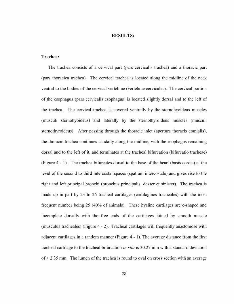
Trachea:
The trachea consists of a cervical part (pars cervicalis trachea) and a thoracic part
(pars thoracica trachea). The cervical trachea is located along the midline of the neck
ventral to the bodies of the cervical vertebrae (vertebrae cervicales). The cervical portion
of the esophagus (pars cervicalis esophagus) is located slightly dorsal and to the left of
the trachea. The cervical trachea is covered ventrally by the sternohyoideus muscles
(musculi sternohyoideus) and laterally by the sternothyroideus muscles (musculi
sternothyroideus). After passing through the thoracic inlet (apertura thoracis cranialis),
the thoracic trachea continues caudally along the midline, with the esophagus remaining
dorsal and to the left of it, and terminates at the tracheal bifurcation (bifurcatio tracheae)
(Figure 4 - 1). The trachea bifurcates dorsal to the base of the heart (basis cordis) at the
level of the second to third intercostal spaces (spatium intercostale) and gives rise to the
right and left principal bronchi (bronchus principalis, dexter et sinister). The trachea is
made up in part by 23 to 26 tracheal cartilages (cartilagines tracheales) with the most
frequent number being 25 (40% of animals). These hyaline cartilages are c-shaped and
incomplete dorsally with the free ends of the cartilages joined by smooth muscle
(musculus tracheales) (Figure 4 - 2). Tracheal cartilages will frequently anastomose with
adjacent cartilages in a random manner (Figure 4 - 1). The average distance from the first
tracheal cartilage to the tracheal bifurcation in situ is 30.27 mm with a standard deviation
of ± 2.35 mm. The lumen of the trachea is round to oval on cross section with an average
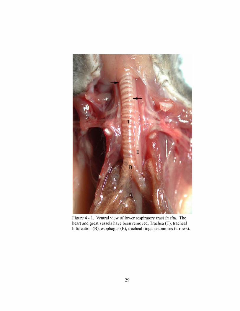
29
30
31
diameter in the cranial third of 3.18 ± 0.28 mm, in the middle third of 2.94 ± 0.20 mm
and the caudal third of 2.05 ± 0.08 mm. The tracheal cartilages are approximately 1.0
mm wide in a cranial to caudal direction.
Lungs:
Each lung (pulmo) has a costal (facies costalis), medial (facies medialis) and
diaphragmatic (facies diaphragmatica) surface, along with dorsal (margo dorsalis) and
ventral (margo ventralis) margins. The costal surfaces are smooth, convex and in contact
with the thoracic wall. The medial surfaces are smooth and concave. Present on the
medial surface of each lung is a cardiac impression (impressio cardiaca) which is created
by the heart. The diaphragmatic surfaces of the lungs are smooth and concave. This
concavity results from the base of the lungs (basis pulmonis) lying against the cranial
surface of the diaphragm. The dorsal margins of the lungs are rounded and located along
the ventrolateral surfaces of the thoracic vertebral bodies (corpus vertebrae thoracicae).
The ventral margin of the right lung is thin and interrupted by interlobar fissures (fissura
interlobaris). The ventral margin of the left lung is thin with several small fissures.
A simple squamous pleural membrane lines the thoracic wall and covers the lungs
and other mediastinal structures within the thoracic cavity (cavum thoracis). The visceral
pleura (pleura pulmonalis) is tightly adhered to the lungs. The parietal pleura (pleura
parietalis) covers the medial surfaces of the ribs (costae), the intercostal muscles (musculi
intercostales), the thoracic portion of the sympathetic trunk (truncus sympathicus), the
cranial surface of the diaphragm and numerous mediastinal structures. A pulmonary
32
ligament (ligamentum pulmonale) attaches the dorsomedial border of each lung to the
middle and caudal mediastinal pleura (pleura mediastinalis). Each ligament extends
along the medial surface of the lung from the hilus (hilus pulmonis) in a caudal direction.
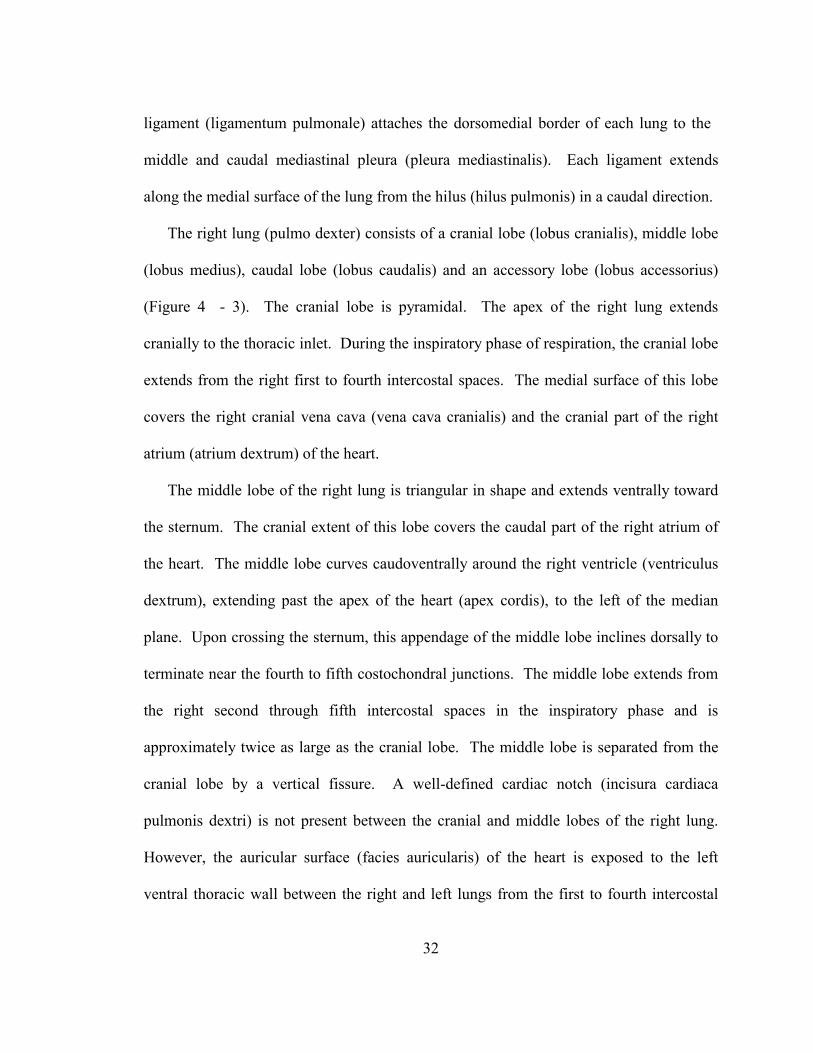
The right lung (pulmo dexter) consists of a cranial lobe (lobus cranialis), middle lobe
(lobus medius), caudal lobe (lobus caudalis) and an accessory lobe (lobus accessorius)
(Figure 4 - 3). The cranial lobe is pyramidal. The apex of the right lung extends
cranially to the thoracic inlet. During the inspiratory phase of respiration, the cranial lobe
extends from the right first to fourth intercostal spaces. The medial surface of this lobe
covers the right cranial vena cava (vena cava cranialis) and the cranial part of the right
atrium (atrium dextrum) of the heart.
The middle lobe of the right lung is triangular in shape and extends ventrally toward
the sternum. The cranial extent of this lobe covers the caudal part of the right atrium of
the heart. The middle lobe curves caudoventrally around the right ventricle (ventriculus
dextrum), extending past the apex of the heart (apex cordis), to the left of the median
plane. Upon crossing the sternum, this appendage of the middle lobe inclines dorsally to
terminate near the fourth to fifth costochondral junctions. The middle lobe extends from
the right second through fifth intercostal spaces in the inspiratory phase and is
approximately twice as large as the cranial lobe. The middle lobe is separated from the
cranial lobe by a vertical fissure. A well-defined cardiac notch (incisura cardiaca
pulmonis dextri) is not present between the cranial and middle lobes of the right lung.
However, the auricular surface (facies auricularis) of the heart is exposed to the left
ventral thoracic wall between the right and left lungs from the first to fourth intercostal
33
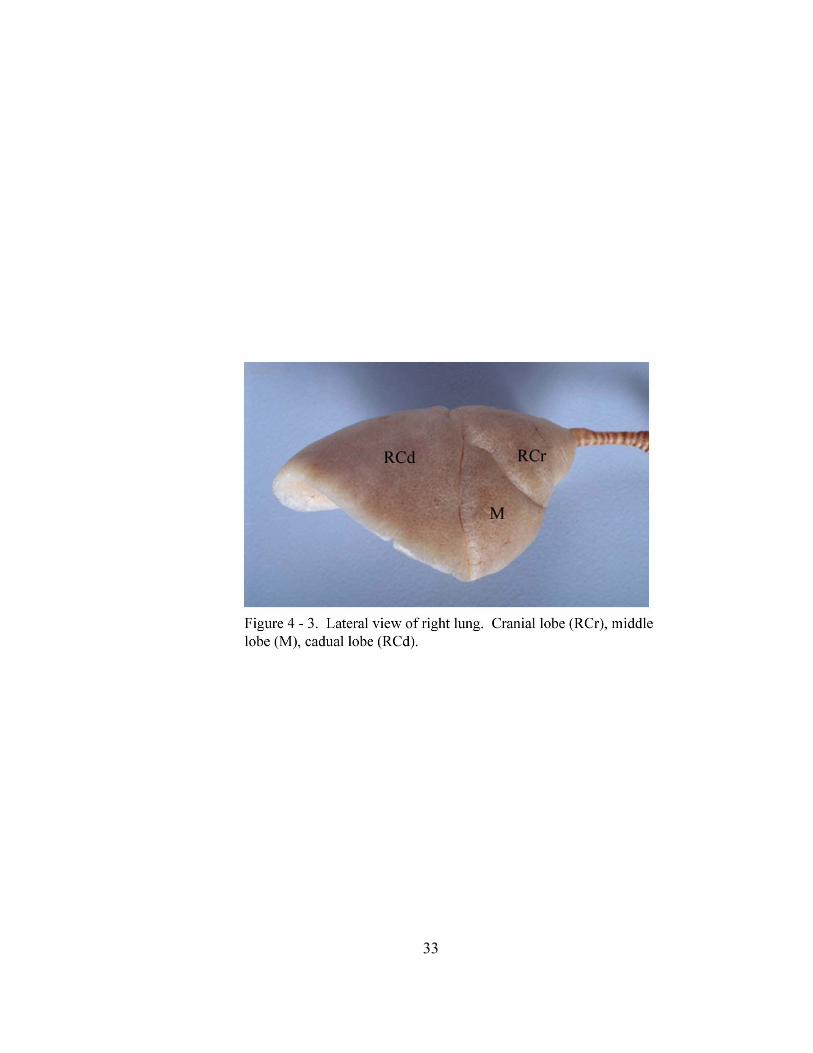
34
spaces. The apex of the heart is directed caudoventrally, as well as 35-40o to the left of
the midline and extends toward the left fourth intercostal space (Figure 4 - 4). The
sternopericardial ligament (ligamentum sternopericardiacum) connects the fibrous
pericardium (pericardium fibrosum) at the apex of the heart to the endothoracic fascia
(fascia endothoracica) at the left fourth intercostal space as well as to the diaphragm.
The caudal lobe of the right lung extends from the right third to sixth intercostal
spaces in the inspiratory phase. This lobe is triangular in shape and is approximately
twice as large as the middle lobe. The caudal lobe is separated from the middle lobe by
an oblique fissure. The base of the caudal lobe is in contact with the cranial surface of
the diaphragm. The caudal vena cava (vena cava caudalis) and the accessory lobe are
located medial to this lobe. The cranioventral margin of the caudal lobe is notched by the
caudal vena cava.
The accessory lobe is the smallest lobe of the right lung and is shaped like an
irregular pyramid. The cranial surface of this lobe rests on the caudodorsal aspect of the
heart resulting in a prominent cardiac impression on this lobe. The caudal surface of this
lobe is molded to the convex, cranial surface of the diaphragm. The right and left
surfaces of the accessory lobe lie adjacent to the medial surfaces of the caudal lobes of
the right and left lungs. The right surface of the accessory lobe has a notch (sulcus venae
cava caudalis) through which the caudal vena cava passes. The left surface of the
accessory lobe is elongated and rests within the mediastinal recess (recessus mediastini)
which is a space between the plica vena cava (plica venae cavae) and the caudal
mediastinal pleura. The caudal vena cava is attached by the triangular shaped plica vena
35
36
cava to the mediastinal pleura. The plica, which is a double fold of mediastinal pleura,
attaches dorsally to the caudal vena cava, cranioventrally to the pericardium of the heart
and caudally to the diaphragm.
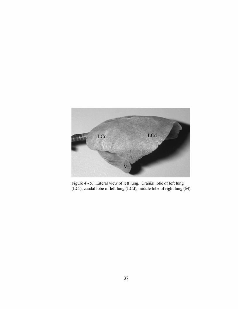
The left lung (pulmo sinister) consists of cranial and caudal lobes (Figure 4 - 5).
These lobes are not separated from one another by a deep fissure. As a result, these lobes
are not identifiable based on superficial features of the lung. This lung covers the left
atrium (atrium sinister) and a portion of the left ventricle (ventriculus sinister) of the
heart. The ventral margin of this lung typically has multiple small fissures. These
fissures are randomly located and usually do not coincide with lobar division (Figure 4 -
5). In the inspiratory phase, the left lung extends from the first through seventh
intercostal spaces. An aortic impression (impressio aortica) is formed on the dorsomedial
surface of the cranial and caudal lobes by the aortic arch (arcus aortae) and descending
aorta (aorta descendens). The caudal lobe also has an esophageal impression (impressio
esophagea) formed by the esophagus on its medial surface.
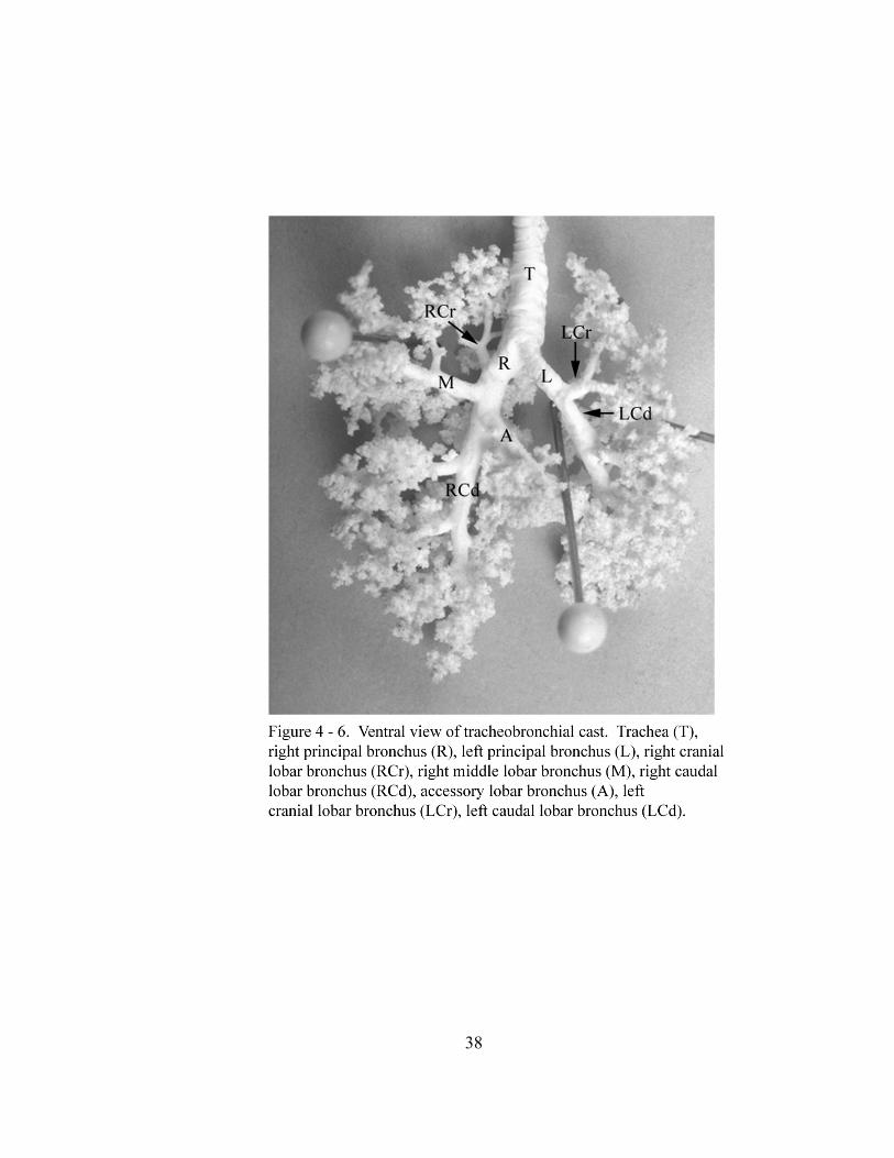
Bronchial Tree:
Bifurcation of the trachea into the right and left principal bronchi marks the beginning
of the bronchial tree (arbor bronchalis). The principal bronchi enter the hilus of the lungs
where they divide into lobar bronchi (bronchi lobares) (Figure 4 - 6). The left principal
bronchus is on average 6.22 ± 0.69 mm in length and the right principal bronchus
averages 6.57 ± 0.6 mm in length.
37
38
39
The right principal bronchus divides into cranial, middle, caudal and accessory lobar
bronchi. The cranial lobar bronchus originates from the dorsolateral surface of the right
principal bronchus and gives off five segmental bronchi (bronchi segmentales) which
radiate in cranial and caudal directions from their origin. The middle lobar bronchus
originates from the ventrolateral surface of the right principal bronchus cranial to the
origin of the accessory and caudal lobar bronchi. The middle lobar bronchus gives off six
segmental bronchi, which radiate cranially, caudally and dorsally. The accessory lobar
bronchus is the last branch to arise from the right principal bronchus at which point the
right principal bronchus continues caudally as the caudal lobar bronchus. The accessory
lobar bronchus originates ventromedially from the right principal bronchus and gives off
five to six segmental bronchi which radiate in cranial and caudal directions. Finally, the
caudal lobar bronchus gives off six to seven segmental bronchi which radiate into the
lung parenchyma.
The left principal bronchus divides into cranial and caudal lobar bronchi. The cranial
lobar bronchus originates ventrolaterally from the left principal bronchus. The cranial
lobar bronchus divides into two bronchi which supply cranial and caudal parts of the left
cranial lung lobe. Arising from each of these bronchi are six segmental bronchi which
radiate into the parenchyma. The caudal lobar bronchus continues the left principal
bronchus caudally and has six to eight segmental bronchi which radiate into the
parenchyma.
40
DISCUSSION:
The trachea of the sexually mature gray short-tailed opossum consists in part of “c”-
shaped tracheal cartilages with the free ends joined together by the trachealis muscle as is
typical of most mammals. Tracheal cartilage number in the gray short-tailed opossum
falls within the range reported in other marsupials which is nineteen rings in the gray
four-eyed opossum (Metachirus opossum) to thirty-four in the bandicoot (Perameles
obesula) and thirty-five in both the lesser gliding opossum (Petaurus sciureus) and tree
kangaroo (Dendrolagsus ursinus) (Sonntag, 1921b).
The lobation of the right lung of the gray short-tailed opossum is similar to previous
descriptions of many marsupials including the mouse opossum (Marmosa elegans), gray
four-eyed opossum, bandicoot, Phalangeridae (Sonntag, 1921a), brush-tailed opossum
(Trichosurus vulpecula) (Sonntag, 1921b), Caenolestes (Osgood, 1921), Dasyuridae
(Owen, 1868; Jones, 1948), Perameles and Petaurists (Owen, 1868). Owen (1868)
describes the right lung of Potoroo as having two to three deep fissures and an azygous
lobe. This may indicate that it also has four lobes with one of them perhaps further
subdivided. A right lung divided into four lobes is a common pattern found in most of
the smaller marsupials.
Several other marsupials possess right lung lobation which differs from that of the
gray short-tailed opossum. The right lung of the tree kangaroo (Dendrolagus ursinus),
eastern grey kangaroo (Macropus giganteus) and wallaroo (Macropus bennetti), which is
trilobate, is described as having a deep median sulcus incompletely dividing the lung into
anterior and posterior parts with the azygous lobe in addition (Sonntag, 1921a). Owen
41
(1868) states that the right lung of the whiptail wallaby (Macropus parryi) has one to
two notches possibly indicating the absence of distinct divisions between lobes based on
external appearance. The right lung of the red-legged short-tailed opossum (Didelphys
brachyura) consists of three lobes and the right lung of the wombat (Vombatus ursinus)
consists of a two lobes. The right lung of the koala (Phascolactos cinereus) is described
by Forbes (1881) as having three lobes without the azygous lobe while Sonntag (1921b)
states that the right lung of the koala consists of only two lobes. The right lung of the
common shrew opossum (Caenolestes obscurus) has three lobes with the anterior lobe
slightly notched which corresponds to where a complete division was found in
Didelphids (Osgood, 1921). Osgood (1921) also states that in a third specimen the right
lung consisted of four lobes thus indicating variability in lung lobation within this
species. While a few of the smaller marsupials possess a right lung exhibiting a pattern
of lobation different from that found in the gray short-tailed opossum, larger marsupials
such as the kangaroos, wallabies and koala consistently have fewer lobes attributed to the
right lung. Earlier reports on marsupial respiratory anatomy often use human anatomy
terms in naming the cranial lobe as the upper lobe, the middle lobe as the ventral lobe, the
caudal lobe as the lower lobe and the accessory lobe as the azygous lobe or intermediate
lobe.
While a cardiac notch is not present on the right lung of the gray short-tailed
opossum, the angulation of the longitudinal axis of the heart to the left of the midline
allows access to the heart via cardiac puncture in the left third and fourth intercostal
spaces near the sternum. The longitudinal axis of the koala heart is described as being
42
parallel to the left side of the sternum with the apex of the heart extending to the left
fourth intercostal space (Sonntag, 1921b). Additional descriptions of marsupial heart
angulation could not be located for further comparisons.
The left lung of the gray short-tailed opossum consists of two lobes with the cranial
lobe being further sub-divided into two parts. The lobes of the left lung are not separated
by an interlobar fissure as are those of the right lung. The only superficial indications
which might be used for lobar demarcation are small, 1.0 – 2.0 mm fissures which are
located along the ventral margin of the lung. These small fissures were found to be
variable in location and number between animals with marked variation between animals
obtained from different sources. Thus, the fissures were of little use in identification of
the two lobes of the left lung from the surface of the organ. Due to the absence of
external lobar demarcation, intercostal landmarks for the cranial and caudal lobes are
difficult to define as well as the cranial and caudal parts of the cranial lobe without
further study.
The left lung of the brush-tailed opossum (Sonntag, 1921b), Phalangeridae (Owen,
1868; Sonntag, 1921a), mouse opossum (Sonntag, 1921a), koala (Owen, 1868; Forbes,
1881), quoll (Dasyurus) (Owen, 1868), Dasycercus cristicauda (Jones, 1948) and
Petaurists (Owen, 1868) is described as having a cranial and caudal lobe. Sonntag
(1921a) and Owen (1852) state that in Macropodidae, in which the tree kangaroo,
wallaroo and eastern grey kangaroo were examined, the left lung has deep median sulci
or clefts dividing it into anterior and posterior parts. The left lung of other marsupials
such as the gray four-eyed opossum, long-nosed bandicoot (Sonntag, 1921a), pig-footed
43
bandicoot (Choeropus castanotis) (Parsons, 1903), Caenolestes (Osgood, 1921),
wombat and American opossum (Owen, 1868) is described as being unilobate apparently
based on the external appearance of the lung as branching of the bronchial tree was not
mentioned. Owen (1868) states that the left lung of Potoroo has a fissure on the anterior
or upper ridge and the left lung of the whiptail wallaby has one to two notches. He
classifies the left lung of these animals as unilobate. These external markings were
probably similar to what we observed in the gray short-tailed opossum giving the
impression of being not lobated. Lobation of the left lung, across all marsupial species,
is described as having one or two lobes. Those described as having one lobe might
actually be found to consist of two lobes should one examine tracheobronchial casts of
those specimens or dissect the bronchial tree.
Lung lobation in the gray short-tailed opossum was based upon the division of the
bronchial tree as described by Nomina Anatomica Veterinaria (International Committees
on Veterinary Gross Anatomical Nomencalture, 1994) rather than on the external
appearance of the lung. We accomplished this by examination of tracheobronchial casts
to identify the lobar bronchi which supply the lung. We were unable to locate data on the
branching pattern of the bronchial tree for other marsupials for comparison.
44
CHAPTER V
MACROSCOPIC ANATOMY OF THE LOWER RESPIRATORY TRACT OF
THE NORTH AMERICAN OPOSSUM (Didelphis virginiana)
ABSTRACT:
This study documents the macroscopic anatomy of the lower respiratory tract of the
North American opossum (Didelphis virginiana). The trachea consists of approximately
28 c-shaped cartilages and extends from the larynx to its bifurcation into right and left
principal bronchi. The right lung consists of cranial, middle, caudal and accessory lobes
which are separated from one another by interlobar fissures. The left lung consists of
cranial and caudal lobes which are not separated by interlobar fissures. Lobation of the
right and left lungs was verified from tracheobronchial casts.
INTRODUCTION:
The North American opossum (Didelphis virginiana), has become more prominent as
a patient in veterinary medicine due to advancements in wildlife rehabilitation.
Therefore, it is essential to have an available source on the normal macroscopic anatomy
of all the organ systems of the North American opossum especially when evaluating
pathological changes.
Additionally, the North American opossum has been used in biomedical research for
over twenty-five years. It has served as a model for embryogenesis (Klima, 1987;
45
Renfree, 1990; Szalay, 1994), cytogenetic studies (Oswaldo-Cruz, 1968) and
physiological studies (Suzuki et al., 1988; Leichus et al., 1997). The first anatomical
descriptions on the osteology, myology (Coues, 1872) and general visceral topography
(Tyson, 1698) of the North American opossum date back three hundred years.
Subsequent anatomical descriptions of the North American opossum have briefly
described the lung (Brewer, 1923) and general visceral topography (Sonntag, 1921a and
1921b; Ellsworth, 1966 and 1976). However, none provide a complete description or
photographic record of the lower respiratory tract. To benefit research and medicine
involving the North American opossum, complete anatomical records of the various
organ systems are a necessity.
MATERIALS AND METHODS:
Eighteen North American opossums (9 males and 9 females) of various ages were
used to study lower respiratory tract anatomy. The lower respiratory tracts from 6
animals were removed and air-dried with laboratory air for 48 hours after which RTV
silicone (Silicone Inc. P.O. Box 363, 211 Woodbine High Point, NC, 27261) was injected
into the trachea to produce tracheobronchial casts (Henry, 1992). The lower respiratory
tracts from the remaining 12 animals were dissected and examined in situ and ex vivo.
Some lungs were inflated in situ to mimic the inspiratory phase for documentation of
intercostal landmarks of the individual lobes.
46
RESULTS:
Trachea:
The cervical trachea (pars cervicalis trachea) is located along the ventral midline of
the cervical region with the esophagus (pars cervicalis esophagus) lying dorsal and to the
left. The cervical trachea is covered ventrally by the right and left sternohyoideus
muscles (musculi sternohyoideus) and laterally by the sternothyroideus muscles (musculi
sternothyroideus). After passing through the thoracic inlet (apertura thoracis cranialis),
the thoracic trachea (pars thoracica trachea) continues caudally along the dorsal midline
and terminates at the tracheal bifurcation (bifurcation tracheae). The trachea maintains
its orientation to the esophagus similar to that in the cervical region. The trachea
bifurcates dorsal to the base of the heart (basis cordis) at the level of the third intercostal
space (spatium intercostale) into the right and left principal bronchi (bronchus principalis
dexter et sinister) (Figure 5 - 1). The average distance from the first tracheal cartilage
(cartilaginous tracheales) to the tracheal bifurcation in situ is 70.0 ± 1.28 mm. The
tracheal cartilages are c-shaped and incomplete dorsally with the ends of the tracheal
cartilages joined by smooth muscle (musculus tracheales). The North American opossum
has 25 to 29 cartilages with the most frequent number being 28 (57% of animals).
Tracheal cartilage anastomosis occurs in the middle one-third of the trachea and can
involve two or three adjacent cartilages.
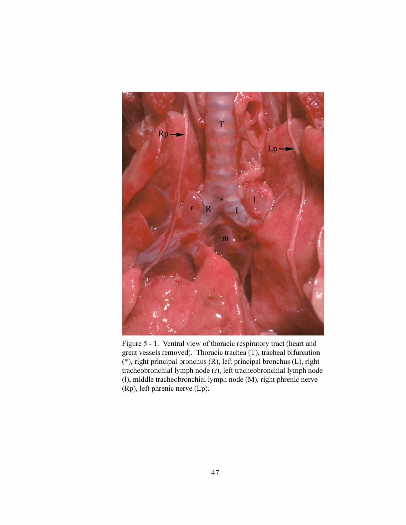
47
48
Lungs:
Each lung (pulmo) has a costal (facies costalis), medial (facies mediales) and
diaphragmatic surface (facies diaphragmatica) along with dorsal and ventral margins
(margo dorsalis et ventralis). The costal surfaces are smooth, convex and in contact with
the thoracic wall. The medial surfaces of the lungs are smooth and concave. Present on
the medial surface of each lung is a cardiac impression (impressio cardiaca) which is
created by the heart. The diaphragmatic surfaces of the lungs are smooth and concave
(Figure 5 - 2). This concavity results from the base of the lungs (basis pulmonis) lying
against the convex surface of the diaphragm. The dorsal margins of the lungs are
rounded and located along the ventrolateral surfaces of the thoracic vertebral bodies
(corpus vertebrae thoracicae). The ventral margin of the right lung (pulmo dexter) is thin
and interrupted by interlobar fissures (fissura interlobaris). The ventral margin of the
cranial lobe (lobus cranialis) extends to the midline. However the ventral margin of the
middle lobe (lobus medius) crosses to the left of the midline and terminates at the left
fourth costochondral junction. The ventral margin of the left lung (pulmo sinister) is thin
with 1 to 2 small fissures. The ventral margin of this lung does not cross the midline and
only extends ventrally to the first through sixth costochondral junctions due to the ventral
attachment of the mediastinal pleura to the left of the midline.
Pleura lines the thoracic wall and covers the lungs and other mediastinal structures
within the thoracic cavity (cavum thoracis). The mediastinal pleura attaches dorsally
along the vertebral bodies. Cranially its ventral attachment is along the midline and
caudally it angles toward the left sixth costochondral junction (Figure 5 - 3). The visceral
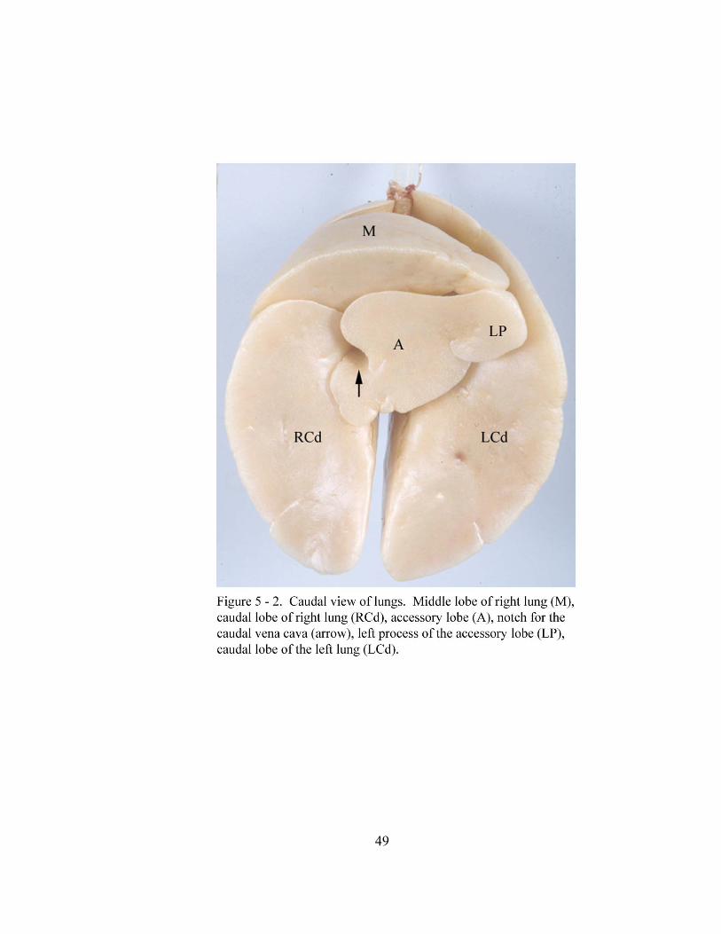
49
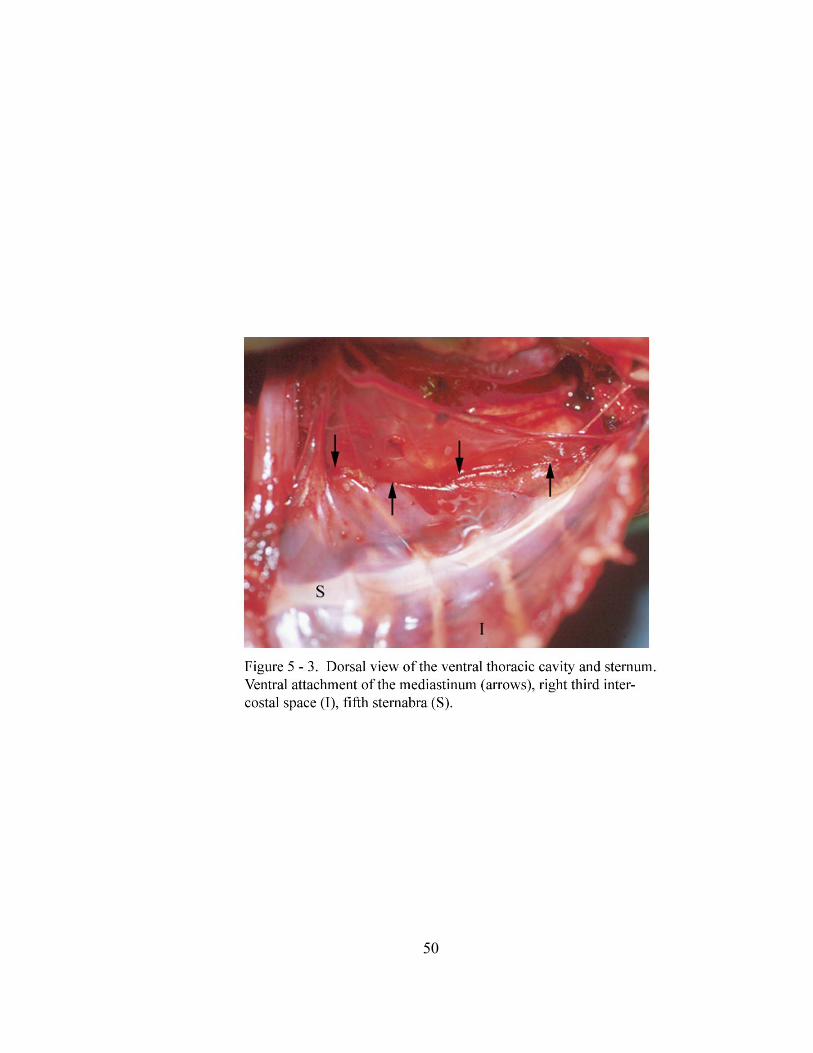
50
51
pleura (pleura pulmonalis) is tightly adhered to the lungs. The parietal pleura (pleura
parietalis) covers the medial surfaces of the ribs (costae), the internal intercostal muscles
(musculi intercostales), the thoracic part of the sympathetic trunks (truncus sympathicus),
the cranial surface of the diaphragm and numerous mediastinal structures. Attaching the
dorsomedial border of each lung to the middle and caudal mediastinal pleura (pleura
mediastinalis) is a pulmonary ligament (ligamentum pulmonale). Each pulmonary
ligament extends along the medial surface of the lung from the hilus (hilus pulmonis) in a
caudal direction.
The right lung consists of a cranial lobe, middle lobe, caudal lobe (lobus caudalis) and
an accessory lobe (lobus accessorius). The cranial lobe extends from the first through
fourth intercostal spaces in the inspiratory phase. The medial surface of this lobe covers
the right cranial vena cava (vena cava cranialis) and the cranial part of the right atrium
(atrium dextrum) of the heart.
The middle lobe of the right lung extends from the second through fifth intercostal
spaces in the inspiratory phase. It is separated from the cranial lobe by an oblique fissure
(Figure 5 - 4). The cranial extent of the middle lobe covers the caudal part of the right
atrium of the heart. This lobe curves caudoventrally around the right ventricle
(ventriculus dexter) and extends past the apex of the heart (apex cordis) to the left of the
median plane. Upon crossing the dorsal surface of the sternum, this elongated portion of
the middle lobe inclines dorsally and terminates at the level of the left fourth
costochondral junction. A small cardiac notch (incisura cardiaca) may be present
between the cranial and middle lobes of the right lung opposite the right second
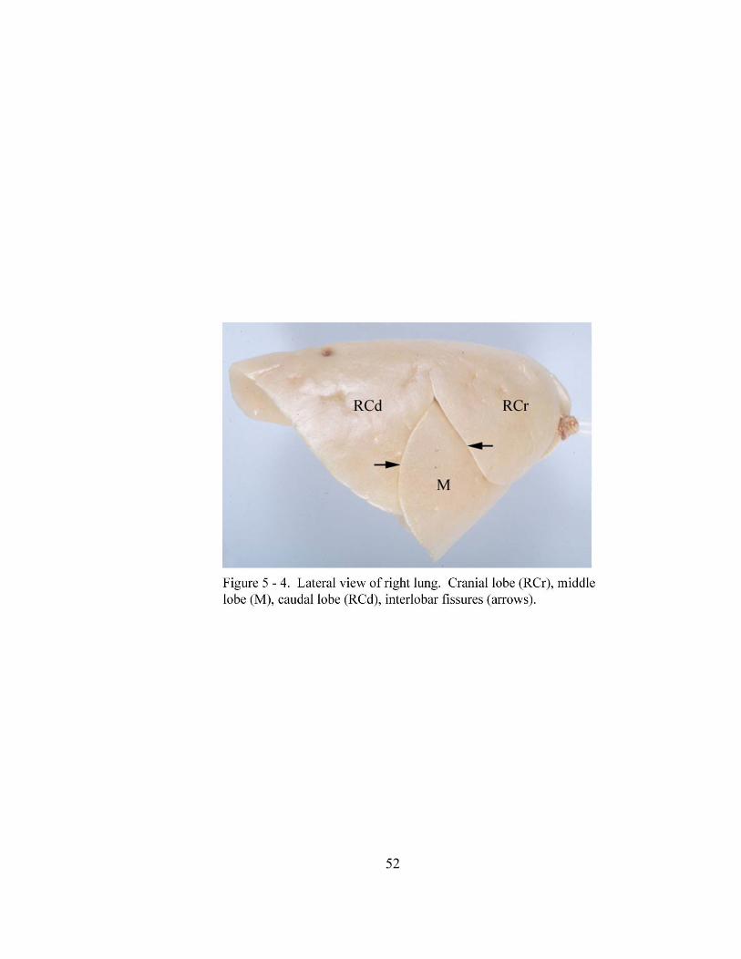
52
53
intercostal space. However the ventral surface of the heart is exposed to the left ventral
thoracic wall between the right and left lungs from the first to fifth intercostal spaces.
The apex of the heart is directed caudoventrally and to the left of the midline (33-37o)
toward the left fourth intercostal space. A sternopericardial ligament (ligamentum
sternopericardiacum), which is continuous with the fibrous pericardium and the
pericardial mediastinal pleura, extends from the apex of the heart to the endothoracic
fascia (fascia endothoracica) and the parietal pleura at the left fifth to sixth costochondral
junctions.
The caudal lobe of the right lung extends from the fourth to eighth intercostal spaces
in the inspiratory phase. This lobe is approximately twice as large as the middle lobe of
the right lung. The caudal lobe is separated from the middle lobe by a vertical fissure
(Figure 5 - 4). Laying medial to the caudal lobe is the caudal vena cava (vena cava
caudalis) and the accessory lobe of the right lung (Figure 5 - 2).
The accessory lobe is the smallest lobe of the right lung and is shaped like an
irregular pyramid. This lobe lies medial to the caudal lobes of the right and left lungs
(Figure 5 - 2). The cranial surface of this lobe rests on the caudodorsal aspect of the heart
resulting in a prominent cardiac impression. The caudal surface of this lobe is molded
against the convex surface of the diaphragm. The right surface of the accessory lobe has a
notch (sulcus venae cava caudalis) through which the caudal vena cava passes (Figure 5 -
2). The left process of the accessory lobe is elongated and rests within the mediastinal
recess (recessus mediastini) which is a space between the plica vena cava (plica venae
cavae) of the caudal vena cava and the caudal mediastinal pleura (pleura mediastinalis).
54
The left lung consists of cranial and caudal lobes. This lung extends from the first to
eighth intercostal spaces in the inspiratory phase. The ventral margin of this lung has 1 to
2 small fissures. These fissures are located on the cranial lobe of the left lung and do not
coincide with the lobar division (Figure 5 - 5). The medial surface of this lung covers the
entire left atrium (atrium sinistrum) and a portion of the left ventricle (ventriculus
sinister) of the heart.
Bronchial Tree:
Bifurcation of the trachea into the right and left principal bronchi marks the beginning
of the bronchial tree (arbor bronchalis) (Figure 5 - 6). The right principal bronchus is on
average 13.08 ± 1.61 mm in length and the left principal bronchus is on average 11.73 ±
2.03 mm in length. The right and left principal bronchi enter the hilus of the lungs at
which point they divide into lobar bronchi (bronchi loabres).
The right principal bronchus divides into cranial, middle, caudal and accessory lobar
bronchi (Figure 5 - 7). The cranial lobar bronchus originates from the dorsolateral
surface of the right principal bronchus and gives off six to seven segmental bronchi
(bronchi segmentales) which radiate in cranial, caudal and dorsal directions. The middle
lobar bronchus originates from the ventral surface of the right principal bronchus cranial
to the origin of the accessory and caudal lobar bronchi. The middle lobar bronchus gives
off ten segmental bronchi which radiate cranially, caudally and laterally. The accessory
lobar bronchus originates ventromedially from the right principal bronchus cranial to the
origin of the caudal lobar bronchus. The accessory lobar bronchus gives off eight
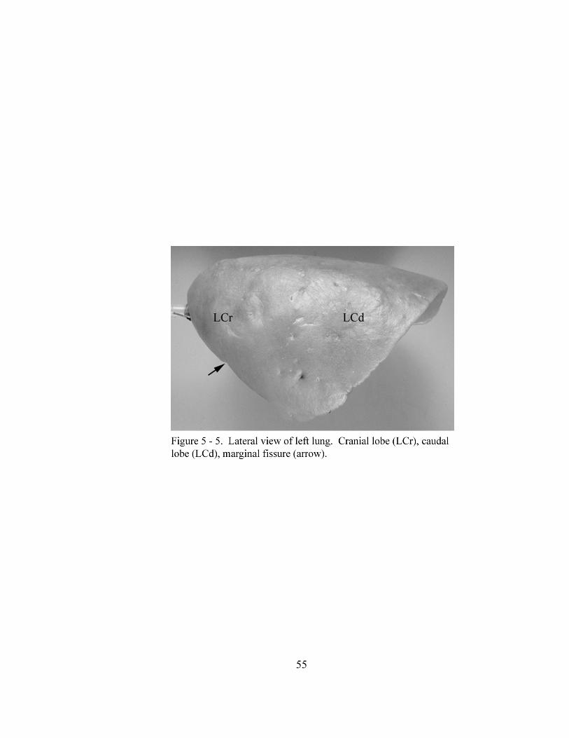
55
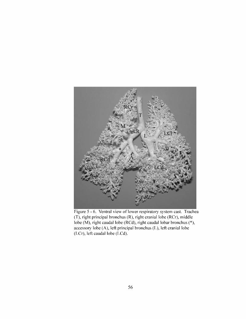
56
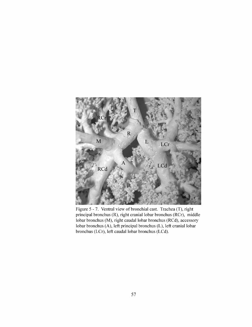
57
58
segmental bronchi which radiate cranially, caudally, dorsally and ventrally. The caudal
lobar bronchus continues the right principal bronchus caudally and gives off nine to ten
segmental bronchi which radiate into the parenchyma.
The left principal bronchus divides into cranial and caudal lobar bronchi (Figures 5 -
6 and 5 - 7). The cranial lobar bronchus originates ventrolaterally from the left principal
bronchus. The cranial lobar bronchus divides into two bronchi, cranial and caudal
branches, which supply cranial and caudal parts of the left cranial lung lobe (Figure 5 -
7). The cranial and caudal branch each give rise to four or five segmental bronchi which
radiate dorsally, cranially and caudally. The caudal lobar bronchus originates caudal to
the origin of the cranial lobar bronchus. Arising from the caudal lobar bronchus are six
segmental bronchi which radiate into the parenchyma.
DISCUSSION:
Tracheal cartilage anastomoses, as seen in the North American opossum, has also
been reported by Sisson and Grossman (Getty, 1975) to occur in domestic mammals.
However, tracheal cartilage anastomosis in these mammals occurs randomly throughout
the length of the trachea as opposed to only the middle third of the opossum trachea. The
free ends of each tracheal cartilage in the North American opossum are joined by the
trachealis muscle to form a complete ring which is also similar to many domestic
mammals (Evans, 1993; Dyce et al., 1996).
The earliest reports on the respiratory anatomy of the North American opossum
(Tyson, 1698; Owen, 1868) state the right lung consist of three lobes. However a
59
subsequent description of the lobation of the right lung of the Didelphyidae (Sonntag,
1921a) reports four lobes. We found that the right lung of the North American consists
of four lobes which are separated from one another by deep interlobar fissures. In
addition to what we observed in the North American opossum, several of the smaller
marsupials such as the brush-tailed opossum (Trichosurus vulpecula) (Sonntag, 1921b),
Phalangeridae and Phalangers (Owen, 1868; Sonntag, 1921a), mulgara (Dasycercus
cristicauda) (Jones, 1948), long nosed bandicoot (Perameles obesula) (Sonntag, 1921a),
Perameles, Petaurists and Dasyures (Owen, 1868) have similar lobation of the right lung.
Variation in the North American opossum lung lobation in previous articles (Tyson,
1698; Owen, 1868; Sonntag, 1921a) may have resulted from using superficial features of
the lung rather than the branching of the bronchial tree to determine lung lobation. This
method of naming lobes was a common practice in previous anatomy texts and articles
and has led to considerable confusion in determining lung lobation (Dyce et al., 1996).
The left lung of the North American opossum extends from the first through eighth
intercostal spaces and consists of a cranial and caudal lobe. Interestingly the left lung
does not have interlobar fissures demarcating the division of cranial and caudal lobes.
This description of lobation of the left lung (as well as the right) is based upon the
division of the bronchial tree as described by Nomina Anatomica Veterinaria
(International Committee on Veterinary Gross Anatomical Nomenclature, 1994). Hence
tracheobronchial casts were utilized to determine lobation since naming lobes based on
superficial features is not reliable and should not be done (Dyce et al., 1996). A previous
description of the North American opossum (Owen, 1868) indicates a similar lobation of
60
the left lung. As well, the Phalangeridae, mouse opossum (Marmosa elegans) (Sonntag,
1921a), brush-tailed opossum (Sonntag, 1921b), Petaurists, Phalangers, Dasyures (Owen,
1868) and mulgara (Jones, 1948) have a similar lobation of the left lung to the North
American opossum. However, no mention is made of how Owen (1868), Sonntag (1921a
and 1921b) or Jones (1948) determined lobation of the lungs. The variability in presence
and location of small fissures on the ventral margin makes it difficult to accurately
approximate the intercostal landmarks for the cranial and caudal lobes of the left lung in
the North American opossum.
The right and left principal bronchi of the North American opossum divide into lobar
bronchi to supply each lobe of the right and left lungs respectively. Within the lobe of
each lung, the lobar bronchi give off numerous segmental bronchi. However the left
cranial lobar bronchus divides into a cranial and caudal branch prior to the emergence of
the segmental bronchi. These two branches supply the cranial and caudal parts of the left
cranial lobe. Previous investigators describe a similar pattern of branching for the left
cranial lobar bronchus in the canine (Getty, 1975; Evans, 1993; Dyce et al., 1996), feline
(Adrian, 1964; Nickel et. al, 1979), sheep (Hare, 1955) and bovine (Stamp, 1948).
However, Hare (1955), Adrian (1964), Getty, (1975) and Nickel et. al (1979) refer to
these branches of the left cranial lobar bronchus as cranial and caudal segmental bronchi.
In addition, Adrian (1964) and Getty (1975) state these segmental bronchi which
ventilate the cranial and caudal bronchopulmonary segments, also give off numerous
subsegmental bronchi. In earlier literature, Hare (1955), Adrian (1964) and Ishaq (1980)
conclude there is lack of agreement on the definition of a bronchopulmonary segment.
61
Kramer and Glass (1932), Hare (1955), Getty, (1975), Nickel (1979), Ishaq (1980) and
Schaller (1992) define a segmental bronchus as that which ventilates a
bronchopulmonary segment and is a self-contained or independent section of lung tissue
within a lobe. Also, Kramer and Glass (1932) and Getty (1975) state the segmental
bronchus supplying a bronchopulmonary segment originates from a lobar bronchus.
Then, according to Nickel (1979), the segmental bronchi dispatch bronchioles. Based on
these definitions, the use of cranial and caudal segmental bronchi does not seem
appropriate in the canine, feline, sheep or the North American opossum. Stamp’s (1948)
suggestion concerning the cranial and caudal branches of the left cranial lobar bronchus
of the bovine lung seems to be the best description. According to him, these branches
dispatch numerous segmental bronchi to supply the bronchopulmonary segments of the
left cranial lobe. In addition, previous articles on the bronchial tree of domestic animals
(Hare, 1955; Adrian, 1964) state that the nomenclature used in these species attempted to
follow that established for human anatomy. However, Hare (1955) states that the
nomenclature adopted for humans is not comparable when in fact it, along with Stamp’s
(1948), seems to be the best description for the branching pattern of the left cranial lobar
bronchus. According to human anatomy terminology (Woodburne, 1973; Netter, 1989;
Federative Committee on Anatomical Terminology, 1998) the left superior lobar
bronchus divides into a superior division and an inferior division from which segmental
bronchi are dispatched to the bronchopulmonary segments. This pattern is similar to
what was observed in the North American opossum.
62
CHAPTER VI
ANATOMY OF STRUCTURES ASSOCIATED WITH THE LOWER
RESPIRATORY TRACT OF THE GRAY SHORT-TAILED OPOSSUM
(Monodelphis domestica).
ABSTRACT:
The present study documents the macroscopic anatomy of structures associated with
the lower respiratory tract of the gray short-tailed opossum (Monodelphis domestica).
Vascularization of lung parenchyma is via the bronchoesophageal artery which originates
from the right fourth dorsal intercostal artery. The right and left pulmonary arteries
divide into pulmonary lobar arteries which are located dorsal to the lobar bronchi and
deliver blood to the lungs. Pulmonary lobar veins are located ventral to the lobar bronchi
and return blood from the lungs to the heart. Cranial deep cervical, cranial mediastinal
and tracheobronchial lymph nodes are located along the trachea and at the tracheal
bifurcation. Sympathetic fibers leave the sympathetic trunks as thoracic splanchnic
nerves to reach the lungs. Parasympathetic innervation to the lungs originates from
branches of the vagus nerves.
INTRODUCTION:
Over the past twenty years, numerous scientific papers have been published
describing the gray short-tailed opossum (Monodelphis domestica) as an ideal model for
63
research (Hubbard, 1997). These marsupials have been used to study embryogenesis
(Baggott and Moore, 1990; Selwood and Vandeberg, 1992; Kuehl-Kovarik, 1995);
reproduction, sexual differentiation, behavior and chemical communication (Vandeberg,
1983 and 1995); and nervous system development, DNA repair mechanisms and
cytogenetic and biochemical genetics (Vandeberg, 1990). Despite the wide use of the
gray short-tailed opossum in research, we were unable to locate any descriptions of
normal anatomical structures associated with the respiratory tract of this opossum. The
focus of this study will be to document the normal macroscopic anatomy of structures
associated with the lower respiratory tract of the gray short-tailed opossum.
MATERIALS AND METHODS:
Ten, 2 - year - old, sexually mature gray short-tailed opossums (Monodelphis
domestica) of both sexes were used for this study. Following anesthesia and
exsanguination, all animals were embalmed with 10% buffered formalin and injected
with epoxy or latex to aid in the visualization and dissection of the lower respiratory tract
vasculature. Two of the ten opossums were used for tracheobronchial vascular casts
(Henry, 1992a and 1992b). The trachea was exposed and transected caudal to the larynx.
A plastic cannula was inserted and ligated in place. Silicone was injected into the trachea
and airways via the plastic cannula. After hardening, the tracheobronchial vascular cast
was removed from the animal and boiled to remove the parenchyma.
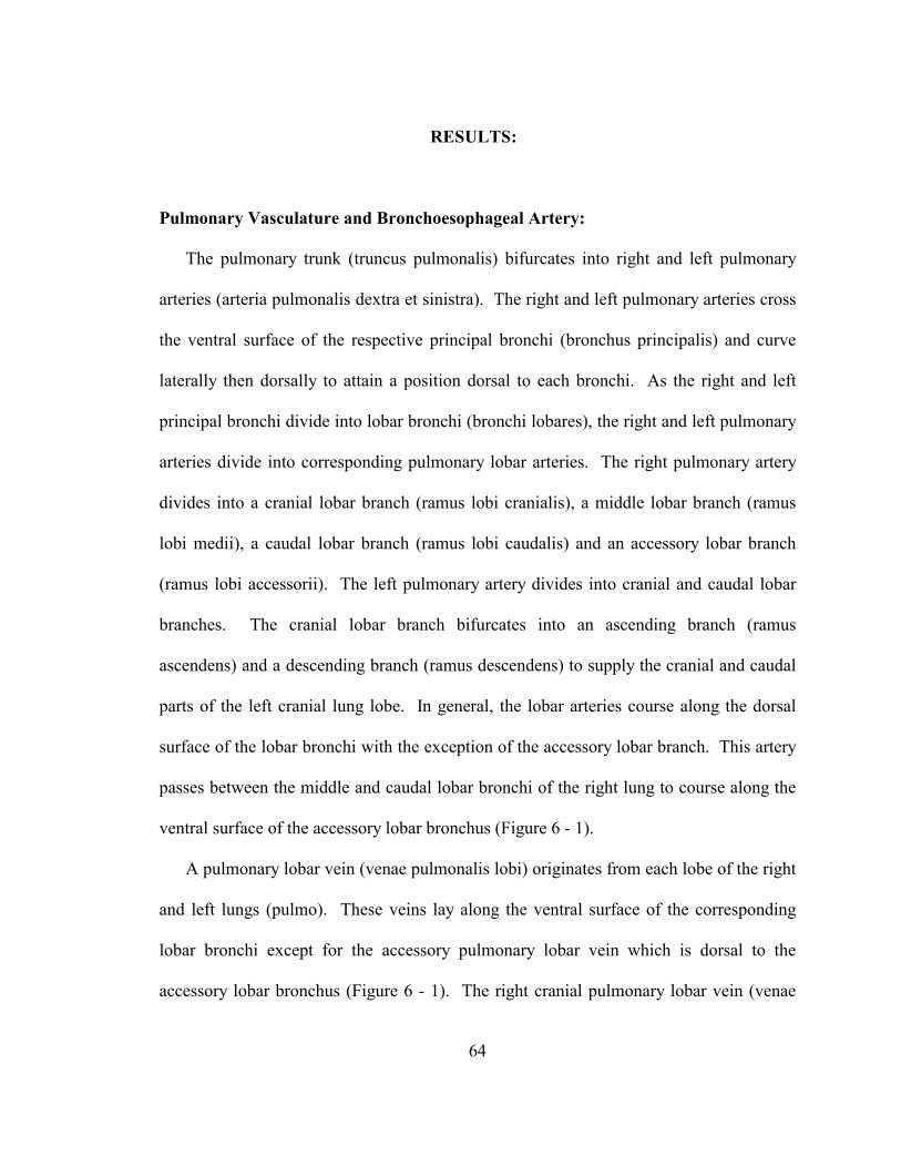
64
RESULTS:
Pulmonary Vasculature and Bronchoesophageal Artery:
The pulmonary trunk (truncus pulmonalis) bifurcates into right and left pulmonary
arteries (arteria pulmonalis dextra et sinistra). The right and left pulmonary arteries cross
the ventral surface of the respective principal bronchi (bronchus principalis) and curve
laterally then dorsally to attain a position dorsal to each bronchi. As the right and left
principal bronchi divide into lobar bronchi (bronchi lobares), the right and left pulmonary
arteries divide into corresponding pulmonary lobar arteries. The right pulmonary artery
divides into a cranial lobar branch (ramus lobi cranialis), a middle lobar branch (ramus
lobi medii), a caudal lobar branch (ramus lobi caudalis) and an accessory lobar branch
(ramus lobi accessorii). The left pulmonary artery divides into cranial and caudal lobar
branches. The cranial lobar branch bifurcates into an ascending branch (ramus
ascendens) and a descending branch (ramus descendens) to supply the cranial and caudal
parts of the left cranial lung lobe. In general, the lobar arteries course along the dorsal
surface of the lobar bronchi with the exception of the accessory lobar branch. This artery
passes between the middle and caudal lobar bronchi of the right lung to course along the
ventral surface of the accessory lobar bronchus (Figure 6 - 1).
A pulmonary lobar vein (venae pulmonalis lobi) originates from each lobe of the right
and left lungs (pulmo). These veins lay along the ventral surface of the corresponding
lobar bronchi except for the accessory pulmonary lobar vein which is dorsal to the
accessory lobar bronchus (Figure 6 - 1). The right cranial pulmonary lobar vein (venae
65
66
pulmonalis lobi cranialis dextra) and the right middle pulmonary lobar vein (venae
pulmonalis lobi medii) join to form a right pulmonary vein. The left cranial pulmonary
lobar vein (venae pulmonalis lobi cranialis sinistri) is formed by a branch from the cranial
part of the left cranial lobe and a branch from the caudal part of the left cranial lobe
(Figure 6 - 2). The left cranial and caudal pulmonary lobar veins (venae pulmonalis lobi
caudalis sinistri) join to form a left pulmonary vein. The right caudal pulmonary lobar
vein (venae pulmonalis lobi caudalis dextra) and the accessory pulmonary lobar vein
(ramus lobi accessorii) may join the left pulmonary vein or the left caudal pulmonary
vein prior to the formation of the left pulmonary vein. The right and left pulmonary veins
join to form a pulmonary venous trunk that opens into the left atrium (atrium sinistrum)
of the heart.
On the right side of the mediastinum, the bronchoesophageal artery (arteria broncho-
esophagea) originates from the right, fourth dorsal intercostal artery (arteria intercostales
dorsales) (Figure 6 - 3). The bronchoesophageal artery courses ventrally through the
mediastinum to the right side of the esophagus where it divides into a bronchial branch
(ramus bronchalis) and an esophageal branch (ramus esophageus) (Figure 6 - 4). The
bronchial branch courses ventrally through the middle mediastinal pleura (mediastinalis
pleura) and divides into branches to the right and left lungs. The right branch courses
through the mediastinum, medial to the right vagus nerve (nervus vagus) to lay along the
dorsal surface of the right principal bronchus. The left branch continues through the
mediastinum to the left side. Here it anastomoses with an aortic mediastinal branch (rami
mediastinales) before coursing along the dorsal surface of the left principal bronchus
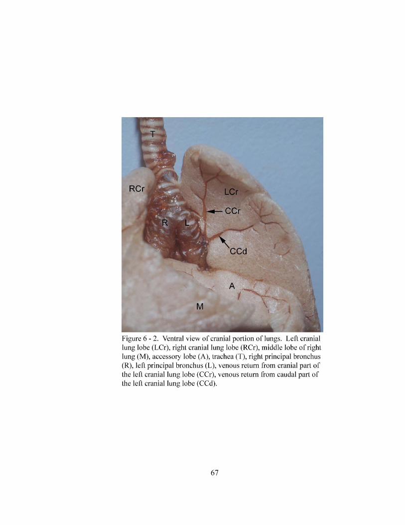
67
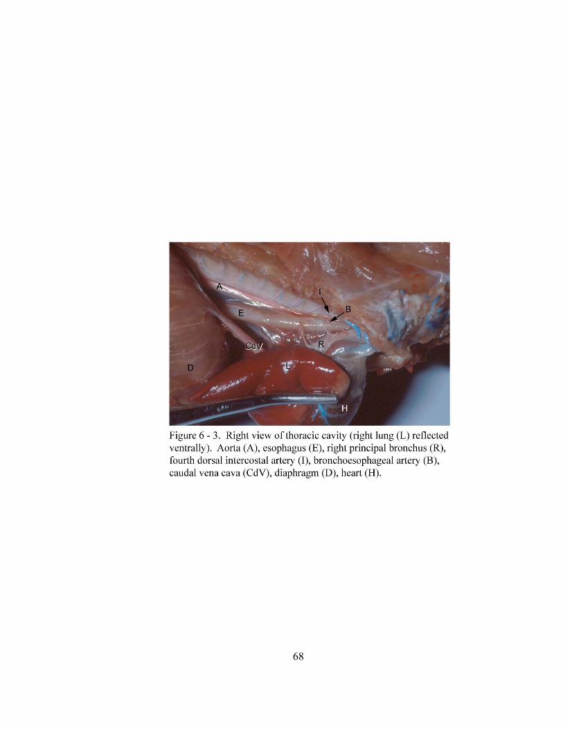
68
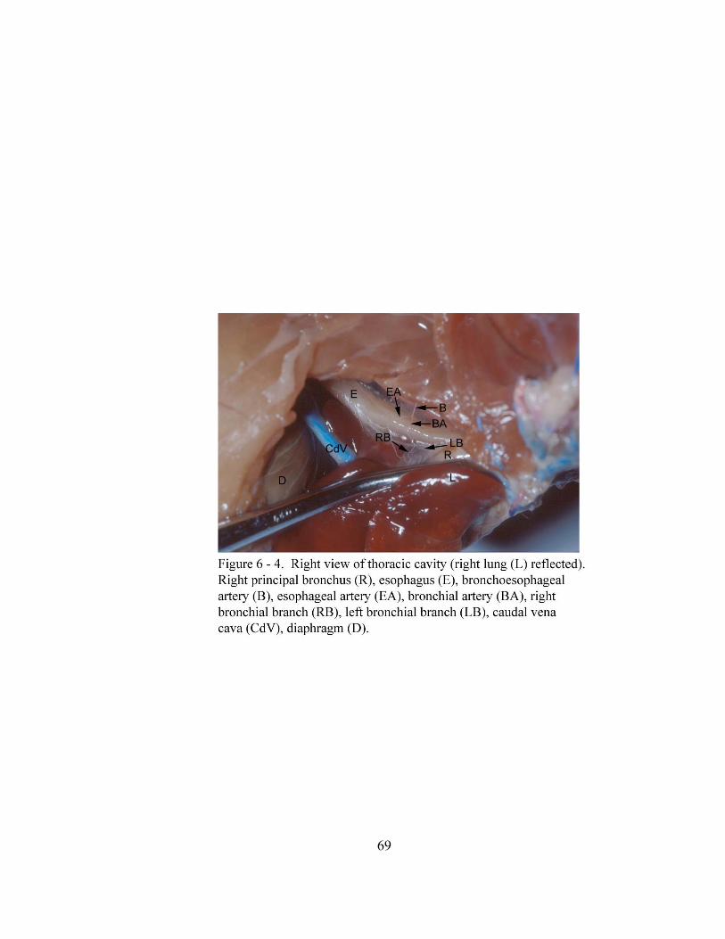
69
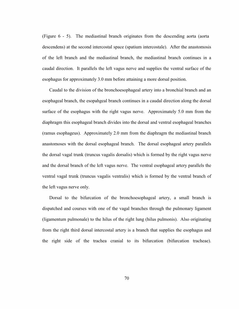
70
(Figure 6 - 5). The mediastinal branch originates from the descending aorta (aorta
descendens) at the second intercostal space (spatium intercostale). After the anastomosis
of the left branch and the mediastinal branch, the mediastinal branch continues in a
caudal direction. It parallels the left vagus nerve and supplies the ventral surface of the
esophagus for approximately 3.0 mm before attaining a more dorsal position.
Caudal to the division of the bronchoesophageal artery into a bronchial branch and an
esophageal branch, the esopahgeal branch continues in a caudal direction along the dorsal
surface of the esophagus with the right vagus nerve. Approximately 5.0 mm from the
diaphragm this esophageal branch divides into the dorsal and ventral esophageal branches
(ramus esophageus). Approximately 2.0 mm from the diaphragm the mediastinal branch
anastomoses with the dorsal esophageal branch. The dorsal esophageal artery parallels
the dorsal vagal trunk (truncus vagalis dorsalis) which is formed by the right vagus nerve
and the dorsal branch of the left vagus nerve. The ventral esophageal artery parallels the
ventral vagal trunk (truncus vagalis ventralis) which is formed by the ventral branch of
the left vagus nerve only.
Dorsal to the bifurcation of the bronchoesophageal artery, a small branch is
dispatched and courses with one of the vagal branches through the pulmonary ligament
(ligamentum pulmonale) to the hilus of the right lung (hilus pulmonis). Also originating
from the right third dorsal intercostal artery is a branch that supplies the esophagus and
the right side of the trachea cranial to its bifurcation (bifurcation tracheae).
71
72
Lymph Nodes of the Lower Respiratory Tract:
Two cranial deep cervical lymph nodes are associated with the cervical part of the
trachea. They are located on the left and right side of the laryngotracheal junction lateral
to the longus capitis muscle. They measure 1.51 ± 0.14 mm in length. Cranial
mediastinal lymph nodes (lymphonodi mediastinales craniales) and tracheobronchial
lymph nodes (lymphonodi tracheobronchales) are associated with the thoracic part of the
lower respiratory tract. Two cranial mediastinal lymph nodes are located on the right and
left ventrolateral surfaces of the trachea, approximately 3.0 mm cranial to the base of the
heart (basis cordis) (Figure 6 - 6). These lymph nodes are oval shaped and measure 1.33
± 0.58 mm in length. Three tracheobronchial lymph nodes are located at the tracheal
bifurcation (Figure 6 - 7). The right and left tracheobronchial lymph nodes [lymphonodi
tracheobronchales bifurcationis (dexter et sinister)] measure 2.31 ± 0.32 mm in length,
are oval shaped and lay on the lateral side of the right and left principal bronchi. The
middle tracheobronchial lymph node (lymphonodi tracheobronchales bifurcationis medii)
is the largest lymph node associated with the lower respiratory system of the gray short-
tailed opossum. It measures 3.62 ± 0.3 mm in length and lies in the angle of the tracheal
bifurcation.
Innervation of the Trachea, Lungs and Diaphragm:
The lower respiratory tract of the gray short-tailed opossum receives innervation from
the sympathetic trunks and the vagus nerves. The right and left sympathetic trunks
(truncus sympathicus) lay behind the costal pleura (costalis pleura) lateral to the thoracic
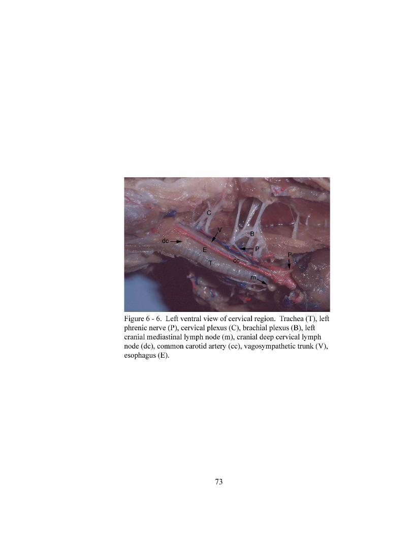
73
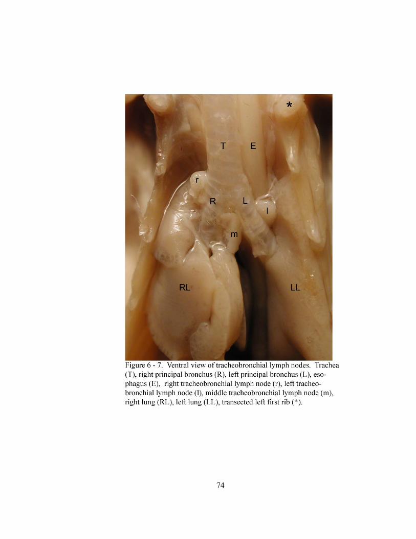
74
75
vertebral bodies (corpus vertebrae). They cross the lateral surfaces of the dorsal
intercostal vessels to reach their ipsilateral cervicothoracic ganglion (ganglion
cervicothoracicum). The right and left cervicothoracic ganglia lay in deep grooves
bordered medially by the ipsilateral longus colli muscle (musculi longus colli) and
laterally by ribs (costae) one and two with their associated intercostal muscles (musculi
intercostales). The sympathetic trunk joins the vagus nerve by the cranial and caudal
loops of the ansa subclavia which emerge from the ventral margin of the cervicothoracic
ganglion as a common trunk. The caudal loop of the ansa courses ventrally to join the
vagus nerve caudal to the subclavian artery (arteria subclavia) thus forming the
vagosympathetic trunk. The cranial loop courses in a cranial direction, dorsal to the
internal jugular vein (venae jugularis interna), and parallels the vagosympathetic trunk
(truncus vagosympathicus) for approximately 4.0 mm before joining it. Cardiac nerves
also emerge from the cervicothoracic ganglion and parallel the caudal loop of the ansa
subclavia for a short distance. These cardiac nerves provide sympathetic innervation to
the heart. Originating from the thoracic part of each sympathetic trunk are two to three
thoracic splanchnic nerves. These nerves course ventrally through the mediastinal pleura
and pulmonary ligament to the hilus of each lung.
As the right vagus nerve crosses the ventrolateral surface of the subclavian artery, the
right recurrent laryngeal nerve (nervus laryngeus recurrens) leaves the vagus nerve,
curves ventromedially around the right subclavian artery and continues cranially along
the right ventral aspect of the trachea. At the thoracic inlet (apertura thoracis cranialis),
the right recurrent laryngeal nerve moves to a dorsal position on the cervical trachea.
76
Near the aortic arch (arcus aortae), the left recurrent laryngeal nerve leaves the left
vagus nerve and curves medially then cranially around the arch to continue along the left
ventral aspect of the trachea. At the thoracic inlet, it attains a position dorsal to the
trachea and continues in a cranial direction.
Caudal to the principal bronchus of the right and left lungs, three to six vagal
branches leave each vagus nerve and course to the hilus of the lung through the
pulmonary ligament. Caudal to the hilus of the right lung, the right vagus nerve
continues caudally along the right dorsal surface of the esophagus. Caudal to the hilus of
the left lung and approximately 6.0 mm from the diaphragm, the left vagus nerve divides
into dorsal and ventral branches which continue caudally along the left dorsal and ventral
surfaces of the esophagus. The right vagus nerve and the dorsal branch of the left vagus
nerve join 1.0 cm cranial to the diaphragm and parallel the dorsal esophageal artery as the
dorsal vagal trunk. Only the ventral branch of the left vagus nerve continues caudally as
the ventral vagal trunk along the ventral surface of the esophagus paralleling the ventral
esophageal artery.
Coursing medial to the right and left lungs are the respective phrenic nerves (nervus
phrenicus). The right and left phrenic nerves are formed by components of the ventral
branches (rami ventrales) of cervical nerves (nervi cervicales) 3, 4, 5, and 6 (Figure 6 -
6). The contributions of cervical nerves 3 and 4 to the phrenic nerve pass through the
cervical plexus. The contribution of cervical nerves 5 and 6 to the phrenic nerve pass
through the brachial plexus (plexus brachialis). In the cervical region, the right and left
phrenic nerves are located immediately lateral to the ipsilateral internal jugular vein. As
77
they course caudally toward the thoracic inlet (apertura thoracis cranialis), they are
medial to the ipsilateral brachial plexus. After passing through the thoracic inlet, the
right phrenic nerve passes ventrolateral to the right subclavian artery and lateral to the
right cranial vena cava (vena cava cranialis). As the nerve continues caudally, it inclines
over the dorsal aspect of the right atrium (atrium dextrum) to continue along the lateral
margin of the caudal vena cava (vena cava caudalis) on its course to the right costal part
(pars costalis) of the diaphragm. The right phrenic nerve is anchored by the mediastinal
pleura to the parietal pericardium of the heart and the caudal vena cava. The left phrenic
nerve follows a course similar to that of the right phrenic nerve in the ventral cervical
region. After passing through the thoracic inlet, the left phrenic nerve passes ventral to
the left subclavian artery, dorsal to the left cranial vena cava and along the left ventral
margin of the heart where it is anchored to the parietal pericardium by the mediastinal
pleura. At the apex of the heart (apex cordis), the left phrenic nerve travels caudodorsally
in the caudal mediastinal pleura en route to the left costal part of the diaphragm (Figure 6
- 8).
The arterial and nervous distribution, as well as lymph node location and number,
described above were seen in all animals studied.
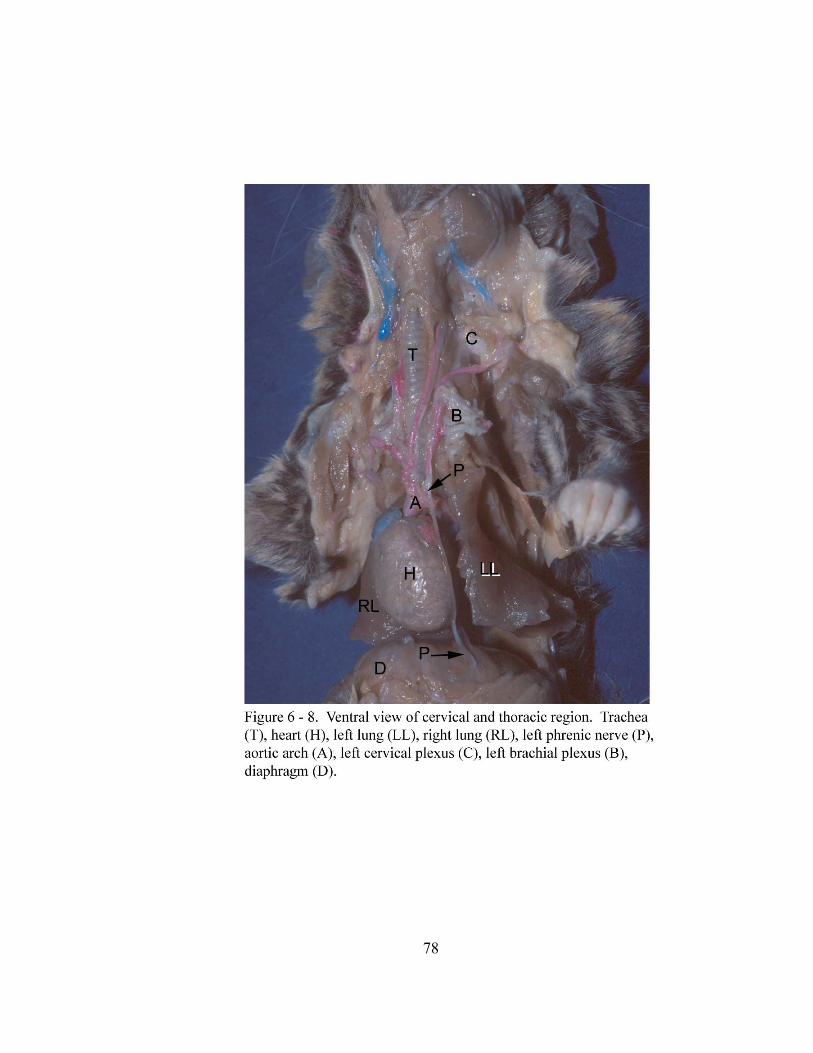
78
79
DISCUSSION:
The branching pattern of the pulmonary lobar arteries as well as their location as
related to the bronchi in the gray short-tailed opossum is similar to that of domestic
mammals (Nickel et al., 1981; Evans, 1993). Descriptions of the distribution of
pulmonary lobar arteries in other marsupials could not be located for further comparison.
Right and left pulmonary veins joining to form a common pulmonary venous trunk as
occurs in the gray short-tailed opossum has also been reported in the North American
opossum (McClure, 1903; Wade and Neely, 1949), the brush-tail opossum (Trichosurus
vulpecula) (Dowd, 1969) and the native cat (Dasyurus viverrinus) (Hill, 1955). The
common pulmonary vein seems to be a distinct characteristic of marsupials whereas
domestic animals exhibit a pattern characterized by multiple pulmonary veins opening
directly into the left atrium.
The bronchial artery of the gray short-tailed opossum is small and its course is
difficult to follow macroscopically beyond the hilus of the lung. Earlier reports on the
bronchial artery in mammals state that the distribution will vary depending on the
thickness of the pleura, the presence of interlobular septa and the presence of bronchial -
pulmonary artery anastomoses (McLaughlin, 1983).
The lymph nodes draining the lower respiratory tract of the gray short-tailed opossum
include the cranial deep cervical lymph nodes, cranial mediastinal lymph nodes and
tracheobronchial lymph nodes. An earlier report on the lymphatic system of two species
of South American opossums, Didelphys azarae and Didelphys marsupislis, describe
80
deep cervical lymph nodes, anterior mediastinal lymph nodes, posterior cranial
mediastinal lymph nodes, a posterior middle mediastinal lymph node and bronchial
lymph nodes (Azzali and Didio, 1965). In the gray short-tailed opossum and Didelphys
(Azzali and Didio, 1965), the cranial deep cervical lymph nodes are located immediately
lateral to the laryngotracheal junction and the longus capitis muscles. Azzali and Didio
(1965) describe two sets of mediastinal lymph nodes as right and left anterior mediastinal
lymph nodes and right and left posterior cranial mediastinal lymph nodes. The anterior
mediastinal lymph nodes were located ventral to the subclavian artery, medial to the
brachiocephalic vein and sometimes found buried in the perithymic fat. These lymph
nodes were not observed in the gray short-tailed opossum. The posterior cranial
mediastinal lymph nodes were described as lying ventral to the longus colli muscle and
medial to the first intercostal space. These lymph nodes are similar in location to the
cranial mediastinal lymph nodes of the gray short-tailed opossum. Azzali and Didio
(1965) describe a single posterior middle mediastinal lymph node located near the arch of
the left azygous vein. Middle mediastinal lymph nodes were not observed in the gray
short-tailed opossum. In the gray short-tailed opossum and the North American
opossum, the tracheobronchial lymph nodes are located at the tracheal bifurcation and on
each side of the right and left principal bronchi. The bronchial lymph nodes of Didelphys
as described by Azzali and Didio (1965) seem to be similar to the tracheobronchial lymph
nodes of the gray short-tailed opossum. One bronchial lymph node of Didelphys is
located cranial to the tracheal bifurcation and the second is located just caudal to the
81
tracheal bifurcation. The South American opossums evidently possess one fewer
tracheobronchial lymph node than the gray short-tailed opossum.
The pattern of innervation to the lung of the gray short-tailed opossum is not
comparativley different from that of domestic animals.
The phrenic nerves of the gray short-tailed opossum are formed by components of
ventral branches of cervical nerves as is seen in domestic animals (Nickel et al., 1981;
Evans, 1993).
The gray short-tailed opossum appears to have a respiratory system less closely
resembling that of other opossums and marsupials previously described. Further studies
would need to be undertaken on these other marsupials to definitively determine this
assumption.
82
CHAPTER VII
ANATOMY OF STRUCTURES ASSOCIATED WITH THE LOWER
RESPIRATORY TRACT OF THE NORTH AMERICAN OPOSSUM
(Didelphis virginiana)
ABSTRACT:
This study documents the macroscopic anatomy of the associated structures of the
lower respiratory tract of the North American opossum (Didelphis virginiana). Cranial
deep cervical, cranial mediastinal and tracheobronchial lymph nodes drain the lower
respiratory tract. Vascularization of the lungs is via the bronchial artery a branch of the
bronchoesophageal artery. The bronchial artery divides into right and left bronchial
arteries which follow the branches of the bronchial tree. The pulmonary arteries divide
into pulmonary lobar arteries which follow the bronchial division. Pulmonary lobar veins
from each lobe of the right and left lungs join to form three pulmonary veins (right, left
and middle). The three pulmonary veins join to form a common pulmonary venous trunk
that opens into the left atrium of the heart. Sympathetic innervation to the lungs comes
from the ipsilateral sympathetic trunks through the mediastinal pleura and pulmonary
ligaments as thoracic splanchnic nerves. Parasympathetic innervation to the lungs is via
branches from the ipsilateral vagus nerves. The right and left phrenic nerves are formed
by components of ventral branches of cervical nerves 2 - 6 which pass through the
cervical (C2-C4) and brachial (C5-C6) plexuses.
83
INTRODUCTION:
For twenty-five years, the North American opossum has been used in biomedical
research in the United States. The extensive use of this marsupial in research has yielded
numerous papers in the area of embryogenesis (Klima, 1987; Renfree, 1990; Szalay,
1994) and yet there have been relatively few anatomical descriptions of macroscopic
anatomy. Two sources provide a detailed description on the origin and development of
the lymphatic system of the North American opossum (Zimmerman, 1940; Kampmeier,
1969). McClure (1903) and Wade and Neely (1949) each briefly describe the pulmonary
vasculature while Bernard’s (1996) work provides a brief description of the bronchial
artery. The goal of the present study is to provide a description of the structures
associated with the respiratory system of the North American opossum.
MATERIALS AND METHODS:
Fourteen (7 females and 7 males) North American opossums (Didelphis virginiana)
were used to study structures associated with the lower respiratory tract. Three of these
opossums were used to produce tracheobronchial vascular casts and one was used for a
whole body arterial injection. The tracheobronchial vascular casts were produced by
injection of latex into the pulmonary vasculature followed by injection of silicone into the
airways (Henry, 1992a and 1992b). Prior to arterial injection, the opossum was
embalmed with 10% buffered formalin. Twenty-four hours after embalming, latex was
injected into the common carotid artery to aid visualization and dissection of the lower
respiratory tract vasculature.
84
RESULTS:
Pulmonary Vasculature and Bronchoesophageal Artery:
The pulmonary trunk (truncus pulmonalis) bifurcates into right and left pulmonary
arteries (arteria pulmonalis dextra et sinistra). The right and left pulmonary arteries cross
the ventral surface of their respective principal bronchi (bronchus principalis) and curve
laterally then dorsally to attain a position dorsal to each bronchus. As the right and left
principal bronchi (bronchus principalis dexter et sinister) divide into lobar bronchi
(bronchi lobares), the right and left pulmonary arteries divide into branches that follow
the bronchial division. The right pulmonary artery divides into a cranial lobar branch
(ramus lobi cranialis), a middle lobar branch (ramus lobi medii), a caudal lobar branch
(ramus lobi caudalis) and an accessory lobar branch (ramus lobi accessorii). The left
pulmonary artery divides unevenly into three branches. Two smaller branches arise from
the left pulmonary artery to supply the cranial and caudal parts of the cranial lobe. The
large, third branch of the left pulmonary artery enters the caudal lobe as the caudal lobar
branch and sends branches along the caudal lobar bronchial division. In general, the
lobar arteries course along the dorsal surface of the lobar bronchi with the exception of
the accessory lobar artery. This artery passes between the middle and caudal lobar
bronchi of the right lung (pulmo dexter) to course along the ventral surface of the
accessory lobar bronchus.
The right, middle and left pulmonary veins, which return blood from the lungs to the
left atrium (atrium sinistrum) of the heart (cordis), are formed by the pulmonary lobar
85
veins (Figure 7 - 1). The pulmonary lobar veins lay along the ventral surface of the
corresponding lobar bronchi except for the accessory pulmonary lobar vein which is
dorsal to the accessory lobar bronchus. The right pulmonary vein is formed by the right
cranial pulmonary lobar vein (venae pulmonalis lobi cranialis dextra) and the right
middle pulmonary lobar vein (venae pulmonalis lobi medii). In addition, an extra vein
from the cranial part of the right caudal lobe may contribute to the right pulmonary vein.
The right caudal pulmonary lobar vein (venae pulmonalis lobi caudalis dextra) and the
accessory pulmonary lobar vein (ramus lobi accessorii) join to form a middle pulmonary
vein. The left pulmonary vein is formed by a branch from the cranial part of the cranial
lobe and a branch from the caudal part of the cranial lobe which join the left caudal
pulmonary lobar vein (venae pulmonalis lobi caudalis sinistri) (Figure 7 - 1). The right,
left and middle pulmonary veins join to form a common pulmonary venous trunk that
opens into the left atrium of the heart.
On the right side of the mediastinum, the bronchoesophageal artery (arteria broncho-
esophagea) originates from the right, third dorsal intercostal artery (arteria intercostales
dorsales) (Figure 7 - 2). The bronchoesophageal artery courses ventrally through the
mediastinum across the right side of the esophagus and divides into a bronchial artery
(ramus bronchalis) and a common trunk that divides into the esophageal artery (ramus
esophageus) and an artery that supplies the right side of the trachea, right vagus nerve
(nervus vagus) and right tracheobronchial lymph node (lymphonodi tracheobronchales
dexter) (Figure 7 - 2). Before the bronchial artery divides into right and left bronchial
arteries, a common branch is given off which passes through the hilus of the right lung
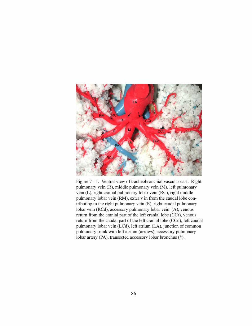
86
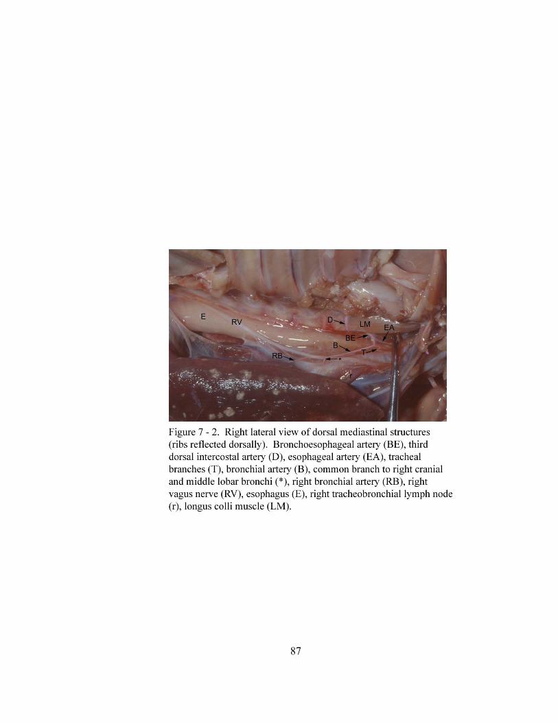
87
88
(hilus pulmonis) and divides to follow the cranial and middle lobar bronchi and their
branches (Figure 7 - 2). After this, the bronchial artery continues caudoventrally through
the middle mediastinal pleura (mediastinalis pleura) and divides caudal to the tracheal
bifurcation (bifurcatio tracheae) into branches to the right and left lungs (pulmo sinister)
(Figure 7 - 3a). The right bronchial artery courses ventrally toward the hilus of the lung
through the mediastinal pleura, medial to the right vagus nerve. The right bronchial
artery enters at the hilus caudal to the right principal bronchus, divides, and follows the
branches of the caudal lobar bronchus. The left bronchial artery continues through the
mediastinum to the left side of the thoracic cavity (Figures 7 - 3a and 7 - 3b). Caudal to
the left principal bronchus it anastomoses with a mediastinal artery (rami mediastinales)
and continues ventrally toward the hilus of the left lung. Here the left bronchial artery
enters the lung and sends branches to the left bronchial division. The mediastinal artery
originates from the caudal surface of the aortic arch (arcus aortae) at the second
intercostal space (spatium intercostale). After the anastomosis of the left bronchial artery
and the mediastinal artery, a small artery arises from the left bronchial artery just prior to
its passage through the hilus of lung. This small artery continues in a caudal direction
through the mediastinal pleura and divides into two branches which parallel the right and
left vagus nerves (Figure 7 - 3a).
Lymph Nodes of the Lower Respiratory Tract:
Cranial deep cervical lymph nodes (lymphonodi cervicales profundi craniales) are
present lateral to the laryngotracheal junction and ventral to the longus capitis muscle
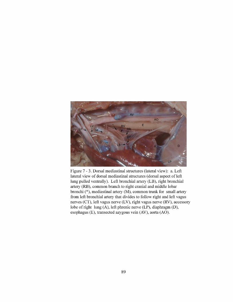
89
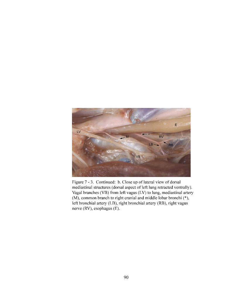
90
91
(Figure 7 - 4). Each lymph node is oval and is 4.31 ± 0.9 mm wide by 11.78 ± 1.3 mm
long. Cranial mediastinal lymph nodes (lymphonodi mediastinales craniales) and
tracheobronchial lymph nodes (lymphonodi tracheobronchales bifurcationis) are present
along the thoracic trachea. The two cranial mediastinal lymph nodes, which lie cranial to
the base of the heart (basis cordis) in adipose tissue, are located respectively one each on
the right and left ventrolateral surfaces of the trachea at the level of the first to second
intercostal spaces (Figure 7 - 5). The cranial mediastinal lymph nodes measure 4.9 ±
0.65 mm in width and 17 ± 0.86 mm in length. The right and left phrenic nerves (nervus
phrenicus) course across these lymph nodes. Three tracheobronchial lymph nodes are
located in the region of the tracheal bifurcation at the level of the third to fourth
intercostal spaces (Figure 7 - 6). The right and left tracheobronchial lymph nodes are
4.84 ± 0.62 mm wide and 9.54 ± 1.69 mm long, oval shaped and lay on the lateral side of
the right and left principal bronchi respectively. The middle tracheobronchial lymph
node (lymphonodi tracheobronchales bifurcationis medii) is the largest of the three lymph
nodes. It is oval shaped and measures 6.3 ± 0.57 mm in width and 14.6 ± 0.54 mm in
length. It is located in the angle of the tracheal bifurcation.
Innervation of the Trachea, Lungs and the Diaphragm:
The lower respiratory tract of the North American opossum receives innervation from
the sympathetic trunks (truncus sympathicus) and the vagus nerves. The right and left
sympathetic trunks lay behind the costal pleura (costalis pleura) lateral to the thoracic
vertebral bodies (corpus vertebrae thoracicae). The sympathetic trunks cross the lateral
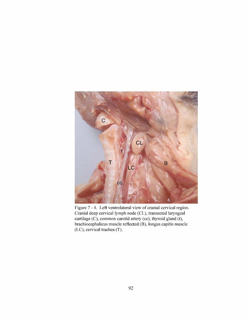
92
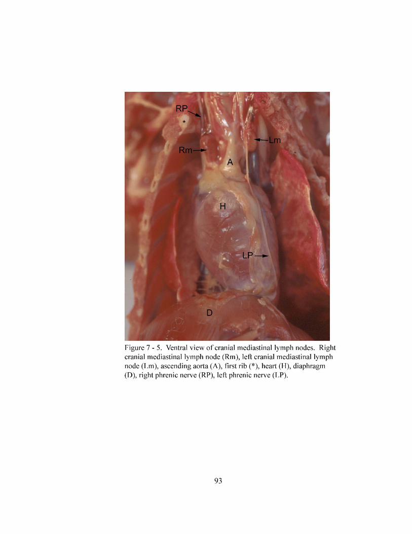
93
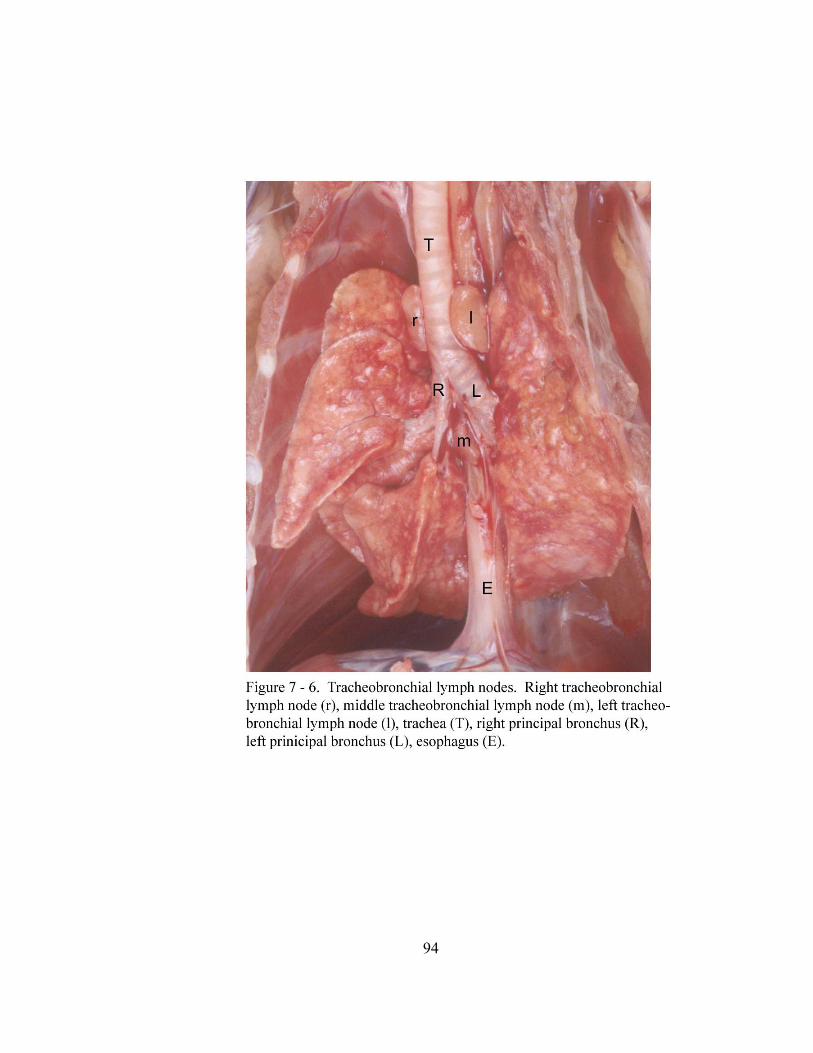
94
95
surfaces of the dorsal intercostals vessels (arteriae intercostalis dorsalis) to reach their
ipsilateral cervicothoracic ganglion (ganglion cervicothoracicum). The right and left
cervicothoracic ganglia each lay deep within the groove formed medially by the
ipsilateral longus colli muscle (musculi longus colli) and laterally by ribs (costae) one
and two and their associated intercostals muscles (musculi intercostals). The sympathetic
trunk continues from the ventral margin of the cervicothoracic ganglion and bifurcates
into cranial and caudal loops of the ansa subclavia. The ansa subclavia joins the vagus
nerve to form the vagosympathetic trunk (truncus vagosympathicus). From the ganglia
of the sympathetic trunk of each side, thoracic splanchnic nerves (nervus splanchnicus)
course ventrally through the mediastinal pleura (mediastinalis pleura). Near the hilus,
they deviate laterally into the pulmonary ligaments (ligamentum pulmonale) and enter
each lung.
As the right vagus nerve crosses the ventral surface of the subclavian artery (arteria
subclavia), the right recurrent laryngeal nerve (nervus laryngeus recurrens) leaves the
vagus nerve and curves ventromedially to continue cranially along the right side of the
trachea. The left recurrent laryngeal nerve leaves the left vagus nerve near the aortic arch
and curves medially then cranially around the aortic arch to continue cranially along the
left side of the trachea. After the right and left recurrent laryngeal nerves are dispatched,
the ipsilateral vagus nerves continue caudally along the dorsal aspect of the thoracic
trachea. Caudal to the principal bronchus of the right and left lungs, three to six vagal
branches leave each vagus nerve (Figure 7 - 3b). These vagal branches course through
the pulmonary ligament to the hilus of each lung. Caudal to the hilus of the lungs, the
96
right and left vagus nerves continue caudally along the dorsal surface of the esophagus.
Approximately 10.0 mm from the diaphragm the left vagus nerve divides into dorsal and
ventral branches which course caudally along the esophagus. The right vagus nerve
remains undivided. Approximately 6.0 mm from the diaphragm, the right vagus nerve
and the dorsal branch of the left vagus nerve join along the dorsal esophageal surface to
form the dorsal vagal trunk (truncus vagalis dorsalis). The ventral branch of the left
vagus nerve continues caudally along the ventral esophageal surface as the ventral vagal
trunk (truncus vagalis ventralis).
Coursing medial to the right and left lungs are the ipsilateral phrenic nerves. The right
and left phrenic nerves are formed by components of the ventral branches (rami
ventrales) of cervical nerves (nervi cervicales) 2 through 6 which pass through the
cervical and brachial plexuses. The contribution of cervical nerves 2, 3 and 4 to the
phrenic nerve pass through the cervical plexus. The contribution of cervical nerves 5 and
6 to the phrenic nerve pass through the brachial plexus. As the phrenic nerves course
caudally toward the thoracic inlet (apertura thoracis cranialis), they are located medial to
the brachial plexus. After passing through the thoracic inlet, the right phrenic nerve
inclines over the dorsal aspect of the right atrium (dextrum atrium) to continue along the
right side of the caudal vena cava (vena cava caudalis) on its course to the right costal
part (pars costalis) of the diaphragm. The right phrenic nerve is anchored by the
mediastinal pleura to the parietal pericardium of the heart and the caudal vena cava. The
left phrenic nerve, after passing through the thoracic inlet courses, dorsal to the left
cranial vena cava (vena cava cranialis). It continues caudally along the left ventral
97
margin of the heart where it is anchored by the mediastinal pleura to the parietal
pericardium (Figure 7 - 5). At the apex (apex cordis) of the heart, the left phrenic nerve
continues caudodorsally in the caudal mediastinal pleura en route to the left costal part of
the diaphragm.
The arterial and nervous supply to the lungs as described was observed in all animals.
Additionally, lymph node location and numbers were consistent among all animals.
DISCUSSION:
The division of the pulmonary arteries into pulmonary lobar arteries in the North
American opossum is similar to that of many domestic mammals as the branching pattern
follows that of the bronchi (Nickel et al., 1981; Evans, 1993). The location of the
pulmonary lobar arteries in relation to the bronchi as well as the branching pattern within
each lung lobe in the North American opossum is similar to that of the canine (Evans,
1993).
In the North American opossum, pulmonary lobar veins from both lungs join to form
three pulmonary veins. These three veins then unite to form a common pulmonary
venous trunk before emptying into the left atrium. Previous descriptions of the
pulmonary vasculature of the North American opossum report finding a common
pulmonary venous trunk in some specimens (Owen, 1868; McClure, 1903; Wade and
Neely, 1949). These articles do not describe the vessels which form the common
pulmonary venous trunk. Wade et al. (1949) did identify two specimens which had two
veins from each lung that joined and formed only two pulmonary veins and in other
98
specimens four pulmonary veins directly entering the left atrium. Hill (1955) and Dowd
(1969) report that the native cat (Dasyurus viverrinus) and the brush-tail opossum
(Trichosurus vulpecula) also have a common pulmonary venous trunk which is formed
by a common right and common left pulmonary vein. Pulmonary venous return as
described in marsupials involves some degree of common pulmonary vein formation.
These common veins then unite to form a common pulmonary venous trunk. The actual
pattern varies slightly among species. We did not find any descriptions which matched
that which we observed in the North American opossum.
Concerning blood supply to the lung parenchyma of the North American opossum,
Bernard et al. (1996) state that either a distinct bronchoesophageal or common bronchial
artery originates from the thoracic aorta at the fifth intercostal space. We observed only
the former description during this study. In either case, the bronchial artery then divides
into right and left branches at the tracheal bifurcation. This division of the bronchial
artery described by Bernard et al. (1996) is similar to that which we observed in the
North American opossum as well as that of many mammalian species (Nickel et al.,
1981; Schaller, 1992; Evans, 1993). The anastomosis of the left bronchial artery with a
mediastinal artery before the former enters the lung is also seen in the canine (Auton et
al., 2000).
The superior cervical lymph nodes identified by Zimmerman (1940) and Kampmeier
(1969) are likely the cranial deep cervical lymph nodes we observed in the North
American opossum. In both cases these lymph nodes are described as lying along the
larynx, medial to the cleido-mastoid, cleido-occipital and sterno-mastoid muscle groups.
99
The location of the cranial deep cervical lymph nodes in the North American opossum is
similar to those of many domestic carnivores (Nickel et al., 1981; Evans, 1993).
The location of the cranial mediastinal lymph nodes in the North American opossum
is similar to the findings of Zimmerman (1940) and Kampmeier (1969) who refer to these
as anterior mediastinal lymph nodes. Kampmeier (1969) and Azzali and Didio (1965)
describe posterior cranial mediastinal lymph nodes in North American opossum and large
American opossums of South America (Didelphis marsupialis and Didelphis azarae).
These nodes were not observed in North American opossums utilized in this study.
The three tracheobronchial lymph nodes of the North American opossum are
positioned one each respectively on the lateral side of the right and left principal bronchi
and in the angle of the tracheal bifurcation. This positioning is similar that found in
many domestic species (Nickel et al., 1981; Schaller, 1992; Evans, 1993). However, in
previous reports on the North American opossum (Kampmeier, 1969) and the large
American opossums of South America (Azzali and Didio, 1965), only two bronchial
lymph nodes were found. One lymph node was cranial to the tracheal bifurcation and
one caudal to the bifurcation.
The innervation to the lung of the North American opossum is unremarkable as it is
similar to that of many domestic mammals (Nickel et al., 1981; Evans, 1993).
100
CHAPTER VIII
MICROSCOPIC ANATOMY OF THE LOWER RESPIRATORY TRACT OF
THE GRAY SHORT-TAILED OPOSSUM (Monodelphis domestica)
ABSTRACT:
This portion of the present study describes the microscopic anatomy of the lower
respiratory tract of the gray short-tailed opossum (Monodelphis domestica). The trachea
and right and left principal bronchi are lined with pseudostratified ciliated columnar
epithelium. Only a few goblet cells are seen and localized to the trachea. The secondary
and tertiary bronchi and the primary and secondary bronchioles are lined by a ciliated
simple columnar epithelium. The terminal bronchioles and a portion of the respiratory
bronchioles are lined by ciliated simple cuboidal epithelium. The terminal portion of the
respiratory bronchioles and the alveolar ducts are lined with simple squamous epithelium.
Alveoli are lined by type I and type II pneumocytes. Compound acinar seromucous
tracheal glands are present in the tela submucosa. The fibromusculocartilaginous tunic of
the trachea consists of c-shaped hyaline cartilge rings and the trachealis muscle. A
lamina muscularis mucosae is not present in the extrapulmonary portion of the principal
bronchus. However, it appears in the intrapulmonary portion of the principal bronchus
and continues into the respiratory bronchioles. The compound acinar seromucous
bronchial glands are present in the propria submucosa and tela submucosa of the
principal bronchi. The bronchial glands do not extend beyond the principal bronchi. The
101
musculocartilaginous tunic is present only in the extrapulmonary portion of the principal
bronchus. The bronchial cartilages are irregular shaped plates and do not extend past the
extrapulmonary portion of the principal bronchus. The visceral pleura is a layer of
simple squamous mesothelium covering the outer surface of the lung. Deep to this layer
is the tunica subserosa, a layer of loose collagenous connective tissue, which gives off
septa into the parenchyma of the lung lobes.
INTRODUCTION:
During the past century, microscopic anatomy of the lower respiratory tract of the rat
(Loosli, 1938; Jeffery, 1975, 1983; Spicer et al., 1982; Scheuermann et al., 1988),
hamster (Becci et al., 1978; Jeffery, 1983), mouse (Hansell and Moretti, 1969;
Greenwood and Holland, 1972; Pack et al., 1981; Jeffery, 1983), rabbit and guinea pig
(Loosli, 1938; Jeffery, 1983), canine (Loosli, 1938; Tucker, 1974; Jeffery, 1983), feline
(Jeffery, 1983; Al-Tikriti et al., 1991), sheep (Alcorn et al., 1981), pig (Clements, 1938;
Ham and Baldwin, 1941; Jeffery, 1983), ox (Tucker, 1974) and nonhuman primates
(Loosli, 1938; Castleman et al., 1975; Jeffery, 1983; Tyler and Plopper, 1985) has been
studied extensively using light and electron microscopy. During the same period, only
seven articles were located on the microscopic anatomy of the lower respiratory tract of
all marsupials such as the North American opossum (Didelphis virgianana) (Bremer,
1904; Sorokin, 1962, 1965; Krause and Leeson, 1973, 1975; Krause et al., 1976), koala
(Phascolarctos cinereus) and phalanger (Trichosurus vulpecula) (Tucker, 1974). These
previous descriptions do not provide many details on the histologic features of the lower
102
respiratory tract of these marsupials. Hence a detailed description documenting the
normal histologic features of the lower respiratory tract of a marsupial would benefit
future researchers. Therefore, this study will provide a more complete description of the
histologic features of the lower respiratory tract of the gray short-tailed opossum
(Monodelphis domestica).
MATERIALS AND METHODS:
Seven gray short-tailed opossums were used to study the microscopic anatomy of the
lower respiratory tract. Each opossum was administered 500 IU of heparin sodium
solution (Heparin Sodium Injection 1,000 units per 1.0 ml, Elkins-Sinn Inc. Cherry Hill,
NJ 08003) by intraperitoneal (IP) injection to facilitate exsanguination. Eighteen hours
later, the opossums were anesthetized with an IP injection of 50 mg of Nembutal
(Nembutal Sodium Solution, Pentobarbital Sodium Injection, Abbott Laboratories, North
Chicago, IL 60064) per 100.0 grams of body weight. When a stage 3 plane of anesthesia
was reached, the left common carotid artery and external jugular vein were catheterized.
The opossums were exsanguinated via these catheters. After euthanasia, in situ fixation
of the trachea and lungs of six of the opossums by intratracheal perfusion was performed
with 10% buffered formalin via a canula from a height of 5.0 cm for 5 minutes.
Following fixation, the trachea and lungs were removed and placed in 10% buffered
formalin for an additional 24-48 hours before sectioning. Tissue samples were taken
from all six lung lobes from four animals for study of the lung parenchyma. The four
tracheas from these animals were divided into cranial, middle and caudal thirds prior to
103
sectioning. The right and left lungs were removed from two opossums for serial
sectioning of the conducting components of the respiratory system and parenchyma of the
lung. All samples were processed for light microscopy using a Tissue TEK VIP 1000
(Floor Model, Mode #4617, Serial #8811895, Ames Division, Miles Laboratories Inc.,
P.O. Box 70, Elkhart, IN 46515) through a series of dehydration and infiltration. The
tissues were dehydrated in a graded series of ethanol (80%, 95%, 95%, 100%, 100%,
100%) followed by two series of xylene under pressure (.35 kg cm2) and vacuum cycle
(50.0 cm Hg) at 40o C for one hour. The samples were infiltrated with paraffin (Paraplast
Tissue embedding Medium, Oxford Labware, Division of Sherwood Medical St. Louis,
MO 63103) under pressure (0.35 kg cm2) and vacuum cycle (50.0 cm Hg) at 60o C for
one hour (repeated twice). Samples were then embedded in paraffin and positioned in
tissue molds to obtain blocks which would yield transverse sections through the trachea
and the bronchi when cut. Random sections 5.0 µm in thickness were taken at 75.0 µm
intervals from the blocks containing the trachea and bronchi. Serial sections (5.0 µm
thick) were made from both lungs. All sections were mounted on glass slides and stained
with either hematoxylin and eosin or Acid Orcein Giemsa (Luna, 1968).
The trachea and lungs of the seventh gray short-tailed opossum were fixed in situ by
perfusion of 2.5% gluteraldehyde (25% Gluteraldehyde EM Grade, Electron Microscopy
Sciences, PO Box 251, 321 Morris Road, Ft. Washington, PA 19034) in 0.1M cacodylate
buffer (pH = 7.4) (Sodium Cacodylate Trihydrate, Electron Microscopy Sciences, PO
Box 251, 321 Morris Road, Ft. Washington, PA 19034) via the catheterized left common
104
carotid artery. The opossum was placed in the cooler at 5oC for 24 hours after which
the thoracic cavity was opened and the trachea and lungs removed and placed in new
2.5% gluteraldehyde in 0.1M cacodylate buffer. It was returned to the cooler until
samples were taken. Tissue samples were taken from the trachea and lungs, cut into 1.0
mm cubes and washed three times each in distilled water. Samples were post - fixed in a
1:1 mixture of 2% aqueous osmium tetroxide and 3% aqueous potassium ferrocyanide at
room temperature for 2 hours or longer until the tissue became dark brown or black in
color. The samples were then removed from the osmium ferrocyanide and were again
washed three times for ten minutes in distilled water. Following this, the samples were
dehydrated for ten minutes each in 50%, 70%, 80% and 90% ethanol followed by two
washes in 100% ethanol for 60 minutes each. Samples were then removed from the
100% ethanol and washed two times, fifteen minutes each, in propylene oxide
(Polysciences Inc. Warrington, PA 18976). After the final washing, the tissue samples
were placed in tissue micromolds (Polysciences Inc. Warrington, PA 18976) and
embedded in epon araldite (Russell and Burguet, 1977). Random sections of 1.0 – 2.0
µm were cut, mounted on glass slides and stained with a mixture of 1:1 mixture of
methylene blue (Methylene Blue, Sigma Chemical Company, PO Box 14508, St. Louis,
MO 63178) and azure blue (Azure II, Sigma Chemical Company, PO Box 14508, St.
Louis, MO 63178).
All sections from the seven gray short-tailed opossum were examined by light
microscopy using an Olympus CH-2 binocular microscope.
105
RESULTS:
Trachea:
The cervical and thoracic parts of the trachea (pars cervicalis et thoracica trachea) of
the gray short-tailed opossum are lined by a low pseudostratified ciliated columnar
epithelium (epithelium pseudostratificatum columnare ciliatum) (Figure 8 - 1). The
height of this epithelium from the basal to the apical surface of the columnar cells is 14
µm. It consists of three cell types including columnar cells (epitheliocytus columnaris
ciliatus), basal cells (epitheliocytus basalis) and goblet cells (exocrinocytus caliciformis).
The columnar cells have a large, round-oval, basophilic staining, vesicular nucleus. The
nucleus is centrally located and surrounded by a foamy and sometimes vacuolated
cytoplasm. These cells rest on the basement membrane (membrana basalis) of the
epithelial layer and their apical surface reaches the lumen of the trachea and is covered
with cilia. Basal cells are wedged between adjacent columnar cells. The shape of the
basal cells is not distinct because the margins of these cells are not visible with routine
H&E staining. These cells are not ciliated and the shape of the nucleus varies from
flattened to round or triangular. The nucleus of the basal cell stains more basophilic than
those of the columnar cells and often has a prominent nucleolus. Only an occasional
goblet cell was found among the epithelial cells. The apical cytoplasm of the goblet cell
is foamy and distended so that its apical surface appears domed and bulges into the lumen
of the trachea. The base of the goblet cell is narrow and occupied by a flattened,
basophilic staining nucleus.
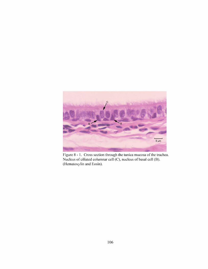
106
107
Beneath the epithelial layer is the lamina propria mucosae which consists of elastic
fibers (fibra elastica) and fibroblasts (fibroblastocytus) (Figure 8 - 2). Deep to the lamina
propria mucosae is the loose connective tissue (textus connectivus collagenosus laxus) of
the tela submucosa. This layer contains numerous small blood vessels and compound
acinar seromucous tracheal glands (glandulae tracheales) (Figures 8 - 2 and 8 - 3). The
tracheal glands are located on the luminal side of the cartilage rings (cartilagines
tracheales) along the right, left and ventral regions of the trachea. The excretory ducts of
the tracheal glands (glandulae tracheales) open onto the luminal surface of the trachea.
These ducts are lined by a simple columnar epithelium (Figure 8 - 4). These columnar
cells have a centrally placed basophilic nucleus which is surrounded by foamy basophilic
cytoplasm. The mucous adenomeres of the tracheal glands consist of pyramidal shaped
mucous cells (exocrinocytus granulum mucigeni) with a foamy cytoplasm and a small,
round to flattened basal nucleus (Figures 8 - 3 and 8 - 5). The serous cells (exocrinocytus
granulum zymogeni) are located both around the periphery of the mucous adenomeres
and form crescent shaped serous demilunes (semiluna serosa) or are individually
interposed between mucous cells. The serous cells have a granular, eosinophilic staining
cytoplasm with a round, basally placed nucleus. The secretory product of the serous
demilunes reaches the lumen of the mucous adenomeres by secretory canaliculi
(canaliculus intercellularis) that extend between adjacent mucous cells (Figure 8 - 5).
Myoepithelial cells (myoepitheliocytus stellatus) are located between the base of the
secretory cells of the tracheal gland adenomeres and the underlying basement membrane
(Figure 8 - 5). The fibromusculocartilaginous tunic (tunica fibromusculocartilaginea) of
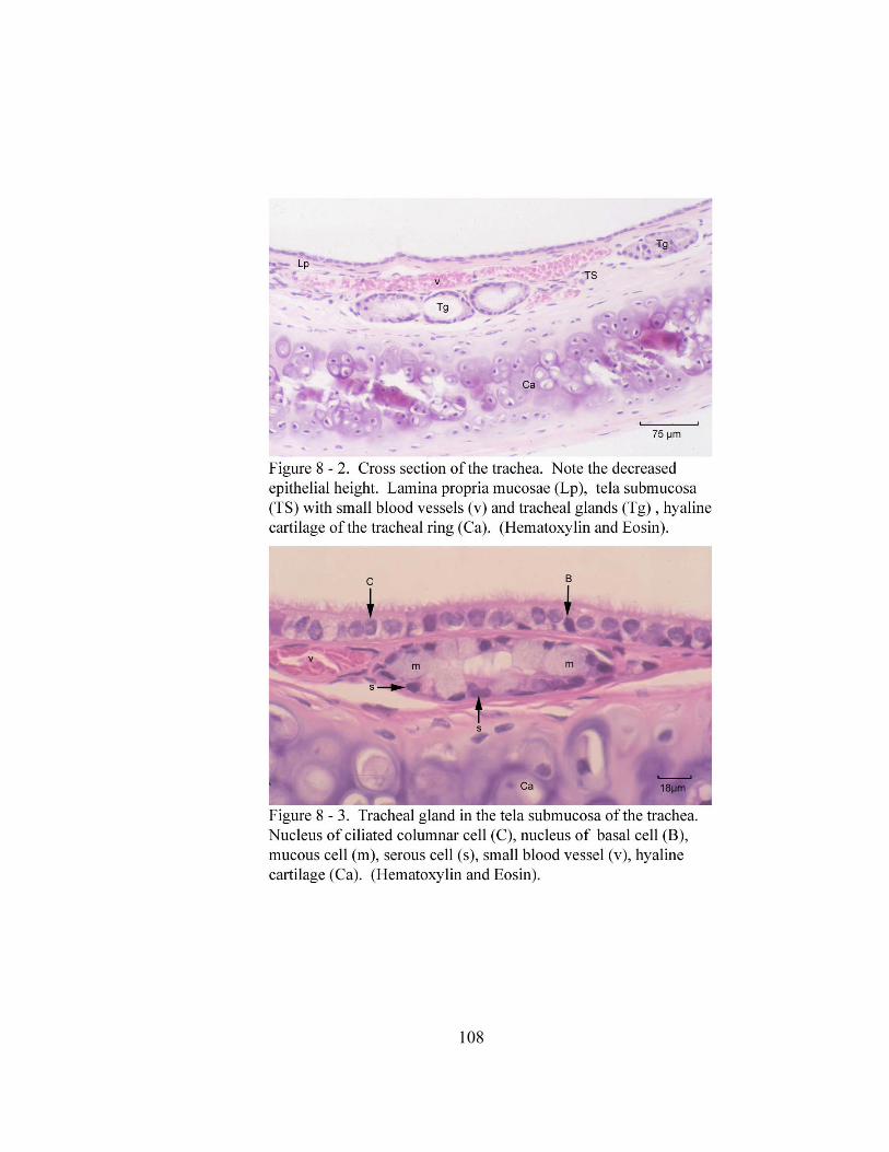
108
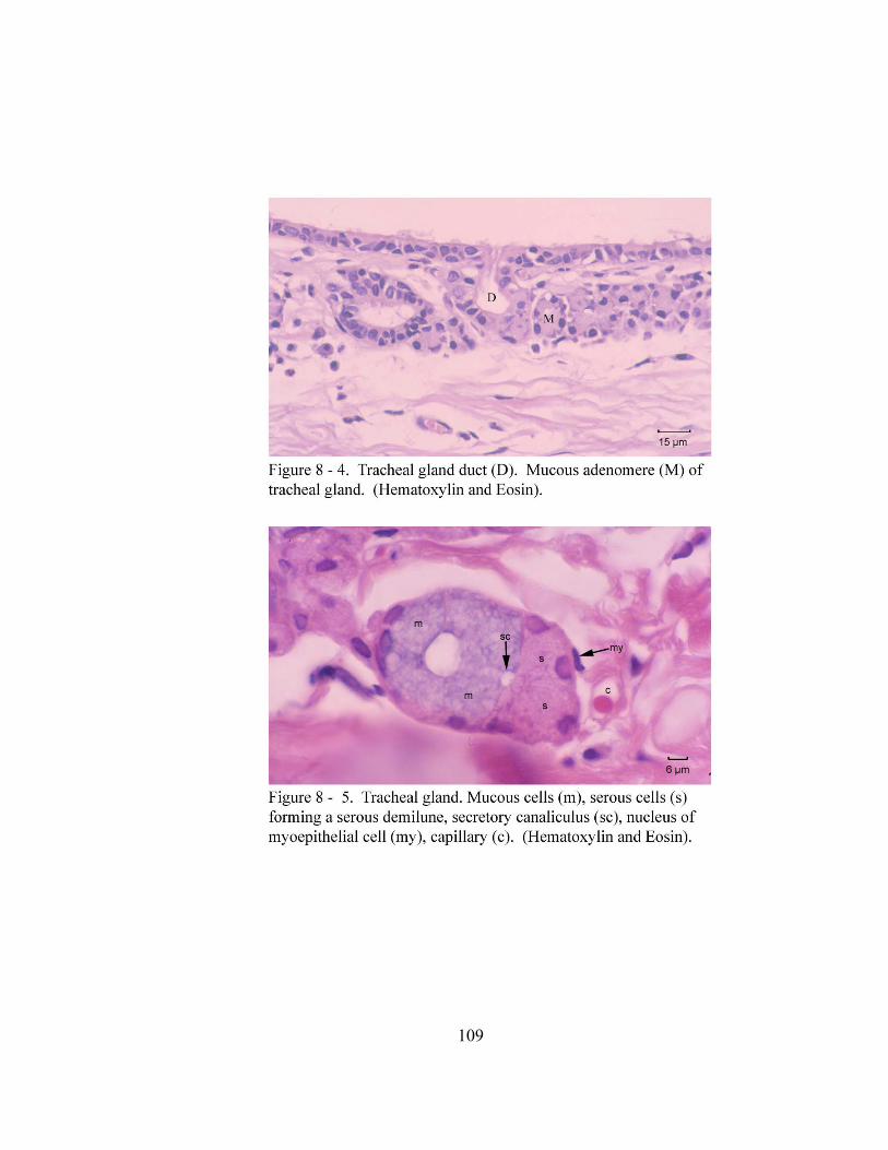
109
110
the trachea consists of a membranous part (paries membranaceus) and a cartilaginous
part. The cartilaginous portion of the fibromusculocartilaginous tunic consists of the
incomplete rings of c-shaped hyaline cartilage (cartilago hyaline). The hyaline cartilage
rings consists of an inner and outer zone (region). The inner zone is basophilic and
formed by clusters of chondrocytes (chondrocytus) and their surrounding matrix (matrix
cartilaginea). The outer zone is the perichondrium. This region stains a pale eosinophilia
and consists of a fibrous and cellular portion. The fibrous portion (stratum fibrosum) is a
layer of dense collagenous connective tissue with fibroblasts. The cellular portion
(stratum chondrogenicum) is composed of chondroblasts. The membranous portion
consists of the transverse bands of smooth muscle cells (myocytus nonstriatus) of the
trachealis muscle (musculus trachealis) (Figure 8 - 6). This muscle is on average eight
cell layers thick. It attaches to the fibrous part of the perichondrium inside the free ends
of each tracheal cartilage. Deep to the trachealis muscle is a layer of dense irregular
collagenous connective tissue (textus connetivus collagenous compactus irregularis) in
which small blood vessels and lymphatic vessels are located. This layer along with the
trachealis muscle bridges the dorsal gap left by the incomplete cartilage rings. The
connective tissue of the membranous portion blends with the tunica adventitia of the
trachea (Figure 8 - 6). The tunica adventitia of the trachea is dense irregular collagenous
connective tissue in which small blood vessels are located.
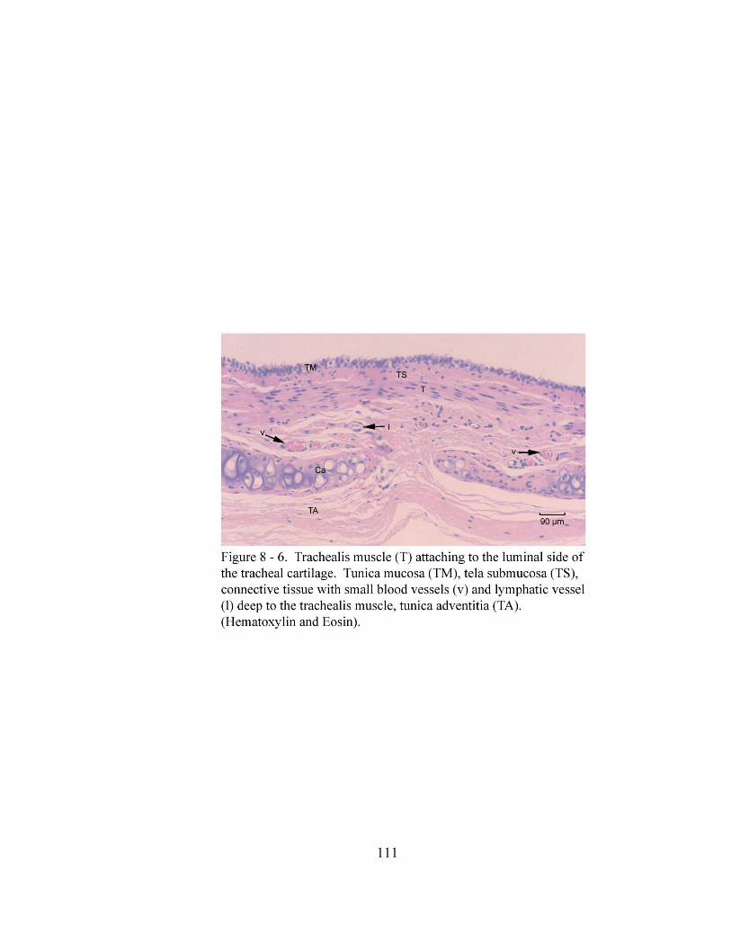
111
112
Bronchial Tree
Projecting into the tracheal lumen at its bifurcation (bifurcation tracheae) into right
and left principal bronchi (bronchus principalis dexter et sinister) are two ridges of tissue.
These ridges are located opposite from one another on the dorsal and ventral aspects of
the tracheal lumen. Each ridge is covered with an epithelium similar to that of the
trachea; however, the ciliated cells appear to be cuboidal (epitheliocytus cuboideus).
Each ridge of tissue has two layers of smooth muscle deep to the tunica mucosa which
are derived from the trachealis muscle. The two ridges unite and form a vertical
membranous tracheal carina (carina tracheae) which marks the bifurcation of the trachea
into right and left principal bronchi (Figure 8 - 7). The carina is covered by a
pseudostratified ciliated cuboidal epithelium with two layers of smooth muscle fibers
deep to the epithelium. These two layers of smooth muscle are separated by a layer of
loose connective tissue (Figure 8 - 7). Within this connective tissue layer are small
blood vessels and compound acinar bronchial glands (glandulae bronchiales). The
excretory ducts of these glands are lined with a simple columnar epithelium (epithelium
simplex columnare). These ducts open into the lumen of the cranial most portion of the
principal bronchi. These bronchial glands consist of mucous cells which have a foamy
cytoplasm and a basally located basophilic staining nucleus. No serous cells are observed
in these glands. Located on the ventral surface of the trachea prior to its bifurcation and
dorsal to the pulmonary trunk (truncus pulmonalis) is an autonomic nervous system
ganglion (ganglion autonomicum) (Figure 8 - 8). The nerve cell bodies (corpus neuroni)
of the ganglion are round and surrounded by satellite cells that are intimately adjacent to

113
114
the nerve cell bodies. The nuclei of the nerve cell bodies are eccentrically placed, large,
round and often have a prominent central nucleolus (Figure 8 - 8). The nuclei of the
nerve cell bodies are surrounded by a granular, basophilic cytoplasm. The nerve cell
bodies are surrounded by a layer of connective tissue which blends with the surrounding
tunica adventitia of the pulmonary trunk and trachea.
The extrapulmonary portion of the principal bronchus is lined by a pseudostratified
ciliated columnar epithelium which is 21.0 µm in height (Figure 8 - 9). This epithelial
layer consists of columnar and basal cells similar to those of the trachea. The columnar
cells have an oval shaped basophilic staining vesicular nucleus and cilia covering the
apical margin (Figure 8 - 9). The basal cells are found between the adjacent columnar
cells and the shape of the nucleus varies. Mucous secreting goblet cells are not observed
in the bronchial epithelium. The propria submucosa is a layer of loose connective tissue
deep to the epithelium of the principal bronchi (Figures 8 - 9 and 8 - 10). This layer
contains compound acinar seromucous bronchial glands. The bronchial glands are
present within the propria submucosa of this portion of the principal bronchus. However,
they are also seen between adjacent bronchial cartilage plates (cartilagines bronchiales)
and peripheral to them (Figure 8 - 10). The excretory ducts of the bronchial glands open
into the lumen of the principal bronchi. The epithelium of this duct is simple columnar
epithelium. The columnar cells are non-ciliated and have a centrally located basophilic
staining vesicular nucleus surrounded by a vacuolated cytoplasm. The bronchial glands
are composed mainly of mixed adenomeres; however, an occassional serous adenomere
is also seen. The mixed adenomeres consist of mucous and serous
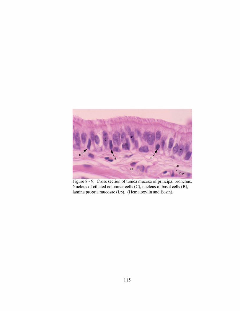
115
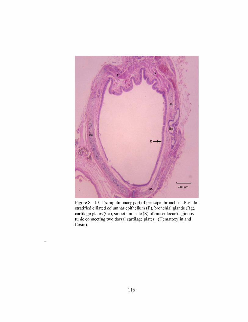
116
117
cells. The mucous cells are pyramidal shaped, have a foamy cytoplasm and a small
round or sometimes flattened, basally positioned basophilic nucleus. The serous cells
have a granular, pale staining eosinophilic cytoplasm with a round, basally located
nucleus. These cells are located in the adenomeres among the mucous cells or around the
periphery of the mucous adenomeres as serous demilunes or form adenomeres
themselves. The musculocartilaginous tunic (tunica musculocartilaginea) consists of a
smooth muscle layer and a cartilaginous part. The cartilaginous part consists of bronchial
cartilage plates that are localized to the extrapulmonary portion of the principal bronchus.
The smooth muscle layer is approximately seven cell layers thick, circularly oriented and
attaches to the inside of the dorsal most right and left bronchial cartilage plates (Figure 8-
10).
The intrapulmonary portion of the principal bronchus is lined by an epithelium
similar to that of the extrapulmonary portion. Additionally, goblet cells are not observed
in the epithelium of this portion of the bronchus. The lamina prorpia mucosae in this
region is a layer of loose connective tissue deep to the epithelium (Figure 8 - 11). It is
separated from the tela submcuosa by the lamina muscularis mucosae. This layer is a
continuation of the smooth muscle of the musculocartilaginous tunic and is arranged as
individual bundles of circularly oriented smooth muscle which are five to six cell layers
thick. The tela submucosa is a layer of loose connective which contains small blood
vessels, compound branched acinar seromucous bronchial glands, lymphatic vessels and
an occasional autonomic ganglion (Figure 8 - 11). The bronchial glands and
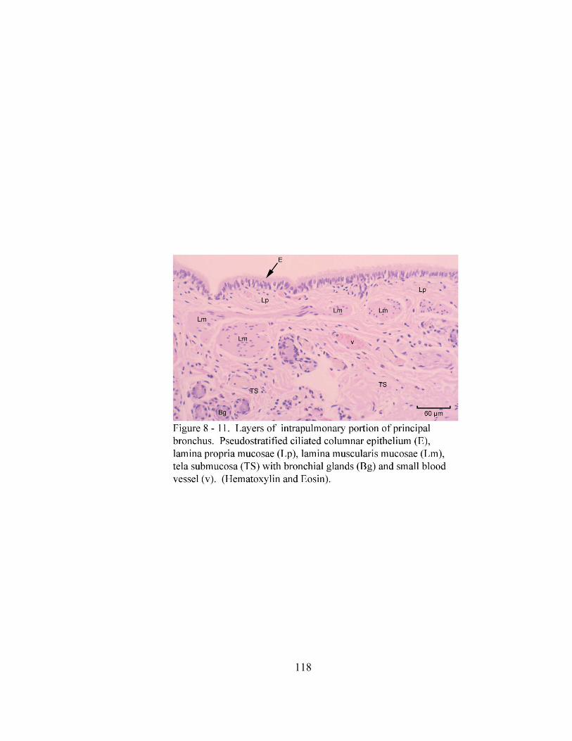
118
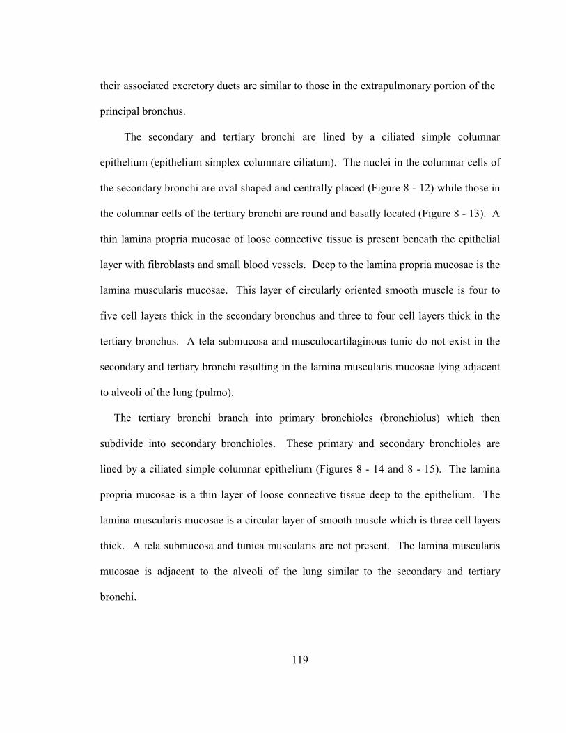
119
their associated excretory ducts are similar to those in the extrapulmonary portion of the
principal bronchus.
The secondary and tertiary bronchi are lined by a ciliated simple columnar
epithelium (epithelium simplex columnare ciliatum). The nuclei in the columnar cells of
the secondary bronchi are oval shaped and centrally placed (Figure 8 - 12) while those in
the columnar cells of the tertiary bronchi are round and basally located (Figure 8 - 13). A
thin lamina propria mucosae of loose connective tissue is present beneath the epithelial
layer with fibroblasts and small blood vessels. Deep to the lamina propria mucosae is the
lamina muscularis mucosae. This layer of circularly oriented smooth muscle is four to
five cell layers thick in the secondary bronchus and three to four cell layers thick in the
tertiary bronchus. A tela submucosa and musculocartilaginous tunic do not exist in the
secondary and tertiary bronchi resulting in the lamina muscularis mucosae lying adjacent
to alveoli of the lung (pulmo).
The tertiary bronchi branch into primary bronchioles (bronchiolus) which then
subdivide into secondary bronchioles. These primary and secondary bronchioles are
lined by a ciliated simple columnar epithelium (Figures 8 - 14 and 8 - 15). The lamina
propria mucosae is a thin layer of loose connective tissue deep to the epithelium. The
lamina muscularis mucosae is a circular layer of smooth muscle which is three cell layers
thick. A tela submucosa and tunica muscularis are not present. The lamina muscularis
mucosae is adjacent to the alveoli of the lung similar to the secondary and tertiary
bronchi.
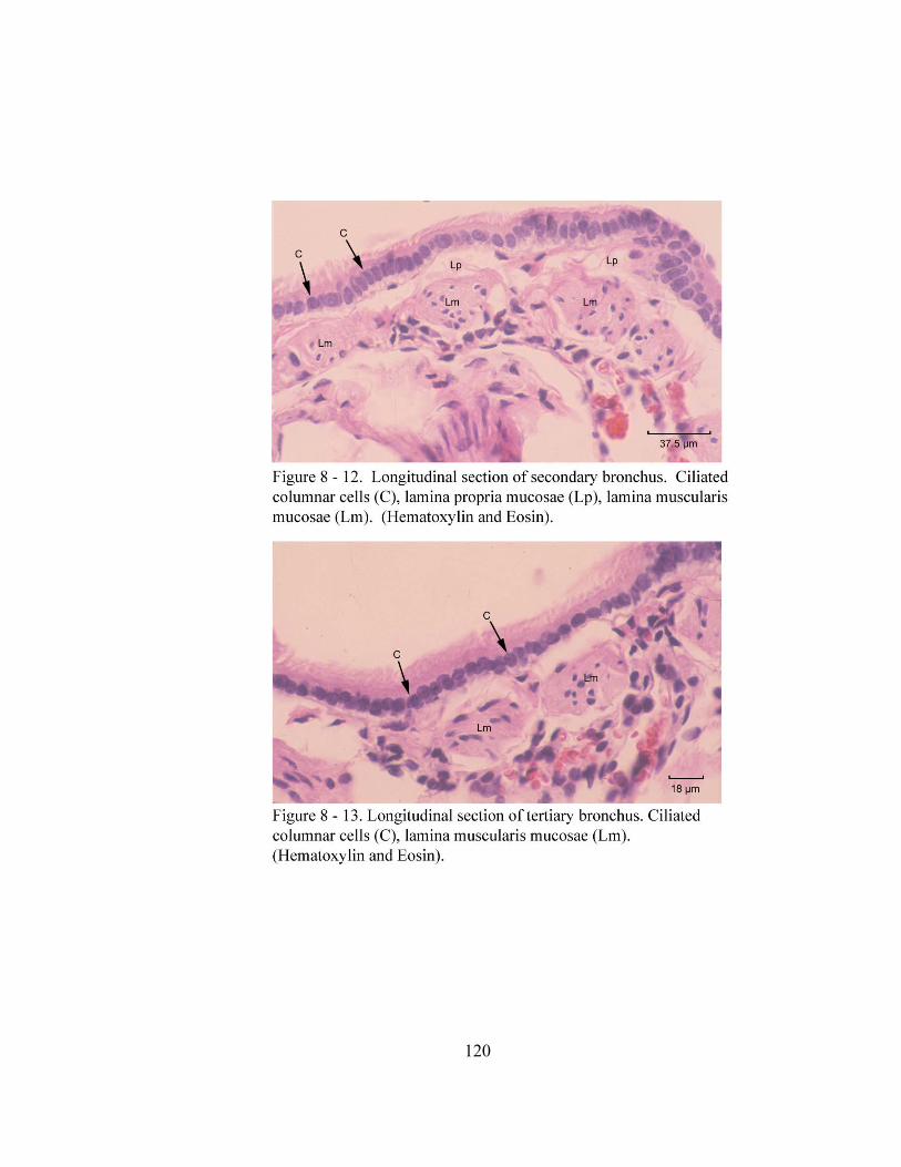
120
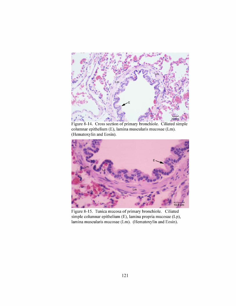
121
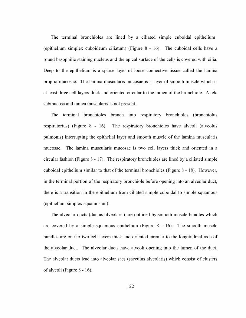
122
The terminal bronchioles are lined by a ciliated simple cuboidal epithelium
(epithelium simplex cuboideum ciliatum) (Figure 8 - 16). The cuboidal cells have a
round basophilic staining nucleus and the apical surface of the cells is covered with cilia.
Deep to the epithelium is a sparse layer of loose connective tissue called the lamina
propria mucosae. The lamina muscularis mucosae is a layer of smooth muscle which is
at least three cell layers thick and oriented circular to the lumen of the bronchiole. A tela
submucosa and tunica muscularis is not present.
The terminal bronchioles branch into respiratory bronchioles (bronchiolus
respiratorius) (Figure 8 - 16). The respiratory bronchioles have alveoli (alveolus
pulmonis) interrupting the epithelial layer and smooth muscle of the lamina muscularis
mucosae. The lamina muscularis mucosae is two cell layers thick and oriented in a
circular fashion (Figure 8 - 17). The respiratory bronchioles are lined by a ciliated simple
cuboidal epithelium similar to that of the terminal bronchioles (Figure 8 - 18). However,
in the terminal portion of the respiratory bronchiole before opening into an alveolar duct,
there is a transition in the epithelium from ciliated simple cuboidal to simple squamous
(epithelium simplex squamosum).
The alveolar ducts (ductus alveolaris) are outlined by smooth muscle bundles which
are covered by a simple squamous epithelium (Figure 8 - 16). The smooth muscle
bundles are one to two cell layers thick and oriented circular to the longitudinal axis of
the alveolar duct. The alveolar ducts have alveoli opening into the lumen of the duct.
The alveolar ducts lead into alveolar sacs (sacculus alveolaris) which consist of clusters
of alveoli (Figure 8 - 16).
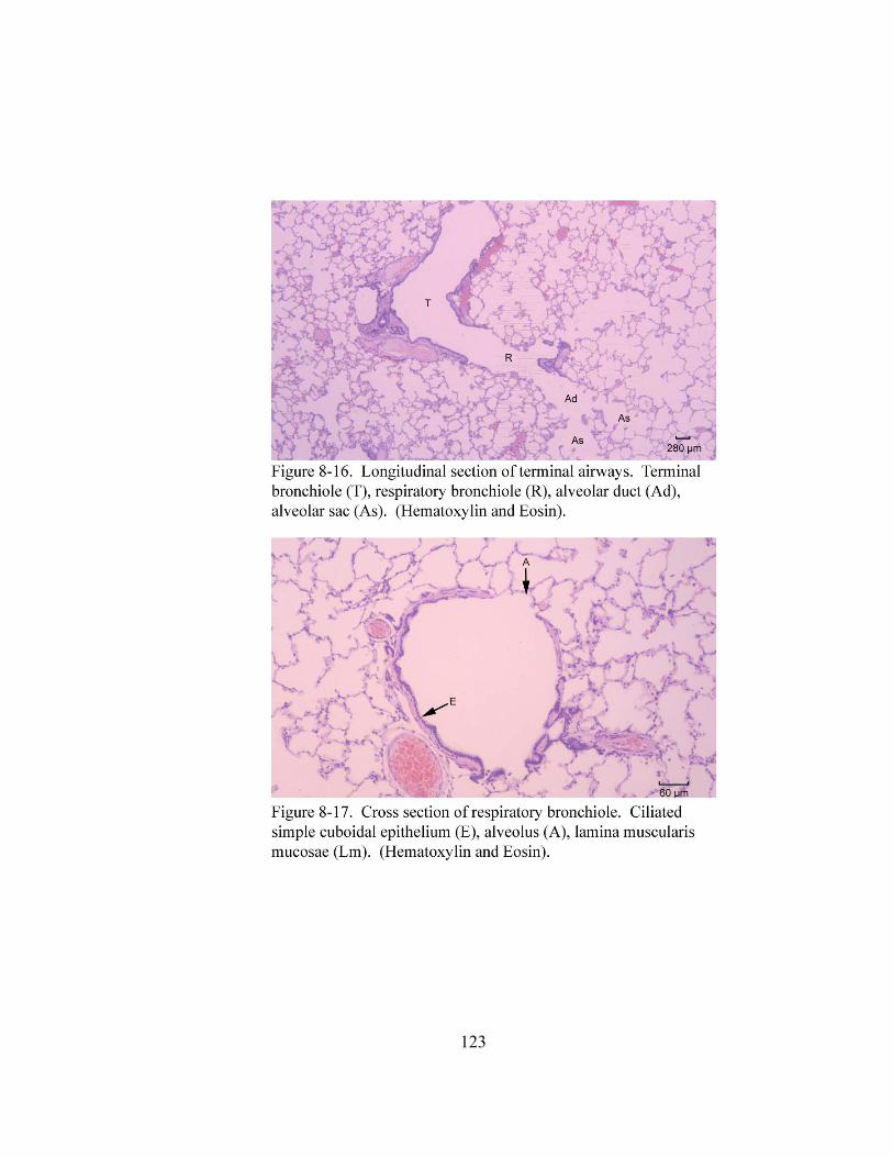
123
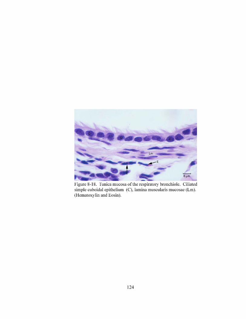
124
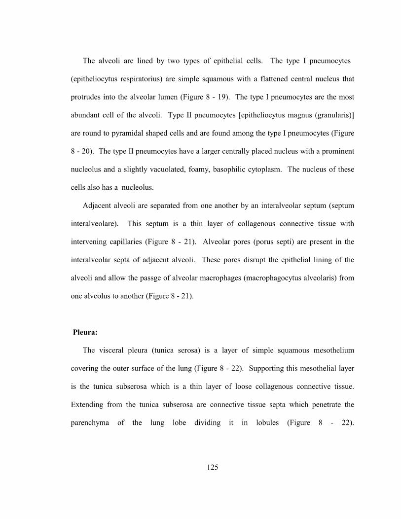
125
The alveoli are lined by two types of epithelial cells. The type I pneumocytes
(epitheliocytus respiratorius) are simple squamous with a flattened central nucleus that
protrudes into the alveolar lumen (Figure 8 - 19). The type I pneumocytes are the most
abundant cell of the alveoli. Type II pneumocytes [epitheliocytus magnus (granularis)]
are round to pyramidal shaped cells and are found among the type I pneumocytes (Figure
8 - 20). The type II pneumocytes have a larger centrally placed nucleus with a prominent
nucleolus and a slightly vacuolated, foamy, basophilic cytoplasm. The nucleus of these
cells also has a nucleolus.
Adjacent alveoli are separated from one another by an interalveolar septum (septum
interalveolare). This septum is a thin layer of collagenous connective tissue with
intervening capillaries (Figure 8 - 21). Alveolar pores (porus septi) are present in the
interalveolar septa of adjacent alveoli. These pores disrupt the epithelial lining of the
alveoli and allow the passge of alveolar macrophages (macrophagocytus alveolaris) from
one alveolus to another (Figure 8 - 21).
Pleura:
The visceral pleura (tunica serosa) is a layer of simple squamous mesothelium
covering the outer surface of the lung (Figure 8 - 22). Supporting this mesothelial layer
is the tunica subserosa which is a thin layer of loose collagenous connective tissue.
Extending from the tunica subserosa are connective tissue septa which penetrate the
parenchyma of the lung lobe dividing it in lobules (Figure 8 - 22).
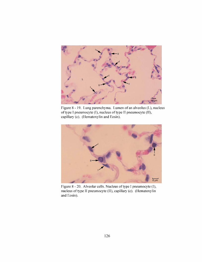
126
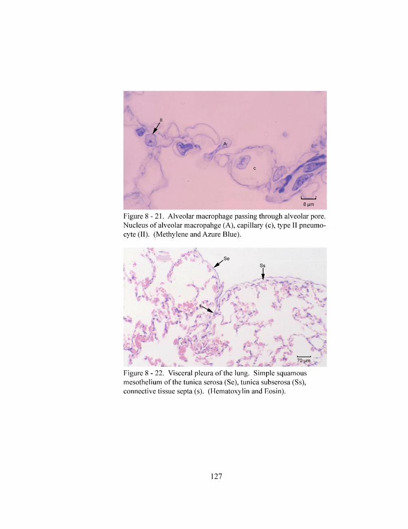
127
128
DISCUSSION:
The epithelium lining the trachea and the principal bronchi of the adult gray short-
tailed opossum (Monodelphis domestica) is classified as pseudostratified ciliated
columnar with goblet cells which is similar to domestic mammals (Banks, 1993;
International Committee on Veterinary Gross Anatomical Nomenclature, 1994; Dellmann
and Eurell, 1998). The height of the epithelium between these two regions of the gray
short-tailed opossum shows some variation which may be a result of the pressure of
intratracheal perfusion. However, samples were taken at various regions of the trachea
and principal bronchi of all animals and all sites yielded consistent results which suggest
a regional difference in the epithelial height of this opossum between the trachea and
principal bronchi.
Three cell types (columnar cells, basal cells and goblet cells) have been identified by
light microscopy in the tracheal epithelium of the gray short-tailed opossum. These cells
are morphologically similar to those described in domestic carnivores (Breeze and
Wheeldon, 1977; Dellmann and Eurell, 1998). In the gray short-tailed opossum, few
goblet cells are observed among the columnar cells in the trachea as compared to
domestic carnivores in which they are found throughout the length of the trachea (Breeze
and Wheeldon, 1977; Dellmann and Eurell, 1998). Similarly Hansell (1968), Jeffery
(1975), Becci (1978) and Pack et al. (1981) also documented decreased numbers of
goblet cells in the tracheal epithelium of the mouse, rat and hamster.
129
The columnar and basal cells of the principal bronchial epithelium are similar to
those found in the tracheobronchial tree of domestic carnivores (Banks, 1993; Dellmann
and Eurell, 1998). However, the goblet cells described in the bronchi of domestic
carnivores are not observed in the principal bronchi of this opossum.
Numerous small blood vessels are observed within the tela submucosa of the gray
short-tailed opossum. A similar observation is also described in the trachea of the koala
(Phascolarctos cinereus) and phalanger (Trichosurus vulpecula) (Tucker, 1974).
According to Tucker (1974), a vascular layer, comprised of two to three rows of
capillaries, lies deep to the epithelium. Also, deeper within the tracheal wall and around
the tracheal glands, numerous arterioles and venules are present. Based on Tucker’s
(1974) description it is difficult to determine if the capillaries are within the lamina
propria mucosae or the tela submucosa. However, we believe the arterioles and venules
Tucker (1974) describes may be similar to the small blood vessels seen in the tela
submucosa of the gray short-tailed opossum. The tracheal glands of the gray short-tailed
opossum are similar to those of domestic carnivores (Banks, 1993; Dellmann and Eurell,
1998) in that they consist of mucous and serous cells. Previous descriptions on domestic
carnivores classify the tracheal glands as compound tubuloacinar. However, in the gray
short-tailed opossum, these glands are classified as compound acinar. The tracheal
glands are numerous in the ventral and lateral walls of the trachea similar to domestic
carnivores (Getty, 1975). The tracheal glands of the gray short-tailed opossum open into
the tracheal lumen by excretory ducts similar to that of the North American opossum
(Krause and Leeson, 1973) and domestic carnivores (Dellmann and Eurell, 1998). In the
130
gray short-tailed opossum, these excretory ducts are lined by a simple columnar
epithelium. The previous article by Krause and Leeson (1973) does not provide a
detailed description on the epithelium of these excretory ducts except to state mucous
secreting cells are often present among the epithelial cells. In addition to Krause and
Leeson’s (1973) observation in the North American opossum, the mucous secreting cells
have also been described in the excretory ducts of domestic carnivores (Dellmann and
Eurell, 1998). Mucous secreting cells are not observed in the excretory ducts of the
tracheal glands of the gray short-tailed opossum.
The carina of the gray short-tailed opossum is membranous in nature. It lacks the
cartilaginous support found in the carina of domestic carnivores (Getty, 1975). Located
within the carina of the gray short-tailed opossum are mucous secreting bronchial glands.
These glands lack the serous components found within the tracheal and bronchial glands
of the principal bronchi of the gray short-tailed opossum, North American opossum
(Krause and Leeson, 1973) and domestic carnivore (Getty, 1975; Banks, 1993; Burkitt et
al., 1993; Dellmann and Eurell, 1998).
The extrapulmonary and intrapulmonary portions of the principal bronchi of the gray
short-tailed opossum have some unique variations in their histologic organization when
compared to that of domestic carnivores (Getty, 1975; Banks, 1993; Dellmann and
Eurell, 1998). The extrapulmonary portion of the principal bronchus has a combined
propria submucosa because of the lack of a lamina muscularis mucosa between these two
layers as in domestic mammals. In the gray short-tailed opossum, this smooth muscle
layer does not appear until the intrapulmonary portion of the principal bronchus.
131
Additionally, the lamina muscularis mucosae of the gray short-tailed opossum is a
discontinuous layer of circularly oriented smooth muscle bundles around the bronchus in
contrast to domestic mammals where the smooth muscle is arranged in a helical fashion
(Getty, 1975; Banks, 1993; Burkitt et al., 1993; Dellmann and Eurell, 1998). The
bronchial glands of the gray short-tailed opossum are found, as they are in the North
American opossum (Krause and Leeson, 1973) and domestic carnivores, in the tela
submucosa. These glands are localized in the principal bronchi only. This differs from
the North American opossum (Sorokin, 1965b; Jeffery, 1983) and domestic feline (Getty,
1975; Jeffery, 1983) in which these glands have been found extending into the
bronchiolar tree while those of other domestic mammals (Getty, 1975; Banks, 1993) only
extend as far caudally as the tertiary bronchi. Similar to the North American opossum
(Krause and Leeson, 1973) and domestic carnivores (Getty, 1975; Banks, 1993; Burkitt et
al., 1993; Dellmann and Eurell, 1998), the bronchial glands of the gray short-tailed
opossum are composed of mucous and serous cells. Through routine staining with H&E
and light microscopy, the mucous and serous cells in the bronchial glands of the gray
short-tailed opossum appear similar to those of domestic carnivores (Banks, 1993; Burkitt
et al., 1993; Dellmann and Eurell, 1998). In addition, a previous article on the bronchial
glands of the North American opossum (Sorokin, 1965b) indicates the mucous cells in
these glands are similar to those of the gray short-tailed opossum. However, this article
also indicates the serous cells of these glands are different from those of domestic
mammals. According to Sorokin (1965b), these serous cells lack the cytoplasmic
secretory granules found in those of domestic mammals. Also, the predominant cellular
132
organelle in these serous cells is the mitochondria and the cell surface has microvilli.
Therefore, these modifications give the serous cells of the North American opossum
bronchial glands characteristics similar to cells involved in ion secretion. Therefore, a
more detailed study of the serous cells in the gray short-tailed opossum bronchial glands
would be needed to determine if these modifications are present. The
musculocartilaginous tunic of the extrapulmonary portion of the principal bronchi of the
gray short-tailed opossum consists of a muscular part and a cartilaginous part. This layer
is present only in the extrapulmonary portion of the principal bronchus. Additionally, the
muscular part is localized dorsally thus attaching to the most dorsal right and left
bronchial cartilage plates similar to the attachment of the trachealis muscle to the dorsal
ends of the tracheal cartilages. The bronchial cartilages of the gray short-tailed opossum
are irregular shaped plates that only surround the perimeter of the extrapulmonary portion
of the principal bronchi. This differs from the pattern in domestic carnivores in which the
extrapulmonary portion of the principal bronchus has cartilaginous rings similar to those
of the trachea while the intrapulmonary bronchial cartilages are irregular shaped plates
(Getty, 1975).
The ciliated simple columnar epithelium of the secondary and tertiary bronchi of the
gray short-tailed opossum differs from the pseudostratified ciliated columnar epithelium
described in domestic carnivores (Getty, 1975; Banks, 1993; Dellmann and Eurell, 1998).
In addition, other variations in the organization of these bronchi in the gray short-tailed
opossum include the lack of bronchial glands within the tela submucosa and the absence
of a distinct musculocartilaginous tunic as described in domestic carnivores (Getty, 1975;
133
Banks, 1993; Burkitt et al., 1993; Dellmann and Eurell, 1998). Therefore, the absence
of these layers results in the alveoli of the lung contacting the lamina muscularis mucosae
of the secondary and tertiary bronchi.
The epithelium of the primary and secondary bronchioles of the gray short-tailed
opossum are lined by an epithelium similar to the large bronchioles of domestic
mammals (Banks, 1993; Dellmann and Eurell, 1998). Previous descriptions of domestic
carnivores describe a tunica submucosa and tunica muscularis in these bronchioles.
However, in the gray short-tailed opossum, the tunica submucosa does not exist and the
only smooth muscle layer present is the lamina muscularis mucosae.
In domestic carnivores, the epithelium of the respiratory bronchioles is classified as
nonciliated simple cuboidal (Banks, 1993; Dellmann and Eurell, 1998). Previous
descriptions of these nonciliated cuboidal cells in domestic mammals refer to them as
bronchiolar exocrine cells. Additionally, ultrastructural studies on this cell indicate it has
a secretory function. However, this cell is not seen in the terminal or respiratory
bronchioles of the gray short-tailed opossum. Also the epithelium of the terminal portion
of the respiratory bronchiole makes a transition from the ciliated simple cuboidal to
simple squamous. Descriptions documenting this transition in the epithelium of this
region of other marsupials or domestic mammals could not be found for comparison.
The alveolar ducts and type I and type II pneumocytes lining the alveoli and the
alveolar pores in the gray short-tailed opossum are unremarkable as they are similar to
those of the North American opossum (Sorokin, 1967; Krause et al., 1976) and domestic
carnivores (Banks, 1993; Dellmann and Eurell, 1998).
134
The visceral pleura covering the lung of the gray short-tailed opossum is similar to
that of the North American opossum (Krause and Leeson, 1973, 1975) and domestic
carnivores (Banks, 1993; Burkitt, 1993). In all three species, the visceral pleura (tunica
serosa) is supported by the loose collagenous connective tissue of the tunica subserosa.
Previous descriptions by Krause and Leeson (1973, 1975), Banks (1993) and Burkitt
(1993) state the tunica subserosa of the North Ameican opossum and domestic carnivore
is also rich in elastic fibers. Using an Acid Orcein Giemsa stain, no elastic fibers were
observed in the tunica subserosa of the gray short-tailed opossum. In addition, the tunica
subserosa of the gray short-tailed opossum gives off septa into the lung similar to the
North American opossum and domestic carnivore.
135
CHAPTER IX
CONCLUSION:
The lobation of the right and left lungs of the gray short-tailed opossum and the North
American opossum is similar to previous descriptions of several small marsupials and
domestic carnivores. However, the variations described in the lung lobation in the
previous chapters among some of the marsupials may in fact be attributed to the use of
the superficial features of the lung instead of tracheobronchial cast examination or
bronchial tree dissection. The division of the pulmonary arteries into pulmonary lobar
arteries and their location in relation to the lobar bronchi are similar to domestic
mammals. The pulmonary veins joining to form a common pulmonary venous trunk in
the gray short-tailed opossum and the North American opossum is also reported in
previous descriptions on the native cat, brush-tailed opossum and the North American
opossum. The formation of a common pulmonary venous trunk seems to be
characteristic of marsupials and differs from the pattern of domestic mammals in which
multiple pulmonary veins open into the left atrium. The bronchial artery of the gray
short-tailed opossum and the North American opossum originates from the
bronchoesophageal artery and divides into right and left branches similar to domestic
mammals. The location of the cranial deep cervical lymph nodes, cranial mediastinal
lymph nodes and tracheobronchial lymph nodes of both opossums are similar to those of
domestic carnivores. The pattern of innervation to the lower respiratory tract of the gray
136
short-tailed opossum and the North American opossum is similar to that described in
domestic mammals.
The trachea and principal bronchi of the gray short-tailed opossum are lined by
pseudostratified ciliated columnar epithelium. The columnar cells and basal cells of this
epithelial layer are similar to those previously described domestic mammals. Goblet cells
are only present in the epithelium of the trachea and similar to those of domestic
carnivores. The tracheal and bronchial glands of the gray short-tailed opossum are
composed of mucous and serous cells similar to those of domestic mammals.
Additionally, the bronchial glands of this opossum do not extend the length of the
bronchial tree as they do in domestic mammals. These glands, along with the bronchial
cartilage plates, are localized to the principal bronchi of the gray short-tailed opossum.
The secondary and tertiary bronchi are lined by ciliated simple columnar epithelium. The
primary, secondary and terminal bronchioles are lined by ciliated simple cuboidal
epithelium. The terminal bronchioles open into respiratory bronchioles. These
respiratory bronchioles are lined by ciliated simple cuboidal epithelium in their proximal
portion and simple squamous in their terminal portion. This epithelial pattern differs
from that of domestic carnivores in which the epithelium of the respiratory bronchiole is
described as simple cuboidal. The alveolar ducts, alveolar sacs, alveoli, type I and type II
pneumocytes are similar to those described in the North American opossum and domestic
carnivores.

137
REFERENCES
138
Adrian, R.W. 1964. Segmental Anatomy of the Cat’s Lung. American Journal of Veterinary Research 25: 1724-1733.
Alcorn, D.G.; Adamson, T.M.; Maloney, J.E.; Robinson, P.M. 1981. A morphologic and morphometeric analysis of fetal lung development in sheep. Anaomical Record 201: 655-667.
Al-Tikriti, M.S.; Henry, R.W.; Eiler, H.; Schultz, T.W.; Breider, M.A.; Cullens, W.C. 1991. Fine structural aspects of postnatal development of feline lung. Anatomia, Histologia, Embryologia 20: 311-319. Ashman, R.B. and J.M. Papadimitriou. 1975. Development of lymphoid tissue in a marsupial Setonix brachyurus (quokka). Acta Anatomica 91: 594-611. Auton, J.M.; G.R Zarzosa; F.G. Cano; R.L. Reviriego; F.M. Medina; O.L. Albors and M.O Hernandez. Atlas De Anatomica Clinica. pp.13-24. Las Palmas De Gran Canaria: U.D. De Anatomía Y Embriología De La Facultad De Veterinaria, 2000. Azzali, Giacomo and Liberato J. DiDio. 1965. The lymphatic system of Didelphys azarae and Didelphys marsupialis. American Journal of Anatomy 116: 449-470. Baggott, L.M. and H.D.M. Moore. 1990. Early development of the gray, short-tailed opossum. (Monodelphis domestica) in vivo and in vitro. Journal of Zoology 222: 623-639. Banks, William J. 1993. Textbook of Veterinary Anatomy. pp.390-407. 3rd edition. St. Louis: Mosby Year Book. Basden, K.; D.W. Cooper and E.M. Deane. 1997. Development of the lymphoid tissues of the tammar wallaby (Macropus eugenii). Reproduction Fertility and Development 9: 243-254. Becci, Peter J.; Elizabeth McDowell and Benjamin F. Trump. 1978. The respiratory epithelium II. Hamster Trachea, Bronchus and Bronchioles. Journal of National Cancer Institute 61(2):551-561.
139
Bernard, S.L.; D.L. Luchtel; R.W. Glenny and S. Lakshimarayan. 1996. Bronchial circulation in the marsupial opossum, Didelphis marsupialis. Respiratory Physiology 105: 77-83.
Bremer, J.L. 1904. On the lung of the opossum. American Journal of Anatomy 3: 67-73.
Breeze, Roger G. and Eric B. Wheeldon. 1977. The cells of the pulmonary airways. American Review Respiratory Diseases 116: 705-777
Breeze , Roger and Margaret Turk. 1984. Cellular structure, function and organization in the lower respiratory tract. Environmental Health Perspectives 55: 3-24. Brewer, A. 1923. Lung of the opossum. Anatomical Record 8: 431-440. Burkitt, H.G.; B. Young and J.W. Heath. 1993. Wheater’s Functional Histology. 3rd edition. New York: Churchill Livingstone Inc. pp.76-92, 220-234. Cisternas, P.A. and P.J. Armati. 1999. Development of the thymus, spleen, lymph nodes and liver in the marsupial, Isoodon macrourus (Northern brown bandicoot, Peramelidae). Anatomy and Embryology 200: 433-443.
Clements, L.P. 1937-38. Embryonic development of the respiratory portion of the pig lung’s. Anatomical. Record 70: 575-595. Coues, E. 1872. Osteology and Myology of Didelphis virginana. Memoirs of Boston Society of Natural History 2: 41-149. Dellmann, H. Dieter and Joann Eurell. 1998. Textbook of Veterinary Histology. pp.19-31, 148-163. 5th edition. Baltimore: Williams and Wilkins. Dowd, David. 1969. Gross features of the heart of a marsupial Trichosurus vulpecula. Acta Anatomica 74: 454-471.
140
Dyce, K.M.; W.O. Sack and C.J.G. Wensing. 1996. Textbook of Veterinary Anatomy. pp.159-167, 403-15. 2nd edition. Philadelphia: W. B. Saunders Company. Ellsworth, A. Didelphis marsupialis virginiana. Dissections. Master's Thesis. University of Connecticut, Storrs. 1966. Ellsworth, A.F. 1976. The North American Opossum An Anatomical Atlas, New York: Robert Krieger Publishing Company; Huntington, 209p. Evans, H.E. 1993. Miller’s Anatomy of the Dog. pp.479-493, 602, 783-795. 3rd edition. Philadelphia: W. B. Saunders Company. Fadem, B.H.; G.L. Trupin; E. Maliniak; J.L. VandeBerg and V. Hayssen. 1982. Care and Breeding of the Gray Short-tailed Opossum (Monodelphis domestica). Laboratory Animal Science 32(4): 405-409. Federative Committee on Anatomical Terminology. 1998. Terminologia Anatomica. pp.60. Germany: Druckhaus Thomas Muntzer. Filan, S. L. 1991. Development of the middle ear region in Monodelphis domestica: Marsupial solutions to an early birth. Journal Zoology 225:557-588. Fobres, W.A. 1881. On some points in the anatomy of the koala (Phascolarctos cinercus). Proceedings of the Zoological Society of London 40: 180-188. Getty, Robert. 1975. Sisson and Grossman’s The Anatomy of the Domestic Animals Vol. 1. pp.128-144. 5th edition. Philadelphia: W.B. Saunders Company. Greenwood, M.F. and Holland, P. 1972. The mammalian respiratory tract surface. Laboratory Investigations 27: 296-304.
Ham, A.W. and Baldwin, K.W. 1941. A histological study of the development of the lung with particular reference to the nature of the alveoli. Anatomical Record 81: 363-379.
141
Hansell, Margaret and Richard Moretti. 1968. Ultrastructure of the mouse tracheal epithelium. Journal of Morphology 128:159-170. Hare, W.C.D. 1955. The broncho-pulmonary segments in the sheep. Journal of Anatomy 89: 387-402. Haynes, Julie I. 1991. Cervical lymph nodes and mast cells in the marsupial Sminthopsis crassicaudata. The Anatomical Record 231: 7-13. Henry, R.W. Silicone tracheobronchial casts. 1992. Journal International Society of Plastination 6 (1): 38-40. Henry, R.W. Silicone pulmonary vascular casts with attached tracheobronchial cast. 1992. Journal International Society of Plastination 6 (1): 41-44. Hill, J.P and W.C. Osman Hill. 1955. The growth stages of the pouch young of the Native cat (Dasyurus vverinus) together with observations on the anatomy of new-born. Transactions of the Zoological Society of London 28 (5): 389-425. Hubbard, G. B.; D.G. Saphire; S. M. Hackelmann. 1991. Ontogeny of the thymus gland of a marsupial (Monodelphis domestica). Lab Animal Science 41:227-232. Hubbard, G.B.; M.C. Mahaney; C.A. Gleiser; D.E. Taylor and J.L. Vandeberg. 1997 Spontaneous Pathology of the Gray Short-Tailed Opossum (Monodelphis domestica), Laboratory Animal Science 47(1): 19-26. International Committee on Veterinary Gross Anatomical Nomenclature. 1994. Nomina Anatomica Veterinaria. pp. 17-18, 51-52, 61, 65, 77, 85. 4th edition. Ithaca, New York: International Committees on Veterinary Gross Anatomical Nomenclature, Veterinary Histological Nomenclature and Veterinary Embryological Nomenclature. Ishaq, M. 1980. A morphological study of the lungs and bronchial tree of the dog: with a suggested system of nomenclature for bronchi. Journal of Anatomy 131(4): 589-610.
142
Jeffery, P.K. and Lynne Reid. 1975. New observations of rat airway epithelium: a quantitative and electron microscopic study. Journal of Anatomy 120 (2): 295-320. Jeffery, P.K. 1983. Morphologic features of airway surface epithelial cells and glands. American Review of Respiratory Diseases (Supplement) 128 (2): S14-S20. Jones, Frederic Wood. 1949. The study of a generalized marsupial (Dasycercus cristicauda). Zoological Society London. 26 (1): 31-48. Kampmeier, Otto Frederic. 1969. Evolution and Comparative Morphology of the Lymphatic System. pp.421-433. Springfield, Illinois: Charles C. Thomas Publisher. Klima, Milan. 1987. Early development of the shoulder girdle and sternum in marsupials. New York: Springer-Verlag. Koch, R.; H. Gasse and H. Wilkens. 1990. Die Topographie der Bauchorgane von Monodelphis domestica (Marsupialia). Zeitschrift fuer Versuchstierkunde. 33: 251-258.
Kramer, R.; Glass, A. 1932. Bronchoscopic localization of lung abscess. Annals of Otology, Rhinology, Laryngology 41: 1210-1240. Krause, William J. and C. R. Leeson. 1973. The postnatal development of the respiratory system of the opossum. I. Light and scanning electron microscopy. American Journal of Anatomy 137: 337-356. Krause, William J. and C.R. Leeson. 1975. Postnatal development of the respiratory system of the opossum. II. Electron microscopy of the epithelium and pleura. Acta Anatomica 92: 28-44. Krause, William J.; Harry Cutts and C. R. Leeson. 1976. Type II pulmonary epithelial cells of the newborn opossum lung. American Journal of Anatomy 146:181-188. Krause, William J. 1992. A scanning electron microscopic study of the opossum nasal cavity prior to or shortly after birth. Anatomy and Embryology 185: 281-289.
143
Kuehl-Kovarik, C.; D.S. Sakaguchi and J. Igabal. 1995. The gray short-tailed opossum: a novel for mammalian development. Lab Animal 24:24-29. Kusewitt, D.F.; L.A. Applegate and R.D. Ley. 1991. Ultravioloet radiation induced skin tumors in a South American opossum (Monodelphis domestica). Veterinary Pathology 28: 55-65. Kusewitt, D.F.; V.A. White; M Rodriguez and R.D. Ley. December 1994. Congestive Heart Failure in a Marsupial (Monodelphis domestica). Laboratory Animal Science 44: 633-639. Leichus, L.S.; R.M. Thomas; J.A. Murray and J.L. Conklin. 1997. Effects of oxygen radicals and radical scavenging on opossum lower esophageal sphincter. Digestive Diseases and Sciences 42(3): 592-596. Ley, R.D.; L.A. Applegate and R.J.M. Fry. 1991. Photoreactivity of ultraviolet radiation- induced skin and eye tumors of Monodelphis domestica. Cancer Research 55: 417-424.
Loosli, C.G. 1938. The structure of the respiratory portion of the mammalian lung with notes on the lining of the frog lung. American Journal of Anatomy 62 (3): 375-415.
Luna, Lee G. 1968. Manual of Histologic Staining Methods of the Armed Forces Institute of Pathology. pp.38-39, 77. 3rd edition. New York: McGraw-Hill Book Company.
McClure, Charles. F. 1903. A contribution to the anatomy and development of the venous system of Didelphys marsupialis- Part I, Anatomy. American Journal of Anatomy: 372-403. McLaughlin, Richard F., Walter S. Tyler and Robert O. Canada. 1961. A study of the subgross pulmonary anatomy in various mammals. American Journal of Anatomy 108: 149-65. McLaughlin, R.F. 1983. Bronchial artery distribution in various mammals and in humans. American Review Respiratory Diseases 128 (2): S57-S58.
144
Morohunfola, K.A., T. E. Jones and B.L. Munger. 1992. The differentiation of the skin and its appendages. I. Normal development of the papillary ridges. Anatomical Record 232: 587-598. Netter, Frank H. 1989. Atlas of Human Anatomy. pp.190-191. First Print, Summit, New Jersey: Ciba-Geigy Corporation, Nickel, R.; A. Schummer and E. Seiferle. 1979. The Viscera of the Domestic Mammals. pp. 238-254. 2nd edition. Berlin: Verlag Paul Parey. Nickel, R.; A. Schummer and E. Seiferle. 1981. The Anatomy of the Domestic Animals Vol. 3-The Circulatory System, the Skin and the Cutaneous Organs of the Domestic Mammals. pp.70, 71-72, 123-24, 126, 131, 184-185, 278-280, 313, 320-321, 341, 342, 344-346. 2nd edition. Berlin: Verlag Paul Parey. Oorschot van, R.A.H.; Sarah Williams-Blangero and J.L. VandeBerg. 1992. Genetic diversity of laboratory gray short-tailed opossums (Monodelphis domestica): effect of newly introduced wild-caught animals. Laboratory Animal Science 42 (3): 255-260. Osgood, Wilfred Hudson. 1921. A Monographic Study of the American Marsupial, Caenolestes. pp. 76-77. The University of Chicago Libraries, Chicago, Illinois. Oswaldo-Cruz, E. and C.E. Miranda Rocha. 1968. The brain of the opossum: a cytoarchitectonic atlas in sterotaxic coordinates. Rio de Janeiro: Instituto de Biofisica, Universidade Federal de Rio de Janeiro. Owen, F.R.S. 1852. Notes on the anatomy of the tree-kangaroo (Dendrolagus inustus). Proceedings of the Zoological Society of London: 103-107. Owen, Richard. 1868. On the anatomy of vertebrates. Vol. III-Mammals. pp. 513-603. London: Longmans, Green and Co. Pack, R.J.; Layla H. Al-Ugaily and G. Morris. 1981. The cells of the tracheobronchial epithelium of the mouse: a quantitative light and electron microscope study. Journal of Anatomy 132 (1): 71-84.
145
Parsons, F.G. 1903. On the anatomy of the pig-footed bandicoot (Choeropus castanotis). Journal of Linnean Society of London 29: 64 – 80. Peukert-Adam, V.I.; H. Gasse and G. Wirth September 1994. Untersuchungen zur Topographie des Pankreas von Monodelphis domestica (Marsupialia). Deutsche. Tieraerztliche. Wochenschrift 101: 341-380. Plopper, Charles; Andrew T. Mariassy and Lila H. Hill, 1980. Ultrastructure of the nonciliated epithelial (clara) cell of mammalian lung: I. A comparison of the rabbit, guinea pig, rat, hamster and mouse. Experimental Lung Research 1:139-154. Plopper, Charles; Andrew T. Mariassy and Lila H. Hill. 1980. Ultrastructure of the nonciliated bronchiolar epithelial (clara) cell of the mammalian lung. II. A comparison of horse, steer, sheep, dog and cat. Experimental Lung Research 1:155-169. Plopper, Charles; Lila H. Hill and Andrew T. Mariassy. 1980. Ultrastructure of the nonciliated epithelial (clara) cell of mammalian lung: III. A study of man with comparison of 15 mammalian species. Experimental Lung Research 1:171-180. Plopper, Charles G. 1983. Comparative Morphologic features of bronchiolar epithelial cells. The Clara cell. American Review of Respiratory Diseases 128 (2): S37-S41.
Renfree, M.B.; E.S. Robinson; R.V. Short and J.L. VandeBerg. 1990 Mammary glands in male marsupials: Primordia in neonatal opossums Didelphis virginiana and Monodelphis domestica. Development 110(2): 385-390. Robinson, R.S.; J.L. Vandeberg and G.B. Hubbard. 1994. Malignant melanoma in U-V irradiated laboratory opossum, initation in suckling young, metastasis in adults and xenograft behavior in nude mice. Cancer Research. 54: 5986-5991. Russell, L.; S. Burguet. 1977. Ultrastructure of Leydig cells as revealed by secondary tissue treatment with a forrocyanide-osmium mixture. Tissue Cell 9: 751-756.
146
Sabourin, C.L.K.; A.G. Freeman and D.F. Kusewitt. 1992. Identification of transforming ras oncogene in an ultraviolet radiation- induced corneal tumor of Monodelphis domestica. Photochemistry 55: 417-424. Schaller, Oskar. 1992. Illustrated Veterinary Anatomical Nomenclature. pp.187-190. Ferdinand Enke Verlag Stuttgart.
Selwood, L. and J.L. VandeBerg. 1992. The influence of incubation temperature on oocyte maturation, parthenogenetic and embryonic development in vitro of the marsupial (Monodelphis domestica). Animal Reproduction Science 29: 99-116. Sonntag, Charles F. 1921a. Contributions to the visceral anatomy and myology of the marsupialia. Proceedings of Zoological Scoiety of London: 851-82. Sonntag, Charles F. 1921b. The comparative anatomy of the koala (Phascolarctos cinereus) vulpine phalanger (Trchosurus vulpecula). Proceedings of Zoological Scoiety of London:. 547-577. Sorokin, Sergei. 1962. A note on the histochemistry of the opossum’s lung. Acta Anatomica 50: 13-21. Sorokin, Sergei P. 1965b. On the cytology and cytochemistry of the opossum’s bronchial glands. American Journal of Anatomy 117: 311-338. Sorokin, Sergei. 1967. A Morphologic and Cytochemical Study on the Great Alveolar Cell. The Journal of Histochemistry and Cytochemistry 14 (12): 884-897. Spicer, S.S.; Setser, M.E.; Mochizuki, I.; Simson, J.A.V. 1982. The histology and fine structure of glands in the rat respiratory tract. Anatomical Record 202: 33-43. Scheuermann, D.W.; Van Meir, F.; Adriaensen, D.; Timmermans, J.P.; De Groodt-Lasseel, M.H.A. 1988. Development of alveolar septa and formation of alveolar pores during the early postnatal period in the rat lung. Acta Anatomica 131: 249-260.
147
Stamp, J.T. 1948. The distribution of the bronchial tree in the Bovine Lung. Journal of. Comparative Pathology 58: 1-8.
Suzuki, T.; W.J. Dodds; S.K. Sarna; W.J. Hogan; R.A. Komorowski and Z. Itoh. 1988 Control mechanisms of sphincter of Oddi contraction rate in the opossum. American Journal of Physiology 255 (5): 619-626.
Szalay, Frederick S. 1994. Evolutionary history of the marsupial and an analysis of ostelogical characters, Cambride, NY: Cambridge Univ. Press. Tucker, R. 1974. Surfaces and Cleansing Mechanism of the Trachea and Bronchi. Anatomia, Histologia, Embryologia 3: 123-141.
Tyler, N.K. and Plopper, C.G. 1985. Morphology of the distal conducting airways on rhesus monkey lungs. Anatomical. Record 211: 295-303. Tyson, E. 1698. Anatomy of an opossum Didelphys. Philosophical Transactions of Royal Society of London 20: 105-164. VandeBerg, J.L. 1983. The gray short-tailed opossum: a new laboratory animal. ILAR News 26: 9-12. Vandeberg, J.L. 1990. The gray short-tailed opossum (Monodelphis domestica) as a model didelphid species for marsupial genetic research. Australian. Journal Zoology 37: 235-247. VandeBerg, J.L.; E.S. Robinson and S. Williams-Blangero. 1992. A new animal model (Monodelphis domestica) for genetic research on skin and eye cancers. Brazilian Journal of Genetics 15 (1): 301-304. Vandeberg, J.L. 1995. Annual Report of Southwest Foundation for Biomedical Research. pp.19-20. Duely Graphics, San Antonio, Texas.
148
Wade, O. and P. Neely. 1949. The heart attached vessels of the opossum, a marsupial. Journal of Mammology 30: 111-116. Woodburne, Russell T. 1973. Essentials of Human Anatomy. pp.343-350. New York: Oxford University Press. Zimmerman, Arnold A. 1940. Origin and Development of the Lymphatic System in the Opossum. pp 164-197. Urbana: The University of Illinois Press.
149
VITA
Lee Anne Cope graduated from Cullowhee High School in May, 1988. She entered Western Carolina University, Cullowhee, North Carolina in August 1988 and graduated in May 1993 with a Bachelor of Science in Biology and a minor in Chemistry. In August 1994, she entered the Master’s program in Animal Science at The University of Tennessee in Knoxville where she concentrated in Comparative and Veterinary Anatomy. Following completion of her Master’s, Lee Anne returned to North Carolina to teach Biology and Anatomy and Physiology at Southwestern Community College. In January 1998, she returned to The University of Tennessee to pursue a Doctorate in Animal Science with a concentration in Comparative and Veterinary Anatomy.

150